Technical Note

Subgingival Zone Detection via Reverse Subgingival Scan
by
Francesco Zingari
1,†,
Matteo Meglioli
1,*,†,
Francesco Gallo
2,
Andrea Toffoli
1 and
Guido Maria Macaluso
1
1
Center of Dental Medicine, Department of Medicine and Surgery, University of Parma, Via Gramsci 14, 43126 Parma, Italy
2
Department of Maxillo-Facial Surgery, University of Milan, 20122 Milan, Italy
*
Author to whom correspondence should be addressed.
These authors contributed equally to the work.
Prosthesis 2022, 4(2), 234-243; https://doi.org/10.3390/prosthesis4020023

Submission received: 23 March 2022 / Revised: 27 April 2022 / Accepted: 10 May 2022 / Published: 14 May 2022
(This article belongs to the Section Prosthodontics)

Abstract

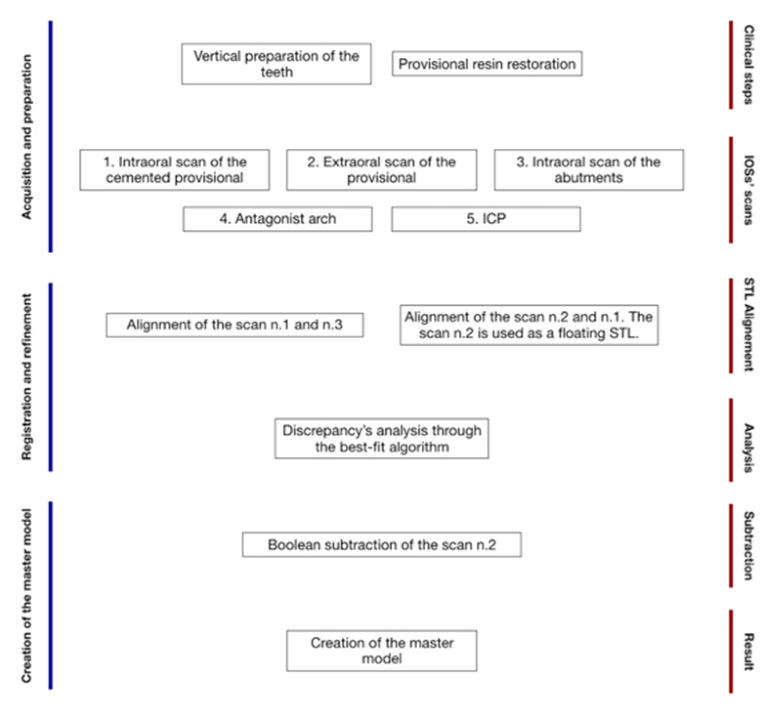
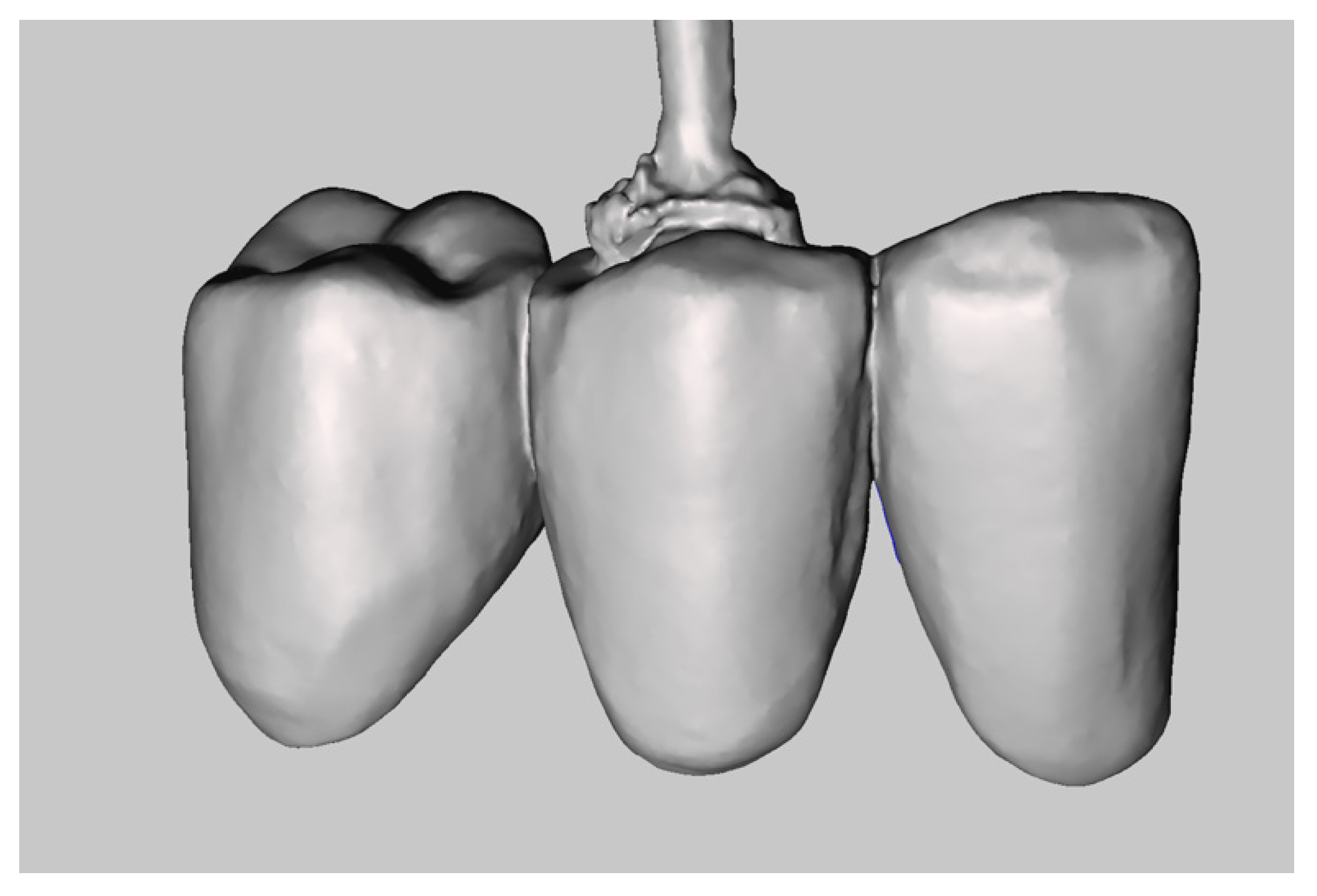
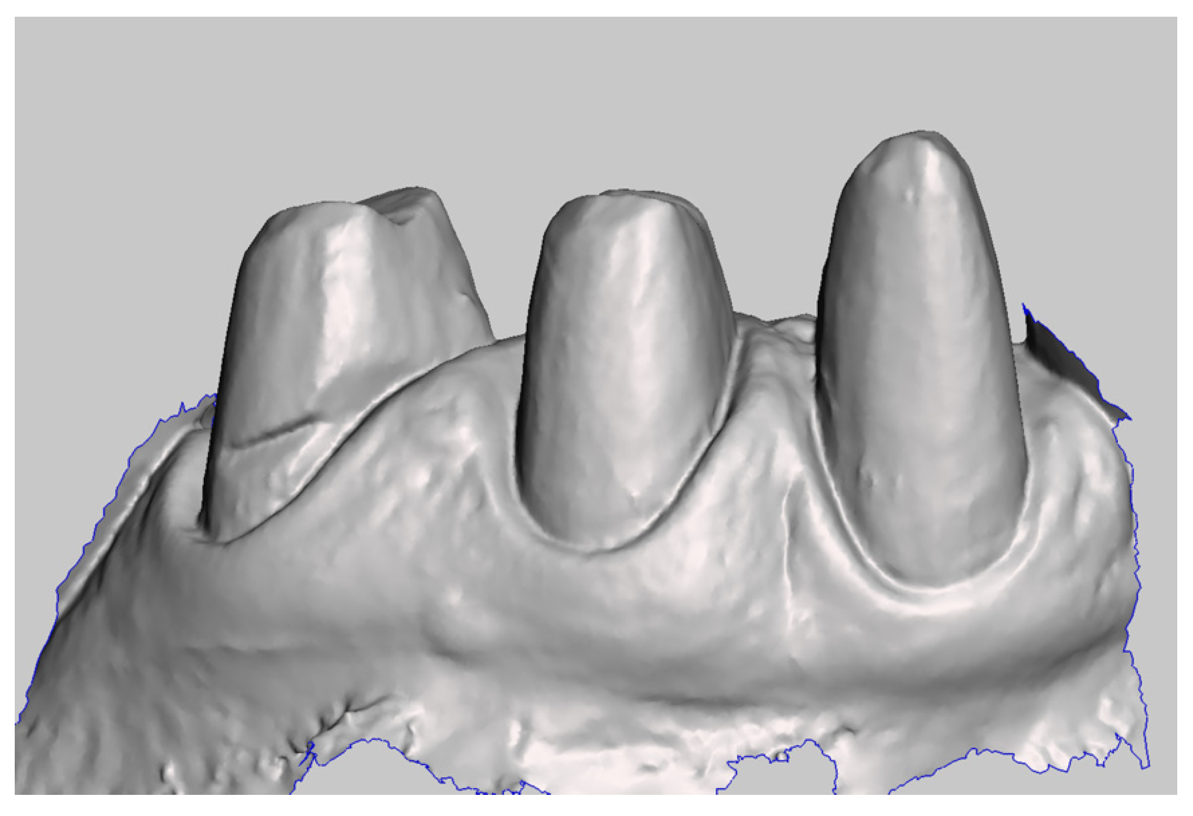
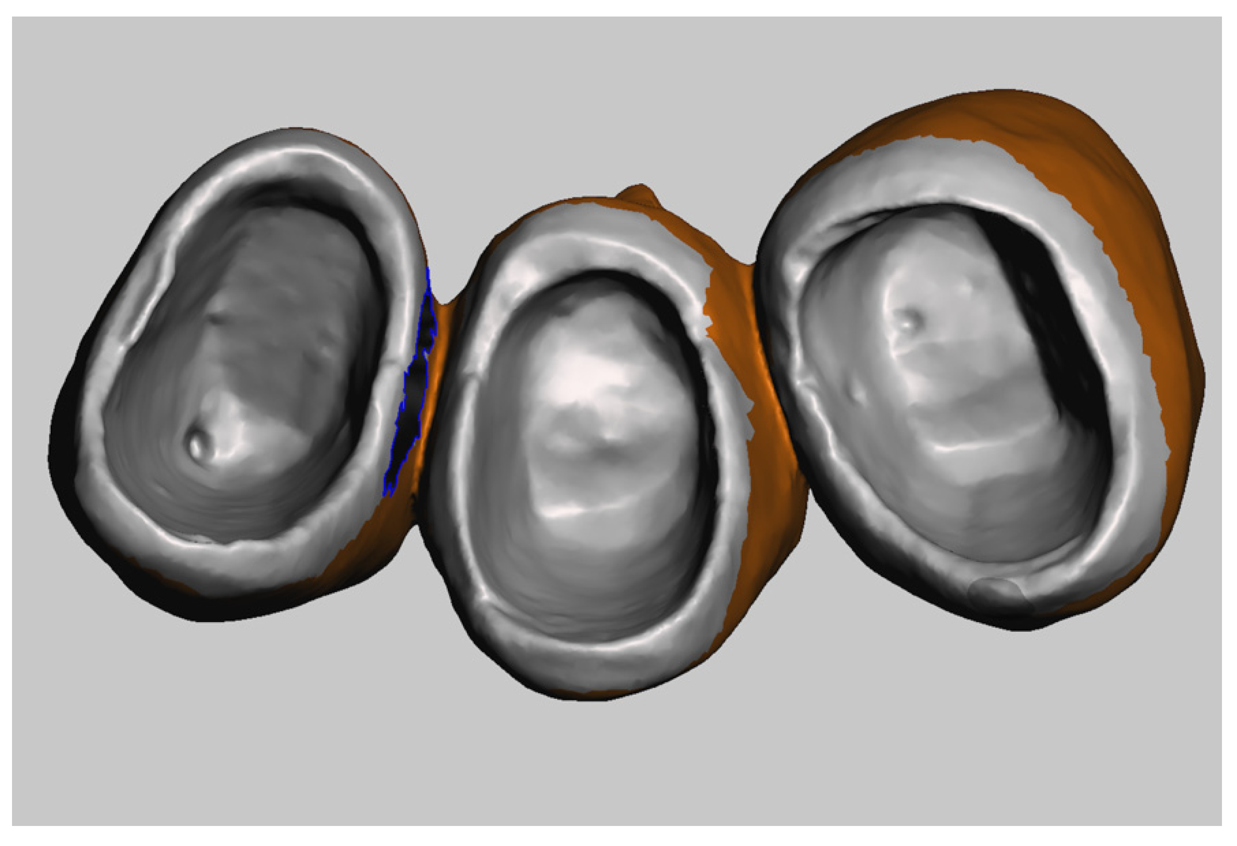
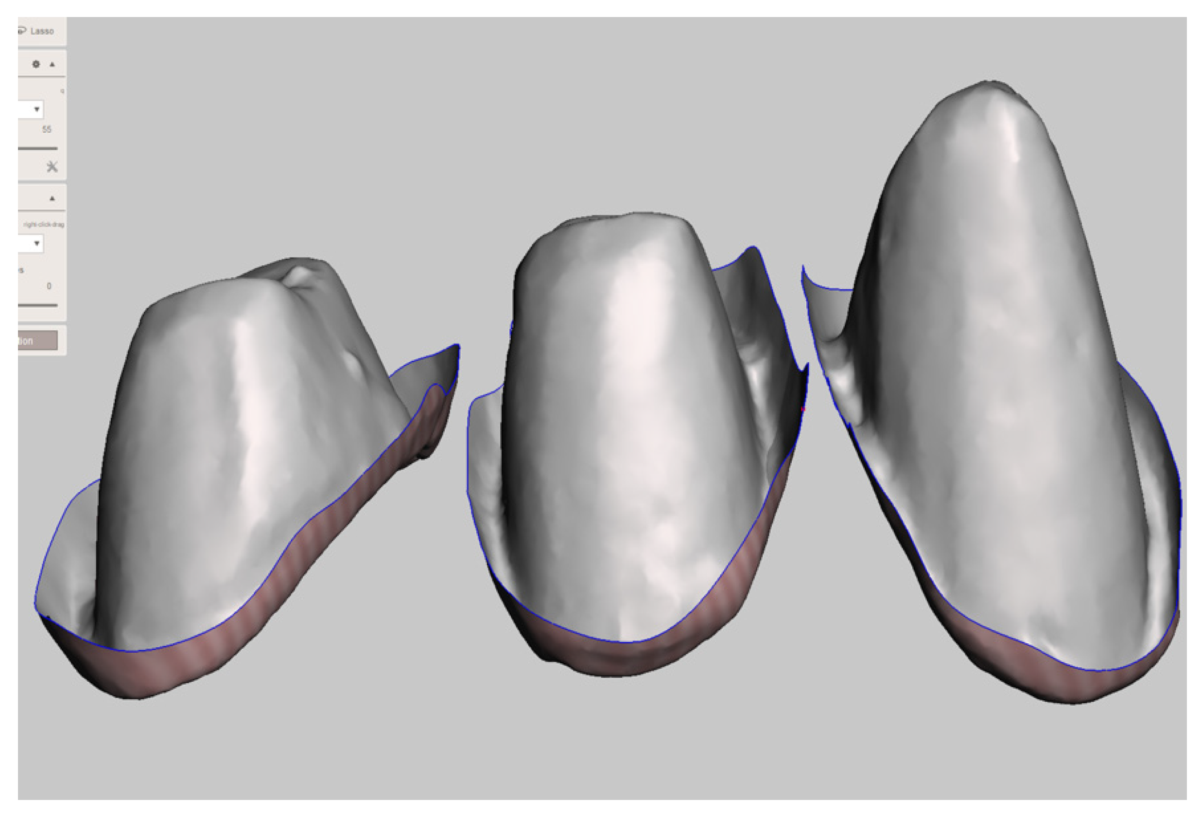
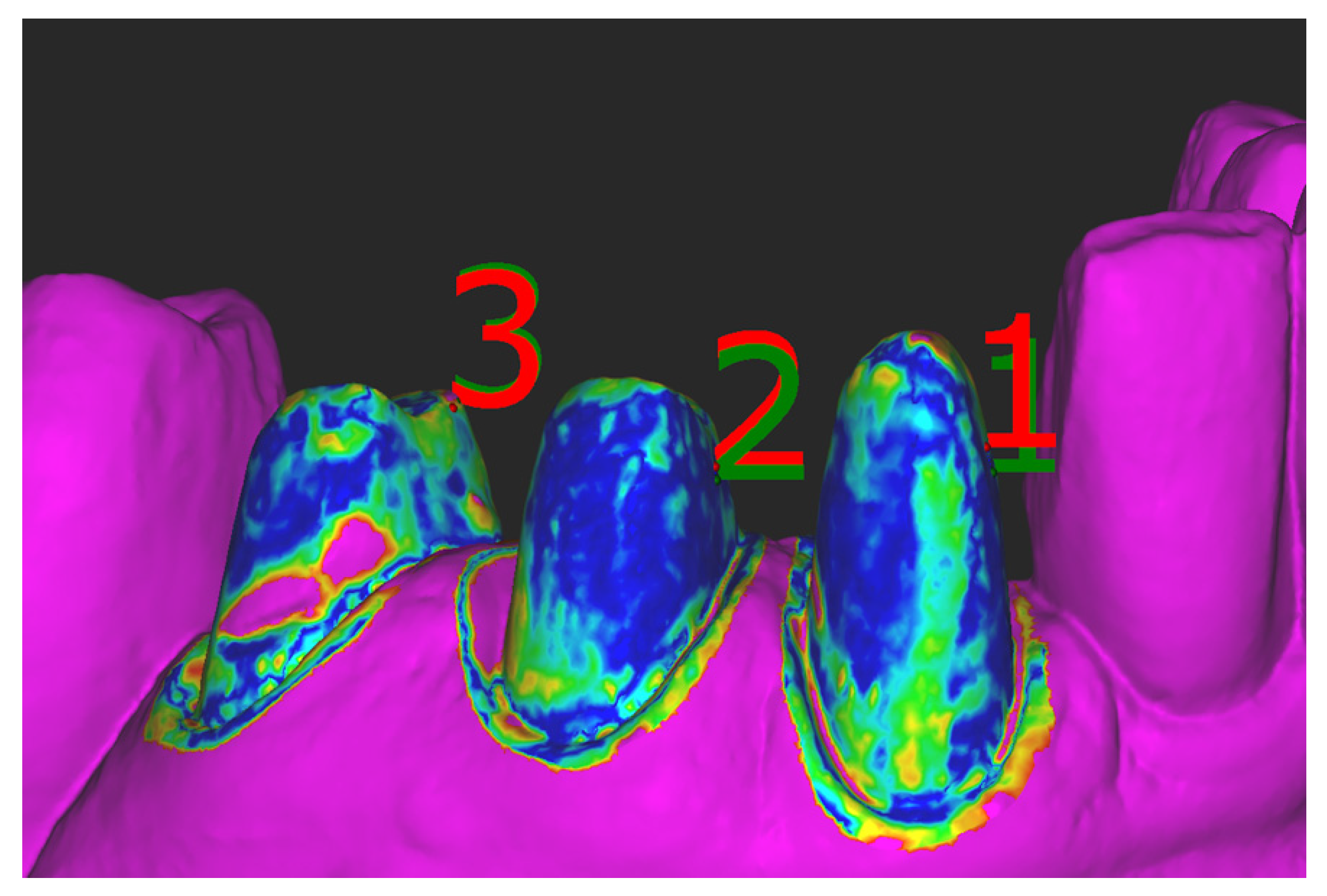
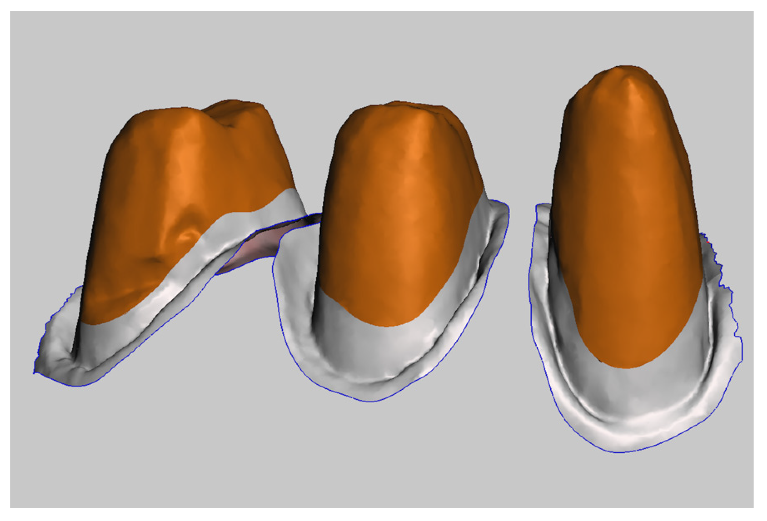
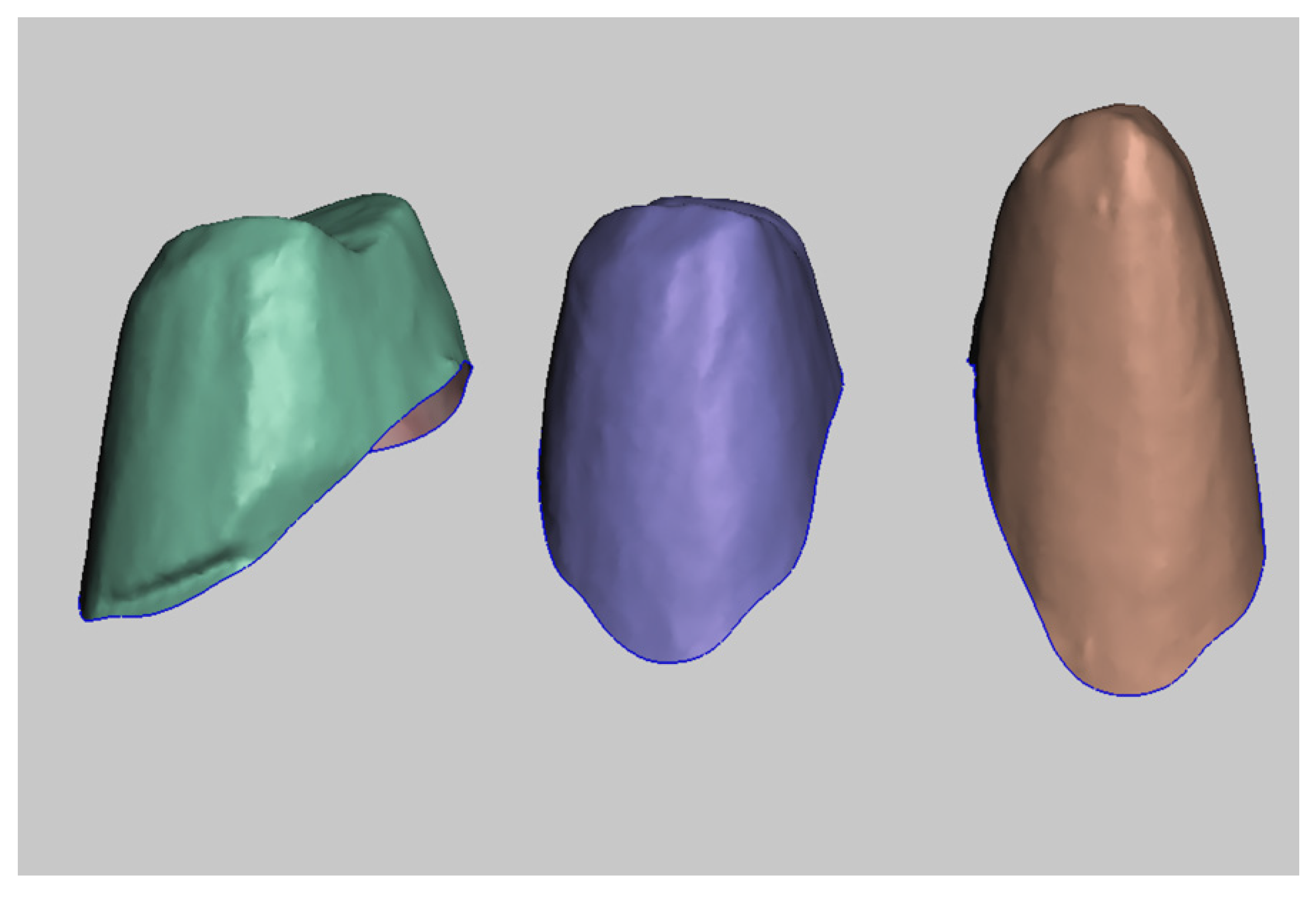
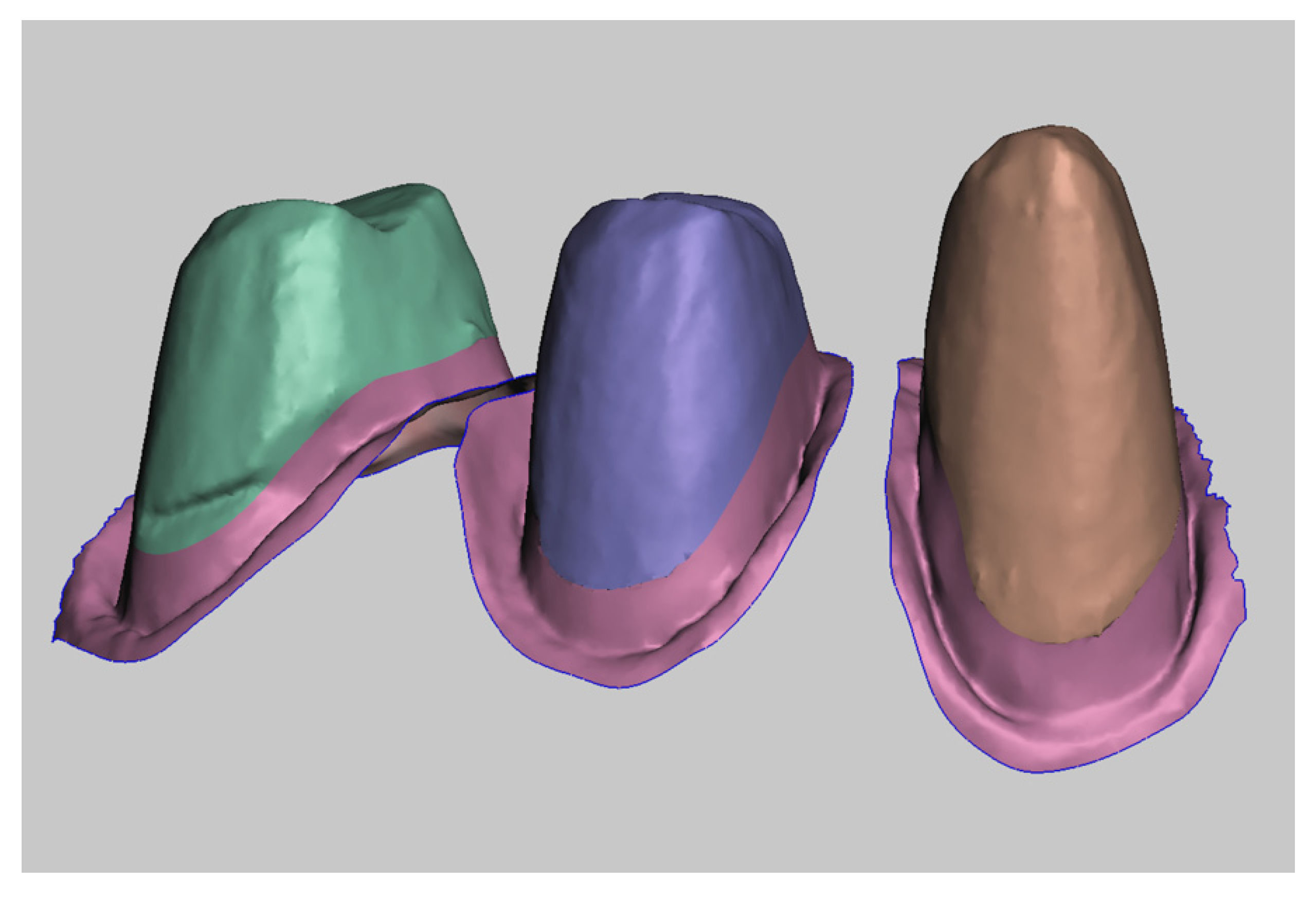
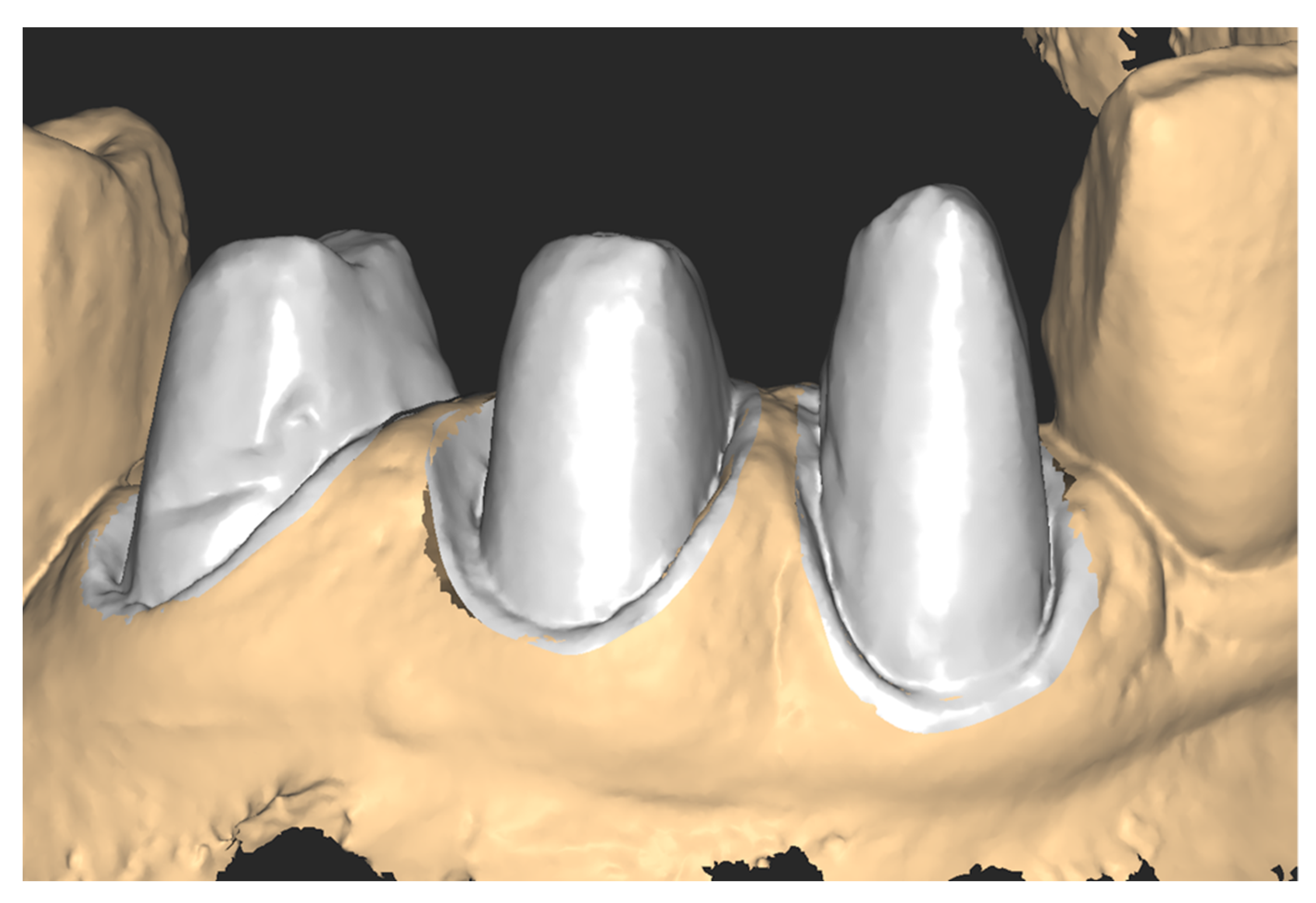
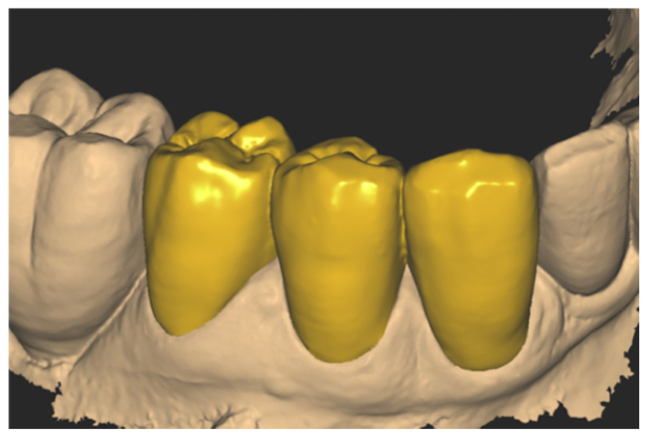
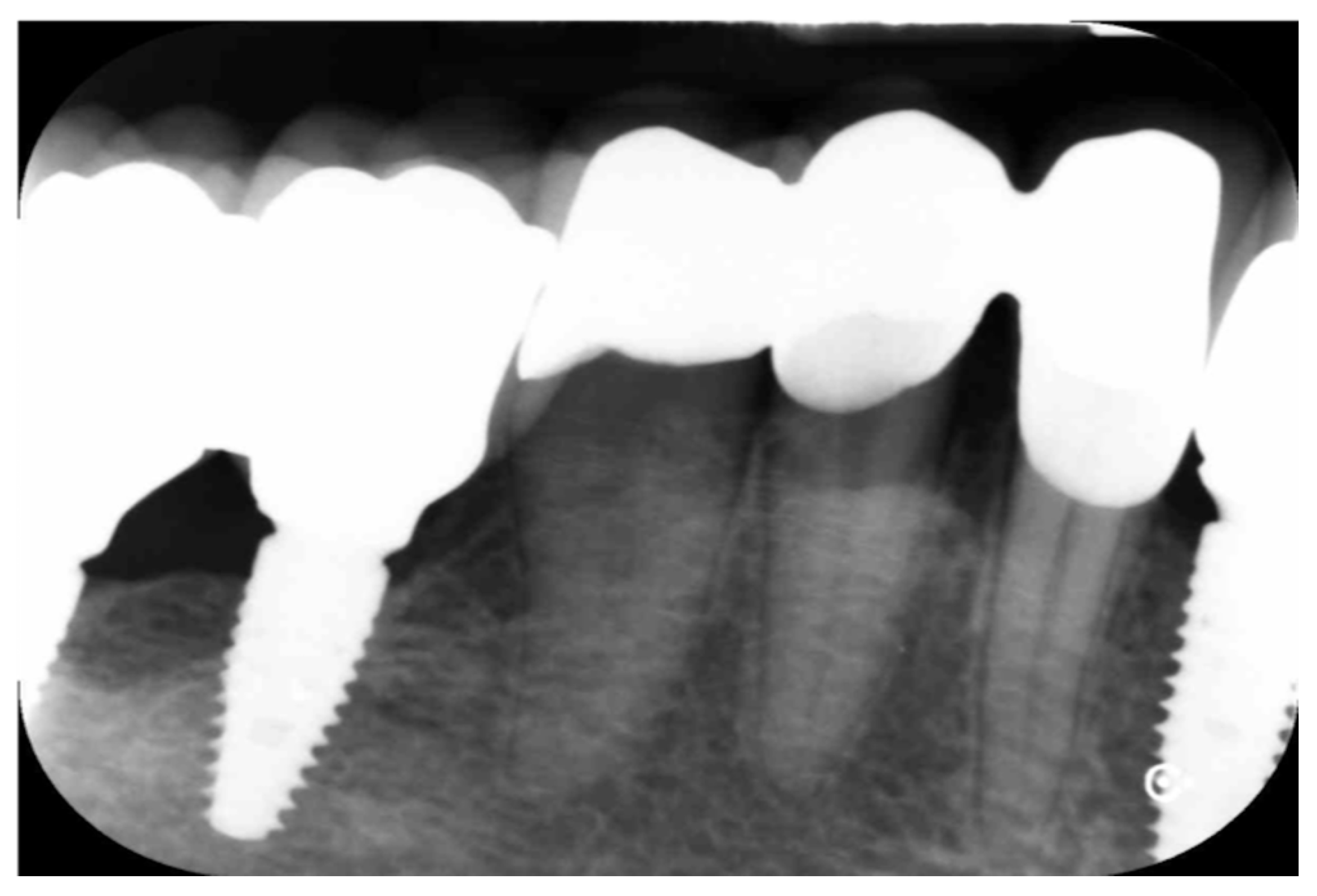
An important issue with digital impression techniques is the difficulty in replicating the subgingival zone, particularly when abutments are prepared with a vertical finish line. We propose a novel procedure, the reverse subgingival scan (RSS), to depict the subgingival area via digital impression without a retraction cord or the distortion of the sulcus. The software workflow, beginning with a digital dental impression, was performed using Exocad® (Align Technology) and Meshmixer® (Autodesk). The approach is based on the direct alignment of the surface of abutments, which is used as a reference point. The efficacy of the RSS approach was confirmed via fit tests, radiographic control assessments, and evaluations of the rate of tissue compression of the final prosthetic restoration, so long as the provisional had a perfect fit. RSS yields information on the transversal dimension and depth of the gingival sulcus. The procedure depends on the quality and characteristics of the provisional restoration, and is suitable only for cases in which the finish line of the final crown is at the same level or more coronal than that of the provisional.
Keywords: digital dentistry; prosthodontics; dental impression technique; intraoral scanners; gingival retraction technique digital dentistry; prosthodontics; dental impression technique; intraoral scanners; gingival retraction technique