Screwdriver Aspiration During Oral Procedures: A Lesson for Dentists and Gastroenterologists

 ,
, 
Abstract
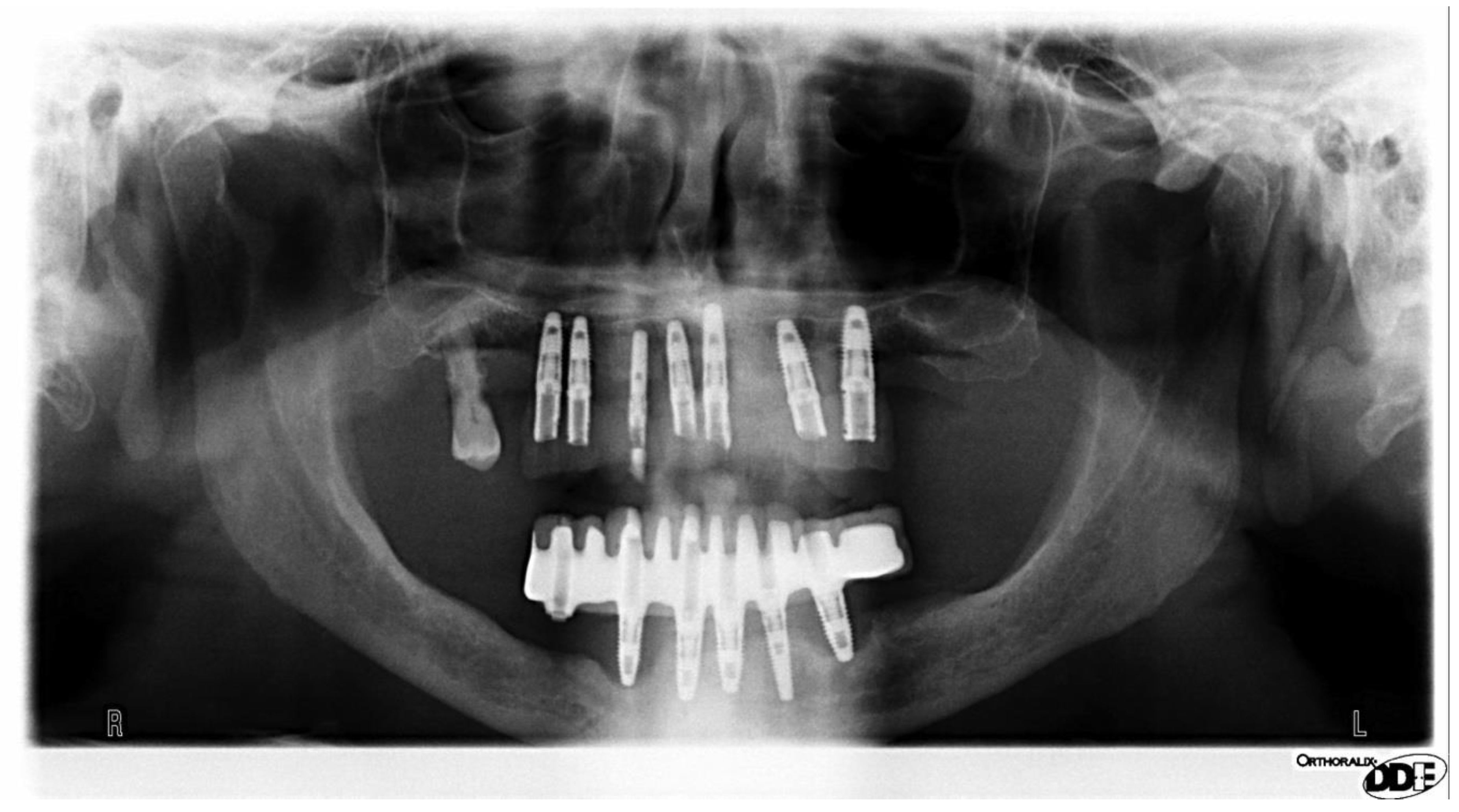
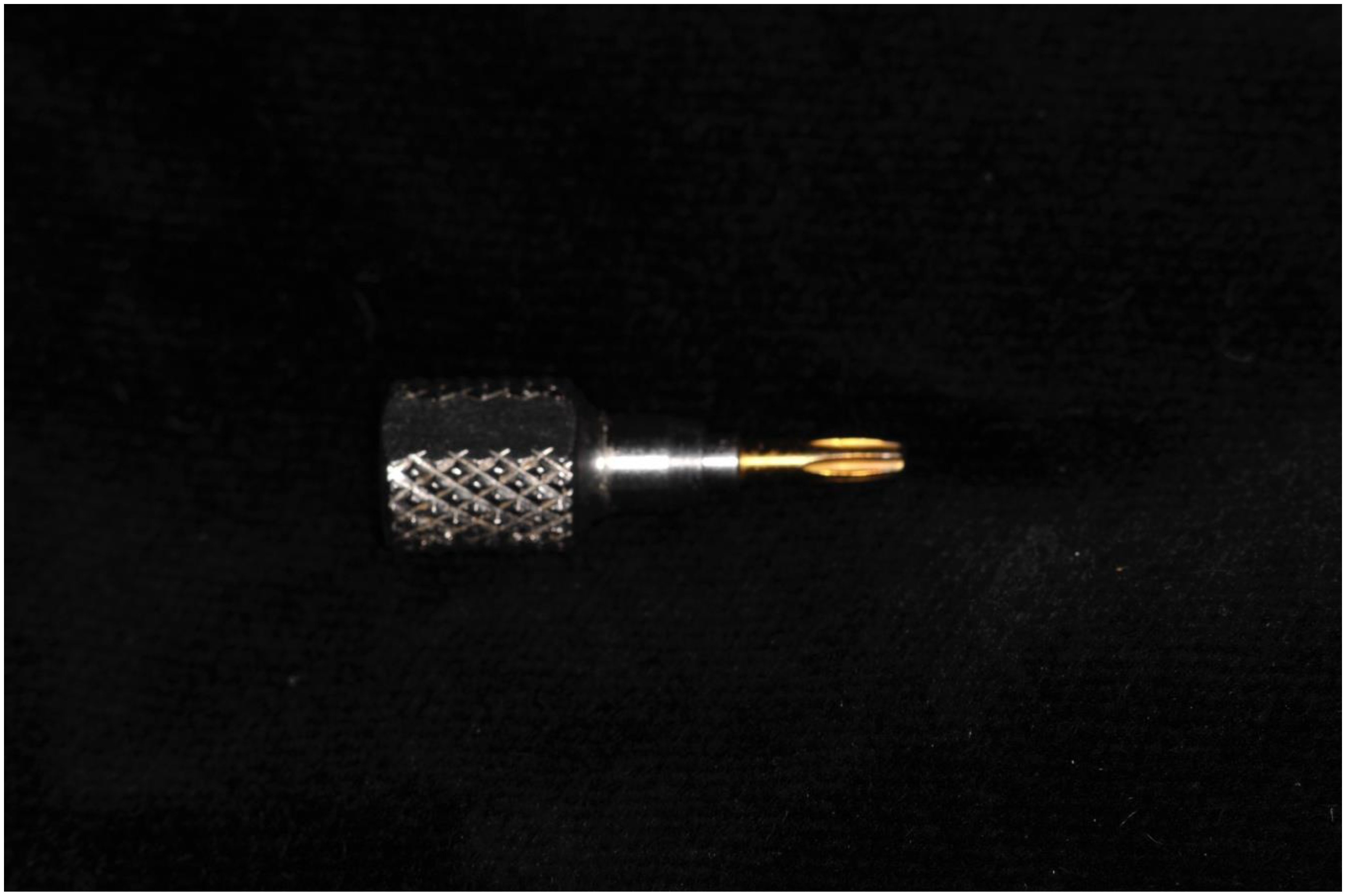
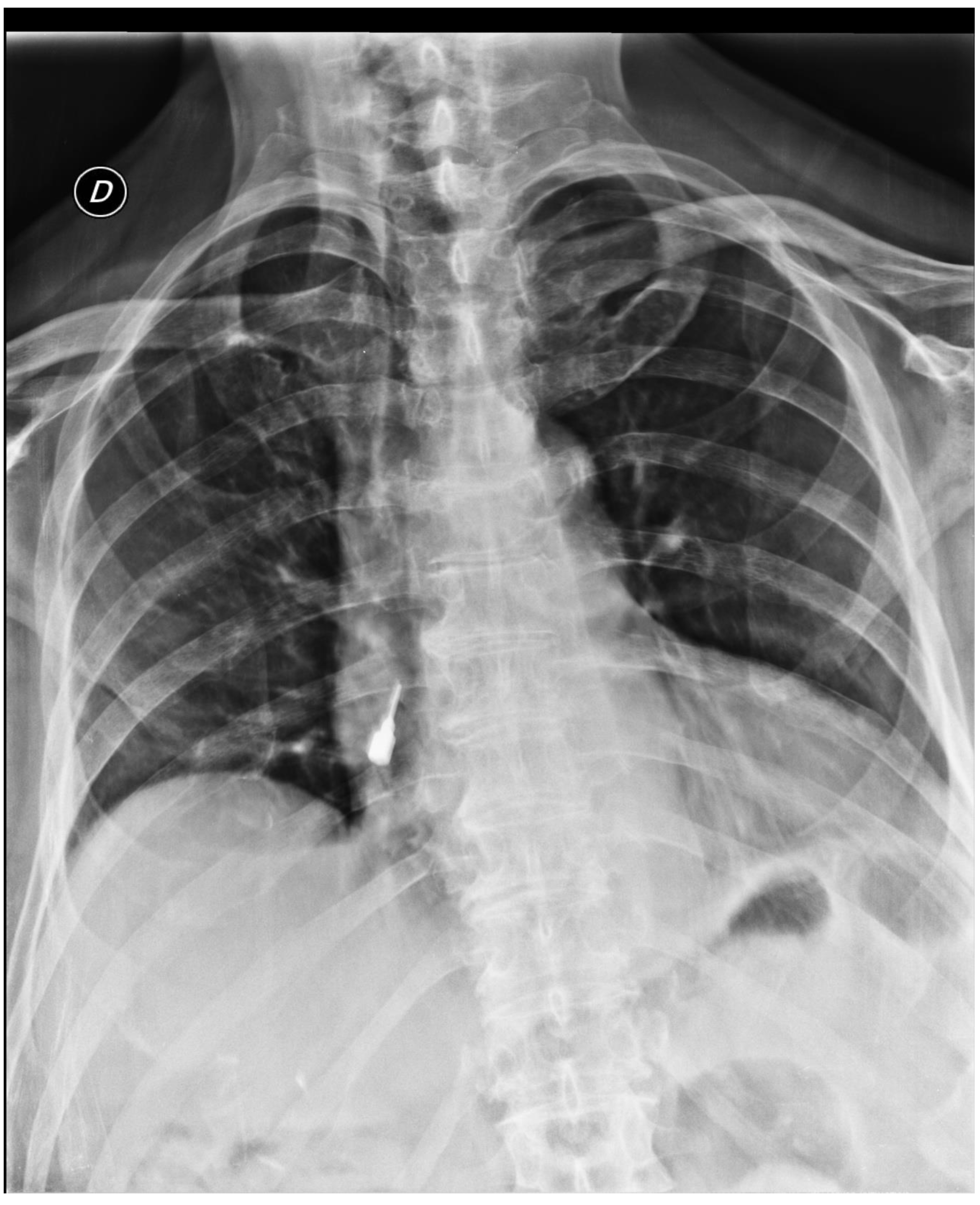
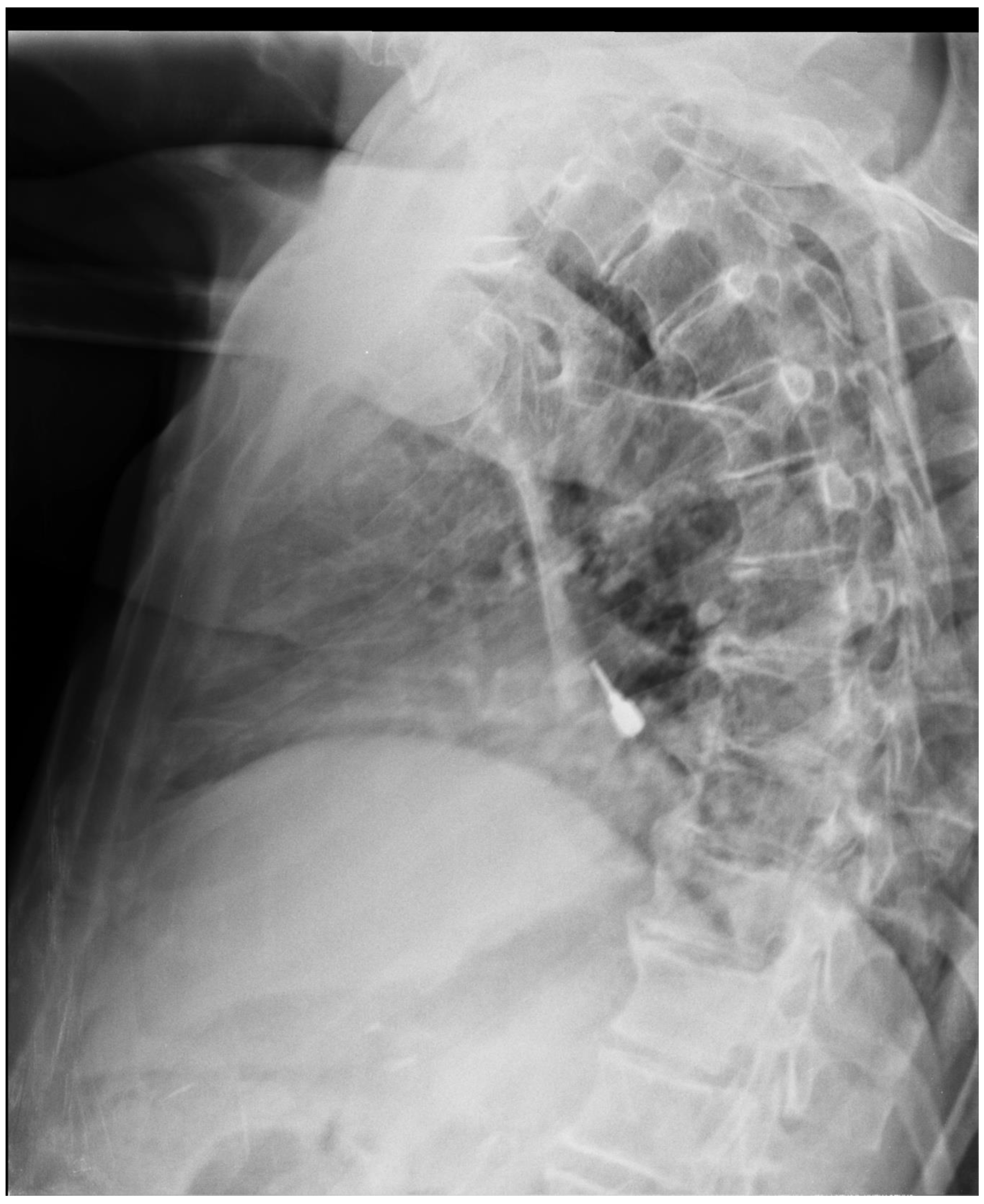
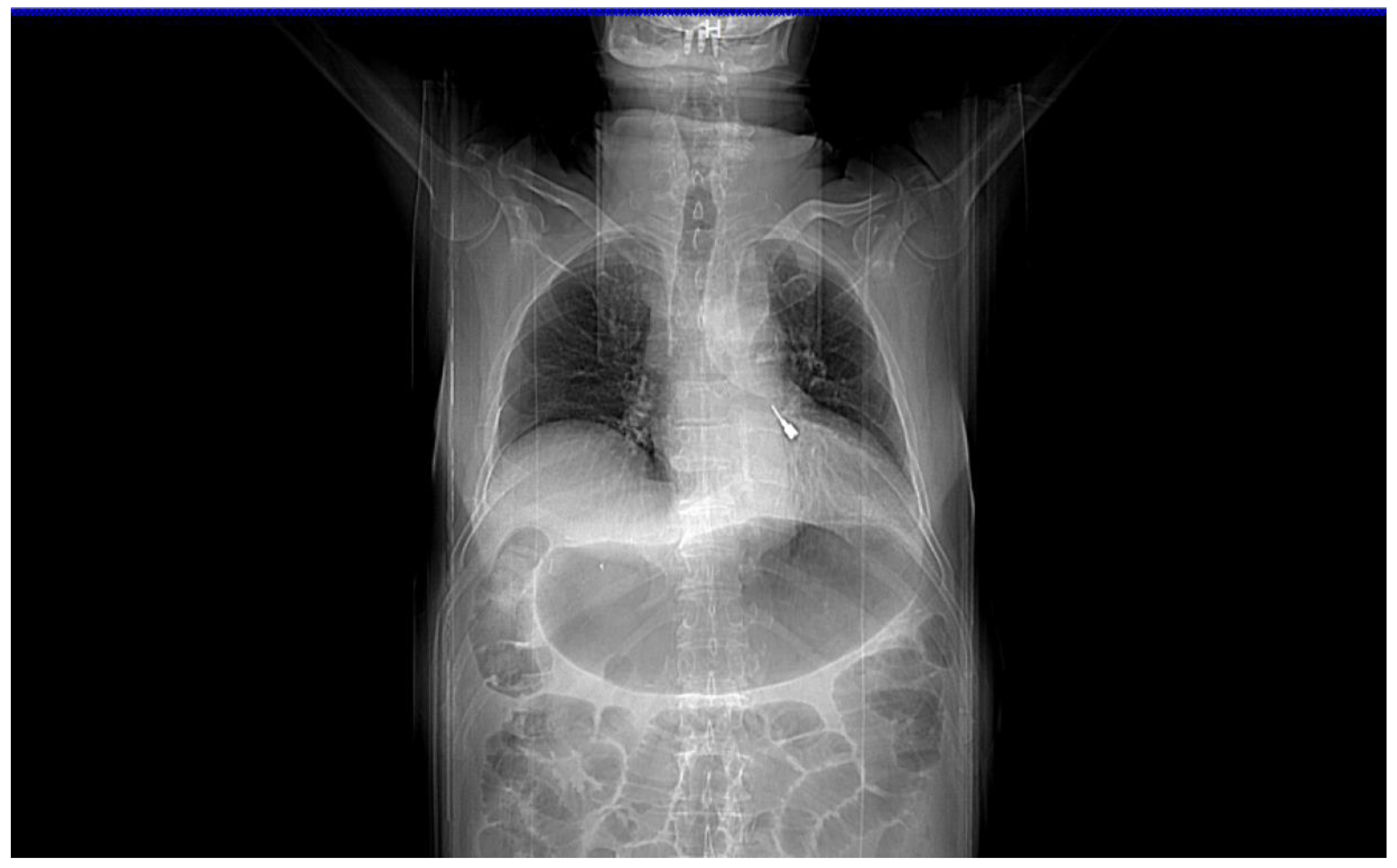
:1. Case Presentation
2. Discussion
3. Conclusions
Author Contributions
Funding
Conflicts of Interest
References
- De Souza, J.G.; Schuldt Filho, G.; Pereira Neto, A.R.; Lyra, H.F., Jr.; Bianchini, M.A.; Cardoso, A.C. Accident in implant dentistry: Involuntary screwdriver ingestion during surgical procedure. A clinical report. J. Prosthodont. 2012, 21, 191–193. [Google Scholar] [CrossRef] [PubMed]
- El Wazani, B.; Nixon, P.; Butterworth, C.J. Accidental Ingestion of an Implant Screwdriver: A Case Report and Literature Review Eur. J. Prosthodont. Restor. Dent. 2018, 26, 184–189. [Google Scholar]
- Jain, A.; Baliga, S.D. Accidental Implant Screwdriver Ingestion: A Rare Complication during Implant Placement. J. Dent. 2014, 11, 711–714. [Google Scholar]
- Pull Ter Gunne, L.; Wismeijer, D. Accidental ingestion of an untethered instrument during implant surgery. Int. J. Prosthodont. 2014, 27, 277–278. [Google Scholar] [CrossRef] [PubMed]
- Kim, A.; Ahn, K.M. Endoscopic removal of an aspirated healing abutment and screwdriver under conscious sedation. Implant Dent. 2014, 23, 250–252. [Google Scholar] [CrossRef] [PubMed]
- Deliberador, T.M.; Marengo, G.; Scaratti, R.; Giovanini, A.F.; Zielak, J.C.; Baratto Filho, F. Accidental aspiration in a patient with Parkinson’s disease during implant-supported prosthesis construction: A case report. Spec. Care Dentist. 2011, 31, 156–161. [Google Scholar] [CrossRef] [PubMed]
- Pingarrón Martín, L.; Morán Soto, M.J.; Sánchez Burgos, R.; Burgueño García, M. Bronchial impaction of an implant screwdriver after accidental aspiration: Report of a case and revision of the literature. Oral Maxillofac. Surg. 2010, 14, 43–47. [Google Scholar] [CrossRef]
- Worthington, P. Ingested foreign body associated with oral implant treatment: Report of a case. Int. J. Oral Maxillofac. Implants 1996, 11, 679–681. [Google Scholar]
- Zitzmann, N.U.; Elsasser, S.; Fried, R.; Marinello, C.P. Foreign body ingestion and aspiration. Oral Surg. Oral Med. Oral Pathol. Oral Radiol. Endodontol. 1999, 88, 657–660. [Google Scholar] [CrossRef]
- Wu, W.T.; Chiu, C.T.; Kuo, C.J.; Lin, C.J.; Chu, Y.Y.; Tsou, Y.K.; Su, M.Y. Endoscopic management of suspected esophageal foreign body in adults. Dis. Esophagus 2011, 24, 131–137. [Google Scholar] [CrossRef]
- Birk, M.; Bauerfeind, P.; Deprez, P.H.; Häfner, M.; Hartmann, D.; Hassan, C.; Hucl, T.; Lesur, G.; Aabakken, L.; Meining, A. Removal of foreign bodies in the upper gastrointestinal tract in adults: European Society of Gastrointestinal Endoscopy (ESGE) Clinical Guideline. Endoscopy 2016, 48, 489–496. [Google Scholar] [CrossRef] [PubMed]
- Weiland, S.T.; Schurr, M.J. Conservative management of ingested foreign bodies. J. Gastrointest. Surg. 2002, 6, 496. [Google Scholar] [CrossRef]
- Li, Z.S.; Sun, Z.X.; Zou, D.W.; Xu, G.M.; Wu, R.P.; Liao, Z. Endoscopic management of foreign bodies in the upper-GI tract: Experience with 1088 cases in China. Gastrointest. Endosc. 2006, 64, 485–492. [Google Scholar] [CrossRef]
- Nandi, P.; Ong, G.B. Foreign body in the oesophagus: Review of 2394 cases. Br. J. Surg. 1978, 65, 5–9. [Google Scholar] [CrossRef] [PubMed]
- Khan, M.A.; Hameed, A.; Choudhry, A.J. Management of foreign bodies in the esophagus. J. Coll. Phys. Surg. Pak. 2004, 14, 218–220. [Google Scholar]
- Sung, S.H.; Jeon, S.W.; Son, H.S.; Kim, S.K.; Jung, M.K.; Cho, C.M.; Tak, W.Y.; Kweon, Y.O. Factors predictive of risk for complications in patients with oesophageal foreign bodies. Dig. Liver Dis. 2011, 43, 632–635. [Google Scholar] [CrossRef]
- Lam, H.C.; Woo, J.K.; van Hasselt, C.A. Management of ingested foreign bodies: A retrospective review of 5240 patients. J. Laryngol. Otol. 2001, 115, 954–957. [Google Scholar] [CrossRef]
- Ahn, D.; Heo, S.J.; Park, J.H.; Sohn, J.H. Tracheoesophageal fistula with tracheal stenosis resulting from retained esophageal foreign body. Auris Nasus Larynx 2011, 38, 753–756. [Google Scholar] [CrossRef]
- Zhang, X.; Liu, J.; Li, J.; Hu, J.; Yu, F.; Li, S.; Yang, X. Diagnosis and treatment of 32 cases with aortoesophageal fistula due to esophageal foreign body. Laryngoscope 2011, 121, 267–272. [Google Scholar] [CrossRef]
- Adams, D.B. Endoscopic removal of entrapped coins from an intraluminal duodenal diverticulum 20 years after ingestion. Gastrointest. Endosc. 1986, 32, 415–416. [Google Scholar] [CrossRef]
- Kirberg, A.E. Long-standing esophageal foreign body. Gastrointest. Endosc. 1986, 32, 304–305. [Google Scholar] [CrossRef]
- Yamamoto, M.; Mizuno, H.; Sugawara, Y. A chopstick is removed after 60 years in the duodenum. Gastrointest. Endosc. 2016, 31, 51. [Google Scholar] [CrossRef]
- Tsui, B.C.; Mossey, J. Occult liver abscess following clinically unsuspected ingestion of foreign bodies. Can. J. Gastroenterol. Hepatol. 1997, 11, 445–448. [Google Scholar] [CrossRef] [PubMed]
- Bucci, C.; Gallotta, S.; Morra, I.; Fortunato, A.; Ciacci, C.; Iovino, P. Anisakis, just think about it in an emergency! Int. J. Infect. Dis. 2013, 17, e1071–e1072. [Google Scholar] [CrossRef] [PubMed]
- Guelfguat, M.; Kaplinskiy, V.; Reddy, S.H.; DiPoce, J. Clinical guidelines for imaging and reporting ingested foreign bodies. AJR Am. J. Roentgenol. 2014, 203, 37–53. [Google Scholar] [CrossRef] [PubMed]


| As recommended by the (ESGE) guidelines [11]: |
|
© 2019 by the authors. Licensee MDPI, Basel, Switzerland. This article is an open access article distributed under the terms and conditions of the Creative Commons Attribution (CC BY) license (http://creativecommons.org/licenses/by/4.0/).
Share and Cite
Iovino, P.; Di Sarno, A.; De Caro, V.; Mazzei, C.; Santonicola, A.; Bruno, V. Screwdriver Aspiration During Oral Procedures: A Lesson for Dentists and Gastroenterologists. Prosthesis 2019, 1, 61-68. https://doi.org/10.3390/prosthesis1010008
Iovino P, Di Sarno A, De Caro V, Mazzei C, Santonicola A, Bruno V. Screwdriver Aspiration During Oral Procedures: A Lesson for Dentists and Gastroenterologists. Prosthesis. 2019; 1(1):61-68. https://doi.org/10.3390/prosthesis1010008
Chicago/Turabian StyleIovino, Paola, Alessandro Di Sarno, Vincenzo De Caro, Cosimo Mazzei, Antonella Santonicola, and Vincenzo Bruno. 2019. "Screwdriver Aspiration During Oral Procedures: A Lesson for Dentists and Gastroenterologists" Prosthesis 1, no. 1: 61-68. https://doi.org/10.3390/prosthesis1010008
APA StyleIovino, P., Di Sarno, A., De Caro, V., Mazzei, C., Santonicola, A., & Bruno, V. (2019). Screwdriver Aspiration During Oral Procedures: A Lesson for Dentists and Gastroenterologists. Prosthesis, 1(1), 61-68. https://doi.org/10.3390/prosthesis1010008