Article

A Bacteria Sol–Gel Template Approach to Form Palladium Core–Shell Catalysts for Suzuki–Miyaura Reactions

by
Vitaliy N. Soromotin
*,
Pavel V. Rybochkin
,
Violetta A. Pertseva
and
Olga A. Kamanina
Laboratory of Biologically Active Compounds and Biocomposites, Tula State University, Lenina Ave. 92, 300012 Tula, Russia
*
Author to whom correspondence should be addressed.
Chemistry 2025, 7(6), 188; https://doi.org/10.3390/chemistry7060188
Submission received: 28 October 2025 / Revised: 15 November 2025 / Accepted: 20 November 2025 / Published: 25 November 2025
(This article belongs to the Special Issue Celebrating the 50th Anniversary of Professor Valentine Ananikov)

Abstract

This study presents a sustainable and efficient method for fabricating core–shell structured palladium catalysts using a bacterial template and sol–gel synthesis. This synthesis aligns with green chemistry principles by minimizing waste and enhancing resource efficiency. Our results demonstrate that the bacterial template effectively stabilizes Pd nanoparticles (NPs), preventing significant agglomeration during synthesis and subsequent calcination under different atmospheres and final temperatures. The catalyst samples were characterized by SEM, TEM, XRD, and TGA. The 1 wt% Pd/R@SiO2 catalyst exhibited high activity in the Suzuki–Miyaura cross-coupling reaction, achieving competitive yields. Furthermore, the catalyst demonstrated a stable performance over five consecutive cycles. This work underscores the potential of biotemplated synthesis as a versatile and eco-friendly platform for producing high-performance, tunable catalysts.

1. Introduction

The Suzuki–Miyaura cross-coupling reaction is a cornerstone of modern organic synthesis due to its mild conditions and high selectivity for forming C-C bonds [1,2]. Conventionally, the Suzuki–Miyaura reaction employs palladium catalysts, which are broadly classified as homogeneous or heterogeneous. This includes systems ranging from ligandless catalysts to supported nanoparticles [3,4]. While homogeneous palladium catalysts enable high activity across diverse substrates, their practical application is hampered by difficulties in separation and recycling, catalyst agglomeration, and consequent cost inefficiencies [5].
Heterogeneous catalysts for the Suzuki–Miyaura reaction offer significant practical advantages, primarily easier separation and reuse compared to their homogeneous counterparts. However, they demonstrate a relatively lower activity and selectivity compared to ligand catalysts [6]. Another issue associated with heterogeneous catalysts lies in the difficulties in characterization, and the structure of their active sites is not well-understood. Core–shell catalyst architectures have been developed to overcome these drawbacks, demonstrating excellent stability, easy recovery, and catalytic performance comparable to homogeneous catalysts. Nevertheless, the intricate and multi-stage manufacturing processes required for these materials lead to high costs, which remains a barrier to their broad adoption [7].
As sustainable alternatives to traditional catalysts, systems incorporating metal nanoparticles synthesized via bio-reduction using biomaterials, microorganisms, plants, their extracts, or enzymes have garnered significant interest [8,9,10,11,12]. The primary advantage of these approaches is their environmental benignity, while the resulting catalysts often demonstrate activity and selectivity comparable to conventional analogs [13]. Despite these merits, significant challenges remain. For instance, catalysts derived from bacterial cells are unsuitable for elevated temperatures due to the thermal degradation of the cellular structure. Furthermore, organic solvents commonly employed in the Suzuki–Miyaura reaction can disrupt cell integrity, which, during prolonged use, promotes the leaching and subsequent loss of metal nanoparticles [8].
Previous research [14,15] has demonstrated the feasibility of forming an organosilicate shell around microorganisms via sol–gel synthesis. For instance, Paracoccus yeei VKM B-3302 bacteria were successfully encapsulated within an organosilicon material using tetraethoxysilane (TEOS) and methyltriethoxysilane (MTES) in a 1:1 volume ratio as precursors. The resulting material was characterized by pores extending to the bacterial surface, with an average diameter of 20 nm. This configuration, where the porous structure serves as a shell encapsulating bacteria-supported metal nanoparticles as the core, provides a foundation for a novel core–shell catalyst.
Herein, we present a core–shell catalyst fabricated by encapsulating bacteria-supported palladium nanoparticles in an organosilicate matrix, followed by thermal treatment. The efficacy of this catalyst was demonstrated in the Suzuki–Miyaura coupling reaction.

2. Materials and Methods

2.1. Bacterial Cell Cultivation

The Paracoccus yeei bacteria are obligate aerobes and Gram-negative cocci with a small cell diameter (approximately 0.5–0.9 μm), high growth rate, ease of cultivation, and maintenance in pure culture. The bacterial cells were cultured in 750 cm3 Erlenmeyer flasks with a nutrient medium (Luria–Bertani media supplemented with 10 g/dm3 peptone, 10 g/dm3 NaCl, and 5 g/dm3 yeast extract) volume of 200 cm3 at a temperature of 28 °C and were aerated in a shaker at 180 rpm. After 48 h, the bacterial culture was harvested by centrifugation at 8000 rpm for 10 min in test tubes. The cell biomass was dried and then stored in test tubes at +4 °C.

2.2. Scanning Electron Microscopy (SEM) Measurements

SEM analysis was performed using a TM4000Plus (Hitachi, Tokyo, Japan) scanning electron microscope in backscattered electron (BSE) detection mode. Images were acquired at an accelerating voltage of 10, 15, and 20 kV. The palladium presence was confirmed by EDX (Bruker, Billerica, MA, USA).

2.3. Transmission Electron Microscopy (TEM) Measurements

Before the measurements, the samples were deposited on 3 mm grids. The sample morphology was studied using Hitachi HT7700 transmission electron microscope. Images were acquired in bright-field TEM mode at an accelerating voltage of 100 kV. The nanoparticle size distribution was determined via ImageJ 1.54g software.

2.4. Thermogravimetric Analysis

Thermogravimetric analysis (TGA) was performed using a NETZSCH STA 449 F5 thermal analyzer. The measurements were conducted under the following conditions: reaction gas mixture of 20 vol% O2 in N2 at a flow rate of 50 cm3/min, with a protective N2 atmosphere maintained at 20 cm3/min. Samples with 50 mg were subjected to a programmed temperature regime consisting of (1) heating from 50 to 130 °C at a constant rate of 10 °C/min, (2) isothermal holding at 130 °C for 30 min, and (3) subsequent heating from 130 to 600 °C (or 800 °C in selected experiments) at 10 °C/min. Experiments under pure N2 atmosphere were performed using identical thermal programming parameters.

2.5. Determination of Pd Content

The quantitative determination of the palladium in the catalyst was carried out using an optical emission spectrometer with inductively coupled plasma (ICP-OES) Varian 710-ES (Agilent, Santa Clara, CA, USA) under the following conditions: plasma power 1.20 kW, plasma flow 15.0 L/min, axial flow 1.5 L/min, and nebulizer pressure 200 kPa. The arithmetic mean of three parallel measurements was taken as the measurement result. To determine the metal content in the catalyst, the sample was calcined at 600 °C for 2 h and then dissolved in a mixture of dilute hydrochloric acid and hydrogen peroxide; the resulting solution was analyzed by the ICP-OES method.

2.6. Pd/R@SiO2 for Suzuki–Miyaura Reaction

A 1 mol. % catalyst (Pd/R@SiO2 1 wt.%) obtained with various temperatures and atmosphere calcination was mixed with other reagents: 0.5 mmol of 1-iodo-benzene; 1-iodo-4-nitrobenzene; 0.6 mmol of phenylboronic acid; and 0.6 mmol of triethylamine in the test tube. The solvent (2.5 cm3 of H2O:EtOH in ratio of 1:1) was added to the placed reagents. The reaction mixture was stirred and heated for 3 h (80 °C; 1200 rpm).
After 3 h, samples were collected from the reaction mixture for GC-MS (0.01 cm3). The GC-MS sample was subsequently diluted 103 times with acetonitrile before analysis.

2.7. Pd/R@SiO2 Recycling Test

The general procedure for the reaction setup is described in Section 2.6, including sample preparation for GC-MS analysis.
Subsequently, a further portion of reagents excluding the catalyst was added to the flask. This consisted of 1-iodobenzene (0.5 mmol), phenylboronic acid (0.6 mmol), triethylamine (0.6 mmol), and a solvent mixture (2.5 cm3 of H2O:EtOH, in ratio 1:1). The reaction mixture was then stirred and heated again for 3 h (80 °C; 1200 rpm).
This cycle of reaction, GC-MS analysis, and reagent addition was repeated five times.

2.8. Pd/R@SiO2 Hot Filtration Test

The general procedure for the reaction setup is described in Section 2.6, except the reaction mixture was stirred and heated for 0.5 h (80 °C; 1200 rpm).
After 0.5 h, samples were collected from the reaction mixtures for GC-MS (0.01 cm3). The GC-MS sample was subsequently diluted 103 times with acetonitrile before analysis. Then reaction mixtures were filtered at 80 °C in new test tubes and stirred for 2.5 h (80 °C; 1200 rpm). New samples were collected from the reaction mixtures for GC-MS analysis.

3. Results

3.1. Catalyst Preparation

The general preparation procedure for Pd/R@SiO2 includes three steps.
Step 1: The synthesis of the Pd/P. yeei biohybrid was performed according to a previously reported method [16,17]. The catalyst was synthesized using bacterial biomass as a support, treated with a palladium (II) acetate solution to target a nominal loading of 5 wt% metallic palladium relative to the dry biomass. This biomass template loses 80% of its initial value during dehydration (upon drying in the air). Briefly, a mixture of bacterial cells and palladium acetate in distilled water was purged with argon for 30 s and subsequently shaken for 10 min (180 rpm, room temperature). Then palladium nanoparticles (Pd NPs) were formed by sparging the mixture with hydrogen gas for 120 s. The content of the palladium on the bacteria support after first preparation step was 4.4 wt%, as determined by ICP-OES.
Step 2: encapsulation in an ORMOSIL shell. The biomass-supported Pd NPs (Pd/P. yeei) were harvested by centrifugation (12,000 rpm, 10 min) and resuspended in a phosphate buffer (pH 6.8) to form an initial suspension with a cell concentration of 150 mg/mL. To this suspension, a 5 wt% polyvinyl alcohol (PVA) solution was added (40 vol%), and the mixture was shaken for 5 min. Methyltriethoxysilane (MTES) and tetraethoxysilane (TEOS) were then added in a 1:1 volume ratio relative to the initial suspension, followed by shaking for 5 min. A NaF solution (200 mmol/dm3) was added as a catalyst, amounting to 10% of the initial suspension volume. After shaking for 15 min, the mixture was transferred to a Petri dish and dried for 48 h at room temperature and atmospheric pressure. The formation of the ORMOSIL shell took approximately 15 min [18], while the total aging time was 5 h [19].
Step 3: thermal treatment. The dried Pd/P. yeei@ORMOSIL material was calcined in a temperature-controlled oven. The temperature was increased at a rate of 20 °C/min to 600 °C and held for 1 h in an air atmosphere. The resulting cooled Pd/R@SiO2 (R—residue) (I) catalyst was washed with water to remove residual NaF and dried. Alternatively, three other catalyst samples were prepared. The difference was the atmosphere of the calcination procedure and the final temperature—air and 800 °C (II), N2 and 600 °C (III), and N2 and 800 °C (IV). The procedure for the catalyst preparation is depicted in Figure 1.
Following the sol–gel synthesis and the subsequent drying of the composite material to a constant mass, the mass ratio of the dehydrated biomass-derived template to the inorganic shell was 1:5. After calcination, the final palladium content in the catalyst was determined to be near 1.0 wt%.

3.2. Catalyst Characterization

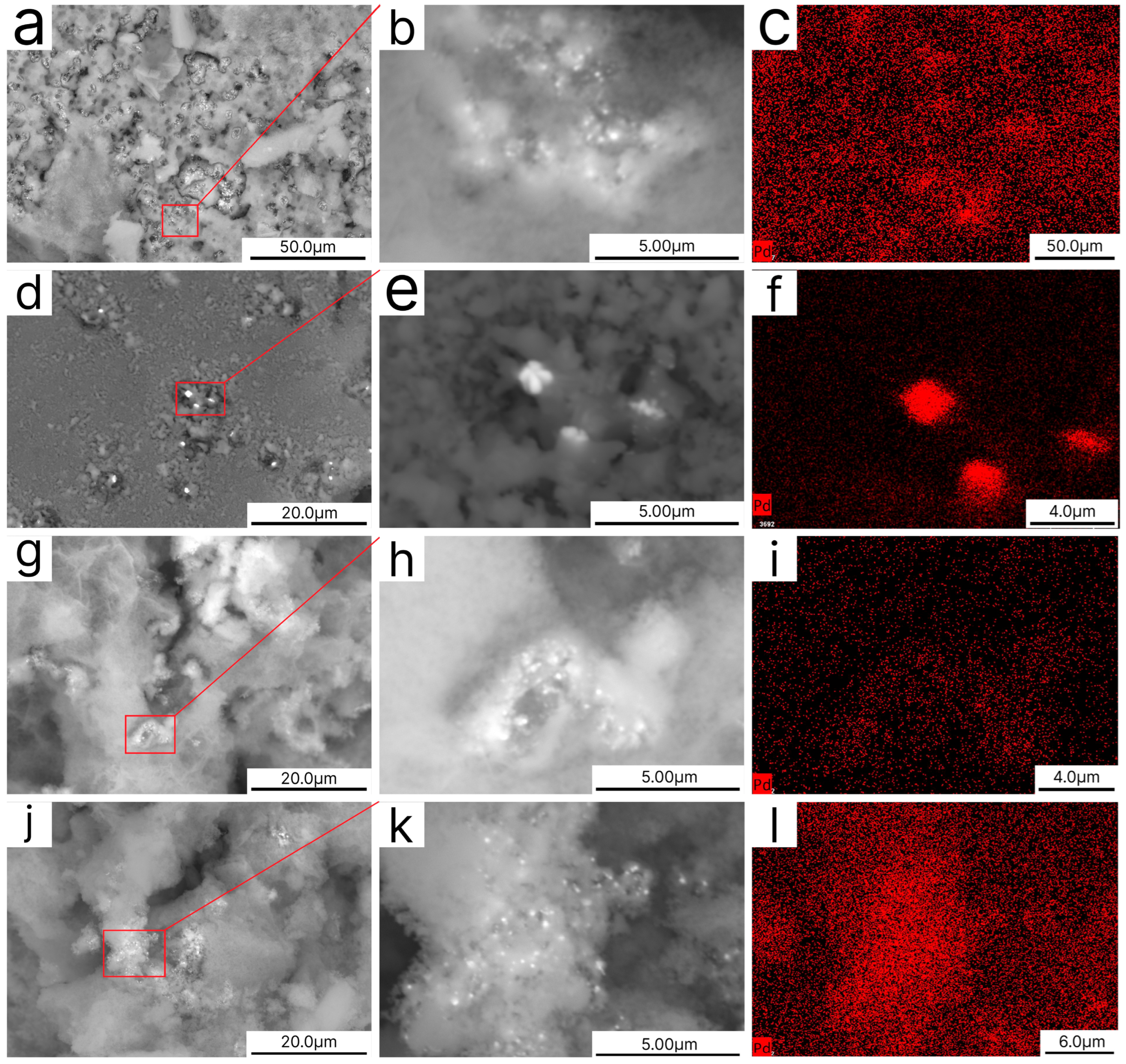
Scanning Electron Microscopy (SEM) micrographs of all catalysts, acquired in backscattered electron (BSE) mode, revealed bright regions attributable to the presence of atoms with a higher density, which exhibit strong electron reflectance. Energy Dispersive X-ray (EDX) spectroscopy confirmed that these regions corresponded to palladium particles (Figure 2). Furthermore, rounded voids, with diameters consistent with the size of the bacterial templates (e.g., up to 1 µm for P. yeei cells, which typically form sarcina clusters with a total size of 2–3 µm), were observed as distributed across the material surfaces. These voids likely originated from the destruction of the silica shells during the thermal treatment for bacterial removal or as a result of the mechanical impact. An analysis of the mapped region in Figure 2f reveals that the deposited silica forms a terrace layer, including the Pd-containing spheres situated beneath it. Therefore, Pd nanoparticles are only detectable in areas where this terrace has been disrupted or where the spheres have been fractured.
Transmission electron microscopy (TEM) of samples I–IV revealed dark clusters, indicative of heavier elements. The complex morphology of the catalysts, wherein palladium was concentrated within silica in places of bacterial templates, hindered an accurate size determination for the majority of Pd nanoparticles (Pd NPs). Nevertheless, the sizes of several isolated nanoparticles could be measured (Figure 3c,d). The average Pd nanoparticle size for all samples was therefore determined from these micrographs, featuring distinct, isolated particles. A previous study [17] reported an average palladium nanoparticle (Pd NP) size of 4 nm on a bacterial Pd/P. yeei catalyst. The present work demonstrates that with the calcination process, the temperature does not significantly influence Pd NP sizes. After the treatment at 600 °C, the average size was 5 ± 1 nm, independent of the calcination atmosphere (Figure 3b,f). A further increase in temperature resulted in the nanoparticles’ growth to an average of 9 nm (Figure 3d,h).
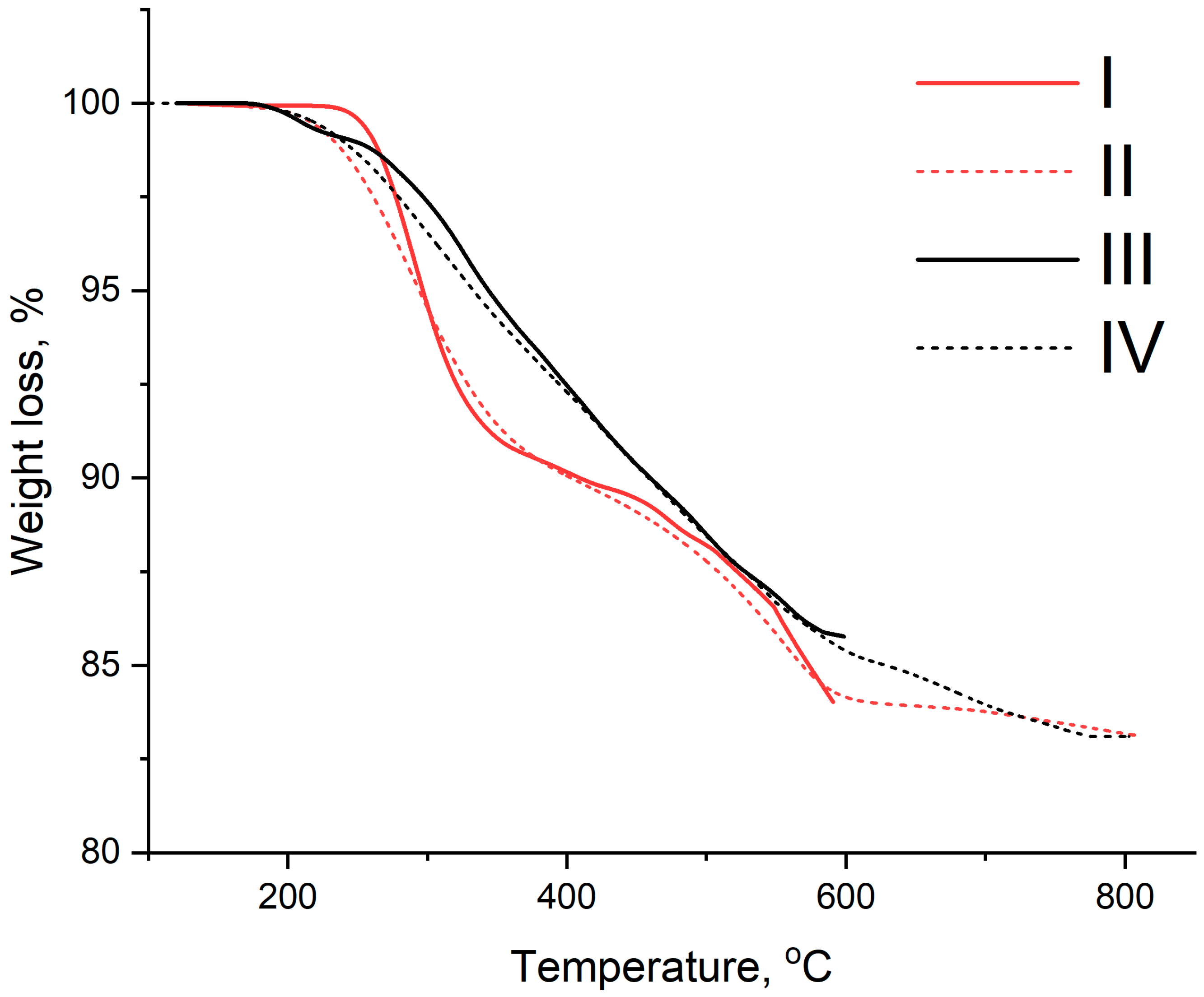
A thermogravimetric analysis (TGA) of the Pd/P. yeei@ORMOSIL samples was performed under conditions replicating the stage 3 heat treatment. Under a nitrogen atmosphere, a uniform weight loss occurred between 280 and 550 °C (Figure 4), attributable to the pyrolysis of bacterial cells, the thermal degradation of organics, and the structural changes in silica with the subsequent formation of a shell. Beyond 550 °C, the weight loss rate decreased significantly, indicating the substantial completion of these primary decomposition processes. In contrast, under an air atmosphere, a more rapid weight loss was observed between 280 and 350 °C, consistent with the combustion of organic components. A slight mass gain between 600 and 800 °C was also noted, likely due to the partial oxidation of palladium to its oxide.
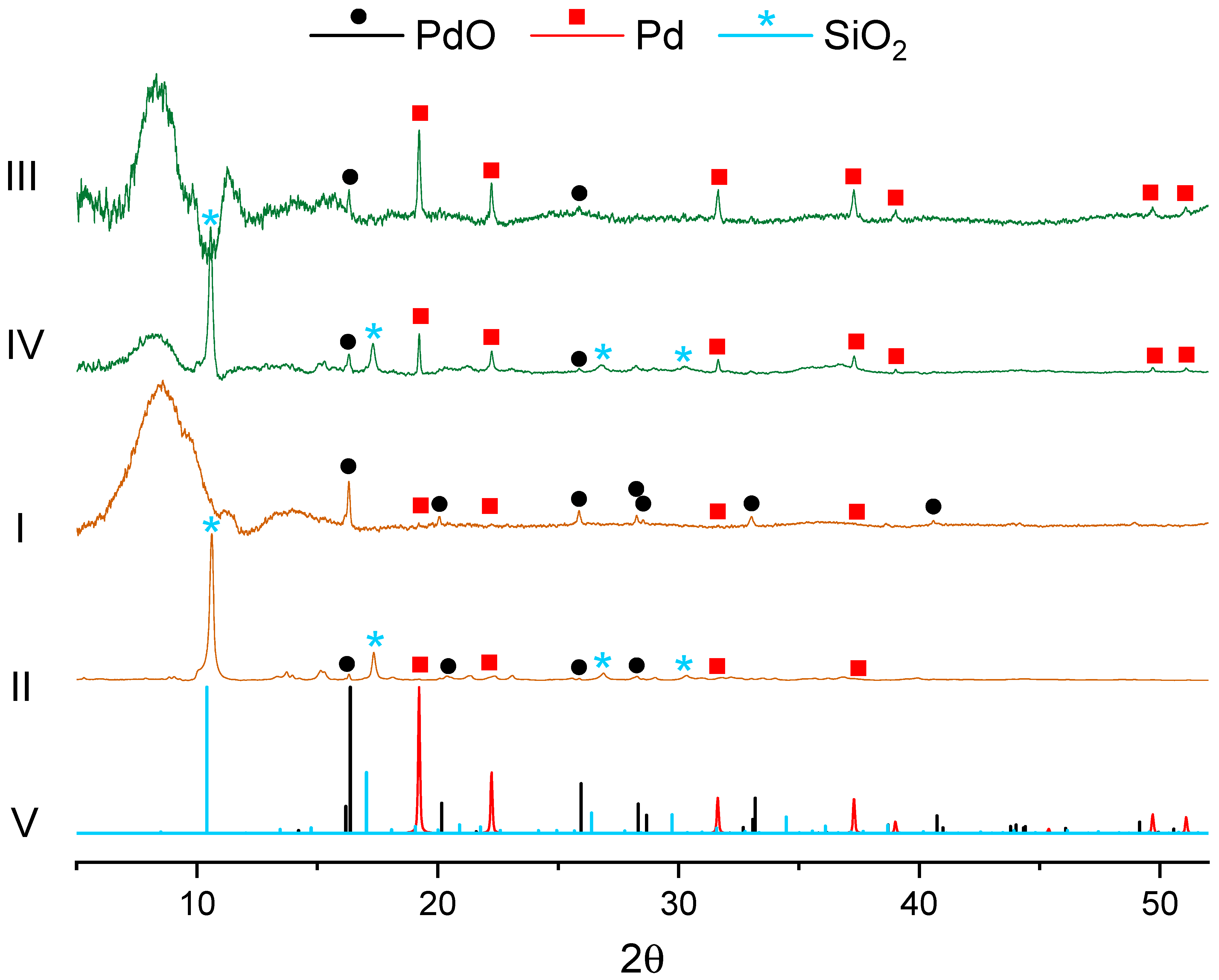
X-ray diffraction patterns of the catalyst (Figure 5) pretreated with nitrogen exhibit distinct reflections attributable to metallic palladium. For catalysts prepared at 600 °C, a broad halo is observed in the 2θ range of 5–10°, indicative of amorphous silica. In the sample pretreated at 800 °C, a decrease in the halo intensity is accompanied by the emergence of a sharp peak at 2θ ≈ 11°, corresponding to crystalline silicon. This phase evolution aligns with observations from a comparable rhodium-based core–shell system [20] and is consistently reproduced in air-pretreated catalysts at equivalent temperatures.
The principal distinction for the air-pretreated samples is the almost complete oxidation of metallic Pd(0) to PdO. Despite this, the most intense diffraction peak (for the model metallic fcc Pd pattern) is visible as traces in the 2θ ≈ 19° in both patterns.

3.3. Catalytic Tests of (I–IV) Pd/R@SiO2 Catalysts in the Suzuki–Miyaura Reaction

The catalytic performance of complexes I–IV was evaluated in the Suzuki–Miyaura coupling of phenylboronic acid with iodobenzene and nitroiodobenzene, as outlined in Scheme 1. The corresponding product yields are summarized in Table 1 (determined by GC-MS). The data indicate that catalysts subjected to the air pretreatment afforded lower yields than those pretreated under a nitrogen atmosphere. Furthermore, the product yield was found to decrease with increasing pretreatment temperatures for the nitrogen atmosphere.
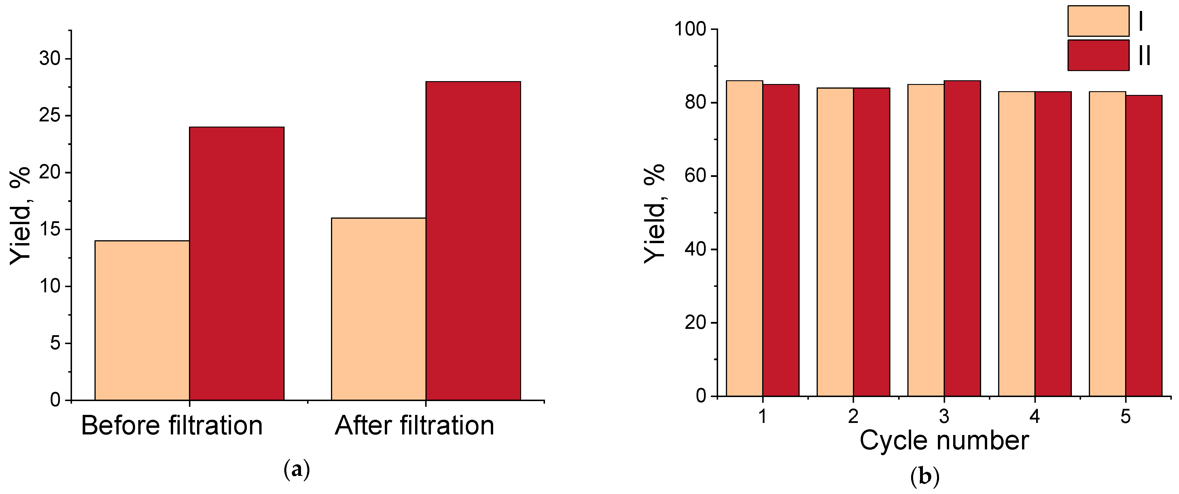
The extent of palladium leaching was evaluated by employing a hot filtration test (Figure 6a). The reaction mixture was stirred at 1200 rpm and heated to 80 °C. Following a 30 min reaction period, the catalyst was separated by hot filtration. To assess the activity of any leached species, the filtrate was subsequently stirred at 80 °C for a further 150 min, and the final yield was determined by GC/MS analysis. The stability of the Pd/R@SiO2 catalysts pretreated in air was evaluated using a “fresh-start” approach [21]. After each trial, a fresh portion of the substrates, base, and solvent was added to the reaction mixture, and the reaction was run again (Figure 6b). Both experiments were conducted using the biphenyl synthesis reaction.
Figure 6a indicates a slight increase in the biphenyl yield after the hot filtration of the air-pretreated catalysts, which points to negligible leaching of palladium nanoparticles. Furthermore, these catalysts maintained a stable performance over five consecutive reaction cycles in the fresh-start mode, with no significant decrease in the biphenyl yield.

4. Discussion

While the concept of core–shell catalysts is well-known, the synthesis methodology presented herein—utilizing bacterial templates for the subsequent formation of a silicate shell—represents a novel approach. This method aligns with green chemistry principles by employing non-toxic substances throughout the preparation, generating only the spent bacterial growth medium as waste. The described method is also energy-efficient, given that key stages such as nanoparticle formation and sol–gel condensation are carried out at room temperature, and only the final calcination step necessitates a thermal treatment. The total preparation time was 30 h, which was predominantly allocated to the sol–gel aging and the heat treatment.
As reported [16,17], palladium nanoparticles were localized extra-/intracellularly prior to encapsulation within the silica matrix. The thermal treatment of this composite material induced the combustion of the bacteria biomass and the formation of residue-supported palladium NPs, as demonstrated by SEM/EDX (Figure 2e,f). The composition of the resulting support was determined by the thermal pretreatment atmosphere [22,23]. Combustion in oxygen removed organics as gaseous CO2, nitrogen oxides, and H2O, yielding an inorganic ash. Conversely, pyrolysis in a nitrogen atmosphere facilitated the conversion of organic matter into a carbon species (soot). Thus, by varying the pretreatment conditions, palladium nanoparticles could be deposited onto different sites, either the inorganic ash or the carbonaceous soot of the resulting material, designated the R-residue.
Following the thermal treatment, palladium nanoparticles increased to average sizes of 5 nm at 600 °C and 9 nm at 800 °C, regardless of the atmosphere used (N2 or air). This modest increase in size stands in contrast to the significant agglomeration to ~30 nm reported for Pd catalysts treated in oxygen [24], which is more typical of thermal sintering [25,26]. However, the present results align with findings under similar high-temperature conditions; for instance, study [27] shows an average Pd NP size of 8 nm at 800 °C. Furthermore, support for limited coarsening comes from paper [28], where Pd nanoparticles on silica were observed to redisperse into smaller entities rather than agglomerate after treatment at 800 °C in oxygen for 4 h. This phenomenon is attributed to the low thermal stability of palladium oxide (PdO), which begins to decompose above 750 °C [25,26]. In the present study, following calcination in air at 800 °C for only 1 h, the catalyst’s XRD pattern exhibited a diminished PdO peak relative to the 600 °C-treated sample and the same trace of metallic Pd(0), which may be explained by several factors. Firstly, the calcination time may have been insufficient for complete PdO decomposition. Alternatively, if redispersion occurred, the resulting Pd(0) crystallites could be below the detection limit of XRD [29]. For crystallites of such small sizes, significant peak broadening occurs; when combined with the low palladium loading (1 wt%), the diffraction signal intensity may be indistinguishable from the background noise [30]. Furthermore, air passivation can form a thicker oxide layer on the nanoparticles. This surface layer can attenuate the intensity of the metallic palladium peaks via the X-rays passing through the oxide layer being absorbed, reducing the diffracted signal [31]. Similarly, a more crystalline and thicker silica shell layer may suppress the signal intensity for both palladium oxide and metallic palladium.
The thermogravimetric profile for the sample pretreated in air at 800 °C exhibited a minor mass gain above 600 °C, which can be attributed to the formation of palladium oxide. The oxidation behavior of palladium nanoparticles (NPs) is temperature-dependent. Below 200 °C, oxidation is limited to the surface of the Pd(0) NPs, whereas above 200 °C, the subsurface Pd(0) begins to oxidize. For a well-defined Pd/Al2O3 catalyst with a Pd NP size of 2.3 nm, the formation of a core–shell PdO@Pd structure was observed up to 400 °C in an oxygen atmosphere [32]. The oxidation of bulk palladium proceeds up to approximately 750 °C, above which the thermal decomposition of palladium oxide occurs [25,26,28]. In the present study, the palladium NPs are larger than those reported in the aforementioned work. Consequently, bulk oxidation is expected to proceed more slowly and at temperatures higher than 400 °C. This is consistent with the features observed in the TGA profiles above 600 °C, which are presumably attributable to this bulk oxidation process.
The catalytic evaluation in the Suzuki–Miyaura reaction revealed that the four catalysts, despite differences in their pretreatment atmosphere (nitrogen vs. oxygen), showed comparable yields. The XRD data confirmed that the pretreatment in oxygen led to the formation of a palladium oxide (PdO) phase. The consistent catalytic activity across the sample series indicates that PdO acts as a pre-catalyst for the Suzuki–Miyaura reaction. The catalytic cycle is initiated by the in situ reduction of PdO to the active Pd(0) species, likely mediated by the organoboronic acid and/or the base in conjunction with the alcohol [33]. This conclusion is reinforced by reference [34], which established that various forms of PdO, including both laboratory-synthesized and commercial varieties, serve as active catalysts for the Suzuki–Miyaura reaction, achieving high yields. The oxidized palladium atoms are more easily released into the solution phase and, at the same time, can favor the oxidative addition of arylboronic acid as reported [35].
Bacterial cells play a crucial role in the catalyst preparation procedure. They produce and stabilize Pd nanoparticles (NPs) of a desired size, facilitate the formation of the organosilicon matrix, and, upon thermal treatment, prevent the NPs from significant aggregation.

5. Conclusions

This work demonstrates a sustainable method for fabricating core–shell catalysts using a bacterial template and sol–gel synthesis. This approach aligns with the principles of green chemistry, particularly in minimizing waste and enhancing resource efficiency. The principal conclusions are as follows:
  • The bacterial template effectively stabilizes the nanoparticle size, preventing significant agglomeration even after the calcination procedure;
  • The sol–gel encapsulation does not significantly change the Pd nanoparticle size distribution, with sizes remaining in the 4–5 nm range before and after the process;
  • The post-synthetic thermal treatment provides a viable pathway for modulating the nanoparticle size, offering a means to tailor the catalyst for reactions beyond cross-coupling;
  • The resulting catalyst, with a low palladium loading of approximately 1 wt%, exhibits high activity in the Suzuki–Miyaura reaction, achieving competitive yields.
Future work will focus on expanding the scope of the pretreatment procedures to systematically correlate them with the catalytic performance. The stability and reusability of the catalyst will be rigorously evaluated over at least 10 catalytic cycles in the Suzuki–Miyaura reaction to assess its leaching resistance. Furthermore, a detailed investigation into the mechanisms responsible for the sintering resistance and the role of the ash in nanoparticle stabilization will be undertaken.

Author Contributions

Conceptualization, V.N.S. and O.A.K.; methodology, V.N.S., P.V.R. and O.A.K.; software, V.N.S. and P.V.R.; validation, V.N.S. and O.A.K.; formal analysis, P.V.R. and V.A.P.; investigation, V.N.S., P.V.R., and V.A.P.; resources, V.N.S.; data curation, V.N.S.; writing—original draft preparation, V.N.S., P.V.R. and O.A.K.; writing—review and editing, V.N.S., P.V.R. and O.A.K.; visualization, P.V.R. and O.A.K.; supervision, V.N.S. and O.A.K.; project administration, V.N.S.; funding acquisition, V.N.S. All authors have read and agreed to the published version of this manuscript.

Funding

This research was funded by the Russian Science Foundation—grant number 25-23-20040, https://rscf.ru/en/project/25-23-20040 (accessed on 19 November 2025), and support from the government of the Tula region.

Data Availability Statement

The raw data supporting the conclusions of this article will be made available by the authors on request.

Acknowledgments

X-ray diffraction studies were performed using the unique scientific facility Kurchatov synchrotron radiation source “KISI-Kurchatov” of the National Research Center “Kurchatov Institute”. The studies using the electron microscopy method were performed by Nina M. Ivanova at the Shared Research Center (Department of Structural Studies) of N.D. Zelinsky Institute of Organic Chemistry RAS, Moscow. The authors thank Valentine P. Ananikov for helpful discussions.

Conflicts of Interest

The authors declare no conflicts of interest.

Abbreviations

The following abbreviations are used in this manuscript:
TEOSTetraethoxysilane
MTESMethyltriethoxysilane
XRDX-Ray Diffraction
TEMTransmission Electronic Microscopy
SEMScanning Electronic Microscopy
EDXEnergy Dispersive X-Ray
TGAThermogravimetric Analysis
NPNanoparticle
BSEBackscattered Electron
ICP-OESOptical Emission Spectrometer with Inductively Coupled Plasma
GC-MSGas Chromatography–Mass Spectrometry

References

  1. Kader, D.A.; Sidiq, M.K.; Taher, S.G.; Aziz, D.M. Recent advances in palladium-catalyzed Suzuki-Miyaura cross-coupling reactions: Exploration of catalytic systems, reaction parameters, and ligand influences: A review. J. Organomet. Chem. 2025, 15, 123569. [Google Scholar] [CrossRef]
  2. Farhang, M.; Akbarzadeh, A.R.; Rabbani, M.; Ghadiri, A.M. A retrospective-prospective review of Suzuki–Miyaura reaction: From cross-coupling reaction to pharmaceutical industry applications. Polyhedron 2022, 227, 116124. [Google Scholar] [CrossRef]
  3. Bourouina, A.; Meille, V.; de Bellefon, C. About Solid Phase vs. Liquid Phase in Suzuki-Miyaura Reaction. Catalysts 2019, 9, 60. [Google Scholar] [CrossRef]
  4. Li, Z.; Gelbaum, C.; Heaner, W.L.; Fisk, J.; Jaganathan, A.; Holden, B.; Pollet, P.; Liotta, C.L. Palladium-Catalyzed Suzuki Reactions in Water with No Added Ligand: Effects of Reaction Scale, Temperature, pH of Aqueous Phase, and Substrate Structure. Org. Process Res. Dev. 2016, 20, 1489–1499. [Google Scholar] [CrossRef]
  5. Marquard, A.N.; Slaymaker, L.E.; Hamers, R.J.; Goldsmith, R.H. Investigation of activity, stability, and degradation mechanism of surface-supported Pd-PEPPSI complexes for Suzuki-Miyaura coupling. Mol. Catal. 2017, 429, 10–17. [Google Scholar] [CrossRef]
  6. Mpungose, P.P.; Vundla, Z.P.; Maguire, G.E.M.; Friedrich, H.B. The Current Status of Heterogeneous Palladium Catalysed Heck and Suzuki Cross-Coupling Reactions. Molecules 2018, 23, 1676. [Google Scholar] [CrossRef] [PubMed]
  7. Das, S.; Perez-Ramirez, J.; Gong, J.; Dewangan, N.; Hidajat, K.; Gates, B.C.; Kawi, S. Core-shell structured catalysts for thermocatalytic, photocatalytic, and electrocatalytic conversion of CO2. Chem. Soc. Rev. 2020, 49, 2937–3004. [Google Scholar] [CrossRef]
  8. Egan-Morriss, C.; Kimber, R.L.; Powell, N.A.; Lloyd, J.R. Biotechnological synthesis of Pd-based nanoparticle catalysts. Nanoscale Adv. 2022, 4, 654–679. [Google Scholar] [CrossRef]
  9. Roy, A.; Elzaki, A.; Tirth, V.; Kajoak, S.; Osman, H.; Algahtani, A.; Islam, S.; Faizo, N.L.; Khandaker, M.U.; Islam, M.N.; et al. Biological Synthesis of Nanocatalysts and Their Applications. Catalysts 2021, 11, 1494. [Google Scholar] [CrossRef]
  10. Qamar, S.U.R.; Ahmad, J.N. Nanoparticles: Mechanism of biosynthesis using plant extracts, bacteria, fungi, and their applications. J. Mol. Liq. 2021, 334, 116040. [Google Scholar] [CrossRef]
  11. Koul, B.; Poonia, A.K.; Yadav, D.; Jin, J.O. Microbe-Mediated Biosynthesis of Nanoparticles: Applications and Future Prospects. Biomolecules 2021, 11, 886. [Google Scholar] [CrossRef] [PubMed]
  12. Coccia, F.; Mascitti, A.; Caporali, S.; Polito, L.; Porcheddu, A.; Di Nicola, C.; Tonucci, L.; d’Alessandro, N. Wool-supported Pd and Rh nanoparticles for selective hydrogenation of maleic acid to succinic acid in batch and flow systems. RSC Adv. 2025, 15, 36760–36768. [Google Scholar] [CrossRef]
  13. Singh, M.; Sinha, A.; Qazi, A.; Soni, R.; Rana, V.; Tyagi, T. Bacteria and their mediated nanoparticles for the environmental sustainable development to support SDGs. In Nanoparticles Synthesis by Soil Microbes; Elsevier: Amsterdam, The Netherlands, 2025; pp. 253–270. [Google Scholar]
  14. Rybochkin, P.V.; Kamanina, O.A.; Lantsova, E.A.; Arlyapov, V.A.; Saverina, E.A. Characterization of the catalytic ability and surface properties of a heterogeneous biocatalyst obtained by the sol-gel method. J. Sol-Gel Sci. Technol. 2022, 108, 310–319. [Google Scholar] [CrossRef]
  15. Lantsova, E.A.; Rybochkin, P.V.; Saverina, E.A.; Kamanina, O.A. Biohybrid silicon-organic materials architecture obtained using various structure-affecting agents. J. Sol-Gel Sci. Technol. 2024, 110, 134–141. [Google Scholar] [CrossRef]
  16. Rybochkin, P.V.; Perchikov, R.N.; Karlinskii, B.Y.; Kamanina, O.A.; Arlyapov, V.A.; Kashin, A.S.; Ananikov, V.P. Aerobic bacteria-supported biohybrid palladium catalysts for efficient cross-coupling reactions. J. Catal. 2024, 429, 115238. [Google Scholar] [CrossRef]
  17. Kamanina, O.A.; Rybochkin, P.V.; Borzova, D.V.; Soromotin, V.N.; Galushko, A.S.; Kashin, A.S.; Ivanova, N.M.; Zvonarev, A.N.; Suzina, N.E.; Holicheva, A.A.; et al. Sustainable catalysts in a short time: Harnessing bacteria for swift palladium nanoparticle production. Nanoscale 2025, 17, 5289–5300. [Google Scholar] [CrossRef]
  18. Kamanina, O.A.; Fedoseeva, D.G.; Rogova, T.V.; Ponamoreva, O.N.; Blokhin, I.V.; Machulin, A.V.; Alferov, V.A. Synthesis of organosilicon sol-gel matrices and preparation of heterogeneous biocatalysts based on them. Russ. J. Appl. Chem. 2014, 87, 761–766. [Google Scholar] [CrossRef]
  19. Ponamoreva, O.N.; Kamanina, O.A.; Alferov, V.A.; Machulin, A.V.; Rogova, T.V.; Arlyapov, V.A.; Alferov, S.V.; Suzina, N.E.; Ivanova, E.P. Yeast-based self-organized hybrid bio-silica sol-gels for the design of biosensors. Biosens. Bioelectron. 2015, 67, 321–326. [Google Scholar] [CrossRef]
  20. Palomo, J.; Caspers, M.; Urakawa, A. Nanostructured Rh/SiC@SiO2 core@shell catalysts for microwave-assisted dry reforming of methane. Catal. Today 2025, 460, 115464. [Google Scholar] [CrossRef]
  21. Mironenko, R.M.; Saybulina, E.R.; Trenikhin, M.V.; Izmailov, R.R.; Lotsman, K.A.; Rodygin, K.S.; Ananikov, V.P. Comparing Separation vs. Fresh Start to Assess Reusability of Pd/C Catalyst in Liquid-Phase Hydrogenation. ChemCatChem 2021, 13, 3656–3661. [Google Scholar] [CrossRef]
  22. Puri, L.; Hu, Y.; Naterer, G. Critical review of the role of ash content and composition in biomass pyrolysis. Front. Fuels 2024, 2, 1378361. [Google Scholar] [CrossRef]
  23. Rong, H.; Wang, T.; Zhou, M.; Wang, H.; Hou, H.; Xue, Y. Combustion Characteristics and Slagging during Co-Combustion of Rice Husk and Sewage Sludge Blends. Energies 2017, 10, 438. [Google Scholar] [CrossRef]
  24. Kalishyn, Y.Y.; Ordynskyi, V.V.; Ishchenko, M.V.; Bychko, I.B.; Kaidanovych, Z.V.; Trypolskyi, A.I.; Strizhak, P.E. Synthesis and Thermal Stability of Palladium Nanoparticles Supported on γ-Al2O3. Curr. Nanomater. 2020, 5, 79–90. [Google Scholar] [CrossRef]
  25. Rahmati, M.; Safdari, M.-S.; Fletcher, T.H.; Argyle, M.D.; Bartholomew, C.H. Chemical and Thermal Sintering of Supported Metals with Emphasis on Cobalt Catalysts During Fischer–Tropsch Synthesis. Chem. Rev. 2020, 120, 4455–4533. [Google Scholar] [CrossRef]
  26. Moulijn, J.A.; Van Diepen, A.; Kapteijn, F. Catalyst deactivation: Is it predictable?: What to do? Appl. Catal. A Gen. 2001, 212, 3–16. [Google Scholar] [CrossRef]
  27. Rostovshchikova, T.N.; Shilina, M.I.; Gurevich, S.A.; Yavsin, D.A.; Veselov, G.B.; Stoyanovskii, V.O.; Vedyagin, A.A. Studies on High-Temperature Evolution of Low-Loaded Pd Three-Way Catalysts Prepared by Laser Electrodispersion. Materials 2023, 16, 3501. [Google Scholar] [CrossRef]
  28. Seo, C.Y.; Chen, X.; Sun, K.; Allard, L.F.; Fisher, G.B.; Schwank, J.W. Palladium redispersion at high temperature within the Pd@SiO2 core@shell structure. Catal. Commun. 2018, 108, 73–76. [Google Scholar] [CrossRef]
  29. Valerio, A.; Trindade, F.J.; Penacchio, R.F.S.; Cisi, B.; Damasceno, S.; Estradiote, M.B.; Rodella, C.B.; Ferlauto, A.S.; Kycia, S.W.; Morelhao, S.L. Implications of size dispersion on X-ray scattering of crystalline nanoparticles: CeO2 as a case study. J. Appl. Crystallogr. 2024, 57, 793–807. [Google Scholar] [CrossRef]
  30. Vorontsov, A.V.; Tsybulya, S.V. Influence of Nanoparticles Size on XRD Patterns for Small Monodisperse Nanoparticles of Cu0 and TiO2 Anatase. Ind. Eng. Chem. Res. 2018, 57, 2526–2536. [Google Scholar] [CrossRef]
  31. Dastoom, Z. Production of Ni0.5Co0.5Fe2O4/activated carbon@chitosan magnetic nanobiocomposite as a novel adsorbent of methylene blue in aqueous solutions. Sci. Rep. 2023, 13, 6137. [Google Scholar] [CrossRef]
  32. Usoltsev, O.; Stoian, D.; Skorynina, A.; Kozyr, E.; Njoroge, P.N.; Pellegrini, R.; Groppo, E.; van Bokhoven, J.A.; Bugaev, A. Restructuring of Palladium Nanoparticles during Oxidation by Molecular Oxygen. Small 2024, 20, e2401184. [Google Scholar] [CrossRef] [PubMed]
  33. Johansson Seechurn, C.C.C.; Sperger, T.; Scrase, T.G.; Schoenebeck, F.; Colacot, T.J. Understanding the Unusual Reduction Mechanism of Pd(II) to Pd(I): Uncovering Hidden Species and Implications in Catalytic Cross-Coupling Reactions. J. Am. Chem. Soc. 2017, 139, 5194–5200. [Google Scholar] [CrossRef]
  34. Amoroso, F.; Colussi, S.; Del Zotto, A.; Llorca, J.; Trovarelli, A. PdO hydrate as an efficient and recyclable catalyst for the Suzuki–Miyaura reaction in water/ethanol at room temperature. Catal. Commun. 2011, 12, 563–567. [Google Scholar] [CrossRef]
  35. Del Zotto, A.; Zuccaccia, D. Metallic palladium, PdO, and palladium supported on metal oxides for the Suzuki–Miyaura cross-coupling reaction: A unified view of the process of formation of the catalytically active species in solution. Catal. Sci. Technol. 2017, 7, 3934–3951. [Google Scholar] [CrossRef]
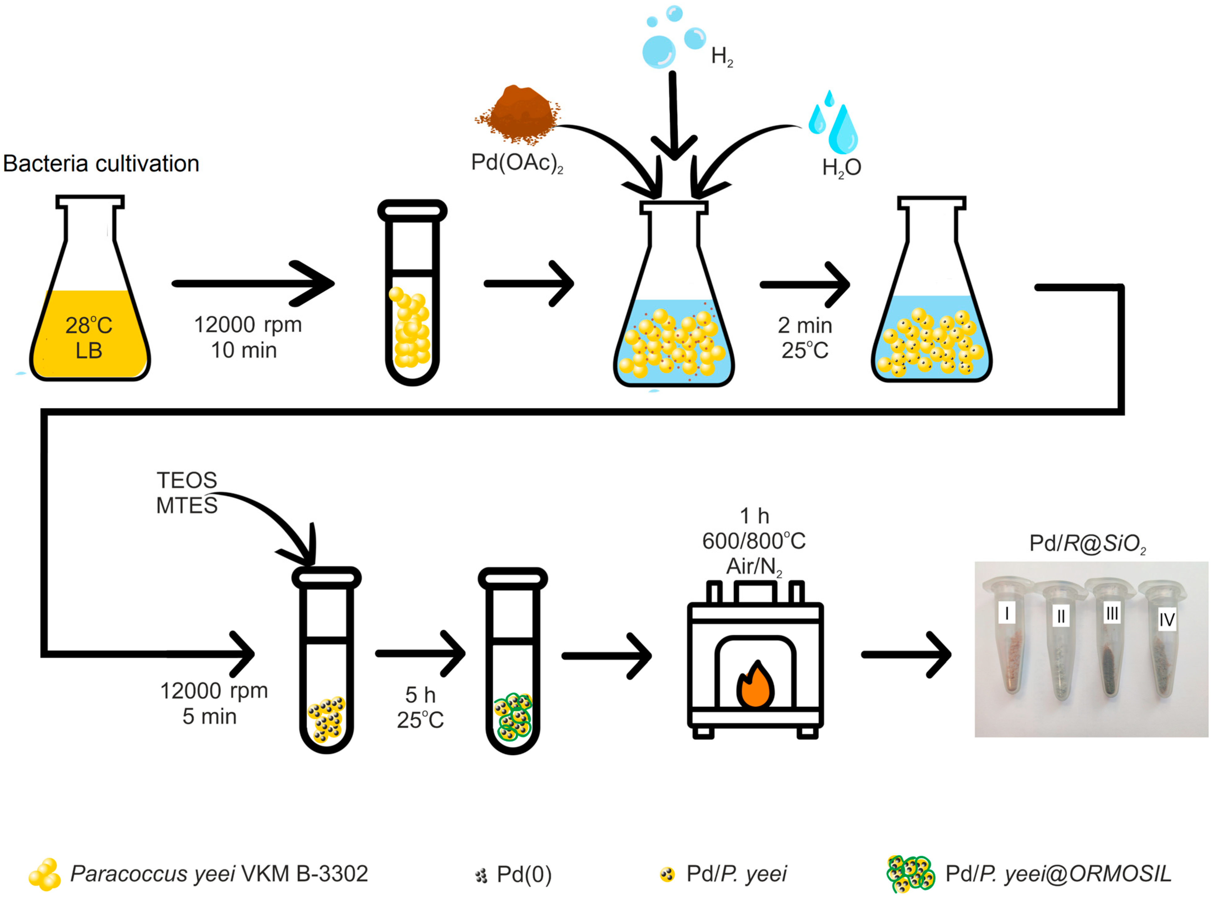
Figure 1. Preparation route of Pd/R@SiO2 catalysts in different atmospheres and temperatures: I—O2, 600 °C; II—O2, 800 °C; III—N2, 600 °C; and IV—N2, 800 °C.
Chemistry 07 00188 g001
Figure 2. SEM images and EDX maps of Pd/R@SiO2 treated catalyst: (ac)—O2 at 600 °C; (df)—O2 at 800 °C; (gi)—N2 at 600 °C; and (jl)—N2 at 800 °C.
Chemistry 07 00188 g002
Figure 3. TEM images of Pd/R@SiO2 catalyst and Pd NP size: (a,b)—O2 at 600 °C; (c,d)—O2 at 800 °C; (e,f)—N2 at 600 °C; and (g,h)—N2 at 800 °C.
Chemistry 07 00188 g003
Figure 4. TGA patterns of Pd/P. yeei@ORMOSIL material during thermal treatment in different conditions: I—O2, 600 °C; II—O2, 800 °C; III—N2, 600 °C; and IV—N2, 800 °C.
Chemistry 07 00188 g004
Figure 5. XRD patterns of Pd/R@SiO2 catalyst pretreated in different conditions: III—N2 at 600 °C; IV—N2 at 800 °C; I—O2 at 600 °C; II—O2 at 800 °C; and V—model patterns for PdO, fcc-Pd, and SiO2 phases.
Chemistry 07 00188 g005
Scheme 1. The Suzuki–Miyaura reaction.
Chemistry 07 00188 sch001
Figure 6. Leaching test (a) and stability test (b) for air-pretreated Pd/R@SiO2 catalysts: I—at 600 °C and II—at 800 °C.
Chemistry 07 00188 g006
Table 1. Product yields of (I–IV) Pd/R@SiO2 catalysts in Suzuki–Miyaura reaction.
I (600 °C, O2)II (800 °C, O2)III (600 °C, N2)IV (800 °C, N2)
Chemistry 07 00188 i00184%85%95%89%
Chemistry 07 00188 i00272%75%84%80%
Disclaimer/Publisher’s Note: The statements, opinions and data contained in all publications are solely those of the individual author(s) and contributor(s) and not of MDPI and/or the editor(s). MDPI and/or the editor(s) disclaim responsibility for any injury to people or property resulting from any ideas, methods, instructions or products referred to in the content.