Article

Synthesis, Dynamic NMR Characterization, and XRD Study of 2,4-Difluorobenzoyl-Substituted Piperazines

by
Martin Köckerling
1 and
Constantin Mamat
2,3,*
1
Institut für Chemie—Festkörperchemie, Universität Rostock, Albert-Einstein-Straße 3a, D-18059 Rostock, Germany
2
Helmholtz-Zentrum Dresden-Rossendorf, Institut für Radiopharmazeutische Krebsforschung, Bautzner Landstraße 400, D-01328 Dresden, Germany
3
Fakultät Chemie und Lebensmittelchemie, Technische Universität Dresden, D-01062 Dresden, Germany
*
Author to whom correspondence should be addressed.
Chemistry 2025, 7(5), 162; https://doi.org/10.3390/chemistry7050162
Submission received: 28 August 2025 / Revised: 29 September 2025 / Accepted: 30 September 2025 / Published: 3 October 2025
(This article belongs to the Section Molecular Organics)

Abstract

Five different 2,4-difluorobenzamide derivatives were synthesized and fully characterized by 1H/13C/19F/2D NMR spectroscopy using DMSO-d6 as solvent and MS. All compounds occur as rotation conformers resulting from the partial amide double bond with a solvent-dependent coalescence point. Temperature-dependent 1H NMR techniques, as well as EXSY, were applied to determine the rate constants of exchange, and the resulting activation energy barriers were calculated. Regarding the N,N-diacylated piperazine, both conformers (syn and anti) were found in solution, whereas only the anti-conformer was found in the crystals. This result was verified by an XRD analysis. Single crystals of N,N-bis(2,4-difluorobenzoyl)piperazine 3b (monoclinic, space group P21/c, a = 7.2687(3), b = 17.2658(8), c = 6.9738(3) Å, β = 115.393(2)°, V = 790.65(6) Å3, Z = 4, Dobs = 1.530 g/cm3) were obtained from a saturated chloroform solution.

1. Introduction

Piperazines belong to saturated six-membered heterocyclic compounds containing two nitrogen atoms [1]. This scaffold has been integrated as a fundamental structural element into bioactive and pharmacologically relevant compounds [2,3,4,5]. N- and N,N-functionalized piperazine derivatives are used as intermediates in organic reactions [6,7] or for polymerization processes, ref. [8] as ligands in coordination chemistry [9,10,11] or as a scaffold for peptide syntheses and modifications [12,13]. Piperazines are also in use for the introduction of fluorescent dyes [14,15] or for the preparation of radiolabeled compounds [16,17,18,19,20]. They serve as skeletons in pesticides, pharmaceuticals [21,22,23], and other drugs [24,25,26] or basic scaffolds in medicinal chemistry [27,28,29]. Furthermore, the N,N-substituted benzamide scaffold is found in various natural products and drugs, such as in Ampakines [30,31].
Especially N-acylated piperazines containing a fluorobenzoyl or difluorobenzoyl group are of interest (Figure 1), as they function as serotonin 5-HT2A receptor antagonists [32], cardiotonic agents [33], adenosine A2A receptor antagonists [34], radiosensitizers for tumor therapy [35], or as a kinase inhibitor for Jun-kinase [36]. The comprehension of the conformational behavior of such functionalized piperazines is mandatory not only to explain biological and/or pharmacological effects, but also for applications in material sciences.
Secondary and tertiary N-acylated piperazines, in general, and especially N-benzoylated piperazines, exhibit a more complex conformational behavior due to the hindered rotation of the C–N amide bond. This phenomenon in amides is well known and has far-reaching implications for peptide and protein folding [42]. Additionally, the conformation of the piperazine ring itself has to be considered. However, NMR properties of such benzamides were only rarely studied in the past. N,N-dimethyl and N,N-diethyl benzamides represent the best characterized compounds [43,44,45,46]. The NMR behavior of selected benzoylpiperazines [47], which were also used as prosthetic groups for 18F-radiolabeling [19], has been investigated. To expand the scope and to obtain a deeper insight into the conformational behavior of N- and N,N-substituted piperazines, two 2,4-difluorobenzoyl-containing derivatives were prepared and fully characterized in terms of their particular NMR behavior and compared with three N,N-disubstituted amides.

2. Materials and Methods

2.1. General

The starting materials were purchased from commercial suppliers and used without further purification unless otherwise specified. Anhydrous THF was purchased from Acros Organics. Mass spectrometric (MS) data was obtained on a mass spectrometer (expression CMS, Advion Interchim, Montluçon, France) by electrospray ionization (ESI). The melting points were determined on a Galen III melting point apparatus (Cambridge Instruments/Leica, Wetzlar, Germany) and are uncorrected. Chromatographic separations were performed using an automated flash chromatography system Selekt (Biotage, Uppsala, Sweden) with Sfär cartridges (Biotage, Uppsala, Sweden). TLC detections were performed using Merck (Darmstadt, Germany) Silica Gel 60 F254 sheets, respectively. TLCs were developed by visualization under UV light (λ = 254 nm).

2.2. VT-NMR Measurements

NMR spectra of all compounds were recorded on an Agilent DD2-400 MHz NMR spectrometer with a 5 mm ProbeOne probe. Deuterated solvents were purchased from deutero. Chemical shifts in the 1H, 19F, and 13C spectra were reported in parts per million (ppm) using TMS as an internal standard for 1H/13C and CFCl3 for 19F spectra at 25 °C unless otherwise stated. Spectra were calibrated to their solvent signals. Dynamic NMR measurements were carried out in a temperature range of 253–393 K using the Agilent VT accessory with a digital temperature controller. The TC was estimated from the NMR spectrum, which showed the collapse of the methylene signals from the piperazine moiety, and Δν values were extracted from the maximum split signals of the piperazine methylene protons, which remained unchanged with decreasing temperature. Values of the exchange rate kexc at TC were calculated using equation kexc = π∙Δν/21/2 [48,49]. Values of the free activation energy, ΔGǂ, were obtained by solving the Eyring equation (1) for ΔGǂ (2) [48,50]. in accordance with other studies on amide cis/trans isomerization, the transmission coefficient κ was assumed to be 1 [51].
k e x c = κ k B T C h e Δ G ǂ R T C
Δ G ǂ = ln κ k B T C h k e x c R T C

2.3. Single Crystal X-Ray Diffraction

Diffraction data was collected with a Bruker-Nonius Apex-II-diffractometer using graphite-monochromated MoKα radiation (λ = 0.71073 Å). The diffraction measurement was performed at –150 °C. The unit cell dimensions were refined using the angular settings of 9936 reflections for 3b. The structure was solved by direct methods and refined against F2 using full-matrix least-squares with the program suites from G. M. Sheldrick [51,52]. All non-hydrogen atoms were refined anisotropically; all hydrogen atoms were placed on geometrically calculated positions and refined using riding models. CCDC 2238312 contains the supplementary crystallographic data for compound 3b. This data can be obtained free of charge from The Cambridge Crystallographic Data Centre via www.ccdc.cam.ac.uk/data_request/cif (accessed on 27 August 2025).

2.4. Synthesis of N-(2,4-Difluorobenzoyl)piperazine (3a)

Piperazine (5 equiv.) was dissolved in absolute chloroform, 2,4-difluorobenzoyl chloride (1 equiv.) was added slowly at 0 °C, and the mixture was allowed to stir at 0 °C for another 5 h and at rt overnight. Afterwards, the solvent was removed and the crude mixture of products was purified via automated column chromatography, yielding N-(2,4-difluorobenzoyl)piperazine (3a) as a colorless solid in a yield of 66% and the N,N-diacylated compound 3b (yield 10%). Mp > 260 °C; 1H NMR (400 MHz, CDCl3): δ = 2.92 (br. s, 2H, NCH2), 3.03 (br. s, 2H, NCH2), 3.39 (br. s, 2H, NCH2), 3.85 (br. s, 2H, NCH2), 6.85 (t, J = 9.0 Hz, 1H, ArH), 6.96 (t, J = 8.3 Hz, 1H, ArH), 7.40 (m, 1H, ArH) ppm; 13C NMR (101 MHz, CDCl3): δ = 42.4, 45.3, 45.8, 47.4 (4 × NCH2), 104.4 (t, JC,F = 26 Hz, CHAr), 112.5 (dd, JC,F = 3 Hz, JC,F = 22 Hz, CHAr), 120.1 (dd, JC,F = 4 Hz, JC,F = 18 Hz, CAr), 130.8 (m, CHAr), 158.7 (dt, JC,F = 253 Hz, JC,F = 11 Hz, CAr), 164.0 (dt, JC,F = 251 Hz, JC,F = 12 Hz, CAr), 164.6 (C = O) ppm; MS (ESI+): m/z = 227 [M+H]+.

2.5. Synthesis of N,N-Bis(2,4-difluorobenzoyl)piperazine (3b)

Piperazine (2 equiv.) was dissolved in absolute chloroform, 2,4-difluorobenzoyl chloride (1 equiv.) was added slowly at 0 °C, and the mixture was allowed to stir at rt overnight. Afterwards, the solvent was removed and the crude mixture of products was purified via automated column chromatography, yielding N,N-bis(2,4-difluorobenzoyl)piperazine (3b) in 44% yield as a colorless solid and the N-monoacylated compound 3a (9% yield) from the one-pot reaction. Mp 245–247 °C; 1H NMR (400 MHz, CDCl3): δ = 3.34 (br. s, 2H, NCH2), 2.44, 3.79, 3.89, 6.78–7.06 (m, 4H, ArH), 7.43 (dd, 3J = 7.5 Hz, 3JH,F = 14.9 Hz, 2H, ArH) ppm; 13C NMR (101 MHz, CDCl3): δ = 42.1, 42.5, 46.9, 47.5 (4 x NCH2), 104.4 (m, CHAr), 112.7 (dd, JC,F = 2 Hz, JC,F = 22 Hz, CHAr), 119.8 (d, JC,F = 18 Hz, CAr), 131.0 (m, CHAr), 158.7 (dt, JC,F = 206 Hz, JC,F = 12 Hz, CAr), 164.0 (dt, JC,F = 254 Hz, JC,F = 10 Hz, CAr), 164.7 (d, JC,F = 18 Hz, C = O) ppm; 19F NMR (378 MHz, CDCl3): δ = −110.7 (ArF), −106.0 (ArF) ppm; MS (ESI+): m/z = 267 [M+H]+.

2.6. Synthesis of N,N-Diethyl-2,4-difluorobenzamide (4)

Diethylamine (2 equiv.) was dissolved in absolute chloroform, 2,4-difluorobenzoyl chloride (1 equiv.) was added slowly at 0 °C, and the mixture was allowed to stir at rt overnight. Afterwards, the solvent was removed and the crude mixture of products was purified via automated column chromatography, yielding N,N-diethyl-2,4-difluorobenzamide (4) in 86% yield as a colorless oil. 1H NMR (400 MHz, DMSO-d6): δ = 1.00 (t, J = 7.1 Hz, 3H, CH3), 1.14 (t, J = 7.1 Hz, 3H, CH3), 3.12 (q, J = 7.1 Hz, 2H, NCH2), 3.45 (q, J = 7.1 Hz, 2H, NCH2), 7.16 (dt, J = 2.4 Hz, J = 8.6 Hz, 1H, ArH), 7.35 (dt, 3J = 2.5 Hz, 3JH,F = 9.8 Hz, 2H, ArH), 7.44 (dd, J = 8.6 Hz, J = 14.9 Hz, 1H, ArH) ppm; 13C NMR (101 MHz, DMSO-d6): δ = 12.8, 13.9 (2 x CH3), 38.7, 42.6 (2 x NCH2), 104.4 (t, JC,F = 25.9 Hz, CHAr), 112.1 (dd, JC,F = 3.5 Hz, JC,F = 21.6 Hz, CHAr), 121.8 (dd, JC,F = 3.9 Hz, JC,F = 18.9 Hz, CAr), 129.6 (dd, JC,F = 5.9 Hz, JC,F = 9.9 Hz, CHAr), 157.8 (dt, JC,F = 246.7 Hz, JC,F = 12.6 Hz, CAr), 162.3 (dt, JC,F = 248.0 Hz, JC,F = 12.1 Hz, CAr), 164.1 (C = O) ppm; 19F NMR (378 MHz, DMSO-d6): δ = −113.1 (ArF), −108.7 (ArF) ppm; MS (ESI+): m/z = 214 [M+H]+.

2.7. Synthesis of (2,4-Difluorophenyl)(piperidin-1-yl)methanone (5)

Piperidine (2 equiv.) was dissolved in absolute chloroform, 2,4-difluorobenzoyl chloride (1 equiv.) was added slowly at 0 °C, and the mixture was allowed to stir at rt overnight. Afterwards, the solvent was removed and the crude mixture of products was purified via automated column chromatography, yielding (2,4-difluorophenyl)(piperidin-1-yl)methanone (5) in 82% yield as a colorless oil. 1H NMR (400 MHz, DMSO-d6): δ = 1.00 (t, J = 7.1 Hz, 3H, CH3), 1.14 (t, J = 7.1 Hz, 3H, CH3), 3.12 (q, J = 7.1 Hz, 2H, NCH2), 3.45 (q, J = 7.1 Hz, 2H, NCH2), 7.16 (dt, J = 2.4 Hz, J = 8.6 Hz, 1H, ArH), 7.35 (dt, 3J = 2.5 Hz, 3JH,F = 9.8 Hz, 2H, ArH), 7.44 (dd, J = 8.6 Hz, J = 14.9 Hz, 1H, ArH) ppm; 13C NMR (101 MHz, DMSO-d6): δ = 12.8, 13.9 (2 x CH3), 38.7, 42.6 (2 x NCH2), 104.4 (t, JC,F = 25.9 Hz, CHAr), 112.1 (dd, JC,F = 3.5 Hz, JC,F = 21.6 Hz, CHAr), 121.8 (dd, JC,F = 3.9 Hz, JC,F = 18.9 Hz, CAr), 129.6 (dd, JC,F = 5.9 Hz, JC,F = 9.9 Hz, CHAr), 157.8 (dt, JC,F = 246.7 Hz, JC,F = 12.6 Hz, CAr), 162.3 (dt, JC,F = 248.0 Hz, JC,F = 12.1 Hz, CAr), 164.1 (C = O) ppm; 19F NMR (378 MHz, DMSO-d6): δ = −113.1 (ArF), −108.7 (ArF) ppm; MS (ESI+): m/z = 226 [M+H]+.

2.8. Synthesis of (2,4-Difluorophenyl)(morpholino)methanone (6)

Morpholine (2 equiv.) was dissolved in absolute chloroform, 2,4-difluorobenzoyl chloride (1 equiv.) was added slowly at 0 °C, and the mixture was allowed to stir at rt overnight. Afterwards, the solvent was removed and the crude mixture of products was purified via automated column chromatography, yielding (2,4-difluorophenyl)(morpholino)methanone (6) in 87% yield as a colorless oil. 1H NMR (400 MHz, DMSO-d6): δ = 3.23 (t, J = 4.5 Hz, 2H, NCH2), 3.52 (t, J = 4.5 Hz, 2H, NCH2), 3.64 (s, 4H, OCH2), 7.19 (dt, J = 2.3 Hz, J = 8.5 Hz, 1H, ArH), 7.36 (dt, 3J = 2.3 Hz, 3JH,F = 9.9 Hz, 2H, ArH), 7.50 (dd, J = 8.3 Hz, J = 14.8 Hz, 1H, ArH) ppm; 13C NMR (101 MHz, DMSO-d6): δ = 41.9, 47.0 (2 x NCH2), 65.9, 66.2 (2 x OCH2), 104.4 (t, JC,F = 26.0 Hz, CHAr), 112.3 (dd, JC,F = 3.5 Hz, JC,F = 21.6 Hz, CHAr), 120.4 (dd, JC,F = 3.9 Hz, JC,F = 18.1 Hz, CAr), 130.5 (dd, JC,F = 5.6 Hz, JC,F = 10.1 Hz, CHAr), 158.0 (dt, JC,F = 248.2 Hz, JC,F = 12.8 Hz, CAr), 162.8 (dt, JC,F = 248.9 Hz, JC,F = 12.1 Hz, CAr), 163.3 (C = O) ppm; 19F NMR (378 MHz, DMSO-d6): δ = −112.0 (ArF), −107.8 (ArF) ppm; MS (ESI+): m/z = 267 [M+K+H]+.

3. Results and Discussion

3.1. Synthesis of the Piperazine Compounds

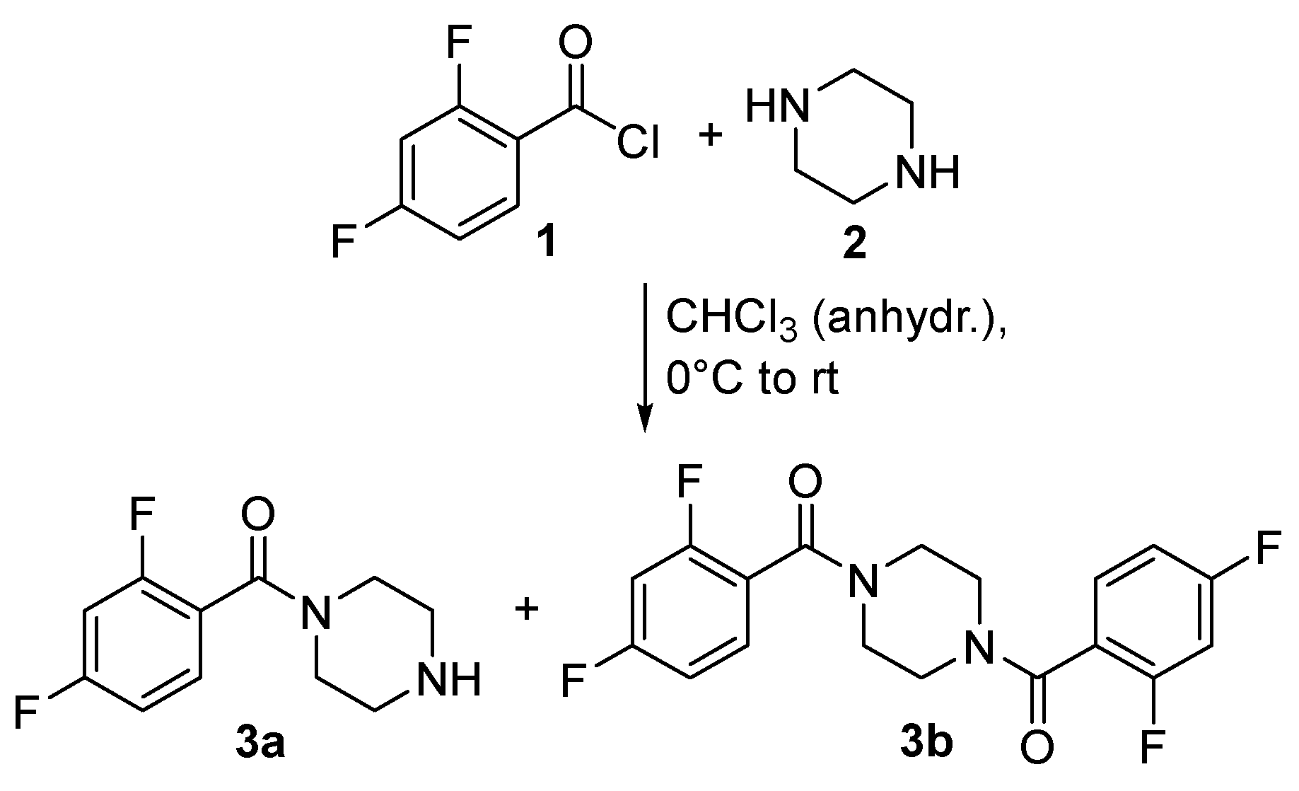
A one-pot reaction procedure using low-cost starting materials was applied. An excess of piperazine (2) was reacted with 2,4-difluorobenzoyl chloride (1) in anhydrous chloroform at 0 °C to obtain both compounds, the N-monoacylated amide 3a and the N,N-diacylated diamide 3b, according to a previously published procedure [47]. Depending on the excess of piperazine used for the reaction, compound 3a was obtained in 66% yield (five-fold excess) and compound 3b in 44% yield (two-fold excess). The reaction for both piperazines, 3a and 3b, is illustrated in Scheme 1.

3.2. Dynamic VT-NMR Study for the Dimer 3b

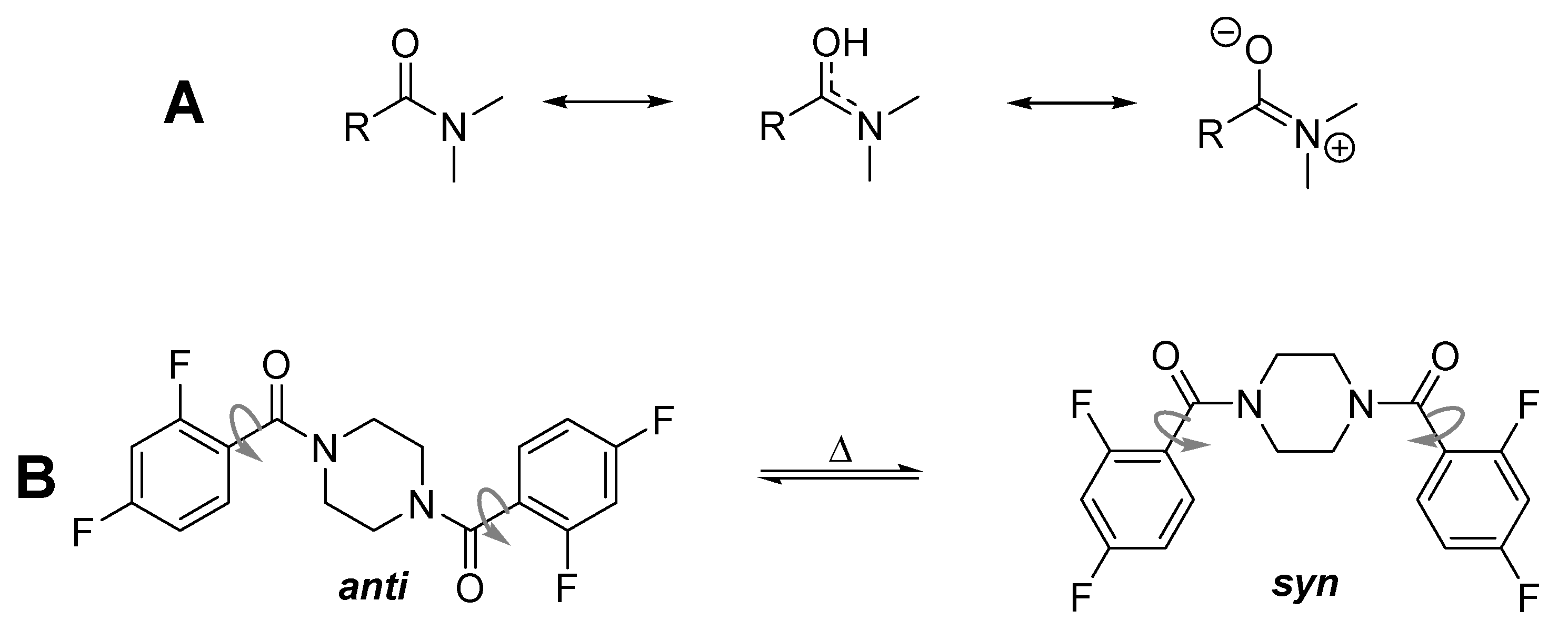
During the full characterization of compound 3b, four broad singlets were observed in the aliphatic region of the 1H NMR spectrum when measured at 25 °C (e.g., for CDCl3: δ = 3.34, 3.44, 3.79, and 3.89 ppm, ratio: 1:1.4:1.4:1 in Figure S10). Normally, one summarized signal could be expected for the NCH2-protons under these conditions due to the symmetry of the molecule. The 1H NMR spectrum is even more complex when measured at −10 °C. The four signals are again split in a more complicated pattern (see SI) due to the unsymmetrical substitution pattern of the benzene ring. Generally, rotation isomers are possible as shown in Figure 2, when additionally considering the rotation around the C(O)-CAr-bond.
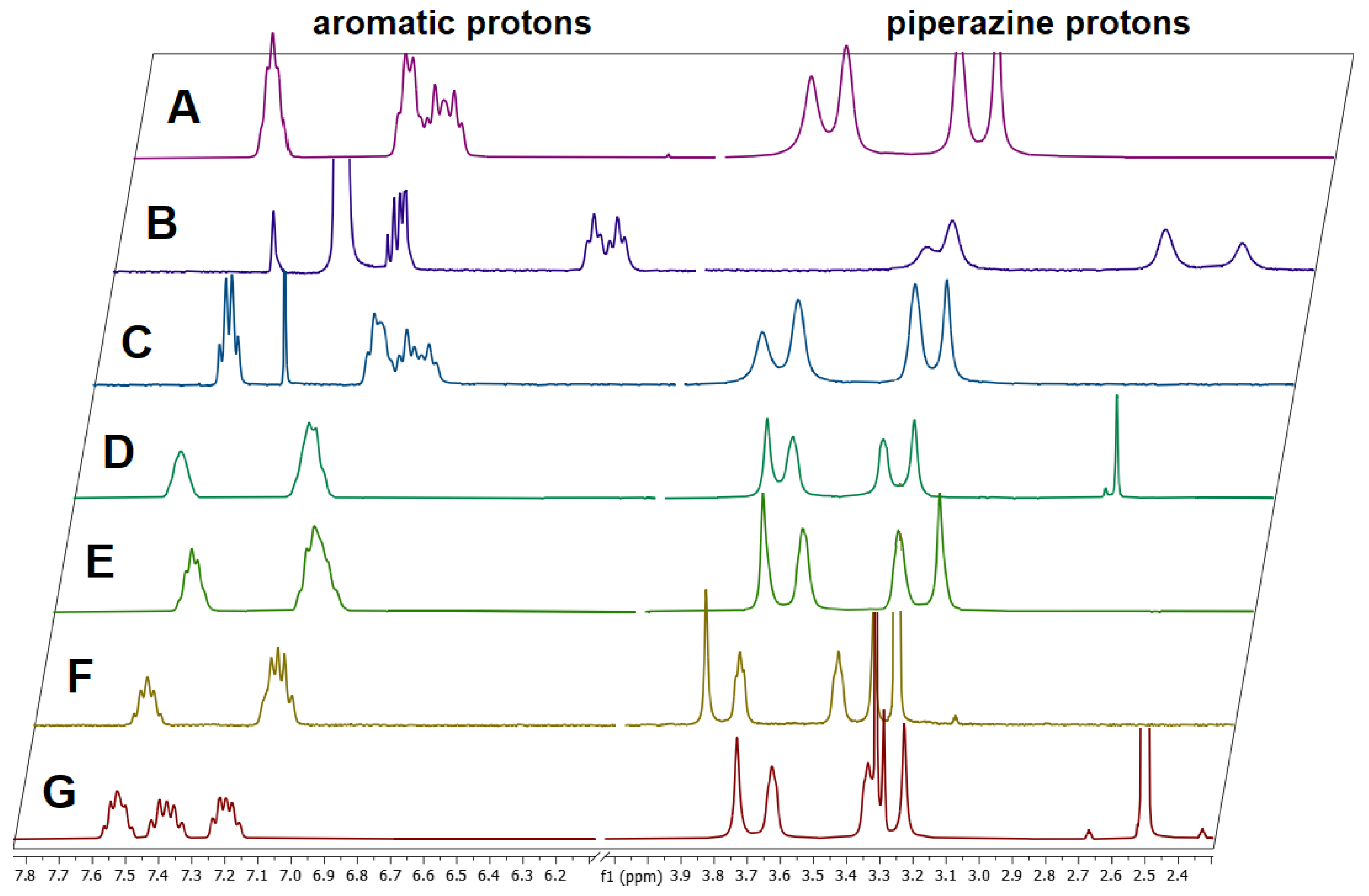
Supplementary 1H NMR spectra were recorded in DMSO-d6, methanol-d4, acetonitrile-d3, acetone-d6, benzene-d6, and tetrachloroethane-d2 (TCE-d2) (Figure 3), all showing four signals for the piperazine protons at 25 °C independent of the solvent but with different Δν values and different integral values. H,H-COSY spectra measured in CDCl3 revealed that only the inner two NCH2 signals couple (Figure S12). The inner two signals belong to the anti (trans) isomer (3.43 and 3.79 ppm), whereas the two outer NCH2 signals belong to the syn (cis) isomer (3.34 and 3.89 ppm), as indicated by recent results [10,47]. This splitting can also be observed in the 13C NMR spectrum (see Figure S11) with δ = 42.1 and 47.5 ppm for the syn isomer and 42.5 and 46.9 ppm for the anti-isomer measured in CDCl3. However, a reduced rotation around the C(O)-CAr-bond has not been detected at temperatures > 25 °C, only the reduced rotation around the amide bond.
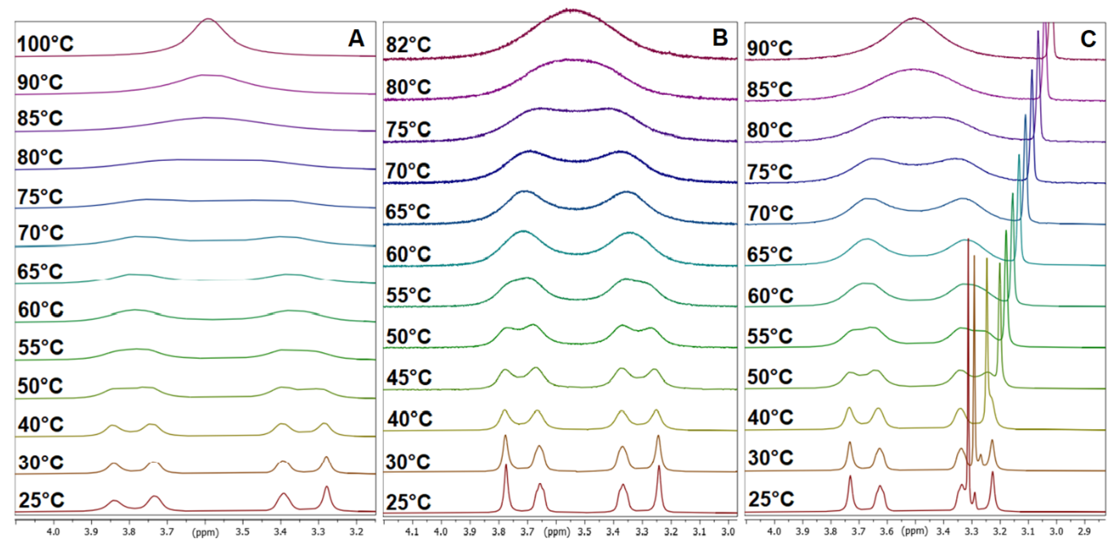
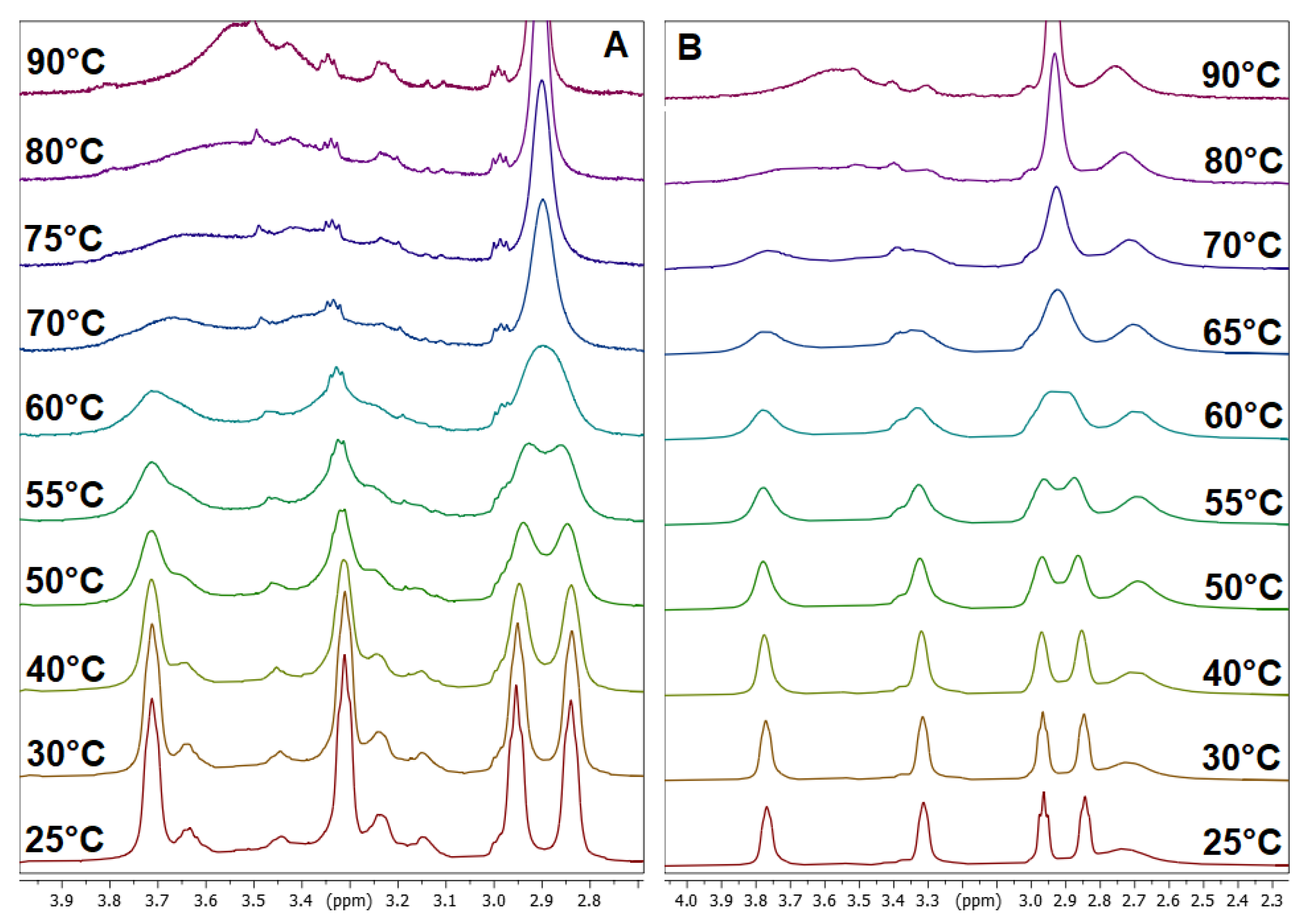
VT-dependent NMR measurements were accomplished (see SI Figures S15–S18) to determine the coalescence point expressed as TC for the partial amide double bond, which is dependent on the solvent. By warming the NMR sample of 3b, all four signals with an integration value of two hydrogen atoms each combine to one signal with a value of 8 at the TC due to the unhindered rotation of the amide bonds beyond this temperature. When using the standard NMR solvent CDCl3, the coalescence point could not be reached due to the boiling point of the solvent (Figure S15). To overcome this, the solvent was changed to tetrachloroethane-d2 (TCE-d2) with similar properties but with a boiling point of 145 °C. The VT-dependent 1H NMR spectra are pointed out in Figure 4A, and the TC was determined at 83 °C. Additionally, VT-1H-NMR spectra were recorded in acetonitrile-d3 with a TC of 80 °C and in DMSO-d6 with a TC of 83 °C (Figure 4B,C).
The TC for compound 3b containing the fluorine 2,4-substitution is higher in all used solvents as for the mono-fluorinated 4-fluorobenzamide compound (TC = 296 K in CDCl3) and the unsubstituted benzamide compound (TC = 306 K in CDCl3) due to the higher electron-withdrawing effect caused by the additional fluorine in the ortho position of the benzoyl moiety, which strengthens the partial double bond. The higher TC also has an influence on ΔG#, which was determined with >68 kJ/mol (anti-isomer) and >70 kJ/mol (syn isomer) for compound 3b (see Table 1).
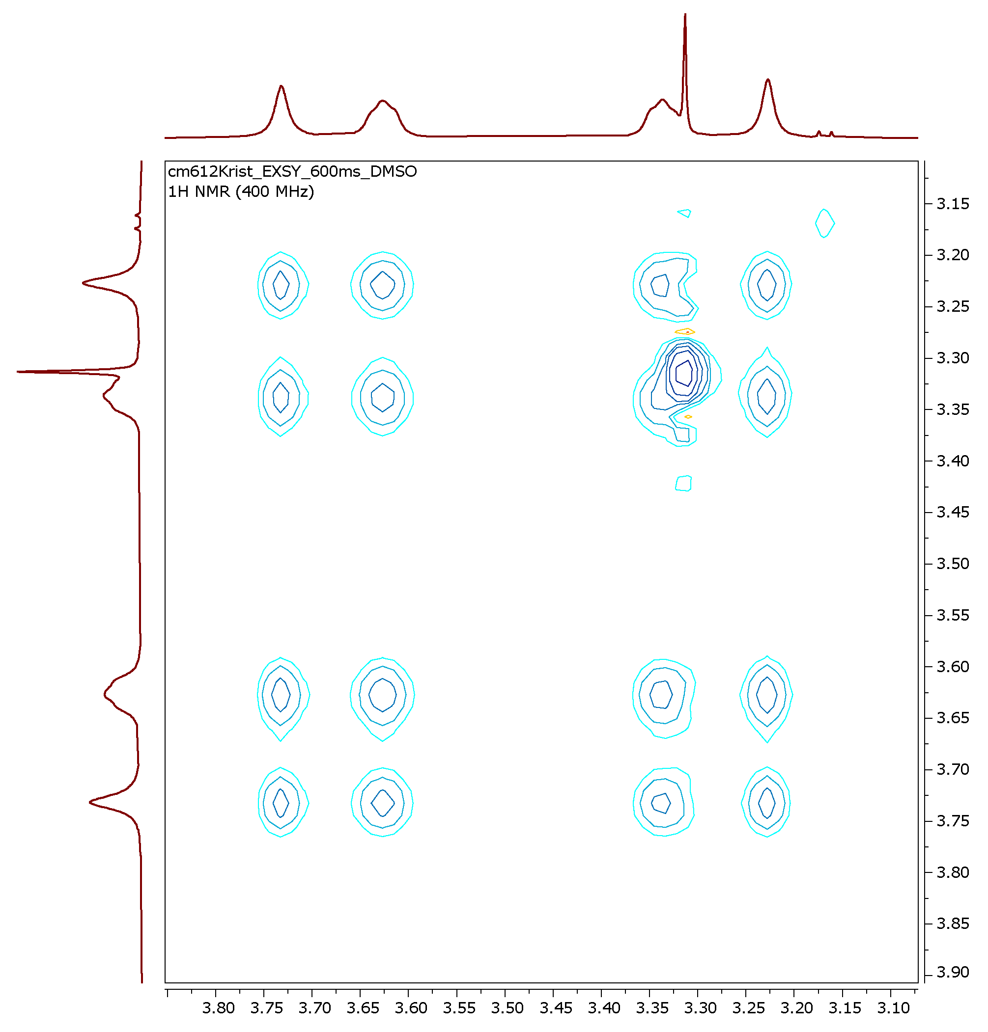
A 2D 1H EXSY spectrum was recorded at 25 °C and 600 ms mixing time, showing the exchange of the protons (representative for compound 3b in Figure 5). At this temperature, the molecule exhibits slow rotation around the N-C bond such that both groups exchange with one another rotationally yet are distinct in the spectrum.

3.3. X-Ray Structure Analysis of 3b

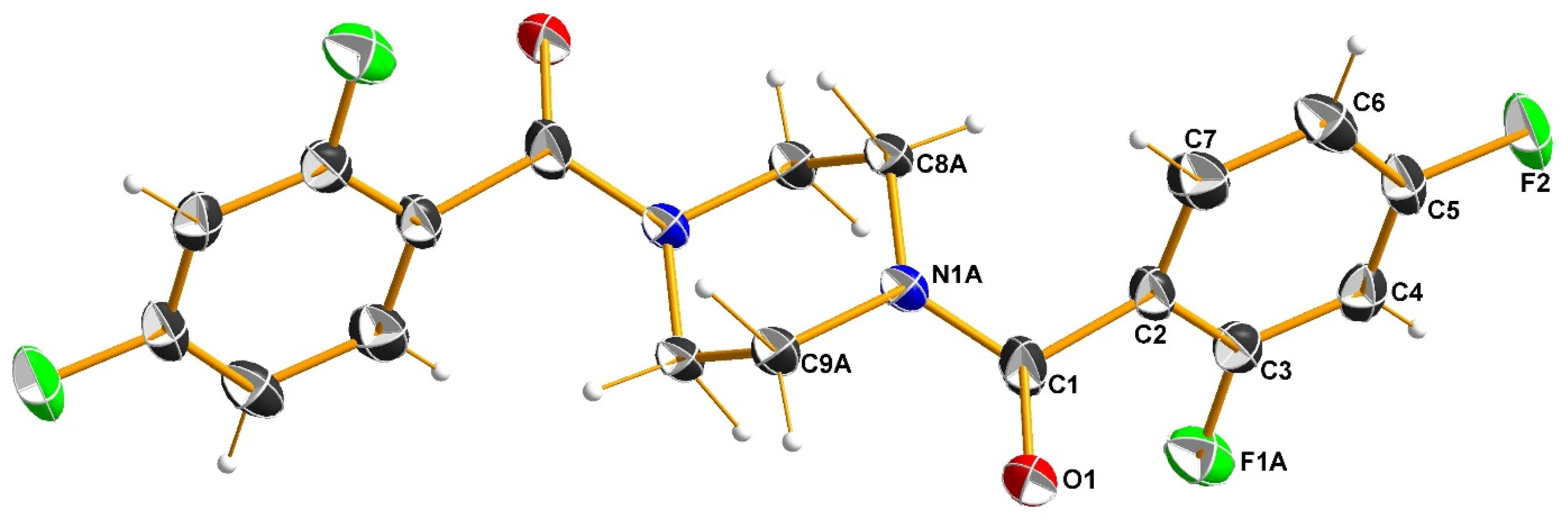
Single crystals of 3b were obtained, and the crystal and molecular structure were determined using single-crystal X-ray structure analysis. The molecular structure of this compound and the atom numbering scheme are shown in Figure 6. Relevant crystallographic data is given in Table 2.
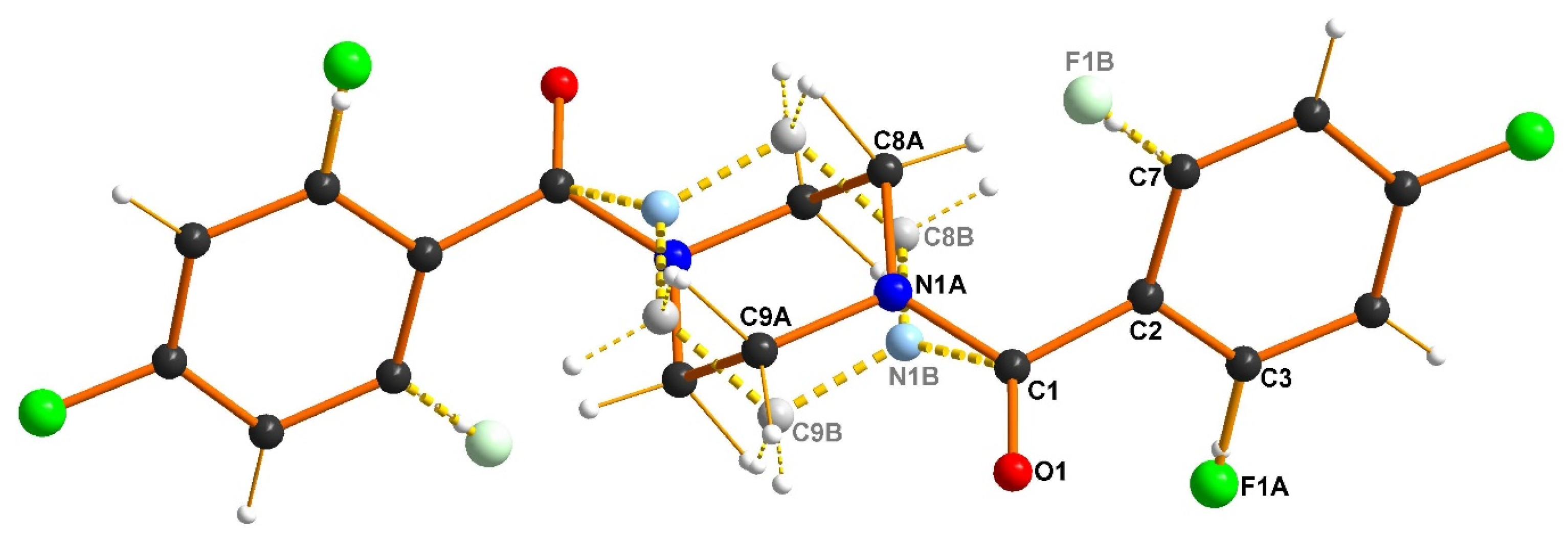
Compound 3b crystallizes in the monoclinic crystal system with the space group P21/c. The asymmetric unit consists of one half of a molecule. It is located such that the center of the piperazine ring is positioned on the inversion center, i.e., the molecule has an inversion symmetry. In the crystal, parts of the molecules of 3b are arranged with different orientations, i.e., they are disordered. The ortho-located fluorine atom (F1) is either cis (F1A) or trans (F2B) oriented with respect to the oxygen atom O1 (see anti-isomers in Figure 2). The occupation of the F1A site has been refined to 26.5%. The piperazine moiety has two different orientations, A and B, as shown in Figure 6, with an occupancy of 56.4% for orientation A. As visible in Figure 6 and Figure 7, only the anti-conformer (with respect to the orientation of the two oxygen atoms in each molecule) exists in the solid state. This probably results from intermolecular interactions preferring this orientation in the solid state, which are not present in solution.
The bonding environment around the nitrogen atom N1A (and also around N1B, respectively) is close to a trigonal planar disposition. The average bond angle of 119.7° and the distance of N1A to the plane of the three surrounding C atoms of only 0.08 Å indicate that the C1−N1A bond has at least a strong partial double bond character. Additionally, this bond is significantly shorter (1.398(2) Å) than comparable aliphatic bonds (i.e., 1.462(4) Å in N-methylpiperazine(sulfan)). These structural features are consistent with the NMR data, as shown above. The CH2 groups of the piperazine moiety have bond angles close to tetrahedral, as expected, with H−C−C−H dihedral angles of 53.3° and −65.2°.

3.4. Dynamic NMR Studies of Monomer 3a

The mono-substituted 2,4-fluorobenzyl compound 3a, with only one amide site and one free secondary amine site, shows a similar splitting behavior. Four signals for NCH2 groups are observed in the 1H NMR spectrum at 25 °C measured in CDCl3 (Figure S1); two for the amide site (δ = 3.39 and 3.85 ppm) and two for the amine site (δ = 2.92 and 3.03 ppm). The evaluation of a H,H-COSY spectrum (Figure S3) showed again the independent coupling of two NCH2 groups.
Two effects can be discussed for the splitting of the piperazine signals. The first effect is related to the limited rotation around the amide bond (as discussed above), and the second effect is related to the interconversion of two piperazine ring chair conformations [47], which occurs at deeper temperatures and thus did not have an influence. When measuring VT-dependent 1H NMR spectra in CDCl3 (Figure S7), only the TC of the amine site was detectable at 57 °C; the coalescence temperature of the amide site is located above the boiling point of the solvent. Thus, TCE-d2 and DMSO-d6 were again used as solvents (Figure 8), having TCs for the amine site of 63 °C and 61 °C, respectively, and for the amide site of 82 °C and 79 °C, respectively. All TC values are higher compared to the respective monosubstituted 4-fluorobenzoyl compound (TC,amine = 25 °C, TC,amide = 36 °C) due to the additional fluorine atom in the 2-position of the ring, which triggers a higher double bond character of the amide bond. As a result, the Gibbs free activation energy ΔG is also higher for the 2,4-difluoro compound 3a in comparison to the respective 4-fluorobenzoyl compound (ΔG = 60.4 kJ/mol), but the solvent seems to have only a small influence on the rotation. All results concerning compound 3a are summarized in Table 3.

3.5. Comparison with Other 2,4-Difluorobenzamides

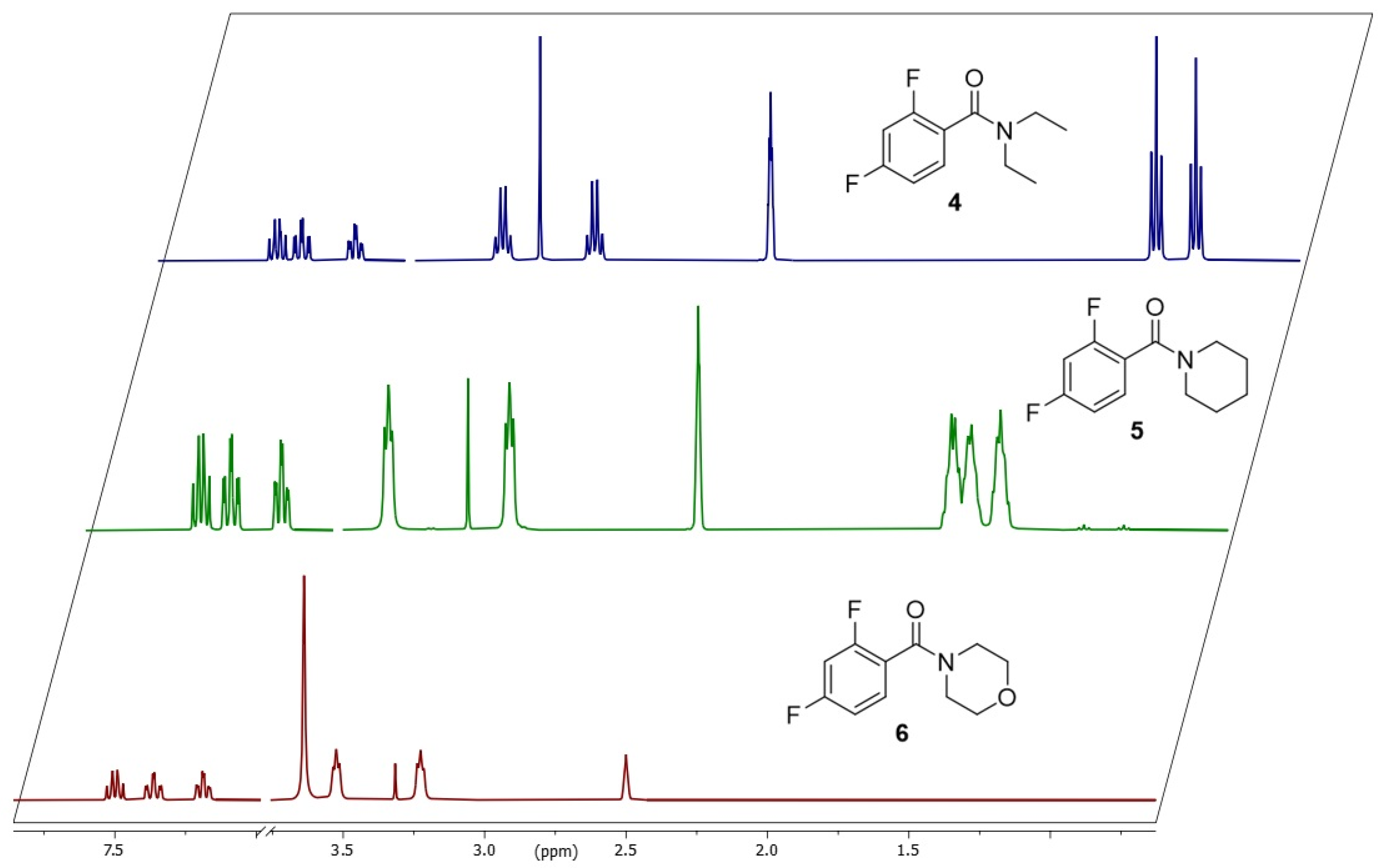
To compare the results of compounds 3a and 3b and to study the influence of the organic groups connected at the amide nitrogen atom (open chain or cyclic or, with or without heteroatom), three additional 2,4-difluorobenzamides 46 were prepared (Figure 9) with diethyl (4), piperidinyl (5) and morpholinyl (6) as substituents according to the synthesis of 3a and 3b with 2,4-difluorobenzyl chloride (1) and the respective amines in anhydrous chloroform.
All NMR spectra were recorded in DMSO-d6 (Figure 10), and all compounds 46 nicely show the splitting of the amide residues due to the hindered rotation around the C(O)-N bond (Figure 10). Again, VT-1H-NMR spectra were recorded for compounds 46 in DMSO-d6 (Figures S27, S36, and S45) to determine the Tc’s of the compounds for the calculation of the rotation barrier according to equations (1) and (2). All recorded data and calculated energy barriers expressed as Gibbs energy ΔG were found in Table 4. The chosen organic groups of the amide site do not have a big influence on the rotation. The highest barrier with 73.9 kJ/mol was found for the open-chain diethylamide 4. Overall, all calculated energy values for the rotation barrier of 3a, 3b, and 46 are in the same range. The additional fluorine in the 2-position of the aromatic ring primarily influences the rotation barrier.

4. Conclusions

Two fluorine-containing piperazine derivatives 3a and 3b were synthesized using a convenient one-pot synthesis procedure. An evaluation of the NMR spectra showed four signals of the NCH2 groups of the piperazine moiety for both compounds, coming from the formation of different rotation isomers. An existing partial double bond in the amide residue led to rotational conformers, whereas the ring conversion of the amine site in 3a and additional reduced rotation around the C(O)-CAr-bond in 3a and 3b are not observed at temperatures > 25 °C. To investigate the influence of the organic groups of the amide, three additional compounds 46 were prepared, and VT-1H-NMR spectra were recorded in DMSO-d6. Activation energies ΔGexc were calculated from solvent-dependent coalescence temperatures TC, which are all in the same range between 69 and 73 kJ/mol. They are higher compared to the mono-fluorinated amide and to the unsubstituted benzamide. The formation of conformers of compound 3b, which result from the partial double bond, was additionally determined and verified using single-crystal X-ray structure analysis for this compound. Only a minor influence of the solvent was observed.

Supplementary Materials

The following supporting information can be downloaded at: https://www.mdpi.com/article/10.3390/chemistry7050162/s1. Figure S1: 1H NMR spectrum of compound 3a in CDCl3; Figure S2: 13C NMR spectrum of compound 3a in CDCl3; Figure S3: H,H-COSY spectrum of compound 3a in CDCl3; Figure S4: H,C-HSQC spectrum of compound 3a in CDCl3; Figure S5: IR spectrum of compound 3a; Figure S6: Mass spectrum (ESI+) of compound 3a; Figure S7: 1H-VT-NMR spectra of 3a measured in CDCl3; Figure S8: 1H-VT-NMR spectra of 3a measured in TCE-d2; Figure S9: 1H-VT-NMR spectra of 3a measured in DMSO-d6; Figure S10: 1H NMR spectrum of compound 3b in CDCl3; Figure S11: 13C NMR spectrum of compound 3b in CDCl3; Figure S12: H,H-COSY spectrum of compound 3b in CDCl3; Figure S13: H,C-HSQC spectrum of compound 3b in CDCl3; Figure S14: EXSY spectrum of compound 3b (mixing time 600 ms, T = 25 °C) in DMSO-d6; Figure S15: 1H-VT-NMR spectra of 3b in CDCl3; Figure S16: 1H-VT-NMR spectra of 3b in TCE-d2; Figure S17: 1H-VT-NMR spectra of 3b in ACN-d3; Figure S18: 1H-VT-NMR spectra of 3b in DMSO-d6; Figure S19: 1H NMR spectrum of compound 4 in DMSO-d6; Figure S20: 13C NMR spectrum of compound 4 in DMSO-d6; Figure S21: H,H-COSY spectrum of compound 4 in DMSO-d6; Figure S22: H,C-HSQC spectrum of compound 4 in DMSO-d6; Figure S23: 19F NMR spectrum of compound 4 in DMSO-d6; Figure S24: EXSY spectrum of compound 4 (mixing time 600 ms, T = 25 °C) in DMSO-d6; Figure S25: IR spectrum of compound 4; Figure S26: Mass spectrum (ESI+) spectrum of compound 4; Figure S27: 1H-VT-NMR spectra of 4 in DMSO-d6; Figure S28: 1H NMR spectrum of compound 5 in DMSO-d6; Figure S29: 13C NMR spectrum of compound 5 in DMSO-d6; Figure S30: H,H-COSY spectrum of compound 5 in DMSO-d6; Figure S31: H,C-HSQC spectrum of compound 5 in DMSO-d6; Figure S32: EXSY spectrum of compound 5 (mixing time 600 ms, T = 25 °C) in DMSO-d6; Figure S33: 19F NMR spectrum of compound 5 in DMSO-d6; Figure S34: IR spectrum of compound 5; Figure S35: Mass spectrum (ESI+) of compound 5; Figure S36: 1H-VT-NMR spectra of 5 in DMSO-d6; Figure S37: 1H NMR spectrum of compound 6 in DMSO-d6; Figure S38: 13C NMR spectrum of compound 6 in DMSO-d6; Figure S39: H,H-COSY spectrum of compound 6 in DMSO-d6; Figure S40: H,C-HSQC spectrum of compound 6 in DMSO-d6; Figure S41: EXSY spectrum of compound 6 (mixing time 600 ms, T = 25 °C) in DMSO-d6; Figure S42: Figure S43: IR spectrum of compound 6; Figure S44: Mass spectrum (ESI+) of compound 6; 19F NMR spectrum of compound 6 in DMSO-d6; Figure S45: 1H-VT-NMR spectra of 6 in DMSO-d6.

Author Contributions

Conceptualization, C.M.; methodology, M.K. and C.M.; validation, M.K. and C.M.; formal analysis, M.K. and C.M.; investigation, C.M.; resources, M.K. and C.M.; data curation, M.K. and C.M.; writing—original draft preparation, M.K. and C.M.; writing—review and editing, M.K. and C.M.; visualization, M.K. and C.M. All authors have read and agreed to the published version of the manuscript.

Funding

This research received no external funding.

Data Availability Statement

The raw data is archived at the HZDR database and will be made available upon request.

Conflicts of Interest

The authors declare no conflicts of interest.

Abbreviations

The following abbreviations are used in this manuscript:
ACNAcetonitrile
COSYCorrelation spectroscopy
DMSODimethyl sulfoxide
EXSYExchange spectroscopy
TCE1,1,2,2-Tetrachloroethane
TLCThin-layer chromatography
VTVariable temperature
XRDX-ray diffraction

References

  1. Gettys, K.E.; Ye, Z.; Dai, M. Recent Advances in Piperazine Synthesis. Synthesis 2017, 49, 2589–2604. [Google Scholar] [CrossRef]
  2. Zhang, R.H.; Guo, H.Y.; Deng, H.; Li, J.; Quan, Z.S. Piperazine skeleton in the structural modification of natural products: A review. J. Enzyme Inhib. Med. Chem. 2021, 36, 1165–1197. [Google Scholar] [CrossRef] [PubMed]
  3. Marshall, C.M.; Federice, J.G.; Bell, C.N.; Cox, P.B.; Njardarson, J.T. An Update on the Nitrogen Heterocycle Compositions and Properties of U.S. FDA-Approved Pharmaceuticals (2013–2023). J. Med. Chem. 2024, 67, 11622–11655. [Google Scholar] [CrossRef] [PubMed]
  4. Faizan, M.; Kumar, R.; Mazumder, A.; Salahuddin; Kukreti, N.; Kumar, A.; Chaitanya, M.V.N.L. The medicinal chemistry of piperazines: A review. Chem. Biol. Drug Des. 2024, 103, e14537. [Google Scholar] [CrossRef] [PubMed]
  5. Heravi, M.M.; Zadsirjan, V. Prescribed drugs containing nitrogen heterocycles: An overview. RSC Adv. 2020, 10, 44247–44311. [Google Scholar] [CrossRef]
  6. Dömling, A.; Huang, Y. Piperazine Scaffolds via Isocyanide-Based Multicomponent Reactions. Synthesis 2010, 17, 2859–2883. [Google Scholar] [CrossRef]
  7. Durand, C.; Szostak, M. Recent Advances in the Synthesis of Piperazines: Focus on C–H Functionalization. Organics 2021, 2, 337–347. [Google Scholar] [CrossRef]
  8. Huang, Z.; Shi, W. UV curing behavior of hyperbranched polyphosphate acrylate/di(hydroxylpropyl methacrylate) piperazine and properties of the cured film. Prog. Org. Coat. 2007, 59, 312–317. [Google Scholar] [CrossRef]
  9. Zhao, B.; Xiao, Y.; Yuan, D.; Lu, C.; Yao, Y. Synthesis and characterization of bridged bis(amidato) rare earth metal amides and their applications in C–N bond formation reactions. Dalton Trans. 2016, 45, 3880–3887. [Google Scholar] [CrossRef]
  10. Sahoo, H.S.; Chand, D.K. Conformation of N,N′-bis(3-pyridylformyl)piperazine and spontaneous formation of a saturated quadruple stranded metallohelicate. Dalton Trans. 2010, 39, 7223–7225. [Google Scholar] [CrossRef]
  11. Barnes, D.J.; Chapman, R.L.; Vagg, R.S.; Watton, E.C. Synthesis of novel bis(amides) by means of triphenyl phosphite intermediates. J. Chem. Eng. Data 1978, 23, 349–350. [Google Scholar] [CrossRef]
  12. Andronati, S.A.; Karaseva, T.L.; Krysko, A.A. Peptidomimetics-antagonists of the fibrinogen receptors: Molecular design, structures, properties and therapeutic applications. Curr. Med. Chem. 2004, 11, 1183–1211. [Google Scholar] [CrossRef] [PubMed]
  13. Horton, D.A.; Bourne, G.T.; Smythe, M.L. Exploring privileged structures: The combinatorial synthesis of cyclic peptides. Mol. Divers. 2000, 5, 289–304. [Google Scholar] [CrossRef]
  14. Wodtke, R.; König, J.; Pigorsch, A.; Köckerling, M.; Mamat, C. Evaluation of novel fluorescence probes for conjugation purposes using the traceless Staudinger Ligation. Dyes Pigment. 2015, 113, 263–273. [Google Scholar] [CrossRef]
  15. Heldt, J.-M.; Kerzendörfer, O.; Mamat, C.; Starke, F.; Pietzsch, H.-J.; Steinbach, J. Synthesis of Short and Versatile Heterobifunctional Linkers for Conjugation Purposes of Bioactive Molecules with (Radio-)Labels. Synlett 2013, 24, 432–436. [Google Scholar] [CrossRef]
  16. Lemoucheux, L.; Rouden, J.; Ibazizene, M.; Sobrio, F.; Lasne, M.C. Debenzylation of Tertiary Amines Using Phosgene or Triphosgene:  An Efficient and Rapid Procedure for the Preparation of Carbamoyl Chlorides and Unsymmetrical Ureas. Application in Carbon-11 Chemistry. J. Org. Chem. 2003, 68, 7289–7297. [Google Scholar] [CrossRef]
  17. Grosse-Gehling, P.; Wuest, F.R.; Peppel, T.; Köckerling, M.; Mamat, C. [18F]1-(3-fluoropropyl)piperazines as model compounds for the radiofluorination of pyrido [2,3-d]pyrimidines. Radiochim. Acta 2011, 99, 365. [Google Scholar] [CrossRef]
  18. Pretze, M.; Mamat, C. Automated preparation of [18F]AFP and [18F]BFP: Two novel bifunctional 18F-labeling building blocks for Huisgen-click. J. Fluor. Chem. 2013, 150, 25–35. [Google Scholar] [CrossRef]
  19. Mamat, C.; Pretze, M.; Gott, M.; Köckerling, M. Synthesis, dynamic NMR characterization and XRD studies of novel N,N’-substituted piperazines for bioorthogonal labeling. Beilstein J. Org. Chem. 2016, 12, 2478–2489. [Google Scholar] [CrossRef]
  20. Wiemer, J.; Steinbach, J.; Pietzsch, J.; Mamat, C. Preparation of a novel radiotracer targeting the EphB4 receptor via radiofluorination using spiro azetidinium salts as precursor. J. Labelled Comp. Radiopharm. 2017, 60, 489–498. [Google Scholar] [CrossRef]
  21. Katz, D.P.; Deruiter, J.; Bhattacharya, D.; Ahuja, M.; Bhattacharya, S.; Clark, C.R.; Suppiramaniam, V.; Dhanasekaran, M. Benzylpiperazine: “A messy drug”. Drug Alcohol Depend. 2016, 164, 1–7. [Google Scholar] [CrossRef]
  22. Shaquiquzzaman, M.; Verma, G.; Marella, A.; Akhter, M.; Akhtar, W.; Khan, M.F.; Tasneem, S.; Alam, M.M. Piperazine scaffold: A remarkable tool in generation of diverse pharmacological agents. Eur. J. Med. Chem. 2015, 102, 487–529. [Google Scholar] [CrossRef] [PubMed]
  23. Shen, L.; Ye, Y.H.; Wang, X.T.; Zhu, H.L.; Xu, C.; Song, Y.C.; Li, H.; Tan, R.X. Structure and total synthesis of aspernigerin: A novel cytotoxic endophyte metabolite. Chem. Eur. J. 2006, 12, 4393–4396. [Google Scholar] [CrossRef] [PubMed]
  24. Irikura, T.; Masuzawa, K.; Nishino, K.; Kitagawa, M.; Uchida, H.; Ichinoseki, N.; Ito, M. New analgetic agents. V. 1-butyryl-4-cinnamylpiperazine hydrochloride and related compounds. J. Med. Chem. 1968, 11, 801–804. [Google Scholar] [CrossRef] [PubMed]
  25. Rathi, A.K.; Syed, R.; Shin, H.S.; Patel, R.V. Piperazine derivatives for therapeutic use: A patent review (2010–present). Expert Opin. Ther. Pat. 2016, 26, 777–797. [Google Scholar] [CrossRef]
  26. Abou El-Ella, D.A.; Hussein, M.M.; Serya, R.A.; Abdel Naby, R.M.; Al-Abd, A.M.; Saleh, D.O.; El-Eraky, W.I.; Abouzid, K.A. Molecular design and synthesis of 1,4-disubstituted piperazines as α(1)-adrenergic receptor blockers. Bioorg. Chem. 2014, 54, 21–30. [Google Scholar] [CrossRef]
  27. Patel, R.V.; Park, S.W. An evolving role of piperazine moieties in drug design and discovery. Mini-Rev. Med. Chem. 2013, 13, 1579–1601. [Google Scholar] [CrossRef]
  28. Miniyar, P.B.; Murumkar, P.R.; Patil, P.S.; Barmade, M.A.; Bothara, K.G. Unequivocal role of pyrazine ring in medicinally important compounds: A review. Mini-Rev. Med. Chem. 2013, 13, 1607–1625. [Google Scholar] [CrossRef]
  29. Font, M.; Ardaiz, E.; Cordeu, L.; Cubedo, E.; Garcia-Foncillas, J.; Sanmartin, C.; Palop, J.A. Structural characteristics of novel symmetrical diaryl derivatives with nitrogenated functions. Requirements for cytotoxic activity. Bioorg. Med. Chem. 2006, 14, 1942–1948. [Google Scholar] [CrossRef]
  30. Radin, D.P.; Lippa, A.; Rana, S.; Fuller, D.D.; Smith, J.L.; Cerne, R.; Witkin, J.M. Amplification of the therapeutic potential of AMPA receptor potentiators from the nootropic era to today. Pharmacol. Biochem. Behav. 2025, 248, 173967. [Google Scholar] [CrossRef]
  31. Rana, S.; Fusco, A.F.; Witkin, J.M.; Radin, D.P.; Cerne, R.; Lippa, A.; Fuller, D.D. Pharmacological modulation of respiratory con-trol: Ampakines as a therapeutic strategy. Pharmacol. Ther. 2025, 265, 108744. [Google Scholar] [CrossRef]
  32. Czopek, A.; Kubacka, M.; Bucki, A.; Siwek, A.; Filipek, B.; Pawłowski, M.; Kołaczkowski, M. Novel serotonin 5-HT2A receptor antagonists derived from 4-phenylcyclohexane-5-spiro-and 5-methyl-5-phenyl-hydantoin, for use as potential antiplatelet agents. Pharmacol. Rep. 2021, 73, 1361–1372. [Google Scholar] [CrossRef]
  33. Liu, X.-K.; Ma, L.-X.; Wei, Z.-Y.; Cui, X.; Zhan, S.; Yin, X.-M.; Piao, H.-R. Synthesis and Positive Inotropic Activity of [1,2,4]Triazolo [4,3-a] Quinoxaline Derivatives Bearing Substituted Benzylpiperazine and Benzoylpiperazine Moieties. Molecules 2017, 22, 273. [Google Scholar] [CrossRef]
  34. Moorman, A.R. Adenosine a2a Receptor Antagonists. U.S. Patent 2008242672A1, 6 April 2010. [Google Scholar]
  35. Dong, H.; Zhang, L. Pyridino-Pyrimidine Compound and Application Thereof. CN Patent 109748914A, 25 May 2025. [Google Scholar]
  36. Arkinstall, S.; Halazy, S.; Church, D.; Camps, M.; Rueckle, T.; Gotteland, J.P.; Biamonte, M. Pharmaceutically Active Sulfonamide Derivatives. U.S. Patent 8012995B1, 6 September 2011. [Google Scholar]
  37. Ielo, L.; Deri, B.; Germanò, M.P.; Vittorio, S.; Mirabile, S.; Gitto, R.; Rapisarda, A.; Ronsisvalle, S.; Floris, S.; Pazy, Y.; et al. Exploiting the 1-(4-fluorobenzyl)piperazine fragment for the development of novel tyrosinase inhibitors as anti-melanogenic agents: Design, synthesis, structural insights and biological profile. Eur. J. Med. Chem. 2019, 178, 380–389. [Google Scholar] [CrossRef] [PubMed]
  38. Wang, J.Q.; Yang, M.D.; Chen, X.; Wang, Y.; Chen, L.Z.; Cheng, X.; Liu, X.H. Discovery of new chromen-4-one derivatives as telomerase inhibitors through regulating expression of dyskerin. J. Enzyme. Inhib. Med. Chem. 2018, 33, 1199–1211. [Google Scholar] [CrossRef] [PubMed]
  39. Ghosh, K.K.; Jeong, Y.M.; Kang, N.Y.; Lee, J.; Diana, W.S.Y.; Kim, J.Y.; Yoo, J.; Kim, D.; Kim, Y.K.; Chang, Y.T. The development of a nucleus staining fluorescent probe for dynamic mitosis imaging in live cells. Chem. Commun. 2015, 51, 9336–9338. [Google Scholar] [CrossRef] [PubMed]
  40. Ha, H.H.; Kim, J.S.; Kim, B.M. Novel heterocycle-substituted pyrimidines as inhibitors of NF-κB transcription regulation related to TNF-α cytokine release. Bioorg. Med. Chem. Lett. 2008, 18, 653–656. [Google Scholar] [CrossRef]
  41. Dugave, C.; Demange, L. Cis−Trans Isomerization of Organic Molecules and Biomolecules:  Implications and Applications. Chem. Rev. 2003, 103, 2475–2532. [Google Scholar] [CrossRef]
  42. Jackman, L.M.; Kavanagh, T.E.; Haddon, R.C. Studies in nuclear magnetic resonance—IX. Rotational barriers in substituted N,N-dimethylbenzamides. Org. Magn. Reson. 1969, 1, 109–123. [Google Scholar] [CrossRef]
  43. Alberghina, G.; Bottino, F.A.; Fisichella, S.; Arnone, C. Solvent effects on the rotational barriers of the N,N-dimethylamides of 2- and 3-furoic and 2- and 3-thenoic acids. J. Chem. Res. (M) 1985, 1201–1217. [Google Scholar]
  44. Alberghina, G.; Bottino, F.A.; Fisichella, S.; Arnone, C. Solvent effects on the rotational barriers of the N,N-dimethylamides of 2- and 3-furoic and 2- and 3-thenoic acids. J. Chem. Res. (S) 1985, 108–109. [Google Scholar]
  45. Turnbull, M.M.; Nelson, D.J.; Lekouses, W.; Sarnov, M.L.; Tartarini, K.A.; Huang, T.-K. Rotational barriers in N,N-diethylbenzamides: Substituent and solvent effects. Tetrahedron 1990, 46, 6613–6622. [Google Scholar] [CrossRef]
  46. Wodtke, R.; Steinberg, J.; Köckerling, M.; Löser, R.; Mamat, C. NMR-based investigations of acyl-functionalized piperazines concerning their conformational behavior in solution. RSC Adv. 2018, 8, 40921–40933. [Google Scholar] [CrossRef]
  47. Friebolin, H. Basic One- and Two-Dimensional NMR Spectroscopy; Wiley-VCH: Weinheim, Germany, 2010; p. 319 ff. [Google Scholar]
  48. Gasparro, F.P.; Kolodny, N.H. NMR determination of the rotational barrier in N,N-dimethylacetamide. A physical chemistry experiment. J. Chem. Educ. 1977, 54, 258. [Google Scholar] [CrossRef]
  49. Jackman, L.M.; Cotton, F.A. (Eds.) Dynamic Nuclear Magnetic Resonance Spectroscopy; Academic Press: New York, NY, USA, 1975; p. 204 ff. [Google Scholar]
  50. Skoglof, A.; Nilsson, I.; Gustafsson, S.; Deinum, J.; Göthe, P.O. Cis-trans isomerization of an angiotensin I-converting enzyme inhibitor. An enzyme kinetic and nuclear magnetic resonance study. Biochim. Biophys. Acta 1990, 1041, 22–30. [Google Scholar] [CrossRef]
  51. Sheldrick, G.M. A short history of SHELX. Acta Cryst. A 2008, 64, 112–122. [Google Scholar] [CrossRef]
  52. Sheldrick, G.M. SHELXL 2014/2; University of Göttingen: Göttingen, Germany, 2014. [Google Scholar]
Figure 1. 2,4-Difluorobenzoylpiperazine moiety present in selected different biologically and pharmacologically active compounds such as TYR inhibitor [37], telomerase inhibitor [38], fluorescent probe for nucleus staining [39], AbTYR inhibitor [40] and GSK-3β inhibitor [41].
Chemistry 07 00162 g001
Scheme 1. Preparation of the 2,4-difluoro derivatives 3a and 3b.
Chemistry 07 00162 sch001
Figure 2. (A) partial double bond of amides (B) syn and anti isomer of compound 3b with free rotation about the single bond (gray arrows).
Chemistry 07 00162 g002
Figure 3. 1H NMR spectra (aliphatic region with piperazine protons from 2.3–4.0 ppm and aromatic region from 6.1–7.8 ppm) of compound 3b measured in five different solvents: (A) TCE-d2, (B) benzene-d6, (C) CDCl3, (D) acetone-d6, (E) acetonitrile-d3, (F) methanol-d4 and (G) DMSO-d6.
Chemistry 07 00162 g003
Figure 4. VT-1H NMR spectra of 3b showing the piperazine protons measured in (A) TCE-d2, (B) acetonitrile-d3, and (C) DMSO-d6 (only the region of the piperazine protons is shown; full spectra see Figures S16–S18).
Chemistry 07 00162 g004
Figure 5. Representative 2D 1H EXSY spectrum of 3b in DMSO-d6 at 25 °C with 600 ms mixing time.
Chemistry 07 00162 g005
Figure 6. Molecular structure of the compound 3b (ORTEP plot with 50% probability level, the respective second orientations of the disordered piperazine moiety and the F1/F1A atom are not shown for better visibility; see Figure 7).
Chemistry 07 00162 g006
Figure 7. Superimposition of the different disordered moieties in crystals of 3b, discriminated by the labels A and B, different color intensities, and solid/dashed lines.
Chemistry 07 00162 g007
Figure 8. VT-1H NMR spectra of 3a measured in (A) DMSO-d6 and (B) TCE-d2 (region of the piperazine protons is shown; see Figures S8 and S9 for full spectra).
Chemistry 07 00162 g008
Figure 9. Selected 2,4-difluorobenzamides 46 with different amide functions (diethyl, piperidinyl, morpholinyl).
Chemistry 07 00162 g009
Figure 10. Comparison of 1H NMR spectra (aliphatic region and aromatic region) of compounds 4 (diethyl), 5 (piperidinyl) and 6 (morpholinyl) measured in DMSO-d6 at 25 °C.
Chemistry 07 00162 g010
Table 1. Physicochemical data for compound 3b in dependence on the solvent. a: anti/s: syn.
SolventRatio syn/antiΔν [Hz]kexc [Hz]TC [K]ΔG [kJ/mol]
Benzene-d61.5:1257 (a)
381 (s)
313 (a)
494 (s)
n.d.-
-
CDCl31.2:1141 (a)
223 (s)
244 (a)
395 (s)
n.d.-
-
TCE-d21.1:1136 (a)
224 (s)
571 (a)
846 (s)
35670.8
69.3
Acetone-d61.2:1110 (a)
223 (s)
302 (a)
498 (s)
n.d.-
-
ACN-d31.1:1116 (a)
213 (s)
258 (a)
473 (s)
35370.7
68.9
CD3OD1.1:1119 (a)
202 (s)
264 (a)
448 (s)
n.d.-
-
DMSO-d61.2:1116 (a)
202 (s)
258 (a)
448 (s)
35370.7
69.0
Table 2. Crystal data and structure refinement for compound 3b.
Compound3b
FormulaC18H14F4N2O2
Formula weight (g·mol−1)366.31
Temperature (K)123
Crystal systemmonoclinic
Space groupP21/c
Unit cell dimensions:
a (Å)7.2687(3)
b (Å)17.2658(8)
c (Å)6.9738(3)
β (°)115.393(2)
Volume (Å3), Z790.65(6), 2
Data/restraints/param.2955/0/157
Measured reflections23,516
2θmax (°)66.0
GoF on F21.08
R1 [I > 2σ(I)]0.058
wR2 (all data)0.154
Larg. diff. peak/hole (e·Å3)0.36/−0.31
Table 3. Physicochemical parameters of compound 3a.
SolventΔν [Hz]kexc [Hz]TC [K]ΔG [kJ/mol]
CDCl345
185
100 (amine)
411 (amide)
330 (amine)
n.d. (amide)
68.5
-
TCE-d248
160
107 (amine)
355 (amide)
336 (amine)
355 (amide)
69.6
70.1
DMSO-d646
160
102 (amine)
355 (amide)
334 (amine)
352 (amide)
69.3
69.5
Table 4. Physicochemical parameters of compounds 3a, 3b, and 46 measured in DMSO-d6.
CompoundΔν [Hz]kexc [Hz]TC [K]ΔG [kJ/mol]
3a116 (anti)
202 (syn)
258 (anti)
448 (syn)
35370.7
69.0
3b48 (amine)
160 (amide)
107 (amine)
355 (amide)
336 (amine)
355 (amide)
69.6
70.1
456 (CH3)
130 (CH2)
124 (CH3)
289 (CH2)
358 (CH3)
370 (CH2)
73.9
73.8
5171 (amide)
41 (CH2)
380 (amide)
91 (CH2)
371
348
73.2
72.6
611926436172.2
Disclaimer/Publisher’s Note: The statements, opinions and data contained in all publications are solely those of the individual author(s) and contributor(s) and not of MDPI and/or the editor(s). MDPI and/or the editor(s) disclaim responsibility for any injury to people or property resulting from any ideas, methods, instructions or products referred to in the content.