Article

In Silico Analysis of Curcumin and Its Analogs MS13 and MS17 Against HSF1 and HSP Family Proteins

1
Jeffrey Cheah School of Medicine and Health Sciences, Monash University Malaysia, Jalan Lagoon Selatan, Bandar Sunway 47500, Selangor, Malaysia
2
Inserm U1086 ANTICIPE (Interdisciplinary Research Unit for Cancer Prevention and Treatment), Université de Caen Normandie, Normandie Univ, 14076 Caen, France
3
Comprehensive Cancer Center François Baclesse, UNICANCER, 14076 Caen, France
4
School of Pharmacy, Monash University Malaysia, Jalan Lagoon Selatan, Bandar Sunway 47500, Selangor, Malaysia
*
Author to whom correspondence should be addressed.
These authors contributed equally to this work.
Chemistry 2025, 7(5), 139; https://doi.org/10.3390/chemistry7050139
Submission received: 9 July 2025 / Revised: 22 August 2025 / Accepted: 26 August 2025 / Published: 28 August 2025
(This article belongs to the Section Biological and Natural Products)

Abstract

Heat shock proteins (HSPs), a family of proteins including HSP27, HSP40, HSP60, HSP70, and HSP90, play critical roles in cellular processes and are often dysregulated in cancer. Heat Shock Factor 1 (HSF1) protein, the master regulator of HSP expression, is also a promising target for cancer therapy due to its involvement in tumorigenesis. This study is the first to investigate the potential of two novel curcumin analogs, MS13 (1,2-bis(4-hydroxy-3-methoxyphenyl)-1,4-pentadiene-3-one) and MS17 (1,5-bis(2-hydroxyphenyl)-1,4-pentadiene-3-one), as modulators of these key targets. Employing molecular docking and molecular dynamics (MD) simulations, we investigated the interactions of MS13 and MS17 with HSF1 and the panel of HSPs. Both compounds demonstrated strong binding affinity for all the proteins, particularly for HSP70, exhibiting greater affinity compared to curcumin. Molecular docking revealed specific binding sites for both compounds on each target protein, which were further investigated using MD simulations. MS17 generally formed more stable complexes with HSP27, HSP40, HSP60, and HSP70, suggesting it might be a more potent modulator of these specific proteins. In contrast, MS13 displayed greater stability when bound to HSF1 and HSP90. These different variations could be attributed to variations in the chemical structures of MS13 and MS17, leading to distinct interactions with each protein’s binding site. MS13 and MS17 exhibit more advantageous ADMET profiles compared to curcumin, particularly in their predicted Blood–Brain Barrier (BBB) permeability and MS17’s superior passive membrane permeability and absorption. These findings highlight the potential of both MS13 and MS17 as promising leads for developing HSP modulators for cancer treatment.

1. Introduction

In recent decades, heat shock proteins (HSPs) have garnered significant attention in cancer research due to their involvement in various aspects of cancer biology, including cell proliferation, differentiation, and survival. In cancer patients, HSP27, HSP60, HSP70, HSP90 and HSP110 are often upregulated, which could serve as cancer biomarkers [1,2,3,4,5,6,7]. HSF1 (heat shock factor 1), the master regulator of heat shock response and HSPs, also plays an important role in tumor development and progression [8,9,10]. HSF1 promotes the expression of HSPs in cellular stress response to maintain the proper folding of proteins, essential in protein folding, ribosome biogenesis and protein degradation pathways [11]. HSF1 overexpression is commonly associated with aggressiveness in multiple cancers, which could be a potential biomarker [12,13,14]. HSF1 can interact with various signaling pathways, such as NF-κB, PI3K-AKT-mTOR, and MAPK pathways, which are essential for cell proliferation, anti-apoptosis, and other cancer-related processes [8]. Thus, HSF1 is a potential therapeutic target for cancer treatment.
HSPs are a group of highly conserved chaperones that play pivotal roles in cellular homeostasis and stress response [15]. HSPs function as molecular chaperones to facilitate the correct protein folding, prevent protein aggregation, and assist in protein transport across cellular membranes [16]. HSPs are categorized into several classes based on their molecular weight (ranging from 8 to 200 kilodaltons), including small HSPs, HSP40s, HSP60s, HSP70s, HSP90s and HSP110s [17]. HSP60, HSP70 and HSP90 are the most widely studied HSP families of 60, 70 and 90 kilodaltons (kDa) in size, respectively [17]. Each HSP class has unique features and functions, but they all share the standard ability to assist in protein folding and protect cells from stress-induced damage. Accumulating evidence suggests that dysregulated HSP levels may affect tumor development and progression. For instance, upregulated HSP27 is associated with cancer progression and metastasis [18]. HSPs interact with oncogenes and tumor suppressor genes, key signaling pathways that regulate cell proliferation, apoptosis and chemoresistance [19,20,21,22,23,24]. Inhibition of HSP60 increases reactive species formation in cancer cells, leading to AMPK activation as well as suppression of mTORC1-mediated phosphorylation and cancer cell growth [25]. HSP90 inhibition downregulates HIF-1α and NF-κB to suppression the migration and invasion of cancer cells [26]. Thus, targeting HSPs presents an intriguing avenue for cancer therapy to revolutionize treatment paradigms.
Curcumin (1,7-bis(4-hydroxy-3-methoxyphenol)-1,6-heptadiene-3,5-dione) is a diferuloyl methane molecule and a major polyphenol of Curcuma longa L. rhizome, which exhibits a wide range of potent antioxidant, anti-inflammatory, and anti-proliferative activities in various types of cancers [27,28,29,30]. Owning two ferulic acid residues connected by a flexible linker, curcumin is known to adopt several conformations to maximize hydrophobic interactions with target proteins. Several molecular structural modifications of curcumin have been attempted to produce synthetic analogs with better stability, bioavailability, and anti-cancer effects [31,32,33]. Figure 1 illustrates the molecular structure of curcumin and several of its synthetic analogs. There has been growing evidence of curcumin and its analogs in regulating HSPs in cancer pathogenesis. Previously, curcumin has shown its inhibitory effects on HSP60-induced cell proliferation [34]. Curcumin analog GO-YO30 has been shown to reduce cancer stem cells by inhibiting the interaction of HSP70/HSP40 [35]. Likewise, Zhou and his colleagues demonstrated the downregulation of HSP90 by EF-24, a curcumin analog, contributing towards its apoptosis activity in papillary thyroid cancer cell lines [36].
Curcumin analog C1206 was reported to bind to the middle domain (co-chaperone binding) of HSP90 via electrostatic interaction to suppress HSP90 ATPase activity, resulting in the growth inhibition and apoptosis of chronic myeloid leukemia (CML) cells [37]. FM807 is a curcumin analog that binds to the N-terminus (ATP binding domain) of HSP90 in nasopharyngeal carcinoma (NPC) cells and subsequently disrupts HSP90/client complexes to degrade EGFR and inhibits the downstream Raf/MEK/ERK and PI3K/AKT pathway, resulting in growth inhibition, apoptosis, and cell cycle arrest [38]. However, there is no direct evidence that demonstrates the binding of curcumin and its analogs towards HSF1, HSP27, HSP60, and HSP70. On the other hand, MS13 (1,2-bis(4-hydroxy-3-methoxyphenyl)-1,4-pentadiene-3-one) and MS17 (1,5-bis(2-hydroxyphenyl)-1,4-pentadiene-3-one), which are synthetic diarylpentanoids (C5 curcumin analogs), have shown cytotoxicity, anti-proliferative effect and induction of apoptosis in human primary and metastatic cancer cells [39,40,41,42]. Proteomic analysis demonstrated that MS13 induced anti-cancer effects via regulating HSP90 in glioblastoma and neuroblastoma cells [43]. Accumulating evidence also shows that curcumin, MS13 and MS17 demonstrate regulatory effects on the expression of HSF1 and HSPs in cancer cells, inhibiting cell proliferation and induction of apoptosis [43,44,45]. However, no reported evidence exists on the molecular binding between MS13/MS17 and HSF1/HSPs (HSP27, HSP60, HSP70 and HSP90). As such, our study aims to investigate the combined regulatory effect and ability of curcumin and its analogs MS13 and MS17 to bind to the HSF1 and selected HSPs (HSP27, HSP60, HSP70 and HSP90).

2. Methodology

2.1. Selection and Preparation of Target Protein Structures

Protein structures of target proteins, HSF1, HSP27, HSP40, HSP60, HSP70, and HSP90 were downloaded from the protein data bank (RCSB.org) using PDB ID: 5D5V, 4MJH, 1NLT, 6MRD, 1HJO, and 2XJX, respectively [46]. After the retrieval of protein structure, the Discovery Studio Client v16.1.0 (BIOVIA, Dassault Systèmes) as used for the preparation of all target protein structures, which involved the removal of water molecules and heteroatoms, assigning charges, and adding hydrogens and missing residues. After the preparation of target structures, the active site was defined using co-crystal ligands and centroids on all residues within 10 Å co-crystal ligands for each targeted protein.

2.2. Generation and Preparation of Compound Structure

2D structures of curcumin, MS-13, and MS-17 were first generated using ChemDraw Professional v15.0 and then imported into Discovery Studio Client v16.1.0 for generating 3D structures and affinity minimization. The pre-requisite multi-conformers of both compounds were generated by OMEGA 3.0.0. OMEGA generates affinity-minimized molecular structures with their tautomers, ionization states, ring conformations, and stereoisomers to produce broad chemical and structural diversity from a single input structure [47]. Therefore, we have used all its utilities to prepare our compounds for molecular docking simulations.

2.3. Molecular Docking Studies

After the generation of pre-requisite compounds and target protein structures, molecular docking studies were carried out to find out the putative binding interaction. The co-crystalized ligand of each targeted protein was redocked and analyzed for the validation of the docking procedure before our subsequent analysis of selected compounds. FRED from OpenEye Scientific Software v4.1.1.0. was used to perform molecular docking calculations [48]. FRED requires a set of input conformers for each compound generated by OMEGA 3.0.0. The default parameters of FRED were used for docking calculation, which generated ten poses for each ligand, and a pose with the lowest Chemguass4 was selected for further analysis [49]. Binding interactions of the best-docked poses were visualized using Discovery Studio visualizer v16.1.0.

2.4. Molecular Dynamics (MD) Simulations

All MD simulations were performed YASARA Structure v24.4.10 (Bioinformatics 30, 2981–2982), assisted with AMBER14 as a force field using the default macro md_run [50,51]. The simulation cell (20 Å) was filled with water, embedding the whole HSP or HSF1 proteins. The target protein model and MS13/MS17 complex in the center of a periodic standard cubic box were added in the counterions, Na+ and Cl, to mimic the physiological condition at pH 7.4 [52]. The experimental condition was set and maintained at constant pressure (3 × 10⁷ Pa) and 298 K for the entire simulation period. The simulation was run at constant pressure and temperature (NPT ensemble) for 100 ns with a time step of 2.5 fs. All simulation steps were run by a pre-established macro script (md_runfast.mcr) in YASARA Structure v24.4.10, and a conformation was output every 100 ps. YASARA Structure v24.4.10 was chosen for its robust and highly automated workflow, which ensures consistency and reproducibility in simulation setup and analysis. Crucially, all simulations were powered by the well-validated and widely benchmarked AMBER14 force field for the protein and the General Amber Force Field (GAFF2) for the ligand. This simulation was observed for an evaluation of the stability of the complex and the interactions with the protein and ligand over time for the final duration of 100 ns. Upon completion, a range of analysis was conducted on each frame of the trajectory using the md_analyzer.mcr script in YASARA Structure v24.4.10. The default analysis includes the calculation of root mean square deviation (RMSD), root mean square fluctuation (RMSF), radius of gyration (Rg), and hydrogen bonding [53].

2.5. Drug Likeness and ADMET Screening for MS13 and MS17

SwissADME (http://www.swissadme.ch, accessed on 19 August 2025), a free webtool was used to predict the Absorption, Distribution, Metabolism, Excretion, and Toxicity (ADMET) properties of MS13, MS17, and Curcumin [54]. Key pharmacokinetic parameters analyzed included lipophilicity (consensus Log P), polar surface area (TPSA), number of hydrogen bond acceptors and donors, and aqueous solubility (ESOL, Ali, and Silicos-IT solubility classes). Additionally, predictions for mutagenicity, tumorigenicity, reproductive effectiveness, and irritancy were also evaluated using the DataWarrior v 06.01.00 tool [55].

3. Results and Discussion

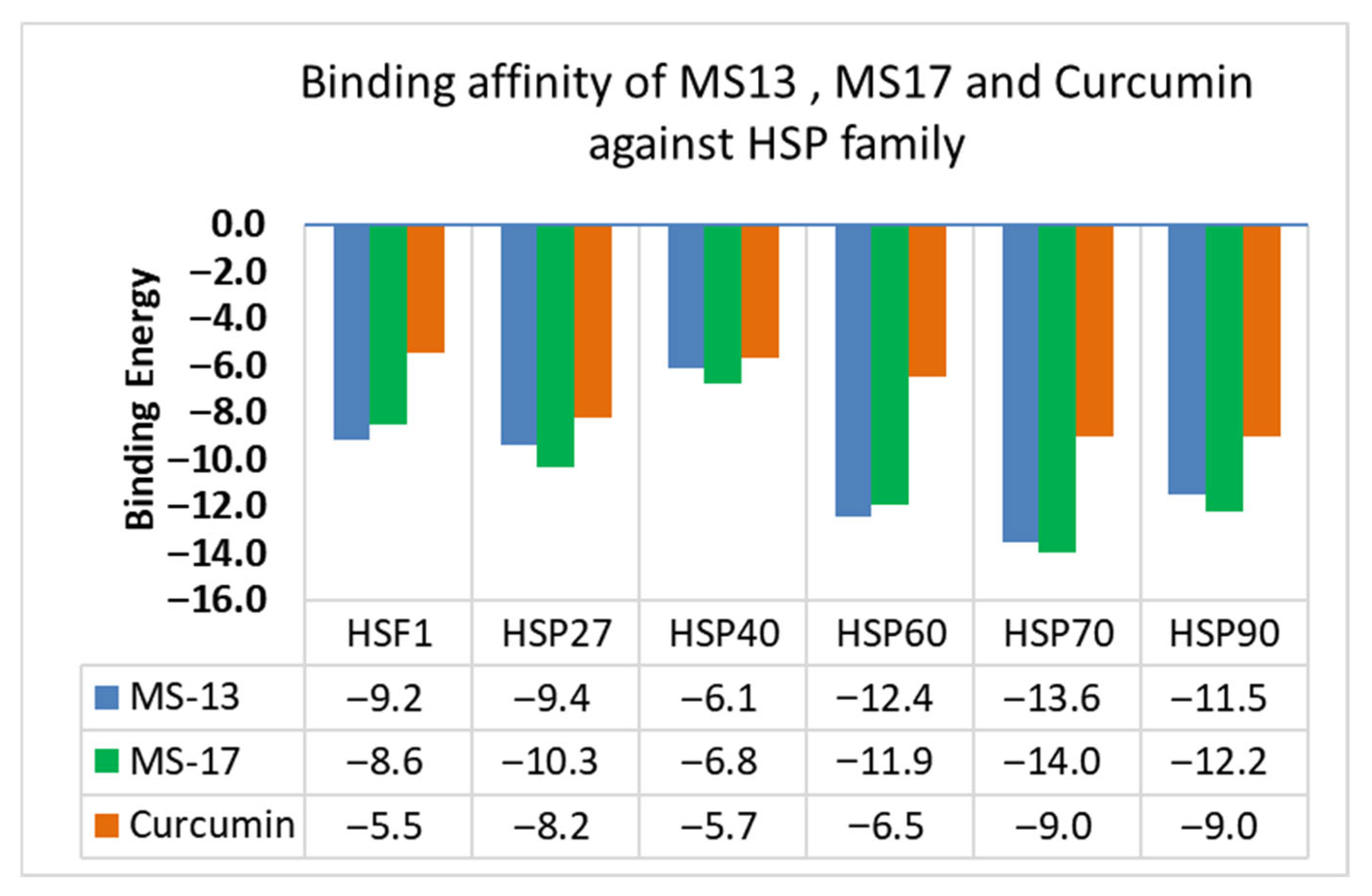
To validate the docking protocol, the first bound co-crystal ligands within all target proteins (HSF1, HSP27, HSP40, HSP60, HSP70, and HSP90) were docked, and RMSD was found to be less than 2 Å. Following validation through redocking, curcumin, MS13 and MS17 were docked. The analysis presented in Table 1 shows the Chemguass4 score for curcumin, MS13 and MS17 against all six target proteins. Analysis of the Chemguass4 score revealed that curcumin analogs, MS13 and MS17 exhibited better binding interaction to all targeted heat shock proteins compared to curcumin. Moreover, the compound MS13 had the highest binding interaction with HSP70 (−13.6), followed by HSP60 (−12.4). Similarly, MS17 had the highest binding affinity with HSP70 (−14.0), followed by HSP90 and HSP60. Both compounds showed relatively lower Chemguass4 scores against HSF1 compared to other target proteins.

3.1. Analysis of MS13 Binding Affinity

Figure 2 illustrates the binding affinity of MS13, MS17, and curcumin against each target protein, respectively. The binding affinity data for MS13 against HSF1 and various HSPs demonstrate its potential as HSF1 and HSPs modulator. With binding energies ranging from −6.10 to −13.56, MS13 exhibited a strong affinity for these proteins. Notably, its highest binding affinity was observed with HSP70 (−13.6), a protein crucial in protein folding and stress response, indicating a particularly strong interaction. This high affinity suggests that MS13 could significantly influence the biological functions of HSP70, potentially making it a candidate for therapeutic applications where modulation of this protein is desired. Our data also showed that MS13 exhibited a lower binding affinity with HSP40 (−6.1) than other HSPs, which might imply a lesser interaction strength and reduced influence on HSP40.

3.2. Analysis of MS17 Binding Affinity

The binding affinity profile of MS17 with HSF1 and HSPs showed a slightly different pattern compared to MS13. MS17 had its highest binding affinity with HSP70 (−14.0), which was slightly stronger than that of MS13, suggesting an even more potent interaction with this protein. This enhanced affinity could have significant implications in therapeutic contexts, especially in diseases where HSP70 is pivotal. Additionally, the binding energies of MS17 with all target proteins were consistently high, with their lowest being (−6.8) with HSP40. This consistent high-affinity binding across various HSPs could indicate a broad-spectrum interaction capability of MS17, which may be advantageous in targeting multiple pathways in different diseases.

3.3. Comparison of Curcumin Binding Affinity with MS13 and MS17

Curcumin demonstrated higher binding energies with HSF1 and HSPs than MS13 and MS17. Its binding energies were between −5.5 to −9.0, with the highest interactions being HSP70 and HSP90. The amino acid residue involved in the binding interaction with curcumin in our study is consistent with previously reported studies. Jun Li et al. reported similar binding energy of (−5.743 kcal/Mol) established from H-bonding with LYS-58 and PHE-138 residues at the curcumin binding site of HSP90AA1 (PDB ID: 1BYQ) [56]. Their results suggest that the downregulation of HSP90AA1 contributes to the anti-cancer effect of curcumin [56]. This suggests a moderate affinity towards these proteins, which could contribute towards curcumin’s broad range of biological activities, such as anti-inflammatory and antioxidant. The comparison of curcumin against its analogs MS13 and MS17 is crucial to highlight the stronger binding affinities of curcumin analogs, which may translate to higher efficacy in modulating the activities of HSPs. Our study further confirmed that MS13 and MS17 bind at the curcumin binding site with similar H-bonding interaction (LYS-58:MS13) as reported in previous studies [56].

3.4. Domain-Wise Binding Orientation of MS13 and MS17 on Heat Shock Proteins

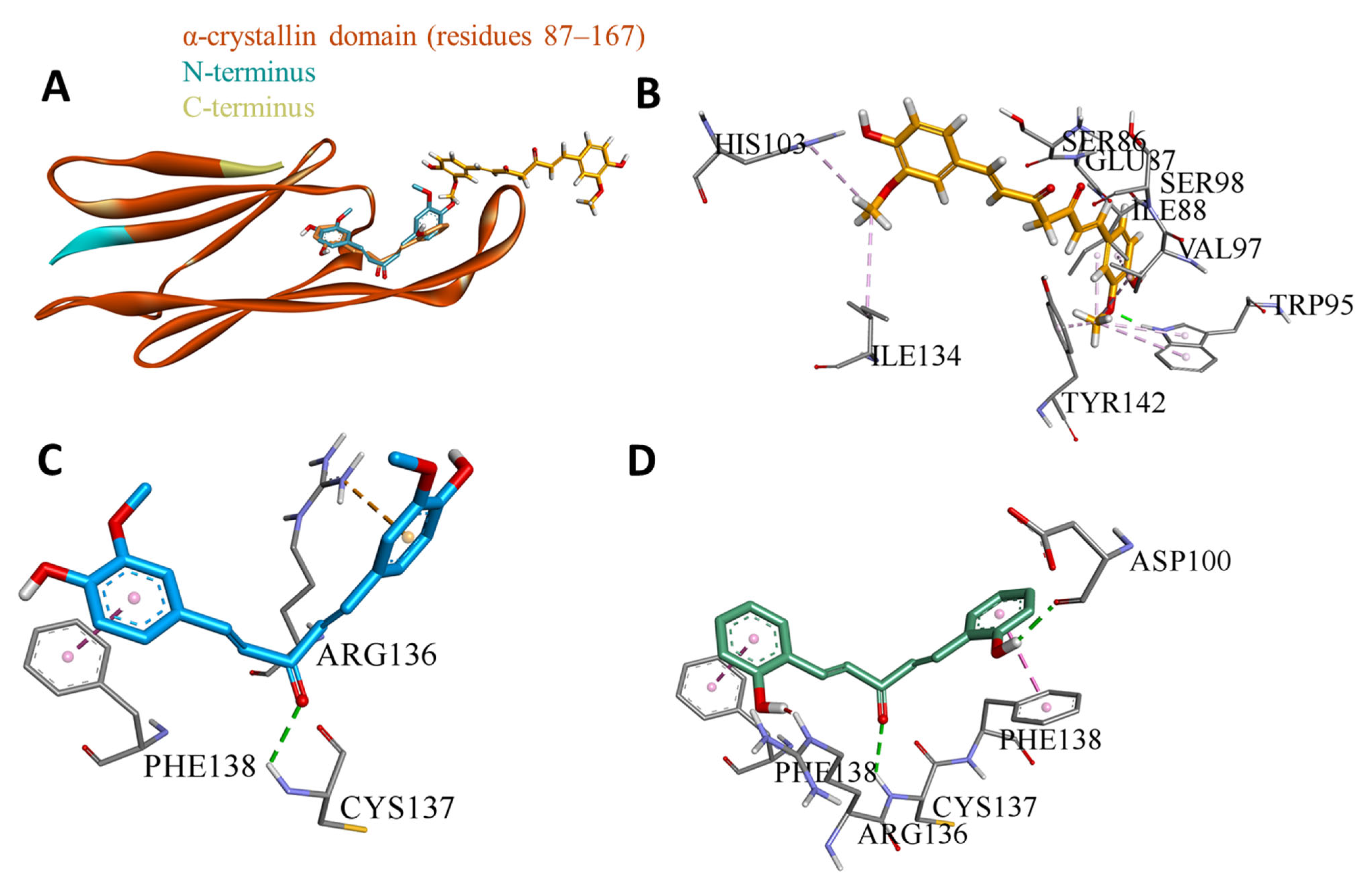
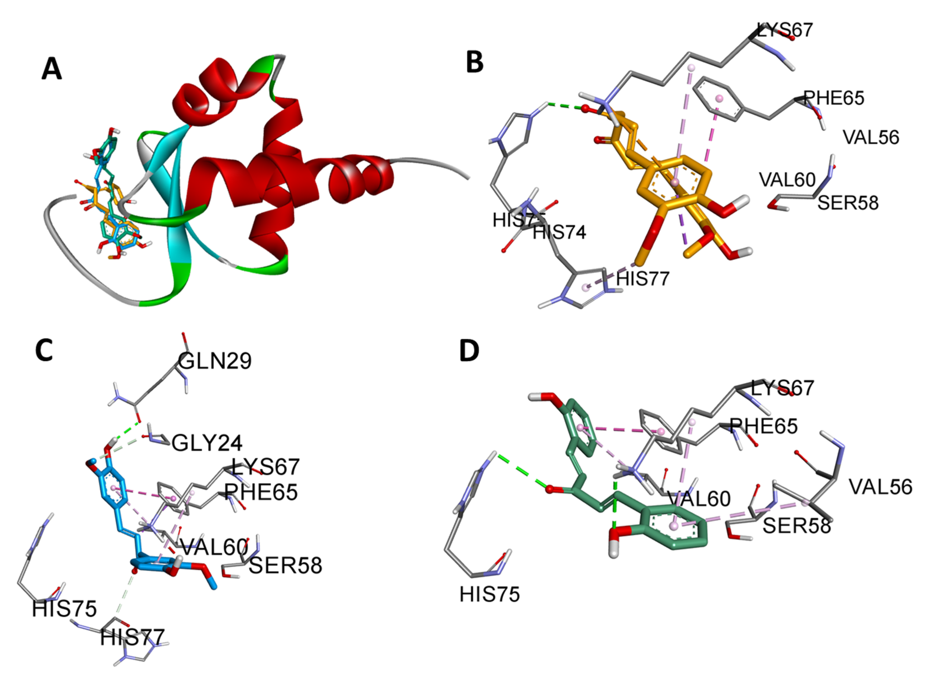
Figure 3, Figure 4, Figure 5, Figure 6, Figure 7 and Figure 8 illustrate the domain-wise binding orientation of MS13 and MS17 on HSF-1 and different HSPs. As shown in Figure 3, MS13 and MS17 were bound to the N-terminal DNA-binding domain (DBD) of HSF1, specifically residues 16–123. This domain was structurally characterized in the Protein Data Bank (PDB) under accession code 5D5V, where it was bound to DNA. In Figure 4, MS13 and MS17 were shown to interact with the alpha-crystallin domain (residues 87–167) of HSP27. The structure of this domain is deposited in the PDB under the accession code 4MJH (residues 85–170). As for HSP40 in Figure 5, MS13 and MS17 were bound to the J-domain (residues 1–80) of this HSP. The HSP40 protein structure is available in the PDB under accession code 2l01 (NMR structure).
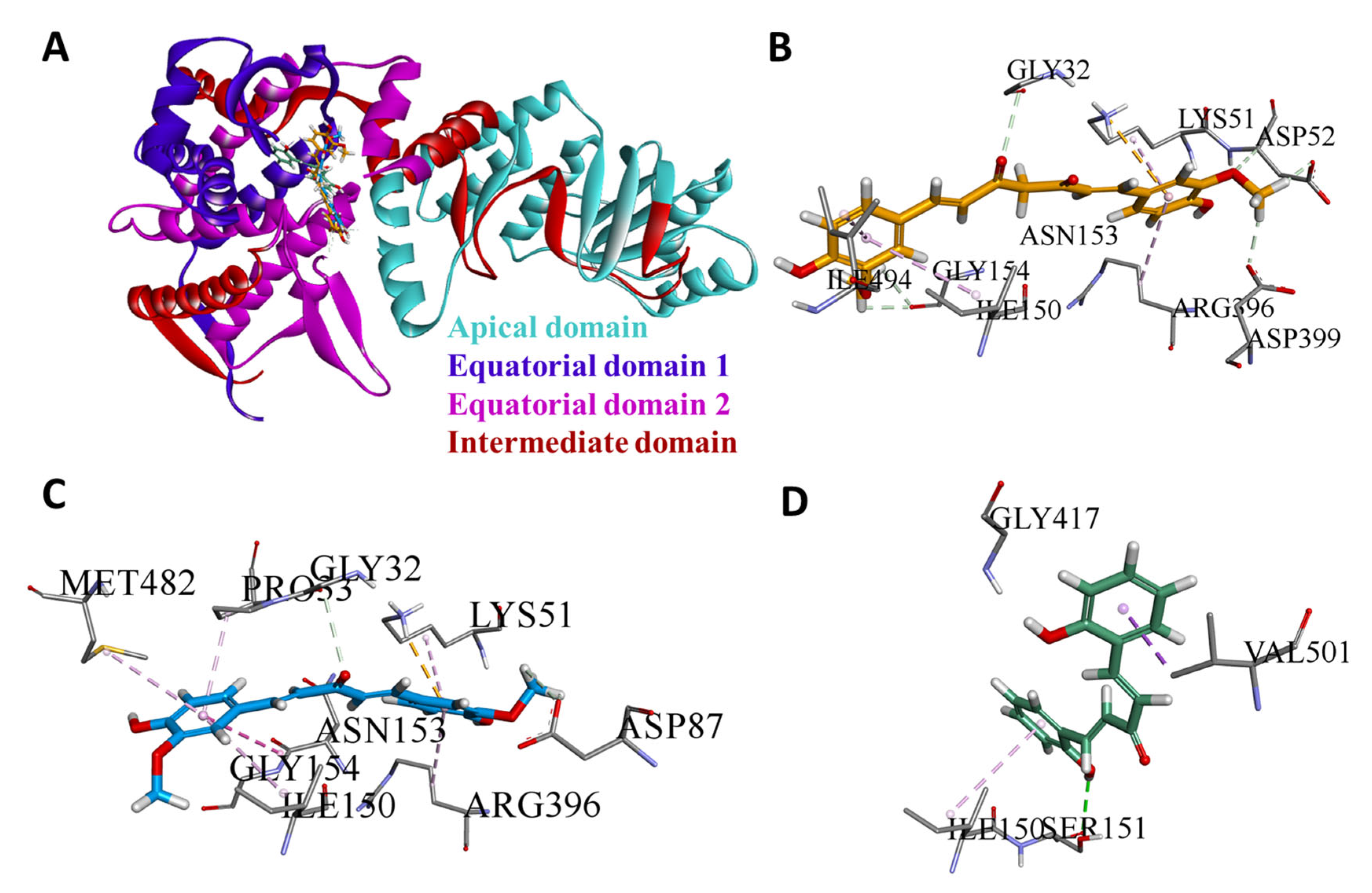
As depicted in Figure 6, MS13 and MS17 targeted a specific pocket within the equatorial domain of HSP60 for interaction. This binding site encompasses residues 30–157 and 434–548. The complete HSP60 structure, including apical, equatorial (1 & 2), and intermediate domains, is revealed in PDB 6MRD. Meanwhile, MS13 and MS17 specifically targeted the N-terminal ATPase domain (NBD) of HSP70 for interaction (Figure 7). A high-resolution crystal structure of the isolated N-terminal ATPase domain (NBD) of HSP70 (residues 3–382) is provided in PDB 1HJO. As for HSP90, shown in Figure 8, MS13 and MS17 were preferentially bound to the N-terminal domain (NTD) of HSP90. The available high-resolution crystal structure (PDB 2XJX) focuses solely on the N-terminal ATPase domain (NBD) of HSP90, encompassing amino acids 11 to 236. However, current data suggests preferential binding to the N-terminal domain. Table 2 summarizes the amino acid residues at the binding site of each target protein and subsequent implication upon binding of MS13 and MS17.

3.5. MD Simulation Analysis

3.5.1. HSF1

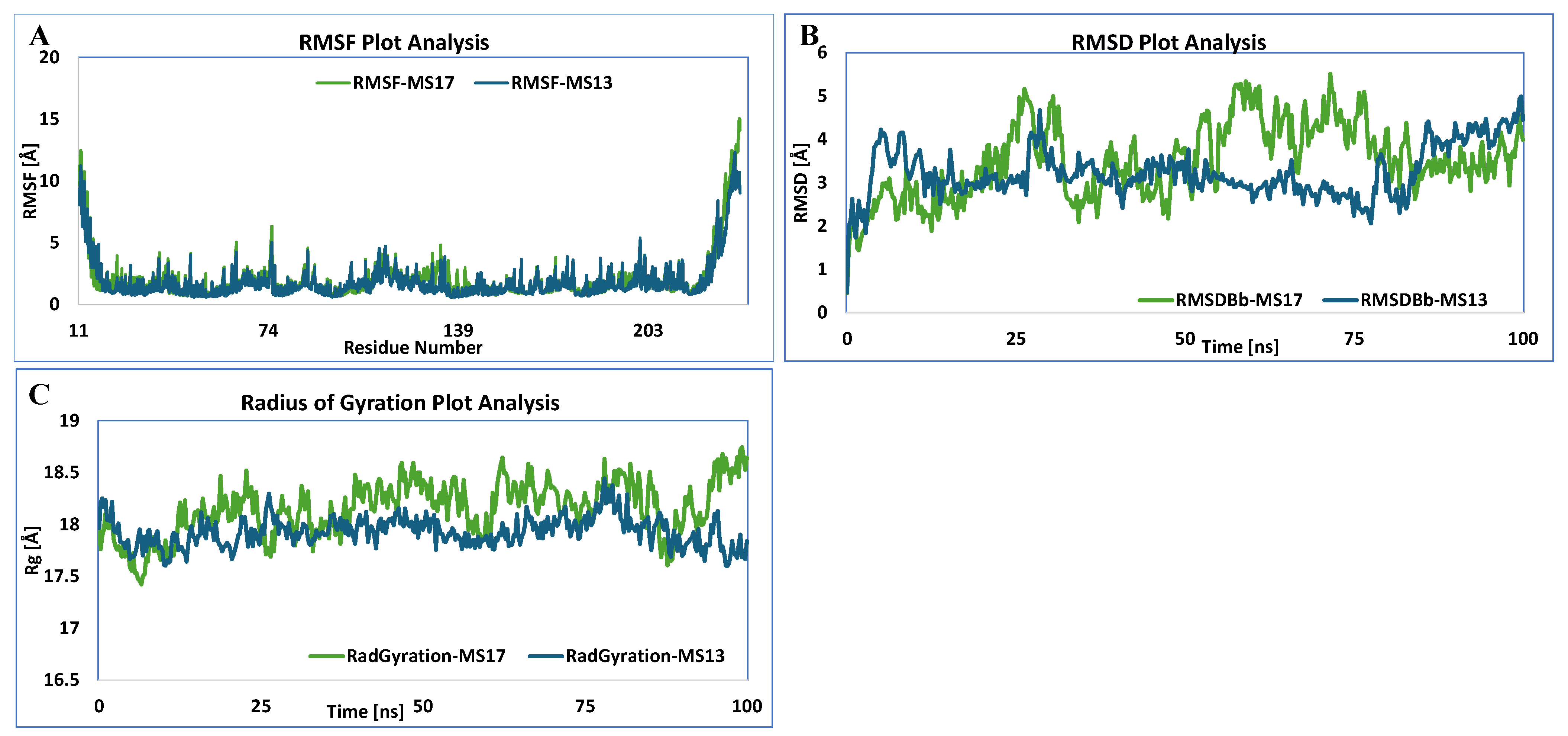
Figure 9 showed that MS13 formed a more stable complex with HSF1 than MS17, as indicated by lower RMSD values. Lower RMSD values indicate less structural fluctuation over time, suggesting a more rigid and stable complex. The binding of MS13 and MS17 to the DNA-binding domain (DBD) region of HSF1 could potentially block the binding between HSF1 and its downstream HSPs. This blockage could inactivate heat shock responses, hindering cancer cell growth and survival. HSF1 is a promising target for cancer therapy due to its overexpression in various cancers and association with advanced stages and poor prognosis.

3.5.2. HSP27

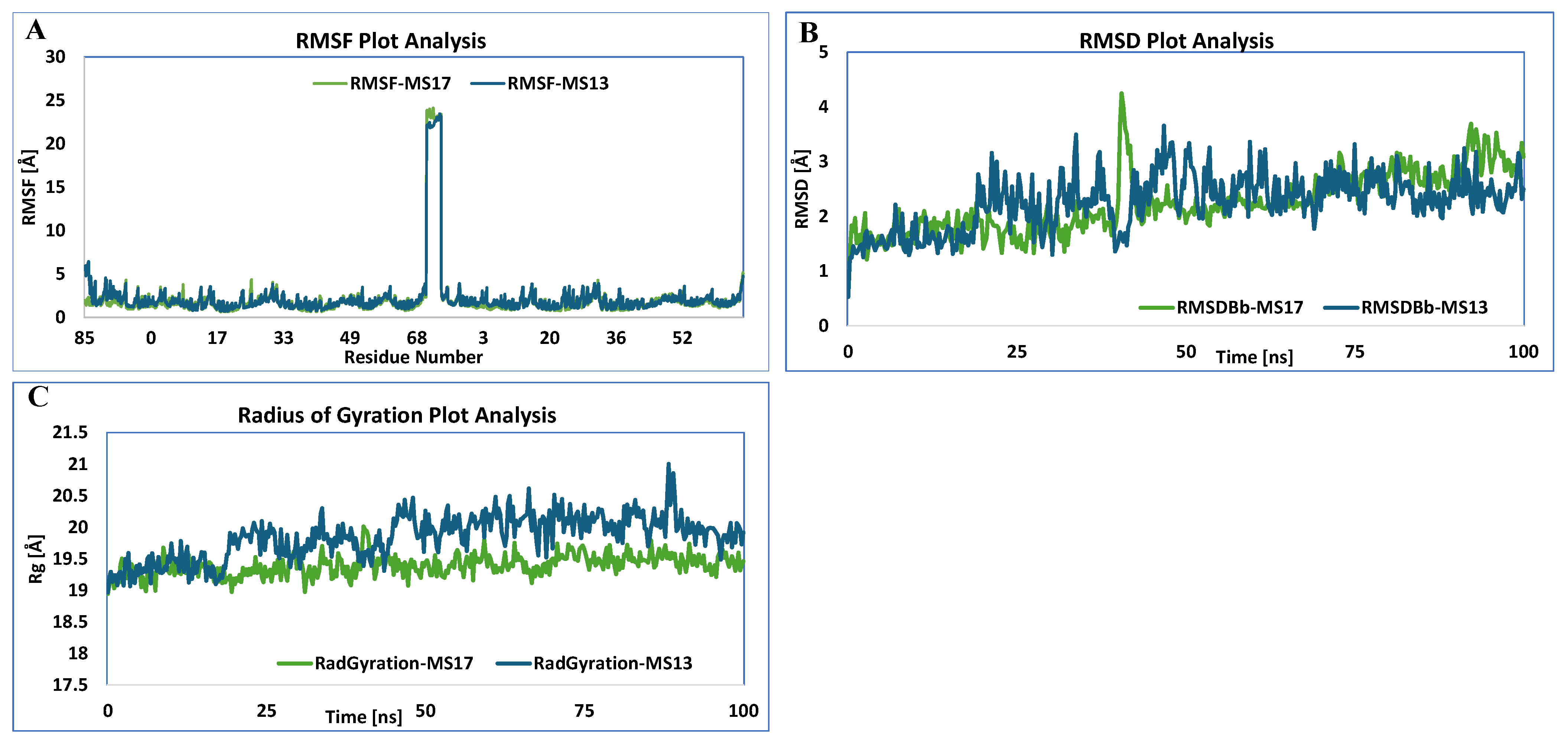
In contrast to HSF1, MD stimulation analysis indicated that MS17 formed a more stable complex with HSP27 compared to MS13, as shown in Figure 10. This finding was supported by lower RMSD values for MS17, except at 40 ns, which shows a bit more fluctuation, but the pose returns to the orientation again after just 42 ns, suggesting overall a more rigid and stable complex. The MS17-HSP27 complex also exhibited a lower radius of gyration (Rg) value, indicating a more compact and potentially more stable structure. MS13 and MS17 bind to the α-Crystallin domain (ACD) of HSP27, which is crucial for the protein’s chaperone activity and dimerization. By binding to this domain, MS13 and MS17 might disrupt HSP27 dimerization and impair its function. Disrupting the dimerization of HSP27 could lead to apoptosis and reduce drug resistance in cancer cells.

3.5.3. HSP40

MS17 demonstrated greater stability when complexed with HSP40 based on MD simulation analysis. This observation was supported by lower RMSD values for MS17, indicating a more rigid and stable complex as depicted in Figure 11. The binding of MS13 and MS17 to the J domain of HSP40 could inhibit the protein’s function. The J domain interacts with HSP70 and stimulates ATP hydrolysis, influencing the chaperone activity of the HSP70-HSP40 complex. Binding to the J domain may interfere with this interaction, potentially affecting protein folding and quality control mechanisms. Inhibiting HSP40 could reduce the amount of misfolded proteins, including mutant p53, potentially leading to anti-cancer effects such as apoptosis and DNA damage.

3.5.4. HSP60

The trend of MS17 forming more stable complexes did not continue with HSP60. MD simulation analysis as depicted in Figure 12, showed lower RMSD values for MS13, suggesting a more stable complex than the MS17. MS13 and MS17 target the Equatorial Domain binding pocket of HSP60, a domain that contains the binding site for ATP and is crucial for HSP60’s chaperone function. Binding to this domain may interfere with the ATP binding and disrupt HSP60’s stability. Disrupting HSP60 could lead to misfolding and aggregation of client proteins, potentially inducing anti-cancer effects.

3.5.5. HSP70

MS17 maintained its trend of forming more stable complexes with HSP70, as shown in Figure 13 by MD simulation analysis. Lower RMSD values were observed for MS17 compared to MS13, indicating a more stable complex. Both MS13 and MS17 exhibited strong binding to the N-terminal ATPase domain (NBD) of HSP70. This domain is responsible for binding and hydrolyzing ATP, providing the energy required for HSP70’s chaperone activity. Inhibition of HSP70’s ATPase activity through binding to the NBD could disrupt its chaperone function, potentially leading to apoptosis and inhibition of drug resistance.

3.5.6. HSP90

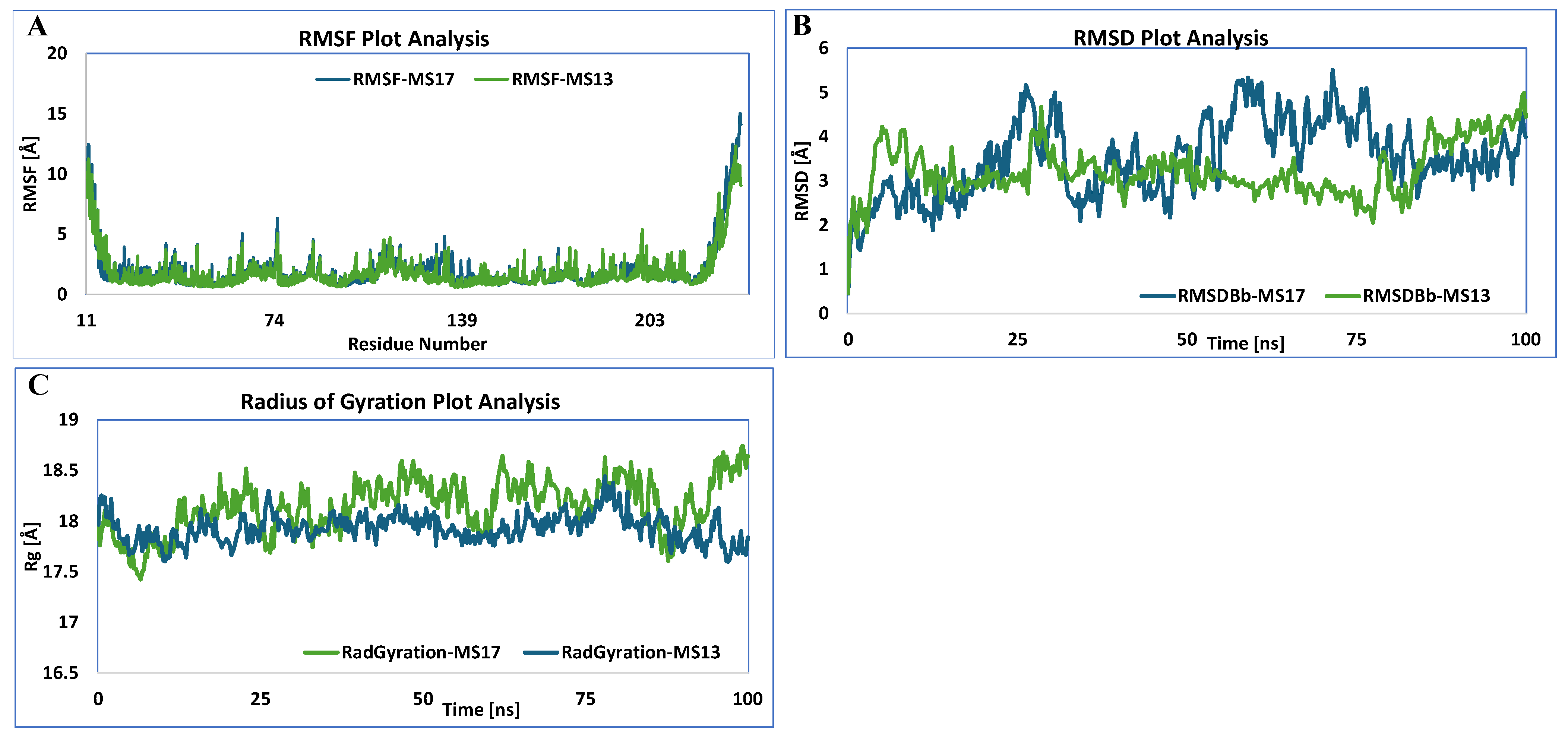
In contrast to the other HSPs, MS13 formed a more stable complex with HSP90 than MS17, as demonstrated by MD simulation analysis in Figure 14. Lower RMSD values for MS13 suggest a more stable complex. Both MS13 and MS17 bind to the N-terminal domain (NTD) of HSP90. This domain plays a critical role in HSP90’s function, including ATP binding and interaction with client proteins. Binding to the NTD may disrupt HSP90’s chaperone activity, potentially affecting the stability of its client proteins and inducing apoptosis in cancer cells.

3.6. ADMET and Drug-likeness Properties of MS13, MS17, and Curcumin

SwissADME, a free webtool was used to predict the Absorption, Distribution, Metabolism, Excretion, and Toxicity (ADMET) properties of MS13, MS17, and curcumin, as shown in Table 3. Key pharmacokinetic parameters analyzed included lipophilicity (consensus Log P), polar surface area (TPSA), number of hydrogen bond acceptors and donors, and aqueous solubility (ESOL, Ali, and Silicos-IT solubility classes). Additionally, predictions for drug-likeness, mutagenicity, tumorigenicity, reproductive effectiveness, and irritancy were also evaluated using the DataWarrior tool. The predicted ADMET profiles reveal key distinctions for MS13 and MS17 compared to curcumin, suggesting enhanced therapeutic potential for the analogs. MS17, with its slightly higher lipophilicity (Consensus Log P 3.08) and significantly lower Polar Surface Area (TPSA 57.53), along with fewer hydrogen bond acceptors, is theoretically poised for superior passive membrane permeability and absorption compared to MS13 and curcumin. Crucially, MS13 and MS17 are predicted to be Blood–Brain Barrier (BBB) permeant, a significant advantage over curcumin. While all three compounds show high gastrointestinal (GI) absorption and are predicted to inhibit CYP2C9 and CYP3A4, MS13 and MS17 uniquely inhibit CYP1A2, unlike curcumin.

3.7. Discussion

Elevated levels of HSF1 and HSPs are associated with poor clinical outcomes, as they contribute to drug resistance and tumor progression. Targeting HSF1 and HSPs is being explored as a potential therapeutic strategy in cancer treatment. In the current study, we explored the binding affinities of curcumin and two other curcumin analogs, MS13 and MS17, towards HSF1 and selected HSPs using molecular docking and molecular dynamics simulations. Overall, both MS13 and MS17 exhibit stronger binding affinities to these target proteins, as compared to the parent compound curcumin. Our comparative analysis also demonstrates that MS17 generally formed more stable complexes with HSP27, HSP40, HSP60, and HSP70, as evidenced by lower RMSD, RMSF values for specific residues, and a smaller Rg. This suggests that MS17 may be a more potent modulation towards HSP27, HSP40, HSP60, and HSP70. On the other hand, MS13 demonstrates higher stability when forming complexes with HSF1 and HSP90, indicating potential for stronger inhibition association with these target proteins. These differences in complex stability are mostly attributed to variations in the chemical structures of MS13 and MS17, leading to distinct interactions with each protein’s binding site.
The DNA-binding domain (DBD) of HSF1 is a winged helix-turn-helix domain located at the N-terminus of the protein [57]. It is the most conserved region in the HSF protein family, which contains a structural motif to recognize a specific DNA sequence [57]. DBD binds to heat-shock elements (HSEs) of heat shock proteins when HSF1 is stressed, to trigger the upregulation of downstream HSPs and induce heat shock responses [58]. Previously, Dong et al. designed a selective small-molecule inhibitor known as Direct Targeted HSF1 Inhibitor (DTHIB) that binds directly to the HSF1 DBD and promotes HSF1 degradation in the nucleus of prostate cancer cells [59]. Our docking analysis showed that MS13 and MS17 bound with the DBD region of HSF1, which could potentially block the binding between HSF1 and its downstream HSPs, resulting in the inactivation of heat shock responses and may hinder cancer cell growth and survival. Based on our findings, MS13 and MS17 could bind to the DBD region of HSF1 with stronger binding energies (−9.2 and −8.6, respectively) than curcumin (−5.5), suggesting that MS13 and MS17 could induce a stronger impact on HSF1 as compared to their parent compound, curcumin. MS13 and MS17 could function as selective inhibitors of HSF1 in cancer cells.
Meanwhile, MS13 and MS17 had been shown to bind with the α-Crystallin domain (ACD) of HSF27. HSP27, also known as HSPB1, is composed of three main structural domains, namely N-terminal Domain (WDPF Domain), α-Crystallin domain (ACD), and C-terminal Domain (IXI/V Motif) [60]. The ACD of HSP27 is highly conserved among small HSPs, encompassing residues 87–167 [60]. The ACD forms stable dimers crucial for HSP27’s chaperone activity and its ability to interact with client proteins, ensuring their proper protein conformation and stability [61]. HSP27 does not depend on ATP for its function, unlike most heat shock proteins [61]. The overexpression of HSP27 is commonly associated with tumor progression and drug resistance in multiple cancers, including CRC, lung, pancreatic and stomach [24,62,63,64,65]. HSP27 interacts with various signaling pathways, such as PI3K/Akt and MAPK, to inhibit apoptosis, promote EMT, and induce angiogenesis, enhancing cell survival and contributing to drug resistance [66]. Several synthetic compounds, namely NA49, OGX-427, RP101 and SW15, have been reported to suppress HSP27 and disrupt its dimer stability, which restores the drug sensitivity in breast, lung and prostate cancers [67,68,69,70]. Peptide aptamers, including PA11 and PA50, can specifically bind to HSP27, interfering with its dimerization and oligomerization to inhibit the anti-apoptotic and oncogenic effects of this HSP in cancer cells [71]. Our docking analysis suggests that MS13 and MS17 could bind strongly to the ACD to disrupt the dimerization of HSP27, which may eventually lead to apoptosis and the downregulation of drug resistance in cancer cells.
HSP40, also known as DNAJ, represents a group of co-chaperones that assist HSP70 in recognizing client proteins, facilitating their folding, and preventing aggregation to maintain cellular proteostasis [72]. DNAJA1 is a member of the HSP40 family that selectively binds to unfolded mutant p53 in cancer cells, promoting its accumulation for its oncogenic gain-of-function activity that may lead to tumor progression and metastasis in head and neck squamous cell carcinoma (HNSCC) [73]. DNAJA1 was significantly downregulated in pancreatic cancer cells, and its overexpression activates a DnaK protein by forming a complex to suppress the JNK pathway [74]. This reduces the hyperphosphorylation of oncogenic transcription factor c-Jun, leading to apoptosis in pancreatic cancer cells [74]. There are three major domains of HSP40 family proteins, namely the N-terminal J domain, the central Cysteine-Rich (C/R) domain, and the C-Terminal (CT) domain [75]. Among all the domains, the interaction between the J domain and HSP70 is crucial for protein folding and cellular homeostasis. The J domain stimulates ATP hydrolysis in HSP70, which stabilizes substrate binding [76]. The J domain perturbs a conserved intramolecular HSP70 network of contacts [77]. This perturbation destabilizes the domain-domain interface, triggering ATP hydrolysis [77].
Variable residues on J domains allow HSP70 to recognize and discriminate among different members of HSP40 family [76]. Researchers have identified a small compound called A11 that binds to the J domain of HSP40 to degrade misfolded mutant p53 (mutp53) and inhibits the malignant properties of cancer cells [78]. This A11 compound has minimal effects on wild-type p53 and other DNAJA1 mutants [78]. In our study, both MS13 and MS17 bound to the J domain of HSP40 with binding energies (−6.1 and −6.8, respectively) slightly higher than that of curcumin (−5.5), suggesting that MS13 and MS17 could inhibit the functions of HSP40 better than curcumin. The binding of MS13 and MS17 onto HSP40 could induce anti-cancer effects by reducing misfolded client proteins, including mutant p53, and eventually lead to anti-cancer effects, such as apoptosis and DNA damage. However, MS13 and MS17 showed the lowest binding energies with HSP40 compared to HSF1 and other HSPs, which merits further investigation.
MS13 and MS17 preferentially bound to the Equatorial Domain binding pocket (residues 30–157 & 434–548) of HSP60 with much higher binding energies (−12.4 and −11.9, respectively), almost double that of curcumin with a binding affinity of −6.4. HSP60 is a mitochondrial chaperonin that assists with proper protein folding and preventing misfolding or aggregation [79]. HSP60 helps cells withstand extreme temperatures (heat stress) by stabilizing client proteins, preventing denaturation [80]. Previous studies show that HSP60 plays intriguing roles in cancer immunity by assisting tumor-infiltrating lymphocytes (TILs) in recognizing transformed cells and inhibiting neoplastic tissue growth [1,81]. HSP60 can be found on the surface of both normal and tumor cells, leading to the activation and maturation of dendritic cells and the generation of antitumor T-cell responses [81,82]. Interestingly, HSP60 could have dual roles in promoting tumorigenesis depending on the type of cancer [83,84]. Previously, overexpression of HSP60 was associated with metastasis in head and neck cancer, as well as drug resistance in CRC cells [85,86]. HSP60 has three structural domains: equatorial, intermediate, and apical [15]. The Equatorial domain contains the binding site for ATP and other heptameric ring components, facilitating interactions critical for HSP60’s chaperone function [84]. The binding of MS13 and MS17 to the Equatorial Domain binding pockets could disrupt the ATP-binding and stability of HSP60, leading to the misfolding and aggregation of the client proteins. Consequently, this may induce anti-cancer effects, such as activating tumor immune response and suppressing proliferation, depending on the context of a particular cancer.
In cancer cells, HSP70 functions as a molecular chaperone to stabilize oncoproteins, allowing them to evade degradation and maintain their function [87]. By chaperoning key proteins involved in cell cycle regulation, apoptosis, and DNA repair, HSP70 supports cancer cell survival and proliferation [87,88]. HSP70 proteins have three major domains: Nucleotide-Binding Domain (NBD), Substrate-binding domain (SBD), and EEVD motif [75]. Based on our results, MS13 and MS17 bound very strongly to the NDB with binding energies of −13.6 and −14.0, respectively, which were about 50–55% higher than curcumin (−9.0). The NBD, also known as the ATPase domain, is located at the N-terminal end of HSP70 [89]. It binds and hydrolyzes ATP to ADP, in which the NBD consists of two lobes with a deep cleft where nucleotides bind [89]. Over the years, researchers have been developing small molecules that specifically inhibit HSP70’s ATPase activity. VER-155008 is a small-molecule inhibitor that specifically binds to the NBD of HSP70, acting as an ATP-competitive inhibitor to prevent the allosteric control between NBD and SBD [90]. VER-155008 inhibits proliferation and cell cycle progression while inducing apoptosis in colon and lung cancer cell lines [90,91,92]. VER-155008 synergistically enhanced the effects of bortezomib in multiple myeloma (MM) cells by significantly suppressing HSP70 to induce apoptosis and reduce proliferation [93]. Current evidence suggests that MS13 and MS17 could function as strong inhibitors of HSP70 in cancer cells, potentially leading to apoptosis and inhibition of drug resistance.
HSP90 family proteins play a crucial role in cancer by regulating intracellular signaling pathways and maintaining the stability of signaling proteins [94]. HSP90α is often significantly upregulated in cancer cells, which is commonly associated with cancer cell growth, survival, and resistance to treatment [7,26,95,96,97]. HSP90 family proteins consist of three distinct structural domains. The N-Terminal Domain (NTD) contains the ATP binding site and plays a crucial role in HSP90’s function [98]. It is connected to the middle domain through a variable charged linker [99]. The Middle Domain (MD) of HSP90 is involved in interactions with cochaperones and client proteins, contributing to the conformational dynamics of HSP90 [98]. The C-Terminal Domain (CTD) is responsible for HSP90 dimerization, ensuring that HSP90 functions as a dimeric complex [98]. In our study, MS13 and MS17 bound strongly to the NTD of HSP90 with binding energies of −11.5 and −12.2, respectively. The ATP-binding domain in NTD is essential for HSP90’s role as a molecular chaperone since HSP90 utilizes ATP binding and hydrolysis to fold client proteins involved in signal transduction, protein trafficking, receptor maturation, and immunity [100,101]. Inhibition of HSP90 through the binding of NTD potentially leads to the degradation of cancer-related client proteins via the ubiquitin-proteasome system [102]. This disruption affects multiple signaling pathways and induces apoptosis in cancer cells [102,103]. Geldanamycin is an antitumor antibiotic that binds to ADP/ATP-binding pocket in the NTD of HSP90, which can disrupt the binding of HSP90 with its client proteins involved in cell survival and cell cycle progression [104,105,106]. Our results suggest that MS13 and MS17 could effectively inhibit HSP90 upon binding to induce anti-cancer activities.
In addition, the HSP family and HSF1 are implicated in the regulation and integration of major oncogenic pathways in several top cancers, including breast, lung, CRC, prostate and stomach, reaffirming their centrality as potential therapeutic targets [13,26,65,68,97,107,108,109]. HSF1 and HSPs support oncogenic signaling pathways in multiple and complementary ways, acting as molecular chaperones that stabilize, fold, and protect key oncoproteins from degradation. Oncogenic RAS activates HSF1 through its effector MEK, leading to phosphorylation and transcriptional activity of HSF1, which in turn drives cancer-specific transcriptional programs beyond classic heat shock proteins [10]. This pathway is frequently mutated in CRC and lung cancer [110,111,112]. HSF1 and mTORC1 interact in a coordinated manner where mTORC1 activates HSF1 by phosphorylation to induce the heat shock response, and HSF1 protects mTORC1 from stress-induced inhibition, enabling sustained oncogenic mTOR signaling and proteostasis in cancer cells [113,114]. HSP27 promotes the activation of Akt and the PI3K/Akt pathway, leading to multidrug resistance and apoptosis evasion in different cancers, such as bladder and colon [18,115]. HSP27 also promotes EMT via IL-6/STAT3/Twist axis, TGF-β1/p38, and β-catenin/MMP3 pathways, which enhance metastasis and invasion [116,117,118]. HSP40 regulates cell cycle progression via CDC45 stabilization, enhances EGFR signaling, and promotes metastasis through the pERK/IQGAP1 signaling axis, which has been reported in CRC, gastric and lung cancers [119,120]. HSP60 supports the growth, proliferation, and chemoresistance of pancreatic, lung, and CRC cells through numerous signaling pathways, such as ROS/AMPK/mTOR signaling, ERK signaling and Wnt signaling [25,83,85,86,121,122,123,124].
HSP70 and HSP90 are associated with PI3K/AKT/mTOR axis signaling, crucial for cancer cell growth, proliferation, angiogenesis, and therapy resistance in several cancer types, including CRC and lung [125,126,127]. Their inhibition destabilizes this pathway, potentially sensitizing tumors to treatment [128]. Computational ADMET and drug-likeness predictions suggest that MS13 and MS17 offer advantages over curcumin, notably their predicted Blood–Brain Barrier (BBB) permeability and MS17’s theoretically superior passive membrane permeability and absorption attributed to its lower TPSA and fewer hydrogen bond acceptors, alongside unique CYP1A2 inhibition profiles. While all three compounds consistently demonstrate excellent drug-likeness with zero violations of Lipinski’s Rule of Five, future planned experimental validation remains crucial to confirm these computational findings and establish their in vivo pharmacological relevance and safety profiles. Our computational results suggest that MS13 and MS17 could, through direct modulation of HSP/HSF1 function, disrupt the stability of oncogenic signaling complexes, impair growth-promoting cascades, and potentially restore sensitivity to anti-cancer therapies. By contextualizing our in silico binding data within the framework of cancer-specific biology and signaling networks, the study could become a solid foundation for the rational design and experimental validation of new anti-cancer therapeutics targeting the HSP/HSF1 axis in cancers.

4. Conclusions

Our study presents the first comparative analysis of curcumin and two novel analogs, MS13 and MS17, in their binding interactions with HSF1 and selected HSPs. MS13 and MS17 demonstrated stronger binding affinity towards HSF1 and the selected HSPs as compared to the parent compound curcumin. Notably, MS13 exhibited stronger complex stability with HSF1 and HSP90, while MS17 displayed better stability with HSP27, HSP40, HSP60, and HSP70. These findings suggest that both analogs may act as more potent inhibitors of their respective target proteins, potentially leading to enhanced anti-cancer effects. The observed differences in complex stability are likely attributed to specific structural variations between MS13 and MS17. Overall, MS13 and MS17 exhibit more advantageous ADMET profiles compared to curcumin, particularly in their predicted Blood–Brain Barrier (BBB) permeability and MS17’s theoretically superior passive membrane permeability and absorption. While our study provides valuable insights into the binding affinity and interaction sites of curcumin and its analogs MS13 and MS17 with HSF1 and various HSP family proteins through in silico approaches, further experimental validation is essential to corroborate these findings.
In vitro binding assays, such as fluorescence-based binding studies, could quantitatively verify the binding affinities and kinetics of MS13 and MS17 with target proteins, particularly HSP70, HSP60, and HSF1, where strong interactions were predicted. Additionally, cell-based functional assays can elucidate the biological consequences of these interactions. These may include assessing changes in HSP expression levels, chaperone activity disruption, and subsequent effects on cancer cell proliferation, apoptosis induction, and cellular stress response using relevant cancer cell lines. Co-immunoprecipitation and protein stability assays can help determine whether these compounds affect HSP-client protein complexes and downstream signaling pathways. Such experimental validations will not only confirm the in silico predictions but also provide mechanistic insights and lay the groundwork for future drug development targeting the HSP system in cancer therapy.

Author Contributions

Conceptualization: R.N.; Methodology and Validation: S.U.K. and T.T.H.; Writing—original draft preparation: K.W.H., S.U.K. and T.T.H.; Writing—review and editing: K.W.H., S.U.K., T.T.H. and R.N.; Visualization and Illustrations: S.U.K. and T.T.H.; Overall Supervision and Project Administration: R.N. All authors have read and agreed to the published version of the manuscript.

Funding

This research did not receive any specific grant from funding agencies in the public, commercial, or non-profit sectors.

Data Availability Statement

The datasets produced throughout this study, as well as those analyzed, are readily obtained from the corresponding author upon the submission of a reasonable request.

Acknowledgments

We gratefully acknowledge the support of the Jeffrey Cheah School of Medicine and the School of Pharmacy, Monash University Malaysia, for providing essential resources and support in this research. Shafi Ullah Khan is the Recipient of the WINNING Normandy Fellowship Programme, which is supported by the European Union’s Horizon 2020 research and innovation programme under the Marie Skłodowska-Curie grant agreement No 101034329 (MSCA). We also thank OpenEye Scientific Software, Inc. for providing a free academic license to perform in silico studies.

Conflicts of Interest

The authors declare no competing conflict of interest.

References

  1. Sun, B.; Li, G.; Yu, Q.; Liu, D.; Tang, X. HSP60 in cancer: A promising biomarker for diagnosis and a potentially useful target for treatment. J. Drug Target. 2022, 30, 31–45. [Google Scholar] [CrossRef]
  2. He, Y.; Wu, Y.; Mou, Z.; Li, W.; Zou, L.; Fu, T.; Zhang, A.; Xiang, D.; Xiao, H.; Wang, X. Proteomics-based identification of HSP60 as a tumor-associated antigen in colorectal cancer. Proteom. Clin. Appl. 2007, 1, 336–342. [Google Scholar] [CrossRef]
  3. Gao, G.; Liu, S.; Yao, Z.; Zhan, Y.; Chen, W.; Liu, Y. The Prognostic Significance of Hsp70 in Patients with Colorectal Cancer Patients: A PRISMA-Compliant Meta-Analysis. BioMed Res. Int. 2021, 2021, 5526327. [Google Scholar] [CrossRef]
  4. Jiang, W.; Pan, X.; Yan, H.; Wang, G. Prognostic Significance of the Hsp70 Gene Family in Colorectal Cancer. Med. Sci. Monit. 2021, 27, e928352. [Google Scholar] [CrossRef]
  5. Kai, M.; Nakatsura, T.; Egami, H.; Senju, S.; Nishimura, Y.; Ogawa, M. Heat shock protein 105 is overexpressed in a variety of human tumors. Oncol. Rep. 2003, 10, 1777–1782. [Google Scholar] [CrossRef] [PubMed]
  6. Hrudka, J.; Jelínková, K.; Fišerová, H.; Matěj, R.; Mandys, V.; Waldauf, P. Heat Shock Proteins 27, 70, and 110: Expression and Prognostic Significance in Colorectal Cancer. Cancers 2021, 13, 4407. [Google Scholar] [CrossRef]
  7. Szczuka, I.; Wierzbicki, J.; Serek, P.; Szczęśniak-Sięga, B.M.; Krzystek-Korpacka, M. Heat Shock Proteins HSPA1 and HSP90AA1 Are Upregulated in Colorectal Polyps and Can Be Targeted in Cancer Cells by Anti-Inflammatory Oxicams with Arylpiperazine Pharmacophore and Benzoyl Moiety Substitutions at Thiazine Ring. Biomolecules 2021, 11, 1588. [Google Scholar] [CrossRef]
  8. Wang, G.; Cao, P.; Fan, Y.; Tan, K. Emerging roles of HSF1 in cancer: Cellular and molecular episodes. Biochim. Biophys. Acta (BBA) Rev. Cancer 2020, 1874, 188390. [Google Scholar] [CrossRef]
  9. Cyran, A.M.; Zhitkovich, A. Heat Shock Proteins and HSF1 in Cancer. Front. Oncol. 2022, 12, 860320. [Google Scholar] [CrossRef]
  10. Dai, C. The heat-shock, or HSF1-mediated proteotoxic stress, response in cancer: From proteomic stability to oncogenesis. Philos. Trans. R. Soc. Lond. B Biol. Sci. 2018, 373, 20160525. [Google Scholar] [CrossRef]
  11. Barna, J.; Csermely, P.; Vellai, T. Roles of heat shock factor 1 beyond the heat shock response. Cell. Mol. Life Sci. 2018, 75, 2897–2916. [Google Scholar] [CrossRef] [PubMed]
  12. Fang, F.; Chang, R.; Yang, L. Heat shock factor 1 promotes invasion and metastasis of hepatocellular carcinoma in vitro and in vivo. Cancer 2012, 118, 1782–1794. [Google Scholar] [CrossRef] [PubMed]
  13. Santagata, S.; Hu, R.; Lin, N.U.; Mendillo, M.L.; Collins, L.C.; Hankinson, S.E.; Schnitt, S.J.; Whitesell, L.; Tamimi, R.M.; Lindquist, S.; et al. High levels of nuclear heat-shock factor 1 (HSF1) are associated with poor prognosis in breast cancer. Proc. Natl. Acad. Sci. USA 2011, 108, 18378–18383. [Google Scholar] [CrossRef] [PubMed]
  14. Mendillo Marc, L.; Santagata, S.; Koeva, M.; Bell, G.W.; Hu, R.; Tamimi, R.M.; Fraenkel, E.; Ince, T.A.; Whitesell, L.; Lindquist, S. HSF1 Drives a Transcriptional Program Distinct from Heat Shock to Support Highly Malignant Human Cancers. Cell 2012, 150, 549–562. [Google Scholar] [CrossRef]
  15. Hu, C.; Yang, J.; Qi, Z.; Wu, H.; Wang, B.; Zou, F.; Mei, H.; Liu, J.; Wang, W.; Liu, Q. Heat shock proteins: Biological functions, pathological roles, and therapeutic opportunities. MedComm 2022, 3, e161. [Google Scholar] [CrossRef]
  16. Lang, B.J.; Guerrero, M.E.; Prince, T.L.; Okusha, Y.; Bonorino, C.; Calderwood, S.K. The functions and regulation of heat shock proteins; key orchestrators of proteostasis and the heat shock response. Arch. Toxicol. 2021, 95, 1943–1970. [Google Scholar] [CrossRef]
  17. Tutar, Y.; Naureen, H.; Farooqi, A.A. Chapter 13—Heat shock proteins in tumor progression and metastasis. In Unraveling the Complexities of Metastasis; Farooqi, A.A., Qureshi, M.Z., Sabitaliyevich, U.Y., Eds.; Academic Press: Cambridge, MA, USA, 2022; pp. 187–201. [Google Scholar]
  18. Liu, Z.; Liu, Y.; Long, Y.; Liu, B.; Wang, X. Role of HSP27 in the multidrug sensitivity and resistance of colon cancer cells. Oncol. Lett. 2020, 19, 2021–2027. [Google Scholar] [CrossRef]
  19. Tao, Y.; Messer, J.S.; Goss, K.H.; Hart, J.; Bissonnette, M.; Chang, E.B. Hsp70 exerts oncogenic activity in the Apc mutant Min mouse model. Carcinogenesis 2016, 37, 731–739. [Google Scholar] [CrossRef]
  20. Yamagishi, N.; Goto, K.; Nakagawa, S.; Saito, Y.; Hatayama, T. Hsp105 reduces the protein aggregation and cytotoxicity by expanded-polyglutamine proteins through the induction of Hsp70. Exp. Cell Res. 2010, 316, 2424–2433. [Google Scholar] [CrossRef]
  21. Berthenet, K.; Bokhari, A.d.; Lagrange, A.; Marcion, G.; Boudesco, C.; Causse, S.; De Thonel, A.; Svrcek, M.; Goloudina, A.R.; Dumont, S.; et al. HSP110 promotes colorectal cancer growth through STAT3 activation. Oncogene 2017, 36, 2328–2336. [Google Scholar] [CrossRef]
  22. Bhasin, N.; Dabral, P.; Senavirathna, L.; Pan, S.; Chen, R. Inhibition of TRAP1 Accelerates the DNA Damage Response, Activation of the Heat Shock Response and Metabolic Reprogramming in Colon Cancer Cells. Front. Biosci. (Landmark Ed.) 2023, 28, 227. [Google Scholar] [CrossRef]
  23. Lettini, G.; Sisinni, L.; Condelli, V.; Matassa, D.S.; Simeon, V.; Maddalena, F.; Gemei, M.; Lopes, E.; Vita, G.; Del Vecchio, L.; et al. TRAP1 regulates stemness through Wnt/β-catenin pathway in human colorectal carcinoma. Cell Death Differ. 2016, 23, 1792–1803. [Google Scholar] [CrossRef]
  24. Musiani, D.; Konda, J.D.; Pavan, S.; Torchiaro, E.; Erriquez, J.; Olivero, M.; Di Renzo, M.F. Heat Shock Protein 27 (HSP27, HSPB1) Is Up-Regulated by Targeted Agents and Confers Resistance to Both Targeted Drugs and Chemotherapeutics. In Heat Shock Protein-Based Therapies; Asea, A.A.A., Almasoud, N.N., Krishnan, S., Kaur, P., Eds.; Springer International Publishing: Cham, Switzerland, 2015; pp. 17–25. [Google Scholar]
  25. Tang, H.; Li, J.; Liu, X.; Wang, G.; Luo, M.; Deng, H. Down-regulation of HSP60 Suppresses the Proliferation of Glioblastoma Cells via the ROS/AMPK/mTOR Pathway. Sci. Rep. 2016, 6, 28388. [Google Scholar] [CrossRef]
  26. Nagaraju, G.P.; Long, T.-E.; Park, W.; Landry, J.C.; Taliaferro-Smith, L.; Farris, A.B.; Diaz, R.; El-Rayes, B.F. Heat shock protein 90 promotes epithelial to mesenchymal transition, invasion, and migration in colorectal cancer. Mol. Carcinog. 2015, 54, 1147–1158. [Google Scholar] [CrossRef] [PubMed]
  27. Wu, G.-Q.; Chai, K.-Q.; Zhu, X.-M.; Jiang, H.; Wang, X.; Xue, Q.; Zheng, A.-H.; Zhou, H.-Y.; Chen, Y.; Chen, X.-C.; et al. Anti-cancer effects of curcumin on lung cancer through the inhibition of EZH2 and NOTCH1. Oncotarget 2016, 7, 26535. [Google Scholar] [CrossRef] [PubMed]
  28. Liu, C.; Rokavec, M.; Huang, Z.; Hermeking, H. Curcumin activates a ROS/KEAP1/NRF2/miR-34a/b/c cascade to suppress colorectal cancer metastasis. Cell Death Differ. 2023, 30, 1771–1785. [Google Scholar] [CrossRef] [PubMed]
  29. Fan, Y.; Zhang, X.; Tong, Y.; Chen, S.; Liang, J. Curcumin against gastrointestinal cancer: A review of the pharmacological mechanisms underlying its antitumor activity. Front. Pharmacol. 2022, 13, 990475. [Google Scholar] [CrossRef]
  30. Zhou, S.; Zhang, S.; Shen, H.; Chen, W.; Xu, H.; Chen, X.; Sun, D.; Zhong, S.; Zhao, J.; Tang, J. Curcumin inhibits cancer progression through regulating expression of microRNAs. Tumor Biol. 2017, 39, 1010428317691680. [Google Scholar] [CrossRef]
  31. Pandya, N.; Khan, E.; Jain, N.; Satham, L.; Singh, R.; Makde, R.D.; Mishra, A.; Kumar, A. Curcumin analogs exhibit anti-cancer activity by selectively targeting G-quadruplex forming c-myc promoter sequence. Biochimie 2021, 180, 205–221. [Google Scholar] [CrossRef]
  32. Khudhayer Oglah, M.; Fakri Mustafa, Y. Curcumin analogs: Synthesis and biological activities. Med. Chem. Res. 2020, 29, 479–486. [Google Scholar] [CrossRef]
  33. Nagaraju, G.P.; Benton, L.; Bethi, S.R.; Shoji, M.; El-Rayes, B.F. Curcumin analogs: Their roles in pancreatic cancer growth and metastasis. Int. J. Cancer 2019, 145, 10–19. [Google Scholar] [CrossRef]
  34. Caruso Bavisotto, C.; Marino Gammazza, A.; Lo Cascio, F.; Mocciaro, E.; Vitale, A.M.; Vergilio, G.; Pace, A.; Cappello, F.; Campanella, C.; Palumbo Piccionello, A. Curcumin Affects HSP60 Folding Activity and Levels in Neuroblastoma Cells. Int. J. Mol. Sci. 2020, 21, 661. [Google Scholar] [CrossRef] [PubMed]
  35. Suzuki, M.; Yamamoto, Y.; Nishijima-Matsunobu, A.; Kawasaki, Y.; Shibata, H.; Omori, Y. A curcumin analogue GO-Y030 depletes cancer stem cells by inhibiting the interaction between the HSP70/HSP40 complex and its substrates. FEBS Open Bio 2023, 13, 434–446. [Google Scholar] [CrossRef] [PubMed]
  36. Zhou, G.; Zhang, Y.; Akbari, A.; Wang, X.; Wei, J. EF-24, a Curcumin Analogue, Could Inhibit HSP90 and Induce Apoptosis-Mediated by Autophagy/Inflammation/Oxidative Stress in Papillary Thyroid Cancer Cell Lines. Nat. Prod. Commun. 2023, 18, 1934578X231166784. [Google Scholar] [CrossRef]
  37. Fan, Y.-j.; Zhou, Y.-x.; Zhang, L.-r.; Lin, Q.-f.; Gao, P.-z.; Cai, F.; Zhu, L.-p.; Liu, B.; Xu, J.-h. C1206, a novel curcumin derivative, potently inhibits Hsp90 and human chronic myeloid leukemia cells in vitro. Acta Pharmacol. Sin. 2018, 39, 649–658. [Google Scholar] [CrossRef]
  38. Ye, M.; Huang, W.; Wu, W.-W.; Liu, Y.; Ye, S.-N.; Xu, J.-H. FM807, a curcumin analogue, shows potent antitumor effects in nasopharyngeal carcinoma cells by heat shock protein 90 inhibition. Oncotarget 2017, 8, 15364. [Google Scholar] [CrossRef]
  39. Paulraj, F.; Abas, F.; Lajis, N.H.; Othman, I.; Hassan, S.S.; Naidu, R. The Curcumin Analogue 1,5-Bis(2-hydroxyphenyl)-1,4-pentadiene-3-one Induces Apoptosis and Downregulates E6 and E7 Oncogene Expression in HPV16 and HPV18-Infected Cervical Cancer Cells. Molecules 2015, 20, 11830–11860. [Google Scholar] [CrossRef]
  40. Ismail, N.I.; Othman, I.; Abas, F.; Lajis, H.N.; Naidu, R. The Curcumin Analogue, MS13 (1,5-Bis(4-hydroxy-3- methoxyphenyl)-1,4-pentadiene-3-one), Inhibits Cell Proliferation and Induces Apoptosis in Primary and Metastatic Human Colon Cancer Cells. Molecules 2020, 25, 3798. [Google Scholar] [CrossRef]
  41. Abd Wahab, N.A.; Abas, F.; Othman, I.; Naidu, R. Diarylpentanoid (1,5-bis(4-hydroxy-3-methoxyphenyl)-1,4-pentadiene-3-one) (MS13) Exhibits Anti-proliferative, Apoptosis Induction and Anti-migration Properties on Androgen-independent Human Prostate Cancer by Targeting Cell Cycle-Apoptosis and PI3K Signalling Pathways. Front. Pharmacol. 2021, 12, 707335. [Google Scholar] [CrossRef]
  42. Citalingam, K.; Abas, F.; Lajis, N.; Othman, I.; Naidu, R. Identification of commonly regulated protein targets and molecular pathways in PC-3 and DU145 androgen-independent human prostate cancer cells treated with the curcumin analogue 1,5-bis(2-hydroxyphenyl)-1,4-pentadiene-3-one. Asian Pac. J. Trop. Biomed. 2018, 8, 436. [Google Scholar] [CrossRef]
  43. Lee, Y.Q.; Rajadurai, P.; Abas, F.; Othman, I.; Naidu, R. Proteomic Analysis on Anti-Proliferative and Apoptosis Effects of Curcumin Analog, 1,5-bis(4-Hydroxy-3-Methyoxyphenyl)-1,4-Pentadiene-3-One-Treated Human Glioblastoma and Neuroblastoma Cells. Front. Mol. Biosci. 2021, 8, 645856. [Google Scholar] [CrossRef] [PubMed]
  44. Chen, Y.-C.; Tsai, S.-H.; Shen, S.-C.; Lin, J.-K.; Lee, W.-R. Alternative activation of extracellular signal-regulated protein kinases in curcumin and arsenite-induced HSP70 gene expression in human colorectal carcinoma cells. Eur. J. Cell Biol. 2001, 80, 213–221. [Google Scholar] [CrossRef]
  45. Guo, M.; Xu, W.; Yamamoto, Y.; Suzuki, T. Curcumin increases heat shock protein 70 expression via different signaling pathways in intestinal epithelial cells. Arch. Biochem. Biophys. 2021, 707, 108938. [Google Scholar] [CrossRef] [PubMed]
  46. Berman, H.M.; Westbrook, J.; Feng, Z.; Gilliland, G.; Bhat, T.N.; Weissig, H.; Shindyalov, I.N.; Bourne, P.E. The Protein Data Bank. Nucleic Acids Res. 2000, 28, 235–242. [Google Scholar] [CrossRef] [PubMed]
  47. Hawkins, P.C.; Skillman, A.G.; Warren, G.L.; Ellingson, B.A.; Stahl, M.T. Conformer generation with OMEGA: Algorithm and validation using high quality structures from the Protein Databank and Cambridge Structural Database. J. Chem. Inf. Model. 2010, 50, 572–584. [Google Scholar] [CrossRef]
  48. McGann, M. FRED Pose Prediction and Virtual Screening Accuracy. J. Chem. Inf. Model. 2011, 51, 578–596. [Google Scholar] [CrossRef]
  49. Khan, S.U.; Ahemad, N.; Chuah, L.-H.; Naidu, R.; Htar, T.T. Natural bioactive compounds as a new source of promising G protein-coupled estrogen receptor (GPER) modulators: Comprehensive in silico approach. J. Biomol. Struct. Dyn. 2022, 40, 1617–1628. [Google Scholar] [CrossRef]
  50. Krieger, E.; Vriend, G. New ways to boost molecular dynamics simulations. J. Comput. Chem. 2015, 36, 996–1007. [Google Scholar] [CrossRef]
  51. Land, H.; Humble, M.S. YASARA: A Tool to Obtain Structural Guidance in Biocatalytic Investigations. In Protein Engineering: Methods and Protocols; Bornscheuer, U.T., Höhne, M., Eds.; Springer: New York, NY, USA, 2018; pp. 43–67. [Google Scholar]
  52. Krieger, E.; Dunbrack, R.L.; Hooft, R.W.W.; Krieger, B. Assignment of Protonation States in Proteins and Ligands: Combining pKa Prediction with Hydrogen Bonding Network Optimization. In Computational Drug Discovery and Design; Baron, R., Ed.; Springer: New York, NY, USA, 2012; pp. 405–421. [Google Scholar]
  53. Baildya, N.; Khan, A.A.; Ghosh, N.N.; Dutta, T.; Chattopadhyay, A.P. Screening of potential drug from Azadirachta Indica (Neem) extracts for SARS-CoV-2: An insight from molecular docking and MD-simulation studies. J. Mol. Struct. 2021, 1227, 129390. [Google Scholar] [CrossRef]
  54. Daina, A.; Michielin, O.; Zoete, V. SwissADME: A free web tool to evaluate pharmacokinetics, drug-likeness and medicinal chemistry friendliness of small molecules. Sci. Rep. 2017, 7, 42717. [Google Scholar] [CrossRef]
  55. Sander, T.; Freyss, J.; von Korff, M.; Rufener, C. DataWarrior: An Open-Source Program for Chemistry Aware Data Visualization and Analysis. J. Chem. Inf. Model. 2015, 55, 460–473. [Google Scholar] [CrossRef]
  56. Li, J.; Wang, X.; Xue, L.; He, Q. Exploring the therapeutic mechanism of curcumin in prostate cancer using network pharmacology and molecular docking. Heliyon 2024, 10, 12. [Google Scholar] [CrossRef] [PubMed]
  57. Li, J.; Labbadia, J.; Morimoto, R.I. Rethinking HSF1 in Stress, Development, and Organismal Health. Trends Cell Biol. 2017, 27, 895–905. [Google Scholar] [CrossRef] [PubMed]
  58. Fujimoto, M.; Takii, R.; Nakai, A. Regulation of HSF1 transcriptional complexes under proteotoxic stress. BioEssays 2023, 45, 2300036. [Google Scholar] [CrossRef]
  59. Dong, B.; Jaeger, A.M.; Hughes, P.F.; Loiselle, D.R.; Hauck, J.S.; Fu, Y.; Haystead, T.A.; Huang, J.; Thiele, D.J. Targeting therapy-resistant prostate cancer via a direct inhibitor of the human heat shock transcription factor 1. Sci. Transl. Med. 2020, 12, eabb5647. [Google Scholar] [CrossRef] [PubMed]
  60. Mehmood, R.; McAlpine, S.R. Heat Shock Protein 27: Structure, Function, Cellular Role and Inhibitors. In Heat Shock Protein Inhibitors: Success Stories; McAlpine, S.R., Edkins, A.L., Eds.; Springer International Publishing: Cham, Switzerland, 2016; pp. 221–234. [Google Scholar]
  61. Moens, U.; Kostenko, S. Hsp27 Phosphorylation Patterns and Cellular Consequences. In Cellular Trafficking of Cell Stress Proteins in Health and Disease; Henderson, B., Pockley, A.G., Eds.; Springer: Dordrecht, The Netherlands, 2012; pp. 43–74. [Google Scholar]
  62. Rashmi, R.; Kumar, S.; Karunagaran, D. Ectopic expression of Hsp70 confers resistance and silencing its expression sensitizes human colon cancer cells to curcumin-induced apoptosis. Carcinogenesis 2004, 25, 179–187. [Google Scholar] [CrossRef]
  63. Rashmi, R.; Santhosh Kumar, T.R.; Karunagaran, D. Human colon cancer cells differ in their sensitivity to curcumin-induced apoptosis and heat shock protects them by inhibiting the release of apoptosis-inducing factor and caspases. FEBS Lett. 2003, 538, 19–24. [Google Scholar] [CrossRef]
  64. Thuringer, D.; Berthenet, K.; Cronier, L.; Solary, E.; Garrido, C. Primary tumor- and metastasis-derived colon cancer cells differently modulate connexin expression and function in human capillary endothelial cells. Oncotarget 2015, 6, 28800–28815. [Google Scholar] [CrossRef]
  65. Vahid, S.; Thaper, D.; Gibson, K.F.; Bishop, J.L.; Zoubeidi, A. Molecular chaperone Hsp27 regulates the Hippo tumor suppressor pathway in cancer. Sci. Rep. 2016, 6, 31842. [Google Scholar] [CrossRef]
  66. Lampros, M.; Vlachos, N.; Voulgaris, S.; Alexiou, G.A. The Role of Hsp27 in Chemotherapy Resistance. Biomedicines 2022, 10, 897. [Google Scholar] [CrossRef]
  67. Heinrich, J.C.; Donakonda, S.; Haupt, V.J.; Lennig, P.; Zhang, Y.; Schroeder, M. New HSP27 inhibitors efficiently suppress drug resistance development in cancer cells. Oncotarget 2016, 7, 68156. [Google Scholar] [CrossRef] [PubMed]
  68. Yoo, H.; Choi, S.-K.; Lee, J.; Park, S.H.; Park, Y.N.; Hwang, S.-Y.; Shin, J.-H.; Na, Y.; Kwon, Y.; Lee, H.J.; et al. Drug-Like Small Molecule HSP27 Functional Inhibitor Sensitizes Lung Cancer Cells to Gefitinib or Cisplatin by Inducing Altered Cross-Linked Hsp27 Dimers. Pharmaceutics 2021, 13, 630. [Google Scholar] [CrossRef] [PubMed]
  69. Kim, J.H.; Jung, Y.J.; Choi, B.; Lee, N.L.; Lee, H.J.; Kwak, S.Y.; Kwon, Y.; Na, Y.; Lee, Y.-S. Overcoming HSP27-mediated resistance by altered dimerization of HSP27 using small molecules. Oncotarget 2016, 7, 53178. [Google Scholar] [CrossRef] [PubMed]
  70. Lamoureux, F.; Thomas, C.; Yin, M.-J.; Fazli, L.; Zoubeidi, A.; Gleave, M.E. Suppression of heat shock protein 27 using OGX-427 induces endoplasmic reticulum stress and potentiates heat shock protein 90 inhibitors to delay castrate-resistant prostate cancer. Eur. Urol. 2014, 66, 145–155. [Google Scholar] [CrossRef]
  71. Gibert, B.; Hadchity, E.; Czekalla, A.; Aloy, M.T.; Colas, P.; Rodriguez-Lafrasse, C.; Arrigo, A.P.; Diaz-Latoud, C. Inhibition of heat shock protein 27 (HspB1) tumorigenic functions by peptide aptamers. Oncogene 2011, 30, 3672–3681. [Google Scholar] [CrossRef]
  72. Pesce, E.-R.; Blatch, G.L.; Edkins, A.L. Hsp40 Co-chaperones as Drug Targets: Towards the Development of Specific Inhibitors. In Heat Shock Protein Inhibitors: Success Stories; McAlpine, S.R., Edkins, A.L., Eds.; Springer International Publishing: Cham, Switzerland, 2016; pp. 163–195. [Google Scholar]
  73. Kaida, A.; Yamamoto, S.; Parrales, A.; Young, E.D.; Ranjan, A.; Alalem, M.A.; Morita, K.; Oikawa, Y.; Harada, H.; Ikeda, T.; et al. DNAJA1 promotes cancer metastasis through interaction with mutant p53. Oncogene 2021, 40, 5013–5025. [Google Scholar] [CrossRef]
  74. Stark, J.L.; Mehla, K.; Chaika, N.; Acton, T.B.; Xiao, R.; Singh, P.K.; Montelione, G.T.; Powers, R. Structure and Function of Human DnaJ Homologue Subfamily A Member 1 (DNAJA1) and Its Relationship to Pancreatic Cancer. Biochemistry 2014, 53, 1360–1372. [Google Scholar] [CrossRef]
  75. Hu, J.; Wu, Y.; Li, J.; Qian, X.; Fu, Z.; Sha, B. The crystal structure of the putative peptide-binding fragment from the human Hsp40 protein Hdj1. BMC Struct. Biol. 2008, 8, 3. [Google Scholar] [CrossRef]
  76. Jiang, Y.; Rossi, P.; Kalodimos, C.G. Structural basis for client recognition and activity of Hsp40 chaperones. Science 2019, 365, 1313–1319. [Google Scholar] [CrossRef]
  77. Tomiczek, B.; Delewski, W.; Nierzwicki, L.; Stolarska, M.; Grochowina, I.; Schilke, B.; Dutkiewicz, R.; Uzarska, M.A.; Ciesielski, S.J.; Czub, J.; et al. Two-step mechanism of J-domain action in driving Hsp70 function. PLoS Comput. Biol. 2020, 16, e1007913. [Google Scholar] [CrossRef]
  78. Nishikawa, S.; Kaida, A.; Parrales, A.; Ranjan, A.; Alalem, M.; Ren, H.; Schoenen, F.J.; Johnson, D.K.; Iwakuma, T. DNAJA1- and conformational mutant p53-dependent inhibition of cancer cell migration by a novel compound identified through a virtual screen. Cell Death Discov. 2022, 8, 437. [Google Scholar] [CrossRef]
  79. Caruso Bavisotto, C.; Alberti, G.; Vitale, A.M.; Paladino, L.; Campanella, C.; Rappa, F.; Gorska, M.; Conway de Macario, E.; Cappello, F.; Macario, A.J.L.; et al. Hsp60 Post-translational Modifications: Functional and Pathological Consequences. Front. Mol. Biosci. 2020, 7, 95. [Google Scholar] [CrossRef]
  80. Malik, J.A.; Lone, R. Heat shock proteins with an emphasis on HSP 60. Mol. Biol. Rep. 2021, 48, 6959–6969. [Google Scholar] [CrossRef]
  81. Gomez, C.R. Hsp60 in Cancer Immunity: Biological Basis, Diagnostic Potential and Therapeutic Opportunities. In Heat Shock Protein 60 in Human Diseases and Disorders; Asea, A.A.A., Kaur, P., Eds.; Springer International Publishing: Cham, Switzerland, 2019; pp. 117–134. [Google Scholar]
  82. Moré, S.H.; Breloer, M.; von Bonin, A. Eukaryotic heat shock proteins as molecular links in innate and adaptive immune responses: Hsp60-mediated activation of cytotoxic T cells. Int. Immunol. 2001, 13, 1121–1127. [Google Scholar] [CrossRef] [PubMed]
  83. Tang, Y.; Zhou, Y.; Fan, S.; Wen, Q. The multiple roles and therapeutic potential of HSP60 in cancer. Biochem. Pharmacol. 2022, 201, 115096. [Google Scholar] [CrossRef] [PubMed]
  84. Basset, C.A.; Cappello, F.; Rappa, F.; Jurjus, A.R.; Conway de Macario, E.; Macario, A.J.L.; Leone, A. Chaperonin Hsp60 and Cancer Therapies. In Heat Shock Proteins in Human Diseases; Asea, A.A.A., Kaur, P., Eds.; Springer International Publishing: Cham, Switzerland, 2021; pp. 31–52. [Google Scholar]
  85. Tsai, Y.-P.; Yang, M.-H.; Huang, C.-H.; Chang, S.-Y.; Chen, P.-M.; Liu, C.-J.; Teng, S.-C.; Wu, K.-J. Interaction between HSP60 and β-catenin promotes metastasis. Carcinogenesis 2009, 30, 1049–1057. [Google Scholar] [CrossRef] [PubMed]
  86. Wong, C.S.-C.; Wong, V.W.-K.; Chan, C.M.-L.; Ma, B.B.-Y.; Hui, E.P.; Wong, M.C.-K.; Lam, M.Y.-Y.; Au, T.C.-C.; Chan, W.-H.; Cheuk, W.; et al. Identification of 5-fluorouracil response proteins in colorectal carcinoma cell line SW480 by two-dimensional electrophoresis and MALDI-TOF mass spectrometry. Oncol. Rep. 2008, 20, 89–98. [Google Scholar] [CrossRef] [PubMed]
  87. Nitika; Zheng, B.; Ruan, L.; Kline, J.T.; Omkar, S.; Sikora, J.; Texeira Torres, M.; Wang, Y.; Takakuwa, J.E.; Huguet, R.; et al. Comprehensive characterization of the Hsp70 interactome reveals novel client proteins and interactions mediated by posttranslational modifications. PLoS Biol. 2022, 20, e3001839. [Google Scholar] [CrossRef]
  88. Yun, C.W.; Kim, H.J.; Lim, J.H.; Lee, S.H. Heat Shock Proteins: Agents of Cancer Development and Therapeutic Targets in Anti-Cancer Therapy. Cells 2019, 9, 60. [Google Scholar] [CrossRef]
  89. Mayer, M.P. Hsp70 chaperone dynamics and molecular mechanism. Trends Biochem. Sci. 2013, 38, 507–514. [Google Scholar] [CrossRef]
  90. Schlecht, R.; Scholz, S.R.; Dahmen, H.; Wegener, A.; Sirrenberg, C.; Musil, D.; Bomke, J.; Eggenweiler, H.-M.; Mayer, M.P.; Bukau, B. Functional Analysis of Hsp70 Inhibitors. PLoS ONE 2013, 8, e78443. [Google Scholar] [CrossRef]
  91. Wen, W.; Liu, W.; Shao, Y.; Chen, L. VER-155008, a small molecule inhibitor of HSP70 with potent anti-cancer activity on lung cancer cell lines. Exp. Biol. Med. 2014, 239, 638–645. [Google Scholar] [CrossRef]
  92. Massey, A.J.; Williamson, D.S.; Browne, H.; Murray, J.B.; Dokurno, P.; Shaw, T.; Macias, A.T.; Daniels, Z.; Geoffroy, S.; Dopson, M.; et al. A novel, small molecule inhibitor of Hsc70/Hsp70 potentiates Hsp90 inhibitor induced apoptosis in HCT116 colon carcinoma cells. Cancer Chemother. Pharmacol. 2010, 66, 535–545. [Google Scholar] [CrossRef]
  93. Huang, L.; Wang, Y.; Bai, J.; Yang, Y.; Wang, F.; Feng, Y.; Zhang, R.; Li, F.; Zhang, P.; Lv, N.; et al. Blockade of HSP70 by VER-155008 synergistically enhances bortezomib-induced cytotoxicity in multiple myeloma. Cell Stress Chaperones 2020, 25, 357–367. [Google Scholar] [CrossRef] [PubMed]
  94. Whitesell, L.; Lindquist, S.L. HSP90 and the chaperoning of cancer. Nat. Rev. Cancer 2005, 5, 761–772. [Google Scholar] [CrossRef] [PubMed]
  95. Slater, C.; de La Mare, J.A.; Edkins, A.L. In vitro analysis of putative cancer stem cell populations and chemosensitivity in the SW480 and SW620 colon cancer metastasis model. Oncol. Lett. 2018, 15, 8516–8526. [Google Scholar] [CrossRef] [PubMed]
  96. Javid, H.; Hashemian, P.; Yazdani, S.; Sharbaf Mashhad, A.; Karimi-Shahri, M. The role of heat shock proteins in metastatic colorectal cancer: A review. J. Cell. Biochem. 2022, 123, 1704–1735. [Google Scholar] [CrossRef]
  97. Niu, M.; Zhang, B.; Li, L.; Su, Z.; Pu, W.; Zhao, C.; Wei, L.; Lian, P.; Lu, R.; Wang, R.; et al. Targeting HSP90 Inhibits Proliferation and Induces Apoptosis Through AKT1/ERK Pathway in Lung Cancer. Front. Pharmacol. 2022, 12, 724192. [Google Scholar] [CrossRef]
  98. Hoter, A.; El-Sabban, M.E.; Naim, H.Y. The HSP90 Family: Structure, Regulation, Function, and Implications in Health and Disease. Int. J. Mol. Sci. 2018, 19, 2560. [Google Scholar] [CrossRef]
  99. Vaughan, C.K.; Gohlke, U.; Sobott, F.; Good, V.M.; Ali, M.M.U.; Prodromou, C.; Robinson, C.V.; Saibil, H.R.; Pearl, L.H. Structure of an Hsp90-Cdc37-Cdk4 Complex. Mol. Cell 2006, 23, 697–707. [Google Scholar] [CrossRef]
  100. Doyle, S.M.; Hoskins, J.R.; Kravats, A.N.; Heffner, A.L.; Garikapati, S.; Wickner, S. Intermolecular Interactions between Hsp90 and Hsp70. J. Mol. Biol. 2019, 431, 2729–2746. [Google Scholar] [CrossRef]
  101. Albakova, Z.; Mangasarova, Y.; Albakov, A.; Gorenkova, L. HSP70 and HSP90 in Cancer: Cytosolic, Endoplasmic Reticulum and Mitochondrial Chaperones of Tumorigenesis. Front. Oncol. 2022, 12, 829520. [Google Scholar] [CrossRef]
  102. Li, Z.-N.; Luo, Y. HSP90 inhibitors and cancer: Prospects for use in targeted therapies (Review). Oncol. Rep. 2023, 49, 6. [Google Scholar] [CrossRef] [PubMed]
  103. Liu, B.; Qian, D. Hsp90α and cell death in cancers: A review. Discov. Oncol. 2024, 15, 151. [Google Scholar] [CrossRef] [PubMed]
  104. Schulte, T.W.; Akinaga, S.; Soga, S.; Sullivan, W.; Stensgard, B.; Toft, D.; Neckers, L.M. Antibiotic radicicol binds to the N-terminal domain of Hsp90 and shares important biologic activities with geldanamycin. Cell Stress. Chaperones 1998, 3, 100–108. [Google Scholar] [CrossRef] [PubMed]
  105. Stebbins, C.E.; Russo, A.A.; Schneider, C.; Rosen, N.; Hartl, F.U.; Pavletich, N.P. Crystal Structure of an Hsp90–Geldanamycin Complex: Targeting of a Protein Chaperone by an Antitumor Agent. Cell 1997, 89, 239–250. [Google Scholar] [CrossRef]
  106. Bedin, M.; Gaben, A.-M.; Saucier, C.; Mester, J. Geldanamycin, an inhibitor of the chaperone activity of HSP90, induces MAPK-independent cell cycle arrest. Int. J. Cancer 2004, 109, 643–652. [Google Scholar] [CrossRef]
  107. Abd El-Fattah, E.E.; Zakaria, A.Y. Targeting HSP47 and HSP70: Promising therapeutic approaches in liver fibrosis management. J. Transl. Med. 2022, 20, 544. [Google Scholar] [CrossRef]
  108. Kosinsky, R.L.; Helms, M.; Zerche, M.; Wohn, L.; Dyas, A.; Prokakis, E.; Kazerouni, Z.B.; Bedi, U.; Wegwitz, F.; Johnsen, S.A. USP22-dependent HSP90AB1 expression promotes resistance to HSP90 inhibition in mammary and colorectal cancer. Cell Death Dis. 2019, 10, 911. [Google Scholar] [CrossRef]
  109. Tian, S.; Peng, P.; Li, J.; Deng, H.; Zhan, N.; Zeng, Z.; Dong, W. SERPINH1 regulates EMT and gastric cancer metastasis via the Wnt/β-catenin signaling pathway. Aging 2020, 12, 3574–3593. [Google Scholar] [CrossRef]
  110. Dhillon, A.S.; Hagan, S.; Rath, O.; Kolch, W. MAP kinase signalling pathways in cancer. Oncogene 2007, 26, 3279–3290. [Google Scholar] [CrossRef]
  111. Yuan, J.; Dong, X.; Yap, J.; Hu, J. The MAPK and AMPK signalings: Interplay and implication in targeted cancer therapy. J. Hematol. Oncol. 2020, 13, 113. [Google Scholar] [CrossRef] [PubMed]
  112. Bahar, M.E.; Kim, H.J.; Kim, D.R. Targeting the RAS/RAF/MAPK pathway for cancer therapy: From mechanism to clinical studies. Signal Transduct. Target. Ther. 2023, 8, 455. [Google Scholar] [CrossRef] [PubMed]
  113. Chou, S.D.; Prince, T.; Gong, J.; Calderwood, S.K. mTOR is essential for the proteotoxic stress response, HSF1 activation and heat shock protein synthesis. PLoS ONE 2012, 7, e39679. [Google Scholar] [CrossRef] [PubMed]
  114. Su, K.-H.; Cao, J.; Tang, Z.; Dai, S.; He, Y.; Sampson, S.B.; Benjamin, I.J.; Dai, C. HSF1 critically attunes proteotoxic stress sensing by mTORC1 to combat stress and promote growth. Nat. Cell Biol. 2016, 18, 527–539. [Google Scholar] [CrossRef]
  115. Chen, N.-G.; Lu, C.-C.; Lin, Y.-H.; Shen, W.-C.; Lai, C.-H.; Ho, Y.-J.; Chung, J.-G.; Lin, T.-H.; Lin, Y.-C.; Yang, J.-S. Proteomic approaches to study epigallocatechin gallate-provoked apoptosis of TSGH-8301 human urinary bladder carcinoma cells: Roles of AKT and heat shock protein 27-modulated intrinsic apoptotic pathways. Oncol. Rep. 2011, 26, 939–947. [Google Scholar] [CrossRef]
  116. Shiota, M.; Bishop, J.L.; Nip, K.M.; Zardan, A.; Takeuchi, A.; Cordonnier, T.; Beraldi, E.; Bazov, J.; Fazli, L.; Chi, K.; et al. Hsp27 regulates epithelial mesenchymal transition, metastasis, and circulating tumor cells in prostate cancer. Cancer Res. 2013, 73, 3109–3119. [Google Scholar] [CrossRef]
  117. Cordonnier, T.; Bishop, J.L.; Shiota, M.; Nip, K.M.; Thaper, D.; Vahid, S.; Heroux, D.; Gleave, M.; Zoubeidi, A. Hsp27 regulates EGF/β-catenin mediated epithelial to mesenchymal transition in prostate cancer. Int. J. Cancer 2015, 136, E496–E507. [Google Scholar] [CrossRef]
  118. Qi, Y.; Cao, J.; Jiang, M.; Lin, Y.; Li, W.; Li, B. HSP27/IL-6 axis promotes OSCC chemoresistance, invasion and migration by orchestrating macrophages via a positive feedback loop. Cell Biol. Toxicol. 2025, 41, 36. [Google Scholar] [CrossRef]
  119. Asgharzadeh, F.; Moradi-Marjaneh, R.; Marjaneh, M.M. The Role of Heat Shock Protein 40 in Carcinogenesis and Biology of Colorectal Cancer. Curr. Pharm. Des. 2022, 28, 1457–1465. [Google Scholar] [CrossRef]
  120. Huang, Y.; Li, G.-M. Role of HSP40 proteins in genome maintenance, insulin signaling and cancer therapy. DNA Repair. 2025, 149, 103839. [Google Scholar] [CrossRef]
  121. Chun, J.N.; Choi, B.; Lee, K.W.; Lee, D.J.; Kang, D.H.; Lee, J.Y.; Song, I.S.; Kim, H.I.; Lee, S.-H.; Kim, H.S.; et al. Cytosolic Hsp60 Is Involved in the NF-κB-Dependent Survival of Cancer Cells via IKK Regulation. PLoS ONE 2010, 5, e9422. [Google Scholar] [CrossRef] [PubMed]
  122. Fucarino, A.; Pitruzzella, A. Role of HSP60/HSP10 in Lung Cancer: Simple Biomarkers or Leading Actors? J. Oncol. 2020, 2020, 4701868. [Google Scholar] [CrossRef] [PubMed]
  123. Min, S.; Kim, J.Y.; Cho, H.M.; Park, S.; Hwang, J.M.; You, H.; Chan Chae, Y.; Lee, W.-J.; Sun, W.; Kang, D.; et al. Heat shock protein 60 couples an oxidative stress-responsive p38/MK2 signaling and NF-κB survival machinery in cancer cells. Redox Biol. 2022, 51, 102293. [Google Scholar] [CrossRef] [PubMed]
  124. Zhou, C.; Sun, H.; Zheng, C.; Gao, J.; Fu, Q.; Hu, N.; Shao, X.; Zhou, Y.; Xiong, J.; Nie, K.; et al. Oncogenic HSP60 regulates mitochondrial oxidative phosphorylation to support Erk1/2 activation during pancreatic cancer cell growth. Cell Death Dis. 2018, 9, 161. [Google Scholar] [CrossRef]
  125. Chatterjee, M.; Andrulis, M.; Stühmer, T.; Müller, E.; Hofmann, C.; Steinbrunn, T.; Heimberger, T.; Schraud, H.; Kressmann, S.; Einsele, H.; et al. The PI3K/Akt signaling pathway regulates the expression of Hsp70, which critically contributes to Hsp90-chaperone function and tumor cell survival in multiple myeloma. Haematologica 2013, 98, 1132–1141. [Google Scholar] [CrossRef]
  126. Zhang, J.; Li, H.; Liu, Y.; Zhao, K.; Wei, S.; Sugarman, E.T.; Liu, L.; Zhang, G. Targeting HSP90 as a Novel Therapy for Cancer: Mechanistic Insights and Translational Relevance. Cells 2022, 11, 2778. [Google Scholar] [CrossRef]
  127. Zhao, K.; Zhou, G.; Liu, Y.; Zhang, J.; Chen, Y.; Liu, L.; Zhang, G. HSP70 Family in Cancer: Signaling Mechanisms and Therapeutic Advances. Biomolecules 2023, 13, 601. [Google Scholar] [CrossRef]
  128. Acquaviva, J.; He, S.; Sang, J.; Smith, D.L.; Sequeira, M.; Zhang, C.; Bates, R.C.; Proia, D.A. mTOR Inhibition Potentiates HSP90 Inhibitor Activity via Cessation of HSP Synthesis. Mol. Cancer Res. 2014, 12, 703–713. [Google Scholar] [CrossRef]
Figure 1. Molecular structure of (1) curcumin and its several analogs. (2) GO-Y030 (3) EF-24 (4) C1206 (5) FM807 (6) MS13 (7) MS17.
Chemistry 07 00139 g001
Figure 2. The binding affinity of curcumin, MS13 and MS17 against HSF1 and HSP family proteins.
Chemistry 07 00139 g002
Figure 3. Domain illustration and binding interactions of Curcumin and its analogs with HSF1. (A) Domain architecture of HSF1, highlighting the N-terminal DNA-Binding Domain (DBD) (residues 16–123) and the overall binding orientation of Curcumin, MS13, and MS17. (BD) 3D representations of individual compound binding pose within the HSF1 DBD (residues 16–123), illustrating key amino acid residues involved in binding. Specifically, (B) depicts Curcumin (golden), (C) depicts MS-13 (blue), and (D) depicts MS-17 (green).
Chemistry 07 00139 g003
Figure 4. Domain illustration and binding interactions of Curcumin, MS13, and MS17 with HSF27. (A) Domain architecture of HSF27, highlighting the α-Crystallin Domain (residues 87–167) along with the overall binding orientation of Curcumin, MS13, and MS17 within the α-Crystallin Domain of HSF27. (BD) 3D representations illustrating the individual binding interactions of each compound with the HSF27 α-Crystallin Domain (residues 87–167), showing key amino acid residues involved in binding. Specifically, (B) depicts Curcumin (golden), (C) depicts MS-13 (blue), and (D) depicts MS-17 (green).
Chemistry 07 00139 g004
Figure 5. Domain illustration and binding interactions of Curcumin, MS13, and MS17 with HSP40. (A) Domain architecture of HSP40, highlighting the J-domain (residues 1–80) along with the overall binding orientation of Curcumin, MS13, and MS17 within the HSP40 J-domain. (BD) 3D representations illustrating the individual binding interactions of each compound with the HSP40 J-domain (residues 1–80), showing key amino acid residues involved in binding. Specifically, (B) depicts Curcumin (golden), (C) depicts MS-13 (blue), and (D) depicts MS-17 (green).
Chemistry 07 00139 g005
Figure 6. Domain illustration and binding interactions of Curcumin, MS13, and MS17 with HSP60. (A) Domain architecture of HSP60, highlighting the Equatorial Domain (residues 30–157 and 434–548) along with the overall binding orientation of Curcumin, MS13, and MS17 within the HSP60 Equatorial Domain binding pockets. (BD) 3D representations illustrating the individual binding interactions of each compound within the HSP60 Equatorial Domain binding pocket (residues 30–157 & 434–548), showing key amino acid residues involved in binding. Specifically, (B) depicts Curcumin (golden), (C) depicts MS-13 (blue), and (D) depicts MS-17 (green).
Chemistry 07 00139 g006
Figure 7. Domain illustration and binding interactions of Curcumin, MS13, and MS17 with HSP70. (A) Domain architecture of HSP70, highlighting the N-terminal ATPase Domain (NBD) (residues 3–382) along with the overall binding orientation of Curcumin, MS13, and MS17 within the HSP70 NBD. (BD) 3D representations illustrating the individual binding interactions of each compound with the HSP70 NBD (residues 3–382), showing key amino acid residues involved in binding. Specifically, (B) depicts Curcumin (golden), (C) depicts MS-13 (blue), and (D) depicts MS-17 (green).
Chemistry 07 00139 g007
Figure 8. Domain illustration and binding interactions of Curcumin, MS13, and MS17 with HSP90. (A) 3D diagram of the HSP90 protein, highlighting the N-terminal Domain (NTD) and the putative overall binding orientation of Curcumin, MS13, and MS17 within HSP90. (BD) 3D representations illustrating the individual binding interactions of each compound with key amino acid residues (residues 11–236) in the NTD of HSP90. Specifically, (B) depicts Curcumin (golden), (C) depicts MS-13 (blue), and (D) depicts MS-17 (green).
Chemistry 07 00139 g008
Figure 9. Stability and flexibility analysis of MS13 and MS17-HSF1 complexes based on the outcome of (A) RMSF, (B) RMSD, and (C) Radius of Gyration.
Chemistry 07 00139 g009
Figure 10. Stability and flexibility analysis of MS13 and MS17-HSF27 complexes based on the outcome of (A) RMSF, (B) RMSD, and (C) Radius of Gyration.
Chemistry 07 00139 g010
Figure 11. Stability and flexibility analysis of MS13 and MS17-HSP40 complexes based on the outcome of (A) RMSF, (B) RMSD, and (C) Radius of Gyration.
Chemistry 07 00139 g011
Figure 12. Stability and flexibility analysis of MS13 and MS17-HSP60 complexes based on the outcome of (A) RMSF, (B) RMSD, and (C) Radius of Gyration.
Chemistry 07 00139 g012
Figure 13. Stability and flexibility analysis of MS13 and MS17-HSP70 complexes based on the outcome of (A) RMSF, (B) RMSD, and (C) Radius of Gyration.
Chemistry 07 00139 g013
Figure 14. Stability and flexibility analysis of MS13 and MS17-HSP90 complexes based on the outcome of (A) RMSF, (B) RMSD, and (C) Radius of Gyration.
Chemistry 07 00139 g014
Table 1. Binding affinity of MS13 and MS17 against HSF-1 and HSP family proteins.
Target ProteinChemguass4 Score
MS13MS17Curcumin
HSF1−9.2−8.6−5.5
HSP27−9.4−10.3−8.2
HSP40−6.1−6.8−5.7
HSP60−12.4−11.9−6.5
HSP70−13.6−14.0−9.0
HSP90−11.5−12.2−9.0
Table 2. Summary of amino acid residues at the binding site of each target protein and subsequent implication upon binding of MS13 and MS17.
Target ProteinAmino Acid Residues at Binding SiteImplications
HSF1
(PDB ID:5d5v)
Residues 16–123This region corresponds to the N-terminal DNA-binding domain (DBD) of HSF1. MS13 and MS17 bind to this domain, which is responsible for recognizing and binding to heat shock elements (HSEs) in DNA, ultimately regulating the expression of HSPs.
HSP27
(PDB ID: 4mjh)
Residues 87–167This region represents the α-Crystallin Domain (ACD) of HSP27. The ACD is crucial for HSP27’s chaperone activity and dimerization. By binding to this domain, MS13 and MS17 could potentially disrupt HSP27 dimerization and impair its function.
HSP40
(PDB ID: 2l01)
Residues 1–80This region corresponds to the J-domain of HSP40. The J-domain interacts with HSP70 and is essential for stimulating ATP hydrolysis in HSP70, thereby influencing the chaperone activity of the HSP70-HSP40 complex. The binding of MS13 and MS17 to the J-domain might interfere with this interaction and potentially affect protein folding and quality control mechanisms.
HSP60
(PDB ID: 6mrd)
Residues 30–157 and 434–548These residues are located within the Equatorial Domain of HSP60. This domain contains the binding site for ATP and facilitates interactions critical for HSP60’s chaperone function. The binding of MS13 and MS17 to this domain may interfere with ATP binding and disrupt the stability of HSP60.
HSP70
(PDB ID: 1hjo)
Residues 3–382This region encompasses the N-terminal ATPase domain (NBD) of HSP70. The NBD is responsible for binding and hydrolyzing ATP, which provides the energy required for HSP70’s chaperone activity. The interaction of MS13 and MS17 with the NBD could inhibit HSP70’s ATPase activity, ultimately disrupting its chaperone function.
HSP90
(PDB ID: 2xjx)
Amino acids 11 to 236This region is found within the N-terminal ATPase domain (NBD) of HSP90. However, the source mentions that current data suggests preferential binding of MS13 and MS17 to the broader N-terminal domain (NTD), although specific residues for this interaction are not provided. NTD plays a critical role in HSP90’s function, including ATP binding and interaction with client proteins. Binding to this domain may disrupt HSP90’s chaperone activity and affect the stability of its client proteins.
Table 3. Predicted ADMET and Drug-likeness Properties of MS13, MS17, and curcumin.
PropertyMS13MS17Curcumin
Consensus Log P3.053.083.03
TPSA75.9957.5393.06
#H-bond acceptors536
#H-bond donors222
GI absorptionHighHighHigh
BBB permeantYesYesNo
CYP1A2 inhibitorYesYesNo
CYP2C9 inhibitorYesYesYes
CYP3A4 inhibitorYesYesYes
Lipinski #violations000
Bioavailability Score0.550.550.55
PAINS #alerts000
Brenk #alerts112
Leadlikeness #violations012
Disclaimer/Publisher’s Note: The statements, opinions and data contained in all publications are solely those of the individual author(s) and contributor(s) and not of MDPI and/or the editor(s). MDPI and/or the editor(s) disclaim responsibility for any injury to people or property resulting from any ideas, methods, instructions or products referred to in the content.