Abstract
Chlorella vulgaris, a unicellular microalga with broad industrial applications, is a valuable source of bioactive compounds, including proteins, pigments, and lipids. However, optimizing its growth and metabolite production remains a challenge. This study investigates the potential of angular 6/6/5/6-annelated pyrrolidine-2,3-diones—structurally complex small molecules resembling alkaloids and 13(14 → 8)abeo-steroids—as novel growth stimulants for C. vulgaris. A series of these compounds (20 structurally diverse derivatives, including 7 previously unreported ones) were synthesized and screened for their ability to enhance microalgal growth. Primary screening identified one compound as a promising candidate, significantly increasing algae cell concentration in microplate cultures. Subsequent validation in flask-scale experiments revealed that this candidate induced a 19% increase in protein content at 1 μmol/L, suggesting potential for protein enrichment in algal biomass. Stability studies of the candidate compound revealed its significant hydrolytic degradation in aqueous media. These findings highlight the potential of angular 6/6/5/6-annelated pyrrolidine-2,3-diones as modulators of microalgal metabolism, offering a new avenue for enhancing C. vulgaris biomass quality, particularly for protein-rich applications in the food and feed industries.
1. Introduction
Chlorella vulgaris, a unicellular microalga, has garnered significant attention due to its extensive industrial applications, driven by its rapid growth rate, adaptability to diverse environmental conditions, and capacity to synthesize a wide array of valuable metabolites. This microalga is a rich source of bioactive compounds, including pigments (chlorophylls, carotenoids), proteins, carbohydrates, dietary fibers, vitamins, and lipids [1]. Its primary commercial applications span human and animal nutrition—such as dietary supplements—as well as cosmetics, while its secondary uses include biofertilizers and pharmaceuticals [2,3]. Despite the growing global market for Chlorella-based products, its utilization in emerging fields such as biofuels, bioremediation, and biodegradable polymer production remains relatively underdeveloped. A major bottleneck lies in the need for innovative strategies to enhance growth rates and increase the accumulation of target metabolites.
To overcome these limitations, various approaches have been explored to stimulate microalgal growth and optimize metabolite production. These strategies include the isolation of novel natural strains, genetic engineering, the optimization of cultivation parameters, and the application of growth stimulants such as phytohormones (for example, auxins [4,5,6], cytokinins [5,6], gibberellins [5,6], strigolactones [6], brassinosteroids [7], etc.) (Figure 1) [8,9,10]. Recently, another promising avenue has emerged: the identification of synthetic small molecules capable of modulating microalgal metabolism [11]. This approach draws inspiration from drug discovery pipelines and involves the high-throughput screening (HTS) of compound libraries to uncover molecules with stimulatory effects [12]. A particularly effective strategy is to investigate compounds with broad biological activity that are structural analogs of molecules known to exhibit growth-promoting properties [13]. In this context, our attention was directed towards angular 6/6/5/6-annelated pyrrolidine-2,3-diones as potential candidates for stimulating the growth of C. vulgaris.
Figure 1.
Selected examples of phytohormones.
Angular 6/6/5/6-annelated pyrrolidine-2,3-diones 1 (Figure 2) are an emerging class of biologically active compounds found to exhibit antinociceptive [14] and antiviral [15] activities. Molecules with such polycyclic annelated structures can be classified as structurally complex small molecules (in terms of 3D shape), which are highly sought after in medicinal chemistry, as their structural complexity can enhance biological activity by optimizing molecular interactions with biological targets [16,17]. Furthermore, annelated pyrrolidine-2,3-diones 1 bear a 6/6/5/6-spiroframework resembling either alkaloids (alkaloid-like [15,18,19]) or 13(14 → 8)abeo-steroids (13(14 → 8)abeo-steroid-like [20,21,22]), rendering them highly promising candidates for the exploration of various biological activities.
Figure 2.
Angular 6/6/5/6-annelated pyrrolidine-2,3-diones 1 and their structural analogs (alkaloids and abeo-steroids).
Interestingly, several phytohormones—such as strigolactones (6/5/5-annelated core) and brassinosteroids (6/7/6/5-annelated core)—also possess angular annelated frameworks (Figure 1). Given the structural parallels between these phytohormones and pyrrolidine-2,3-diones 1, coupled with the latter’s demonstrated bioactivity, we proposed that these compounds warranted investigation for potential plant growth-stimulating effects.
In this study, we investigate the growth regulation activity of a series of angular 6/6/5/6-annelated pyrrolidine-2,3-diones 1 in C. vulgaris.
2. Results and Discussion
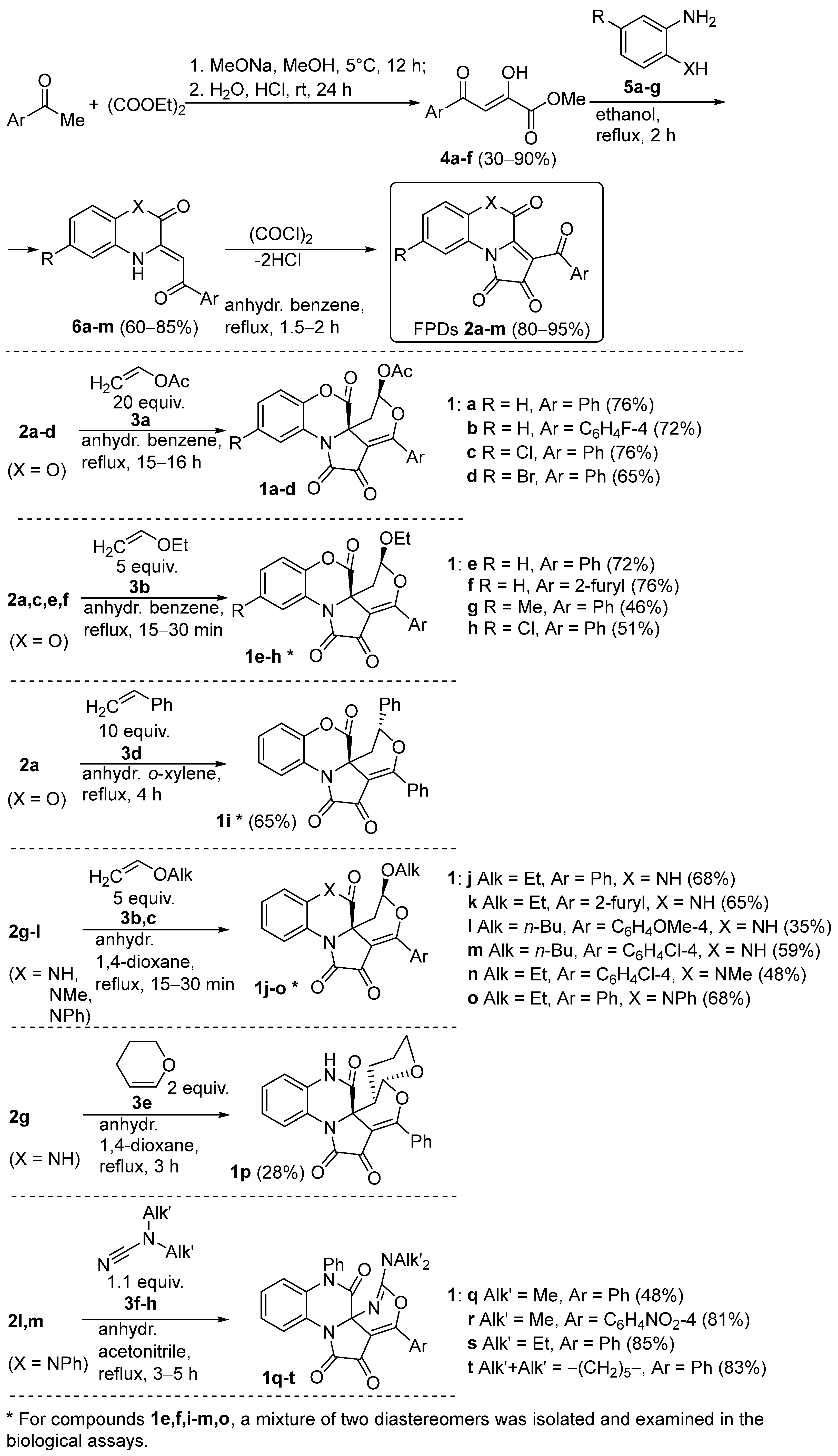
2.1. Synthesis of 6/6/5/6-Annelated Pyrrolidine-2,3-Diones 1
[e]-Fused 1H-pyrrole-2,3-diones (FPDs) are well-known precursors for the rapid construction of diverse heterocyclic scaffolds, including angular polycyclic ones [23,24,25,26]. Therefore, as a key step to prepare a series of diversely substituted angular 6/6/5/6-annelated pyrrolidine-2,3-diones 1a–t, we used a [4+2]-cycloaddition reaction of FPDs 2a–m with electron-rich dienophiles 3a–h (vinyl acetate 3a [27], alkoxyethylenes 3b,c [20,28,29], styrene 3d [30], 3,4-dihydro-2H-pyran 3e [31]) and N,N-dialkylcyanamides 3f–h [32] (Scheme 1, Supplementary Materials, S37).
Scheme 1.
Synthesis of angular 6/6/5/6-annelated pyrrolidine-2,3-diones 1a–t (isolated yields are given).
A series of angular 6/6/5/6-annelated pyrrolidine-2,3-diones 1a–t were obtained in several steps via earlier-reported synthetic protocols (Scheme 1, Supplementary Materials, S37) [20,27,28,29,30,31,32]. In brief, initially, methyl esters of aroylpyruvic acids 4a–f were obtained via the Claisen condensation of the corresponding acetophenones and diethyl oxalate in the presence of sodium methoxide [33]. Then, the reaction of compounds 4a–f with anilines 5a–g afforded heterocyclic enaminones 6a–m [29,34,35,36,37,38,39]. After that, the acylation of compounds 6a–m by oxalyl chloride resulted in FPDs 2a–m [29,34,35,37,38,39,40]. And finally, the hetero-Diels-Alder reaction of FPDs 2a–m and dienophiles 3a–h afforded the desired compounds 1a–t [20,27,28,29,30,31,32]. Compounds 1e,f,i–m,o were isolated as mixtures of two diastereomers. It should be mentioned that compounds 1b,d,f,g,k,n,o were not reported earlier.
2.2. Biology
The identification of bioactive compounds capable of stimulating the growth of C. vulgaris was carried out through a two-stage screening approach. In the initial stage, microalgae were cultivated in 96-well plates for five days in the presence of various concentrations (0.1–10 μmol/L) of the tested compounds. A culture medium without additives served as the negative control, while 2 g/L of glucose was used as the positive control. Optical density at 750 nm (OD750), a parameter reflecting both cell concentration and, to some extent, cell size and composition, was employed to evaluate growth. Compounds that induced OD750 values exceeding the mean OD750 of the negative control plus three standard deviations at two or more concentrations were shortlisted for further investigation.
The primary screening results (Table 1) identified four compounds (1n,q,s,t) that enhanced OD750 beyond the threshold, with only compound 1s meeting the selection criteria for further analysis.
Table 1.
The difference in algae cell concentrations between the cultures containing the compounds under study and the control cultures.
To validate and explore the effects of compound 1s in greater detail, secondary experiments were conducted in 50 mL flasks to ensure sufficient biomass for biochemical analyses (Table 2). The concentration range of compound 1s was extended to 0.1–100 μmol/L, with experimental controls identical to those in the primary screening. Cell concentrations were quantified via direct cell counting, while metabolite content was assessed using standard biochemical methods.
Table 2.
The effect of compound 1s on the growth and accumulation of metabolites in C. vulgaris cells.
Interestingly, despite its promising performance in microplate screening, compound 1s did not significantly impact the cell concentration in the flask cultures compared to the negative control (Table 2). This discrepancy may be attributed to differences in mixing conditions and oxygen availability between the microplate and flask setups. Nevertheless, biochemical analysis revealed a statistically significant 19% increase in protein content at 1 μmol/L of compound 1s.
A comparative review of the literature data on phytohormones demonstrated variability in their reported effects on protein accumulation in C. vulgaris. For instance, only salicylic acid at 5–10 mg/L induced a moderate 20–60% increase in protein content in some studies [5,41], whereas 2,4-epibrassinolide, benzyladenine, ethylene, gibberellin, indoleacetic acid, and naphthaleneacetic acid had no or little effect. At the same time, others reported markedly higher enhancements promoted by 2,4-epibrassinolide and indoleacetic acid [42].
The observed effect of compound 1s on protein accumulation suggests its potential as an alternative protein-enhancing agent for C. vulgaris. Given the high nutritional quality of C. vulgaris proteins, which include all essential amino acids [43,44], this finding holds promise for its application in the food and feed industries where protein enrichment is a key objective.
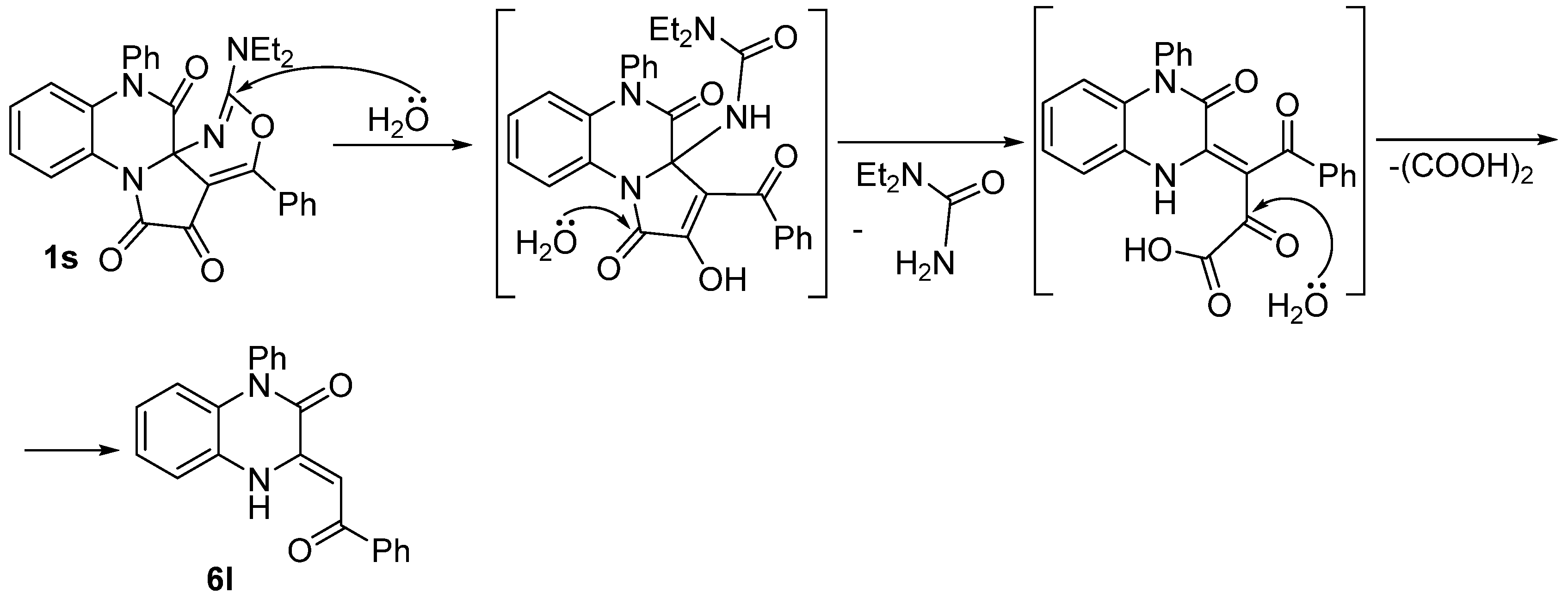
2.3. Hydrolysis Study of Compound 1s
Chemical instability in aquatic environments—driven by hydrolysis, oxidation, and other transformation reactions—can critically influence the stability and efficacy of algal growth regulators, including both natural phytohormones and synthetic analogs. Given this challenge, we evaluated the environmental stability of compound 1s, which has demonstrated superior activity in biological assays, under simulated aquatic conditions (Table 3). As a result, we found that compound 1s underwent a decomposition under all tested conditions, yielding a mixture of unidentified transformation products (HPLC-UV chromatogram plots are available in Supplementary Materials). While demonstrating moderate stability in DMSO with traces of water, the decomposition of compound 1s accelerated in aqueous mixtures. Notably, intense illumination triggered the complete degradation of compound 1s in all tested media.
Table 3.
Conversion of compound 1s under aquatic conditions.
Although compound 1s decomposed under all tested conditions to yield complex mixtures of transformation products, we successfully identified one of these products. An analysis of the reaction mixtures obtained under ambient conditions (Entries 1–3, Table 3) revealed the presence of heterocyclic enaminone 6l (Scheme 2, HPLC-UV chromatogram plots are available in Supplementary Materials). However, this compound was absent in the mixtures exposed to the culture chamber conditions (Entries 4–6, Table 3), suggesting that enaminone 6l could undergo photodegradation under a more intense illumination regime.
Scheme 2.
Possible hydrolysis pathway for the formation of compound 6l from compound 1s under ambient conditions.
3. Materials and Methods
3.1. Synthetic Methods and Analytic Data of Compounds
3.1.1. General Information
1H and 13C NMR spectra (Supplementary Materials) were acquired on a Bruker Avance III 400 HD spectrometer (Bruker BioSpin AG, Faellanden, Switzerland) (at 400 and 100 MHz, respectively) in CDCl3 or DMSO-d6, using TMS (in 1H NMR, 0.00 ppm) or solvent residual signals (in 13C NMR, 77.00 ppm for CDCl3, 39.52 ppm for DMSO-d6; in 1H NMR, 7.26 ppm for CDCl3, 2.50 ppm for DMSO-d6) as internal standards. 19F NMR spectra (Supplementary Materials) were acquired on a Bruker Avance III 400 HD spectrometer (Bruker BioSpin AG, Faellanden, Switzerland) (at 376 MHz) in CDCl3, using C6F6 (in 19F NMR, −162.90 ppm) as an internal standard. IR spectra were recorded on a Perkin-Elmer Spectrum Two spectrometer (PerkinElmer Inc., Waltham, MA, USA) from mulls in mineral oil. Melting points were measured on a Mettler Toledo MP70 apparatus (Mettler-Toledo (MTADA), Schwerzenbach, Switzerland). Elemental analyses were carried out on a Vario MICRO Cube analyzer (Elementar Analysensysteme GmbH, Langenselbold, Germany). The study of the hydrolysis of compound 1s was carried out using HPLC-UV on a Hitachi Chromaster (Hitachi High-Tech, Tokyo, Japan) [NUCLEODUR C18 Gravity column (particle size 3 μm; eluent acetonitrile–water, flow rate 1.5 mL/min); Hitachi Chromaster 5430 diode array detector (λ 210–750 nm)] (Supplementary Materials). Thin-layer chromatography (TLC) was performed on ALUGRAM Xtra SIL G/UV254 silica gel 60 plates (Macherey-Nagel, Düren, Germany) using EtOAC/toluene, 1:5 v/v, EtOAc, and toluene as eluents; spots were visualized with iodine vapor and/or UV light (254, 365 nm) in the light of a TLC viewing cabinet Petrolaser TLC-254/365 Thin Layer Chromatography Dark Room (Petrolaser, St. Petersburg, Russia).
FPDs 2a–m were obtained according to reported procedures [29,34,35,37,38,39,40]. Enaminones 6a–m were obtained according to the reported procedures [29,34,35,36,37,38,39]. Methyl aroylpyruvates 4a–f were obtained according to the reported procedures [33]. Dienophiles 3a–e were distilled before their use. The benzene, toluene, o-xylene, and 1,4-dioxane for the procedures with compounds 2a–m were distilled over Na before use. The acetonitrile for the procedures with FPDs 2a–m was dehydrated over molecular sieves 4Å before its use. All procedures with FPDs 2a–m were performed in oven-dried glassware. All other solvents and reagents were purchased from commercial vendors and were used as received.
3.1.2. Analytic Data of Compounds 1a–t
Compounds 1a,c were reported earlier [27]. Compounds 1b,d were not reported.
General procedure for compounds 1a–d. A suspension of the corresponding FPD 2a–d (3.1 mmol) and vinyl acetate 3a (5.7 mL, 62 mmol) in 10 mL of anhydrous benzene was refluxed for 15–16 h (until the violet color typical of starting compound 2 disappeared). Then, the mixture was cooled and evaporated to dryness. The resulting residue was ground with toluene, and the precipitate was filtered off and recrystallized from toluene (5–15 mL) to afford the corresponding compound 1a–d.
(5R*,6aR*)-1,2,7-Trioxo-3-phenyl-1,2,5,6-tetrahydro-7H-benzo[b]pyrano[4′,3′:2,3]pyrrolo[1,2-d][1,4]oxazin-5-yl acetate (1a) [27]. Yield: 76% (0.95 g); yellow solid; mp 256–258 °C (decomp., toluene) (Lit.mp 257–258 °C (decomp., toluene) [27]). 1H NMR (400 MHz, CDCl3): δ = 8.01 (m, 2H), 7.91 (m, 1H), 7.65 (m, 1H), 7.52 (m, 2H), 7.34 (m, 3H), 6.80 (d.d, 1H, 3Jeq.eq. = 1.0 Hz, 3Jax.eq. = 4.4 Hz), 2.71 (d.d, 1H, 2J = 14.1 Hz, 3Jeq.eq. = 0.9 Hz), 2.18 (d.d, 1H, 2J = 14.1 Hz, 3Jax.eq. = 4.5 Hz), 2.14 (s, 3H) ppm.
(5R*,6aR*)-3-(4-Fluorophenyl)-1,2,7-trioxo-1,2,5,6-tetrahydro-7H-benzo[b]pyrano[4′,3′:2,3]pyrrolo[1,2-d][1,4]oxazin-5-yl acetate (1b). Yield: 72% (0.94 g); yellow solid; mp 278–279 °C (decomp., toluene). 1H NMR (400 MHz, CDCl3): δ = 8.07 (m, 2H), 7.90 (m, 1H), 7.39 (m, 1H), 7.31 (m, 2H), 7.20 (m, 2H), 6.79 (d.d, 1H, 3Jeq.eq. = 0.7 Hz, 3Jax.eq. = 4.4 Hz), 2.71 (d.d, 1H, 2J = 14.1 Hz, 3Jeq.eq. = 0.9 Hz), 2.18 (d.d, 1H, 2J = 14.1 Hz, 3Jax.eq. = 4.5 Hz), 2.14 (s, 3H) ppm. 13C NMR (100 MHz, DMSO-d6): δ = 176.1, 168.0, 164.8 (d, 1JCF = 252.0 Hz), 163.9, 160.8, 158.7, 143.0, 133.2 (d, 2C, 3JCF = 9.2 Hz), 127.4, 126.5 (d, 4JCF = 2.3 Hz), 124.8, 122.3, 121.2, 116.7, 115.1 (d, 2C, 2JCF = 24.7 Hz), 102.8, 90.9, 54.1, 33.6, 20.3 ppm. 19F NMR (376 MHz, CDCl3): δ = −103.55 ppm. IR (mineral oil): 1796, 1770, 1735, 1717 cm−1. Anal. Calcd (%) for C22H14FNO7: C 62.42; H 3.33; N 3.31. Found: C 62.69; H 3.38; N 3.51.
(5R*,6aR*)-11-Chloro-1,2,7-trioxo-3-phenyl-1,2,5,6-tetrahydro-7H-benzo[b]pyrano[4′,3′:2,3]pyrrolo[1,2-d][1,4]oxazin-5-yl acetate (1c) [27]. Yield: 76% (1.03 g); yellow solid; mp 278–280 °C (decomp., toluene) (Lit.mp 278–280 °C (decomp., toluene) [27]). 1H NMR (400 MHz, CDCl3): δ = 8.00 (m, 2H), 7.94 (m, 1H), 7.65 (m, 1H), 7.52 (m, 2H), 7.36 (m, 1H), 7.23 (m, 1H), 6.81 (d.d, 1H, 3Jeq.eq. = 0.9 Hz, 3Jax.eq. = 4.3 Hz), 2.70 (d.d, 1H, 2J = 14.1 Hz, 3Jeq.eq. = 0.9 Hz), 2.18 (d.d, 1H, 2J = 13.9 Hz, 3Jax.eq. = 4.4 Hz), 2.13 (s, 3H) ppm.
(5R*,6aR*)-11-Bromo-1,2,7-trioxo-3-phenyl-1,2,5,6-tetrahydro-7H-benzo[b]pyrano[4′,3′:2,3]pyrrolo[1,2-d][1,4]oxazin-5-yl acetate (1d). Yield: 65% (0.98 g); yellow solid; mp 263–265 °C (decomp., toluene). 1H NMR (400 MHz, CDCl3): δ = 8.09 (m, 1H), 8.00 (m, 2H), 7.66 (m, 1H), 7.52 (m, 3H), 7.17 (m, 1H), 6.81 (d.d, 1H, 3Jeq.eq. = 1.0 Hz, 3Jax.eq. = 4.4 Hz), 2.70 (d.d, 1H, 2J = 13.9 Hz, 3Jeq.eq. = 1.0 Hz), 2.17 (d.d, 1H, 2J = 14.2 Hz, 3Jax.eq. = 4.4 Hz), 2.13 (s, 3H) ppm. 13C NMR (100 MHz, DMSO-d6): δ = 175.6, 168.0, 165.4, 160.2, 158.8, 142.3, 133.2, 130.4, 130.0, 129.9, 127.8, 124.5, 122.6, 118.8, 115.8, 102.6, 90.8, 53.9, 33.6, 20.3 ppm. IR (mineral oil): 1796, 1758, 1731, 1714 cm−1. Anal. Calcd (%) for C22H14BrNO7: C 54.57; H 2.91; N 2.89. Found: C 54.87; H 2.58; N 2.55.
Compounds 1e,h were reported earlier [20]. Compounds 1f,g were not reported.
General procedure for compounds 1e–h. A suspension of the corresponding FPD 2a,c,e,f (3.1 mmol) and ethoxyethylene 3b (1.5 mL, 15.5 mmol) in 10 mL of anhydrous benzene was refluxed for 15–30 min (until the violet color typical of starting compound 2 disappeared). Then, the mixture was cooled to room temperature. The formed precipitate was filtered off and recrystallized from toluene (5–20 mL) to afford the corresponding compound 1e–h.
(5S*,6aR*)- and (5R*,6aR*)-5-Ethoxy-3-phenyl-5,6-dihydro-7H-benzo[b]pyrano[4′,3′:2,3]pyrrolo[1,2-d][1,4]oxazine-1,2,7-triones (1e) [20]. Obtained as a mixture of diastereomers, (5S*,6aR*):(5R*,6aR*) ~ 5:2 (by NMR). Yield: 72% (0.87 g); yellow solid; mp 246–249 °C (decomp., toluene) (Lit.mp 248–250 °C (decomp., toluene) [20]). 1H NMR (400 MHz, DMSO-d6) of (5S*,6aR*)-diastereomer (major): δ = 7.92 (m, 2H), 7.81 (m, 1H), 7.68 (m, 1H), 7.58 (m, 2H), 7.40 (m, 2H), 7.36 (m, 1H), 5.79 (m, 1H, X-part of ABX system), 3.86–3.67 (m, 2H), 2.39 (m, 2H, AB-part of ABX system), 1.11 (t, 3H, 3J = 7.0 Hz) ppm. 1H NMR (400 MHz, DMSO-d6) of (5R*,6aR*)-diastereomer (minor): δ = 7.86 (m, 2H), 7.77 (m, 1H), 7.68 (m, 1H), 7.58 (m, 2H), 7.40 (m, 1H), 7.36 (m, 2H), 5.60 (d.d, 1H, 3Jax.ax. = 10.2 Hz, 3Jax.eq. = 4.1 Hz), 4.09–4.01 (m, 1H), 3.84–3.77 (m, 1H), 2.61 (d.d, 1H, 2J = 13.0 Hz, 3Jax.eq. = 4.2 Hz), 2.18 (d.d, 1H, 2J = 13.0 Hz, 3Jax.ax. = 10.4 Hz), 1.21 (t, 3H, 3J = 7.2 Hz) ppm.
(5S*,6aR*)- and (5R*,6aR*)-5-Ethoxy-3-(furan-2-yl)-5,6-dihydro-7H-benzo[b]pyrano[4′,3′:2,3]pyrrolo[1,2-d][1,4]oxazine-1,2,7-triones (1f). Obtained as a mixture of diastereomers, (5S*,6aR*):(5R*,6aR*) ~ 5:2 (by NMR). Yield: 76% (0.90 g); yellow solid; mp 199–201 °C (decomp., toluene). 1H NMR (400 MHz, DMSO-d6) of (5S*,6aR*)-diastereomer (major): δ = 8.26 (m, 1H), 8.13 (m, 1H), 7.81 (m, 1H), 7.37 (m, 3H), 6.89 (m, 1H), 5.77 (m, 1H, X-part of ABX system), 3.92–3.71 (m, 2H), 2.38 (m, 2H, AB-part of ABX system), 1.14 (t, 3H, 3J = 7.1 Hz) ppm. 1H NMR (400 MHz, DMSO-d6) of (5R*,6aR*)-diastereomer (minor): δ = 8.26 (m, 1H), 8.13 (m, 1H), 7.78 (m, 1H), 7.36 (m, 3H), 6.88 (m, 1H), 5.61 (d.d, 1H, 3Jax.ax. = 10.0 Hz, 3Jax.eq. = 4.2 Hz), 4.08–4.00 (m, 1H), 3.84–3.77 (m, 1H), 2.61 (d.d, 1H, 2J = 13.2 Hz, 3Jax.eq. = 4.4 Hz), 2.15 (d.d, 1H, 2J = 13.0 Hz, 3Jax.ax. = 10.0 Hz), 1.21 (t, 3H, 3J = 7.1 Hz) ppm. 13C NMR (100 MHz, DMSO-d6) of (5S*,6aR*)-diastereomer (major): δ = 174.7, 160.7, 159.1, 153.8, 147.9, 144.9, 143.1, 127.4, 124.7, 122.1, 121.7, 121.2, 116.6, 112.9, 101.5, 100.1, 64.8, 54.2, 34.8, 14.6 ppm. 13C NMR (100 MHz, DMSO-d6) of (5R*,6aR*)-diastereomer (minor): δ = 174.9, 162.7, 159.9, 154.4, 147.8, 144.9, 142.8, 127.2, 124.8, 122.7, 121.5, 121.1, 116.7, 112.8, 101.7, 101.5, 65.5, 57.0, 34.0, 14.9 ppm. IR (mineral oil): 1789, 1771, 1723, 1699 cm−1. Anal. Calcd (%) for C20H15NO7: C 62.99; H 3.96; N 3.67. Found: C 63.26; H 3.87; N 3.95.
(5S*,6aR*)-5-Ethoxy-11-methyl-3-phenyl-5,6-dihydro-7H-benzo[b]pyrano[4′,3′:2,3]pyrrolo[1,2-d][1,4]oxazine-1,2,7-trione (1g). Yield: 46% (0.58 g); yellow solid; mp 191–193 °C (decomp., toluene). 1H NMR (400 MHz, DMSO-d6): δ = 7.93 (m, 2H), 7.68 (m, 1H), 7.62 (m, 1H), 7.57 (m, 2H), 7.28 (m, 1H), 7.21 (m, 1H), 5.78 (m, 1H, X-part of ABX system), 3.87–3.68 (m, 2H), 2.38 (m, 5H, AB-part of ABX system + Me), 1.11 (t, 3H, 3J = 7.1 Hz) ppm. 13C NMR (100 MHz, DMSO-d6): δ = 175.8, 166.4, 160.8, 159.0, 141.0, 134.1, 132.9, 130.7, 130.2, 127.9, 127.8, 122.1, 120.9, 116.3, 102.9, 100.2, 64.6, 54.4, 35.0, 20.4, 14.5 ppm. IR (mineral oil): 1789, 1724, 1709 cm−1. Anal. Calcd (%) for C23H19NO6: C 68.14; H 4.72; N 3.46. Found: C 68.01; H 4.83; N 3.53.
(5S*,6aR*)-11-Chloro-5-ethoxy-3-phenyl-5,6-dihydro-7H-benzo[b]pyrano[4′,3′:2,3]pyrrolo[1,2-d][1,4]oxazine-1,2,7-trione (1h) [20]. Yield: 51% (0.67 g); yellow solid; mp 243–244 °C (decomp., toluene) (Lit.mp 243–245 °C (decomp., toluene) [20]). 1H NMR (400 MHz, DMSO-d6): δ = 7.91 (m, 2H), 7.79 (m, 1H), 7.69 (m, 1H), 7.58 (m, 2H), 7.46 (m, 2H), 5.80 (m, 1H, X-part of ABX system), 3.86–3.67 (m, 2H), 2.42 (m, 2H, AB-part of ABX system), 1.10 (t, 3H, 3J = 7.0 Hz) ppm.
Compound 1i was reported earlier [30].
Procedure for compound 1i. A suspension of FPD 2a (3.1 mmol, 1.0 g) and styrene 3c (3.6 mL, 31.0 mmol) in 15 mL of anhydrous o-xylene was refluxed for 4 h (until the reaction mixture became dark yellow). Then, the mixture was cooled to room temperature. The formed precipitate was filtered off and recrystallized from toluene (10 mL) to afford compound 1i.
(5R*,6aR*)- and (5S*,6aR*)-3,5-Diphenyl-5,6-dihydro-7H-benzo[b]pyrano[4′,3′:2,3]pyrrolo[1,2-d][1,4]oxazine-1,2,7-triones (1i) [30]. Obtained as a mixture of diastereomers, (5R*,6aR*):(5S*,6aR*) ~ 5:1 (by NMR). Yield: 65% (0.85 g); yellow solid; mp 275–277 °C (decomp., toluene) (Lit.mp 278–279 °C (decomp., toluene) [30]). 1H NMR (400 MHz, DMSO-d6) of (5R*,6aR*)-diastereomer (major): δ = 7.75 (m, 3H), 7.61 (m, 5H), 7.36 (m, 6H), 5.53 (d.d, 1H, 3Jax.ax. = 12.7 Hz, 3Jax.eq. = 4.1 Hz), 2.60 (d.d, 1H, 2J = 12.9 Hz, 3Jax.eq. = 3.9 Hz), 2.47 (m, 1H) ppm. 1H NMR (400 MHz, DMSO-d6) of (5S*,6aR*)-diastereomer (minor): δ = 7.99 (m, 2H), 7.75 (m, 1H), 7.61 (m, 5H), 7.36 (m, 6H), 5.92 (m, 1H, X-part of ABX system), 2.79 (m, 2H, AB-part of ABX system) ppm.
Compounds 1j,l,m were reported earlier [28]. Compounds 1k,n,o were not reported.
General procedure for compounds 1j–o. A suspension of the corresponding FPD 2g–l (3.1 mmol) and alkoxyethylenes 3b,d (15.5 mmol) in 10 mL of anhydrous 1,4-dioxane was refluxed for 15–30 min (until the violet color typical of starting compound 2 disappeared). Then, the mixture was cooled to room temperature. The formed precipitate was filtered off and recrystallized from 1,4-dioxane (5–10 mL) to afford the corresponding compound 1j–o.
(5S*,6aR*)- and (5R*,6aR*)-5-Ethoxy-3-phenyl-5,6-dihydropyrano[4′,3′:2,3]pyrrolo[1,2-a]quinoxaline-1,2,7(8H)-triones (1j) [28]. Obtained as a mixture of diastereomers, (5S*,6aR*):(5R*,6aR*) ~ 5:1 (by NMR). Yield: 68% (0.82 g); yellow solid; mp 243–244 °C (decomp., 1,4-dioxane) (Lit.mp 243–244 °C (decomp., 1,4-dioxane) [28]). 1H NMR (400 MHz, DMSO-d6) of (5S*,6aR*)-diastereomer (major): δ = 10.88 (s, 1H), 7.94 (m, 2H), 7.72 (m, 1H), 7.67 (m, 1H), 7.56 (m, 2H), 7.30 (m, 1H), 7.14 (m, 2H), 5.73 (m, 1H, X-part of ABX system), 3.88–3.67 (m, 2H), 2.30 (m, 2H, AB-part of ABX system), 1.12 (t, 3H, 3J = 7.1 Hz) ppm. 1H NMR (400 MHz, DMSO-d6) of (5R*,6aR*)-diastereomer (minor): δ = 11.04 (s, 1H), 7.87 (m, 2H), 7.72 (m, 1H), 7.67 (m, 1H), 7.56 (m, 2H), 7.30 (m, 1H), 7.14 (m, 2H), 5.60 (d.d, 1H, 3Jax.ax. = 10.3 Hz, 3Jax.eq. = 3.9 Hz), 4.08–4.01 (m, 1H), 3.84–3.77 (m, 1H), 2.42 (d.d, 1H, 2J = 12.7 Hz, 3Jax.eq. = 4.2 Hz), 2.05 (d.d, 1H, 2J = 12.7 Hz, 3Jax.ax. = 10.3 Hz), 1.22 (t, 3H, 3J = 7.1 Hz) ppm.
(5S*,6aR*)- and (5R*,6aR*)-5-Ethoxy-3-(furan-2-yl)-5,6-dihydropyrano[4′,3′:2,3]pyrrolo[1,2-a]quinoxaline-1,2,7(8H)-triones (1k). Obtained as a mixture of diastereomers, (5S*,6aR*):(5R*,6aR*) ~ 5:1 (by NMR). Yield: 65% (0.77 g); yellow solid; mp 225–227 °C (decomp., 1,4-dioxane). 1H NMR (400 MHz, DMSO-d6) of (5S*,6aR*)-diastereomer (major): δ = 10.87 (s, 1H), 8.24 (m, 1H), 8.12 (m, 1H), 7.71 (m, 1H), 7.30 (m, 1H), 7.17 (m, 1H), 7.10 (m, 1H), 6.86 (m, 1H), 5.71 (m, 1H, X-part of ABX system), 3.93–3.68 (m, 2H), 2.29 (m, 2H, AB-part of ABX system), 1.15 (t, 3H, 3J = 7.1 Hz) ppm. 1H NMR (400 MHz, DMSO-d6) of (5R*,6aR*)-diastereomer (minor): δ = 11.03 (s, 1H), 8.27 (m, 1H), 8.12 (m, 1H), 7.71 (m, 1H), 7.30 (m, 1H), 7.17 (m, 1H), 7.10 (m, 1H), 6.86 (m, 1H), 5.61 (d.d, 1H, 3Jax.ax. = 10.3 Hz, 3Jax.eq. = 4.4 Hz), 4.08–4.00 (m, 1H), 3.84–3.77 (m, 1H), 2.42 (d.d, 1H, 2J = 12.7 Hz, 3Jax.eq. = 3.9 Hz), 2.03 (d.d, 1H, 2J = 12.7 Hz, 3Jax.ax. = 10.3 Hz), 1.21 (t, 3H, 3J = 7.1 Hz) ppm. 13C NMR (100 MHz, DMSO-d6) of (5S*,6aR*)-diastereomer (major): δ = 175.0, 163.8, 159.5, 154.1, 147.7, 145.3, 130.4, 127.1, 122.5, 122.3, 121.5, 121.4, 115.8, 112.8, 102.3, 100.9, 64.8, 55.4, 36.0, 14.7 ppm. 13C NMR (100 MHz, DMSO-d6) of (5R*,6aR*)-diastereomer (minor): δ = 174.9, 165.4, 160.1, 154.7, 150.7, 145.0, 130.0, 127.1, 122.8, 122.7, 121.5, 121.4, 115.9, 112.7, 102.0, 101.6, 65.4, 58.0, 34.7, 14.9 ppm. IR (mineral oil): 1715, 1696 cm−1. Anal. Calcd (%) for C20H16N2O6: C 63.16; H 4.24; N 7.37. Found: C 62.86; H 4.36; N 7.41.
(5S*,6aR*)- and (5R*,6aR*)-5-Butoxy-3-(4-methoxyphenyl)-5,6-dihydropyrano[4′,3′:2,3]pyrrolo[1,2-a]quinoxaline-1,2,7(8H)-triones (1l) [28]. Obtained as a mixture of diastereomers, (5S*,6aR*):(5R*,6aR*) ~ 1:2 (by NMR). Yield: 35% (0.47 g); yellow solid; mp 256–258 °C (decomp., 1,4-dioxane) (Lit.mp 250–251 °C (decomp., 1,4-dioxane) [28]). 1H NMR (400 MHz, DMSO-d6) of (5S*,6aR*)-diastereomer (minor): δ = 10.91 (s, 1H), 7.97 (m, 2H), 7.71 (m, 1H), 7.30 (m, 1H), 7.17 (m, 1H), 7.12 (m, 3H), 5.67 (m, 1H, X-part of ABX system), 3.89 (s, 3H), 3.81 (m, 1H), 3.65 (m, 1H), 2.28 (m, 2H, AB-part of ABX system), 1.47 (m, 2H), 1.31 (m, 2H), 0.84 (t, 3H, 3J = 7.2 Hz) ppm. 1H NMR (400 MHz, DMSO-d6) of (5R*,6aR*)-diastereomer (major): δ = 11.08 (s, 1H), 7.89 (m, 2H), 7.71 (m, 1H), 7.30 (m, 1H), 7.17 (m, 1H), 7.12 (m, 3H), 5.56 (d.d, 1H, 3Jax.ax. = 10.2 Hz, 3Jax.eq. = 4.3 Hz), 4.01 (m, 1H), 3.89 (s, 3H), 3.73 (m, 1H), 2.42 (d.d, 1H, 2J = 12.7 Hz, 3Jax.eq. = 4.1 Hz), 2.02 (d.d, 1H, 2J = 12.5 Hz, 3Jax.ax. = 10.6 Hz), 1.56 (m, 2H), 1.37 (m, 2H), 0.89 (t, 3H, 3J = 7.2 Hz) ppm.
(5S*,6aR*)- and (5R*,6aR*)-5-Butoxy-3-(4-chlorophenyl)-5,6-dihydropyrano[4′,3′:2,3]pyrrolo[1,2-a]quinoxaline-1,2,7(8H)-triones (1m) [28]. Obtained as a mixture of diastereomers, (5S*,6aR*):(5R*,6aR*) ~ 10:1 (by NMR). Yield: 59% (0.83 g); yellow solid; mp 243–245 °C (decomp., 1,4-dioxane) (Lit.mp 241–242 °C (decomp., 1,4-dioxane) [28]). 1H NMR (400 MHz, DMSO-d6) of (5S*,6aR*)-diastereomer (major): δ = 10.93 (s, 1H), 7.94 (m, 2H), 7.71 (m, 1H), 7.65 (m, 2H), 7.30 (m, 1H), 7.14 (m, 2H), 5.71 (m, 1H, X-part of ABX system), 3.79 (m, 1H), 3.64 (m, 1H), 2.28 (m, 2H, AB-part of ABX system), 1.47 (m, 2H), 1.30 (m, 2H), 0.83 (t, 3H, 3J = 7.4 Hz) ppm. 1H NMR (400 MHz, DMSO-d6) of (5R*,6aR*)-diastereomer (minor): δ = 11.09 (s, 1H), 7.85 (m, 2H), 7.71 (m, 1H), 7.65 (m, 2H), 7.30 (m, 1H), 7.14 (m, 2H), 5.60 (d.d, 1H, 3Jax.ax. = 10.2 Hz, 3Jax.eq. = 3.9 Hz), 3.99 (m, 1H), 3.72 (m, 1H), 2.42 (d.d, 1H, 2J = 12.5 Hz, 3Jax.eq. = 3.9 Hz), 2.07 (d.d, 1H, 2J = 12.7 Hz, 3Jax.ax. = 10.4 Hz), 1.55 (m, 2H), 1.37 (m, 2H), 0.88 (t, 3H, 3J = 7.2 Hz) ppm.
(5S*,6aR*)-3-(4-Chlorophenyl)-5-ethoxy-8-methyl-5,6-dihydropyrano[4′,3′:2,3]pyrrolo[1,2-a]quinoxaline-1,2,7(8H)-trione (1n). Yield: 48% (0.65 g); yellow solid; mp 235–236 °C (decomp., 1,4-dioxane). 1H NMR (400 MHz, DMSO-d6): δ = 7.95 (m, 2H), 7.76 (m, 1H), 7.65 (m, 2H), 7.42 (m, 2H), 7.25 (m, 1H), 5.72 (m, 1H, X-part of ABX system), 3.81 (m, 1H), 3.66 (m, 1H), 3.35 (s, 3H), 2.25 (m, 2H, AB-part of ABX system), 1.10 (t, 3H, 3J = 6.8 Hz) ppm. 13C NMR (100 MHz, DMSO-d6): δ = 176.2, 165.3, 163.2, 158.8, 137.6, 132.4, 132.0, 129.8, 128.0, 127.3, 122.9, 122.5, 122.1, 116.0, 104.6, 101.1, 64.3, 55.4, 35.9, 30.1, 14.5 ppm. IR (mineral oil): 1720, 1698, 1683 cm−1. Anal. Calcd (%) for C23H19ClN2O5: C 62.95; H 4.36; N 6.38. Found: C 63.18; H 4.26; N 6.55.
(5S*,6aR*)- and (5R*,6aR*)-5-Ethoxy-3,8-diphenyl-5,6-dihydropyrano[4′,3′:2,3]pyrrolo[1,2-a]quinoxaline-1,2,7(8H)-triones (1o). Obtained as a mixture of diastereomers, (5S*,6aR*):(5R*,6aR*) ~ 5:2 (by NMR). Yield: 68% (0.98 g); yellow solid; mp 236–238 °C (decomp., 1,4-dioxane). 1H NMR (400 MHz, DMSO-d6) of (5S*,6aR*)-diastereomer (major): δ = 7.93 (m, 2H), 7.84 (m, 1H), 7.67 (m, 1H), 7.56 (m, 3H), 7.49 (m, 2H), 7.30 (m, 2H), 7.23 (m, 2H), 6.51 (m, 1H), 5.82 (d, 1H, 3Jax.eq. = 4.4 Hz), 3.85 (m, 1H), 3.73 (m, 1H), 2.53 (d, 1H, 2J = 13.2 Hz), 2.37 (d.d, 1H, 2J = 13.7 Hz, 3Jax.eq. = 4.9 Hz), 1.15 (t, 3H, 3J = 7.1 Hz) ppm. 1H NMR (400 MHz, DMSO-d6) of (5R*,6aR*)-diastereomer (minor): δ = 7.87 (m, 2H), 7.80 (m, 1H), 7.67 (m, 1H), 7.56 (m, 5H), 7.49 (m, 1H), 7.30 (m, 1H), 7.23 (m, 2H), 6.47 (m, 1H), 5.63 (d.d, 1H, 3Jax.ax. = 10.3 Hz, 3Jax.eq. = 3.9 Hz), 4.08 (m, 1H), 3.82 (m, 1H), 2.73 (d.d, 1H, 2J = 13.0 Hz, 3Jax.eq. = 4.2 Hz), 2.14 (d.d, 1H, 2J = 12.7 Hz, 3Jax.ax. = 10.3 Hz), 1.25 (t, 3H, 3J = 6.8 Hz) ppm. 13C NMR (100 MHz, DMSO-d6) of (5S*,6aR*)-diastereomer (major): δ = 176.2, 166.6, 163.1, 159.0, 137.7, 133.1, 132.9, 131.0, 130.2, 130.0, 129.6, 128.8, 128.4, 127.8, 126.8, 123.2, 122.4, 122.2, 116.7, 103.8, 101.1, 64.6, 55.9, 35.7, 14.6 ppm. 13C NMR (100 MHz, DMSO-d6) of (5R*,6aR*)-diastereomer (minor): δ = 170.1, 167.2, 164.7, 159.8, 137.0, 132.9, 132.7, 130.9, 129.6, 128.7, 128.6, 128.1, 127.8, 126.7, 123.2, 122.8, 122.2, 122.2, 116.7, 103.2, 102.0, 65.4, 58.5, 34.7, 15.0 ppm. IR (mineral oil): 1715 cm−1. Anal. Calcd (%) for C28H22N2O5: C 72.09; H 4.75; N 6.01. Found: C 71.93; H 4.89; N 6.15.
Compound 1p was reported earlier [31].
Procedure for compound 1p. A suspension of FPD 2g (3.1 mmol, 1.0 g) and 3,4-dihydro-2H-pyran 3e (5.7 mL, 6.2 mmol) in 10 mL of anhydrous 1,4-dioxane was refluxed for 3 h (until the violet color typical of starting compound 2 disappeared). Then, the mixture was cooled to room temperature. The formed precipitate was filtered off to afford compound 1i. Compound 1i was used in further studies without additional purification.
(9aR*,13aR*,13bR*)-8-Phenyl-9a,12,13,13a-tetrahydro-11H-pyrano[3″,2″:5′,6′]pyrano[4′,3′:2,3]pyrrolo[1,2-a]quinoxaline-6,7,14(15H)-trione (1p) [31]. Yield: 28% (0.35 g); yellow solid; mp 270–271 °C (decomp., 1,4-dioxane) (Lit.mp 278–280 °C (decomp., 1,4-dioxane) [31]). 1H NMR (400 MHz, DMSO-d6): δ = 11.15 (s, 1H), 7.91 (m, 2H), 7.69 (m, 2H), 7.57 (m, 2H), 7.33 (m, 1H), 7.19 (m, 1H), 7.13 (m, 1H), 5.77 (d, 1H, 3Jax.eq. = 3.9 Hz), 3.79 (m, 2H), 2.26 (d.t, 1H, 3J = 12.5 Hz, 3Jax.eq. = 4.3 Hz), 1.60 (m, 2H), 1.42 (m, 1H), 1.11 (m, 1H) ppm.
Compounds 1q–t were reported earlier [32].
General procedure for compounds 1q–t. A suspension of the corresponding FPD 2l,m (3.1 mmol) and the corresponding cyanamide 3f–h (3.2 mmol) in 15 mL of anhydrous acetonitrile was refluxed for 3–5 h (until the violet color typical of starting compound 2 disappeared). Then, the reaction mixture was cooled to room temperature, and the resulting precipitate was filtered off to afford the corresponding compound 1q–t. Compounds 1q–t were used in further studies without additional purification.
5-(Dimethylamino)-3,8-diphenyl-[1,3]oxazino[4′,5′:2,3]pyrrolo[1,2-a]quinoxaline-1,2,7(8H)-trione (1q) [32]. Yield: 78% (1.14 g); yellow solid; mp 220–221 °C (decomp., acetonitrile) (Lit.mp 220–221 °C (decomp., acetonitrile) [32]). 1H NMR (400 MHz, DMSO-d6): δ = 8.08 (m, 2H), 7.81 (m, 1H), 7.74 (m, 1H), 7.65 (m, 2H), 7.59 (m, 2H), 7.51 (m, 1H), 7.27 (m, 2H), 7.20 (m, 2H), 6.41 (m, 1H), 3.01 (s, 6H) ppm.
5-(Dimethylamino)-3-(4-nitrophenyl)-8-phenyl-[1,3]oxazino[4′,5′:2,3]pyrrolo[1,2-a]quinoxaline-1,2,7(8H)-trione (1r) [32]. Yield: 81% (1.26 g); yellow solid; mp 210–211 °C (decomp., acetonitrile) (Lit.mp 210–211 °C (decomp., acetonitrile) [32]). 1H NMR (400 MHz, DMSO-d6): δ = 8.46 (m, 2H), 8.29 (m, 2H), 7.81 (m, 1H), 7.60 (m, 2H), 7.51 (m, 1H), 7.27 (m, 2H), 7.22 (m, 2H), 6.43 (m, 1H), 3.02 (s, 6H) ppm.
5-(Diethylamino)-3,8-diphenyl-[1,3]oxazino[4′,5′:2,3]pyrrolo[1,2-a]quinoxaline-1,2,7(8H)-trione (1s) [32]. Yield: 85% (1.30 g); yellow solid; mp 200–204 °C (decomp., acetonitrile) (Lit.mp 200–204 °C (decomp., acetonitrile) [32]). 1H NMR (400 MHz, DMSO-d6): δ = 8.07 (m, 2H), 7.82 (m, 1H), 7.74 (m, 1H), 7.66 (m, 2H), 7.59 (m, 2H), 7.51 (m, 1H), 7.23 (m, 4H), 6.43 (m, 1H), 3.56–3.47 (m, 2H), 3.36–3.26 (m, 2H), 1.15 (t, 6H, 3J = 7.1 Hz,) ppm.
3,8-Diphenyl-5-(piperidin-1-yl)-[1,3]oxazino[4′,5′:2,3]pyrrolo[1,2-a]quinoxaline-1,2,7(8H)-trione (1t) [32]. Yield: 83% (1.30 g); yellow solid; mp 221–224 °C (decomp., acetonitrile) (Lit.mp 221–224 °C (decomp., acetonitrile) [32]). 1H NMR (400 MHz, DMSO-d6): δ = 8.04 (m, 2H), 7.82 (m, 1H), 7.74 (m, 1H), 7.65 (m, 2H), 7.59 (m, 2H), 7.51 (m, 1H), 7.22 (m, 4H), 6.41 (m, 1H), 3.49 (m, 4H), 1.57 (m, 6H) ppm.
3.2. Biology
3.2.1. Screening of Substances in 96-Well Plates
Chlorella vulgaris (strain IMBR-19, obtained from the A.O. Kovalevsky Institute of Biology of the Southern Seas, RAS, Sevastopol, Russia) was cultivated in BG-11 medium [45] under aseptic conditions. The only exception to sterility was the use of DMSO solutions of the tested compounds, which were not sterilized after dilution. The test compounds were initially dissolved in 99% DMSO. For poorly soluble compounds, the solutions were either incubated on a rotator for 16 h or subjected to ultrasonic treatment using an ultrasound homogenizer with a 3 mm probe (VCX-130, Sonics and Materials, Newtown, CT, USA).
For the screening experiments, 96-well culture plates were prepared by combining BG-11 medium, a starter culture of C. vulgaris, and DMSO-diluted test compounds to a final volume of 300 µL per well. The initial cell density was set at 5 × 10⁴ cells/well, with a final DMSO concentration of 1% (v/v) across all wells. The test compounds were evaluated at final concentrations of 0.1, 1, and 10 μmol/L. Negative control wells received an equivalent volume of pure DMSO without test compounds, while positive control wells contained BG-11 medium supplemented with 2 g/L glucose in addition to DMSO. Each test condition was performed in duplicate (two wells per concentration). To minimize edge effects, sterile distilled water was added to all edge and corner wells of the plates. The plates were sealed with gas-permeable films to maintain sterility and allow gas exchange.
The cultures were incubated for five days in a humidified chamber at 28 °C with orbital shaking at 150 rpm. Illumination was provided in a 12 h light/12 h dark cycle at an intensity of 100 μmol m−2 s−1 using an array of evenly distributed white LEDs positioned below the culture plates for bottom illumination. The lighting system included a cooling mechanism to prevent heat transfer to the culture plates.
At the conclusion of the incubation period, the contents of each well were thoroughly mixed using a multichannel pipette to ensure uniform cell suspension. Cell growth was assessed by measuring the OD750 using a microplate spectrophotometer [46].
3.2.2. Evaluation of Compound 1s in 50 mL Flasks
The experiments were conducted in 50 mL Erlenmeyer flasks containing BG-11 medium, a starter culture of C. vulgaris, and compound 1s dissolved in DMSO. The final volume in each flask was adjusted to 30 mL, with the initial cell density standardized at 1 × 10⁷ cells per flask. The DMSO concentration in all flasks, including the controls, was maintained at 1% (v/v). The test compound was evaluated at final concentrations of 0.1, 1, 10, and 100 µmol/L. Negative control flasks received an equivalent volume of DMSO without any test compound, while positive control flasks were supplemented with 2 g/L glucose in addition to DMSO. All treatments, including the controls, were prepared in triplicate (three flasks per condition). To ensure gas exchange while minimizing contamination, the flasks were sealed with gas-permeable cellulose caps. The cultures were incubated for five days under conditions identical to those described in prior studies.
Cell Count. At the end of the incubation period, the contents of each flask were thoroughly mixed to ensure homogeneity. A 10 mL aliquot of the culture was transferred into a 15 mL centrifuge tube and subjected to two washing steps with distilled water (10 mL per wash) by centrifugation at 450× g for 15 min. The washed cell pellet was resuspended in 5 mL of distilled water (the resulting cell pellet was concentrated two-fold and vortexed for 1 min to ensure uniformity). Cell counts were determined using a hemocytometer.
Pigment Analysis. To determine the concentration of cellular pigments, 2 mL of washed cells obtained from the cell count experiment were subjected to centrifugation at 7000× g for 10 min. Following centrifugation, 1.8 mL of the supernatant was removed, and an equal volume (1.8 mL) of 99% methanol was added to achieve a final methanol concentration of 90%. The samples were incubated at 60 °C for 30 min in a solid-state thermostat, followed by cooling to room temperature. After cooling, the samples were centrifuged at 10,000× g for 10 min to remove cellular debris. The absorbance of the supernatant containing the extracted pigments was measured spectrophotometrically at 665 nm, 652 nm, and 470 nm. Chlorophyll and carotenoid concentrations were calculated using established equations [47,48]. The pigment content was normalized to the cell count and expressed as μg per 1 × 10⁷ cells.
Preparation of C. vulgaris biomass for protein and carbohydrate analysis. Cells were washed in deionized water and concentrated two-fold. Cell suspensions washed from BG-11 medium were transferred to 2 mL screw-cap tubes. Only the pellet (cell biomass) was left in the tubes. Then, 90% methanol (v/v) was added to the pellet for pigment extraction. The samples were incubated at 60 °C for 30 min in a solid-state thermostat and then cooled to room temperature. After cooling, the samples were centrifuged at 10,000× g for 10 min to remove the supernatant. The pellet was treated with 500 μL of 2 mol/L hydrochloric acid and boiled at 100 °C for 2 h.
Protein Analysis. A modified Bradford colorimetric assay was employed to quantify total protein content [49]. Acid hydrolysates (160 μL) were loaded into deep-well 96-well plates, followed by the sequential addition of 2 mol/L NaOH (160 μL) and deionized water (480 μL) with thorough mixing. A bovine serum albumin (BSA) standard curve (8–32 μg/mL in 8 μg/mL increments) and blank (deionized water) were prepared in parallel, each receiving 160 μL of 2 mol/L NaCl and being brought to an 800 μL final volume with deionized water. Using a multichannel pipette, 160 μL aliquots from the deep-well plate were transferred to a flat-bottom 96-well plate, combined with 40 μL of Bradford reagent (Bio-Rad Protein Assay Dye Concentrate), and incubated (30 °C, 850 rpm, 5 min). Absorbance at 595 nm was measured using a Synergy H1 microplate reader (BioTek Instruments). Protein concentrations were determined by referencing a calibration curve and normalized to cellular content as μg per 1 × 10⁶ cells.
Carbohydrate Analysis. A modified colorimetric anthrone assay was employed to quantify the total carbohydrate content [50]. Anthrone (150 mg) was dissolved in 3.75 mL of 95% ethanol under magnetic stirring (80 °C, 5 min) in a 100 mL beaker. Sulfuric acid (75%, 71.25 mL) was added dropwise to the mixture with continuous stirring, followed by heating for an additional 5 min. The final reagent volume was 75 mL. The reagent was stored at 4 °C for ≤24 h prior to use. Acid hydrolysates (80 μL for compound 1s/DMSO-treated cells; 10 μL for glucose-treated cells) were diluted to 200 μL with distilled water in 2.0 mL screw-cap micro-centrifuge tubes. A calibration curve was prepared using glucose standards (0, 50, 100, 150, and 200 μg/mL in distilled water). To each tube, 1.0 mL of freshly prepared anthrone reagent (prepared as described above) was added, followed by gently mixing all tubes for 20 s. The reaction mixtures were heated at (100 ± 1) °C for 15 min in a water bath and then cooled in an ice-water bath for 5 min. The absorbance was measured at 625 nm using a spectrophotometer. All technical replicates were performed in triplicate. Carbohydrate concentrations were determined by referencing a calibration curve and normalized to cellular content as μg per 1 × 10⁶ cells.
3.2.3. Data Analysis
For the screening experiments, two wells were used for each concentration of the tested compounds, while six wells were allocated for both the negative and positive controls. Compounds were selected for further analysis if their mean OD750 exceeded the mean OD750 of the negative control by more than three standard deviations.
In the detailed investigation of compound 1s bioactivity, the biological replicates consisted of three independently cultured flasks. For the positive controls, the biological replicates were derived from a single culture flask per condition. Protein quantification was performed using triplicate technical replicates (n = 3) from each biological replicate (flask), resulting in nine data points per condition. For pigment and carbohydrate analyses, duplicate technical replicates (n = 2) were obtained from each biological replicate (flask), yielding six data points per condition. Statistical comparisons between groups were conducted using nested one-way ANOVA.
4. Conclusions
The present study explored the potential of angular 6/6/5/6-annelated pyrrolidine-2,3-diones as growth-stimulating agents for C. vulgaris. We identified compound 1s as a promising candidate capable of modulating microalgal metabolism. While our initial microplate assays suggested a stimulatory effect on biomass accumulation, flask-scale experiments revealed that compound 1s did not significantly enhance cell concentration but instead induced a 19% increase in protein content at 1 μmol/L. While demonstrating promising bioactivity, compound 1s exhibited substantial hydrolytic instability in aqueous media. This finding aligns with the growing demand for sustainable protein sources, as C. vulgaris is already recognized for its high-quality protein profile. Overall, this work contributes to the development of novel small-molecule strategies for enhancing microalgal biomass composition, with potential applications in nutraceuticals, aquaculture feed, and sustainable protein production.
Supplementary Materials
The following supporting information can be downloaded at https://www.mdpi.com/article/10.3390/chemistry7040102/s1. The following are available online: copies of NMR spectra for compounds 1a–t; copies of HPLC-UV chromatogram plots for hydrolysis study of compound 1s; and clarified substituents in compounds 1–6.
Author Contributions
Conceptualization, E.E.K. and P.V.K.; investigation, E.E.K. (chemistry), A.D.N. and P.V.K. (biology); writing—original draft preparation, E.E.K., P.V.K. and A.D.N.; writing—review and editing, E.E.K., P.V.K. and A.D.N.; supervision, E.E.K. and P.V.K.; project administration, E.E.K. and P.V.K. All authors have read and agreed to the published version of the manuscript.
Funding
This study was performed in the framework of State Assignment No. 124020500028-4.
Institutional Review Board Statement
Not applicable.
Informed Consent Statement
Not applicable.
Data Availability Statement
The presented data are available in this article.
Acknowledgments
The authors thank Ekaterina A. Lystsova (Perm State University, Perm, Russia) for their help with the HPLC and NMR experiments.
Conflicts of Interest
The authors declare no conflicts of interest.
References
- Mendes, A.R.; Spínola, M.P.; Lordelo, M.; Prates, J.A.M. Chemical Compounds, Bioactivities, and Applications of Chlorella vulgaris in Food, Feed and Medicine. Appl. Sci. 2024, 14, 10810. [Google Scholar] [CrossRef]
- Al-Hammadi, M.; Güngörmüşler, M. New insights into Chlorella vulgaris applications. Biotechnol. Bioeng. 2024, 121, 1486–1502. [Google Scholar] [CrossRef]
- Gallego, I.; Medic, N.; Pedersen, J.S.; Ramasamy, P.K.; Robbens, J.; Vereecke, E.; Romeis, J. The microalgal sector in Europe: Towards a sustainable bioeconomy. New Biotechnol. 2025, 86, 1–13. [Google Scholar] [CrossRef]
- Lin, H.; Li, Y.; Hill, R.T. Microalgal and bacterial auxin biosynthesis: Implications for algal biotechnology. Curr. Opin. Biotechnol. 2022, 73, 300–307. [Google Scholar] [CrossRef] [PubMed]
- Xie, Z.; Ma, S.; Cao, Y.; Peng, S.; Zhang, X.; Kong, W. Effects of six phytohormones on the growth behavior and cellular biochemical components of Chlorella vulgaris 31. J. Appl. Phycol. 2023, 35, 1589–1602. [Google Scholar] [CrossRef]
- Wang, Q.; Zhou, X.; Jin, W.; Zhang, C.; Liang, Y.; He, Z.; Chen, Y.; Han, W.; Jiang, G. Enhancing cultivation of biodiesel-promising microalgae Chlorella pyrenoidosa using plant hormones in municipal wastewater. Biomass Conv. Bioref. 2023, 13, 9753–9763. [Google Scholar] [CrossRef]
- Bajguz, A. Brassinosteroid enhanced the level of abscisic acid in Chlorella vulgaris subjected to short-term heat stress. J. Plant Physiol. 2009, 166, 882–886. [Google Scholar] [CrossRef]
- Mendes, A.R.; Spínola, M.P.; Lordelo, M.; Prates, J.A. Advances in Bioprocess Engineering for Optimising Chlorella vulgaris Fermentation: Biotechnological Innovations and Applications. Foods 2024, 13, 4154. [Google Scholar] [CrossRef]
- Zhang, T.Y.; Hu, H.Y.; Wu, Y.H.; Zhuang, L.L.; Xu, X.Q.; Wang, X.X.; Dao, G.H. Promising solutions to solve the bottlenecks in the large-scale cultivation of microalgae for biomass/bioenergy production. Renew. Sustain. Energy Rev. 2016, 60, 1602–1614. [Google Scholar] [CrossRef]
- Tate, J.J.; Gutierrez-Wing, M.T.; Rusch, K.A.; Benton, M.G. The effects of plant growth substances and mixed cultures on growth and metabolite production of green algae Chlorella sp.: A review. J. Plant Growth Regul. 2013, 32, 417–428. [Google Scholar] [CrossRef]
- Wase, N.; Black, P.; DiRusso, C. Innovations in improving lipid production: Algal chemical genetics. Prog. Lipid Res. 2018, 71, 101–123. [Google Scholar] [CrossRef]
- Wase, N.; Tu, B.; Allen, J.W.; Black, P.N.; DiRusso, C.C. Identification and metabolite profiling of chemical activators of lipid accumulation in green algae. Plant Physiol. 2017, 174, 2146–2165. [Google Scholar] [CrossRef]
- Bro, F.S.; Laraia, L. Unifying principles for the design and evaluation of natural product-inspired compound collections. Chem. Sci. 2025, 16, 2961–2979. [Google Scholar] [CrossRef] [PubMed]
- Maslivets, A.N.; Makhmudov, R.R.; Stepanova, E.E. Method of Producing 9-aryl-6,8-dioxa-13,20-diazapentacyclo [11.8.0.01,10.02,7014,19]heneicosa-9,14,16,18-tetraene-11,12,21-triones. Russia Patent RU2581271C1, 20 April 2016. [Google Scholar]
- Khramtsova, E.E.; Dmitriev, M.V.; Bormotov, N.I.; Serova, O.A.; Shishkina, L.N.; Maslivets, A.N. Alkaloid-like annulated pyrano [4,3-b]pyrroles: Antiviral activity and hydrolysis. Chem. Heterocycl. Compd. 2021, 57, 483–489. [Google Scholar] [CrossRef]
- Lovering, F. Escape from Flatland 2: Complexity and promiscuity. MedChemComm 2013, 4, 515–519. [Google Scholar] [CrossRef]
- Li Petri, G.; Raimondi, M.V.; Spanò, V.; Holl, R.; Barraja, P.; Montalbano, A. Pyrrolidine in drug discovery: A versatile scaffold for novel biologically active compounds. Top. Curr. Chem. 2021, 379, 34. [Google Scholar] [CrossRef] [PubMed]
- de Andrade, J.P.; Pigni, N.B.; Torras-Claveria, L.; Berkov, S.; Codina, C.; Viladomat, F.; Bastida, J. Bioactive alkaloid extracts from Narcissus broussonetii: Mass spectral studies. J. Pharm. Biomed. Anal. 2012, 70, 13–25. [Google Scholar] [CrossRef] [PubMed]
- Singh, A.N.; Pande, H.; Bhakuni, D.S. Structure and stereochemistry of coccuvine (Cocculus laurifolius DC). Experientia 1976, 32, 1368–1369. [Google Scholar] [CrossRef]
- Stepanova, E.E.; Aliev, Z.G.; Maslivets, A.N. Five-membered 2,3-dioxoheterocycles: XCVIII. [4+2]-Cycloaddition of alkyl vinyl ethers to 3-aroyl-1H-pyrrolo[2,1-c][1,4]benzoxazine-1,2,4-triones. A new synthetic approach to heteroanalogs of 13(14 → 8)-abeo steroids. Russ. J. Org. Chem. 2013, 49, 1762–1767. [Google Scholar] [CrossRef]
- Miyata, Y.; Diyabalanage, T.; Amsler, C.D.; McClintock, J.B.; Valeriote, F.A.; Baker, B.J. Ecdysteroids from the Antarctic tunicate Synoicum adareanum. J. Nat. Prod. 2007, 70, 1859–1864. [Google Scholar] [CrossRef]
- Amagata, T.; Tanaka, M.; Yamada, T.; Doi, M.; Minoura, K.; Ohishi, H.; Yamori, T.; Numata, A. Variation in cytostatic constituents of a sponge-derived Gymnascella dankaliensis by manipulating the carbon source. J. Nat. Prod. 2007, 70, 1731–1740. [Google Scholar] [CrossRef] [PubMed]
- Sano, T. Syntheses of heterocyclic compounds containing nitrogen utilizing dioxopyrrolines. J. Synth. Org. Chem. 1984, 42, 340–354. [Google Scholar] [CrossRef]
- Konovalova, V.V.; Shklyaev, Y.V.; Maslivets, A.N. Reactions of fused pyrrole-2,3-diones with dinucleophiles. ARKIVOC Online J. Org. Chem. 2015, 2015, 48–69. [Google Scholar] [CrossRef]
- Konovalova, V.V.; Maslivets, A.N. Synthesis of spiro compounds based on 1H-pyrrole-2,3-diones. Mini-Rev. Org. Chem. 2019, 16, 173–192. [Google Scholar] [CrossRef]
- Lystsova, E.A.; Khramtsova, E.E.; Maslivets, A.N. Acyl(imidoyl)ketenes: Reactive Bidentate Oxa/Aza-Dienes for Organic Synthesis. Symmetry 2021, 13, 1509. [Google Scholar] [CrossRef]
- Stepanova, E.E.; Maslivets, A.N. [4+2]-Cycloaddition of vinyl acetate to pyrrolobenzoxazinetriones. Diastereoselective synthesis of angularly fused pyrano[4,3-b]pyrroles. Russ. J. Org. Chem. 2016, 52, 879–882. [Google Scholar] [CrossRef]
- Kasatkina, S.O.; Titov, M.S.; Stepanova, E.E.; Dmitriev, M.V.; Maslivets, A.N. Synthesis of Pyrano[4′,3′:2,3]pyrrolo[1,2-a]quinoxalines by Reaction of Aroylpyrroloquinoxalines with Alkyl Vinyl Ethers. Russ. J. Org. Chem. 2018, 54, 626–632. [Google Scholar] [CrossRef]
- Stepanova, E.E.; Babenysheva, A.V.; Maslivets, A.N. Five-membered 2,3-dioxoheterocycles: LXXVII. [4+2]-Cycloaddition of alkyl vinyl ethers to 3-aroylpyrrolo[2,1-c][1,4]benzoxazine-1,2,4(4H)-triones. Russ. J. Org. Chem. 2011, 47, 937–940. [Google Scholar] [CrossRef]
- Stepanova, E.E.; Dmitriev, M.V.; Maslivets, A.N. Hetero-Diels–Alder reaction of 3-aroylpyrrolo[2,1-c][1,4]benzoxazines with styrene. Synthesis of pyrano[4′,3′:2,3]pyrrolo[2,1-c][1,4]benzoxazines. Russ. J. Org. Chem. 2017, 53, 1851–1856. [Google Scholar] [CrossRef]
- Kasatkina, S.O.; Stepanova, E.E.; Dmitriev, M.V.; Maslivets, A.N. Reactions of 3-Aroylpyrrolo[1,2-a]quinoxaline-1,2,4(5H)-triones with 2,3-Dihydrofuran and 3,4-Dihydro-2H-pyran. Russ. J. Org. Chem. 2018, 54, 1055–1060. [Google Scholar] [CrossRef]
- Khramtsova, E.E.; Krainov, A.D.; Dmitriev, M.V.; Maslivets, A.N. Cycloaddition of 4-Acyl-1H-pyrrole-2,3-diones Fused at [e]-Side and Cyanamides: Divergent Approach to 4H-1,3-Oxazines. Molecules 2022, 27, 5257. [Google Scholar] [CrossRef] [PubMed]
- Beyer, C.; Claisen, L. Ueber die Einführung von Säureradicalen in Ketone. Ber. Dtsch. Chem. Ges. 1887, 20, 2178–2188. [Google Scholar] [CrossRef]
- Bozdyreva, K.S.; Smirnova, I.V.; Maslivets, A.N. Five-Membered 2,3-Dioxoheterocycles: L. Synthesis and Thermolysis of 3-Aroyl-and 3-Hetaroyl-5-phenyl-1,2,4,5-tetrahydropyrrolo[1,2-a]quinoxalin-1,2,4-triones. Russ. J. Org. Chem. 2005, 41, 1081–1088. [Google Scholar] [CrossRef]
- Mashevskaya, I.V.; Mokrushin, I.G.; Bozdyreva, K.S.; Maslivets, A.N. Five-membered 2,3-dioxoheterocycles: LXXIII. Synthesis and thermolysis of 3-acylpyrrolo[1,2-a]quinoxaline-1,2,4(5H)-triones. Russ. J. Org. Chem. 2011, 47, 253–257. [Google Scholar] [CrossRef]
- Stepanova, E.E.; Lukmanova, D.N.; Kasatkina, S.O.; Dmitriev, M.V.; Maslivets, A.N. Facile Synthesis of Regioisomeric N-Alkyl Substituted 3-Methylene-3,4-dihydroquinoxalin-2(1H)-ones. ChemistrySelect 2019, 4, 12774–12778. [Google Scholar] [CrossRef]
- Maslivets, A.N.; Mashevskaya, I.V.; Smirnova, L.I.; Krasnykh, O.P.; Shurov, S.N.; Andreichikov, Y.S. Five-membered 2,3-dioxoheterocycles. Synthesis of 3-aroyl-1,2-dihydro-4H-pyrrolo[5,1-c][1,4]benzoxazine-1,2,4-triones and their reaction with water and alcohols. Zh. Org. Khim. 1992, 28, 2545–2553. (In Russian) [Google Scholar]
- Tolmacheva, I.A.; Mashevskaya, I.V.; Maslivets, A.N. Recyclization of 3-heteroylpyrrolo[2,1-c][1,4]benzoxazine-1,2,4-triones at treatment with o-phenylenediamine. Russ. J. Org. Chem. 2001, 37, 596–597. [Google Scholar] [CrossRef]
- Tolmacheva, I.A.; Mashevskaya, I.V.; Maslivets, A.N. Nucleophilic Transformations of Heterocyclic Derivatives of 4-Heteryl-2,4-dioxobutanoic Acids. Russ. J. Org. Chem. 2002, 38, 281–285. [Google Scholar] [CrossRef]
- Kasatkina, S.; Stepanova, E.; Dmitriev, M.; Mokrushin, I.; Maslivets, A. Divergent synthesis of (quinoxalin-2-yl)-1,3-oxazines and pyrimido[1,6-a]quinoxalines via the cycloaddition reaction of acyl(quinoxalinyl)ketenes. Tetrahedron Lett. 2019, 60, 151088. [Google Scholar] [CrossRef]
- Lin, B.; Ahmed, F.; Du, H.; Li, Z.; Yan, Y.; Huang, Y.; Cui, M.; Yin, Y.; Li, B.; Wang, M.; et al. Plant growth regulators promote lipid and carotenoid accumulation in Chlorella vulgaris. J. Appl. Phycol. 2018, 30, 1549–1561. [Google Scholar] [CrossRef]
- Bajguz, A.; Piotrowska-Niczyporuk, A. Synergistic effect of auxins and brassinosteroids on the growth and regulation of metabolite content in the green alga Chlorella vulgaris (Trebouxiophyceae). Plant Physiol. Biochem. 2013, 71, 290–297. [Google Scholar] [CrossRef] [PubMed]
- Spínola, M.P.; Costa, M.M.; Prates, J.A. Enhancing digestibility of Chlorella vulgaris biomass in monogastric diets: Strategies and insights. Animals 2023, 13, 1017. [Google Scholar] [CrossRef]
- Wang, C.A.; Onyeaka, H.; Miri, T.; Soltani, F. Chlorella vulgaris as a food substitute: Applications and benefits in the food industry. J. Food Sci. 2024, 89, 8231–8247. [Google Scholar] [CrossRef] [PubMed]
- Touloupakis, E.; Tartari, G.; Zittelli, G.C.; Torzillo, G. Growth and photosynthetic performance of Chlamydopodium fusiforme cells cultivated in BG11 and Bristol media. J. Appl. Phycol. 2020, 32, 145–152. [Google Scholar] [CrossRef]
- Chioccioli, M.; Hankamer, B.; Ross, I.L. Flow cytometry pulse width data enables rapid and sensitive estimation of biomass dry weight in the microalgae Chlamydomonas reinhardtii and Chlorella vulgaris. PLoS ONE 2014, 9, e97269. [Google Scholar] [CrossRef] [PubMed]
- Kondzior, P.; Butarewicz, A. Effect of heavy metals (Cu and Zn) on the content of photosynthetic pigments in the cells of algae Chlorella vulgaris. J. Ecol. Eng. 2018, 19, 18–28. [Google Scholar] [CrossRef]
- Lichtenthaler, H.K. [34] Chlorophylls and carotenoids: Pigments of photosynthetic biomembranes. Methods Enzymol. 1987, 148, 350–382. [Google Scholar] [CrossRef]
- Kruger, N.J. The Bradford Method for Protein Quantitation. In The Protein Protocols Handbook; Walker, J.M., Ed.; Humana Press Inc.: Totowa, NJ, USA, 2002; pp. 15–21. [Google Scholar] [CrossRef]
- Weber, S.; Grande, P.M.; Blank, L.M.; Klose, H. Insights into cell wall disintegration of Chlorella vulgaris. PLoS ONE 2022, 17, e0262500. [Google Scholar] [CrossRef]
Disclaimer/Publisher’s Note: The statements, opinions and data contained in all publications are solely those of the individual author(s) and contributor(s) and not of MDPI and/or the editor(s). MDPI and/or the editor(s) disclaim responsibility for any injury to people or property resulting from any ideas, methods, instructions or products referred to in the content. |
© 2025 by the authors. Licensee MDPI, Basel, Switzerland. This article is an open access article distributed under the terms and conditions of the Creative Commons Attribution (CC BY) license (https://creativecommons.org/licenses/by/4.0/).