Article

Ultrasound-Assisted Synthesis of Microcrystalline Lanthanide Terephthalates: Insights into Morphology and Structural Properties

by
Yulia N. Toikka
1,*,
Polina B. Guseva
1,
Nikita A. Bogachev
1,
Stefaniia S. Kolesnik
1,
Nikita A. Glukhoedov
1,
Sergey N. Orlov
2,
Mikhail N. Ryazantsev
1,3,
Mikhail Yu. Skripkin
1 and
Andrey S. Mereshchenko
1,*
1
Institute of chemistry, Saint Petersburg State University, 7/9 Universitetskaya Emb., Saint Petersburg 199034, Russia
2
Institute of Nuclear Industry, Peter the Great St. Petersburg Polytechnic University (SPbSU), 29, Polytechnicheskaya Street, Saint Petersburg 195251, Russia
3
Institute of Biomedical Systems and Biotechnology, Peter the Great St. Petersburg Polytechnic University (SPbSU), 29, Polytechnicheskaya Street, Saint Petersburg 195251, Russia
*
Authors to whom correspondence should be addressed.
Chemistry 2025, 7(2), 49; https://doi.org/10.3390/chemistry7020049
Submission received: 3 February 2025 / Revised: 16 March 2025 / Accepted: 24 March 2025 / Published: 28 March 2025
(This article belongs to the Section Inorganic and Solid State Chemistry)

Abstract

:
Crystalline lanthanide terephthalates, Ln2bdc3‧nH2O (Ln = La–Lu, excluding Pm), were synthesized using a surfactant-free, ultrasound-assisted method. This approach yielded microcrystals with diverse shapes and sizes ranging from 2 to 10 μm. Notably, under these conditions, lutetium terephthalate uniquely crystallized as Lu2(1,4-bdc)3·2.5H2O, while the remaining lanthanides formed tetrahydrate terephthalates, Ln2bdc3‧4H2O (Ln = La–Nd, Sm–Yb).

1. Introduction

Metal–organic frameworks (MOFs) are highly promising materials for applications in size-selective separation, catalysis, gas storage, and sensor design due to their well-defined crystallinity, porosity, high stability, and diverse structures and topologies [1,2,3,4,5,6,7]. Nano- or micro-sized MOFs are particularly noteworthy because of their large surface-to-volume ratio [8,9,10]. Rare earth-element (REE) MOFs are especially interesting due to their unique luminescence properties, which are largely determined by the type of lanthanide ion [11]. As a result, REE-MOFs are considered as strong candidates for use in LEDs and sensors development, bioimaging, and medical applications [12,13,14,15,16,17,18,19,20,21]. Benzene–polycarboxylate ligands are among the most commonly used linkers in MOFs due to their chemical and thermal stability, as well as their ability to form various structures through multiple coordination modes of carboxylate groups [6,22,23]. The luminescent properties of rare earth terephthalates depend on the ligand environment of the emitter ion and the particle size, both of which are influenced by synthesis conditions [24,25,26,27]. Therefore, controlling synthesis conditions is essential for tailoring MOF properties.
Carboxylate REE-MOFs can be synthesized using several approaches. The solvothermal (or hydrothermal) method is the most widely used, producing REE-MOFs with specific morphologies (size and shape) through direct coordination between lanthanide ions and organic linkers at high temperatures (100–300 °C) [28]. In this method, the rate of nucleation and crystal growth depends on the temperature, which is a key factor affecting the coordination between REE ions and organic ligands, ultimately determining particle size and shape [29].
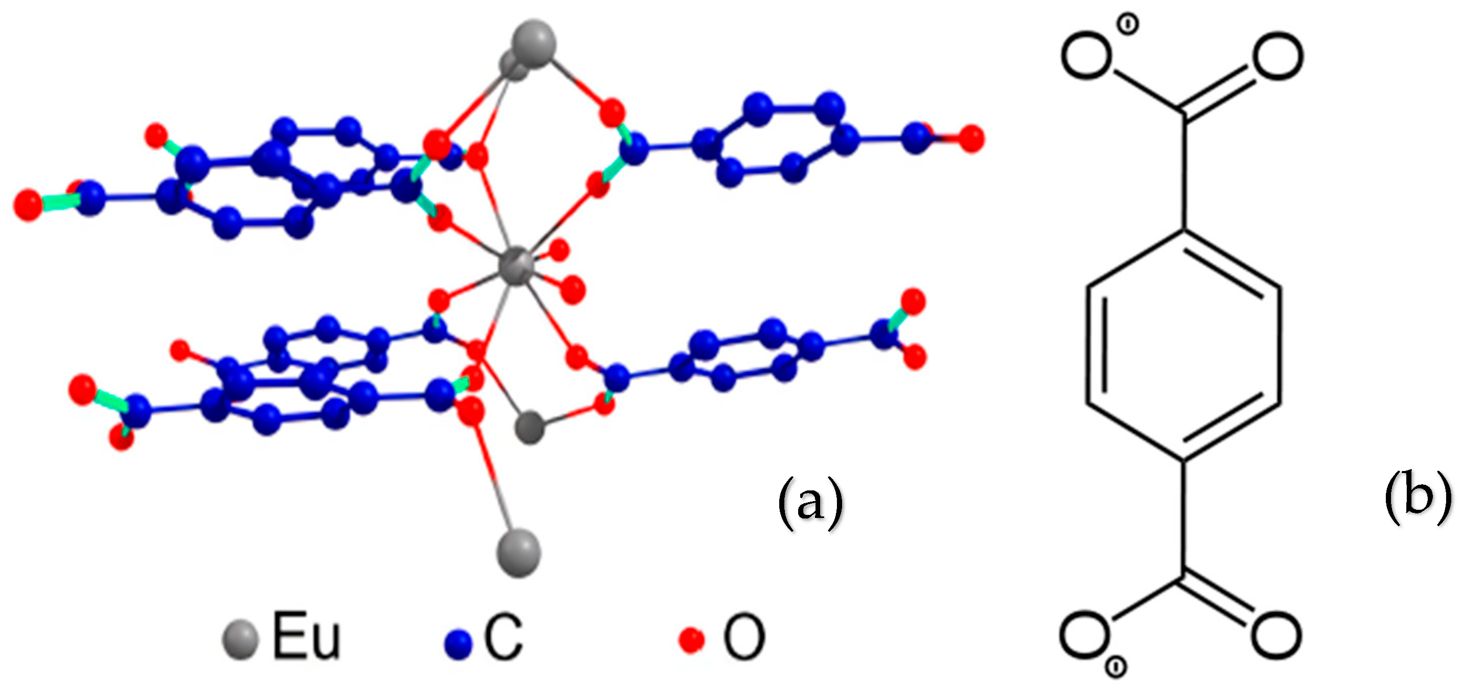
The synthesis of bulky lanthanide terephthalates was first reported by Reineke [30], where Tb2bdc3‧4H2O MOF (Scheme 1, bdc2− = 1,4-benzene-1,4-dicarboxylate or terephthalate ion) was obtained via a hydrothermal reaction between H2bdc and Tb(NO3)3‧5H2O in a Parr Teflon-lined stainless steel vessel. Triethylamine was added to the mixture, which was then sealed, heated at 140 °C for 12 h, and cooled to room temperature at 0.1 °C/min. Thermal removal of water yielded anhydrous Tb2(bdc)3, a microporous phase with extended 1-D channels. However, solvothermal synthesis is energy- and time-consuming, often taking hours to days, prompting the exploration of alternative methods.
Scheme 1. Structure of Eu2bdc3‧4H2O MOF [31] (a) and bdc2− (1,4-bdc, 1,4-benzene-1,4-dicarboxylate) (b).
Chemistry 07 00049 sch001
Non-solvothermal, eco-friendly synthesis methods, such as mechanical, ultrasound, emulsion, nanoprecipitation, and electrochemical techniques, enable the rapid production of nanoscale RE-MOFs [32,33,34]. These methods are energy-efficient, as they can be conducted at near-ambient temperatures and atmospheric pressure. The ultrasound-assisted (sonochemical) method is particularly versatile, offering a green chemistry approach by reducing reaction times, often using water as a solvent, and increasing product yields. Sonochemical synthesis has been successfully applied to produce various materials, including mesoporous silicas, metal oxides, perovskites, activated carbons, and diverse MOFs [35,36,37,38,39,40,41,42,43,44,45,46,47,48,49,50,51,52,53,54]. The intensity of ultrasonic irradiation significantly affects the chemical composition, size, and shape of particles. For example, gold nanoparticles synthesized under different ultrasound intensities exhibit varying shapes and size distributions, with smaller, more uniform particles forming at higher power levels [55]. However, the impact of ultrasonic intensity on complex structures like MOFs remains underexplored.
This article focuses on the ultrasonic synthesis of MOFs, examining how ultrasound affects the particle size. As an example, MIL-53(Fe), an iron terephthalate MOF, was synthesized using conventional heating, ultrasonication, and microwave methods. Ultrasonication resulted in the fastest crystallization rates, followed by microwave and conventional heating [56]. Similarly, nano-sized Tb(BTC)(H2O)6 (BTC = benzene-1,3,5-tricarboxylate) particles were synthesized by optimizing ultrasound-assisted reaction times [57]. These examples suggest that ultrasound-assisted methods can be extended to other micro- and nanomaterials, including lanthanide terephthalates. Previous work by the authors demonstrated the ultrasound-assisted synthesis of europium, gadolinium, lanthanum, and lutetium terephthalates [58,59].
Surfactant-based methods are another common approach for synthesizing micro- and nanomaterials. For example, mixing lanthanide chlorides with disodium terephthalate in water yields Ln2bdc3‧4H2O (Ln = La–Nd, Sm–Tm) particles with sizes ranging from 0.3 to 1.4 μm [60]. Nanoparticles of Ln2bdc3‧4H2O (Ln = La, Eu, Tb) were also prepared using polyvinylpyrrolidone (PVP) as a surfactant, with particle sizes ranging from 10 to 20 nm in various solvents [60]. However, SEM analysis in these studies was limited to lanthanum and terbium terephthalates, and the particle size distribution for other lanthanides was inferred indirectly, assuming spherical shapes. In reality, lanthanide terephthalates often have irregular shapes and exhibit luminescence, complicating accurate size determination. Additionally, ytterbium and lutetium terephthalates were not thoroughly studied, likely due to their ability to form MOFs with varying compositions and structures, such as Lu2bdc3·10H2O or Yb2bdc3·2H2O, depending on reagent concentrations [61,62]. Erbium terephthalates with different water contents were also synthesized in gel media, but their morphologies were not examined due to low yields and polymer removal challenges [63]. Thus, previous studies on the effects of surfactants and ultrasound on particle morphology are incomplete and inconclusive.
In this work, we synthesized microparticles of all lanthanide terephthalates (excluding promethium) using a surfactant-free, ultrasound-assisted method. The particles were characterized for composition, morphology, and structural features. These highly dispersed materials, with their large specific surface areas, hold potential as sorbents, sensors, or other advanced materials.

2. Materials and Methods

The following reagents were employed in this study: Ln(NO3)3∙nH2O (99.999%) (Ln = La, Ce, Pr, Nd, Sm, Eu, Gd, Tb, Dy, Ho, Er, Tm, Yb, Lu, Chemcraft, Kaliningrad, Russia); nickel(II) chloride hexahydrate and sodium hydroxide (>99%, Nevareactiv, St. Petersburg, Russia); benzene-1,4-dicarboxylic (terephthalic acid, H2(1,4-bdc) acid) (>98%); EDTA disodium salt (0.1 M aqueous solution); and murexide from Sigma-Aldrich Chemie GmbH (Taufkirchen, Germany). All reagents were used without further purification. For the synthesis of terephthalates, 0.2 M solutions of lanthanide nitrates were prepared and standardized via reverse complexometric reversed titration where the excess EDTA were added, and then titration was performed with nickel chloride in the presence of an ammonia buffer (pH = 9) and murexide; at the equivalence point, a transition from lilac to yellow color was observed [64]. A 0.2 M solution of sodium terephthalate, prepared by dissolving terephthalic acid in a standardized sodium hydroxide solution, served as the source of terephthalate ions.
To synthesize MOFs, 20 mL of a 0.005 M Ln(NO3)3 solution, obtained by diluting the initial 0.2 M solution by a factor of 40 (0.01 mmol), was placed in a beaker immersed in an ultrasonic bath. Then, 20 mL of a 0.01 M sodium terephthalate solution (Na2(1,4-bdc), 0.02 mmol), was added dropwise (one drop per 1–2 s) under ultrasonication (resulting pH = 7). An ultrasonic bath “GRANBO GA008G” (40 kHz, 60 W) was used for the synthesis. After completing the addition of the sodium terephthalate solution, the mixture was left in the bath for an additional 10–15 min, during which a white precipitate of the target compound formed. The solid was separated by centrifugation at 4000 rpm for 5 min and washed three times with 3 mL portions of distilled water. The resulting white solid was air-dried for 24 h at 20 °C, with average product yields ranging from 55% to 70%, Table S1 (Supplementary Materials).
The presence of specific lanthanide ions in the synthesized terephthalates was confirmed using energy-dispersive X-ray spectroscopy (EDX-spectrometer EDX-800P, Shimadzu, Kyoto, Japan) or an SEM-EDX module (Oxford Instruments INCAx-act, Oxford, UK). EDX spectra, provided in Figure S1 (Supplementary Materials), displayed characteristic X-ray emission bands for the corresponding lanthanides. X-ray powder diffraction (PXRD) analysis was performed using a D2 Phaser diffractometer (Bruker, Warwic, RI, USA, Cu Kα, λ = 1.54056 Å). Thermogravimetric analysis (TGA) was conducted on a NETZSCH TG 209 F1 Libra thermal analyzer (NETZSCH Group, Selb, Germany) under an argon atmosphere. Particle morphology was examined using scanning electron microscopy (SEM) on a Zeiss Merlin electron microscope (Zeiss, Jena, Germany). The CNH analysis was performed by means of a LECO TruSpec MICRO Elemental Determinator (LECO, St. Joseph, MI, USA)

3. Results and Discussion

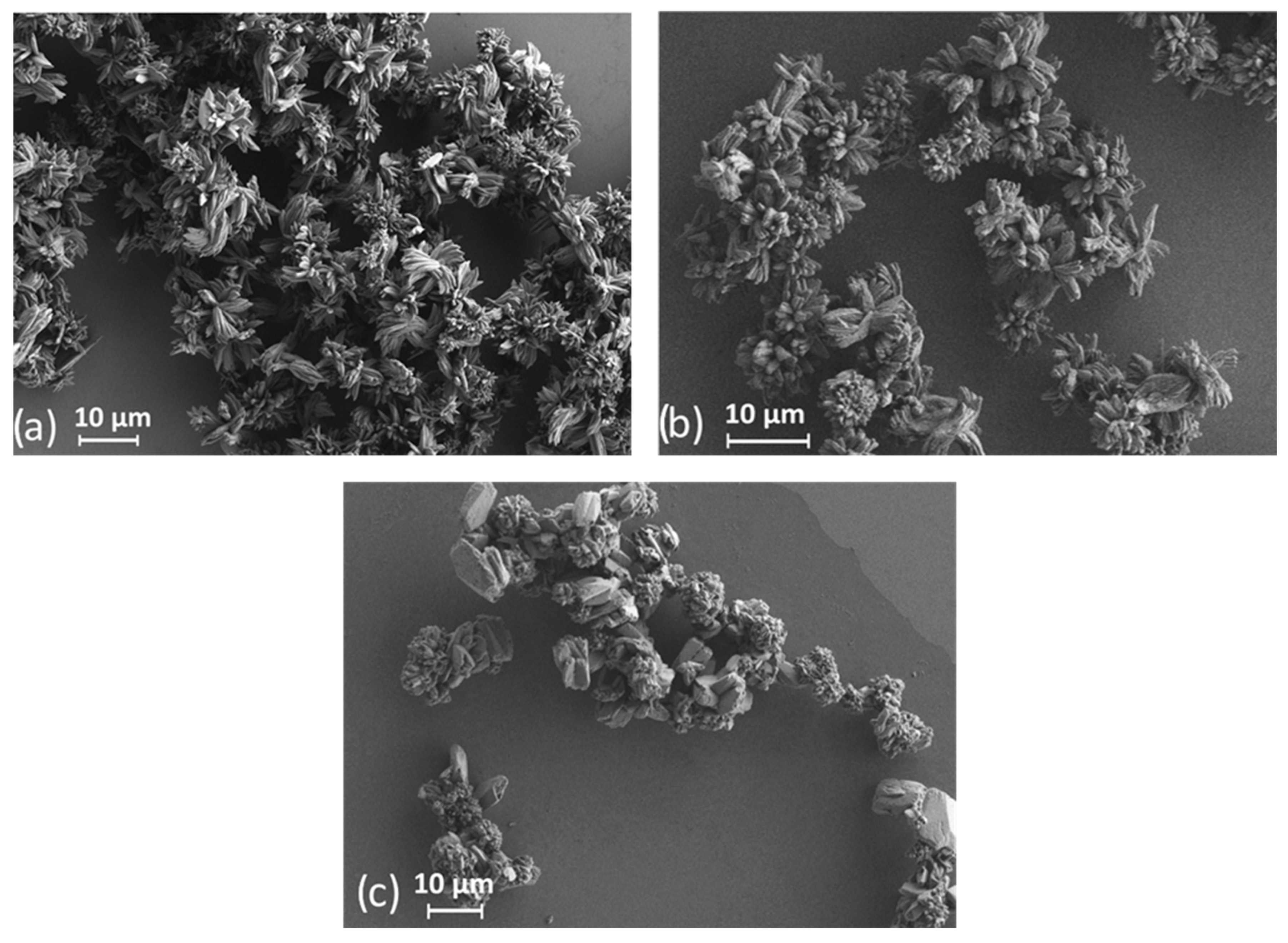
The effect of ultrasonic irradiation on the chemical and phase composition as well as on the morphology of particles of the obtained REE terephthalate MOFs was carefully studied in this work. The morphology and the size of the resulting particles were determined using scanning electron microscopy (SEM). The analysis of high-quality SEM images of lanthanide terephthalates particles obtained as a result of ultrasound-assisted synthesis showed that the samples consist of oval platelet-shaped particles with an approximate length–width ratio of 3:1 ((4.9 × 1.5)–(8.9 × 3.1) μm) and the thickness of about 0.5 μm. More careful analysis of the SEM micrographs showed that the microparticles consist of fused rods less than 100 nm in size (Figure 1a–f and Figure S2, Supplementary Materials). For comparison, Figure 2 shows SEM photographs of some lanthanide terephthalates (La, Gd, Lu) obtained without the use of ultrasound. The shape of the particles is practically independent of the composition of the compounds, with the exception of lutetium terephthalate. The particles of the later compound are brick-shaped with dimensions of 7.7 × 5.3 × 4.9 μm (Figure 1f). SEM micrographs for all obtained compounds are given in Figure S2, Supplementary Materials. In Figure 1e of ytterbium terephthalate, we can see two types of particles—the oval platelets, which are similar in shape to other lanthanide terephthalate particles, and a small admixture of brick-shaped particles similar to lutetium terephthalate particles (Figure 1f). This observation probably indicates the presence of a second chemical phase. The particle size distribution diagrams obtained from the analysis of SEM images are shown in Figure 3 (main text) and in Figure S3 (Supplementary Materials). The particle size distribution corresponds to the normal distribution with close size dispersion values for all studied materials, Figure 4. Therefore, it can be noted that the size of particles obtained by ultrasound-assisted synthesis is practically independent of the composition of the compounds, which can be seen from the obtained size values in Table 1. For comparison, lanthanum, gadolinium, and lutetium terephthalates were additionally synthesized using a similar procedure without the use of ultrasound, but with intense stirring. As can be seen from Figure 2, in the absence of ultrasound, the particles stick together, forming much larger aggregates of several tens of micrometers in size. Also, lutetium terephthalate does not form full-fledged cubic particles in distinction from the synthesis method implementing ultrasound.
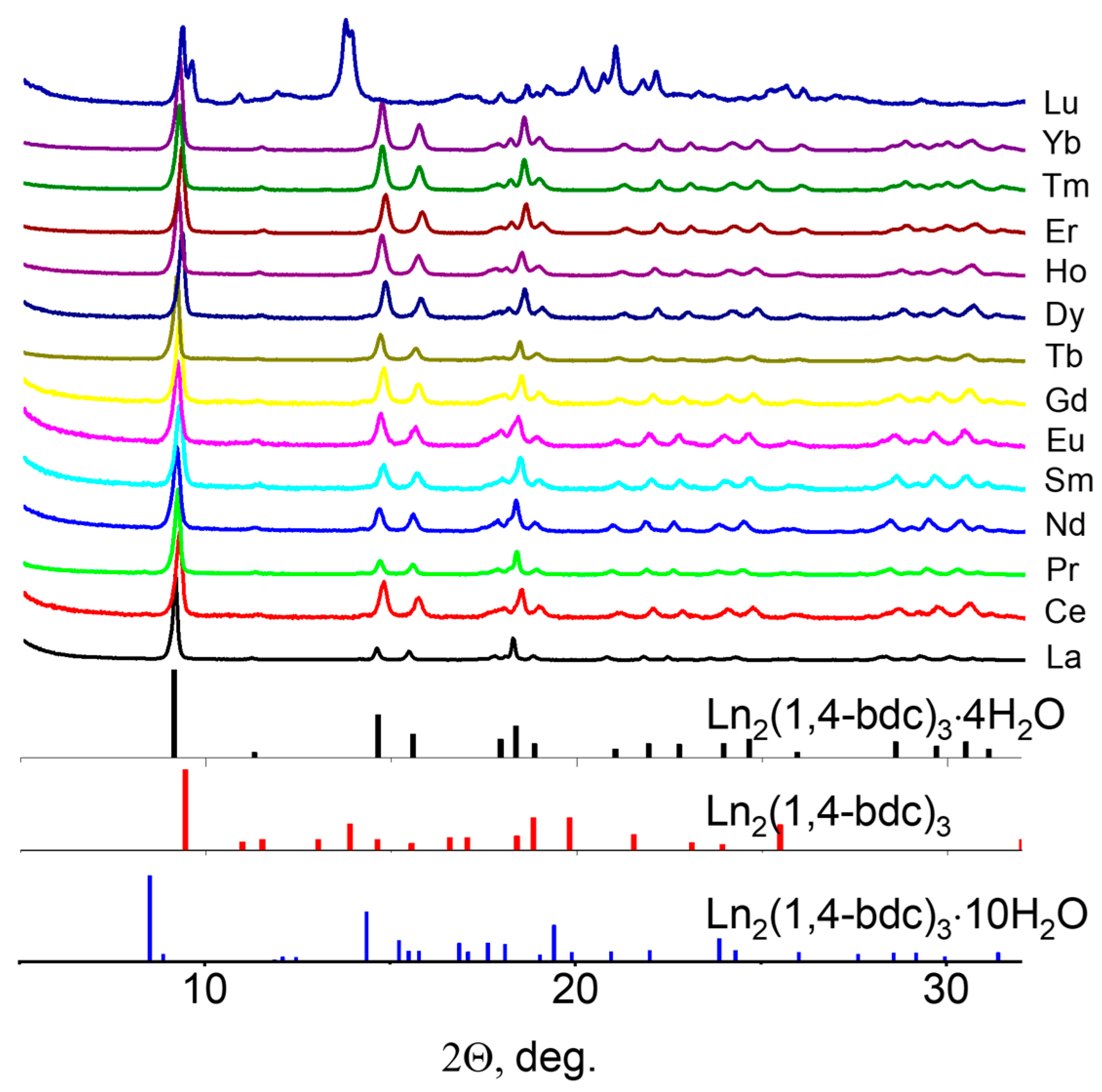
To study the chemical and phase composition in detail, several methods were employed, with powder X-ray diffraction (PXRD) being the primary technique. The PXRD patterns of the synthesized compounds are presented in Figure 5. The diffraction peak positions for various lanthanide terephthalates, such as Eu2(1,4-bdc)3·4H2O, Lu2(1,4-bdc)3·10H2O, and Tb2(1,4-bdc)3, simulated from cif-files [30,58] are also included in Figure 5 alongside the PXRD patterns. Analysis of the PXRD patterns indicated that the terephthalates of all lanthanides, except lutetium, exhibit a similar crystalline structure, specifically Ln2(1,4-bdc)3·4H2O, and are isomorphic to Eu2(1,4-bdc)3·4H2O. In contrast, lutetium terephthalate displays a distinct structure that does not align with the reference data. The diffraction peak positions of its PXRD pattern matched those of Lu2(1,4-bdc)3·2.5H2O, as reported in our earlier work [64].
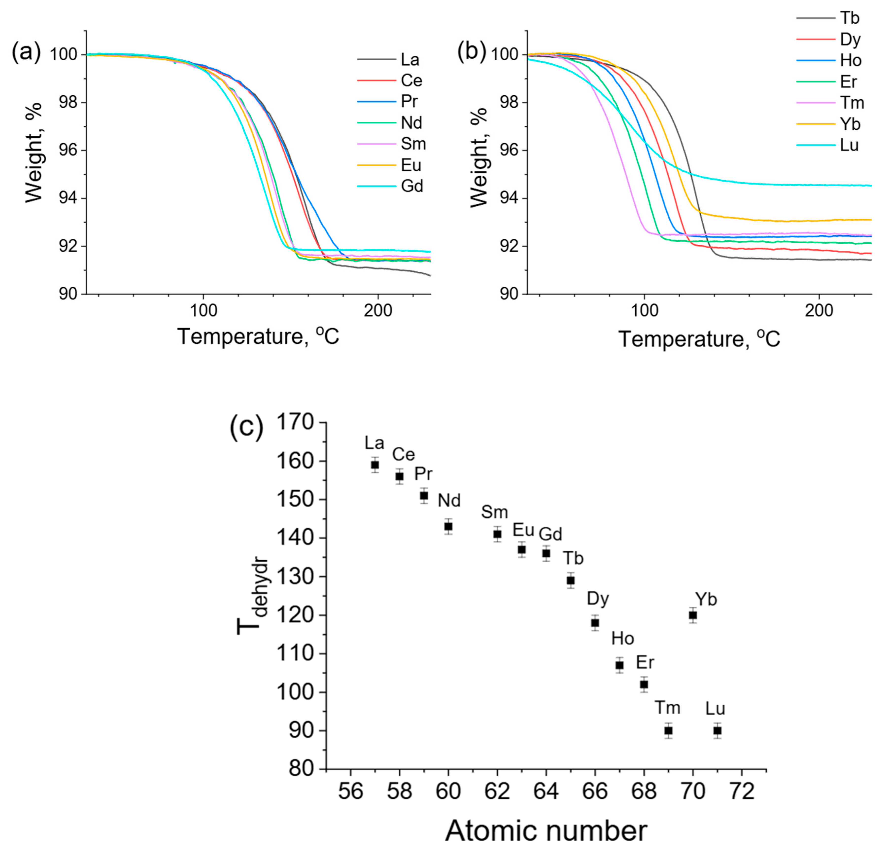
Thermogravimetric analysis (TGA) was performed to confirm the water content in all compounds. TGA curves (Figure 6) were recorded in the temperature range of 25–230 °C (heating rate 2°/min). For all lanthanide terephthalates, the mass loss was between 8 and 8.5 percent, which corresponds to about four water molecules. The derivative of the TGA curve of Figure 6a,b was also taken and the decomposition temperatures for each terephthalate were found (Figure 6c). From Figure 6c, it is evident that the decomposition temperature monotonically decreases from lanthanum to lutetium. The ytterbium terephthalate shows an unusually high dehydration temperature which can be possibly associated with the defects in the structure of this compound. A mass loss for lutetium terephthalate of approximately 5.5% was observed below 150 °C. The PXRD pattern of the solid residue after 24 h calcination at 200 °C corresponds to the anhydrous terephthalate Lu2(1,4-bdc)3, which is isomorphic to Tb2(1,4-bdc)3 [30] (Figure 7). Thus, it was concluded that the weight loss corresponds to the removal of two and a half water molecules during the thermal decomposition of lutetium terephthalate hydrate, Lu2(1,4-bdc)3·2.5H2O, consistent with our previous findings [64]. The reduced coordination number and, consequently, lower water content in lutetium terephthalate compared to other lanthanides are likely due to its smallest ionic radius, a result of lanthanide contraction. Therefore, the morphological differences between Lu2(1,4-bdc)3·2.5H2O and other samples in the series are primarily attributed to the distinct phase composition of lutetium terephthalate compared to other lanthanide terephthalates. The results for ytterbium terephthalate are also noteworthy. Although XRD (Figure 5) indicates the monophase nature of ytterbium terephthalate, SEM images (Figure 1) suggest the possible presence of a 2.5-hydrate phase of ytterbium terephthalate, as XRD cannot reliably reveal the presence of crystalline phases with the mole fraction less than 5%. However, a slightly smaller mass loss can be observed on the TGA curve of ytterbium than for the other lanthanide terephthalates (except lutecium), which is also probably associated with the presence of an admixture of Yb2(1,4-bdc)3·2.5H2O. Like lutetium, ytterbium has a small ionic radius, which may allow it to form a stable Yb2(1,4-bdc)3·2.5H2O phase, though its fraction appears minimal under the chosen synthesis conditions. The results of the CNH analysis (Table S2, Supplementary Materials) are in agreement with the proposed terephthalate compositions.
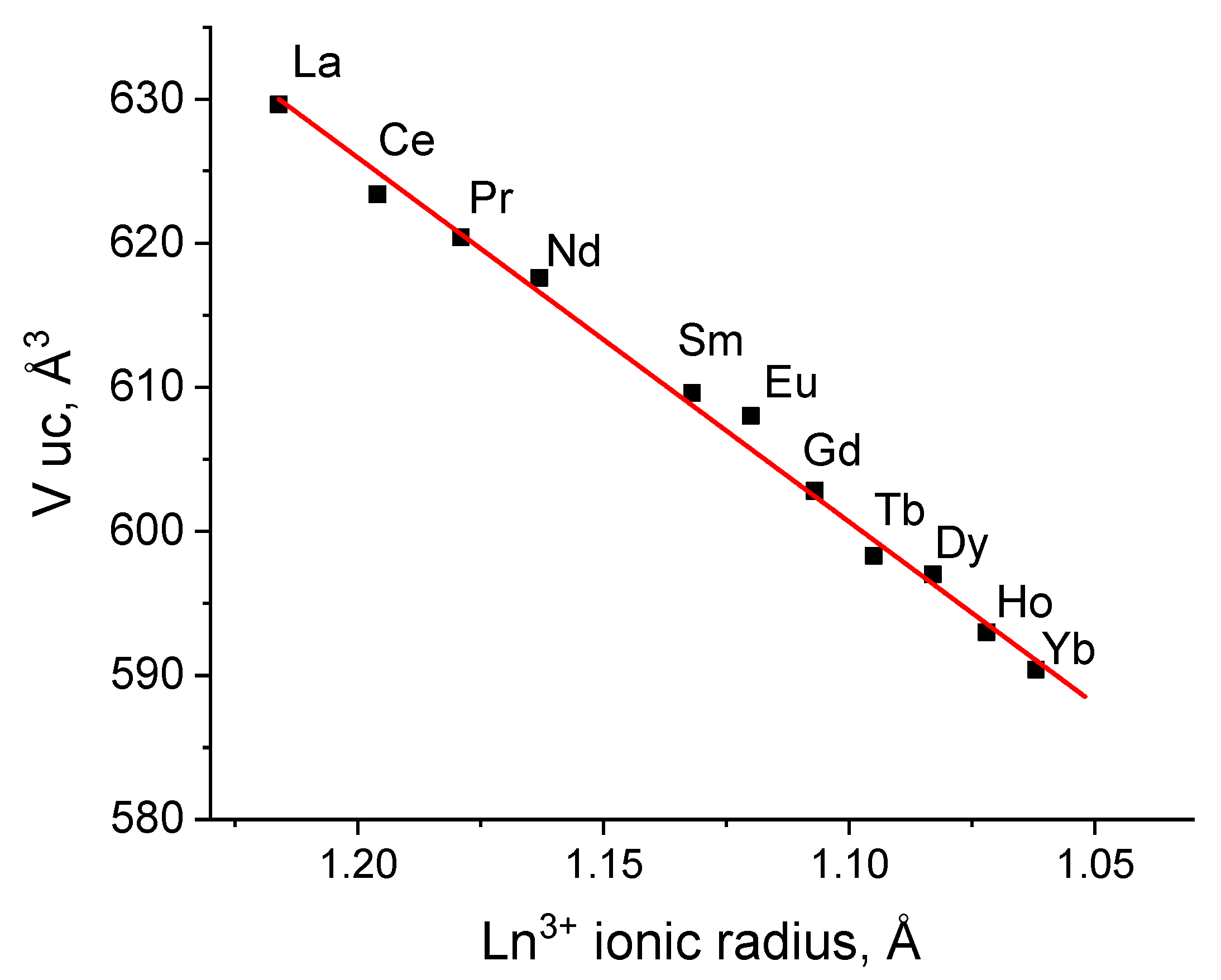
This study also aimed to assess the effect of lanthanide nature on particle size. The relationship between particle distribution by length and width and the lanthanide number is illustrated in Figure 4. It can be noted that the size of the microparticles slightly decreases as the lanthanide shifts from the beginning to the middle of the series, while closer to the end, an increase in size is observed, even as the unit cell volume decreases monotonically (Figure 8). Consequently, it is challenging to explain the cause of this phenomenon, and it can be suggested that the shape and size of the obtained microparticles depend only weakly on the nature of the lanthanide within the same crystalline phase, specifically Ln2(1,4-bdc)3∙4H2O.
The effect of lanthanide nature on the unit cell volume of Ln2(1,4-bdc)3∙4H2O was also monitored in this study (Figure 8). The unit cell parameters of all synthesized compounds were refined from PXRD patterns refined using TOPAS software (Version 3.2.0) [65], Table S3, Supplementary Materials. This program can retrieve unit cell parameters from diffraction data using a least-squares method from the positions of the indexed diffraction maxima of PXRD patterns (Pawley method [66]). It was found that the unit cell volumes decrease linearly as the lanthanide ionic radius decreases [67], a consequence of lanthanide contraction. However, it is noteworthy that the size of the resulting particles (Figure 5) does not exhibit a correlation with the unit cell volume within the Ln2(1,4-bdc)3∙4H2O crystalline phase.

4. Conclusions

A detailed study of the particle morphology, the phase, and the chemical composition of the microcrystalline lanthanide terephthalates obtained by the ultrasound-assisted method is reported. It was shown that the ultrasound-assisted synthesis of the abovementioned MOFs from aqueous solutions results in the formation of sub 10 μm particles. The particle shape and the composition of the synthesized materials are practically independent of the nature of the lanthanide ion. Meanwhile, if ultrasound is not applied during synthesis, significantly larger MOF particles are formed. The particle morphology and the structure of lutetium terephthalate obtained by the ultrasound-assisted method differ from other lanthanide terephthalates. It is formed as Lu2(1,4-bdc)3·2.5H2O consisting of brick-shaped particles, whereas other compounds crystallize in the form of tetrahydrate Ln2bdc3‧4H2O (Ln = La–Nd, Sm–Yb), and the particles have the shape of oval platelets. In summary, in the current work, we demonstrated the possibility of the synthesis of small-particle-size MOF materials with a large specific surface area by ultrasound-assisted method without the use of additional reagents (such as surfactants, organic solvents, etc.) which makes the synthesis process more environmentally friendly. Thus, the microparticles of all lanthanide terephthalates, several micrometers in size, were obtained for the first time (similar particles of only some lanthanide terephthalates were previously described) and particles of all lanthanides with this size were obtained for the first time using ultrasound without the addition of surfactants. Therefore, the developed ultrasound-assisted method can likely be scaled for other MOFs and for the design of materials such as sorbents, sensors etc.

Supplementary Materials

The following supporting information can be downloaded at: https://www.mdpi.com/article/10.3390/chemistry7020049/s1, Figure S1: EDX spectra of the lanthanide terephthalates. Figure S2: SEM images of lanthanide terephthalates. Figure S3: Particle length and width distribution for lanthanide terephthalates. Table S1: Yields of lanthanide terephthalates for ultrasonic synthesis. Table S2: CHN-analysis for lanthanide terephthalates. Table S3: Crystallographic data for Ln2(1,4-bdc)3∙4H2O (Ln = La, Ce, Pm, Pr, Nd, Sm, Eu, Gd, Tb, Dy, Ho, Er, Tm, Yb, Lu).

Author Contributions

Conceptualization, A.S.M., N.A.B. and Y.N.T.; methodology, Y.N.T. and S.S.K.; validation, P.B.G.; formal analysis, A.S.M. and N.A.B.; investigation, Y.N.T., N.A.G., P.B.G. and S.S.K.; resources, A.S.M.; data curation, N.A.G. and M.N.R.; writing—original draft preparation, Y.N.T. and A.S.M.; writing—review and editing, Y.N.T., N.A.B., A.S.M., S.N.O. and M.Y.S.; visualization, N.A.G.; supervision, A.S.M.; project administration, A.S.M.; funding acquisition, A.S.M. All authors have read and agreed to the published version of the manuscript.

Funding

This work was supported by the Russian Science Foundation under grant no. 22-73-10040 (https://rscf.ru/en/project/22-73-10040/, accessed on 2 July 2024).

Data Availability Statement

The original contributions presented in the study are included in the article and Supplementary Materials; further inquiries can be directed to the corresponding authors.

Acknowledgments

The measurements were performed at the Research Park of Saint-Petersburg State University (Magnetic Resonance Research Centre, Chemical Analysis and Materials Research Centre, Cryogenic Department, Interdisciplinary Resource Centre for Nanotechnology, Centre for X-ray Diffraction Studies, Centre for Optical and Laser Materials Research, Thermogravimetric and Calorimetric Research Centre, and Centre for Innovative Technologies of Composite Nanomaterials).

Conflicts of Interest

The authors declare no conflicts of interest.

References

  1. Yang, J.; Yue, Q.; Li, G.-D.; Cao, J.-J.; Li, G.-H.; Chen, J.-S. Structures, Photoluminescence, Up-Conversion, and Magnetism of 2D and 3D Rare-Earth Coordination Polymers with Multicarboxylate Linkages. Inorg. Chem. 2006, 45, 2857–2865. [Google Scholar] [CrossRef] [PubMed]
  2. Wang, Z.; Jin, C.-M.; Shao, T.; Li, Y.-Z.; Zhang, K.-L.; Zhang, H.-T.; You, X.-Z. Syntheses, Structures, and Luminescence Properties of a New Family of Three-Dimensional Open-Framework Lanthanide Coordination Polymers. Inorg. Chem. Commun. 2002, 5, 642–648. [Google Scholar] [CrossRef]
  3. Zhang, L.-Z.; Gu, W.; Li, B.; Liu, X.; Liao, D.-Z. {[Nd4(ox)4(NO3)2(OH)2(H2O)2] 5H2O} n: A Porous 3D Lanthanide-Based Coordination Polymer with a Special Luminescent Property. Inorg. Chem. 2007, 46, 622–624. [Google Scholar] [CrossRef]
  4. Yan, B.; Bai, Y.; Chen, Z. Synthesis, structure and luminescence of novel 1D chain coordination polymers [Ln(isophth)(Hisophth)(H2O)4·4H2O]n (Ln = Sm, Dy). J. Mol. Struct. 2005, 741, 141–147. [Google Scholar] [CrossRef]
  5. Cai, G.; Yan, P.; Zhang, L.; Zhou, H.-C.; Jiang, H.-L. Metal–Organic Framework-Based Hierarchically Porous Materials: Synthesis and Applications. Chem. Rev. 2021, 121, 12278–12326. [Google Scholar] [CrossRef] [PubMed]
  6. Chowdhuri, D.S.; Rana, A.; Bera, M.; Zangrando, E.; Dalai, S. 3D Zinc(II) Coordination Polymers Built up by Triazole and Benzene-Polycarboxylate Anions: Synthesis, Crystal Structure, Thermal and Photoluminescence Characterization. Polyhedron 2009, 28, 2131–2136. [Google Scholar] [CrossRef]
  7. Ye, Z.; Jiang, Y.; Li, L.; Wu, F.; Chen, R. Rational Design of MOF-Based Materials for Next-Generation Rechargeable Batteries. Nanomicro Lett. 2021, 13, 203. [Google Scholar] [CrossRef]
  8. Annamalai, J.; Murugan, P.; Ganapathy, D.; Nallaswamy, D.; Atchudan, R.; Arya, S.; Khosla, A.; Barathi, S.; Sundramoorthy, A.K. Synthesis of Various Dimensional Metal Organic Frameworks (MOFs) and Their Hybrid Composites for Emerging Applications—A Review. Chemosphere 2022, 298, 134184. [Google Scholar] [CrossRef]
  9. Meng, Z.; Qiu, Z.; Shi, Y.; Wang, S.; Zhang, G.; Pi, Y.; Pang, H. Micro/Nano Metal–Organic Frameworks Meet Energy Chemistry: A Review of Materials Synthesis and Applications. eScience 2023, 3, 100092. [Google Scholar] [CrossRef]
  10. Xu, G.-R.; An, Z.-H.; Xu, K.; Liu, Q.; Das, R.; Zhao, H.-L. Metal Organic Framework (MOF)-Based Micro/Nanoscaled Materials for Heavy Metal Ions Removal: The Cutting-Edge Study on Designs, Synthesis, and Applications. Coord. Chem. Rev. 2021, 427, 213554. [Google Scholar] [CrossRef]
  11. Le, S.-K.; Jin, Q.-J.; Han, J.-A.; Zhou, H.-C.; Liu, Q.-S.; Yang, F.; Miao, J.; Liu, P.-P.; Zhu, C.-Z.; Xu, H.-T. Rare Earth Element-Modified MOF Materials: Synthesis and Photocatalytic Applications in Environmental Remediation. Rare Metals 2024, 43, 1390–1406. [Google Scholar] [CrossRef]
  12. Younis, S.A.; Bhardwaj, N.; Bhardwaj, S.K.; Kim, K.-H.; Deep, A. Rare Earth Metal–Organic Frameworks (RE-MOFs): Synthesis, Properties, and Biomedical Applications. Coord. Chem. Rev. 2021, 429, 213620. [Google Scholar] [CrossRef]
  13. Neufeld, M.J.; Winter, H.; Landry, M.R.; Goforth, A.M.; Khan, S.; Pratx, G.; Sun, C. Lanthanide Metal–Organic Frameworks for Multispectral Radioluminescent Imaging. ACS Appl. Mater. Interfaces 2020, 12, 26943–26954. [Google Scholar] [CrossRef] [PubMed]
  14. Xia, N.; Chang, Y.; Zhou, Q.; Ding, S.; Gao, F. An Overview of the Design of Metal-Organic Frameworks-Based Fluorescent Chemosensors and Biosensors. Biosensors 2022, 12, 928. [Google Scholar] [CrossRef] [PubMed]
  15. Yu, Z.; Kang, S.; Tai, M.; Wang, J.; Tian, Q.; Jin, D.; Wang, L. Synthesis, Modulation, and Characterization of Ln3+ Ions Doped Metal−organic Frameworks for WLED Applications. Dye. Pigment. 2023, 209, 110897. [Google Scholar] [CrossRef]
  16. Meng, S.; Li, G.; Wang, P.; He, M.; Sun, X.; Li, Z. Rare Earth-Based MOFs for Photo/Electrocatalysis. Mater. Chem. Front. 2023, 7, 806–827. [Google Scholar] [CrossRef]
  17. Sun, S.; Zhao, Y.; Wang, J.; Pei, R. Lanthanide-Based MOFs: Synthesis Approaches and Applications in Cancer Diagnosis and Therapy. J. Mater. Chem. B 2022, 10, 9535–9564. [Google Scholar] [CrossRef]
  18. Li, C.; Wang, P.; He, M.; Yuan, X.; Fang, Z.; Li, Z. Rare Earth-Based Nanomaterials in Electrocatalysis. Coord. Chem. Rev. 2023, 489, 215204. [Google Scholar] [CrossRef]
  19. Crawford, S.E.; Ohodnicki, P.R.; Baltrus, J.P. Materials for the Photoluminescent Sensing of Rare Earth Elements: Challenges and Opportunities. J. Mater. Chem. C Mater. 2020, 8, 7975–8006. [Google Scholar] [CrossRef]
  20. Shen, G.; Zhong, L.; Liu, G.; Yang, L.; Wen, X.; Chen, G.; Zhao, J.; Hou, C.; Wang, X. Synthesis of Rare-Earth Metal-Organic Frameworks to Construct High-Resolution Sensing Array for Multiplex Anions Detection, Cell Imaging and Blood Phosphorus Monitoring. J. Colloid. Interface Sci. 2023, 652, 1925–1936. [Google Scholar] [CrossRef]
  21. Wu, N.; Bo, C.; Guo, S. Luminescent Ln-MOFs for Chemical Sensing Application on Biomolecules. ACS Sens. 2024, 9, 4402–4424. [Google Scholar] [CrossRef]
  22. Zheng, X.-J.; Zheng, T.-T.; Jin, L.-P. Self-Assembly of Lanthanide Mixed-Carboxylates Coordination Polymers. J. Mol. Struct. 2005, 740, 31–35. [Google Scholar] [CrossRef]
  23. Rodríguez-Cortiñas, R.; Avecilla, F.; Platas-Iglesias, C.; Imbert, D.; Bünzli, J.-C.G.; de Blas, A.; Rodríguez-Blas, T. Structural and Photophysical Properties of Heterobimetallic 4f-Zn Iminophenolate Cryptates. Inorg. Chem. 2002, 41, 5336–5349. [Google Scholar] [CrossRef] [PubMed]
  24. Zhao, S.-N.; Wang, G.; Poelman, D.; Voort, P. Luminescent Lanthanide MOFs: A Unique Platform for Chemical Sensing. Materials 2018, 11, 572. [Google Scholar] [CrossRef]
  25. Dou, X.; Sun, K.; Chen, H.; Jiang, Y.; Wu, L.; Mei, J.; Ding, Z.; Xie, J. Nanoscale Metal-Organic Frameworks as Fluorescence Sensors for Food Safety. Antibiotics 2021, 10, 358. [Google Scholar] [CrossRef]
  26. Puglisi, R.; Pellegrino, A.L.; Fiorenza, R.; Scirè, S.; Malandrino, G. A Facile One-Pot Approach to the Synthesis of Gd-Eu Based Metal-Organic Frameworks and Applications to Sensing of Fe3+ and Cr2O72− Ions. Sensors 2021, 21, 1679. [Google Scholar] [CrossRef] [PubMed]
  27. Duan, L.; Zhang, C.; Cen, P.; Jin, X.; Liang, C.; Yang, J.; Liu, X. Stable Ln-MOFs as Multi-Responsive Photoluminescence Sensors for the Sensitive Sensing of Fe3+, Cr2O72−, and Nitrofuran. CrystEngComm 2020, 22, 1695–1704. [Google Scholar] [CrossRef]
  28. Li, S.; Tan, L.; Meng, X. Nanoscale Metal-Organic Frameworks: Synthesis, Biocompatibility, Imaging Applications, and Thermal and Dynamic Therapy of Tumors. Adv. Funct. Mater. 2020, 30, 1908924. [Google Scholar] [CrossRef]
  29. Sun, Y.; Zhou, H.-C. Recent Progress in the Synthesis of Metal–Organic Frameworks. Sci. Technol. Adv. Mater. 2015, 16, 054202. [Google Scholar] [CrossRef]
  30. Reineke, T.M.; Eddaoudi, M.; Fehr, M.; Kelley, D.; Yaghi, O.M. From Condensed Lanthanide Coordination Solids to Microporous Frameworks Having Accessible Metal Sites. J. Am. Chem. Soc. 1999, 121, 1651–1657. [Google Scholar] [CrossRef]
  31. Butorlin, O.S.; Petrova, A.S.; Toikka, Y.N.; Kolesnikov, I.E.; Orlov, S.N.; Ryazantsev, M.N.; Bogachev, N.A.; Skripkin, M.Y.; Mereshchenko, A.S. The Structure and Optical Properties of Luminescent Europium Terephthalate Antenna Metal–Organic Frameworks Doped by Yttrium, Gadolinium, and Lanthanum Ions. Molecules 2024, 29, 3558. [Google Scholar] [CrossRef] [PubMed]
  32. Hu, D.; Song, Y.; Wang, L. Nanoscale Luminescent Lanthanide-Based Metal–Organic Frameworks: Properties, Synthesis, and Applications. J. Nanopart. Res. 2015, 17, 310. [Google Scholar] [CrossRef]
  33. Kukkar, D.; Vellingiri, K.; Kim, K.-H.; Deep, A. Recent Progress in Biological and Chemical Sensing by Luminescent Metal-Organic Frameworks. Sens. Actuators B Chem. 2018, 273, 1346–1370. [Google Scholar] [CrossRef]
  34. Ren, X.-Y.; Lu, L.-H. Luminescent Nanoscale Metal–Organic Frameworks for Chemical Sensing. Chin. Chem. Lett. 2015, 26, 1439–1445. [Google Scholar] [CrossRef]
  35. Vetrivel, S.; Chen, C.-T.; Kao, H.-M. The Ultrafast Sonochemical Synthesis of Mesoporous Silica MCM-41. New J. Chem. 2010, 34, 2109. [Google Scholar] [CrossRef]
  36. Jabariyan, S.; Zanjanchi, M.A. A Simple and Fast Sonication Procedure to Remove Surfactant Templates from Mesoporous MCM-41. Ultrason. Sonochem. 2012, 19, 1087–1093. [Google Scholar] [CrossRef]
  37. Palani, A.; Wu, H.-Y.; Ting, C.-C.; Vetrivel, S.; Shanmugapriya, K.; Chiang, A.S.T.; Kao, H.-M. Rapid Temperature-Assisted Sonochemical Synthesis of Mesoporous Silica SBA-15. Microporous Mesoporous Mater. 2010, 131, 385–392. [Google Scholar] [CrossRef]
  38. Gnana Sundara Raj, B.; Asiri, A.M.; Qusti, A.H.; Wu, J.J.; Anandan, S. Sonochemically Synthesized MnO2 Nanoparticles as Electrode Material for Supercapacitors. Ultrason. Sonochem. 2014, 21, 1933–1938. [Google Scholar] [CrossRef]
  39. Sakthipandi, K.; Rajendran, V. On-Line Phase Transitions of Bulk and Nanocrystalline La1−xPbxMnO3 (X = 0.3, 0.4, and 0.5) Perovskite Manganite Materials Using Ultrasonic Measurements. Mater. Chem. Phys. 2013, 138, 581–592. [Google Scholar] [CrossRef]
  40. Sharifalhoseini, Z.; Entezari, M.H.; Jalal, R. Direct and Indirect Sonication Affect Differently the Microstructure and the Morphology of ZnO Nanoparticles: Optical Behavior and Its Antibacterial Activity. Ultrason. Sonochem. 2015, 27, 466–473. [Google Scholar] [CrossRef]
  41. Altaf, A.R.; Teng, H.; Gang, L.; Adewuyi, Y.G.; Zheng, M. Effect of Sonochemical Treatment on Thermal Stability, Elemental Mercury (Hg0) Removal, and Regenerable Performance of Magnetic Tea Biochar. ACS Omega 2021, 6, 23913–23923. [Google Scholar] [CrossRef]
  42. Dong, D.; Zhang, Y.; Xiao, Y.; Wang, T.; Wang, J.; Pan, W. Synthesis of O-Doped Coal-Based Carbon Electrode Materials by Ultrasound-Assisted Bimetallic Activation for Application in Supercapacitors. Appl. Surf. Sci. 2020, 529, 147074. [Google Scholar] [CrossRef]
  43. Teng, Z.; Han, K.; Li, J.; Gao, Y.; Li, M.; Ji, T. Ultrasonic-Assisted Preparation and Characterization of Hierarchical Porous Carbon Derived from Garlic Peel for High-Performance Supercapacitors. Ultrason. Sonochem. 2020, 60, 104756. [Google Scholar] [CrossRef] [PubMed]
  44. Liu, W.; Tang, Y.; Sun, Z.; Gao, S.; Ma, J.; Liu, L. A Simple Approach of Constructing Sulfur-Containing Porous Carbon Nanotubes for High-Performance Supercapacitors. Carbon. N. Y 2017, 115, 754–762. [Google Scholar] [CrossRef]
  45. Ho, P.H.; Salles, F.; Di Renzo, F.; Trens, P. One-Pot Synthesis of 5-FU@ZIF-8 and Ibuprofen@ZIF-8 Nanoparticles. Inorganica Chim. Acta 2020, 500, 119229. [Google Scholar] [CrossRef]
  46. Bergaoui, M.; Khalfaoui, M.; Awadallah-F, A.; Al-Muhtaseb, S. A Review of the Features and Applications of ZIF-8 and Its Derivatives for Separating CO2 and Isomers of C3- and C4- Hydrocarbons. J. Nat. Gas. Sci. Eng. 2021, 96, 104289. [Google Scholar] [CrossRef]
  47. Razavi, S.A.A.; Morsali, A. Ultrasonic-Assisted Linker Exchange (USALE): A Novel Post-Synthesis Method for Controlling the Functionality, Porosity, and Morphology of MOFs. Chem. A Eur. J. 2019, 25, 10876–10885. [Google Scholar] [CrossRef]
  48. Yu, K.; Lee, Y.-R.; Seo, J.Y.; Baek, K.-Y.; Chung, Y.-M.; Ahn, W.-S. Sonochemical Synthesis of Zr-Based Porphyrinic MOF-525 and MOF-545: Enhancement in Catalytic and Adsorption Properties. Microporous Mesoporous Mater. 2021, 316, 110985. [Google Scholar] [CrossRef]
  49. Kim, J.; Yang, S.-T.; Choi, S.B.; Sim, J.; Kim, J.; Ahn, W.-S. Control of Catenation in CuTATB-n Metal–Organic Frameworks by Sonochemical Synthesis and Its Effect on CO2 Adsorption. J. Mater. Chem. 2011, 21, 3070. [Google Scholar] [CrossRef]
  50. Jung, D.-W.; Yang, D.-A.; Kim, J.; Kim, J.; Ahn, W.-S. Facile Synthesis of MOF-177 by a Sonochemical Method Using 1-Methyl-2-Pyrrolidinone as a Solvent. Dalton Trans. 2010, 39, 2883. [Google Scholar] [CrossRef]
  51. Wang, X.; Hu, X.; Shao, Y.; Peng, L.; Zhang, Q.; Zhou, T.; Xiang, Y.; Ye, N. Ambient Temperature Fabrication of a Covalent Organic Framework from 1,3,5-Triformylphloroglucinol and 1,4-Phenylenediamine as a Coating for Use in Open-Tubular Capillary Electrochromatography of Drugs and Amino Acids. Microchim. Acta 2019, 186, 650. [Google Scholar] [CrossRef]
  52. Zhao, W.; Yan, P.; Yang, H.; Bahri, M.; James, A.M.; Chen, H.; Liu, L.; Li, B.; Pang, Z.; Clowes, R.; et al. Using Sound to Synthesize Covalent Organic Frameworks in Water. Nat. Synth. 2022, 1, 87–95. [Google Scholar] [CrossRef]
  53. Yang, S.-T.; Kim, J.; Cho, H.-Y.; Kim, S.; Ahn, W.-S. Facile Synthesis of Covalent Organic Frameworks COF-1 and COF-5 by Sonochemical Method. RSC Adv. 2012, 2, 10179. [Google Scholar] [CrossRef]
  54. Duan, K.; Wang, J.; Zhang, Y.; Liu, J. Covalent Organic Frameworks (COFs) Functionalized Mixed Matrix Membrane for Effective CO2/N2 Separation. J. Memb. Sci. 2019, 572, 588–595. [Google Scholar] [CrossRef]
  55. Yusof, N.S.M.; Ashokkumar, M. Sonochemical Synthesis of Gold Nanoparticles by Using High Intensity Focused Ultrasound. Chemphyschem 2015, 16, 775–781. [Google Scholar] [CrossRef] [PubMed]
  56. Haque, E.; Khan, N.A.; Park, J.H.; Jhung, S.H. Synthesis of a Metal–Organic Framework Material, Iron Terephthalate, by Ultrasound, Microwave, and Conventional Electric Heating: A Kinetic Study. Chem. A Eur. J. 2010, 16, 1046–1052. [Google Scholar] [CrossRef]
  57. Hu, S.-M.; Niu, H.-L.; Qiu, L.-G.; Yuan, Y.-P.; Jiang, X.; Xie, A.-J.; Shen, Y.-H.; Zhu, J.-F. Facile Synthesis of Highly Luminescent Nanowires of a Terbium-Based Metal–Organic Framework by an Ultrasonic-Assisted Method and Their Application as a Luminescent Probe for Selective Sensing of Organoamines. Inorg. Chem. Commun. 2012, 17, 147–150. [Google Scholar] [CrossRef]
  58. Kolesnik, S.S.; Nosov, V.G.; Kolesnikov, I.E.; Khairullina, E.M.; Tumkin, I.I.; Vidyakina, A.A.; Sysoeva, A.A.; Ryazantsev, M.N.; Panov, M.S.; Khripun, V.D.; et al. Ultrasound-Assisted Synthesis of Luminescent Micro- and Nanocrystalline Eu-Based MOFs as Luminescent Probes for Heavy Metal Ions. Nanomaterials 2021, 11, 2448. [Google Scholar] [CrossRef]
  59. Kolesnik, S.S.; Bogachev, N.A.; Kolesnikov, I.E.; Orlov, S.N.; Ryazantsev, M.N.; González, G.; Skripkin, M.Y.; Mereshchenko, A.S. Microcrystalline Luminescent (Eu1−XLnx)2bdc3·nH2O (Ln = La, Gd, Lu) Antenna MOFs: Effect of Dopant Content on Structure, Particle Morphology, and Luminescent Properties. Molecules 2024, 29, 532. [Google Scholar] [CrossRef]
  60. Daiguebonne, C.; Kerbellec, N.; Guillou, O.; Bünzli, J.-C.; Gumy, F.; Catala, L.; Mallah, T.; Audebrand, N.; Gérault, Y.; Bernot, K.; et al. Structural and Luminescent Properties of Micro- and Nanosized Particles of Lanthanide Terephthalate Coordination Polymers. Inorg. Chem. 2008, 47, 3700–3708. [Google Scholar] [CrossRef] [PubMed]
  61. Nosov, V.G.; Toikka, Y.N.; Petrova, A.S.; Butorlin, O.S.; Kolesnikov, I.E.; Orlov, S.N.; Ryazantsev, M.N.; Kolesnik, S.S.; Bogachev, N.A.; Skripkin, M.Y.; et al. Brightly Luminescent (TbxLu1−x)2bdc3·nH2O MOFs: Effect of Synthesis Conditions on Structure and Luminescent Properties. Molecules 2023, 28, 2378. [Google Scholar] [CrossRef] [PubMed]
  62. Toikka, Y.N.; Badikov, A.R.; Bogachev, N.A.; Kolesnikov, I.E.; Skripkin, M.Y.; Orlov, S.N.; Mereshchenko, A.S. Luminescent Properties and Thermal Stability of (Lu0.98Eu0.02)2bdc3·10H2O Metal–Organic Frameworks. Mendeleev Commun. 2024, 34, 634–636. [Google Scholar] [CrossRef]
  63. Zehnder, R.A.; Renn, R.A.; Pippin, E.; Zeller, M.; Wheeler, K.A.; Carr, J.A.; Fontaine, N.; McMullen, N.C. Network Dimensionality and Ligand Flexibility in Lanthanide Terephthalate Hydrates. J. Mol. Struct. 2011, 985, 109–119. [Google Scholar] [CrossRef]
  64. Schwarzenbach, G.; Flaschka, H.A. Complexometric Titrations; Methuen: London, UK, 1969; ISBN 9780389011439. [Google Scholar]
  65. Coelho, A.A. TOPAS and TOPAS-Academic: An optimization program integrating computer algebra and crystallographic objects written in C++. J. Appl. Cryst. 2018, 51, 210–218. [Google Scholar] [CrossRef]
  66. Pawley, G.S. Unit-Cell Refinement from Powder Diffraction Scans. J. Appl. Crystallogr. 1981, 14, 357–361. [Google Scholar] [CrossRef]
  67. Shannon, R.D. Revised Effective Ionic Radii and Systematic Studies of Interatomic Distances in Halides and Chalcogenides. Acta Crystallogr. Sect. A 1976, 32, 751–767. [Google Scholar] [CrossRef]
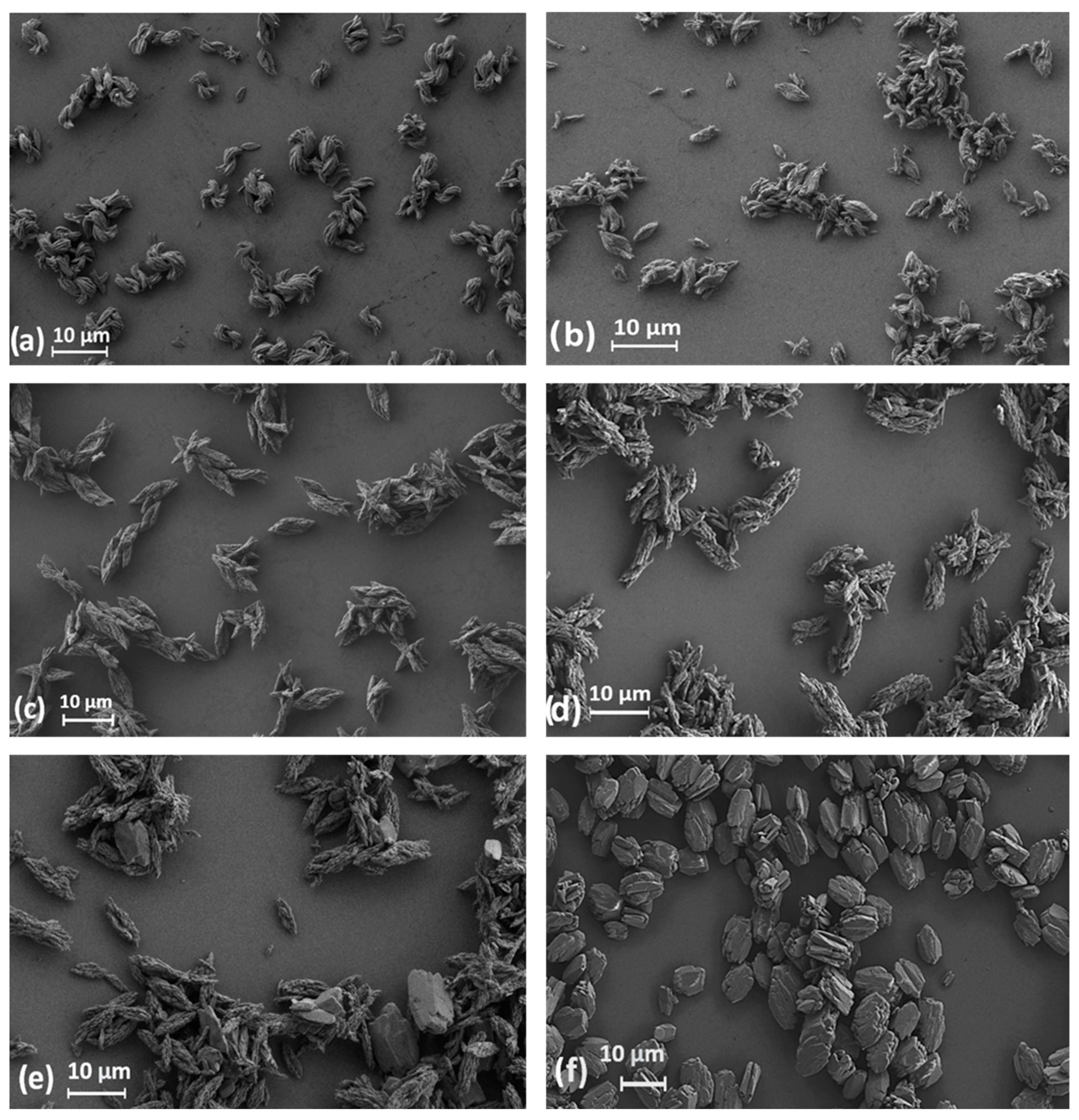
Figure 1. SEM images of microcrystalline La (a), Gd (b), Er (c), Tm (d), Yb (e), and Lu (f) terephthalates obtained by ultrasound-assisted synthesis.
Chemistry 07 00049 g001
Figure 2. SEM images of lanthanum (a), gadolinium (b), and lutetium (c) terephthalates obtained by stirring-only method without ultrasonication.
Chemistry 07 00049 g002
Figure 3. Size distribution (length and width) of microcrystalline lanthanide terephthalates obtained from SEM images: La (a), Gd (b), Lu (c).
Chemistry 07 00049 g003aChemistry 07 00049 g003b
Figure 4. The correlation of average size (length and width) of Ln2(1,4-bdc)3∙4H2O microparticles with the ionic radii of lanthanide ions.
Chemistry 07 00049 g004
Figure 5. Experimental PXRD patterns of microcrystalline terephthalates Ln2bdc3‧nH2O (Ln = La–Lu, excluding Pm) and the positions of the reflexes simulated for Ln2(1,4-bdc)3∙nH2O (n = 0, 4, 10) compounds [30].
Chemistry 07 00049 g005
Figure 6. TGA curves of lanthanide terephthalates measured in the temperature range of 35–230 °C (a,b) and the dehydration temperatures defined from the TGA curves as the first derivative extremum point. (c) Figure 6c shows dependence of lanthanide terephthalate dehydration temperature on lanthanide atomic number.
Chemistry 07 00049 g006
Figure 7. PXRD pattern of Lu2(1,4-bdc)3∙nH2O after thermal decomposition (24 h at 200 °C) compared with the positions of diffraction maxima of Tb2(1,4-bdc)3 simulated from the structure given in [30].
Chemistry 07 00049 g007
Figure 8. The effect of the ionic radii of lanthanide ions on the unit cell volumes of Ln2bdc3‧4H2O (Ln = La–Yb, excluding Pm) microcrystals.
Chemistry 07 00049 g008
Table 1. Average size (length and width) of microcrystalline lanthanide terephthalates Ln2(1,4-bdc)3∙nH2O.
LnAverage Length, μmAverage Width, μm
La6.7 ± 1.32.8 ± 0.7
Ce5.5 ± 1.12.6 ± 0.6
Pr4.9 ± 0.92.1 ± 0.5
Nd5.9 ± 1.12.6 ± 0.5
Sm3.9 ± 0.61.6 ± 0.3
Eu4.7 ± 0.91.9 ± 0.4
Gd5.1 ± 1.12.2 ± 0.6
Tb3.9 ± 1.01.5 ± 0.5
Dy5.5 ± 2.02.2 ± 0.9
Ho5.8 ± 1.32.3 ± 0.6
Er9.3 ± 2.03.1 ± 0.6
Tm7.1 ± 1.92.4 ± 0.6
Yb8.9 ± 1.93.1 ± 0.8
Lu7.7 ± 1.35.3 ± 1.1
Disclaimer/Publisher’s Note: The statements, opinions and data contained in all publications are solely those of the individual author(s) and contributor(s) and not of MDPI and/or the editor(s). MDPI and/or the editor(s) disclaim responsibility for any injury to people or property resulting from any ideas, methods, instructions or products referred to in the content.