


Anti-Orthopoxvirus Activity of Amantadine and Rimantadine Derivatives—In Vitro Testing and Molecular Modeling

 ,
,  ,
,  , , , , ,
Abstract
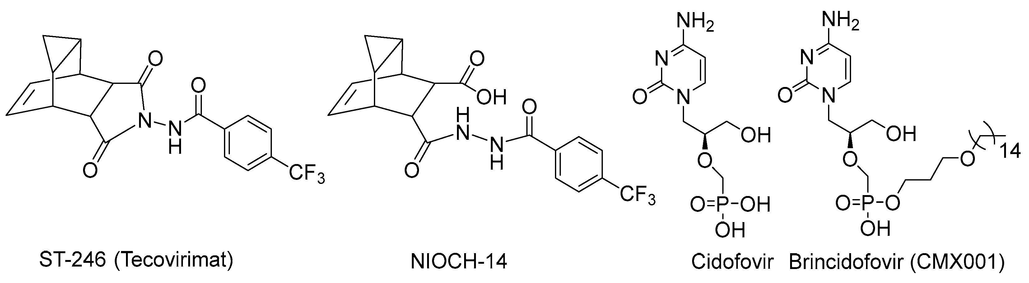
1. Introduction
2. Materials and Methods
2.1. Chemistry
General Information
2.2. Biology
2.3. Molecular Modeling
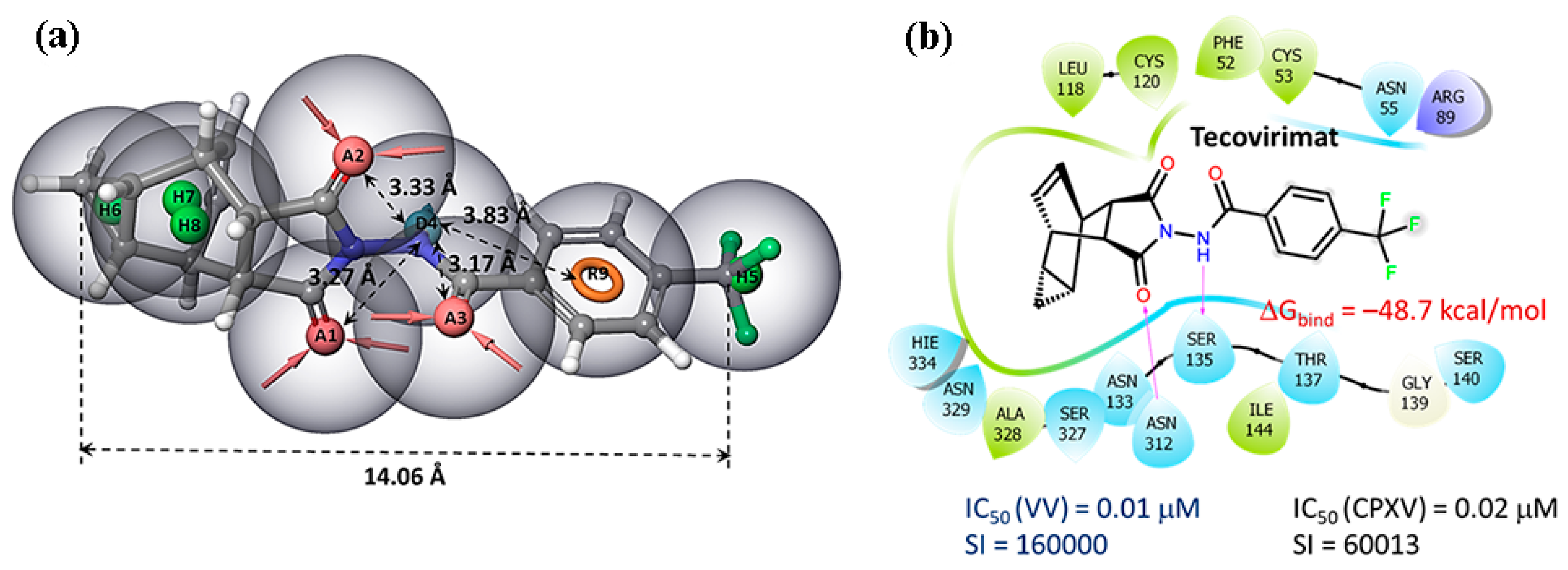
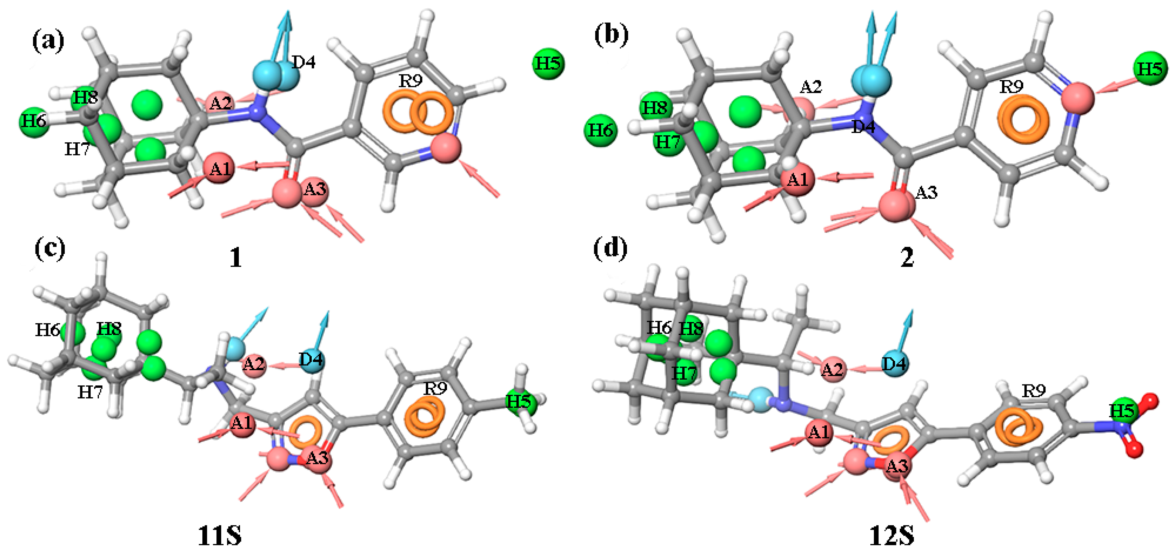
2.3.1. The Pharmacophore Model
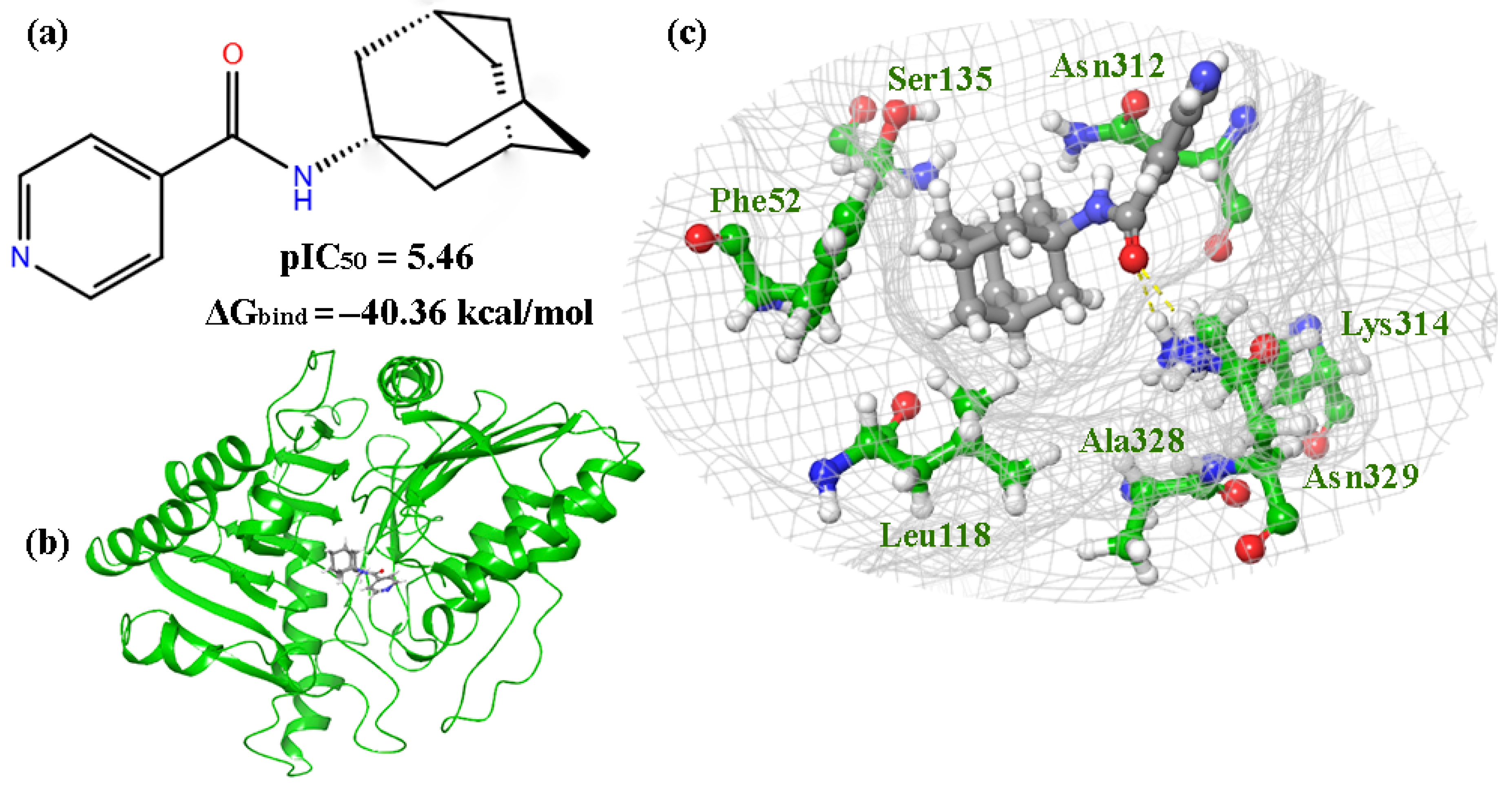
2.3.2. Molecular Docking
2.3.3. Molecular Dynamics
2.4. Single-Crystal X-Ray Diffraction
3. Results
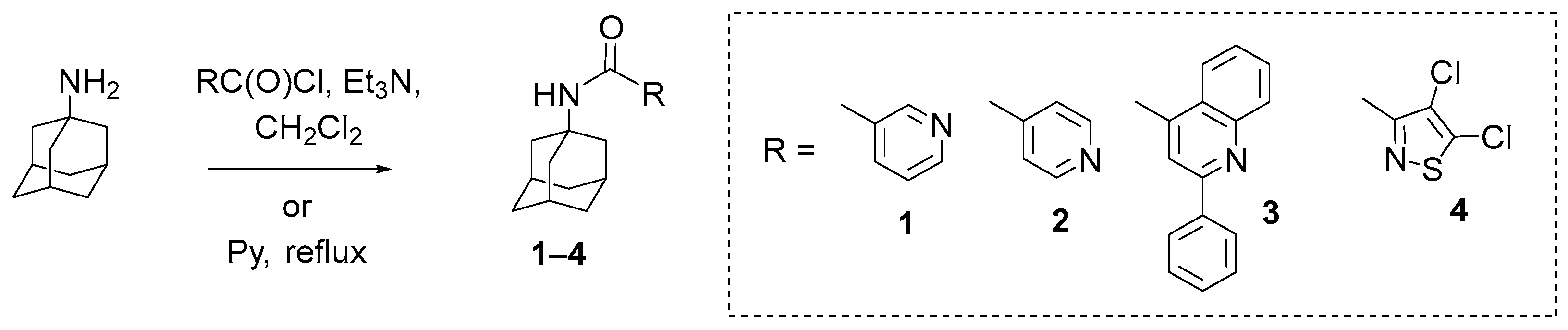
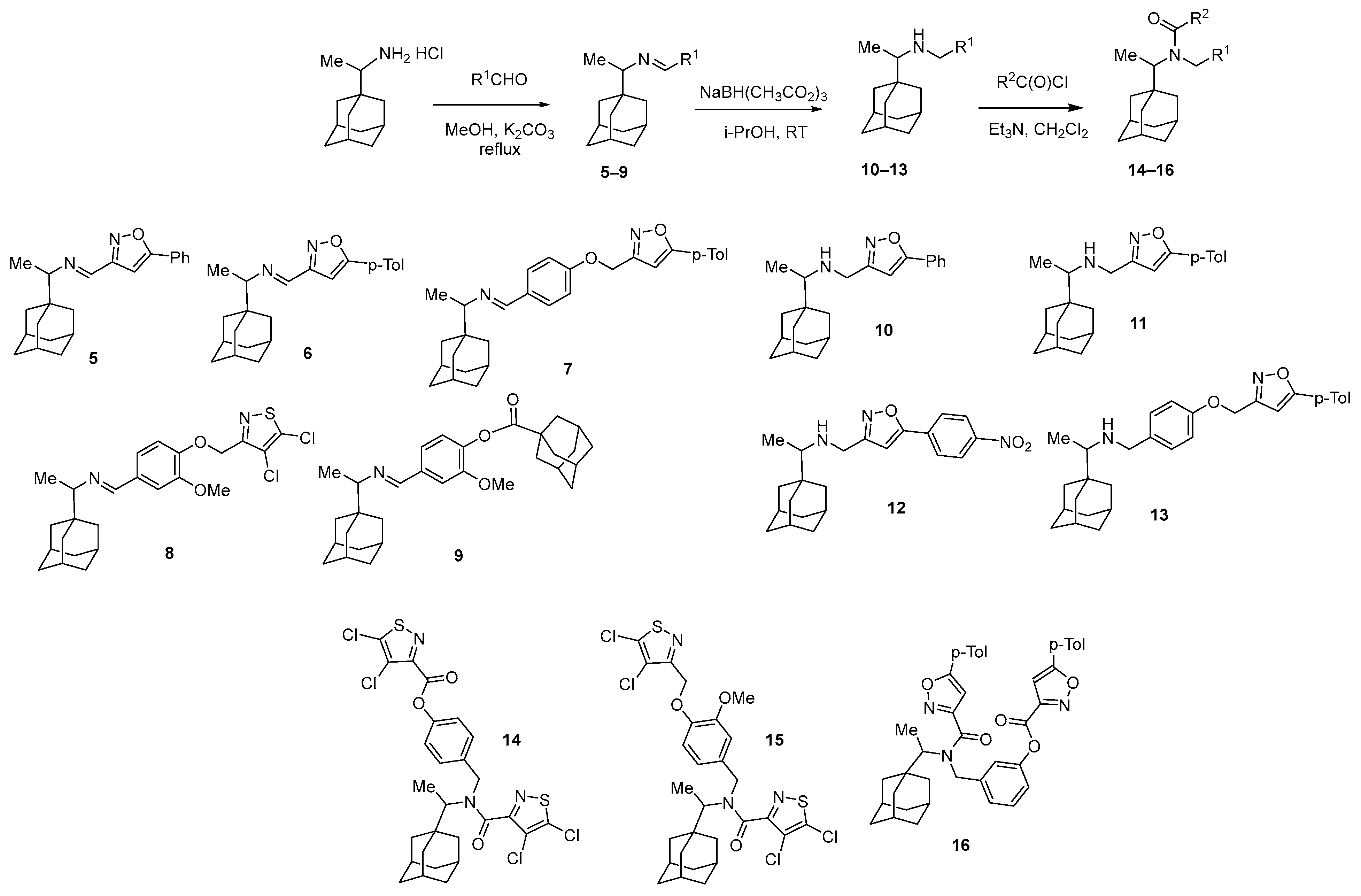
3.1. Chemistry
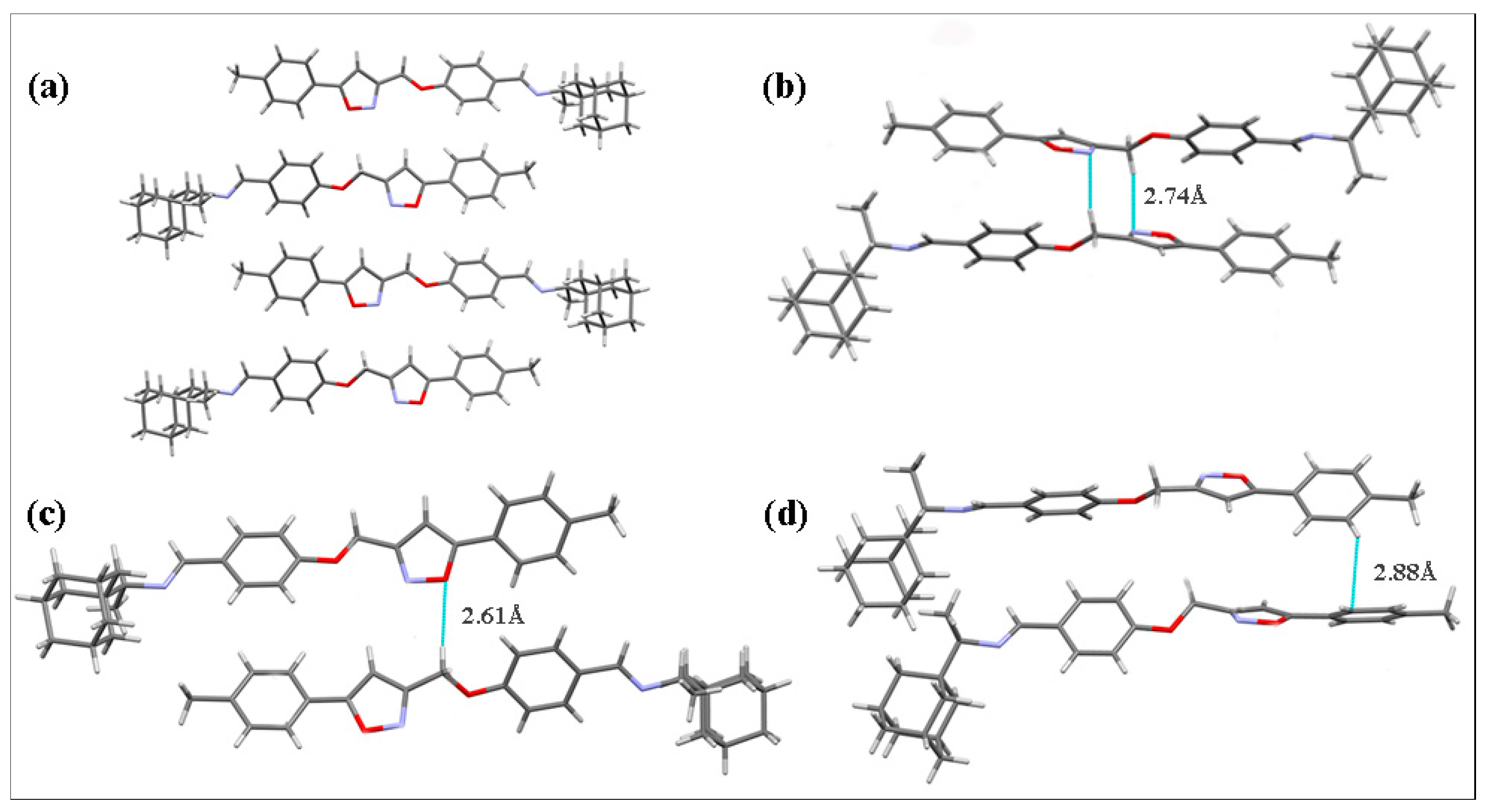
3.2. Crystal Structure Analysis
3.3. Biology
3.4. Molecular Modeling Study
4. Conclusions
Supplementary Materials
Author Contributions
Funding
Data Availability Statement
Acknowledgments
Conflicts of Interest
References
- Boehm, E.; Summermatter, K.; Kaiser, L. Orthopox Viruses: Is the Threat Growing? Clin. Microbiol. Infect. 2024, 30, 883–887. [Google Scholar] [CrossRef]
- Adetifa, I.; Muyembe, J.-J.; Bausch, D.G.; Heymann, D.L. Mpox Neglect and the Smallpox Niche: A Problem for Africa, a Problem for the World. Lancet 2023, 401, 1822–1824. [Google Scholar] [CrossRef]
- Velásquez, J.S.; Herrera-Echeverría, F.B.; Porres-Paredes, H.S.; Rodríguez-Cerdeira, C. Understanding the Epidemiology of Monkeypox Virus to Prevent Future Outbreaks. Microorganisms 2024, 12, 2576. [Google Scholar] [CrossRef]
- De Pascali, A.M.; Brandolini, M.; Peli, L.; Sambri, V.; Cricca, M.; Scagliarini, A. Zoonotic Orthopoxviruses after Smallpox Eradication: A Shift from Crisis Response to a One Health Approach. IJID One Health 2024, 2, 100018. [Google Scholar] [CrossRef]
- Protopapas, K.; Dimopoulou, D.; Kalesis, N.; Akinosoglou, K.; Moschopoulos, C.D. Mpox and Lessons Learned in the Light of the Recent Outbreak: A Narrative Review. Viruses 2024, 16, 1620. [Google Scholar] [CrossRef] [PubMed]
- Kumar, S.; Guruparan, D.; Karuppanan, K.; Kumar, K.J.S. Comprehensive Insights into Monkeypox (Mpox): Recent Advances in Epidemiology, Diagnostic Approaches and Therapeutic Strategies. Pathogens 2024, 14, 1. [Google Scholar] [CrossRef] [PubMed]
- Schneider, C.P.; McDonald, M.D. “The King of Terrors” Revisited: The Smallpox Vaccination Campaign and Its Lessons for Future Biopreparedness. J. Law Med. Ethics 2003, 31, 580–589. [Google Scholar] [CrossRef]
- Wang, J.; Shahed-AI-Mahmud, M.; Chen, A.; Li, K.; Tan, H.; Joyce, R. An Overview of Antivirals against Monkeypox Virus and Other Orthopoxviruses. J. Med. Chem. 2023, 66, 4468–4490. [Google Scholar] [CrossRef]
- FDA News Release. FDA Approves the First Drug with an Indication for Treatment of Smallpox. Available online: https://www.fda.gov/news-events/press-announcements/fda-approves-first-drug-indication-treatment-smallpox (accessed on 24 December 2024).
- Chen, Y.; Amantana, A.; Tyavanagimatt, S.R.; Zima, D.; Yan, X.S.; Kasi, G.; Weeks, M.; Stone, M.A.; Weimers, W.C.; Samuel, P.; et al. Comparison of the Safety and Pharmacokinetics of ST-246® after IV Infusion or Oral Administration in Mice, Rabbits and Monkeys. PLoS ONE 2011, 6, e23237. [Google Scholar] [CrossRef]
- Mazurkov, O.Y.; Kabanov, A.S.; Shishkina, L.N.; Sergeev, A.A.; Skarnovich, M.O.; Bormotov, N.I.; Skarnovich, M.A.; Ovchinnikova, A.S.; Titova, K.A.; Galahova, D.O.; et al. New Effective Chemically Synthesized Anti-Smallpox Compound NIOCH-14. J. Gen. Virol. 2016, 97, 1229–1239. [Google Scholar] [CrossRef]
- Hoy, S.M. Tecovirimat: First Global Approval. Drugs 2018, 78, 1377–1382. [Google Scholar] [CrossRef] [PubMed]
- Chimerix Receives, U.S. Food and Drug Administration Approval for TEMBEXA® (Brincidofovir) for the Treatment of Smallpox. Available online: https://Ir.Chimerix.Com/News-Releases/News-Release-Details/Chimerix-Receives-Us-Food-and-Drug-Administration-Approval (accessed on 24 December 2024).
- World Health Organization. Scientific Review of Variola Virus Research, 1999–2010. 2010. Available online: https://iris.who.int/bitstream/handle/10665/70508/WHO_HSE_GAR_BDP_2010.3_eng.pdf?sequence=1 (accessed on 24 December 2024).
- Prichard, M.; Kern, E. Orthopoxvirus Targets for the Development of Antiviral Therapies. Curr. Drug Target-Infect. Disord. 2005, 5, 17–28. [Google Scholar] [CrossRef]
- Parker, S.; Chen, N.G.; Foster, S.; Hartzler, H.; Hembrador, E.; Hruby, D.; Jordan, R.; Lanier, R.; Painter, G.; Painter, W.; et al. Evaluation of Disease and Viral Biomarkers as Triggers for Therapeutic Intervention in Respiratory Mousepox—An Animal Model of Smallpox. Antiviral Res. 2012, 94, 44–53. [Google Scholar] [CrossRef]
- Ragshaniya, A.; Kumar, V.; Tittal, R.K.; Lal, K. Nascent Pharmacological Advancement in Adamantane Derivatives. Arch. Pharm. 2024, 357, 2300595. [Google Scholar] [CrossRef] [PubMed]
- Stockdale, T.P.; Williams, C.M. Pharmaceuticals That Contain Polycyclic Hydrocarbon Scaffolds. Chem. Soc. Rev. 2015, 44, 7737–7763. [Google Scholar] [CrossRef]
- Singh, Y.P.; Kumar, H. Recent Advances in Medicinal Chemistry of Memantine Against Alzheimer’s Disease. Chem. Biol. Drug Des. 2024, 104, e14638. [Google Scholar] [CrossRef] [PubMed]
- Jalily, P.H.; Duncan, M.C.; Fedida, D.; Wang, J.; Tietjen, I. Put a Cork in It: Plugging the M2 Viral Ion Channel to Sink Influenza. Antiviral Res. 2020, 178, 104780. [Google Scholar] [CrossRef]
- Kuznetsov, N.Y.; Tikhov, R.M.; Godovikov, I.A.; Medvedev, M.G.; Lyssenko, K.A.; Burtseva, E.I.; Kirillova, E.S.; Bubnov, Y.N. Stereoselective Synthesis of Novel Adamantane Derivatives with High Potency against Rimantadine-Resistant Influenza A Virus Strains. Org. Biomol. Chem. 2017, 15, 3152–3157. [Google Scholar] [CrossRef]
- Kumar, G.; Sakharam, K.A. Tackling Influenza A Virus by M2 Ion Channel Blockers: Latest Progress and Limitations. Eur. J. Med. Chem. 2024, 267, 116172. [Google Scholar] [CrossRef]
- Lim, S.; Guo, Z.; Liu, P.; Mckay, L.G.A.; Storm, N.; Qu, M.D.; Finberg, R.W.; Somasundaran, M.; Wang, J.P. Anti-SARS-CoV-2 Activity of Adamantanes In Vitro and in Animal Models of Infection. COVID 2023, 2, 1551–1563. [Google Scholar] [CrossRef]
- Brown, E.; Swinscoe, G.; Lefteri, D.A.; Singh, R.; Moran, A.; Thompson, R.F.; Maskell, D.; Beaumont, H.; Bentham, M.J.; Donald, C.; et al. Inhibitors of the Small Membrane (M) Protein Viroporin Prevent Zika Virus Infection. eLife 2024, 13, e68404. [Google Scholar] [CrossRef] [PubMed]
- Kreutzberger, A.; Schröders, H.-H. Die Aliphatische Säureamidgruppierung Als Partialstruktur in Virustatika 3. Mitt. Antivirale Wirkstoffe. Arch. Pharm. 1974, 307, 766–774. [Google Scholar] [CrossRef] [PubMed]
- Suslov, E.V.; Mozhaytsev, E.S.; Korchagina, D.V.; Bormotov, N.I.; Yarovaya, O.I.; Volcho, K.P.; Serova, O.A.; Agafonov, A.P.; Maksyutov, R.A.; Shishkina, L.N.; et al. New Chemical Agents Based on Adamantane-Monoterpene Conjugates against Orthopoxvirus Infections. RSC Med. Chem. 2020, 11, 1185–1195. [Google Scholar] [CrossRef]
- Shiryaev, V.A.; Skomorohov, M.Y.; Leonova, M.V.; Bormotov, N.I.; Serova, O.A.; Shishkina, L.N.; Agafonov, A.P.; Maksyutov, R.A.; Klimochkin, Y.N. Adamantane Derivatives as Potential Inhibitors of P37 Major Envelope Protein and Poxvirus Reproduction. Design, Synthesis and Antiviral Activity. Eur. J. Med. Chem. 2021, 221, 113485. [Google Scholar] [CrossRef] [PubMed]
- Mozhaitsev, E.S.; Suslov, E.V.; Rastrepaeva, D.A.; Yarovaya, O.I.; Borisevich, S.S.; Khamitov, E.M.; Kolybalov, D.S.; Arkhipov, S.G.; Bormotov, N.I.; Shishkina, L.N.; et al. Structure-Based Design, Synthesis, and Biological Evaluation of the Cage–Amide Derived Orthopox Virus Replication Inhibitors. Viruses 2022, 15, 29. [Google Scholar] [CrossRef]
- Sokolova, A.S.; Kovaleva, K.S.; Kuranov, S.O.; Bormotov, N.I.; Borisevich, S.S.; Zhukovets, A.A.; Yarovaya, O.I.; Serova, O.A.; Nawrozkij, M.B.; Vernigora, A.A.; et al. Design, Synthesis, and Biological Evaluation of (+)-Camphor- and (−)-Fenchone-Based Derivatives as Potent Orthopoxvirus Inhibitors. ChemMedChem 2022, 17, e202100771. [Google Scholar] [CrossRef]
- Chen, Y.; Honeychurch, K.M.; Yang, G.; Byrd, C.M.; Harver, C.; Hruby, D.E.; Jordan, R. Vaccinia Virus P37 Interacts with Host Proteins Associated with LE-Derived Transport Vesicle Biogenesis. Virol. J. 2009, 6, 44. [Google Scholar] [CrossRef]
- Sen Gupta, P.S.; Panda, S.K.; Nayak, A.K.; Rana, M.K. Identification and Investigation of a Cryptic Binding Pocket of the P37 Envelope Protein of Monkeypox Virus by Molecular Dynamics Simulations. J. Phys. Chem. Lett. 2023, 14, 3230–3235. [Google Scholar] [CrossRef]
- Duraffour, S.; Lorenzo, M.M.; Zöller, G.; Topalis, D.; Grosenbach, D.; Hruby, D.E.; Andrei, G.; Blasco, R.; Meyer, H.; Snoeck, R. ST-246 Is a Key Antiviral to Inhibit the Viral F13L Phospholipase, One of the Essential Proteins for Orthopoxvirus Wrapping. J. Antimicrob. Chemother. 2015, 70, 1367–1380. [Google Scholar] [CrossRef]
- Jordan, R.; Leeds, J.M.; Tyavanagimatt, S.; Hruby, D.E. Development of ST-246® for Treatment of Poxvirus Infections. Viruses 2010, 2, 2409–2435. [Google Scholar] [CrossRef]
- Akishina, E.A.; Dikusar, Е.А.; Kurman, P.V.; Potkin, V.I. Synthesis of Novel Rimantadine and Adamantane-1-Carboxylic Acid Derivatives with 1,2-Azole Fragments. Proc. Natl. Acad. Sci. Belarus Chem. Ser. 2023, 59, 211–224. [Google Scholar] [CrossRef]
- Selivanov, B.A.; Tikhonov, A.; Belanov, E.F.; Bormotov, N.I.; Kabanov, A.S.; Mazurkov, O.Y.; Serova, O.A.; Shishkina, L.N.; Agafonov, A.P.; Sergeev, A.N. Synthesis and Antiviral Activity of 1-Aryl-3-{3,5-Dioxo-4-Azatetracyclo[5.3.2.02,6.08,10]-Dodec-11-En-4-Yl}ureas. Khimiko-Farmatsevticheskii Zhurnal 2017, 51, 13–17. [Google Scholar] [CrossRef]
- Lu, C.; Wu, C.; Ghoreishi, D.; Chen, W.; Wang, L.; Damm, W.; Ross, G.A.; Dahlgren, M.K.; Russell, E.; Von Bargen, C.D.; et al. OPLS4: Improving Force Field Accuracy on Challenging Regimes of Chemical Space. J. Chem. Theory Comput. 2021, 17, 4291–4300. [Google Scholar] [CrossRef] [PubMed]
- Johnston, R.C.; Yao, K.; Kaplan, Z.; Chelliah, M.; Leswing, K.; Seekins, S.; Watts, S.; Calkins, D.; Chief Elk, J.; Jerome, S.V.; et al. Epik: P K a and Protonation State Prediction through Machine Learning. J. Chem. Theory Comput. 2023, 19, 2380–2388. [Google Scholar] [CrossRef]
- Mirdita, M.; Schütze, K.; Moriwaki, Y.; Heo, L.; Ovchinnikov, S.; Steinegger, M. ColabFold: Making Protein Folding Accessible to All. Nat. Methods 2022, 19, 679–682. [Google Scholar] [CrossRef]
- Rothkirch, A.; Gatta, G.D.; Meyer, M.; Merkel, S.; Merlini, M.; Liermann, H.-P. Single-Crystal Diffraction at the Extreme Conditions Beamline P02.2: Procedure for Collecting and Analyzing High-Pressure Single-Crystal Data. J. Synchrotron Radiat. 2013, 20, 711–720. [Google Scholar] [CrossRef]
- CrysAlisPro 171.43.143a, Rigaku Oxford Diffraction, 2023. Available online: https://www.rigaku.com/products/crystallography/crysalis (accessed on 9 December 2024).
- Sheldrick, G.M. SHELXT–Integrated Space-Group and Crystal-Structure Determination. Acta Crystallogr. Sect. A Found. Adv. 2015, 71, 3–8. [Google Scholar] [CrossRef]
- Sheldrick, G.M. Crystal Structure Refinement with SHELXL. Acta Crystallogr. Sect. C Struct. Chem. 2015, 71, 3–8. [Google Scholar] [CrossRef]
- Dolomanov, O.V.; Bourhis, L.J.; Gildea, R.J.; Howard, J.A.K.; Puschmann, H. OLEX2: A Complete Structure Solution, Refinement and Analysis Program. J. Appl. Crystallogr. 2009, 42, 339–341. [Google Scholar] [CrossRef]
- Spek, A.L. Structure Validation in Chemical Crystallography. Acta Crystallogr. Sect. D Biol. Crystallogr. 2009, 65, 148–155. [Google Scholar] [CrossRef]
- Macrae, C.F.; Sovago, I.; Cottrell, S.J.; Galek, P.T.A.; McCabe, P.; Pidcock, E.; Platings, M.; Shields, G.P.; Stevens, J.S.; Towler, M.; et al. Mercury 4.0: From Visualization to Analysis, Design and Prediction. J. Appl. Crystallogr. 2020, 53, 226–235. [Google Scholar] [CrossRef] [PubMed]
- Fridman, A.L.; Sivkova, M.P.; Zalesov, V.S.; Dolbilkin, K.V.; Moiseev, I.K.; Doroshenko, R.I.; Manzhelevskaya, É.V. Synthesis and Biological Activity of Adamantane Derivatives. Pharm. Chem. J. 1979, 13, 1241–1246. [Google Scholar] [CrossRef]
- Okawara, T.; Islam, R.; Hossain, M.I.; Okamoto, Y.; Nagamatsu, T.; Anraku, K. ChemInform Abstract: Facile Synthesis of 2-Phenylquinoline-4-carboxamide Derivatives with Variant Structural Features. ChemInform 2014, 45, 32. [Google Scholar] [CrossRef]
- Borisevich, S.S.; Gorokhov, Y.V.; Arkhipov, S.G. Binding Site of Tecovirimat, Inhibitor of the P37 Membrane Protein of Orthopox Viruses. J. Struct. Chem. 2024, 65, 776–785. [Google Scholar] [CrossRef]

| Compound | CC50, µM a | EC50, µM b | SI c |
|---|---|---|---|
| 1 | 100.67 ± 21.1 | 16.78 ± 3.7 | 6 |
| 2 | 390.33 ± 26.1 | 3.45 ± 0.9 | 115 |
| 3 | 350.31 ± 38.9 | NA | - |
| 4 | 75.50 ± 8.7 | 2.91 ± 0.15 | 26 |
| 5 | 299.40 ± 41.0 | 75.53 ± 8.1 | 4 |
| 6 | 287.35 ± 11.7 | NA | - |
| 7 | 66.08 ± 5.1 | 40.8 ± 3.8 | 2 |
| 8 | 52.31 ± 2.5 | NA | - |
| 9 | 56.85 ± 2.7 | NA d | - |
| 10 | 15.47± 0.7 | 13.91 ± 2.5 | - |
| 11 | 15.14 ± 1.1 | NA | - |
| 12 | 204.72 ± 10.8 | 9.44 ± 1.5 | 22 |
| 13 | 10.47 ± 2.1 | NA | - |
| 14 | 8.87 ± 0.7 | NA | - |
| 15 | 21.31 ± 1.3 | NA | - |
| 16 | 150.37 ± 11.1 | NA | - |
| Cidofovir | 201.33 ± 60.10 | 7.63 ± 3.15 | 26 |
Disclaimer/Publisher’s Note: The statements, opinions and data contained in all publications are solely those of the individual author(s) and contributor(s) and not of MDPI and/or the editor(s). MDPI and/or the editor(s) disclaim responsibility for any injury to people or property resulting from any ideas, methods, instructions or products referred to in the content. |
© 2025 by the authors. Licensee MDPI, Basel, Switzerland. This article is an open access article distributed under the terms and conditions of the Creative Commons Attribution (CC BY) license (https://creativecommons.org/licenses/by/4.0/).
Share and Cite
Moskalev, I.A.; Akishina, E.A.; Dikusar, E.A.; Yarovaya, O.I.; Borisevich, S.S.; Khamitov, E.M.; Fedorov, A.Y.; Arkhipov, S.G.; Bormotov, N.I.; Serova, O.A.; et al. Anti-Orthopoxvirus Activity of Amantadine and Rimantadine Derivatives—In Vitro Testing and Molecular Modeling. Chemistry 2025, 7, 34. https://doi.org/10.3390/chemistry7020034
Moskalev IA, Akishina EA, Dikusar EA, Yarovaya OI, Borisevich SS, Khamitov EM, Fedorov AY, Arkhipov SG, Bormotov NI, Serova OA, et al. Anti-Orthopoxvirus Activity of Amantadine and Rimantadine Derivatives—In Vitro Testing and Molecular Modeling. Chemistry. 2025; 7(2):34. https://doi.org/10.3390/chemistry7020034
Chicago/Turabian StyleMoskalev, Ivan A., Ekaterina A. Akishina, Evgenij A. Dikusar, Olga I. Yarovaya, Sophia S. Borisevich, Edward M. Khamitov, Alexey Yu. Fedorov, Sergey G. Arkhipov, Nikolay I. Bormotov, Olga A. Serova, and et al. 2025. "Anti-Orthopoxvirus Activity of Amantadine and Rimantadine Derivatives—In Vitro Testing and Molecular Modeling" Chemistry 7, no. 2: 34. https://doi.org/10.3390/chemistry7020034
APA StyleMoskalev, I. A., Akishina, E. A., Dikusar, E. A., Yarovaya, O. I., Borisevich, S. S., Khamitov, E. M., Fedorov, A. Y., Arkhipov, S. G., Bormotov, N. I., Serova, O. A., Shishkina, L. N., Potkin, V. I., & Salakhutdinov, N. F. (2025). Anti-Orthopoxvirus Activity of Amantadine and Rimantadine Derivatives—In Vitro Testing and Molecular Modeling. Chemistry, 7(2), 34. https://doi.org/10.3390/chemistry7020034