Article

Evaluation of the Anticancer and DNA-Binding Characteristics of Dichloro(diimine)zinc(II) Complexes

by
Bandar A. Babgi
1,*,
Doaa Domyati
2,
Magda H. Abdellattif
3 and
Mostafa A. Hussien
1,4
1
Department of Chemistry, Faculty of Science, King Abdulaziz University, P.O. Box 80203, Jeddah 21589, Saudi Arabia
2
Department of Chemistry, College of Science, University of Jeddah, Jeddah 21589, Saudi Arabia
3
Department of Chemistry, Faculty of Science, Taif University, Al-Haweiah, P.O. Box 888, Taif 21974, Saudi Arabia
4
Department of Chemistry, Faculty of Science, Port Said University, Port Said 42521, Egypt
*
Author to whom correspondence should be addressed.
Chemistry 2021, 3(4), 1178-1188; https://doi.org/10.3390/chemistry3040086
Submission received: 10 August 2021 / Revised: 8 September 2021 / Accepted: 5 October 2021 / Published: 11 October 2021
(This article belongs to the Section Bioinorganics)

Abstract

:
Several metal diimine complexes have been reported to possess anticancer properties. To evaluate the anticancer properties of tetrahedral zinc(II) diimine complexes, six complexes were synthesized with the general formula M(N^N)Cl2 {where M = Zn, Pt and N^N = 2,2’-biquinoline (1), 2,2’-dipyridylketone (2) and 4-((pyridine-2-ylmethylene)amino)phenol (3)}. In general, the intrinsic DNA-binding constants for the different compounds exhibited values within close proximity; the changes in the viscosity of the CT-DNA upon binding to the compounds suggest intercalation-binding mode. Molecular docking study predicted that complexes containing the highly planar ligand 2,2’-biquinoline are capable to establish π–π interactions with nucleobases of the DNA; the other four complexes engaged in donor–acceptor interactions with DNA nucleobases. The six complexes and two reference drugs (cisplatin and sunitinib) were tested against two cancer cell lines (COLO 205 and RCC-PR) and one normal cell line (LLC-MK2), highlighting the better performance of the zinc(II) complexes compared to their platinum(II) analogues. Moreover, zinc(II) complexes have higher selectivity index values than the reference drugs, with promising anticancer properties.

Graphical Abstract

1. Introduction

Cisplatin (cis-diamminedichloridoplatinum(II)), carboplatin (cis-diammine(1,1-cyclobutanedicarboxylato)platinum(II)) and oxaliplatin (trans-R,R-cyclohexane-(1,2-diamine)oxalatoplatinum(II)) are clinically approved metal-based drugs used for the chemotherapeutic treatment of several cancer types [1]. Pt(II) drugs are believed to be activated by the hydrolysis of anionic ligands and the aqua-adducts bind to DNA, cross-linking the DNA strands; this process leads to block replication and prevents transcription [2]. Although platinum-based complexes possess excellent anticancer properties, there are several problems associated with them, including sever side-effects (e.g., nausea and kidney toxicity) and acquired resistance developed by some tumor types [3,4,5]. These drawbacks motivate further research to synthesize other transition metal complexes that exhibit comparable anticancer properties to that of cisplatin, with fewer side-effects. Several metal complexes have been tested for their antitumor properties, especially metal complexes employing derivatives of 2,2’-bipyridines or 1,10-phenanthrolines as ligands [6,7]. The interest in this type of ligands is due to their ability to interact non-covalently with various biomolecules [8,9,10]. Among diimine complexes, vanadium diimine complexes have received considerable attention. Metvan is a peroxovanadium compound (1 in Figure 1), which induces apoptosis in cancer cell lines and exhibits considerable anticancer activity against human glioblastoma (GBM) and breast cancer xenograft models in SCID mice [11]. Another peroxovanadium complex (2 in Figure 1) has been reported and found active in vitro and in vivo toward transplanted breast cancer [12]. Another class of diimine metal complexes that have been widely studied are copper(I) and copper(II) diimine complexes. In a subsequent report, [Cu(phen)(LL-threonine)(H2O)]+ was shown to exhibit strong cytotoxic effects against human leukemia (HL-60) and human stomach cancer (SGC-7901) cell lines, with DNA binding by intercalation being observed for this coordination compound [13]. Several octahedral compounds (3 in Figure 1) have been reported as potential anticancer agents, with IC50 values revealing that a sample containing 3,4,7,8-tetramethyl-1,10-phenanthroline exhibits anticancer activity superior to cisplatin against the human cervical epidermoid carcinoma cell line (ME180) [14]. Recent studies have reported the design of numerous copper(II) diimine complexes, highlighting significant antitumor properties. Complex [Cu(L)2]2+, where L is 2-phenyl-1H-imidazo [4,5-f][1,10]phenanthroline, exhibited remarkable intercalation with DNA and showed potent cytotoxicity towards a range of cancer cell lines, apparently through the formation of intracellular reactive oxygen species (ROS) and the induction of apoptosis [15]. For copper(I) diimine complexes, [CuBr(1,10-phenanthroline)(PPh3)] demonstrated cytotoxic effects similar to cisplatin against breast and prostate (MFC-7 and PC3) tumor cell lines [16]. Varying the diimine ligand in synthesizing compounds with the general composition [CuBr(PPh3)(diimine)] leads to pronounced changes in their cytotoxicity, observing the best anticancer activities with the dipyrido[3,2-a:2’,3’-c]phenazine (dppz) ligand and its functionalized analogues against several cell lines [17,18]. The trend in increasing the cytotoxicity seems to follow the same trend in the DNA-binding capabilities [17,18]. The anticancer properties of Ru(II) polyimine (e.g., polypyridyls) have been widely investigated and their anticancer activity is predominantly due to interactions with DNA and/or the production of ROS [19,20]. Despite the large number of potential Ru(II) therapeutic agents, many compounds have not progressed for further trials due to solubility problems, instability, or low membrane permeability. These physio-chemical properties are currently under intensive investigation [19,20]. There is considerable interest in studying Pd(II) polypyridyls as anticancer drugs due to similarities with Pt(II) [21]. For instance, the Pd(II) complex (complex 4 in Figure 1) exhibited IC50 values less than those of cisplatin against MCF-7, A549, K562, and HT29 cancer cell lines [22]. The complex establishes its interaction with CT-DNA via a combination of covalent, hydrogen bonding and intercalation [22]. Gold(III) polypyridyls are among the most intensively studied complexes due to their structural similarity to Pt-based antitumor drugs. However, gold(III) complexes are characterized by low stability under physiological conditions and their tendencies for reduction [23]. Employing multidentate ligands in synthesizing Au(III) complexes such as [Au(phen)Cl2]Cl and [Au(terpy)Cl]Cl2 proved to be a good strategy to develop reasonably stable complexes in physiological conditions. The abovementioned Au(III) complexes exhibited good anticancer properties against human ovarian (A2780) cancer cell lines [24].
Zinc is an essential trace element for human nutrition and is a vital part of many enzymes, including DNA polymerase complexes [25]. In some studies, zinc supplementation exhibited inhibition of the proliferation of esophageal carcinoma cells [26,27]. Zinc(II) complexes containing polydentate Schiff bases showed promising anticancer properties [28]. Many thiosemicarbazone-containing ligands have been utilized in synthesizing Zn(II) complexes with inhabitation capabilities against several cancer cell lines [29,30,31]. However, tetrahedral zinc diimine complexes have scarcely been examined. The objective of the current work is to highlight the DNA-binding behavior and anticancer properties of a range of zinc(II) complexes with the general formula [ZnCl2(diimine)]. This class of complexes has structural flexibility by increasing the coordination number from four to six. Parallel examination of the DNA-binding and anticancer activities of the analogue PtCl2(diimine) complexes would allow the initial evaluation the role of the zinc(II).

2. Materials and Methods

2.1. Materials

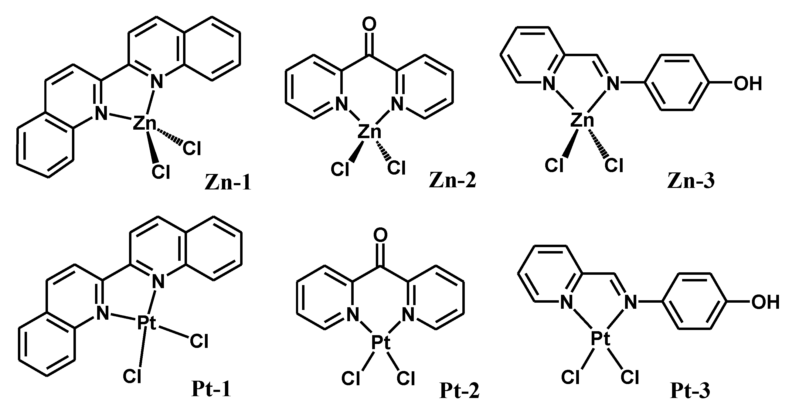
All reactions were performed in oven-dried glassware using the standard Schlenk technique under an inert gas atmosphere, and performed utilizing solvents dried over A4 molecular sieves. All reagents were obtained commercially and used as received without further purification. All chemicals were purchased from Sigma Aldrich and used without further purification. The Schiff base ligand 4-((pyridine-2-ylmethylene)amino)phenol (L) was synthesized according to a previously reported procedure [32]. ZnCl2(2,2’-biquinoline) (Zn-1), ZnCl2(2,2’-dipyridylketone) (Zn-2) and ZnCl2(L) (Zn-3) (Figure 2) were synthesized by refluxing equimolar amounts of ZnCl2 and the ligands in absolute ethanol (minimum amounts of chloroform could be used to solubilize the ligand in the reaction mixture). The products were precipitated form the reaction mixture in a few minutes. PtCl2(2,2’-biquinoline) (Pt-1), PtCl2(2,2’-dipyridylketone) (Pt-2) and PtCl2(L) (Pt-3) (Figure 2) were synthesized by stirring equimolar amounts of PtCl2(dmso)2 and the ligands in dichloromethane. The products were precipitated by the addition of diethylether. 1H NMR data of the complexes are available in the supporting information (Figures S1–S6). Full characterizations and detailed procedures are presented in previously published works [32,33,34,35,36,37].

2.2. Methods and Instrumentation

Electronic absorption spectroscopy was collected using a Genesys-10s UV-VIS spectrophotometer and 1 cm path-length quartz cells; bands are reported in the form wavelength (nm). 1H NMR (850 MHz) spectra were obtained in DMSO-D6 solutions using a Bruker Avance 850 MHz spectrometer.

2.3. DNA Binding Studies

Absorption spectra of the CT-DNA in the presence of different compounds were recorded, subtracting the compound absorption. Six solutions in 5 mM Tris-HCl (pH = 7.4) and 50 mM NaCl were synthesized by maintaining the CT-DNA at 20 µM (5% v/v DMSO was used to support the solubility of the complexes in aqueous media) and varying the ratio of [compounds]/[CT-DNA] (from 0 to ~0.1). The quantitative affinities of the compounds toward CT-DNA are expressed in terms of the intrinsic-binding constant (Kb), which can be calculated from the Benesi–Hildebrand equation:
([compound])/((εA − εF)) = ([compound])/((εB − εF)) + 1/Kb (εB − εF)
where εA, εB and εF correspond to the extinction coefficient for the CT-DNA at the specific addition of the compound, before the addition of the compound, and at the fully bound mode, respectively. From the plotting of [compound]/(εA − εF) versus [compound], Kb is obtainable by dividing the slope on the intercept [38]. The viscosity measurements were established by utilizing an Ostwald viscometer. The mode of binding of each complex was evaluated by micro-additions (10 µL) of the complex to a CT-DNA/buffer solution while maintaining the [complex]/[DNA] ratio in the range 0.02 to 0.2. The solutions were allowed to stand for 15 min at 25 °C in a water bath before measurements. The flow times of each addition to the solutions were recorded and replicated four times. The relative viscosity (η/ηo)1/3 values were plotted against [complex]/[DNA], where ηo and η represent the specific viscosity of the CT-DNA and the CT-DNA-complex adduct, respectively. The specific viscosity values, η and ηo, were obtained using the formulation [(t − tb)/tb], where t is the observed flow time and tb is the buffer flow time [38,39].

2.4. Molecular Docking Studies

Molecular docking simulations were performed in the Molecular Operating Environment (MOE) 2008.10 (Chemical Computing Group Inc., Montreal, QC, Canada, 2008). The docking scores were attained in two steps: using the London dG scoring function in MOE software; and improved utilizing two unrelated refinement methods. To confirm that the refined poses of the complexes were of acceptable geometry, Grid min pose and Force-field were employed. Bond rotations were restricted to avoid changes in the geometry while examining the best five binding positions. To evaluate the binding free energy of the compounds against DNA, docking was performed for positions of the complexes and the co-crystallized structure of the B-DNA (RSCP PDB code: 1BNA); RMSD values were employed to assess the best binding position.

2.5. Anticancer Activity and Cytotoxicity

The cells were sourced from the Egyptian Holding Company for Biological Products and Vaccines (VACSERA), Giza, Egypt. The cells were maintained in the tissue culture unit and grown in RBMI-1640 medium, supplemented with 10% heat-inactivated FBS, 50 units/mL of penicillin and 50 mg/mL of streptomycin, maintaining them in a humidified atmosphere containing 5% CO2 [40,41]. Serial sub-culturing was used to keep the cells as a monolayer culture. Cell culture reagents were sourced from Lonza (Basel, Switzerland). The antitumor properties of the examined complexes were assessed against COLO 205 [colon cancer], RCC-PR [kidney cancer], and LLC-MK2 [normal] cell lines. Cytotoxicity was identified using the sulforhodamine B (SRB) assay method, as described in the literature [42]. Exponentially growing cells were gathered, employing 0.25% Trypsin–EDTA, and seeded in 96-well plates at 1000–2000 cells/well in RBMI-1640-supplemented medium for 24 h. Cells were incubated for 72 h with various concentrations of the examined compounds in DMSO. Then, the cells were set up with 10% trichloroacetic acid for 1 h at 4 °C. Wells were stained for 10 min at ambient temperature with 0.4% SRBC (Sulphorhodamine B) dissolved in 1% CH3COOH. After drying the plates in air for 24 h, the dye was solubilized with Tris-HCl for 5 min on a shaker at 1600 rpm. The optical density (OD) of each well was quantified spectrophotometrically at 564 nm with an ELISA microplate reader (ChroMate-4300, FL, USA). The minimum inhibitory concentration (IC50) values were evaluated according to the equation for the Boltzmann sigmoidal concentration response curve using the nonlinear regression fitting models (Graph Pad, Prism Version 9).

3. Results

3.1. DNA-Binding Studies

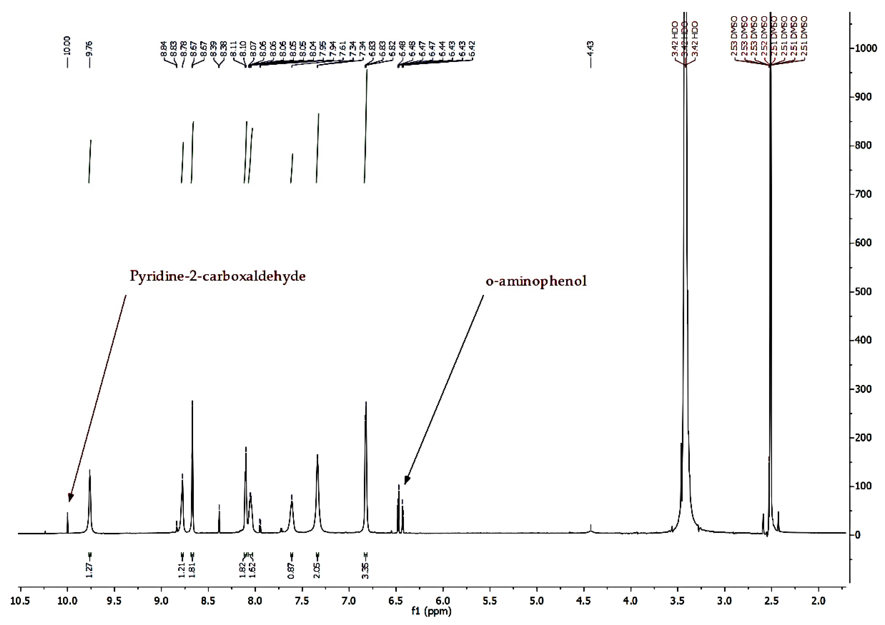
The current study was undertaken to evaluate the DNA-binding capabilities of tetrahedral zinc(II) diimine. Analogue platinum(II) complexes were tested as benchmarks, motivated by the fact that several platinum(II) complexes are known for their strong antitumor activities originating from targeting DNA. The insolubility of our complexes in aqueous medium necessitated the use of minimum amounts of DMSO to prepare solutions for biological studies. The stabilities of our complexes in DMSO and DMSO/buffer mixture are important for their biological applications. Tetrahedral zinc(II) complexes are quite labile and can undergo ligand substitution. Hence, their stabilities were justified in DMSO and D2O/DMSO mixtures over time. Specifically, 1H NMR data for all the complexes were collected after 72 h in DMSO and in D2O/DMSO, without any indication of degradation. However, Zn-3 and Pt-3 showed signs of hydrolysis after 24 h on the Schiff base ligand, but the hydrolysis products were estimated to be around 8% over 72 h, as illustrated in Figure 3. Further confirmation was attained by measuring the conductivity of the complexes in DMSO and DMSO/H2O; no conductivity was observed, which demonstrates there was no chloro/solvent exchange.
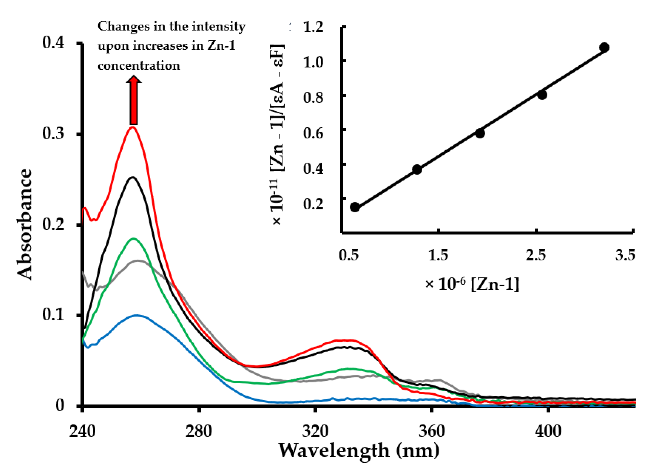
Upon the titration process described in the experimental section, spectroscopic changes in the absorption band of CT-DNA at ca. 270 nm were used to determine intrinsic-binding constants (Kb) of the complexes (Figure 4). The binding affinities provide information on the strength of the interaction with DNA, resulting from the different interaction modes (Table 1). In general, the binding affinities of the zinc complexes and their analogue platinum complexes were within a narrow range, highlighting their strong binding to CT-DNA. Apparently, the tetrahedral geometry of zinc(II) does not causing any notable steric hinderance, which explains the proximity of the results to those of square planar analogue platinum(II) complexes. The best binding affinities were obtained for Zn-1 and Pt-1, which contain the 2,2’-biquinoline ligand, which has the capability to establish π–π interactions with nucleobases of the DNA; the binding constants for most of our complexes were within the range of classical intercalators (104–107) [20,43,44].
The trend in the changes in viscosity of DNA upon interacting with a compound can indicate its mode of interaction with DNA. If the compound interacts electrostatically, no change in the relative viscosity of the DNA solution would be observed [32]. In a similar manner, groove-binder compounds do not affect the relative viscosity of the DNA. In contrast, intercalating compounds produce an elongation of the DNA polymer, leading to an increase in relative viscosity [39]. Partial intercalators cause kinking of the DNA chain, which decreases the relative viscosity [45]. Similarly, molecule binding covalently decrease the relative viscosity of the DNA solution by unwinding the DNA double-helix [46]. Therefore, viscosity measurements were performed for our complexes and ethidium bromide (as a positive control) to identify the modes of binding (Figure 5). From the trends in the changes in the relative viscosity, all the complexes induced an increase in the relative viscosity of CT-DNA, which suggests their binding via intercalation. However, the increase in the relative viscosity of CT-DNA seems to be ligand-dependent (Figure 5). The highly π-delocalized planar ligand (2,2’-biquinoline) induced the strongest increase in the relative viscosity compared to the other ligands, whereas the impact of the metal fragment was negligible.

3.2. Docking Studies

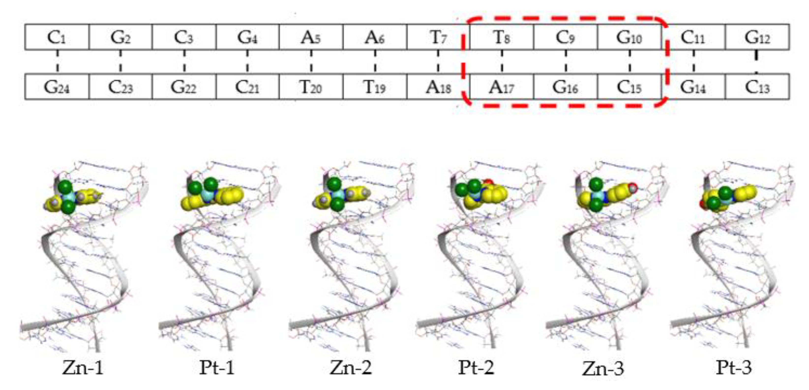
Docking studies were directed to identify the possible sites and the possible modes of interactions, to aid rationalizing the binding information obtained by the DNA-binding measurements. A cartoon representation of the docked B-DNA is illustrated in Figure 6, with approximate binding sites of Zn-1, Zn-2, Zn-3, Pt-1, Pt-2, and Pt-3 as obtained by the docking study, showing that all the studied complexes intercalated in the major groove. The binding scores are listed in Table 1; the docking scores for 2,2’-biquinoline complexes showed that Zn-1 has a higher score than that of Pt-1, which is in agreement with the experimentally measured binding affinities. Docking modelling suggests that Zn-1 can establish π–H interactions with guanine (G16) and adenine (A17), whereas Pt-1 does not show any notable stacking interaction by docking, due to the slight out-of-plane twist caused by the steric hinderance (hydrogens of the biquinoline with the chloro ligands). For the ligand 2,2’-dipyridylketone, the presence of the carbonyl group allows the ligand to establish hydrogen bonding with the nucleobases. Zn-2 forms a hydrogen bond with N2 in guanine (G10), whereas Pt-2 forms it with N2 in guanine (G14). The slight difference in the position is due to the difference in geometrical structure between the two complexes. Zn-3 and Pt-3 bind to B-DNA in almost the same position; however, Zn-3 establishes a hydrogen–acceptor interaction.

3.3. Anticancer Activities

The aim of the current study was to explore the potential of dichloro(diimine)zinc(II) in chemotherapy in comparison to dichloro(diimine)platinum(II) complexes. The complexes were examined against human colon adenocarcinoma (COLO 205) and human kidney clear cell carcinoma (RCC-PR) cancer cell lines, as well as a normal rhesus monkey kidney epithelial (LLC-MK2) cell line. Cisplatin and sunitinib were chosen as benchmark drugs; cisplatin is structurally relevant to the currently studied compounds, whereas sunitinib is an effective drug for treating kidney cancer. Scanning through the minimum inhibitory concentration (IC50) values (Table 2), zinc complexes have much lower cytotoxicity than their platinum analogues and cisplatin against COLO 205; however, their cytotoxicity is similar to that of sunitinib. Zinc complexes have slightly lower cytotoxicity than their platinum analogues, but their IC50 values are 20% to 40% higher than that of cisplatin, and 50% to 75% higher than that of sunitinib. The cytotoxicity of the zinc complexes towards the normal cell line was lower than that of their platinum counterparts, cisplatin and sunitinib. The selectivity index (SI) was calculated according to the equation (SI = IC50 against normal cell line ÷ IC50 against cancer cell lines) [47]: SI values were in the range 40–77 for zinc complexes, whereas it was in the range 20–32 for platinum complexes. Compared to cisplatin (SI = ~32) and sunitinib (SI = ~60), zinc complexes have comparable or better selectivity toward cancer cell lines over normal cell lines.

4. Conclusions

Several metal diimine complexes have been reported to exhibit anticancer properties. Our current work was dedicated to exploring the potential of tetrahedral zinc(II) diimine complexes in chemotherapy. Studies have suggested that the tetrahedral structure of zinc(II) is more favored in biological systems than octahedral structures [48]. Three dichloro(diimine)zinc(II) and platinum(II) complexes were synthesized. In general, the DNA-binding properties, as expressed by the intrinsic binding constants of the zinc(II) complexes and their analogue platinum(II) complexes, are within a narrow range, highlighting their strong binding to CT-DNA by intercalation. Apparently, the tetrahedral geometry of the zinc(II) and square planar geometry of platinum(II) are sterically similar which explain the proximity of the results. As expected, the highly planar ligand 2,2’-biquinoline produces complexes with high capability to establish π–π interactions with nucleobases of the DNA and cause optimum binding conditions. The six complexes and two reference drugs (cisplatin and sunitinib) were examined against two cancer cell lines and one normal cell line; zinc(II) complexes showed better anticancer properties than their platinum(II) counterparts. Moreover, zinc(II) complexes have comparable cytotoxicity to and higher selectivity index values than the reference drugs. The current study highlights the strong potential of tetrahedral zinc(II) complexes for cancer treatment, which could motivate further research in the near future to identify their mechanisms of action.

Supplementary Materials

The following are available online at https://www.mdpi.com/article/10.3390/chemistry3040086/s1, 1H NMR data (Figures S1–S6); IR data (Figures S7–S12); DNA–compound titration curves to obtain binding affinities of the compounds toward ct-DNA (Figures S13–S17); 2D illustrations of interactions of different compounds with B-DNA as predicted by MOE (Figures S18 and S19).

Author Contributions

Conceptualization, B.A.B.; Data curation, D.D. and M.H.A.; Formal analysis, D.D., M.H.A. and M.A.H.; Funding acquisition, B.A.B.; Investigation, B.A.B., D.D., M.H.A. and M.A.H.; Methodology, B.A.B., M.H.A. and M.A.H.; Project administration, B.A.B.; Resources, B.A.B.; Software, M.H.A. and M.A.H.; Supervision, B.A.B.; Visualization, B.A.B. and M.A.H.; Writing—Original draft, B.A.B.; Writing—Review and editing, B.A.B. All authors have read and agreed to the published version of the manuscript.

Funding

This research received no external funding.

Data Availability Statement

The data presented in this study are available on request from the authors.

Acknowledgments

B.A.B. and M.A.H. would like to thank K.A.U. technical and financial support.

Conflicts of Interest

The authors declare no conflict of interest.

Abbreviations

CT-DNAcalf-thymus DNA
COLO 205human colon adenocarcinoma cancer cell line
RCC-PRhuman kidney clear cell carcinoma cancer cell line
LLC-MK2rhesus monkey kidney epithelial normal cell line

References

  1. Wang, D.; Lippard, S.J. Cellular processing of platinum anticancer drugs. Nat. Rev. Drug Discov. 2005, 4, 307–320. [Google Scholar] [CrossRef]
  2. Pasetto, L.M.; Andrea, M.R.D.; Brandes, A.A.; Rossi, E.; Monfardini, S. The development of platinum compounds and their possible combination. Crit. Rev. Oncol. Hematol. 2006, 60, 59–75. [Google Scholar] [CrossRef]
  3. Wong, E.; Giandomenico, C.M. Current Status of Platinum-Based Antitumor Drugs. Chem. Rev. 1999, 99, 2451–2466. [Google Scholar] [CrossRef]
  4. Rabik, C.A.; Dolan, M.E. Molecular mechanisms of resistance and toxicity associated with platinating agents. Cancer Treat. Rev. 2007, 33, 9–23. [Google Scholar] [CrossRef] [Green Version]
  5. Heffeter, P.; Jungwirth, U.; Jakupec, M.; Hartinger, C.; Galanski, M.; Elbling, L.; Micksche, M.; Keppler, B.; Berger, W. Resistance against novel anticancer metal compounds: Differences and similarities. Drug Resist. Updates 2008, 11, 1–16. [Google Scholar] [CrossRef]
  6. Schatzschneider, U. Chapter 14: Metallointercalators and metalloinsertors: Structural requirements for DNA recognition and anticancer activity. In Metallo-Drugs: Development and Action of Anticancer Agents; Sigel, A., Sigel, H., Freisinger, E., Sigel, R.K.O., Eds.; De Gruyter: Berlin, Germany; Boston, MA, USA, 2018; pp. 387–436. [Google Scholar]
  7. Pages, B.J.; Ang, D.L.; Wright, E.P.; Aldrich-Wright, J.R. Metal complex interactions with DNA. Dalton Trans. 2015, 44, 3505–3526. [Google Scholar] [CrossRef]
  8. Lo, K.K.-W. Luminescent Rhenium (I) and Iridium (III) Polypyridine Complexes as Biological Probes, Imaging Reagents, and Photocytotoxic Agents. Acc. Chem. Res. 2015, 48, 2985–2995. [Google Scholar] [CrossRef] [PubMed]
  9. Sullivan, M.P.; Holtkamp, H.U.; Meier, S.M.; Hartinger, C.G. Chapter 10: The Analysis of Therapeutic Metal Complexes and Their Biomolecular Interactions. In Inorganic and Organometallic Transition Metal Complexes with Biological Molecules and Living Cells; Lo, K.K.-W., Ed.; Academic Press: Cambridge, MA, USA, 2017; pp. 355–386. [Google Scholar]
  10. Simpson, D.H.; Scott, P. Chapter 7: Antimicrobial Metallodrugs. In Inorganic and Organometallic Transition Metal Complexes with Biological Molecules and Living Cells; Lo, K.K.-W., Ed.; Academic Press: Cambridge, MA, USA, 2017; pp. 205–243. [Google Scholar]
  11. D’Cruz, O.J.; Uckun, F.M. Metvan: A novel oxovanadium (IV) complex with broad spectrum anticancer activity. Expert. Opin. Investig. Drugs 2002, 11, 1829–1836. [Google Scholar] [CrossRef] [PubMed]
  12. Scrivens, P.J.; Alaoui-Jamali, M.A.; Giannini, G.; Wang, T.; Loignon, M.; Batist, G.; Sandor, V.A. Cdc25A-inhibitory properties and antineoplastic activity of bisperoxovanadium analogues. Mol. Cancer Ther. 2003, 2, 1053–1059. [Google Scholar] [PubMed]
  13. Zhang, S.; Zhu, Y.; Tu, C.; Wei, H.; Yang, Z.; Lin, L.; Ding, J.; Zhang, J.; Guo, Z. A novel cytotoxic ternary copper (II) complex of 1,10-phenanthroline and l-threonine with DNA nuclease activity. J. Inorg. Biochem. 2004, 98, 2099–2106. [Google Scholar] [CrossRef] [PubMed]
  14. Rajendiran, V.; Karthik, R.; Palaniandavar, M.; Stoeckli-Evans, H.; Periasamy, V.S.; Akbarsha, M.A.; Srinag, B.S.; Krishnamurthy, H. Mixed-ligand copper (II)-phenolate complexes: Effect of coligand on enhanced DNA and protein binding, DNA cleavage, and anticancer activity. Inorg. Chem. 2007, 46, 8208–8221. [Google Scholar] [CrossRef] [PubMed]
  15. Nagababu, P.; Barui, A.K.; Thulasiram, B.; Devi, C.S.; Satyanarayana, S.; Patra, C.R.; Sreedhar, B. Antiangiogenic Activity of Mononuclear Copper (II) Polypyridyl Complexes for the Treatment of Cancers. J. Med. Chem. 2015, 58, 5226–5241. [Google Scholar] [CrossRef]
  16. Mashat, K.; Babgi, B.A.; Hussien, M.A.; Arshad, M.N.; Abdellattif, M. Synthesis, structures, DNA-binding and anticancer activities of some copper(I)-phosphine complexes. Polyhedron 2019, 158, 164–172. [Google Scholar] [CrossRef]
  17. Babgi, B.A.; Mashat, K.H.; Abdellattif, M.H.; Arshad, M.N.; Alzahrani, K.A.; Asiri, A.M.; Du, J.; Humphrey, M.G.; Hussien, M.A. Synthesis, structures, DNA-binding, cytotoxicity and molecular docking of CuBr (PPh3)(diimine). Polyhedron 2020, 192, 114847. [Google Scholar] [CrossRef]
  18. Alsaedi, S.; Babgi, B.A.; Abdellatif, M.H.; Arshad, M.N.; Emwas, A.; Jaremko, M.; Humphrey, M.G.; Asiri, A.M.; Hussein, M.A. DNA-Binding and Cytotoxicity of Copper(I) Complexes Containing Functionalized Dipyridylphenazine Ligands. Pharmaceutics 2021, 13, 764. [Google Scholar] [CrossRef]
  19. Mital, M.; Ziora, Z. Biological applications of Ru (II) polypyridyl complexes. Coord. Chem. Rev. 2018, 375, 434–458. [Google Scholar] [CrossRef]
  20. Li, X.; Gorle, A.K.; Sundaraneedi, M.K.; Keene, F.R.; Collins, J.G. Kinetically-inert polypyridylruthenium (II) complexes as therapeutic agents. Coord. Chem. Rev. 2018, 375, 134–147. [Google Scholar] [CrossRef]
  21. Kapdi, A.R.; Fairlamb, I.J.S. Anti-cancer palladium complexes: A focus on PdX2L2, palladacycles and related complexes. Chem. Soc. Rev. 2014, 43, 4751–4777. [Google Scholar] [CrossRef]
  22. Darabi, F.; Hadadzadeh, H.; Simpson, J.; Shahpiri, A. A water-soluble Pd(ii) complex with a terpyridine ligand: Experimental and molecular modeling studies of the interaction with DNA and BSA; and in vitro cytotoxicity investigations against five human cancer cell lines. New J. Chem. 2016, 40, 9081–9097. [Google Scholar] [CrossRef]
  23. Ott, I. On the medicinal chemistry of gold complexes as anticancer drugs. Coord. Chem. Rev. 2009, 253, 1670–1681. [Google Scholar] [CrossRef]
  24. Messori, L.; Abbate, F.; Marcon, G.; Orioli, P.; Fontani, M.; Mini, E.; Mazzei, T.; Carotti, S.; O’Connell, T.; Zanello, P. Gold (III) Complexes as Potential Antitumor Agents: Solution Chemistry and Cytotoxic Properties of Some Selected Gold (III) Compounds. J. Med. Chem. 2000, 43, 3541–3548. [Google Scholar] [CrossRef] [PubMed]
  25. Markov, D.; Naryshikina, T.; Mustaev, A.; Severinov, K. A zinc-binding site in the largest subunit of DNA-dependent RNA polymerase is involved in enzyme assembly. Genes Dev. 1999, 13, 2439–2448. [Google Scholar] [CrossRef] [PubMed] [Green Version]
  26. Choi, S.; Cui, C.; Luo, Y.; Kim, S.H.; Ko, J.K.; Huo, X.; Ma, J.; Fu, L.W.; Souza, R.F.; Korichneva, I.; et al. Selective inhibitory effects of zinc on cell proliferation in esophageal squamous cell carcinoma through Orai1. FASEB J. 2018, 32, 404–416. [Google Scholar] [CrossRef] [PubMed] [Green Version]
  27. Fong, L.Y.; Nguyen, V.T.; Farber, J.L. Esophageal cancer prevention in zinc-deficient rats: Rapid induction of apoptosis by replenishing zinc. J. Natl. Cancer Inst. 2001, 93, 1525–1533. [Google Scholar] [CrossRef]
  28. Asadizadeh, S.; Amirnasr, M.; Tirani, F.F.; Mansouri, A.; Schenk, K. DNA-BSA interaction, cytotoxicity and molecular docking of mononuclear zinc complexes with reductively cleaved N2S2 Schiff base ligands. Inorg. Chim. Acta. 2018, 483, 310–320. [Google Scholar] [CrossRef]
  29. Vikneswaran, R.; Eltayeb, N.E.; Ramesh, S.; Yahya, R. New alicyclic thiosemicarbazone chelated zinc (II) antitumor complexes: Interactions with DNA/protein, nuclease activity and inhibition of topoisomerase-I. Polyhedron 2016, 105, 89–95. [Google Scholar] [CrossRef]
  30. Kovala-Demertzi, D.; Yadav, P.N.; Wiecek, J.; Skoulika, S.; Varadinova, T.; Demertzis, M.A. Zinc (II) complexes derived from pyridine-2-carbaldehyde thiosemicarbazone and (1E)-1-pyridin-2-ylethan-1-one thiosemicarbazone. Synthesis, crystal structures and antiproliferative activity of zinc (II) complexes. J. Inorg. Biochem. 2006, 100, 1558–1567. [Google Scholar] [CrossRef]
  31. Arion, V.B. Coordination chemistry of S-substituted isothiosemicarbazides and isothiosemicarbazones. Coord. Chem. Rev. 2019, 387, 348–397. [Google Scholar] [CrossRef]
  32. Al-Khathami, N.D.; Al-Rashdi, K.S.; Babgi, B.A.; Hussien, M.A.; Arshad, M.N.; Eltayeb, N.E.; Elsilk, S.E.; Lasri, J.; Basaleh, A.S.; Al-Jahdali, M. Spectroscopic and biological properties of platinum complexes derived from 2-pyridyl Schiff bases. J. Saudi Chem. Soc. 2019, 23, 903–915. [Google Scholar] [CrossRef]
  33. Muranishi, Y.; Wang, Y.; Odoko, M.; Okabe, N. (2,2’-Biquinoline-κ2N,N′)dichloropalladium(II), -copper(II) and -zinc(II). Acta Crystallograph. C: Cryst. Struct. Commun. 2005, C61, 307–310. [Google Scholar] [CrossRef]
  34. Ivanov, M.A.; Puzyk, M.V.; Balashev, K.P. Spectroscopic and electrochemical properties of dichlorobiquinoline complexes of Au (III), Pt (II), and Pd (II). Opt. Spectrosc. 2006, 101, 105–108. [Google Scholar] [CrossRef]
  35. Ishaq, M.; Baghlaf, A.O.; Al-Mouty, A.; Al-Dousry, M. Preparation and characterization of 2,2’-dipyridylamine, 2,2’-dipyridylketone and 2,2’-dithiodipyridine complexes of copper (II), zinc (II), nickel (II) and uranium (VI) metal ions. J. Chem. Soc. Pak. 1992, 14, 115–117. [Google Scholar]
  36. Annibale, G.; Canovese, L.; Cattalini, L.; Natile, G.; Biagini-Cingi, M.; Manotti-Lanfredi, A.M.; Tiripicchio, A. Reversible addition of protic molecules to coordinated di-2-pyridyl ketone in palladium (II), platinum (II), and gold (III) complexes. X-ray crystal structures of dichloro(dihydroxydi-2-pyridylmethane) palladium (II) and dichloro(dihydroxydi-2-pyridylmethane) gold (III) chloride. J. Chem. Soc. Dalton Trans. Inorg. Chem. 1981, 12, 2280–2287. [Google Scholar]
  37. Roy, A.S.; Saha, P.; Mitra, P.; Maity, S.S.; Ghosh, S.; Ghosh, P. Unsymmetrical diimine chelation to M(II) (M = Zn, Cd, Pd): Atropisomerism, pi-pi stacking and photoluminescence. Dalton Trans. 2011, 40, 7375–7384. [Google Scholar]
  38. Kalantari, R.; Asadi, Z. DNA/BSA binding of a new oxovanadium (IV) complex of glycylglycine derivative Schiff base ligand. J. Mol. Struct. 2020, 1219, 128664. [Google Scholar] [CrossRef]
  39. Seferoğlu, Z.; Mahmoud, M.M.A.; Ihmels, H. Studies of the binding interactions of dicationic styrylimidazo [1, 2-a] pyridinium dyes with duplex and quadruplex DNA. Dyes Pigment. 2016, 125, 241–248. [Google Scholar] [CrossRef]
  40. Muanza, D.N.; Kim, B.W.; Euler, K.L.; Williams, L. Antibacterial and Antifungal Activities of Nine Medicinal Plants from Zaire. Int. J. Pharmacog. 1994, 32, 337–345. [Google Scholar] [CrossRef]
  41. Pezzuto, J.M.; Che, C.-T.; McPherson, D.D.; Zhu, J.-P.; Topcu, G.; Erdelmeier, C.A.J.; Cordell, G.A. DNA as an Affinity Probe Useful in the Detection and Isolation of Biologically Active Natural Products. J. Natur. Prod. 1991, 54, 1522–1530. [Google Scholar] [CrossRef]
  42. Skehan, P.; Storeng, R.; Scudiero, D.; Monks, A.; McMahon, J.; Vistica, D.; Warren, J.; Bokesch, H.; Kenney, S.; Boyd, M. New Colorimetric Cytotoxicity Assay for Anticancer-Drug Screening. J. Natl. Cancer Inst. 1990, 82, 1107–1112. [Google Scholar] [CrossRef] [PubMed]
  43. Rescifina, A.; Zagni, C.; Varrica, M.G.; Pistara, V.; Corsaro, A. Recent advances in small organic molecules as DNA intercalating agents: Synthesis, activity, and modeling. Eur. J. Med. Chem. 2014, 74, 95–115. [Google Scholar] [CrossRef] [PubMed]
  44. Barone, G.; Terenzi, A.; Lauria, A.; Almericom, A.M.; Leal, J.M.; Busto, N.; Garcia, B. DNA-binding of nickel (II), copper (II) and zinc (II) complexes: Structure–affinity relationships. Coord. Che. Rev. 2013, 257, 2848–2862. [Google Scholar] [CrossRef]
  45. Selvakumar, B.; Rajendiran, V.; Maheswari, P.U.; Stoeckli-Evans, H.; Palaniandavar, M. Structures, spectra, and DNA-binding properties of mixed ligand copper (II) complexes of iminodiacetic acid: The novel role of diimine co-ligands on DNA conformation and hydrolytic and oxidative double strand DNA cleavage. J. Inorg. Biochem. 2006, 100, 316–330. [Google Scholar] [CrossRef] [PubMed] [Green Version]
  46. Busto, N.; Martínez-Alonso, M.; Leal, J.M.; Rodríguez, A.M.; Domínguez, F.; Acuña, M.I.; Espino, G.; García, B. Monomer–Dimer Divergent Behavior toward DNA in a Half-Sandwich Ruthenium (II) Aqua Complex. Antiproliferative Biphasic Ac-tivity. Organometallics 2015, 34, 319–327. [Google Scholar] [CrossRef]
  47. Peña-Morán, O.A.; Villarreal, M.L.; Álvarez-Berber, L.; Meneses-Acosta, A.; Rodríguez-López, V. Cytotoxicity, Post-Treatment Recovery, and SelectivityAnalysis of Naturally Occurring Podophyllotoxins from Bursera fagaroides var. fagaroides on BreastCancer Cell Lines. Molecules 2016, 21, 1013. [Google Scholar] [CrossRef]
  48. Dudev, T.; Lim, C. Tetrahedral vs Octahedral Zinc Complexes with Ligands of Biological Interest:  A DFT/CDM Study. J. Am. Chem. Soc. 2000, 122, 11146–11153. [Google Scholar] [CrossRef]
Figure 1. Some examples of reported polypyridyl metal complexes with potential anticancer activities.
Chemistry 03 00086 g001
Figure 2. Complexes examined in this study.
Chemistry 03 00086 g002
Figure 3. 1H NMR of Zn-3 in the DMSO/D2O solvent mixture over 72 h, showing slight amount of Schiff base hydrolysis.
Chemistry 03 00086 g003
Figure 4. Changes in the electronic spectra of CT-DNA after increased concentrations of Zn-1 (blue spectrum/0.6 µM, grey spectrum/1.3 µM, green spectrum/1.9 µM, black spectrum/2.6 µM and red spectrum/3.2 µM) and the plotting of [Zn-1]/[εA − εF] against [Zn-1] to determine the binding affinity.
Chemistry 03 00086 g004
Figure 5. Changes in the relative viscosity of CT-DNA upon the addition of the different zinc(II) complexes.
Chemistry 03 00086 g005
Figure 6. The sequence of B-DNA used in the docking, highlighting the binding site of all the complexes and 3D representations of compound–DNA adducts.
Chemistry 03 00086 g006
Table 1. DNA-binding affinities (Kb) toward ct-DNA obtained from spectroscopic titration and docking scores obtained by MOE.
CompoundKb (Intrinsic Binding Constant)Docking ScoresProposed Interaction by Docking
Zn-16.67 × 10⁶−3.45H-π (G16—biquinoline)
H-π (A17—biquinoline)
Pt-14.44 × 10⁶−3.34None
Zn-25.00 × 10⁵−4.78H-acceptor (G10—dipyridylketone)
H-π (A17—dipyridylketone)
Pt-21.00 × 10⁶−5.52H-acceptor (G14—dipyridylketone)
Zn-31.00 × 10⁶−5.42H-acceptor (G16—Cl)
Pt-35.00 × 10⁵−4.58None
Table 2. Minimum inhibitory concentrations (IC50) of the different compounds and reference drugs against COLO 205, RCCD-PR and LLC-MK2 cell lines.
CompoundCOLO 205RCC-PRLLC-MK2
Zn-19.46 ± 0.0215.54 ± 0.77695.37 ± 42.89
Pt-121.43 ± 0.0417.81 ± 0.14453.77 ± 6.99
Zn-28.10 ± 0.0814.41 ± 0.10627.17 ± 37.36
Pt-214.82 ± 0.0618.69 ± 0.04397.30 ± 12.26
Zn-38.18 ± 0.0113.54 ± 0.03534.37 ± 37.23
Pt-314.69 ± 0.0215.47 ± 0.04472.87 ± 17.81
Cisplatin12.59 ± 0.0411.07 ± 0.07378.50 ± 31.84
Sunitinib9.22 ± 0.018.74 ± 0.04548.13 ± 61.18
Publisher’s Note: MDPI stays neutral with regard to jurisdictional claims in published maps and institutional affiliations.