Article

Monofluorophosphates—New Examples and a Survey of the PO3F2− Anion

Institute for Chemical Technologies and Analytics, Division of Structural Chemistry, TU Wien, Getreidemarkt 9/164-SC, A-1060 Vienna, Austria
Chemistry 2021, 3(1), 45-73; https://doi.org/10.3390/chemistry3010005
Submission received: 14 December 2020 / Revised: 29 December 2020 / Accepted: 1 January 2021 / Published: 7 January 2021
(This article belongs to the Special Issue 2020 Profile Papers by Chemistry' Editorial Board Members)

Abstract

:
During a systematic study of monofluorophosphates, i.e., compounds comprising the tetrahedral anion PO3F2−, twelve, for the most part new, compounds were obtained from aqueous solutions. Crystal structure refinements based on single crystal X-ray diffraction data revealed the previously unknown crystal structures of CdPO3F(H2O)2, Cr2(PO3F)3(H2O)18.8, Pb2(PO3F)Cl2(H2O), (NH4)2M(PO3F)2(H2O)2 (M = Mg, Mn, Co), NH4Cr(PO3F)2(H2O)6, NH4Cu2(H3O2)(PO3F)2, (NH4)2Zn(PO3F)2(H2O)0.2, and (NH4)2Zn3(PO3F)4(H2O), as well as redeterminations of ZnPO3F(H2O)2.5 and (NH4)2Ni(PO3F)2(H2O)6. From the previously unknown crystal structures, CdPO3F(H2O)2 (space group P 1 ¯ ), Cr2(PO3F)3(H2O)18.8 (P 1 ¯ ), Pb2(PO3F)Cl2(H2O) (Pnma), NH4Cr(PO3F)2(H2O)6 (R 3 ¯ m), (NH4)2Zn(PO3F)2(H2O)0.2 (C2/c), and (NH4)2Zn3(PO3F)4(H2O) (I 4 ¯ 3d) each crystallizes in an unique crystal structure, whereas compounds (NH4)2M(PO3F)2(H2O)2 (M = Mg, Co) crystallize in the (NH4)2Cu(PO3F)2(H2O)2 type of structure (C2/m) and (NH4)2Mn(PO3F)2(H2O)2 in a subgroup thereof (P21/n, with a klassengleiche relationship of index 2), and NH4Cu2(H3O2)(PO3F)2 (C2/m) crystallizes isotypically with natrochalcite-type KCu2(H3O2)(SO4)2. A survey on the PO3F2 anion, including database entries of all inorganic compounds comprising this group, revealed mean bond lengths of P–O = 1.506(13) Å, P–F = 1.578(20) Å, and angles of O–P–O = 113.7(1.7)° and O–P–F = 104.8(1.7)°, using a dataset of 88 independent PO3F2 anions or entities. For those crystal structures of monofluorophosphates where hydrogen bonding is present, in the vast majority of cases, hydrogen bonds of the type D–H···F–P (D = O, N) are not developed.

1. Introduction

The family of monofluorophosphates comprising the PO3F2− anion was introduced by Lange more than 90 years ago [1]. In the PO3F2− anion, the fluorine atom is directly bound to the phosphorus atom. However, in the literature, compounds with discrete PO43− and F anions are also sometimes incorrectly described as “fluorophosphates”, e.g., Mn2PO4F [2] or Na2FePO4F [3]. These compounds correctly belong to the family of ‘phosphate fluorides’. Various preparation methods for monofluorophosphates as well as applications of this family of compounds as additives in toothpastes, wood preservatives, corrosion inhibitors, solubility inhibitors for lead in potable water sources, or as active agents against osteoporosis or caries during biomineralization of fluoroapatite were summarized some time ago [4]. More recently, some monofluorophosphates were also shown to exhibit excellent nonlinear optical (NLO) behaviour [5].
In his seminal paper, Lange emphasized the chemical relationships between monofluorophosphates and sulfates in terms of solubilities and reaction behaviours. In fact, the PO3F2− anion is isoelectronic with the SO42− anion, and both anions have a tetrahedral shape, as later evidenced by the very first structure determination of a monofluorophosphate [6]. Prior to this first experimental proof about the structure and shape of the PO3F2− anion, it was assumed that monofluorophosphates are isomorphic with corresponding sulfates [7]. It should be noted that also the terms isomorphic/isomorphism still are found in literature to express structural relationships, but their use is not recommended any longer [8]. More appropriate terms are isostructural/isostructurality or synonymous isotypic/isotypism. Meanwhile, numerous other monofluorophosphates were synthesized and structurally determined, but not for all reported monofluorophosphates corresponding sulfates even exist or show isotypism with existing sulfates.
The current study was undertaken to add more examples of structurally determined monofluorophosphates with inorganic cations to the already existing list of this family of compounds. Although some of the monofluorophosphates investigated during this study have previously been reported and their powder diffraction data deposited in the International Centre for Diffraction Data’s (ICDD) powder diffraction file (PDF) [9], structural details of corresponding phases are still missing up to now. As it turned out, some of the data compiled in the PDF at that time are incorrect (wrong space groups, wrong unit cell volume) and were revised during the current study. Moreover, results of the present crystal structure analyses were used under special emphasis to review structural characteristics (bond lengths and angles, point group symmetries) and hydrogen-bonding features of the PO3F2− anions as well as their structural relationships to corresponding sulfates. O and F atoms differ only in one electron and thus have very similar atomic form factors for X-rays. Consequently, a distinction of the two atom types on the basis of X-ray diffraction methods alone is not free from ambiguity, as was recently shown for minerals that were claimed to comprise monofluorophosphate groups [10]. Nevertheless, the result of the current structure evaluation for the PO3F2− anion is a useful tool to correctly assign F and O atoms in monofluorophosphates, as exemplified by using structure data of a published crystal structure with incorrectly assigned F and O atoms.

2. Materials and Methods

2.1. Syntheses and Single Crystal Growth

For syntheses of NH4+-containing monofluorophosphates, the starting compound (NH4)2PO3F(H2O) was prepared according to Schülke and Kayser [11] and its purity checked with X-ray powder diffraction (XRPD). One gram of this material was dissolved in 10 mL of a methanol/water mixture (1:1 v:v). Then, 80 mg solid AgNO3 were added to this solution to precipitate the phosphate anions present due to incomplete conversion or partial hydrolysis of the PO3F2− anion. The yellow Ag3PO4 was filtered off, and the filtrate was repeatedly checked for PO43− anions by adding a few drops of an AgNO3 solution until no more clouding was observed in the filtrate, ensuring that all PO43− anions were removed. Then, 10 mL of a solution consisting of 500 mg of the respective metal chloride in methanol/water (1:1 v:v) were added to the monofluorophosphate solution. The excessive Ag+ ions were precipitated as AgCl and filtered off. To the remaining clear filtrate 100 mL of an acetone/methanol solution (2:1 v:v) were slowly added, resulting in flocculent precipitates in all cases. The respective suspensions were stirred for one hour and then were filtered. The obtained solids were washed with methanol and acetone and then dried in an exsiccator overnight. XRPD revealed an amorphous state for the obtained materials, except for (NH4)2Mg(PO3F)2(H2O)2 that was obtained as a pure polycrystalline phase (cf. PDF entry #00-059-0045).
For single crystal growth of NH4+-containing monofluorophosphates, 100 mg of the as-precipitated solids were dissolved in 10 mL of a methanol/water mixture (1:1 v:v), to some extent under mild warming. The clear solutions were allowed to evaporate for 2–4 days until full dryness. In all cases, the majority of material was still amorphous, and only few single crystals were found to be suitable for X-ray analysis. This way, single crystals of (NH4)2M(PO3F)2(H2O)2 (M = Mg, Co, Mn), (NH4)2Ni(PO3F)2(H2O)6, NH4Cr(PO3F)2(H2O)6 and NH4Cu2(H3O2)(PO3F)2 could be obtained from the respective metal salt solution. Single crystals of Cr2(PO3F)3(H2O)18.8 were likewise harvested from the batch containing the ammonium-chromium monofluorophosphate solution. From the original ammonium zinc monofluorophosphate solution, three different types of single crystals were isolated, viz. ZnPO3F(H2O)2.5, (NH4)2Zn(PO3F)2(H2O)0.2 and (NH4)2Zn3(PO3F)4(H2O).
Single crystals of CdPO3F(H2O)2 and Pb2(PO3F)Cl2(H2O) were obtained from metathesis reactions. For this purpose, 200 mg Ag2PO3F (prepared according to [4]) were dissolved in 10 mL of water; equimolar amounts of CdCl2 and PbCl2, respectively, were added to the solution, resulting in an immediate precipitation of AgCl. The suspension was stirred for two hours, AgCl filtered off, and the filtrate allowed to evaporate until complete dryness. CdPO3F(H2O)2 was obtained as a single phase material, whereas only few single crystals of Pb2(PO3F)Cl2(H2O) could be isolated. In the latter batch, polycrystalline 2PbCO3·Pb(OH)2 was also determined by XRPD next to a dark-brown to metallic film deposited at the surface of the glass. The formation of the film points to silver that apparently was also present in the filtrate and was reduced to its metallic form during evaporation.

2.2. Single Crystal Diffraction and Structure Analysis

Single crystals were optically preselected under a polarising microscope, embedded in perfluorinated polyether for protection from air and humidity and mounted on MiTeGen MicroLoopsTM for the diffraction studies. Experimental details of the data collections and refinements are collated in Table 1.
All crystal structures were initially solved with SHELXS [12] and refined with SHELXL [13]. For the renewed refinement of ZnPO3F(H2O)2.5 and (NH4)2Ni(PO3F)2(H2O)6, the original atom labelling and atomic coordinates (as starting parameters) were resumed from the original structure reports [14,15]. For NH4Cu2(H3O2)(PO3F)2, atom labelling and coordinates were adopted from isotypic KCu2(H3O2)3(SO4)2 [16]. In cases where H atom positions were clearly discernible from difference Fourier maps, the corresponding sites were refined with soft restraints on N–H or O–H bond lengths. In cases where H atom positions could not be unambiguously located, H atoms were not considered in the final model, but are part of the chemical formula, X-ray density, etc. These cases apply to Pb2(PO3F)Cl2(H2O), Cr2(PO3F)3(H2O)18.8 and (NH4)2Zn(PO3F)2(H2O)0.2. In the crystal structure model of the chromium compound severe disorder of the free water molecules (i.e., the non-coordinating or structural water molecules) is observed, both in terms of occupational and positional disorder; the same applies for the partly hydrated zinc compound. Site occupation factors (s.o.f.) for these O sites were refined freely without restraints or constrains. Disorder was also observed for NH4Cr(PO3F)2(H2O)6 and NH4Cu2(H3O2)(PO3F)2 where the N atom of the ammonium cation is situated on a position with site symmetry 3 ¯ m and 2/m, respectively, which results in a symmetry-restricted disorder of the corresponding ammonium H atoms. Finally, in the crystal structure of (NH4)2Zn3(PO3F)4(H2O), the N site of the ammonium cation and the O site of the water molecule share the same fully occupied site with a 2/3 occupation by N and a 1/3 occupation by O.
Further details of the crystal structure investigations may be obtained from The Cambridge Crystallographic Data Centre (CCDC) on quoting the depository numbers listed at the end of Table 1. The data can be obtained free of charge via www.ccdc.cam.ac.uk/structures.
Table 2 lists selected bond lengths and angles for all crystal structures, and Table 3 gives numerical details of hydrogen bonding.

2.3. Vibrational Spectroscopy

The infrared (IR) spectrum of a powdered CdPO3F(H2O)2 sample was recorded as a KBr pellet in the spectral range between 4000 and 400 cm−1 employing a Bruker-EQUINOX-55 FTIR-instrument (Billerica, MA, USA). Raman spectra down to 100 cm−1, were measured using the FRA 106 Raman accessory of an IF66 Bruker spectrophotometer (Billerica, MA, USA). Radiation from a Nd:YAG solid-state laser (1064 nm) was used for excitation. The spectral resolution was ± 4 cm−1 in both measurements.

2.4. Thermogravimetry (TG)

A Netzsch TG209 F1 thermobalance (Selb, Germany) was used for measurement using a corundum crucible in flowing nitrogen atmosphere and a heating rate of 20 °C/min.

3. Results

In the current study, only (NH4)2Mg(PO3F)2(H2O)2 and CdPO3F(H2O)2 were obtained as pure and crystalline phases and in amounts sufficient for the application of other current analytical methods (vibrational spectroscopy, thermogravimetry). All other monofluorophosphates either were obtained in form of multi-phase material or in form of only few single crystals next to amorphous material. This allowed in all cases the determination of the crystal structure but prevented further analytical measurements.

3.1. CdPO3F(H2O)2

All atoms in the crystal structure of CdPO3F(H2O)2 are located on general sites. The cadmium cation exhibits a distorted octahedral coordination sphere defined by two cis-aligned water molecules (OW1, OW2) and four O atoms from four PO3F2− anions. Two [CdO4(OH2)2] octahedra share an edge to form a dimer {Cd2O6(OH2)4}; adjacent dimers are linked by corner-sharing with PO3F2− groups into layers extending parallel (001). An intralayer hydrogen bond between two water molecules (OW1 and OW2) consolidates this arrangement. Neighbouring layers are held together by medium-strong to weak and partly bifurcated hydrogen bonds between both water molecules and O1 and O2 atoms of the monofluorophosphate anions (Figure 1).
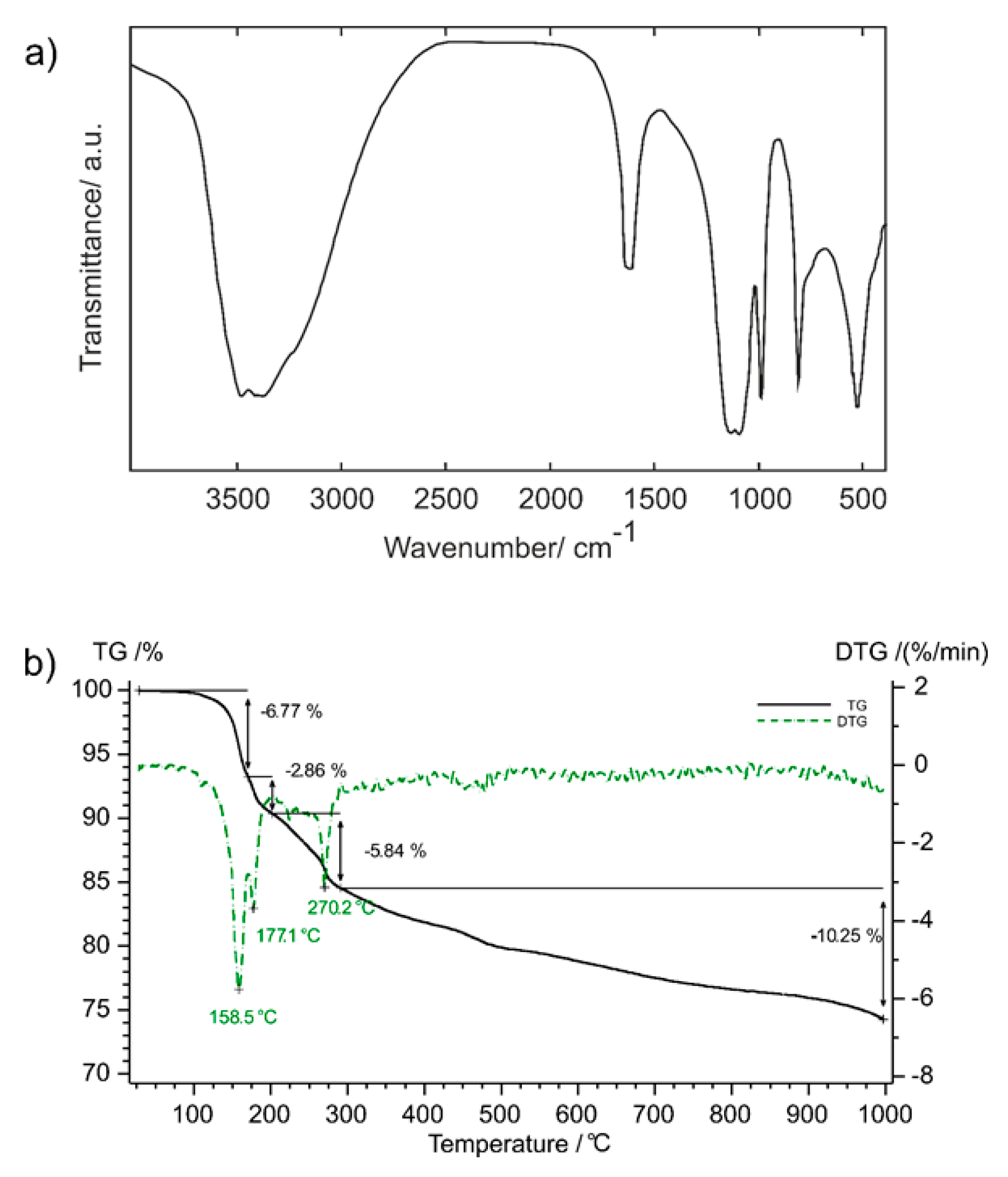
On the basis of the known structural data, it is possible to perform an analysis of the vibrational-spectroscopic behaviour of the PO3F2− anion present in the CdPO3F(H2O)2 crystal structure, using the simple site-symmetry approximation [17,18,19,20]. Since the monofluorophosphate anion is located on a general C1 position, the symmetry of the “free” PO3F2− anion (C3v) was correlated with its site symmetry (C1), as shown in Table 4. From these results, it becomes evident that, under site symmetry conditions, the three double degenerated E modes are split, and all vibrations present IR and Raman activity. The FTIR spectrum of CdPO3F(H2O)2 is quite simple (Figure 2a) and can be clearly correlated with the results of this analysis. In the Raman spectrum, a more reduced number of bands was observed which, notwithstanding, was useful to additionally support the performed assignments, which are shown in Table 4 and briefly commented on as follows:
-
Regarding the vibrations of the water molecules, the O–H stretchings are seen as a relatively broad and clearly splitted band due to the presence of two crystallographically different water molecules. The positions of these bands are characteristic for the presence of hydrogen bridges of medium strength [20], in agreement with the results of the structure analysis. Interestingly, the corresponding deformational mode, δ(H2O), shows also splitting signals.
-
The antisymmetric ν(PO3) vibration was not observed in the Raman spectrum, whereas in the IR spectrum it is very strong and broad. In accord with the predictions of the site-symmetry analysis two components can be seen. The corresponding symmetric stretching vibration is the strongest Raman band in both compounds and is also relatively strong in the IR spectrum.
-
The ν(P–F) vibration can be clearly identified in the spectra, lying at somewhat higher energy than that observed in the solution Raman spectrum (795 cm−1) [20].
-
For the deformational modes only δ(PO3) could be identified, clearly split in the IR spectra as predicted (cf. Table 4), whereas no signals for the δ(FPO3) mode could be found. In the Raman spectrum of a PO3F2− solution, both vibrations are reported at the same energy (520 cm−1) [20], although in the case of crystalline Hg2PO3F, both vibrations were identified at slightly different wavenumbers, with ν5 > ν3 [21].
-
The corresponding ν6-PO3-rocking mode was only identified in the Raman spectrum, as a very weak band.
The TG curve of CdPO3F(H2O)2 and the associated difference curve ist depicted in Figure 2b. The dihydrate starts to decompose with an onset temperature of 144 °C accompanied with a first dehydration step that can be grouped into two separated events. Considering a mass loss of 7.3% per water molecule in the formula unit, the first dehydration event is associated with the loss of about one water molecule (maximum in the difference curve at 159 °C and a mass loss of 6.8%), followed by a second event (maximum in the difference curve at 177 °C) with the release of about one-third of a water molecule (2.9%) relative to the formula unit. The second dehydration step (maximum in the difference curve at 270 °C) is indicated by the release of about two-thirds of a water molecule per formula unit with a further mass loss of 5.8%. The formation of the anhydrous compound is completed at 280 °C (expected overall mass loss 14.6%, observed 15.5%). Above this temperature, the remaining phase(s) gradually decompose(s), and Cd2P2O7 [22] was identified by PXRD as the only crystalline reaction product obtained at 1000 °C. For a clear interpretation of this last decomposition step, coupled mass-spectroscopic studies of the gaseous products released during the TG experiment and temperature-dependent PXRD would have been required. In general, monofluorophosphates show a rather complex thermal decomposition behaviour, as exemplified by the cases of (NH4)2Mg(PO3F)2(H2O)2 [23] with Mg2P4O12, of Ag2PO3F [4] with Ag4P2O7 and Ag3PO4, and of SrPO3F(H2O) [24] with Sr2P2O7 and Sr5(PO4)3F, respectively, as the final products.

3.2. Cr2(PO3F)3(H2O)18.8

The crystal structure of Cr2(PO3F)3(H2O)18.8 is rather complex, with five Cr sites (two of which (Cr1, Cr2) are situated on inversion centres on Wyckoff positions 1a and 1h, respectively), with six PO3F2− anions (one of which (P6) shows positional disorder of the F6 atom over two set of sites), with 24 O atoms associated with aqua ligands of the Cr(III) atoms, and with 14 crystal water molecules (five of which (O1W–O5W) are positionally and occupationally disordered over multiple sites).
The five chromium(III) atoms are solely ligated by water molecules in an octahedral manner and are isolated in the crystal structure. The corresponding Cr–Owater distances are normal and agree with other [Cr(OH2)6] octahedra, e.g., like those found in alums [25].
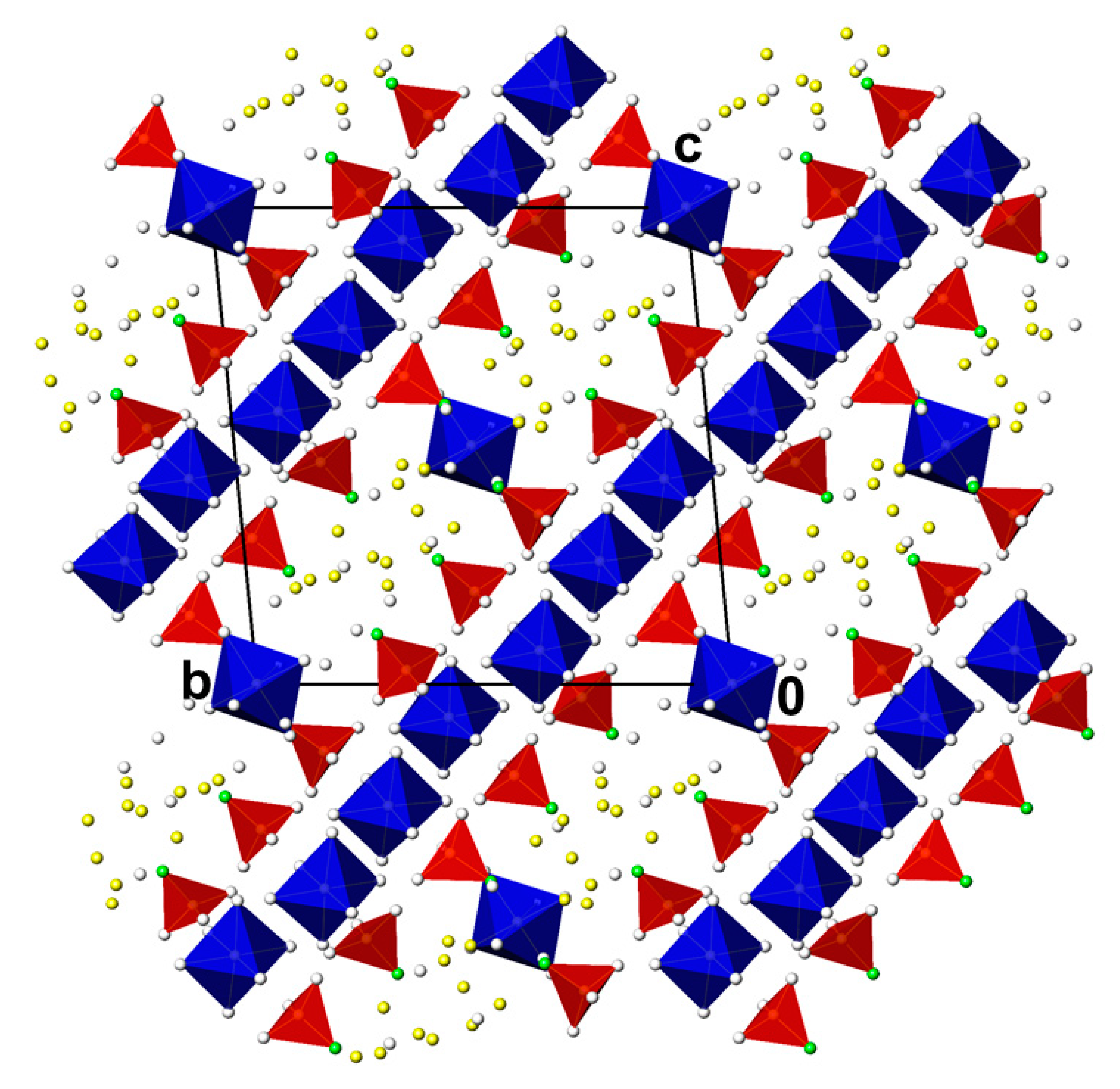
[Cr(OH2)6] octahedra associated with Cr3, Cr4, and Cr5 are arranged in (011) layers that are sandwiched by the six monofluorophosphate anions, giving an overall composition of {[Cr(OH2)6]3(PO3F)6}3−. Interestingly, all F atoms of the monofluorophosphate tetrahedra point away from the intermediate cationic layer. The two remaining [Cr(OH2)6] octahedra, associated with the two chromium sites Cr1 and Cr2, lie in-between adjacent {[Cr(OH2)6]3(PO3F)6}3− layers. Together with the partly disordered water molecules O1W-O5W they define an own layer with composition {[Cr(OH2)6](H2O)13.6}3+. Both types of layers alternate and stack along [011] (Figure 3). Although H atoms could not be located for the water molecules, it is evident that hydrogen bonding is the crucial force for stabilising the stacking arrangement in this crystal structure. Within an anionic {[Cr(OH2)6]3(PO3F)6}3− layer the aqua ligands most probably are involved in hydrogen bonding to the monofluorophosphate O atoms and also among each other; in the intermediate {[Cr(OH2)6](H2O)13.6}3+ layer hydrogen bonds might occur between aqua ligands and crystal water molecules. From the same orientation of all monofluorophosphate F atoms towards the {[Cr(OH2)6](H2O)13.6}3+ layers one might expect also O–H···F hydrogen bonds in this crystal structure. However, a clear localisation of (disordered) H atoms will be possible only by the application of neutron diffraction, provided that crystals large enough for this diffraction technique can be grown.
The formula of this compound can also be written as [Cr(H2O)6]2(PO3F)3·6.8H2O. Considering full occupancy of the positionally and occupationally disordered five crystal water sites, this would result in a ‚19-hydrate‘, i.e., [(Cr(H2O)6]2(PO3F)3·7H2O. This amount of water is close to that reported for highly hydrated violet chromium(III) sulfates that are described to contain about 18 water molecules [26]. However, crystal structure determinations of corresponding chromium(III) sulfate hydrates have not been performed up to now.

3.3. Pb2(PO3F)Cl2(H2O)

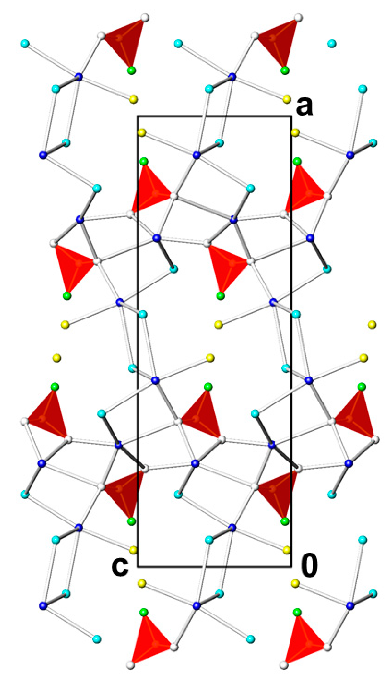
The crystal structure of Pb2(PO3F)Cl2(H2O) comprises two unique lead(II) atoms, two chloride anions, one monofluorophosphate anion and one water molecule of an aqua ligand. Except one oxygen atom of the latter (O2; H atom(s) could not be determined), all other atoms are situated on a mirror plane (Wyckoff position 4c). The two lead(II) atoms exhibit different coordination environments whereby in each case the F atom of the monofluorophosphate anion is not part of the first coordination sphere (shortest Pb–F distances are 3.624(3) Å for Pb1 and 3.912(3) Å for Pb2). Pb1 has a coordination number of 7 (considering distances less than 3.5 Å) and is bonded to two monofluorophosphate O atoms, the O atom of the aqua ligand and four chloride anions. Pb2 has a coordination number of 9, with one very short and two short distances to monofluorophosphate O atoms, two bonds to chloride anions and two pairs of long bonds to monofluorophosphates O atoms. The corresponding [Pb1O2(H2O)Cl4] and [Pb2O7Cl2] polyhedra are irregular and share vertices and edges to build up a three-dimensional framework structure (Figure 4). The water molecules protrude into the interstices present in this framework. The monofluorophosphate tetrahedron shares all its O atoms with the framework, whereby the P and F atoms are also oriented towards the interstices of the framework. The next nearest distance between two water molecules in this section of the structure amounts to 3.102(7) Å; three shorter distances to the monofluorophosphate F atom (2.848(8) Å and twice 3.007(4) Å) are also present, making O–H···O and also O–H···F hydrogen bonding interactions possible. Since H atoms could not be determined, detailed hydrogen bonding interactions cannot be provided.

3.4. ZnPO3F(H2O)2.5

The crystal structure of ZnPO3F(H2O)2.5 has been determined previously from a single crystal X-ray data set at room temperature, using a CAD-4 four-circle diffractometer equipped with a point detector. Since only parts of the water hydrogen atoms could be located at that time [14], the crystal structure model remained incomplete, in particular in terms of hydrogen-bonding interactions. The current re-refinement unambiguously revealed all hydrogen atoms, making a complete assignment of hydrogen-bonding interactions possible.
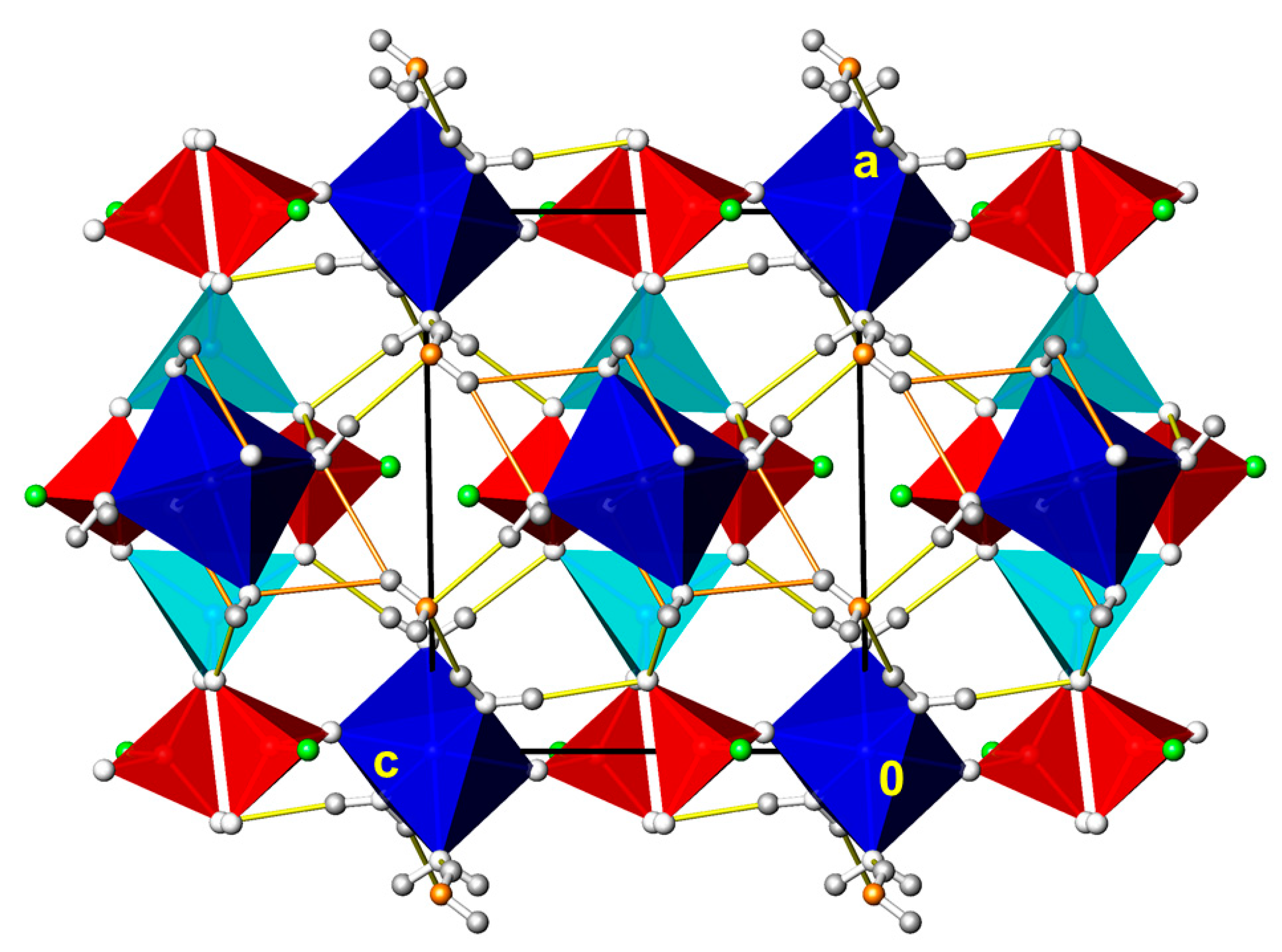
Two of the three unique zinc cations (Zn2, Wyckoff position 1h and Zn3, Wyckoff position 1a) are located on inversion centres; all other atoms are in general sites. Zn1 has a tetrahedral coordination environment and is bonded to the O atoms of four monofluorophosphate tetrahedra. Two such units dimerise into an inversion-symmetric {Zn12(PO3F)6} unit. Both Zn2 and Zn3 atoms have an octahedral coordination environment, being bound to two monofluorophosphate O and four water O atoms. The corresponding [ZnO2(OH2)4] octahedra share the non-water O atoms with the {Zn12(PO3F)6} units to define a three-dimensional framework structure. O–H···O hydrogen bonding of medium strengths between the coordinating water molecules and the monofluorophosphate O atoms reinforces this arrangement. There is an additional crystal water molecule (O2) present in the structure acting both as a donor and an acceptor group. O2 donates weak hydrogen bonds (partly bifurcated) to two ligand water O and one monofluorophosphate O atoms, and accepts medium-strong hydrogen bonds of two coordinating water molecules (Figure 5).

3.5. (NH4)2M(PO3F)2(H2O)2 (M = Co, Mg)

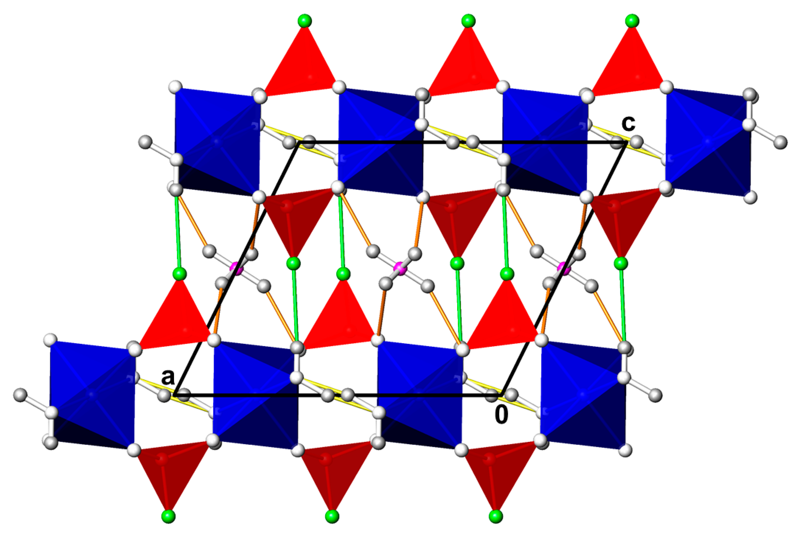
(NH4)2Co(PO3F)2(H2O)2 and (NH4)2Mg(PO3F)2(H2O)2 crystallize isotypically with the copper member (NH4)2Cu(PO3F)2(H2O)2 in space group C2/m [27]. Instead of the C-centred unit cell for this structure type, PDF entry #00-045-0355 for (NH4)2Co(PO3F)2(H2O)2 reports a primitive unit cell (without further assignment of possible space groups) with lattice parameters of a = 12.3817(1), b = 5.3449(5), c = 7.3894(6) Å, β = 98.930(8)°, V = 483.10 Å3. Comparison with the current single crystal X-ray study (Table 1) revealed virtually the same lengths of the b and c axes and the same unit cell volume. Preparation, chemical analysis as well as infra-red spectroscopic measurements and thermal behaviour of (NH4)2Mg(PO3F)2(H2O)2 were already reported some time ago, without determination of the crystal structure. The originally given lattice parameters (a = 15.476(4), b = 5.372(1), c = 13.416(3) Å, β = 118.76(1)°; determined from polycrystalline material using a Guinier camera) and two possible space groups (Cc or C2/c) [23] do not match with the current single crystal data with a halved unit cell volume (978 Å3 for [23] versus 484 Å3 in the current singly crystal study) and space group C2/m. Nevertheless, the deposited X-ray powder diffraction data (PDF entry #00-039-029) can be indexed with the actual halved cell. Rietveld refinement of (NH4)2Mg(PO3F)2(H2O)2 unambiguously showed the correctness of the halved cell in space group C2/m (PDF entry #00-059-0045; no structure data given).
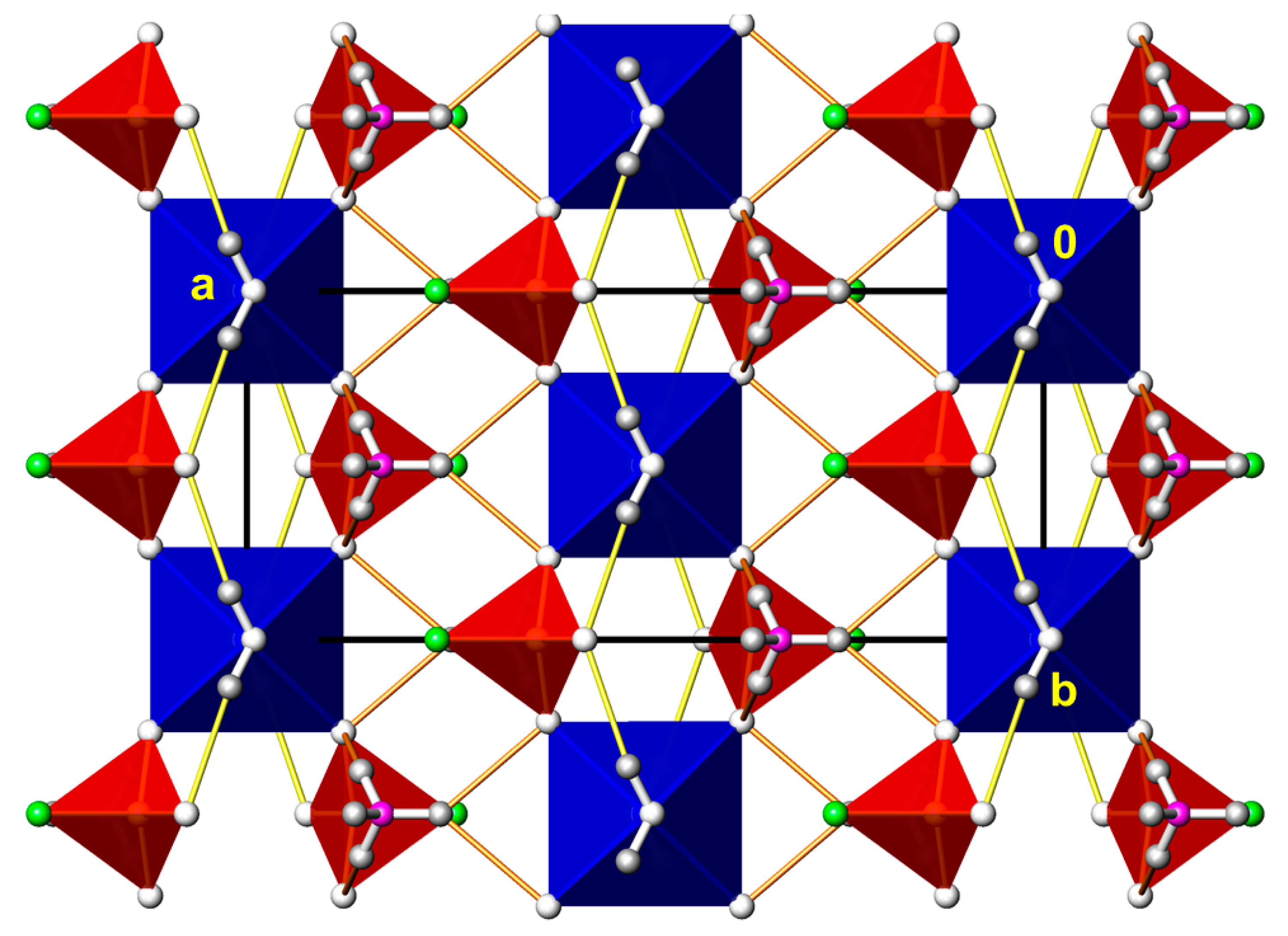
The metal cation in the (NH4)2M(PO3F)2(H2O)2 (M = Co, Cu, Mg) structure is situated on Wyckoff position 2c with site symmetry 2/m and thus has four equal M–O bonds to the O2 atoms of four PO3F2− anions and two equal bonds to two trans-aligned water molecules (O1W). The monofluorophosphate anion is located about a mirror plane, just like the ammonium cation. Adjacent [MO4(OH2)2] octahedra are linked by corner-sharing with the PO3F2− anions into {Mg(OH2)(PO3F)2)2-}n strands running along [010]. Adjacent strands are aligned in parallel and are arranged into layers along (001). Within a strand, medium-strong hydrogen bonds between the water molecules and the non-coordinating O1 atom of the PO3F2− anion are established. The ammonium cations are situated between the strands and are hydrogen-bonded to the O1 and O2 atoms of the anions into a three-dimensional network (Figure 6).

3.6. (NH4)2Mn(PO3F)2(H2O)2

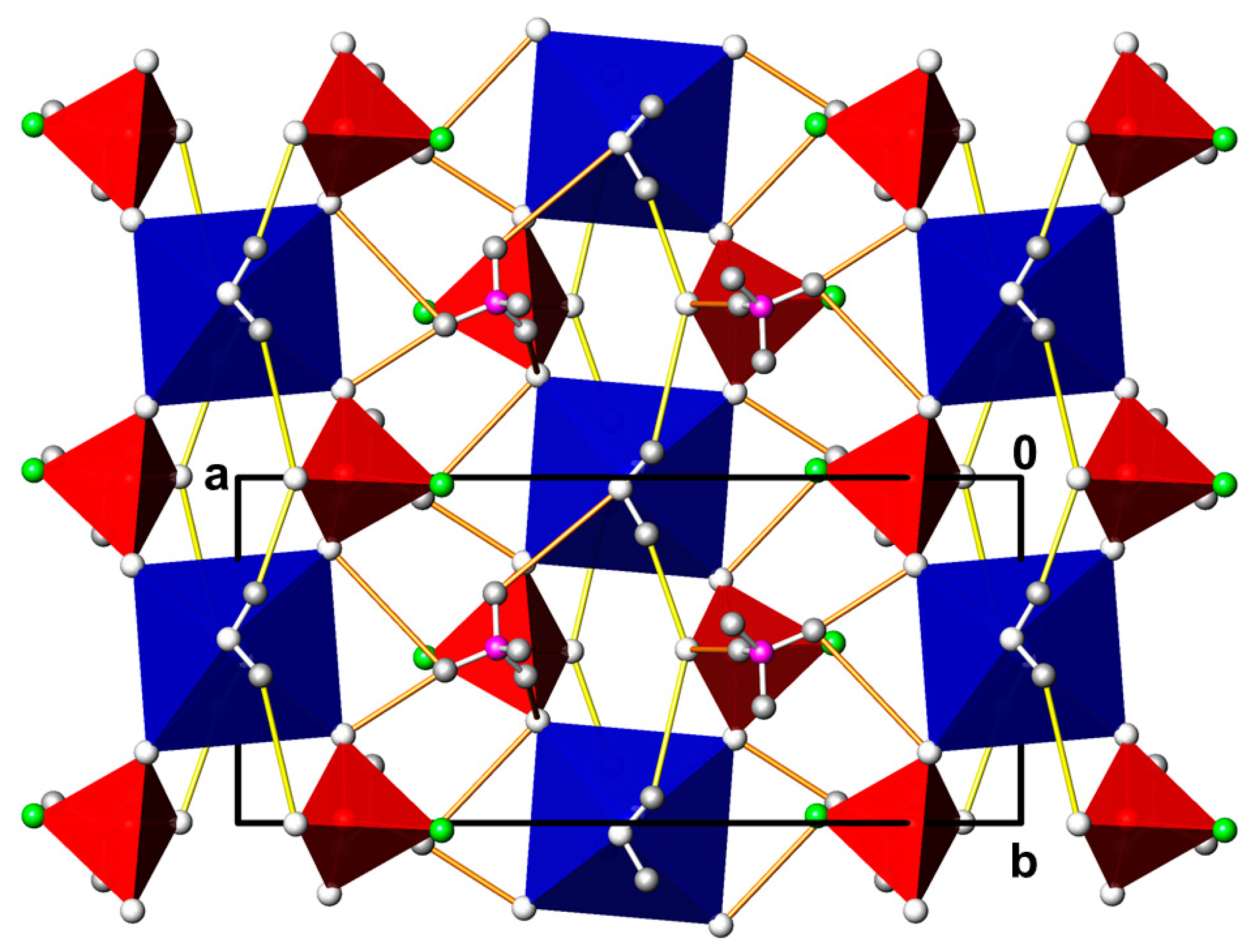
Although manganese is part of the first-row transition metals, (NH4)2Mn(PO3F)2(H2O)2 does not adopt the (NH4)2Cu(PO3F)2(H2O)2 structure type in space group C2/m described above for the transition metals cobalt and copper. The manganese compound shows a group-subgroup relation with the (NH4)2Cu(PO3F)2(H2O)2 structure type, crystallizing in space group P21/n that is a klassengleiche subgroup of index 2 [28]. Hence, some of the sites and/or groups in the higher-symmetric space group C2/m have a reduced symmetry or split into two positions in the P21/n structure. The divalent metal position (Wyckoff position 2c) now shows site symmetry 1 ¯ , and all other atoms are located on general sites (Wyckoff positions 4e). The general features of the (NH4)2Mn(PO3F)2(H2O)2 crystal structure (Figure 7) are the same as for the (NH4)2Cu(PO3F)2(H2O)2 structure type. {Mn(OH2)(PO3F)2)2-}n strands run along [010] with the water molecule hydrogen-bonded to the non-coordinating O atom of the monofluorophosphate anions; adjacent strands are organised into layers parallel (101). The ammonium cations again are hydrogen-bonded to the strands by weak hydrogen bonds. As expected, the Mn–O bond lengths are the longest in all four (NH4)2M(PO3F)2(H2O)2 structures because Mn has the largest ionic radius of all divalent transition metal cations. Most probably, the large ionic radius of Mn2+ is the driving force for the symmetry reduction from C2/m to P21/n.

3.7. (NH4)2Ni(PO3F)2(H2O)6

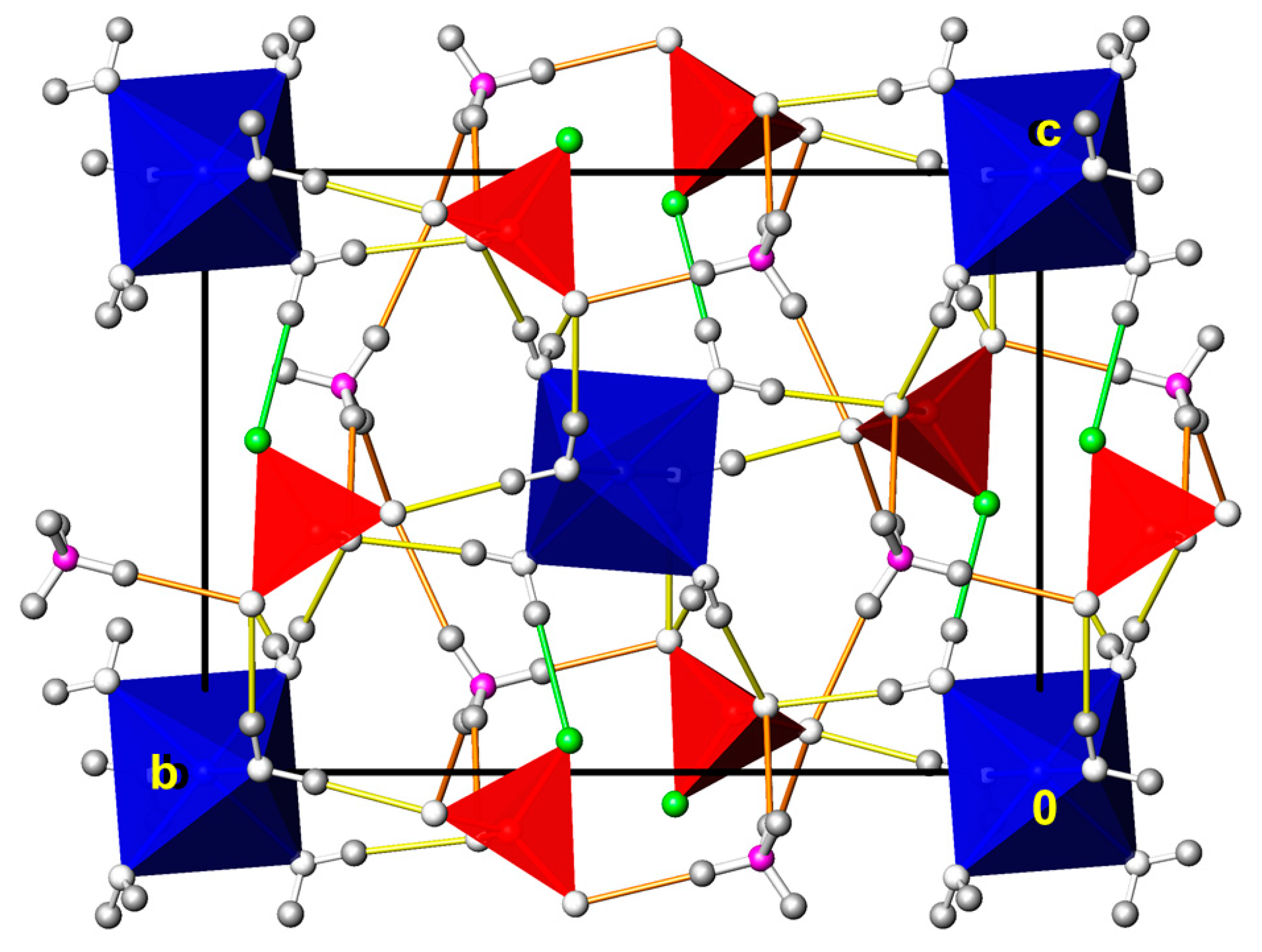
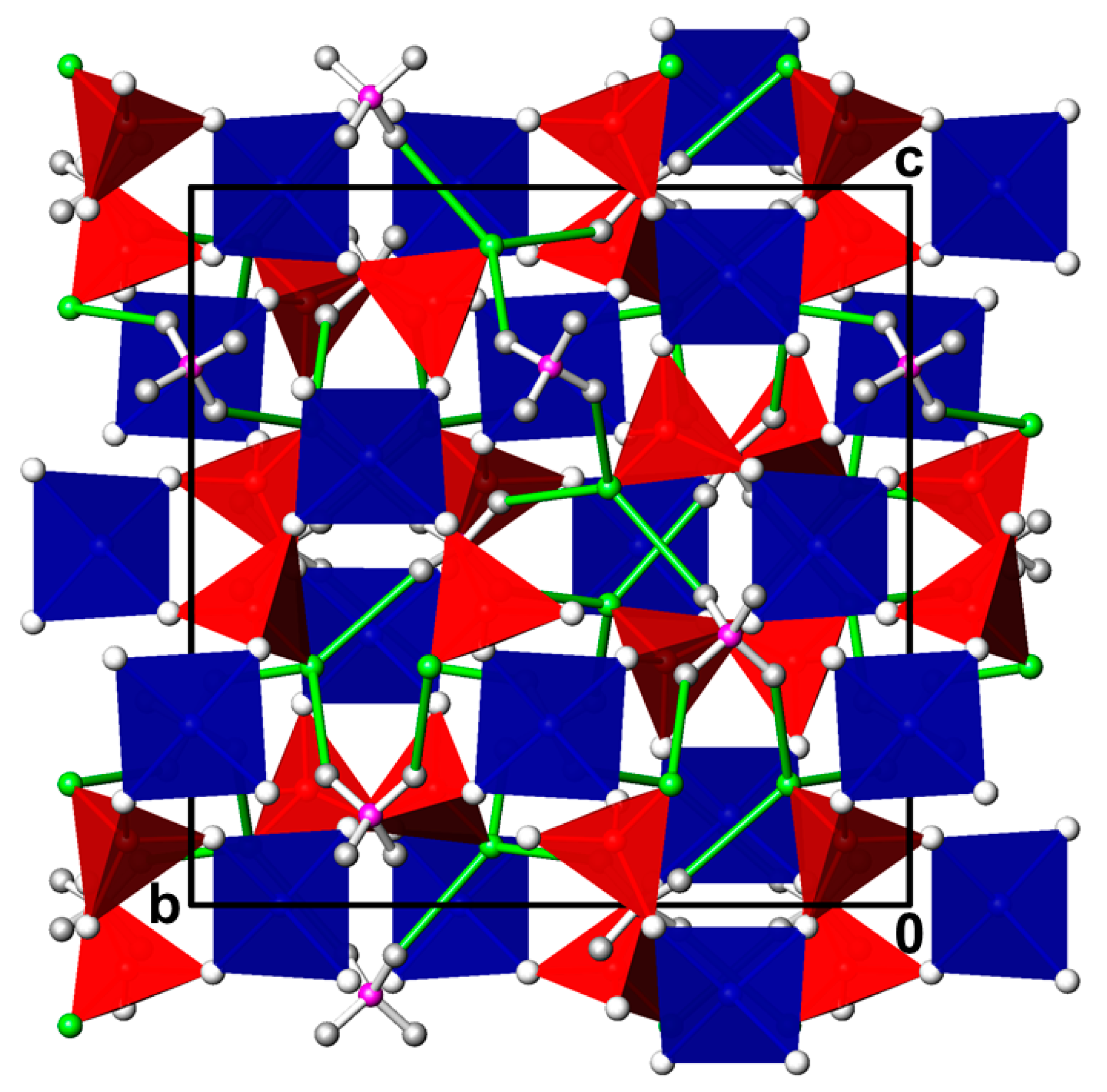
In contrast to the ammonium transition metal monofluorophosphate dihydrates (NH4)2M(PO3F)2(H2O)2 (M = Mg, Mn, Co, Cu) described in the preceding sections, the nickel compound crystallizes with six water molecules. (NH4)2Ni(PO3F)2(H2O)6 is a member of the vast family of Tutton salts with general formula MI2MII(XO4)2(H2O)6. Typical crystal-chemical features of Tutton salts have been reviewed in various reports, e.g., ([29], and references therein). In short, the unit cell of a Tutton salt comprises two formula units and is made up of one MII site (here Ni) located on a centre of inversion (Wyckoff position 2a) and surrounded by six water molecules in the form of a slightly distorted octahedron, one XO4 tetrahedron (here PO3F), and one ammonium cation (for cases with other MI cations distorted MIO8 polyhedron are present). Hydrogen bonds of medium strengths between the building units of the type O–H···O and, as a peculiarity in the case of (NH4)2Ni(PO3F)2(H2O)6, also of the type O–H···F generate a three-dimensional network structure. The crystal structure of (NH4)2Ni(PO3F)2(H2O)6 has previously been determined based on a X-ray diffraction data set recorded at room temperature using a CAD-4 four-circle diffractometer and a point detector. Since the crystal intensities dropped by up to 73% of their initial values during the long-lasting data collection [15], it was decided to re-refine the crystal structure with CCD data at 100 K for an improved model. In principle, the current low-temperature data confirm the previous room-temperature data, however with much higher precision as indicated by standard uncertainties for bond lengths and angles about three to five times smaller. The crystal structure of (NH4)2Ni(PO3F)2(H2O)6 is depicted in Figure 8.

3.8. NH4Cr(PO3F)2(H2O)6

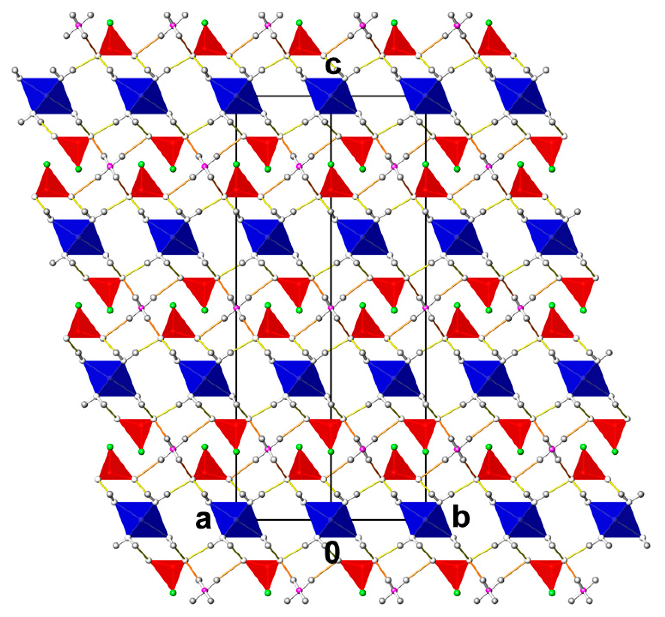
PDF entry #00-044-0535 reports the same R-centred cell for NH4Cr(PO3F)2(H2O)6 but with space group R3 instead of R 3 ¯ m determined from the present single crystal X-ray data. In the crystal structure, isolated [Cr(OH2)6] octahedra (point group symmetry 3 ¯ m) are organised in layers parallel (001) and are sandwiched by double layers of PO3F2− anions (point group symmetry 3m) along the [001] stacking direction. The disordered ammonium cations (site symmetry 3 ¯ m) are situated between the PO3F2− anions in the middle of the monofluorophosphate double layer. Strong hydrogen bonds between the [Cr(OH2)6] octahedra and the O atoms of the monofluorophosphate groups link the chromium and monofluorophosphate layers together. Ammonium cations additionally hydrogen-bond to the O atoms within a monofluorophosphate double layer (Figure 9).

3.9. NH4Cu2(H3O2)(PO3F)2

The ammonium copper compound crystallizes isotypically with KCu2(H3O2)(PO3F)2 [30] in the natrochalcite structure type [16]. The copper cation (site symmetry 2/m; Wyckoff position 4e) is surrounded by six O atoms and shows its characteristic tetragonally distorted octahedral coordination owing to the Jahn-Teller effect. Neighbouring [CuO6] polyhedra share common edges to form chains parallel to [010]. Adjacent chains are bridged by the monofluorophosphate tetrahedra (site symmetry m), sharing exclusively the O atoms into (001) layers. The disordered ammonium cation (with the N atom situated on Wyckoff position 2d with site symmetry 2/m) is located between adjacent layers and links them through hydrogen bonding to the monofluorophosphate O atoms. Additional hydrogen bonds, albeit of weak nature, develop between the non-disordered part of the {H3O2} group and the F atom of the monofluorophosphate anion. The crystal structure is shown in Figure 10. It is well known that natrochalcite-type compounds contain such {H3O2} groups where a positionally disordered H atom with half-occupation (here H2O) sits between two OH groups. Since the features of the resulting hydrogen bonding system, including a clear location of hydrogen atoms by neutron diffraction, was reported for isotypic KCu2(H3O2)(SO4)2, we refer to the original description [16] for further details.

3.10. (NH4)2Zn(PO3F)2(H2O)0.2

Three ammonium cations, two zinc cations, three monofluorophosphate anions and one positionally and occupationally disordered water molecule are present in the asymmetric unit. Except one Zn site (Zn2) located on a twofold rotation axis (Wyckoff position 4e), all other atoms in this structure are located on general positions (Wyckoff position 8f). The O atoms of four monofluorophosphate anions tetrahedrally surround both zinc cations. The latter do not share common atoms but are bridged by the monofluorophosphate units into (10 1 ¯ ) layers. Within a layer disorder of the four O atoms around Zn2 over two sets of sites is observed, with atoms O4 and O8 having an occupational ratio of 0.65(3):0.35(3) for the split pair A:B. This disorder also affects the monofluorophosphate anions associated with P2 and P3 that share these O atoms with Zn2. Additionally, the water molecule (O1W) shows positional and occupational disorder. It is disordered over an inversion centre and shows an occupancy of 0.309(12); full occupation of this site would result in a value of 0.5, leading to a formula of (NH4)2Zn(PO3F)2(H2O)0.33. Neighbouring layers are linked through intermediate ammonium cations by medium to weak hydrogen bonds to the monofluorophosphate O atoms. The crystal structure of (NH4)2Zn(PO3F)2(H2O)0.2 is displayed in Figure 11.

3.11. (NH4)2Zn3(PO3F)4(H2O)

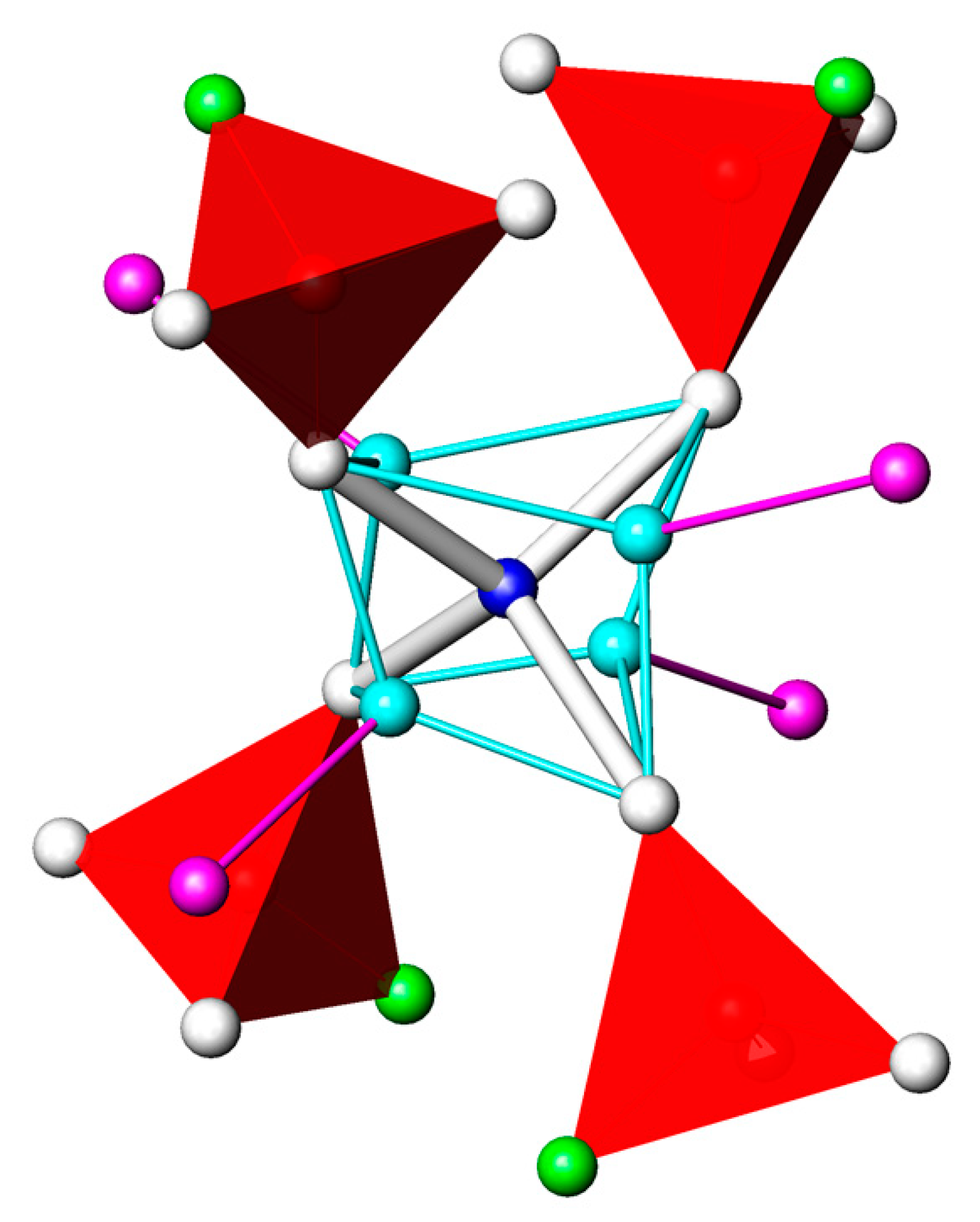
(NH4)2Zn3(PO3F)4(H2O) has a higher Zn and PO3F content than (NH4)2Zn(PO3F)2(H2O)0.2 (ratio NH4:Zn:PO3F = 2:3:4 versus 2:1:2) but together with ZnPO3F(H2O)2.5 also crystallized from the same solution. A very similar I-centred cubic unit cell (without further assignments of possible space groups) was reported for anhydrous (NH4)2Zn3(PO3F)4 at room temperature (PDF entry #00-044-0539; a = 11.4769(5) Å). In the crystal structure of (NH4)2Zn3(PO3F)4(H2O) disorder is observed, affecting the zinc and the ammonium sites. The major part of the cation (ZnA; occupancy 0.75) is situated on Wyckoff position 12b with 4 ¯ site symmetry. Due to disorder around this axis, the remaining Zn cations split into four equivalent sites (ZnB) with an occupancy of 0.0625 each. The N atom of the ammonium cation (N1H) and the O atom of a water molecule (O1W) simultaneously occupy Wyckoff position 12a (located on a 4 ¯ axis) in a ratio of 0.67:0.33. The site symmetry of the PO3F tetrahedron is .3 with the P atom situated on Wyckoff position 16c. The disordered part of the crystal structure is shown in Figure 12.
Whereas ZnA is exclusively bonded to four O atoms (O1) of symmetry-related monofluorophosphate anions with an equal bond length of 1.934(3) Å, ZnB is coordinated by only three monofluorophosphate O atoms at two shorter and one longer Zn–O distances. The fourth coordination site, completing a distorted tetrahedron, is occupied by the water molecule at the longest distance of 2.204(14) Å. Again, the F atom of the monofluorophosphate tetrahedron does not take part in constructing the framework structure because it is not part of the coordination spheres around the two zinc sites (Figure 13). However, it is involved in weak hydrogen bonding interactions as the acceptor atom with the disordered (N1H/O1W) donor group. Two more hydrogen bonding interactions of similar strength are present between the donor group and the monofluorophosphate O atoms.

3.12. Survey on the PO3F2− Group

  • Mean bond lengths and angles in the PO3F tetrahedron
For the statistical analysis of bond lengths and angles within a PO3F tetrahedron in inorganic monofluorophosphates that are compiled in the most recent version of the Inorganic Structure Database (ICSD, [31]), reliability factors R1 ≤ 0.08 for the structure model and only ordered PO3F groups were considered as criteria, disregarding different measurement temperatures or redeterminations. HPO3F tetrahedra present in hydrogenmonofluorophosphates were not taken into account. In summary, 88 independent PO3F tetrahedra from 63 different monofluorophosphate phases (including the examples of the current study) were used (Table 5). As a result, the P–F bond of 1.578(20) Å is significantly longer than the three P—O bonds with 1.506(13) Å, and relative to the ideal tetrahedral angle of 109.47°, the three O–P–O angles of 113.7(1.7)° are enlarged by about 4° and the O—P—F angle of 104.8(1.7)° reduced by about the same value. The averaged values for bond lengths and angles in the monofluorophosphate PO3F tetrahedron differ markedly from those of the difluorophosphate PO2F2 tetrahedron. Here, the two P–O and the two P–F bonds are shortened with mean values of 1.459 (27) and 1.530 (21) Å, respectively, and the O–P–O angle once more is widened to 121.2 (2.9)°, whereas the O—P—F angle of 108.7 (6)° now is closer to the ideal value (the F–P–F angle is the smallest in the PO2F2 tetrahedron with 98.5 (2.6)°) [32]).
The computed mean values of the monofluorophosphate tetrahedron can be used as a simple tool for evaluation of crystal structures with this entity. In one case (Table 5), a significant deviation in terms of bond lengths and angles was observed for the crystal structure of (NH4)3Fe(PO3F)2F2 [33] where one of the two distinct monofluorophosphate anions has one of the P–O bonds as the longest in the tetrahedron, a very short P–F bond, and with O–P–O and O–P–F angles unexpected: P2–O1 = 1.562(5) Å, P2–F4 = 1.498(5) Å, P2–O4 = 1.485(6) Å (2x); O1–P2–F4 = 106.7(3)°, O1–P2–O4 = 102.7(2)° (2x), F4–P2–O4 = 115.0(2)° (2x), O4–P2–O4 = 112.8(3)°. Based on the current averaged data for a PO3F tetrahedron, it is clear that atoms O1 and F4 were wrongly assigned and must be interchanged.
  • Symmetry of the PO3F group in crystal structures
Possible point group symmetries of a PO3F group in a crystal structure are 1, 3, m and 3m, the latter being the highest possible point group symmetry for this tetrahedron in the crystalline state. The vast majority of monofluorophosphate groups exhibits point group symmetry 1 (70 examples), followed by point group symmetry m (15 examples), 3m (two examples) and 3 (one example). The reported point group symmetry of 4 ¯ 2m for the PO3F group in K3(PO3F)F [34] is incompatible with its molecular symmetry and consequently, this group is disordered.
  • Isotypism with sulfates
From the numerous phases compiled in Table 5, only nine show isotypism with the corresponding sulfate, viz. Na2PO3F(H2O)10, NaK3(PO3F)2, K3(PO3F)F, M2PO3F (M = K, Rb, Cs, NH4), (NH4)2Ni(PO3F)(H2O)6 and CuK(OH)(PO3F)(H2O). Eleven monofluorophosphate phases have equivalent sulfate phases but with different crystal structures, viz. Li(NH4)PO3F, Na2PO3F, CaPO3F(H2O)2, SrPO3F, BaPO3F, (NH4)Mn(PO3F)F2, Fe2(PO3F)3, NaFe(PO3F)2, SnPO3F, Ag2PO3F, and Hg2PO3F, but the majority of monofluorophosphate phases has no sulfate counterpart.
  • Hydrogen bonding with the monofluorophosphate F atom as an acceptor
As discussed briefly for appropriate structures above and detailed in Table 3, hydrogen bonding involving the F atom of the monofluorophosphate anion occurs only occasionally and then only as a comparatively weak interaction. A review of the crystal structures where hydrogen bonding is possible and where all H atoms were determined revealed that this situation holds also for most other monofluorophosphates. Considering a D···F distance (D = donor atom: N, O) less than 3.2 Å and D–H···F angles greater than 130°, as relevant for a significant hydrogen bonding interaction [35], then only for Li(NH4)PO3F, Na2PO3F(H2O)10, CaPO3F(H2O)2, Cu2K(OH)(H2O)(PO3F)2, K2Mn3(HPO4)2(PO3F)F, and (NH4)2Ni(PO3F)2(H2O)6 is this kind of interaction realized, albeit of weak nature (Table 5). In all other monofluorophosphates capaple of hydrogen-bonding interactions either the D···F–P distances are much greater than the threshold of 3.2 Å, or the D–H···F angles are much smaller than 140°. In these structures, D–H···O hydrogen bonds dominate or are the only hydrogen-bonding interactions.

4. Conclusions

Single crystals of twelve and partly unknown monofluorophosphate phases were grown from aqueous solutions. Crystal structure refinements of these compounds extend our knowledge about the PO3F2− anion. Based on the present crystal structure data and a complete literature search addressing monofluorophosphate structures of inorganic compounds, the following structural characteristics for the tetrahedral PO3F group were obtained: The P—F bond has a mean value of 1.578(20) Å and is considerably longer than the mean of the three P—O bonds of 1.506(13) Å, and the mean O–P–O angles of 113.7(1.7)° are considerably larger than the mean O–P–F angle of 104.8(1.7)°. The point group symmetry of the “free” PO3F group (C3v in Schoenflies or 3m in Hermann–Maugin notation) is found with this symmetry in the solid state only in two examples. In most cases (70 examples) the point group symmetry is reduced to C1 (1) followed by point group symmetry Cs (m) with 15 examples and C3 (3) with one example. The monofluorophosphate F atom is characterized by its isolated state in the crystal structure. In the vast majority of cases, it is not part of the coordination sphere of the cation and/or is not engaged in hydrogen bonding as an acceptor atom. Only in exceptional cases are weak interactions realized, i.e., for large cations with high coordination numbers in form of long metal–F bonds or as hydrogen bonds with long donor···F distances between 2.8 and 3.2 Å.

Funding

This research received no external funding.

Institutional Review Board Statement

Not applicable.

Informed Consent Statement

Not applicable.

Data Availability Statement

The data presented in this study are available in The Cambridge Crystallographic Data Centre (CCDC) and can be obtained free of charge via www.ccdc.cam.ac.uk/structures.

Acknowledgments

The author thanks Enrique J. Baran (Consejo Nacional de Investigaciones Científicas y Técnicas de la República Argentina (CONICET) and the Universidad Nacional de La Plata, Argentina) for recording and interpreting the vibrational spectra of CdPO3F(H2O)2, and Ekkehard Füglein (Netzsch GmbH, Selb, Germany) for recording the TG of CdPO3F(H2O)2.

Conflicts of Interest

The author declares no conflict of interest.

References

  1. Lange, W. Über die Monofluorphosphorsäure und die Ähnlichkeit ihrer Salze mit den Sulfaten. Ber. Dtsch. Chem. Ges. 1929, 62B, 793–801. [Google Scholar] [CrossRef]
  2. Rea, J.R.; Kostiner, E. The crystal structure of manganese fluorophosphate, Mn2(PO4)F. Acta Crystallogr. 1972, B28, 2525–2529. [Google Scholar] [CrossRef]
  3. Jin, D.; Qiu, H.; Du, F.; Wei, Y.; Meng, X. Co-doped Na2FePO4F fluorophosphates as a promising cathode material for rechargeable sodium-ion batteries. Solid State Sci. 2019, 93, 62–69. [Google Scholar] [CrossRef]
  4. Weil, M.; Puchberger, M.; Füglein, E.; Baran, E.J.; Vannahme, J.; Jakobsen, H.J.; Skibsted, J. Single-Crystal Growth and Characterization of Disilver(I) Monofluorophosphate(V), Ag2PO3F: Crystal Structure, Thermal Behavior, Vibrational Spectroscopy, and Solid-State 19F, 31P, and 109Ag MAS NMR Spectroscopy. Inorg. Chem. 2007, 46, 801–808. [Google Scholar] [CrossRef]
  5. Xiong, L.; Chen, J.; Lu, J.; Pan, C.-Y.; Wu, L.-M. Monofluorophosphates: A New Source of Deep-Ultraviolet Nonlinear Optical Materials. Chem. Mater. 2018, 30, 7823–7830. [Google Scholar] [CrossRef]
  6. Robinson, M.T. The crystal structures of β-K2SO4 and β-K2PO3F. J. Phys. Chem. 1958, 62, 925–928. [Google Scholar] [CrossRef]
  7. Rây, R.C. Isomorphism and Chemical Homology. Nature 1930, 126, 310–311. [Google Scholar] [CrossRef]
  8. Lima-de-Faria, J.; Hellner, E.; Liebau, F.; Makovicky, E.; Parthé, E. Nomenclature of Inorganic Structure Types—Report of the International Union of Crystallography Commission on Crystallographic Nomenclature Subcommittee on the Nomenclature of Inorganic Structure Types. Acta Crystallogr. 1990, A46, 1–11. [Google Scholar] [CrossRef] [Green Version]
  9. Gates-Rector, S.D.; Blanton, T.N. The Powder Diffraction File: A Quality Materials Characterization Database. Powder Diffr. 2019, 34, 352–360. [Google Scholar] [CrossRef] [Green Version]
  10. McCubbin, F.M.; Phillips, B.L.; Adcock, C.T.; Tait, K.T.; Steele, A.; Vaughn, J.S.; Fries, M.D.; Atudorei, V.; Vander Kaaden, K.E.; Hausrath, E.M. Discreditation of bobdownsite and the establishment of criteria for the identification of minerals with essential monofluorophosphate (PO3F2). Am. Mineral. 2018, 103, 1319–1328. [Google Scholar] [CrossRef]
  11. Schülke, U.; Kayser, R. Herstellung von Fluorophosphaten, Difluorophosphaten, Fluorophosphonaten und Fluorophosphiten in fluoridhaltigen Harnstoffschmelzen. Z. Anorg. Allg. Chem. 1991, 600, 221–226. [Google Scholar] [CrossRef]
  12. Sheldrick, G.M. A short history of SHELX. Acta Crystallogr. 2008, 64, 112–122. [Google Scholar] [CrossRef] [Green Version]
  13. Sheldrick, G.M. Crystal structure refinement with SHELXL. Acta Crystallogr. 2015, 71, 3–8. [Google Scholar]
  14. Durand, J.; Larbot, A.; Cot, L.; Duprat, M.; Dabosi, F. Etude Structurale de ZnPO3F·2.5H2O, Nouvel Inhibiteur de Corrosion. Z. Anorg. Allg. Chem. 1983, 504, 163–172. [Google Scholar] [CrossRef]
  15. Berraho, M.; R’Kha, C.; Vegas, A.; Rafiq, M. Structure of Ni(H2O)6(NH4)2(PO3F)2. Acta Crystallogr. 1992, C48, 1350–1352. [Google Scholar] [CrossRef]
  16. Chevier, G.; Giester, G.; Jarosch, D.; Zemann, J. Neutron Diffraction Study of the Hydrogen-Bond System in Cu2K(H3O2)(SO4)2). Acta Crystallogr. 1990, C46, 175–177. [Google Scholar] [CrossRef]
  17. Ross, S.D. Inorganic Infrared and Raman Spectra; McGraw Hill: London, UK, 1972. [Google Scholar]
  18. Müller, A.; Baran, E.J.; Carter, R.O. Vibrational spectra of oxo-, thio-, and selenometallates of transition elements in the solid state. Struct. Bond. 1976, 26, 81–139. [Google Scholar]
  19. Fadini, A.; Schnepel, F.M. Vibrational Spectroscopy: Methods and Applications; Ellis Horwood: Chichester, UK, 1989. [Google Scholar]
  20. Siebert, H. Anwendungen der Schwingungsspektroskopie in derAnorganischen Chemie; Springer: Berlin, Germany, 1966. [Google Scholar]
  21. Weil, M.; Puchberger, M.; Baran, E.J. Preparation and characterization of dimercury(I) monofluorophosphate(V), Hg2PO3F: Crystal structure, thermal behavior, vibrational spectra, and solid state 31P and 19F NMR spectra. Inorg. Chem. 2004, 43, 8330–8335. [Google Scholar] [CrossRef]
  22. Calvo, C.; Au, P.K.L. Crystal structure of Cd2P2O7. Can. J. Chem. 1969, 47, 3409–3416. [Google Scholar] [CrossRef]
  23. Menz, D.-H.; Kolditz, L. Synthese und thermisches Verhalten von Mg(NH4)2(PO3F)2·2H2O. Z. Chem. 1985, 25, 189–190. [Google Scholar] [CrossRef]
  24. Menz, D.-H.; Kolditz, L.; Heide, K.; Kunert, C.; Mensing, C. Zur Thermischen Zersetzung von SrPO3F·H2O. Z. Anorg. Allg. Chem. 1986, 540, 191–197. [Google Scholar] [CrossRef]
  25. Nyburg, S.C.; Steed, J.W.; Aleksovska, S.; Petruševski, V.M. Structure of the alums. I. On the sulfate group disorder in the α-alums. Acta Crystallogr. 2000, B56, 204–209. [Google Scholar] [CrossRef] [PubMed]
  26. Gmelin Handbook of Inorganic Chemistry. Cr-Chrom (Systemnummer 52); Springer-Verlag: Berlin/Heidelberg, Germany, 1962; Teil B; p. 306ff.
  27. Berraho, M.; Vegas, A.; Martinez-Ripoll, M.; Rafiq, M. A copper monofluorophosphate, Cu(H2O)2(NH4)2(PO3F)2. Acta Crystallogr. 1994, C50, 666–668. [Google Scholar] [CrossRef]
  28. Müller, U. Symmetry Relationships between Crystal Structures; IUCr/Oxford Science Publications: Oxford, UK, 2013. [Google Scholar]
  29. Weil, M. On the (non-)existence of Tutton salts with formula types [M(H2O)6](ClO4)2(H2O)2 and Na2M(SO4)2(H2O)6 (M is a first-row transition metal). Acta Crystallogr. 2013, C69, 990–994. [Google Scholar]
  30. Moewius, F.; Ziemer, B.; Reck, G.; Meisel, M.; Grunze, H. Darstellung und Kristallstruktur von Kalium-dikupferhydroxid-bis(monofluorophosphat)-Monohydrat Cu2K(OH)(PO3F)2·H2O. Z. Anorg. Allg. Chem. 1987, 547, 75–82. [Google Scholar] [CrossRef]
  31. Zagorac, D.; Müller, H.; Ruehl, S.; Zagorac, J.; Rehme, S. Recent developments in the Inorganic Crystal Structure Database: Theoretical crystal structure data and related features. J Appl. Crystallogr. 2019, 52, 918–925. [Google Scholar] [CrossRef] [Green Version]
  32. Weil, M.; Fürst, M. Crystal structure of (1,4-diphenyl-4H-1,2,4-triazol-3-yl)phenylamine difluorophosphate, and a survey of the difluorophosphate anion (PO2F2). Acta Crystallogr. 2020, E76, 1003–1006. [Google Scholar] [CrossRef]
  33. Keates, A.C.; Armstrong, J.A.; Weller, M.T. Iron fluorophosphates. Dalton Trans. 2013, 42, 10715–10724. [Google Scholar] [CrossRef]
  34. Skakle, J.M.S.; Fletcher, J.G.; West, A.R. The crystal structures of potassium oxyfluorides, K3SeO4F and K3PO3F2. An. Quim. Int. Ed. 1996, 92, 358–361. [Google Scholar]
  35. Steiner, T. The Hydrogen bond in the Solid State. Angew. Chem. Int. Ed. 2002, 41, 48–76. [Google Scholar] [CrossRef]
  36. Galigne, J.L.; Durand, J.; le Cot, L. Etudes structurales de composes oxyfluores du P(V). I. Structure crystalline de LiKPO3FH2 O. Acta Crystallogr. 1974, B30, 697–701. [Google Scholar] [CrossRef]
  37. Durand, J.; le Cot, L.; Galigne, J.L. Etudes structurales de composes oxyfluores du P(V). V. Structure crystalline de LiNH4PO3F. Acta Crystallogr. 1978, B34, 388–391. [Google Scholar] [CrossRef]
  38. Durand, J.; le Cot, L.; Galigne, J.L. Etudes structurales de composes oxyfluores du P(V) II. Structure cristalline de Na2PO3F beta. Acta Crystallogr. 1974, B30, 1565–1569. [Google Scholar] [CrossRef] [Green Version]
  39. Prescott, H.A.; Troyanov, S.I.; Kemnitz, E. The synthesis and crystal structures of two new hydrated sodium monofluorophosphates: NaHPO3F·2.5H2O (I) and Na2PO3F·10H2O (II). J. Solid State Chem. 2001, 156, 415–421. [Google Scholar] [CrossRef]
  40. Durand, J.; Granier, W.; le Cot, L.; Galigne, J.L. Etudes structurales de composes oxyfluores du P(V). III. Structure crystalline de NaK3(PO3F)2. Acta Crystallogr. 1975, B31, 1533–1535. [Google Scholar] [CrossRef] [Green Version]
  41. Fabry, J.; Dušek, M.; Krupkova, R. Ammonium sodium fluorotrioxophosphate monohydrate. Acta Crystallogr. 2007, E63, i92–i94. [Google Scholar] [CrossRef]
  42. Payen, J.; Durand, J.; le Cot, L.; Galigne, J.L. Etude structurale du monofluorophosphate de potassium K2PO3F. Can. J. Chem. 1979, 57, 886–889. [Google Scholar] [CrossRef]
  43. Fabry, J.; Dušek, M.; Fejfarova, K.; Krupkova, R.; Vanek, P.; Cisarova, I. Dirubidum fluorotrioxophosphate, Rb2PO3F, at 290 and 130 K, and dicesium fluorotrioxophosphate, Cs2PO3F, at 240 and 100 K. Acta Crystallogr. 2006, C62, i49–i52. [Google Scholar]
  44. Prescott, H.; Troyanov, S.; Kemnitz, E. The crystal structures of two hydrogen monofluorophosphates: CsHPO3F and Cs3(NH4)2(HPO3F)3(PO3F). Z. Kristallogr. 2000, 215, 240–245. [Google Scholar]
  45. Krupkova, R.; Fabry, J.; Cisarova, I.; Vanek, P. Bis(ammonium) fluorophosphate at room temperature. Acta Crystallogr. 2002, C58, i66–i68. [Google Scholar] [CrossRef]
  46. Perloff, A. The crystal structures of hydrated calcium and ammonium monofluorophosphates: CaPO3(H2O)2 and (NH4)2PO3F(H2O). Acta Crystallogr. 1972, B28, 2183–2191. [Google Scholar] [CrossRef]
  47. Berndt, A.F.; Sylvester, J.M. The crystal structure of ammonium monofluorophosphate: (NH4)2PO3F(H2O). Acta Crystallogr. 1972, B28, 2191–2193. [Google Scholar] [CrossRef]
  48. Durand, J.; Beys, L.; Hillaire, P.; Aleonard, S.; le Cot, L. Etude structurale de (NH4)2PO3F, H2O a 120 K par diffraction des rayons X et spectroscopie Raman entre 293 et 83 K. Spectrochim. Acta 1978, A34, 123–127. [Google Scholar] [CrossRef]
  49. Jantz, S.G.; van Wuellen, L.; Fischer, A.; Libowitzky, E.; Baran, E.J.; Weil, M.; Höppe, H.A. Syntheses, crystal structures, NMR spectroscopy, and vibrational spectroscopy of Sr(PO3F).H2O and Sr(PO3F). Eur. J. Inorg. Chem. 2016, 7, 1121–1128. [Google Scholar] [CrossRef]
  50. Stöger, B.; Weil, M.; Skibsted, J. The crystal structure of BaPO3F revisited—A combined X-ray diffraction and solid-state 19F, 31P MAS NMR study. Dalton Trans. 2013, 42, 11672–11682. [Google Scholar] [CrossRef]
  51. Marshall, K.L.; Weller, M.T. Synthesis of titanium fluorophosphates and fluorosulfates from hexafluorotitanic acid. Z. Anorg. Allg. Chem. 2014, 640, 2766–2770. [Google Scholar] [CrossRef]
  52. Weil, M.; Baran, E.J.; Kremer, R.K.; Libowitzky, E. Synthesis, crystal structure, and properties of Mn(PO3F)(H2O)2. Z. Anorg. Allg. Chem. 2015, 641, 184–191. [Google Scholar] [CrossRef]
  53. Armstrong, J.A.; Williams, E.R.; Weller, M.T. Manganese(III) fluorophosphate frameworks. Dalton Trans. 2013, 42, 2302–2308. [Google Scholar] [CrossRef]
  54. Wang, G.; Valldor, M.; Dorn, K.V.; Wilk-Kozubek, M.; Smetana, V.; Mudring, A.-V. Ionothermal Synthesis Enables Access to 3D Open Framework Manganese Phosphates Containing Extra-Large 18-Ring Channels. Chem. Mater. 2019, 31, 7329–7339. [Google Scholar] [CrossRef]
  55. Armstrong, J.A.; Williams, E.R.; Weller, M.T. Fluoride-rich, hydrofluorothermal routes to functional transition metal (Mn, Fe, Co, Cu) fluorophosphates. J. Am. Chem. Soc. 2011, 133, 8252–8263. [Google Scholar] [CrossRef]
  56. Jiang, J.; Zhu, B.; Zhu, T.; Yang, H.; Jin, Y.; Lü, M. Open-framework ammonium transition metal fluorophosphates with a Kagomé lattice network: Synthesis, structure and magnetic properties. Dalton Trans. 2020, 49, 841–849. [Google Scholar] [CrossRef] [PubMed]
  57. Zhu, B.; Jiang, J.; Zhu, T.; Yang, H.; Jin, Y.; Choi, K.-Y.; Lü, M. Transition-Metal Monofluorophosphate Ba2M2(PO3F)F6 (M = Mn, Co, and Ni): Varied One-Dimensional Transition-Metal Chains and Antiferromagnetism. Inorg. Chem. 2020, 59, 3794–3804. [Google Scholar] [CrossRef] [PubMed]
  58. Durand, J.; le Cot, L.; Berraho, M.; Rafiq, M. Structure du monofluorophosphate de cobalt trihydrate. Acta Crystallogr. 1987, C43, 611–613. [Google Scholar]
  59. Zeibig, M.; Wallis, B.; Moewius, F.; Meisel, M. Darstellung und Kristallstruktur von Kupfer(II)-monofluorophosphat-Dihydrat CuPO3F·2H2O. Z. Anorg. Allg. Chem. 1991, 600, 231–238. [Google Scholar] [CrossRef]
  60. Berndt, A.F. The crystal structure of SnPO3F. Acta Crystallogr. 1974, B30, 529–530. [Google Scholar] [CrossRef]
  61. Weil, M. NH4Ag3(PO3F)2, a layered monofluorophosphate(V) with seven different Ag sites. Acta Crystallogr. 2007, C63, i31–i33. [Google Scholar]
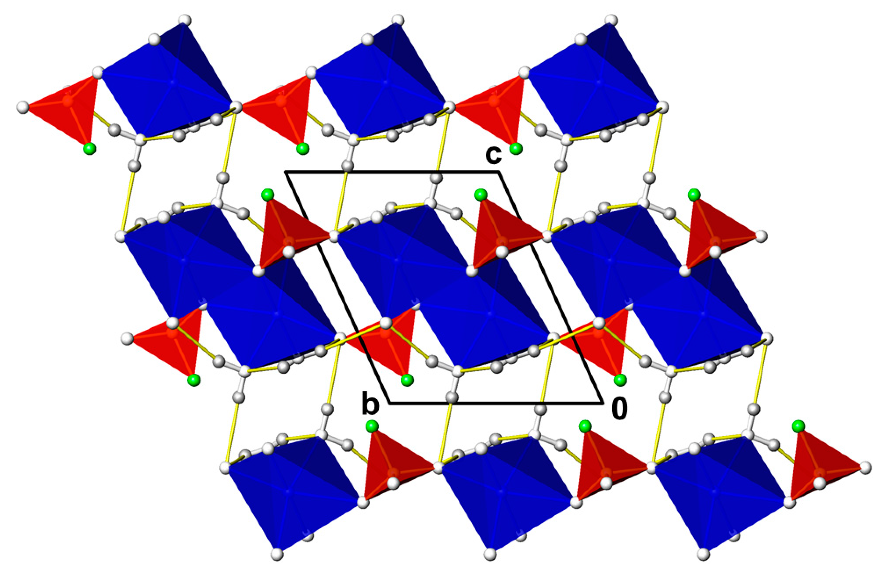
Figure 1. The crystal structure of CdPO3F(H2O)2 in a projection along [ 1 ¯ 00]. PO3F tetrahedra are given in red (O atoms as colourless, F atoms as green spheres), [CdO6] octahedra are given in blue, and H atoms are given as grey spheres. O–H···O hydrogen bonding is indicated by yellow lines.
Chemistry 03 00005 g001
Figure 2. CdPO3F(H2O)2. (a) IR spectrum; (b) TG curve (black) with its fist derivative (DTG curve, green).
Chemistry 03 00005 g002
Figure 3. The crystal structure of Cr2(PO3F)3(H2O)18.8 in a projection along [ 1 ¯ 00]. Colour code of PO3F tetrahedra as in Figure 1; [CrO6] octahedra are given in blue, O atoms of crystal water molecules with full occupation are given as colourless spheres and those of disordered crystal water molecules as yellow spheres. For clarity, disorder of one of the PO3F groups is not shown.
Chemistry 03 00005 g003
Figure 4. The crystal structure of Pb2(PO3F)Cl2(H2O) in a projection along [0 1 ¯ 0]. Colour code of PO3F tetrahedra as in Figure 1; Pb atoms are represented by dark-blue spheres, Cl atoms as turquoise spheres and O atoms of the water molecules as yellow spheres.
Chemistry 03 00005 g004
Figure 5. The crystal structure of ZnPO3F(H2O)2.5 in a projection along [010]. Colour code of PO3F tetrahedra and H atoms as in Figure 1; [ZnO6] octahedra are given in dark-blue, [ZnO4] tetrahedra in turquoise and the O atom of the crystal water molecule as an orange sphere. O–H···O hydrogen bonding with the aqua ligands as donor groups is given as yellow lines, and involving the crystal water molecules as donor groups as orange lines.
Chemistry 03 00005 g005
Figure 6. The crystal structure of (NH4)2M(PO3F)2(H2O)2 (M = Mg, Co; data used from the Mg-compound) in a projection along [001]. Colour code of PO3F tetrahedra, H atoms and O–H···O hydrogen bonding as in Figure 1; [MO6] octahedra are given in blue, N atoms of the ammonium groups as magenta spheres. N–H···O hydrogen bonding is indicated by orange lines.
Chemistry 03 00005 g006
Figure 7. The crystal structure of (NH4)2Mn(PO3F)2(H2O)2 in a projection along [001]. Colour code as in Figure 6.
Chemistry 03 00005 g007
Figure 8. The crystal structure of (NH4)2Ni(PO3F)2(H2O)6 in a projection along [ 1 ¯ 00]. Colour code as in Figure 6; O–H···F hydrogen bonding is indicated by green lines.
Chemistry 03 00005 g008
Figure 9. The crystal structure of NH4Cr(PO3F)2(H2O)6 in a projection along [110]. Colour code as in Figure 6.
Chemistry 03 00005 g009
Figure 10. The crystal structure of NH4Cu2(H3O2)(PO3F)2 in a projection along [010]. Colour code as in Figure 6.
Chemistry 03 00005 g010
Figure 11. The crystal structure of (NH4)2Zn(PO3F)2(H2O)0.2 in a projection along [010]. Colour code of PO3F tetrahedra and H atoms as in Figure 1; O atoms of disordered crystal water molecules are given as yellow spheres. For clarity, disorder involving parts of the [ZnO4] and PO3F tetrahedra is not shown.
Chemistry 03 00005 g011
Figure 12. Disorder in the crystal structure of (NH4)2Zn3(PO3F)4(H2O). Colour code of the PO3F tetrahedra as in Figure 1. The major part of the disordered Zn site (ZnA) is given as a blue sphere, the minor part (ZnB) as a turquoise sphere. The statistically occupied (NH4/H2O) site is given in magenta. For clarity, H atoms are not shown.
Chemistry 03 00005 g012
Figure 13. The crystal structure of (NH4)2Zn3(PO3F)4(H2O) in a projection along [ 1 ¯ 00] giving only the major part of the disordered Zn site. Colour code of PO3F tetrahedra as in Figure 1; [ZnO4] tetrahedra are given in blue and (N,O)–H···F hydrogen bonding is indicated by green lines. For clarity, (N,O)–H···O hydrogen bonding is not shown.
Chemistry 03 00005 g013
Table 1. Details of data collections and structure refinements.
CompoundCdPO3F(H2O)2Cr2(PO3F)3(H2O)18.8Pb2(PO3F)Cl2(H2O)ZnPO3F(H2O)2.5(NH4)2Co(PO3F)2(H2O)2(NH4)2Mg(PO3F)2(H2O)2
Formula weight246.40736.88601.27208.38326.99292.37
Temperature/°C23−17323−1732323
Radiation; λMo K a ¯ ; 0.71073Mo K a ¯ ; 0.71073Mo K a ¯ ; 0.71073Mo K a ¯ ; 0.71073Mo K a ¯ ; 0.71073Mo K a ¯ ; 0.71073
DiffractometerSMART CCDAPEX-II CCDSMART CCDAPEX-II CCDCAD-4SMART CCD
Crystal dimensions / mm0.14 × 0.10 × 0.040.10 × 0.10 × 0.010.35 × 0.05 × 0.040.12 × 0.02 × 0.020.05 × 0.05 × 0.010.04 × 0.04 × 0.01
Crystal colour; shapecolourless; fragmentgreen; platecolourless; needlecolourless; rodviolet; platecolourless; plate
Space group, no.P 1 ¯ , 2P 1 ¯ , 2Pnma, 62P 1 ¯ , 2C2/m, 12C2/m, 12
Formula units Z244422
a5.27680(10)11.5937(16)20.4864(14)7.6020(2)13.386(3)13.374(2)
b6.66970(10)15.292(2)5.3967(4)7.6490(2)5.3476(9)5.3541(8)
c7.7037(2)15.360(2)6.9722(5)9.4671(3)7.390(2)7.3852(11)
α65.5060(10)83.804(6)9088.633(2)9090
β85.9190(10)84.203(6)9088.888(2)114.02(2)113.758(3)
γ75.3940(10)82.579(6)9087.182(2)9090
V3238.584(9)2674.1(6)770.84(10)549.58(3)483.2(2)484.01(13)
μ/mm−14.8671.11844.4964.7332.1690.576
X-ray density/g·cm–33.4301.8305.1812.5182.2472.006
Absorption correctionmulti-scan; SADABSmulti-scan; SADABSmulti-scan; SADABSmulti-scan; SADABSnumerical; HABITUSmulti-scan; SADABS
Trans. coef. Tmin; Tmax0.549; 0.8290.778; 0.9720.023; 0.2690.545; 0.7490.884; 0.9340.887; 0.922
Range θminθmax2.91–35.511.34–26.003.09–30.492.15–43.263.02–29.963.01–28.29
Rangeh−8–7−14–14−25–29−14–14−18–18−17–17
k−10–10−18–18−7–7−14–14−7–7−7–7
l−12–12−18–18−9–9−18–18−10–10−9–9
Measured reflections209508092685397790427993204
Independent reflections20551050412918190772660
Obs.reflections [I > 2σ(I)]1927672011767431540517
Ri0.02350.07310.03900.03290.06680.0525
Number of parameters87681591985555
Ext. coef. (SHELXL)0.0173(9)-0.00298(15)0.0042(4)--
Diff. elec. dens. max; min0.51 (0.70, Cd1);1.24 (0.94, O8W);2.33 (0.80, Pb1);1.34 (1.52, O8);0.44 (0.49, O1);0.57 (0.71, O2);
[e·Å–3] (dist./Å, atom)−0.56 (0.65, Cd1)−0.97 (0.43, O14W)−1.51 (0.26, Pb2)−0.48 (0.48, O4)−0.34 (0.51, H2)−0.34 (1.23, H2)
R[F2 > 2σ(F2)]0.01360.06050.02370.02200.02800.0462
wR2(F2 all)0.02970.20100.05540.05230.06620.1153
Goof1.0811.0361.0621.2250.9891.031
CSD number2,048,1402,048,1412,048,1442,048,1452,048,1342,048,135
Compound(NH4)2Mn(PO3F)2(H2O)2(NH4)2Ni(PO3F)2(H2O)6NH4Cr(PO3F)2(H2O)6NH4Cu2(H3O2)(PO3F)2(NH4)2Zn(PO3F)2(H2O)0.2(NH4)2Zn3(PO3F)4(H2O)
Formula weight323.00398.83374.08376.09301.10642.09
Temperature/°C−173−173−173−17323−173
Radiation; λMo K a ¯ ; 0.71073Mo K a ¯ ; 0.71073Mo K a ¯ ; 0.71073Mo K a ¯ ; 0.71073Mo K a ¯ ; 0.71073Mo K a ¯ ; 0.71073
DiffractometerAPEX-II CCDAPEX-II CCD APEX-II CCDAPEX-II CCDAPEX-II CCDAPEX-II CCD
Crystal dimensions/mm0.08 × 0.06 × 0.010.44 × 0.32 × 0.150.15 × 0.15 × 0.080.20 × 0.10 × 0.080.10 × 0.10 × 0.010.08 × 0.08 × 0.08
Crystal colour; shapelight-pink; plateblue; fragmentgreen; platelight-blue; parallelepipedcolourless; platecolourless; octahedron
Space group, no.P21/n, 14P21/c, 14R 3 ¯ m, 166C2/m, 12C2/c, 15I 4 ¯ 3d, 220
Formula units Z2232124
a12.558(3)6.2700(3)6.5491(2)9.1012(8)18.9363(17)11.3693(4)
b5.4559(19)12.2845(6)6.5491(2)6.4121(5)7.6955(7)11.3693(4)
c7.4215(18)9.1894(4)25.4381(14)7.8506(7)20.5276(18)11.3693(4)
α909090909090
β99.918(5)106.033(2)90116.277(3)108.641(2)90
γ9090120909090
V3500.9(2)680.27(6)944.88(8)410.80(6)2834.4(4)1469.61(16)
μ/mm−11.6971.7451.2455.6312.9765.415
X-ray density/g·cm–32.1421.9471.9723.0402.1172.902
Absorption correctionmulti-scan; SADABSmulti-scan; SADABSmulti-scan; SADABSmulti-scan; SADABSmulti-scan; SADABSmulti-scan; SADABS
Trans. coef. Tmin; Tmax0.636; 0.7470.662; 0.7490.661; 0.7470.631; 0.7480.889; 0.9950.656; 0.746
Range θminθmax2.98–32.992.34–45.972.40–34.712.89–39.982.88–31.004.39–30.00
Range h−18–19−12–12−10–10−16–16−27–19−15–12
k−8–8−24–24−9–10−11–11−11–11−15–15
l−11–11−18–18−40–40−14–13−23–29−16–16
Measured reflections10,452105,6486844761911,8286077
Independent reflections1886591055713564382362
Obs.reflections [I > 2σ(I)]1059565946111833079328
Ri0.08810.02270.05260.03360.03920.0516
Number of parameters94129305723433
Ext. coef. (SHELXL)-0.0022(9)----
Flack parameter-----0.026(15)
Diff. elec. dens. max; min 0.56 (0.63, O1)0.53 (0.56, Ni1);0.51 (0.77, Cr1);1.07 (1.52, H1W);0.78 (0.75, O1);0.34 (0.78, O1);
[e·Å−3] (dist./Å, atom)−0.63 (0.72, P1)−0.79 (0.55, Ni1)−0.51 (0.0, Cr1)−0.83 (0.60, Cu1)−0.45 (1.36, Zn2)−0.33 (0.41, Zn1A)
R[F2 > 2σ(F2)]0.04040.01420.02320.02390.04670.0265
wR2(F2 all)0.10090.04000.06210.05750.11330.0533
Goof1.0121.1601.1401.1091.0091.114
CSD number2,048,1362,048,1372,048,1422,048,1432,048,1382,048,139
Table 2. Selected interatomic distances/Å and angles/°.
CdPO3F(H2O)2Cr2(PO3F)3(H2O)18.8Pb2(PO3F)Cl2(H2O)
Cd1OW12.2474(10)Cr1O11.956(3)2xPb1O12.570(3) 2x
Cd1O22.2505(9)Cr1O21.960(3)2xPb1O1W2.703(7)
Cd1O12.2545(9)Cr1O31.973(3)2xPb1Cl12.875(2)
Cd1O32.2867(9)Cr2O41.949(3)2xPb1Cl22.9423(10)2x
Cd1O32.3310(9)Cr2O51.959(3)2xPb1Cl23.048(2)
Cd1OW22.3865(10)Cr2O61.971(3)2xPb2O22.301(5)
P1O11.5058(9)Cr3O71.939(3)Pb2O12.503(3)2x
P1O21.5073(9)Cr3O81.950(4)Pb2Cl13.1402(10)2x
P1O31.5155(9)Cr3O91.952(3)Pb2O23.142(3)2x
P1F11.5747(8)Cr3O101.969(4)Pb2O13.167(3) 2x
Cr3O111.975(4)P1O21.493(6)
O−P1−O 113.89(5)–114.93(5)Cr3O121.984(3)P1O11.517(4) 2x
O−P1−F1103.37(5)–104.33(5) Cr4O131.947(3)P1F11.564(5)
Cr4O141.950(4)
Cr4O151.955(3)O−P1−O111.92(17)–113.9(2)
ZnPO3F(H2O)2.5 Cr4O161.972(4)O−P1−F1105.12(19)–108.2(3)
Zn1O101.9480(7)Cr4O171.977(3)
Zn1O81.9614(8)Cr4O181.983(3)
Zn1O71.9647(7)Cr5O191.942(4)(NH4)2Co(PO3F)2(H2O)2
Zn1O51.9915(7)Cr5O201.959(4)Co1O22.0714(16) 4x
Zn1P22.9235(3)Cr5O211.960(4)Co1O1W2.161(3) 2x
Zn2O92.0199(8)Cr5O221.967(4)P1O11.505(3)
Zn2O92.0199(8) 2xCr5O241.974(4)P1O21.5056(17)2x
Zn2O62.0941(8) 2xCr5O231.976(4)P1F11.579(2)
Zn2O42.1306(8) 2xP1O11P1.505(3)
Zn3O31.9979(8) 2xP1O12P1.507(3)O−P1−O112.54(15)–115.25(8)
Zn3O112.1130(9) 2xP1O13P1.518(3)O−P1−F1103.27(9)–105.39(14)
Zn3O12.1308(8) 2xP1F11.568(3)
P1O31.4866(8)P2O21P1.501(4)
P1O81.5123(8)P2O22P1.503(4)(NH4)2Mg(PO3F)2(H2O)2
P1O71.5342(7)P2O23P1.521(3)Mg1O22.0561(18)4x
P1F21.5604(7)P2F21.564(3)Mg1O1W2.124(3)2x
P2O91.4906(8)P3O31P1.498(4)P1O11.501(3)
P2O101.5167(8)P3O32P1.511(3)P1O21.5045(19)
P2O51.5302(7)P3O33P1.521(3)P1F11.586(2)
P2F11.5595(7)P4F31.563(3)
P4O41P1.497(4)O−P1−O113.31(16)–114.74(9)
O−P−O109.98(5)–115.78(5)P4O42P1.503(4)O−P1−F1103.47(9)–105.35(6)
O−P−F103.61(4)–108.61(4) P4O43P1.523(3)
P5F41.564(3)
P5O51P1.494(4)(NH4)2Ni(PO3F)2(H2O)6
NH4Cr(PO3F)2(H2O)6P5O52P1.507(4)Ni1O42.0295(3)
Cr1O2W1.9622(10)6xP5O53P1.518(3)Ni1O62.0624(3)
P1O11.5106(10)3xP6F51.564(4)Ni1O52.0747(3)
P1F11.5676(17)P6O61P1.480(4)P1O11.5082(3)
P6O62P1.483(4)P1O21.5109(3)
O1-P-O1113.44(4)P6O63P1.519(3)P1O31.5113(3)
O1-P-F1105.13(4)P6F6B1.585(7)P1F11.6054(3)
P6F6A1.591(9)
O−P1−O113.599(17)–115.710(17)
(NH4)2Mn(PO3F)2(H2O)2O−P−O113.1(2)–114.6(2) O−P1−F1103.168(16)–104.160(18)
Mn1O32.151(2)2xO−P−F90.4(4)–118.9(4)
Mn1O12.1650(19)2x
Mn1O1W2.240(2)2x (NH4)2Zn3(PO3F)4(H2O)
P1O11.512(2)(NH4)2Zn(PO3F)2(H2O)0.2Zn1AO11.934(3)4x
P1O21.5140(19)Zn1O91.911(3)Zn1BO11.766(11)
P1O31.517(2)Zn1O51.920(3)Zn1BO11.830(11)
P1F11.5944(19)Zn1O31.929(2)Zn1BO12.199(10)
Zn1O21.940(2)Zn1BO1W2.204(14)
O-P-O112.73(12)–114.98(12)Zn2O8B1.835(11)P1O11.497(3) 3x
O-P-F1103.15(10)–105.12(10)Zn2O8B1.835(11)P1F11.554(5)
Zn2O4A1.872(5)
NH4Cu2(H3O2)(PO3F)2Zn2O4A1.872(5)O−P1−O113.24(13)
Cu1O21.9493(10) 2xZn2O8A1.958(6)O−P1−F1105.37(16)
Cu1O1H2.0217(8) 2xZn2O8A1.958(6)
Cu1O12.3642(10) 2xZn2O4B1.988(12)
P1O11.5052(14)Zn2O4B1.988(12)
P1O21.5138(10) 2xP1O11.484(3)
P1F11.5934(12)P1O21.512(2)
P1O31.512(2)
O−P1−O114.36(8)–114.85(5)P1F11.575(2)
O−P1−F1103.15(5)–104.34(7)P2O4B1.469(12)
P2O51.488(3)
P2O61.489(3)
P2O4A1.532(7)
P2F21.556(3)
P3O71.472(3)
P3O8A1.485(6)
P3O91.498(3)
P3F31.556(3)
P3O8B1.597(13)
O−P−O101.6(4)–127.3(10)
O−P−F86.8(9)–116.2(6)
Table 3. Details of hydrogen bonding/Å, °.
DHAD–HH···AD···AD–H···ADHAD–HH···AD···AD–H···A
CdPO3F(H2O)2 ZnPO3F(H2O)2.5
OW1H1OW20.83(2)1.99(2)2.7983(15)164(2)O1H1O100.819(9)1.960(9)2.7778(11)177(2)
OW1H2O20.81(2)2.34(3)2.9231(14)129(2)O1H2O50.819(10)2.004(10)2.8212(11)175(3)
OW1H2O10.81(2)2.36(3)2.9635(14)133(2)O2H3O10.818(10)2.25(2)2.9854(14)149(3)
OW2H3O20.90(2)2.00(2)2.8740(15)162(2)O2H4O40.814(10)2.496(15)3.2526(14)155(3)
OW2H3F10.90(2)2.57(3)3.1411(14)121(2)O2H4O60.814(10)2.52(2)3.1410(13)134(2)
OW2H4O10.84(2)2.18(2)3.0032(15)167(2)O4H5O20.823(9)1.992(11)2.8053(13)170(3)
OW2H4F10.84(2)2.60(2)3.0887(13)118(2)O4H6O50.828(9)2.047(10)2.8707(11)173(2)
O6H7O80.816(9)1.900(10)2.7136(11)175(2)
(NH4)2Co(PO3F)2(H2O)2 O6H8O70.818(9)2.018(10)2.8336(11)174(2)
N1HH1O10.92(4)1.88(4)2.800(4)180(5)O11H9O70.819(10)2.059(11)2.8641(11)168(3)
N1HH2O20.86(4)2.22(3)2.924(3)139.9(8)O11H10O20.822(10)2.059(10)2.8742(13)171(3)
N1HH3O20.85(3)2.32(3)3.031(4)142(3)
O1WH4O10.81(3)2.14(3)2.8702(14)149(3)(NH4)2Ni(PO3F)2(H2O)6
N1H1NO30.849(12)2.159(12)2.9613(5)157.6(11)
(NH4)2Mg(PO3F)2(H2O)2 N1H2NO30.917(11)1.921(11)2.8283(4)169.8(10)
N1HH1O10.90(4)1.90(4)2.801(5)175(5)N1H3NO10.896(12)1.921(12)2.8134(5)173.7(10)
N1HH2O20.88(4)2.21(3)2.933(4)139.7(9)N1H4NO20.849(11)2.115(11)2.9053(5)154.7(10)
N1HH3O20.82(3)2.34(4)3.040(4)143(4)O4H5WO30.834(10)1.866(10)2.6969(4)174.0(11)
O1WH4O10.76(3)2.15(3)2.8732(16)160(4)O4H6WO10.826(11)1.938(11)2.7489(4)167.0(10)
O5H7WO20.799(11)1.883(11)2.6792(4)173.7(11)
(NH4)2Mn(PO3F)2(H2O)2 O5H8WF10.804(11)2.010(11)2.8146(4)178.6(12)
O1WH1WO20.850(10)1.930(14)2.748(3)161(3)O6H9WO10.805(12)1.956(12)2.7589(4)175.3(11)
O1WH2WO20.849(10)2.44(3)3.103(3)136(4)O6H10WO20.789(11)2.001(11)2.7874(4)174.1(11)
N1HH1NO10.899(10)2.131(19)2.945(3)150(3)
N1HH2NF10.897(10)2.50(3)3.041(3)119(3)NH4Cu2(H3O2)(PO3F)2
N1HH2NO1W0.897(10)2.59(3)3.209(3)127(3)O1HH1OF10.837(10)2.344(10)3.1797(18)177(4)
N1HH3NO20.902(10)1.909(11)2.810(3)176(3)O1HH2OO1H0.999(10)1.50(4)2.489(3)169(20)
N1HH4NO30.900(10)1.912(12)2.803(3)170(3)N1H1NO20.8999(10)2.021(18)2.8448(10)152(3)
N1H2NF10.9000(10)2.24(6)2.9730(13)139(8)
N1H2NO10.9000(10)2.03(4)2.8554(14)152(8)
NH4Cr(PO3F)2(H2O)6
O2WH1O10.864(16)1.753(16)2.6124(9)173.1(16)(NH4)2Zn3(PO3F)4(H2O)
N1HH2O10.95(4)2.00(4)2.9493(10)176(3)N1H/O1WH1F11.02(8)2.31(8)3.1703(11)141(6)
N1H/O1WH1O11.02(8)2.40(8)3.007(3)117(6)
(NH4)2Zn(PO3F)2(H2O)0.2 N1H/O1WH1O11.02(8)2.38(9)3.190(4)136(7)
N1H1O70.897(10)1.874(16)2.756(5)167(5)
N1H2O10.891(10)2.17(3)2.955(5)147(5)
N1H3O30.898(10)2.130(18)3.000(4)163(5)
N1H4O60.894(10)1.992(14)2.878(5)171(5)
N2H5O60.897(10)2.015(18)2.890(4)164(5)
N2H6O20.896(10)2.16(2)3.000(4)156(4)
N2H6O90.896(10)2.74(4)3.321(5)123(4)
N2H7O60.899(10)1.925(13)2.817(5)172(5)
N2H8O8A0.897(10)1.96(2)2.811(8)158(5)
N2H8O8B0.897(10)2.49(4)3.28(3)148(5)
N3H11O10.894(10)2.08(3)2.869(5)146(4)
N3H9O70.899(10)1.89(2)2.743(5)158(5)
N3H10O4A0.897(10)2.165(16)3.055(9)171(4)
N3H10O8B0.897(10)2.55(5)3.03(2)114(4)
N3H12O10.892(10)1.949(13)2.837(5)173(5)
Table 4. Site symmetry analysis of the PO3F2− vibrations in the lattice of CdPO3F(H2O)2 and assignment of IR and Raman bands (band positions in cm−1).
Vibrational ModeFree Anion
(C3v)
Site Symmetry
(C1)
IRRaman
ν1ν(P–F)A1A825 vs826 m
ν2νs(PO3)A1A1006 vs1022 vs
ν3δ(FPO3)A1A
ν4νas(PO3)E2A1142 vs, 1106 vs---
ν5δ(PO3)E2A560 sh, 541 s540 w
ν6ρ(PO3)E2A---395 w
ν(OH) 3496 vs,
3393 vs,
3223 sh
---
δ(H2O) 1648 sh,
1626 m
---
Activity: A1, E: IR and Raman; A: IR and Raman. Intensity: vs: very strong; s: strong; m: medium; w: weak; sh: shoulder.
Table 5. Structural details of monofluorophosphates, site symmetries of corresponding PO3F tetrahedra, relation to the sulfate analogues and details of D–H···P–F hydrogen bonding.
Monofluorophosphate
(Reference)
Space Group, ZSite Symmetry PO3F Group(s)D–H···F–P Hydrogen Bonding with D···F/Å and D–H···F/°Corresponding SulfateRelationshipRemark
LiKPO3F(H2O) [36]P21/c, 413.15; 120---
Li(NH4)PO3F [37]P21/c, 412.98; 147Li(NH4)(SO4)
P21cn, Z = 4; P21/c Z = 8; Pmcn, Z = 8.
-Sulfate shows poly-morphism.
Na2PO3F [38]P212121, 81, 1-Na2SO4
Fddd, Z = 8; P63/mmc, Z = 2.
-Sulfate shows dimorphism.
Na2PO3F(H2O)10 [39]P21/c, 412.83, 149
3.01, 178
Na2SO4(H2O)10 (Glauber salt)Isotypic-
NaK3(PO3F)2 [40]P 3 ¯ m1, 13m-NaK3(SO4)2 (glaserite)Isotypic-
(NH4)Na(PO3F)(H2O) [41]Pn, 21----
K2PO3F [6,42]Pnam, 4m-K2SO4 (arcanite); high-temperature form:
P63/mmc; Z = 2.
IsotypicIsotypic with low-temperature form;
K3(PO3F)F [34]I4/mcm, 4 4 ¯ 2m-K3(PO3F)FIsotypic,PO3F disordered. Phase transition reported for the sulfate.
Rb2PO3F [43]Pnma, 4m-Rb2SO4IsotypicK2SO4 structure type.
Cs2PO3F [43]Pnma, 4m-Cs2SO4IsotypicK2SO4 structure type.
Cs3(NH4)2(HPO3F)3(PO3F) [44]P21/c, 81,1,1,1----
(NH4)2(PO3F) [45]Pna21, 41-(NH4)2(SO4)IsotypicFerroelectric phase; K2SO4 structutre type.
(NH4)2(PO3F)(H2O) [46,47,48]P21/c, 41----
(NH4)2Mg(PO3F)2(H2O)2 [this work]C2/m, 2m----
CaPO3F(H2O)2 [46]P 1 ¯ , 213.12, 147CaPO4(H2O)2 (gypsum) C2/m, Z = 4.--
SrPO3F [49]P21/c, 41-Barite-type SrSO4; Pnma,
Z = 4;
-SrPO3F adopts the monazite structure type.
SrPO3F(H2O) [49]P21/c, 412.77, 108
2.95, 110
--X-ray powder data.
BaPO3F [50]P21/c, 81, 1-BaSO4 (barite), Pnma, Z = 4;
F 4 ¯ 3m, Z = 4 (HT)
-Sulfate shows dimorphism.
CsTi2F2(PO4)(PO3F)2 [51]P2/c, 21---
Cr2(PO3F)3(H2O)18.8 [this work]P 1 ¯ , 41, 1, 1, 1, 1, 1?--H atoms not determined.
NH4Cr(PO3F)2(H2O)6 [this work]R 3 ¯ m, 33m----
MnPO3F(H2O)2 [52]P 1 ¯ , 212.91, 111
3.11, 128
3.18, 127
---
Li3Mn(PO3F)2F2 [53]P21/c, 21----
KMnF2(PO3F) [53]P21/n, 41----
K2Mn3(HPO4)2(PO3F)F [54] P21/c, 413.09, 142
2.93, 122
---
Rb3Mn3(PO4)(PO3F)2F5 [53]Cc, 41, 1----
Cs2Mn2F4(PO3F)2 [53]P21, 21, 1----
(NH4)2Mn(PO3F)2(H2O)2 [this work]P21/n, 21----
(NH4)Mn(PO3F)F2 [53,55]P21/n, 41-NH4Mn(SO4)F2, Pnna, Z = 8.--
(NH4)2Mn3(HPO4)2(PO3F)F2 [54]P21/c, 41N–H 3.11, 160
O–H 2.87, 104
---
(NH4)Mn3(PO3F)2(H2PO4)F2 [54]C2/c, 41O–H 2.97, 128--Same unit cell as
(NH4)Mn3(PO3F)2(PO2F2)F2 *.
(NH4)Mn3(PO3F)2(PO2F2)F2 [56]C2/c, 41---Same unit cell as
(NH4)Mn3(PO3F)2(H2PO4)F2 *.
Ba2Mn2(PO3F)F6 [57]P21/c, 41----
Fe2(PO3F)3 [33]P63/m, 6m, m, m-Fe2(SO4)3,
P21/n, Z = 4;
R 3 ¯ , Z = 6.
-Sulfate shows dimorphism.
NaFe(PO3F)2 [33]P21/c, 41, 1-NaFe(SO4)2, C2/m, Z = 2--
KFe(PO3F)F2 [54]P21/c, 41----
KFe2(PO2F2)(PO3F)2F2 [33]P 1 ¯ , 21, 1----
RbFe3(PO3F)((PO2)2(F1.5(OH)0.5)2)F2 [33]C2/c, 41----
Cs2Fe2F3(PO3F)2(PO2F2) [33]Aea2, 41----
(NH4)2Fe2(PO3F)2FCl2 [33]Pca21, 41, 1---H atoms not determined.
(NH4)3Fe(PO3F)2F2 [33]P21/m, 4m, m---F and O wrongly assigned for P2.
CoPO3F(H2O)3 [58]P 1 ¯ , 212.98, 118---
(NH4)2Co(PO3F)2(H2O)2 [this work]C2/m, 2m----
(NH4)Co3(PO3F)2(PO2F2)F2 [56]C2/c, 412.96, 115---
Ba2Co2(PO3F)F6 [55]P21/c, 41----
(NH4)2(Ni(H2O)6)(PO3F)2 [15]P21/c, 212.84, 176(NH4)2(Ni(H2O)6(SO4)2IsotypicPicromerite structure type.
(NH4)2(Ni(H2O)6)(PO3F)2 [this work]P21/c, 212.81, 179(NH4)2(Ni(H2O)6 (SO4)2Isotypic-
Ba2Ni2(PO3F)F6 [57]P21/c, 41----
CuPO3F(H2O)2 [59]P21/c, 41----
Cu2K(OH)(PO3F)2(H2O) [30]C2/m, 2m3.00, 174Cu2K(H3O2) (SO4)2Isotypic
KCu3(PO2F2)(PO3F)2F2 [55]C2/c, 41----
RbCu3(PO2F2)(PO3F)2F2 [55]C2/c, 41----
NH4Cu2(H3O2)(PO3F)2 [this work]C2/m. 2mO–H 3.18, 177
N–H 2.97, 139
-KCu2(H3O2)(SO4)2 Natrochalcite structure type.
(NH4)2(Cu(H2O)2(PO3F)2) [27]C2/m, 2m----
Ba2Cu2(PO3F)F6 [57]P21/c, 41---
ZnPO3F(H2O)2.5 [14]P 1 ¯ , 41, 1-- H atoms not reliably determined.
ZnPO3F(H2O)2.5
[this work]
P 1 ¯ , 41, 1----
(NH4)2Zn(PO3F)2(H2O)0.2 [this work]C2/c, 121, 1, 1?--H atoms of water not determined.
(NH4)2Zn3(PO3F)4(H2O) [this work]I 4 ¯ 3d, 43(N,O)– 3.17, 141- -
SnPO3F [60]P21/c, 41-Barite-type SnSO4,Pnma, Z = 4--
Ag2PO3F [4]C2/c, 81-Ag2SO4,
Fddd, Z = 8; P63/mmc, Z = 2
-Sulfate shows dimorphism.
(NH4)Ag3(PO3F)2 [61]I2, 81, 1, 1, 1 ?- H atoms not determined
CdPO3F(H2O)2 [this work]P- 1 ¯ , 213.14, 121
3.09, 118
- -
Hg2PO3F [21]Ibam, 8m-Hg2SO4,
P2/c, Z = 2
--
Pb2PO3FCl2(H2O) [this work]Pnma, 4m?- H atoms not determined.
* It is most unlikely that (NH4)Mn3(PO3F)2(PO2F2)F2 and (NH4)Mn3(PO3F)2(H2PO4)F2 crystallize in the same type of structure with virtually the same unit cell and the same space group symmetry and differ only in one of the anions, i.e., PO2F2- and PO2(OH)2. In all likelihood, one of the crystal structure models (and the respective composition) is incorrect. Based on the available data, an evaluation was, however, not possible.
Publisher’s Note: MDPI stays neutral with regard to jurisdictional claims in published maps and institutional affiliations.