Review

Inorganic Nanoparticle-Based Theranostics for Pulmonary Diseases

by
Balaji Yadav
1,
Sannidhi Poojary
2 and
Alekha K. Dash
1,*
1
Department of Pharmacy Sciences, School of Pharmacy and Health Professions, Creighton University, 2500 California Plaza, Omaha, NE 68178, USA
2
Department of Pharmacology and Toxicology, School of Pharmacy, The University of Utah, 201 Presidents’ Cir, Salt Lake City, UT 84112, USA
*
Author to whom correspondence should be addressed.
J. Nanotheranostics 2026, 7(3), 21; https://doi.org/10.3390/jnt7030021
Submission received: 10 July 2026 / Revised: 9 September 2026 / Accepted: 12 September 2026 / Published: 15 September 2026

Abstract

Pulmonary diseases, including tuberculosis (TB), chronic obstructive pulmonary disease (COPD), asthma, lung cancer, and pulmonary fibrosis, remain a leading cause of morbidity and mortality worldwide, and each presents a different combination of diagnostic and therapeutic problems. Theranostic nanoparticles, which carry diagnostic and therapeutic functions in a single construct, have been proposed as a way to address both at once. Inorganic nanoparticles are attractive for this purpose because their optical, magnetic, and structural properties are tunable and because their surfaces tolerate extensive modification. The term theranostic, however, is applied inconsistently: much of the literature described as theranostic reports particles characterized for imaging or for therapy, but not for both on the same construct. This review applies an explicit three-tier definition, distinguishing shared-mechanism platforms, integrated platforms whose diagnostic and therapeutic modes act independently, and single-function platforms with theranostic potential. We use it to assess gold nanoparticles (GNPs), silver nanoparticles (AgNPs), mesoporous silica nanoparticles (MSNs), superparamagnetic iron oxide nanoparticles (SPIONs), and quantum dots (QDs) in pulmonary disease. Attention is given to lung-specific constraints: the difference between inhaled and systemic administration, aerodynamic requirements for deposition, mucus and surfactant interactions, alveolar macrophage uptake, mucociliary clearance, and barriers that differ between fibrotic, infective, inflammatory, and malignant disease. We address limitations by material class rather than generically, because the dominant risk differs among silver ion release, gold persistence, iron-mediated redox chemistry, silica dissolution, and heavy metal leaching from QD cores. Several inorganic nanoparticle formulations have regulatory approval, but pulmonary theranostic applications remain preclinical, and we outline what would be required to change that.

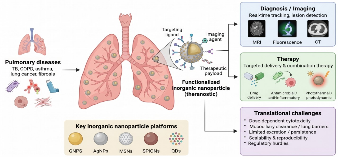
Graphical Abstract

1. Introduction

Pulmonary diseases are among the leading causes of morbidity and mortality worldwide. They include tuberculosis (TB), chronic obstructive pulmonary disease (COPD), asthma, interstitial lung diseases, and pulmonary fibrosis. Their health and economic costs stem from three shared features: high prevalence, a chronic course, and management that consumes clinical resources over years rather than weeks. TB and COPD carry the largest global burden, though asthma and lung cancer are not far behind. Reducing that burden requires better diagnosis, better therapy, and prevention, and progress in any one area is limited by the other two [1].
TB remains a persistent global threat, especially in low- and middle-income countries with limited healthcare resources. According to the World Health Organization, TB caused 1.23 million deaths and about 10.7 million cases in 2024 [2]. The emergence of multidrug-resistant TB (MDR-TB) and extensively drug-resistant TB (XDR-TB) has further complicated control efforts [3]. Similarly, COPD, driven primarily by smoking, air pollution, and occupational hazards, is the third leading cause of death globally and affects millions of people. Unlike TB, COPD is not reversible but is treatable, often leading to substantial long-term disability and a heavy economic burden on healthcare systems. By contrast, asthma disproportionately affects children and young adults, causing significant morbidity and an overall reduction in quality of life. Pulmonary fibrosis and interstitial lung diseases are less common but often devastating, as they are associated with progressive scarring of lung tissue and limited treatment options [4].
Despite advances in medicine, diagnosing pulmonary disease remains difficult. These conditions share early symptoms, including chronic cough, shortness of breath, and fatigue, so misdiagnosis and delay are common, and the first presentation of TB or lung cancer is easily mistaken for a milder respiratory illness. Each available test has a weakness. Spirometry, the standard for COPD and asthma, is underused outside well-resourced settings. Sputum microscopy for TB lacks both sensitivity and specificity. GeneXpert improved TB detection but costs more and depends on infrastructure many centers lack. CT and MRI, on which the diagnosis of lung cancer and interstitial disease rests, are too expensive for routine use in much of the world [5,6]. Treatment is no simpler. TB requires at least six months of antibiotics, which strains adherence and selects for resistance, and MDR-TB requires second-line drugs that are toxic and expensive [7]. COPD is managed with inhaled therapy and lifestyle change, and in severe cases with lung volume reduction surgery or transplantation, none of which is curative. Asthma control improves with inhaled corticosteroids and bronchodilators, but uncontrolled disease persists, particularly in low-income regions [8]. Pulmonary fibrosis and interstitial diseases are limited by poorly understood pathogenesis [9]. Theranostics has been proposed to address diagnostic and therapeutic challenges simultaneously.
Theranostics couples a diagnostic function, such as imaging or biomarker detection, with a therapeutic one, such as a drug, radiation, or gene therapy, in a single construct. The appeal for respiratory disease is that a lesion can be located and treated in the same step. The diseases in question span bacterial and viral infections, including pneumonia, TB, and COVID-19; chronic inflammatory conditions such as asthma and COPD; and malignancy. They share limited treatment efficacy, antibiotic resistance, adverse effects, and diagnostic delays. Nanoparticles are the usual vehicle, chosen for high surface area, tunable size, and surfaces that accept drugs or targeting ligands. Anti-TB drugs can be directed to infected lung tissue, lowering systemic exposure; nanoparticle-based inhalers may improve airway delivery in COPD and asthma; and in lung cancer, particles can be designed to bind to tumor cells and spare surrounding tissue [10].
Nanoparticles have diagnostic uses as well. Magnetic nanoparticles improve imaging of lung tumors and fibrotic regions. Functionalized particles can act as biosensors for asthma or COPD biomarkers, enabling non-invasive testing. Theranostic constructs are also proposed for following treatment response over time. Three obstacles stand between these approaches and the clinic. Production cost and scalability are the first, and they bite hardest in the resource-limited settings where the disease burden is greatest. Regulatory validation is the second, since trials must establish safety and efficacy for a product class with little precedent [11,12]. Third, using these technologies on a scale would require investment in infrastructure and staff training that few health systems have done.
Nanotechnology has been applied to both halves of this problem. On the diagnostic side, nanomaterial-based platforms combining quantum dots, dendrimers, microfluidics, and smartphone readouts have been developed for rapid point-of-care testing, an approach validated earlier for HIV antibody detection and now extended to respiratory pathogens [13]. On the therapeutic side, nanoparticles are classified as inorganic, organic, lipid-based, or polymeric, each with a different profile. Inorganic nanoparticles, which include metals (gold, silver, copper), metal oxides (iron, titanium, zinc, magnesium oxides), and semiconductors, are chemically durable, tolerate sterilization and aerosolization, and kill bacteria through mechanisms that resistance genes do not readily counter membrane disruption, ROS generation, and metal ion release [14,15]. These same optical, magnetic, and structural properties allow a single inorganic particle to carry a diagnostic function alongside a therapeutic one.
Reviews of inorganic nanoparticles in respiratory medicine are not scarce, and another descriptive survey would add little. We therefore organized this review around two questions that published work rarely answers. First, are the diagnostic and therapeutic functions of a given construct genuinely coupled, or merely present on the same particle? Second, was the evidence for a given claim generated under conditions that resemble the lungs, or imported from subcutaneous tumor models, cell culture, or unrelated organ systems? Section 2 sets out the classification we use for the first question and Section 3 the pulmonary constraints relevant to the second. Section 4 and Section 5 apply to both surface modification chemistry and the five platform classes. Section 6 reviews clinical status, Section 7 treats limitations material by material rather than as a single list, and Section 8 sets out what would need to change for these platforms to reach patients.

2. Defining a Theranostic Platform

The word theranostic is used loosely. A substantial fraction of studies carrying the label report a particle that images well, or a particle that treats well, characterized in separate experiments and then described together. Others use it for a carrier that could in principle be loaded with a contrast agent. If the term is to do any work in a review, it needs a definition that some studies fail to provide. We use three tiers and indicate throughout which tier the primary evidence supports.
Type I platforms are those in which one physical property produces both the diagnostic signal and the therapeutic effect. Gold is the clearest case: localized surface plasmon resonance generates the photoacoustic and computed tomography (CT) contrast and also the photothermal heating, so imaging and treatment are inseparable consequences of the same particle under the same illumination [16,17]. SPIONs qualify on the same grounds, since T2 contrast, magnetic hyperthermia, and magnetically guided accumulation all derive from the magnetite core [18,19]. In these systems, the imaging signal reports on the therapeutic agent, which makes dose guidance possible in principle.
Type II platforms carry diagnostic and therapeutic cargo on one construct, but the two act through unrelated mechanisms. An MSN loaded with a fluorophore and a chemotherapeutic is the standard example. The imaging readout tracks where the carrier went; it does not report whether the drug was released, how much was released, or whether the tumor or lesion responded. This distinction is often blurred in claims about real-time therapeutic monitoring.
Type III covers particles demonstrated in a single role so far, with a plausible but unrealized second function. Most AgNP work in the lungs falls in this category. Antibacterial and anti-inflammatory evidence is substantial, whereas imaging evidence is thin and largely non-pulmonary. Table 1 assigns five platforms to these tiers based on pulmonary evidence rather than what is achievable in principle.

3. Pulmonary-Specific Design Constraints

Most inorganic nanoparticle chemistry is developed in subcutaneous tumor models, in cell culture, or for systemic infection, and then proposed for respiratory use. The lungs impose constraints that those systems do not, and they determine whether a particle that performs well elsewhere is usable at all. Four of them are decisive.

3.1. Inhaled Versus Systemic Administration

The two routes are not interchangeable, and evidence generated under one should not support claims about the other. Systemic dosing relies on the enhanced permeability and retention (EPR) effect for tumor accumulation [30]. EPR was characterized in rapidly growing subcutaneous xenografts; in orthotopic and human lung tumors the effect is heterogeneous, varying between and within lesions, and in fibrotic, infective, and inflammatory lung disease there is no comparable vascular defect to exploit. Arguments built on EPR therefore do not transfer to the non-oncological pulmonary indications that make up most of the disease burden discussed in Section 1.
Inhaled delivery bypasses first-pass metabolism and systemic dilution and raises local concentration by one to two orders of magnitude relative to an equivalent intravenous dose [31,32]. Lung deposition is controlled by aerodynamics rather than colloidal particle dimensions. Aerodynamic particle sizes between approximately 1 and 5 µm preferentially target the peripheral airways and alveolar deposition, while particles beyond 5 µm affect the oropharynx. Below around 500 nm particle size, deposition is mostly dictated by Brownian diffusion as opposed to impaction or sedimentation. Deposition and retention levels of particles smaller than 500 nm are influenced by aerodynamic particle size, aggregation, breathing pattern, and airway dimensions. A 30 nm gold nanoparticle is therefore not an inhalable medicine. It becomes one only after formulation, typically by spray drying or spray freeze drying into porous microparticles that disperse into their nanoscale components after deposition, or by nebulization of a suspension in which droplet size, not particle size, sets the deposition pattern [24,33]. The design problem has two scales that must be satisfied at once, and papers that report only nanoscale characterization have addressed only half of it.

3.2. Mucus, Surfactant, and the Acquired Surface

A deposited particle immediately meets a fluid layer that traps foreign material. In the conducting airways, this is mucus, a crosslinked mucin mesh with pore sizes on the order of hundreds of nanometers in health, and considerably smaller and more adhesive in cystic fibrosis, COPD, and severe asthma, where sputum is dehydrated and enriched in DNA and actin [34]. Trapping is as chemical as steric: hydrophobic and cationic surfaces bind mucin domains and are cleared, whereas dense coatings of low-molecular-weight PEG produce near-neutral, hydrophilic surfaces that diffuse through the mesh. Coating chemistry and density are not interchangeable variables. Coating identity defines the nature of the interface and regulates corona composition, colloidal stability, and cellular contacts, whereas density dictates whether this chemistry manifests as a uniform hydrophilic interface or a sparse layer that reveals mucoadhesive core. For PEG, mucus penetration is only documented above a threshold coating density, which is rarely reported.
In the alveolar region, the relevant interface is pulmonary surfactant. Phospholipids and surfactant proteins adsorb within seconds of deposition, so the surface presented to epithelium and macrophages is a lipid and protein corona, not the engineered surface. This layer can bury targeting ligands, and SP-A and SP-D opsonize particles and accelerate macrophage recognition. Gold nanoparticles deposited directly from the aerosol phase into lung lining fluid acquire a larger and compositionally different corona than the same particles suspended in that fluid, so corona measurements made in suspension do not describe what happens on deposition [35]. Surfactant protein D agglomerates gold nanoparticles in vitro, although its effect on early clearance and translocation in vivo was small [36]. Any targeting claim based on ligand density measured in buffer should therefore be treated as provisional until repeated in surfactant or bronchoalveolar lavage fluid.

3.3. Macrophage Uptake and Clearance

Alveolar macrophages remove deposited particles efficiently, and the consequence depends entirely on the disease. In TB, macrophages are where Mycobacterium tuberculosis lives, so avid uptake is the delivery mechanism rather than an obstacle, and the design goal is uptake followed by drug release in the phagolysosome [25]. In lung cancer, fibrosis, and inflammatory airway disease, the same uptake removes the dose from where it is needed. Statements that a particle should evade macrophages, or should be captured by them, are therefore indication-specific, and a general recommendation for either is not meaningful.
Clearance operates on two timescales. The mucociliary escalator moves material deposited in the conducting airways out within hours, limiting the airway compartment’s utility for sustained release. Alveolar deposition is retained for much longer, on the order of weeks to months for poorly soluble material, which supports sustained delivery but is the same property that produces the persistence and chronic toxicity concerns discussed in Section 7 [31,37]. Retention is both an advantage and a hazard through one mechanism, and formulation alone cannot separate the two.

3.4. Disease-Specific Barriers

The barriers differ sufficiently across pulmonary diseases that a single optimized particle is unlikely to serve them all. In cystic fibrosis and advanced COPD, mucus plugging blocks access to entire regions, so deposition follows ventilation and concentrates in the healthier lung rather than the diseased lung, the opposite of what is desired. In pulmonary fibrosis, extracellular matrix deposition stiffens tissue and lengthens diffusion paths, and fibrotic regions are the least ventilated. Intratracheal gadolinium-based nanoparticles combined with ultrashort echo time MRI produced 120% signal enhancement in fibrotic lesions against 50% in healthy tissue in bleomycin-treated mice. The nanoparticle elimination constant was 54% higher in fibrotic regions, so altered retention in diseased tissue is measurable and can itself serve as a marker [38]. In TB, the granuloma presents a fibrous cuff and, in caseating lesions, a necrotic avascular core. High-resolution mapping of fluoroquinolone distribution in rabbit lesions showed that macrophage content, distance from the lesion border, and the extent of necrosis together determine how far the drug penetrates, leaving the center of caseous lesions least well exposed [39]. Binding to caseum macromolecules is the dominant chemical driver and correlates inversely with free diffusion into the necrotic core, with lipophilicity and poor solubility the main predictors [40]. These findings are relevant to both nanoparticle carriers and free drugs, and simply increasing particle delivery to the airway does not address these issues. In lung cancer, blood supply and tumor pressure vary between the primary tumor and metastases, meaning a particle reaching one may not reach the other. Listing these four diseases highlights how each uniquely impedes delivery, and a solution effective for one does not necessarily work for the others. Therefore, a particle should be specifically designed for a particular disease from the start. Work labeled only as pulmonary theranostics without a specified indication lacks a clear target for tailored design.

4. Surface Modification of Inorganic Nanoparticles

The surface, not the core, determines most of what a nanoparticle does in a biological setting. It determines colloidal stability, which protein corona forms, which cells take up the particle, how quickly it is cleared, and whether an attached ligand remains accessible. Functionalization involves attaching chemical groups, commonly thiols, disulfides, amines, nitriles, carboxylic acids, phosphines, or biomolecules, to give a surface with the properties the application requires [41,42].
Functionalization attaches a molecule to the particle surface whose chemistry suits the intended use, substantially changing the particle’s surface characteristics and, with them, its behavior in biological fluid [42]. Table 2 lists the modifications applied to each platform in this review.
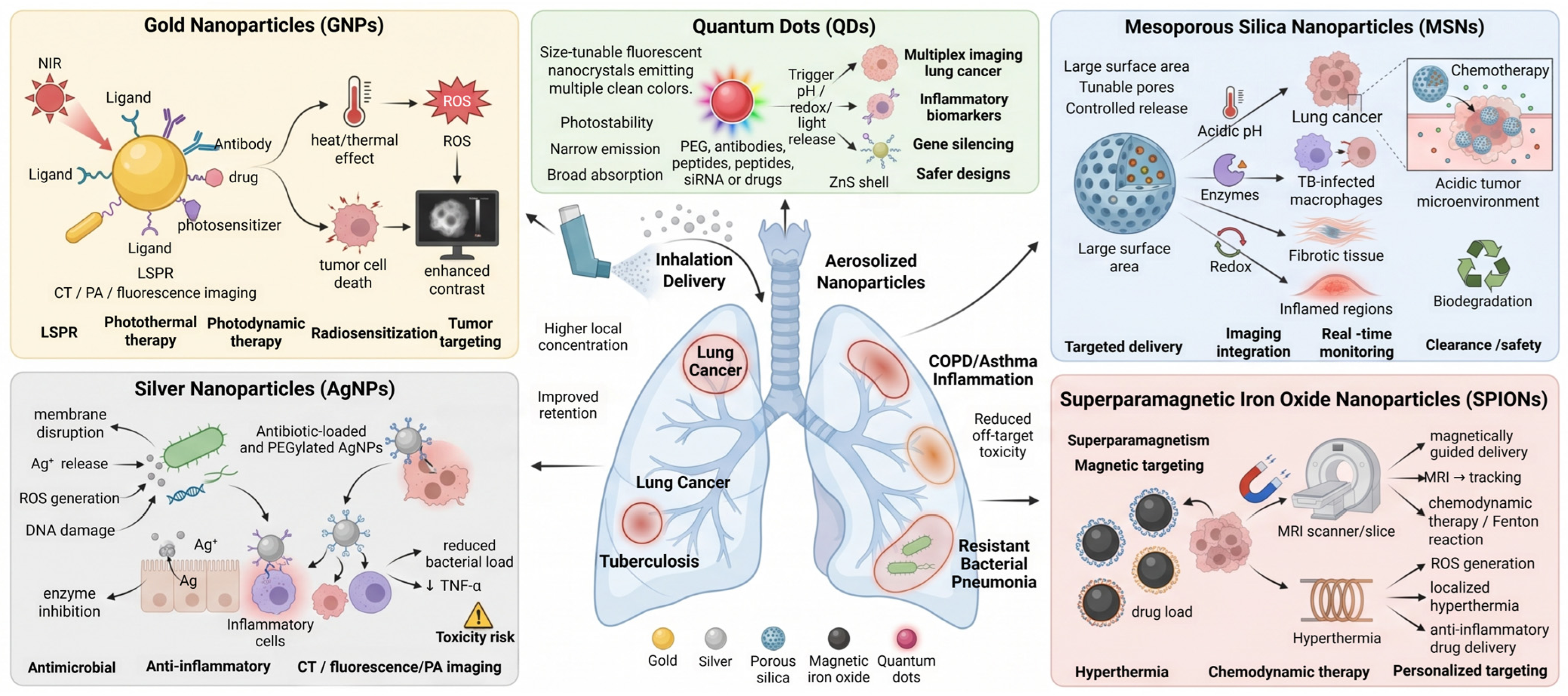
Functionalization strategies for NPs are based on their surface affinity for specific chemical groups. Noble metals such as gold (Au) and silver (Ag) are typically functionalized with thiols and, less often, amines and cyanides. Oxides, including magnetic iron oxides, are coated via oxygen bonding with acidic and hydroxyl groups. Binary compounds, including fluorescent semiconductor NPs like quantum dots from Groups 12 to 16, have a high affinity for thiols and hydroxyl groups, and amino groups are also frequently used. Molecules such as polyethylene glycol (PEG), zwitterionic ligands, glycans, and aptamers are used to preserve NP stability in biological fluids, with the anchoring strategy chosen based on NP composition and binding affinities. Generally, thiols, amines, and hydroxyls are the key functional groups used to attach ligands to NP surfaces. Table 2 summarizes the modifications applied to each platform and the reported pulmonary applications, with the primary evidence indicated for each entry. Figure 1 shows a graphical summary of the platforms and their pulmonary applications.

Comparative Effects of Modification Strategies

Listing modifications is of limited use without comparing their costs. Each strategy trades one property for another, and in the pulmonary context the trade-offs differ from those in systemic delivery. Table 3 sets out the main classes against six criteria; several points deserve comment.
PEGylation is the default choice and remains the best-characterized, but its two main benefits work against each other in the lungs. Dense, low-molecular-weight PEG improves mucus penetration; longer chains extend systemic circulation but entangle with mucin. PEGylation also delays macrophage uptake, which is desirable in oncology and counterproductive for intracellular infections such as TB, where the macrophage is the target compartment. No PEG configuration is simultaneously optimal for both, so the choice must follow the indication.
Ligand-based targeting generally enhances in vitro specificity more reliably than in vivo, as the protein corona that forms on deposits can obscure the ligand. Therefore, targeting improvements observed in buffer or serum-free medium are weak indicators of pulmonary performance. Additionally, antibody conjugation often decreases colloidal stability and increases manufacturing costs and batch variability, posing challenges for translation. While aptamers can help lower some costs, they have limited experience in pulmonary applications.
Inorganic shells present the clearest conflict between safety and clearance. A ZnS shell on a CdSe core suppresses Cd2+ leaching and improves quantum yield, and a silica shell on gold or magnetite does the same for ion release, but both add non-degradable material to a compartment where persistence is already the main long-term concern. The shell reduces acute toxicity and worsens chronic retention. Similarly, doping SPIONs with cerium oxide to scavenge ROS lowers oxidative toxicity while directly antagonizing the ROS-dependent chemodynamic mechanism that the same particles are proposed to exploit [18,58], a design conflict that is not usually acknowledged when both functions are listed as advantages.
Stimuli-responsive gating requires careful consideration. Many pH-triggered systems are tailored to the acidic tumor environment, typically around pH 6.5 to 6.8. However, airway surface liquid in inflamed or infected airways is only mildly acidic, so gates designed for tumors may not function reliably in conditions like COPD, asthma, or bronchiectasis. Enzyme-responsive gates that use proteases common in inflamed airways are more suitable for these cases. Additionally, redox gating depends on an intracellular glutathione gradient that only forms after cellular uptake [53].

5. Role of Inorganic Nanoparticles in Pulmonary Theranostics

5.1. Gold Nanoparticles

Gold nanoparticles (GNPs) are among the most studied materials for theranostics, which combines diagnostic and therapeutic functions on a single platform. The pulmonary diseases at issue, lung cancer, TB, COPD, and viral respiratory infection, together account for a large share of global disease burden [62,63,64]. Gold suits this role because it is biocompatible, optically active, and readily functionalized [65,66]. Localized surface plasmon resonance enables strong light absorption and scattering [16], supporting fluorescence, photoacoustic, and computed tomography (CT) imaging [67]. Below 100 nm, GNPs gather in tumor tissue through the enhanced permeability and retention (EPR) mechanism, which is the primary basis for most GNP oncology research [30]. The same surface that carries a targeting ligand can also carry a drug or photosensitizer, enabling dual functionality.

5.1.1. Systemic Administration and EPR-Dependent Accumulation

Lung cancer is the principal focus of GNP-based theranostics, and most reported work uses intravenous administration with EPR-driven accumulation [68]. Epidermal growth factor receptor (EGFR)-targeted GNPs combined with radiotherapy have improved tumor regression through radiosensitization, with gold’s high atomic number increasing local dose deposition [46]. Gold nanoprisms functionalized with PD-L1 binding peptides and chlorin e6 have been used for imaging-guided photothermal therapy (PTT) and photodynamic therapy (PDT), with selective accumulation in PD-L1-high lung cancer cells and an additive effect from the two light-driven modalities [20]; the same system reported photothermal conversion efficiency of approximately 81% [20]. Related antibody-modified and multifunctional gold constructs have been reviewed elsewhere [69,70].
These results were obtained by systemic dosing, with the following limitations. EPR-dependent accumulation varies in orthotopic lung tumors and is largely absent in inflammatory and infective lung disease, and the fraction of an intravenous gold dose reaching a lung lesion is typically low, with the remainder distributed to the liver and spleen. Efficacy demonstrated this way supports the photophysics and the targeting chemistry. It does not establish that the same construct would behave comparably when delivered to the airway.
Photothermal therapy exploits GNPs’ ability to convert absorbed light into localized heat, killing cells in the illuminated volume [17]. Combined with PDT, in which a photosensitizer generates ROS under light exposure, the two mechanisms target different pathways, and their effects are additive [71]. Gold nanoprisms have reached photothermal conversion efficiencies of approximately 81% with effective tumor ablation [20].
Gold has also been applied to inflammatory and infective pulmonary diseases. Anti-inflammatory activity has been used to moderate cytokine-driven injury in severe respiratory infection, including COVID-19 [72], and antibiotic-loaded GNPs have shown activity against drug-resistant respiratory pathogens [73,74]. Multifunctional composites extend this further: gold–silica constructs doped with photosensitizers combining imaging with PTT and PDT [75], and gold-magnetic hybrids allow dual-modal imaging with magnetically guided delivery [76].

5.1.2. Inhaled Delivery

Inhaled GNP formulations take a different approach, depositing particles directly in the lungs and increasing local concentration without relying on tumor vasculature [48]. This route is more relevant for TB, COPD, and localized infection, where no EPR-like mechanism exists. Because free gold nanoparticles in the inhalable size range are largely exhaled, the reported formulations pair GNPs with carriers, embedding them in liposomes, polymeric microparticles, or porous excipient matrices that deposit by aerodynamic size and then release the nanoparticles [49]. These composites have shown improved lung retention and controlled release compared with solution instillation.
Aerosolized gold nanoparticle studies mostly serve a single function, reporting either deposition and imaging contrast or a therapeutic endpoint. So, the coupled Type I behavior that gold nanoparticles support in principle has rarely been demonstrated after inhalation. Photothermal and photodynamic activation also requires light delivery to the lesion, which for peripheral lung disease means bronchoscopic or interstitial fiber placement rather than surface illumination. This is feasible for central airway tumors and impractical for diffuse or peripheral disease, and it constrains where gold-based PTT and PDT can realistically be used regardless of how the particle is delivered.
Several problems stand between these results and clinical use. Gold is not biodegradable, and inhaled gold has been shown to cause pneumonia, fibrosis, chronic inflammatory infiltration, and vascular changes in rodent lungs, depending on size, dose, and coating [77]. Synthesis variability and scale-up remain unresolved, particularly for anisotropic shapes whose optical properties depend on tight dimensional control, and residual cetyltrimethylammonium bromide from nanorod synthesis is cytotoxic if incompletely exchanged. Biodegradable coatings and designs that permit clearance, rather than only reducing acute toxicity, are the more useful strategies [78].

5.2. Silver Nanoparticles

Silver nanoparticles (AgNPs) have attracted significant attention recently for their unique physical, chemical, and biological properties. As nanotechnology continues to evolve, these tiny metallic particles have found extensive use in biomedical research, particularly in theranostics, a hybrid discipline that combines diagnostic imaging and therapeutic interventions [79]. Pulmonary diseases remain a leading cause of morbidity and mortality worldwide, and innovative strategies are needed to improve both diagnosis and treatment outcomes. Silver nanoparticles show promise, and with their antimicrobial, anti-inflammatory, and imaging-enhancing properties, AgNPs hold substantial potential as theranostic agents for a broad spectrum of lung pathologies [80,81,82].
AgNPs are nanostructures of elemental silver with at least one dimension between roughly 30 and 200 nm [83]. Their electrical, thermal, and optical properties are size-dependent and differ from those of bulk silver [84,85]. In biomedicine, they are used mainly for antimicrobial and anti-inflammatory activity, and they can be conjugated to biomolecules or imaging agents [86]. Size and shape are controllable, with spheres, rods, cubes, triangles, and wires all reported. Surfaces are modified with PEG, antibodies, small molecules, or fluorescent dyes to improve biocompatibility, stability, and targeting; these modifications also govern release rate, toxicity, and tissue selectivity [87].
The synthesis method matters primarily because it determines the surface left behind, which controls the dissolution rate that affects both efficacy and toxicity. Chemical reduction with borohydride or ascorbate provides good size control but leaves residual reductant and capping agents whose alveolar tolerance is rarely assessed. Physical methods such as laser ablation produce ligand-free surfaces that dissolve faster and are more difficult to stabilize in the ionic, protein-rich alveolar environment. Green synthesis using plant extracts or microbial systems yields biocompatible capping layers, but their composition is poorly defined and batch-variable, a substantial regulatory obstacle for an inhaled product where the capping layer is part of the mechanism [88]. For pulmonary use, capping-layer reproducibility is a more relevant selection criterion than the synthesis route, and none of the three methods currently delivers it to the level an inhaled formulation would require.
The therapeutic case for AgNPs in the lungs rests on four mechanisms. They disrupt microbial membranes, generate ROS that damage cellular components, interfere with DNA replication, and inhibit microbial enzymes. Because these act in parallel, resistance develops more slowly than against a single-target antibiotic, which is the main argument for using silver against resistant respiratory pathogens. Silver also suppresses pro-inflammatory signaling, including cytokine and tumor necrosis factor alpha (TNFα) release, which matters in COPD and asthma, where the inflammatory cycle rather than the pathogen drives tissue damage [82,89,90].
Size, shape, and surface chemistry can be tuned to slow alveolar clearance and extend residence in lung tissue [22], and therapeutic agents, antibiotics, anti-inflammatories, or targeting ligands can be attached to the same surface [51]. Imaging capability is the weaker half of the theranostic claim for silver. AgNPs have optical and photoacoustic properties that, in principle, support CT, optical coherence tomography, photoacoustic, and fluorescence contrast, but the demonstrations supporting this claim come from non-pulmonary settings, including dental applications [21], and we are not aware of a study establishing AgNP-based imaging in a pulmonary disease model. Where silver has been followed in the lungs, it has been through radiolabelling rather than its own optical properties: aerosolized 111Ag-labelled silver-carrying nanoparticles retained 62 to 68 percent of the one-hour lung dose at 24 h in mice, a pharmacokinetic readout rather than a diagnostic image [91]. Silver’s imaging role in the lungs should therefore be treated as an open possibility rather than a demonstrated capability, and the pulmonary evidence base supports AgNPs as therapeutic agents with theranostic potential rather than as working theranostic platforms.
AgNPs have been proposed for MDR-TB, pneumonia, COPD, and lung cancer because of their antimicrobial and anti-inflammatory activity. They can be aerosolized or formulated for inhalation, which raises local concentration and limits systemic exposure [21,23]. The same chemistry sets the limits. Excess ROS production, Ag+ release, and accumulation in lung tissue all scale with dose and with the dissolution rate that the coating controls [50]. Safety evaluation therefore has to address particle size and surface modification together rather than dose alone, since these determine whether chronic inflammation and oxidative injury follow repeated administration [92].
Consistent, large-scale synthesis with a reproducible capping layer, guided by disease-specific biomarkers, is a practical precondition for AgNP use in the lungs, and cost-effective production matters in low- and middle-income settings where the relevant infection burden is concentrated.

5.3. Mesoporous Silica Nanoparticles

Mesoporous silica nanoparticles (MSNs) are used in pulmonary theranostics for their structure rather than for any intrinsic optical or magnetic property [93]. The porous framework provides a large surface area and adjustable pore size, so one particle can carry therapeutic agents, imaging molecules, or multiple drugs [94]. Surface functional groups then determine what the particle does in the lungs, from targeted delivery to imaging [95].
The main practical advantage of MSNs is delivery to lung tissue without passing through the systemic circulation. Inhalable formulations reach the deep lungs with limited off-target exposure, which matters most in pulmonary fibrosis, TB, COPD, and lung cancer, where early and localized intervention changes outcome. MSNs loaded with anti-inflammatory or antifibrotic agents have reduced inflammation and scarring in animal models [24,33,96].
Controlled release prevents the payload from escaping before the particle reaches the target [97]. Stimuli-responsive designs use pH, enzyme, or redox triggers to hold the drug in the pores until the particle reaches the target site [53]. In lung cancer, the acidic tumor microenvironment can release a chemotherapeutic held in the pores, increasing local cytotoxicity while limiting exposure of healthy tissue [96,98]. The comparative effects of modification strategies section discusses why triggers calibrated for tumor pH transfer poorly to inflamed airways.
MSNs can also carry diagnostic components. Fluorescent dyes, MRI contrast materials, and radionuclides can be incorporated into the framework or attached to the surface [99], allowing imaging to follow nanoparticle accumulation in tumors or inflamed regions [55,56]. It is important to state precisely what this does and does not show. Tracking the carrier reports biodistribution; it does not report drug release, because the imaging agent and the drug are physically distinct and are not coupled to the same trigger. A better design would let the imaging signal change when the drug is released. One way to do this is dye quenching. While the pore is closed, the dye sits close enough to the particle that its fluorescence is switched off. When the gate opens and the drug comes out, the dye is freed and starts to glow. The light appearing tells you the drug has been released, not merely that the particle arrived. However, there is no report of such a system in a lung model. Therefore, real-time monitoring of drug release from MSNs in the lungs is something researchers are aiming for in pulmonary delivery.
Targeting specificity is a further advantage of MSN-based formulations. Coating the surface with ligands, antibodies, or peptides that bind markers overexpressed on diseased lung cells directs the particles to the intended site, increases uptake there, and reduces systemic exposure [100,101]. The most relevant pulmonary demonstration is in TB: mannose-functionalized MSNs releasing isoniazid and rifampicin inside Mycobacterium tuberculosis-infected macrophages achieved intracellular bacterial killing at substantially lower drug concentrations than free drug in the same model [25], and inhalable anti-tubercular MSN carriers have been reviewed more broadly [102]. Whether improved intracellular delivery translates into shorter therapy in humans has not been tested. Treatment duration in TB depends on sterilizing persistent bacterial populations across multiple anatomical compartments, so shortening it is a hypothesis that would require controlled trial evidence and cannot be presented as an expected benefit of MSN delivery.
Two problems stand between MSNs and clinical use. The first is retention. The framework that protects the cargo also resists degradation, so particles not designed to break down accumulate in lung tissue across repeated doses; biodegradable frameworks and coatings that degrade after release address this [97,103,104]. The second is the lung’s own clearance. Mucociliary transport removes deposited material quickly, so particle size, surface charge, and hydrophilicity must be set against that rather than optimized in isolation [22,34].
MSN development for the lungs is limited less by chemistry than by evidence. Refinements to biodegradation rate, ligand design, and manufacturing reproducibility are all in progress, but the decisive step is repeat-dose inhalation studies that establish whether framework degradation outpaces cumulative deposition at a clinically plausible dosing interval.

5.4. Superparamagnetic Iron Oxide Nanoparticles

Superparamagnetic iron oxide nanoparticles (SPIONs) work as an imaging agent and drug carrier in the same particle [19]. In an external magnetic field, they magnetize strongly, and they lose that magnetization once the field is removed, so they do not aggregate through residual magnetism afterward. This is what allows an external magnet to concentrate them in a chosen lung region and then release them to deliver their payload [105,106,107].
SPIONs act as magnetic resonance imaging (MRI) contrast agents [108]. By shortening T2 relaxation times, they produce dark contrast on MRI scans, separating inflamed or cancerous lung tissue from surrounding healthy tissue. That sensitivity matters in the lungs, where air–tissue interfaces complicate imaging [109,110,111]. Magnetic particle imaging avoids the air–tissue problem altogether by detecting the tracer directly rather than through surrounding protons, and aerosolized magnetic nano-tracers with anisotropic shape have produced high-resolution spatiotemporal images of the mouse lung, currently the most convincing demonstration of inorganic nanoparticle imaging in the respiratory tract [112]. SPIONs also support chemodynamic therapy (CDT) by catalyzing ROS formation inside tumor cells: in the acidic environment of some lung tumors, they release iron ions, which drive the Fenton reaction and generate hydroxyl radicals [113,114]. In an alternating magnetic field, they induce localized hyperthermia, which weakens malignant cells further and adds to the effect of CDT [59,114].
In asthma and COPD, SPIONs have been used to modulate ROS rather than generate it [26]. Doping with cerium oxide adds antioxidant capacity and reduces inflammation-driven tissue damage [115]. A particle that delivers an anti-inflammatory drug and scavenges ROS at the same time would avoid the systemic exposure that long-term corticosteroid use entails, though, as the section comparative effects of modification strategies notes, antioxidant function works against the chemodynamic mechanism described above.
Inhalers and nebulizers deliver SPIONs to the deep lungs, where release can be controlled [116]. Combining them with radiation or conventional chemotherapy raises oxidative stress in tumor cells and increases the effect of both [117]. In cystic fibrosis, SPIONs have been used to deliver antibiotics or gene therapy vectors into mucus-obstructed regions, with MRI tracking dose distribution [118,119].
Several problems still remain before their clinical application. SPIONs are generally biocompatible, but at high concentrations, the ROS they generate damage tissue [120], so the therapeutic window must be established rather than assumed [59,121]. Ultrasmall SPIONs clear faster through physiological pathways, which reduces long-term accumulation in the lungs [122]. PEG and biodegradable polymer coatings improve stability and biocompatibility [123].
Targeting requires disease-specific ligands on the particle surface. Antibodies and peptides against markers restricted to the target tissue improve delivery precision and reduce systemic effects [124]. In inflammatory lung disease, ligands against inflammatory proteins would concentrate particles carrying antioxidants or anti-inflammatory drugs in the affected regions [125]. Where mucus hypersecretion is the barrier, SPION formulations have been designed to cross it while providing both anti-infective activity and imaging contrast [126].
SPIONs combine MRI contrast, magnetically guided accumulation, hyperthermia, and chemodynamic activity in one material, which places them, with gold, in the small group of genuinely shared-mechanism platforms. The pulmonary literature nonetheless reports the imaging and therapeutic arms largely in separate studies, and the coupling that makes them Type I platforms has yet to be demonstrated in a lung disease model.

5.5. Quantum Dots

Inorganic quantum dots (QDs) are semiconductor crystals of 2 to 10 nm that carry imaging and therapeutic function on the same particle [127]. Compared with organic dyes, they are more photostable; their emission wavelength is set by size, and they absorb across a broad range while emitting in narrow peaks, allowing several targets to be imaged at once in lung tissue [128,129]. Common compositions include cadmium selenide, cadmium telluride, zinc sulfide, and indium phosphide [60,130]. Researchers can administer QDs by inhalation or intravenously and functionalize them with PEG, antibodies, or peptides for targeting [28,60,130].
QDs resist photobleaching, so the same particle can be imaged repeatedly over the course of a disease or a treatment [131,132]. In lung cancer, QDs conjugated to tumor-specific ligands have been used to locate malignant lesions, track metastasis, and measure how much chemotherapeutic reached its target [28,132]. In asthma and COPD, QD probes label inflammatory biomarkers, allowing visualization of inflamed regions and grading disease severity [127,132]. Because emission wavelength depends on particle size, one sample can yield multiple biomarkers [60,132,133].
QDs also carry therapeutic cargo that may include drugs, gene-silencing constructs, or small interfering RNA (siRNA) [61]. A high surface-area-to-volume ratio allows a substantial payload, adsorbed onto the surface or chemically linked to it [134]. Delivered by inhaler or nebulizer, these particles release the drug in the lungs and limit systemic exposure [28]. pH, redox conditions, or an external stimulus can trigger release at the diseased site while sparing healthy tissue [135]. Imaging and therapy in one construct place QDs in the theranostic category; however, as Table 1 shows, these two functions have not yet been fully demonstrated together in a pulmonary model.
Toxicity is the central problem. Cadmium leaches from CdSe and CdTe cores and damages cells and organs, so researchers use alternative cores such as InP and protective ZnS shells [60,136]. Clearance must also be established, since alveolar macrophages internalize QDs and can redistribute them to other organs, so biodistribution studies cannot stop at the lungs [37].
The obstacle for QDs is not imaging performance, but the toxicity of the cores that produce them, and progress depends on whether cadmium-free compositions can reach comparable quantum yield and photostability [60,137]. Emerging tools, including CRISPR-based sensing, organ-on-chip airway models, and near-infrared imaging in the second window, may extend what QDs can do in respiratory disease [138], but current pulmonary evidence supports them as imaging agents with delivery capability rather than as demonstrated theranostic platforms.

6. Clinical Status

Inorganic nanoparticles have reached the clinic in some forms and not in others, and the distinction matters for judging the application of these nano carriers in the field of pulmonary diseases. From CosmoFer, the first inorganic nanoparticle approved by the FDA in 1974, to Hensify, approved by the European Medicines Agency (EMA) in 2019, the approved products are systemic or locally injected agents rather than theranostic constructs [139].
Clinical trials using inorganic nanoparticles currently focus mostly on anticancer and antibacterial medicines. Iron, silica, gold, and silver nanoparticles have shown therapeutic promise in cancer and infectious disorders [139,140,141]. In lung cancer, the Hafnium Oxide radioenhancer NBTXR3, marketed as Hensify and now developed as JNJ-1900 under license to Johnson & Johnson, has advanced furthest. The randomized phase 2 CONVERGE study (NCT06667908) is evaluating intratumoral or intranodal injection alongside concurrent chemoradiotherapy and durvalumab consolidation in stage III unresectable NSCLC; part 1 data reported in 2026 indicate that injection into lung lesions is feasible and tolerable, with complete responses in four of seven patients who completed the full regimen. A separate phase 1 study is examining reirradiation with NBTXR3 in locally recurrent NSCLC (NCT04505267). Both are therapeutic studies. The particle increases the radiation dose deposited in the tumor and carries no diagnostic function, so it is not theranostic under the definition in Section 2, but it is the clearest demonstration that an inorganic nanoparticle can be placed into a lung lesion in humans and tolerated there. IONPs such as Resovist/Cliavist and Feridex/Endorem, approved by the FDA and EMA as MRI contrast agents for liver cancer, have been discontinued in the USA and/or EU because of low commercial and clinical adoption [142].
For theranostic applications in pulmonary disorders specifically, inorganic nanoparticles remain preclinical. Ferumoxytol, an approved iron oxide preparation, is used as an MRI contrast agent across a range of indications, including pulmonary vascular imaging, and gold nanoparticles appear in respiratory studies as breath-sensor arrays for lung cancer detection and as a vaccine scaffold. Each of these uses one half of the theranostic pair, and none combines imaging and therapy in a single lung construct, which is what the classification in Section 2 would predict from the preclinical literature. The gap between preclinical results and human testing is dominated by two unresolved questions: toxicity after repeated pulmonary dosing, and in vivo behavior in lungs whose architecture and clearance differ substantially from rodent models. Neither is likely to be settled by further single-dose efficacy studies. What would move the field is a smaller set of well-characterized constructs tested in repeat-dose inhalation toxicology in a large-animal model, with biodistribution and clearance measured over months rather than days.

7. Limitations

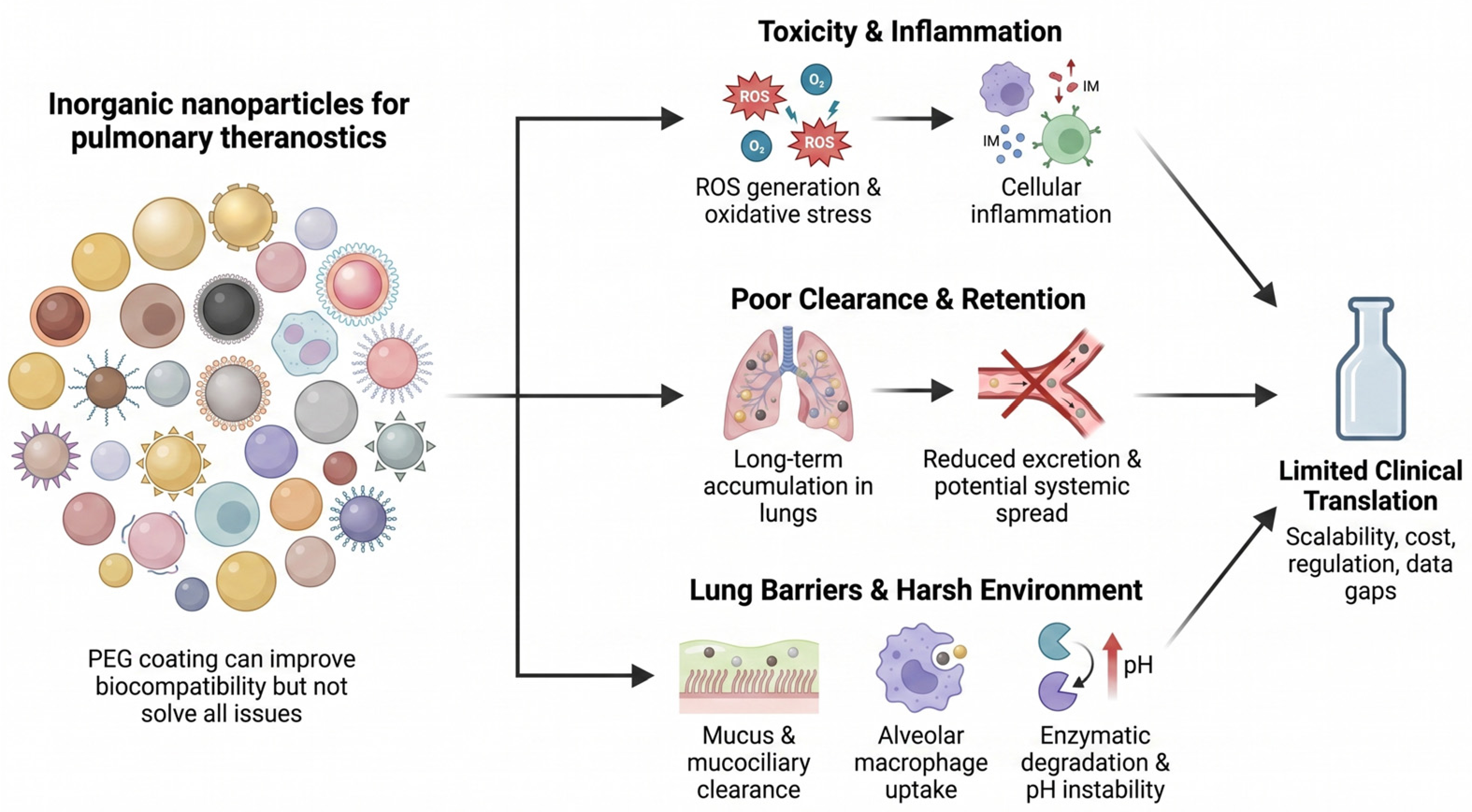
Inorganic nanoparticles are attractive theranostic agents because their nanoscale properties support imaging, sensing, and delivery. Gold supports photoacoustic detection, fluorescence, hyperthermia, and surface-enhanced Raman scattering; iron oxide serves as an MRI contrast agent [143]. Obstacles to clinical use are often presented as a single list, but the dominant risk differs by material, and mitigation strategies that suit one class are irrelevant or harmful for another. Figure 2 provides a schematic overview of the shared translation barriers; the material-specific risks are set out below.

7.1. Material-Specific Risks

Silver: Toxicity is driven principally by dissolution to Ag+ rather than by redox cycling at the particle surface, a mechanism sometimes described as the Trojan horse effect: particles are internalized, then release ions intracellularly at concentrations that free silver could not achieve extracellularly [50]. This matters because ion release is also antibacterial, so the therapeutic and toxic effects share a single origin, and the therapeutic window is intrinsically narrow. Coating strategies that slow dissolution reduce both toxicity and antibacterial potency. Prolonged exposure also produces silver deposition in tissue, and inhaled exposure raises the additional question of pulmonary retention of a slowly dissolving agent that acts as a reservoir [92].
Gold: Metallic gold is chemically inert and does not release ions or catalyze Fenton chemistry; its problem is the opposite one. It is not biodegradable, and particles above the renal filtration threshold of roughly 6 nm are retained in the liver, spleen, and, after inhalation, in alveolar macrophages and lung interstitium for extended periods. Rodent studies of pulmonary gold exposure report pneumonia, fibrotic change, chronic inflammatory infiltrate, and vascular congestion [77]. Residual cationic surfactants from anisotropic synthesis contribute further cytotoxicity that is a synthesis artifact rather than a property of gold. Mitigation therefore means designing for clearance, either through ultrasmall renally clearable cores or through assemblies that disassemble into clearable subunits, not through inert coatings alone [37].
Iron oxide: SPIONs are the one class in this review for which Fenton chemistry is the correct mechanism. Fe2+ released during lysosomal degradation reacts with hydrogen peroxide to generate hydroxyl radicals, which underlie chemodynamic therapy, off-target oxidative injury, and ferroptotic cell death at higher doses [113,114,125]. Iron has an advantage; unlike other materials, its degradation products enter physiological iron handling and are metabolized rather than accumulated, which is why iron oxide agents have reached the clinic. The corresponding risks are local iron overload, oxidative stress in lung tissue already under oxidative burden in COPD and asthma, and the hypersensitivity reactions historically associated with some coated formulations [120].
Mesoporous silica: Silica degrades by hydrolysis of the siloxane framework to soluble silicic acid, which is excreted renally, so silica is biodegradable in a way gold and QD cores are not. The degradation rate depends strongly on framework condensation, pore geometry, and doping, and densely condensed frameworks persist far longer than the literature average suggests [104]. The specific toxicity concern is not redox chemistry but surface silanol groups, which interact with membrane phospholipids and cause hemolysis and membrane damage in a manner that depends on silanol density and can be reduced by surface capping. Repeated inhaled dosing raises the question of cumulative burden if the degradation half-life exceeds the dosing interval, a calculation that is seldom presented.
Quantum dots: The dominant risk is leaching of heavy metals, Cd2+ from CdSe and CdTe cores and Pb2+ from PbS, following oxidative or photolytic degradation of the shell [144,145]. Toxicity therefore tracks core composition and shell integrity rather than particle size or surface charge, and shell integrity must be demonstrated under use conditions, including light exposure during fluorescence imaging, rather than in storage. Cadmium-free alternatives based on InP and Ag2S or Ag2Se cores avoid the worst of this but currently offer lower quantum yield and less chemical stability [60,144,145]. Because alveolar macrophages internalize QDs and can redistribute them systemically, researchers must track biodistribution beyond the lungs [37].

7.2. Shared Translational Barriers

Some problems are shared. Formulating a particle that carries diagnostic and therapeutic function while retaining acceptable size, stability, and manufacturability is demanding and expensive. Small-animal models under-represent the deposition and distribution behavior of larger lungs; rodent inhalation studies with radiolabelled gold indicate that air–blood barrier translocation is broadly comparable to human values for similarly sized particles, but airway geometry, breathing pattern, and clearance rates are not [31,45]. Few studies follow stability or toxicity in chronic respiratory disease models. Regulatory approval has been delayed or refused for nanomedicines because of inadequate study design, poor justification of the clinical comparator, and weak data analysis rather than the underlying science, and early engagement with regulators addresses more of this than is generally assumed [139,142]. Real-world efficacy data across diverse patient populations does not yet exist for any inorganic theranostic platform in respiratory disease.
The lung’s own defenses constrain all five classes. Thick mucus in chronic airway disease limits penetration, the mucociliary escalator removes airway-deposited material within hours, and alveolar macrophages sequester what reaches the periphery. Enzymatic activity, pH shifts, and biomolecule adsorption can also compromise the particle’s structural integrity and functional performance after deposition [146,147]. Section 3 covers these; they are repeated here because they interact with the material-specific risks above, most directly by extending residence time for materials whose main hazard is persistence.

8. Conclusions and Future Directions

Inorganic nanoparticles combine diagnostic and therapeutic function in ways that suit several unmet needs in respiratory medicine, and the underlying chemistry is well developed. Gold and iron oxide couple imaging and therapy through a single physical property and are theranostic in the strict sense. MSNs offer high loading capacity, controllable release, and genuine biodegradability, with their diagnostic function supplied by co-loaded agents rather than intrinsic to the material. QDs have optical properties that no organic fluorophore matches, but their cores are constrained by toxicity. Silver has strong therapeutic credentials in pulmonary infection and inflammation and, on current evidence, no established role in pulmonary imaging.
Applying the classification of Section 2 across the literature yields a consistent result: pulmonary work is concentrated in Type II and Type III categories, and demonstrations of coupled diagnostic and therapeutic function in a relevant lung model are rare. This is the major gap that could be identified as the field’s central problem. It is not a materials problem, since the required photophysics and magnetism already work. However, the studies that could demonstrate coupling both imaging a delivered dose and then showing that the imaged signal predicts the therapeutic outcome in the same animal are harder to achieve than separate demonstrations of individual component effects.
Designing for clearance, not just reduced acute toxicity, would address the persistence problem that affects gold and dense silica most severely; ultrasmall, renally clearable cores and disassembling constructs are two credible routes. Formulation research should treat aerodynamic behavior as a primary design variable rather than a downstream step, since a particle that cannot be delivered to the intended region of the lung is not a candidate regardless of its properties. Repeat-dose inhalation toxicology in large-animal models, with biodistribution monitored over months, is the evidence gap most likely to determine whether any of these platforms will reach trials.

8.1. Computational Approaches

Machine learning is often invoked as a way past these obstacles, usually without specifying what it would do. Its realistic near-term contribution is narrower and is worth stating precisely. Models trained on existing physicochemical and biodistribution datasets can predict protein corona composition from surface properties, which is useful because corona formation determines targeting performance and is expensive to measure empirically. Computational fluid dynamics models of airway deposition, already used in inhaler development, can be coupled to formulation parameters to screen carrier designs before synthesis. Quantitative structure–activity relationship approaches can rank candidate compositions for cytotoxicity screening. They share one thing in common: they can narrow the experimental search space but are not able to remove the need for in vivo work. The binding constraint is data: pulmonary nanoparticle datasets are small, heterogeneously reported, and biased toward positive results, and no model trained on them will generalize well until reporting standards and shared datasets improve [148]. Framing machine learning as a route to de novo nanoparticle design overstates what current data can support, whereas using it to prioritize formulations for testing is now achievable.

8.2. Outlook

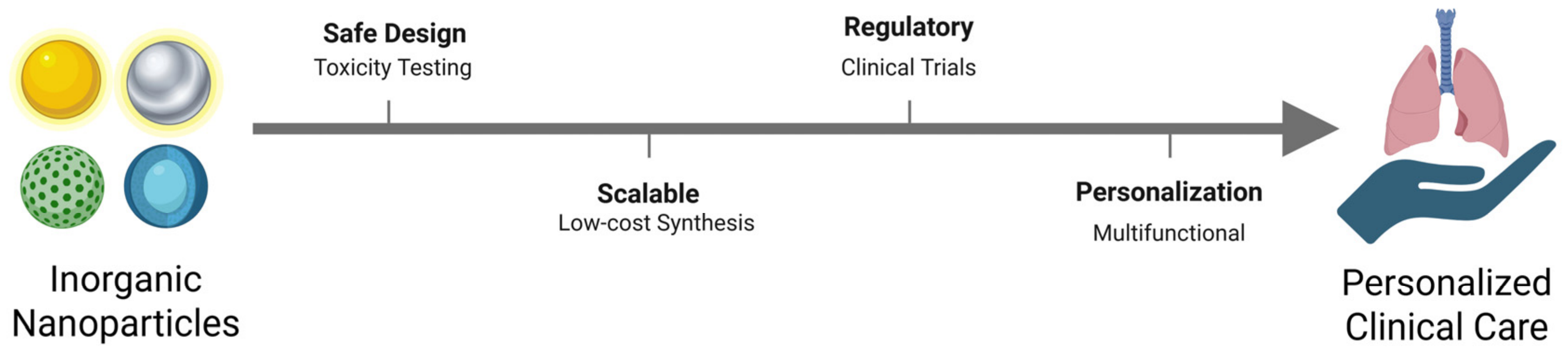
The gap between preclinical results and clinical use in this field is real and is not closing quickly. Closing it depends less on new materials than on taking a smaller number of well-characterized constructs through the unglamorous work of repeat-dose toxicology, deposition modeling, and manufacturing reproducibility, with a nominated indication and a defined comparator from the start. Figure 3 places those steps in the order they must be taken, from safety and toxicity characterization and scalable synthesis through regulatory approval to use in individual patients.

Funding

This research received no external funding.

Institutional Review Board Statement

Not applicable.

Informed Consent Statement

Not applicable.

Data Availability Statement

No new data were created or analyzed in this study. Data sharing is not applicable to this article.

Conflicts of Interest

The authors declare no conflict of interest.

References

  1. Agusti, A.; Vogelmeier, C.F.; Halpin, D.M.G. Tackling the global burden of lung disease through prevention and early diagnosis. Lancet Respir. Med. 2022, 10, 1013–1015. [Google Scholar] [CrossRef] [Scilit] [PubMed]
  2. WHO. Global Tuberculosis Report 2025; World Health Organization: Geneva, Switzerland, 2025. [Google Scholar]
  3. Dheda, K.; Mirzayev, F.; Cirillo, D.M.; Udwadia, Z.; Dooley, K.E.; Chang, K.C.; Omar, S.V.; Reuter, A.; Perumal, T.; Horsburgh, C.R.; et al. Multidrug-resistant tuberculosis. Nat. Rev. Dis. Prim. 2024, 10, 22. [Google Scholar] [CrossRef] [Scilit] [PubMed]
  4. Soriano, J.B.; Lopez-Campos, J.L. The World’s Burden of Chronic Respiratory Disease: An Epidemiological Review of Asthma, COPD, Interstitial Lung Disease, and Smoking in the 21st Century. Arch. Bronconeumol. 2026; in press. [CrossRef] [Scilit] [PubMed]
  5. Song, C.; Xie, Y.; Feng, Z.; Xue, Z.; Panagiotakos, D.; Hao, X. Cost-effectiveness of screening for chronic obstructive pulmonary disease: A systematic review and meta-analysis. BMC Public Health 2025, 25, 3779. [Google Scholar] [CrossRef] [Scilit] [PubMed] [PubMed Central]
  6. Ryu, Y.J. Diagnosis of Pulmonary Tuberculosis: Recent Advances and Diagnostic Algorithms. Tuberc. Respir. Dis. 2015, 78, 64–71. [Google Scholar] [CrossRef] [Scilit] [PubMed] [PubMed Central]
  7. Motta, I.; Boeree, M.; Chesov, D.; Dheda, K.; Günther, G.; Horsburgh, C.R.; Kherabi, Y.; Lange, C.; Lienhardt, C.; McIlleron, H.M.; et al. Recent advances in the treatment of tuberculosis. Clin. Microbiol. Infect. 2024, 30, 1107–1114. [Google Scholar] [CrossRef] [Scilit] [PubMed]
  8. Quint, J.K.; Ariel, A.; Barnes, P.J. Rational use of inhaled corticosteroids for the treatment of COPD. npj Prim. Care Respir. Med. 2023, 33, 27. [Google Scholar] [CrossRef] [Scilit] [PubMed]
  9. Glass, D.S.; Grossfeld, D.; Renna, H.A.; Agarwala, P.; Spiegler, P.; DeLeon, J.; Reiss, A.B. Idiopathic pulmonary fibrosis: Current and future treatment. Clin. Respir. J. 2022, 16, 84–96. [Google Scholar] [CrossRef] [Scilit] [PubMed]
  10. Feyzbakhsh, H. Nanoparticle theranostics for respiratory diseases: Advances and challenges in asthma, lung cancer, and tuberculosis. Egypt. J. Bronchol. 2025, 19, 144. [Google Scholar] [CrossRef] [Scilit]
  11. Yi, M.; Li, Y.; Jie, H.; Deng, S. Nanotechnology-Driven Drug Delivery Systems for Lung Cancer: Computational Advances and Clinical Perspectives. Thorac. Cancer 2025, 16, e70134. [Google Scholar] [CrossRef] [Scilit] [PubMed]
  12. Gulati, N.; Saini, A.; Nagar, L.; Awasthi, R.; Dua, K.; Dureja, H. Nanotechnology-based diagnosis of COPD. In Nanotechnology-Based Innovations in Chronic Obstructive Pulmonary Disease (COPD) Treatment and Management; Academic Press: Cambridge, MA, USA, 2026; pp. 71–86. [Google Scholar] [CrossRef] [Scilit]
  13. Muthukrishnan, L. Multidrug resistant tuberculosis—Diagnostic challenges and its conquering by nanotechnology approach—An overview. Chem.-Biol. Interact. 2021, 337, 109397. [Google Scholar] [CrossRef] [Scilit] [PubMed]
  14. Aguilera-Correa, J.J.; Esteban, J.; Vallet-Regí, M. Inorganic and Polymeric Nanoparticles for Human Viral and Bacterial Infections Prevention and Treatment. Nanomaterials 2021, 11, 137. [Google Scholar] [CrossRef] [Scilit] [PubMed]
  15. Poon, C.; Patel, A.A. Organic and inorganic nanoparticle vaccines for prevention of infectious diseases. Nano Express 2020, 1, 012001. [Google Scholar] [CrossRef] [Scilit]
  16. Mayer, K.M.; Hafner, J.H. Localized Surface Plasmon Resonance Sensors. Chem. Rev. 2011, 111, 3828–3857. [Google Scholar] [CrossRef] [Scilit] [PubMed]
  17. Gupta, N.; Malviya, R. Understanding and advancement in gold nanoparticle targeted photothermal therapy of cancer. Biochim. Biophys. Acta(BBA)—Rev. Cancer 2021, 1875, 188532. [Google Scholar] [CrossRef] [Scilit] [PubMed]
  18. Wahajuddin Arora, S. Superparamagnetic iron oxide nanoparticles: Magnetic nanoplatforms as drug carriers. Int. J. Nanomed. 2012, 7, 3445–3471. [Google Scholar] [CrossRef] [Scilit] [PubMed] [PubMed Central]
  19. Zarepour, A.; Zarrabi, A.; Khosravi, A. SPIONs as Nano-Theranostics Agents. In SPIONs as Nano-Theranostics Agents; Zarepour, A., Zarrabi, A., Khosravi, A., Eds.; Springer: Singapore, 2017; pp. 1–44. [Google Scholar] [CrossRef] [Scilit]
  20. Liu, B.; Qiao, G.; Han, Y.; Shen, E.; Alfranca, G.; Tan, H.; Wang, L.; Pan, S.; Ma, L.; Xiong, W.; et al. Targeted theranostics of lung cancer: PD-L1-guided delivery of gold nanoprisms with chlorin e6 for enhanced imaging and photothermal/photodynamic therapy. Acta Biomater. 2020, 117, 361–373. [Google Scholar] [CrossRef] [Scilit] [PubMed]
  21. Das, A.; Raposo, G.C.C.; Lopes, D.S.; da Silva, E.J.; Carneiro, V.S.M.; Mota, C.C.B.d.O.; Amaral, M.M.; Zezell, D.M.; Barbosa-Silva, R.; Gomes, A.S.L. Exploiting Nanomaterials for Optical Coherence Tomography and Photoacoustic Imaging in Nanodentistry. Nanomaterials 2022, 12, 506. [Google Scholar] [CrossRef] [Scilit] [PubMed]
  22. Cojocaru, E.; Petriș, O.R.; Cojocaru, C. Nanoparticle-Based Drug Delivery Systems in Inhaled Therapy: Improving Respiratory Medicine. Pharmaceuticals 2024, 17, 1059. [Google Scholar] [CrossRef] [Scilit] [PubMed]
  23. Siafaka, P.I.; Özcan Bülbül, E.; Miliotou, A.N.; Karantas, I.D.; Okur, M.E.; Üstündağ Okur, N. Nano-based carriers for pulmonary drug delivery: A review on the available drug delivery applications and toxicity issues. J. Drug Deliv. Sci. Technol. 2024, 92, 105381. [Google Scholar] [CrossRef] [Scilit]
  24. Li, X.; Xue, M.; Raabe, O.G.; Aaron, H.L.; Eisen, E.A.; Evans, J.E.; Hayes, F.A.; Inaga, S.; Tagmount, A.; Takeuchi, M.; et al. Aerosol droplet delivery of mesoporous silica nanoparticles: A strategy for respiratory-based therapeutics. Nanomed. Nanotechnol. Biol. Med. 2015, 11, 1377–1385. [Google Scholar] [CrossRef] [Scilit] [PubMed]
  25. Clemens, D.L.; Lee, B.Y.; Xue, M.; Thomas, C.R.; Meng, H.; Ferris, D.; Nel, A.E.; Zink, J.I.; Horwitz, M.A. Targeted Intracellular Delivery of Antituberculosis Drugs to Mycobacterium tuberculosis-Infected Macrophages via Functionalized Mesoporous Silica Nanoparticles. Antimicrob. Agents Chemother. 2012, 56, 2535–2545. [Google Scholar] [CrossRef] [Scilit] [PubMed]
  26. Al Faraj, A.; Shaik, A.S.; Afzal, S.; Al-Muhsen, S.; Halwani, R. Specific targeting and noninvasive magnetic resonance imaging of an asthma biomarker in the lung using polyethylene glycol functionalized magnetic nanocarriers. Contrast Media Mol. Imaging 2016, 11, 172–183. [Google Scholar] [CrossRef] [Scilit] [PubMed]
  27. Sadhukha, T.; Wiedmann, T.S.; Panyam, J. Inhalable magnetic nanoparticles for targeted hyperthermia in lung cancer therapy. Biomaterials 2013, 34, 5163–5171. [Google Scholar] [CrossRef] [Scilit] [PubMed]
  28. Mehta, P.P.; Dhapte-Pawar, V. Resourceful Quantum Dots for Pulmonary Drug Delivery: Facts, Frontiers, and Future. In Pulmonary Drug Delivery Systems: Material and Technological Advances; Mehta, P.P., Dhapte-Pawar, V., Eds.; Springer Nature: Singapore, 2023; pp. 345–368. [Google Scholar] [CrossRef] [Scilit] [PubMed]
  29. Cai, X.; Luo, Y.; Zhang, W.; Du, D.; Lin, Y. pH-sensitive ZnO quantum dots–doxorubicin nanoparticles for lung cancer targeted drug delivery. ACS Appl. Mater. Interfaces 2016, 8, 22442–22450. [Google Scholar] [CrossRef] [Scilit] [PubMed]
  30. Kalyane, D.; Raval, N.; Maheshwari, R.; Tambe, V.; Kalia, K.; Tekade, R.K. Employment of enhanced permeability and retention effect (EPR): Nanoparticle-based precision tools for targeting of therapeutic and diagnostic agent in cancer. Mater. Sci. Eng. C 2019, 98, 1252–1276. [Google Scholar] [CrossRef] [Scilit] [PubMed]
  31. Jin, Z.; Gao, Q.; Wu, K.; Ouyang, J.; Guo, W.; Liang, X.J. Harnessing inhaled nanoparticles to overcome the pulmonary barrier for respiratory disease therapy. Adv. Drug Deliv. Rev. 2023, 202, 115111. [Google Scholar] [CrossRef] [Scilit] [PubMed]
  32. Feng, X.; Shi, Y.; Zhang, Y.; Lei, F.; Ren, R.; Tang, X. Opportunities and challenges for inhalable nanomedicine formulations in respiratory diseases: A review. Int. J. Nanomed. 2024, 19, 1509–1538. [Google Scholar] [CrossRef] [Scilit] [PubMed]
  33. Campos Pacheco, J.E.; Yalovenko, T.; Riaz, A.; Kotov, N.; Davids, C.; Persson, A.; Falkman, P.; Feiler, A.; Godaly, G.; Johnson, C.M.; et al. Inhalable porous particles as dual micro-nano carriers demonstrating efficient lung drug delivery for treatment of tuberculosis. J. Control. Release 2024, 369, 231–250. [Google Scholar] [CrossRef] [Scilit] [PubMed]
  34. Watchorn, J.; Clasky, A.J.; Prakash, G.; Johnston, I.A.E.; Chen, P.Z.; Gu, F.X. Untangling Mucosal Drug Delivery: Engineering, Designing, and Testing Nanoparticles to Overcome the Mucus Barrier. ACS Biomater. Sci. Eng. 2022, 8, 1396–1426. [Google Scholar] [CrossRef] [Scilit] [PubMed]
  35. Svensson, C.R.; Messing, M.E.; Lundqvist, M.; Schollin, A.; Deppert, K.; Pagels, J.H.; Rissler, J.; Cedervall, T. Direct deposition of gas phase generated aerosol gold nanoparticles into biological fluids—Corona formation and particle size shifts. PLoS ONE 2013, 8, e74702. [Google Scholar] [CrossRef] [Scilit] [PubMed]
  36. Schleh, C.; Holzwarth, U.; Hirn, S.; Wenk, A.; Simonelli, F.; Schäffler, M.; Möller, W.; Gibson, N.; Kreyling, W.G. Biodistribution of inhaled gold nanoparticles in mice and the influence of surfactant protein D. J. Aerosol Med. Pulm. Drug Deliv. 2013, 26, 24–30. [Google Scholar] [CrossRef] [Scilit] [PubMed]
  37. Wang, X.; Zhong, X.; Li, J.; Liu, Z.; Cheng, L. Inorganic nanomaterials with rapid clearance for biomedical applications. Chem. Soc. Rev. 2021, 50, 8669–8742. [Google Scholar] [CrossRef] [Scilit] [PubMed]
  38. Tassali, N.; Bianchi, A.; Lux, F.; Raffard, G.; Sanchez, S.; Tillement, O.; Crémillieux, Y. MR imaging, targeting and characterization of pulmonary fibrosis using intra-tracheal administration of gadolinium-based nanoparticles. Contrast Media Mol. Imaging 2016, 11, 396–404. [Google Scholar] [CrossRef] [Scilit] [PubMed]
  39. Blanc, L.; Daudelin, I.B.; Podell, B.K.; Chen, P.-Y.; Zimmerman, M.; Martinot, A.J.; Savic, R.M.; Prideaux, B.; Dartois, V. High-resolution mapping of fluoroquinolones in TB rabbit lesions reveals specific distribution in immune cell types. eLife 2018, 7, e41115. [Google Scholar] [CrossRef] [Scilit] [PubMed]
  40. Sarathy, J.P.; Zuccotto, F.; Hsinpin, H.; Sandberg, L.; Via, L.E.; Marriner, G.A.; Masquelin, T.; Wyatt, P.; Ray, P.; Dartois, V. Prediction of drug penetration in tuberculosis lesions. ACS Infect. Dis. 2016, 2, 552–563. [Google Scholar] [CrossRef] [Scilit] [PubMed]
  41. Colino, C.I.; Lanao, J.M.; Gutierrez-Millan, C. Recent advances in functionalized nanomaterials for the diagnosis and treatment of bacterial infections. Mater. Sci. Eng. C 2021, 121, 111843. [Google Scholar] [CrossRef] [Scilit] [PubMed]
  42. Subbiah, R.; Veerapandian, M.; Yun, K.S. Nanoparticles: Functionalization and Multifunctional Applications in Biomedical Sciences. Curr. Med. Chem. 2010, 17, 4559–4577. [Google Scholar] [CrossRef] [Scilit] [PubMed]
  43. Zhuang, L.; Lian, Y.; Zhu, T. Multifunctional gold nanoparticles: Bridging detection, diagnosis, and targeted therapy in cancer. Mol. Cancer 2025, 24, 228. [Google Scholar] [CrossRef] [Scilit] [PubMed] [PubMed Central]
  44. Singh, P.; Pandit, S.; Mokkapati, V.R.S.S.; Garg, A.; Ravikumar, V.; Mijakovic, I. Gold Nanoparticles in Diagnostics and Therapeutics for Human Cancer. Int. J. Mol. Sci. 2018, 19, 1979. [Google Scholar] [CrossRef] [Scilit] [PubMed] [PubMed Central]
  45. Kreyling, W.G.; Möller, W.; Holzwarth, U.; Hirn, S.; Wenk, A.; Schleh, C.; Schaeffler, M.; Haberl, N.; Gibson, N.; Schittny, J.C. Age-dependent rat lung deposition patterns of inhaled 20 nanometer gold nanoparticles and their quantitative biokinetics in adult rats. ACS Nano 2018, 12, 7771–7790. [Google Scholar] [CrossRef] [Scilit] [PubMed]
  46. Bromma, K.; Chithrani, D.B. Advances in Gold Nanoparticle-Based Combined Cancer Therapy. Nanomaterials 2020, 10, 1671. [Google Scholar] [CrossRef] [Scilit] [PubMed]
  47. Jeong, S.Y.; Park, S.J.; Yoon, S.M.; Jung, J.; Woo, H.N.; Yi, S.L.; Song, S.Y.; Park, H.J.; Kim, C.; Lee, J.S.; et al. Systemic delivery and preclinical evaluation of Au nanoparticle containing beta-lapachone for radiosensitization. J. Control. Release 2009, 139, 239–245. [Google Scholar] [CrossRef] [Scilit] [PubMed]
  48. Anderson, C.F.; Grimmett, M.E.; Domalewski, C.J.; Cui, H. Inhalable nanotherapeutics to improve treatment efficacy for common lung diseases. WIREs Nanomed. Nanobiotechnol. 2020, 12, e1586. [Google Scholar] [CrossRef] [Scilit] [PubMed]
  49. Kole, E.; Jadhav, K.; Shirsath, N.; Dudhe, P.; Verma, R.K.; Chatterjee, A.; Naik, J. Nanotherapeutics for pulmonary drug delivery: An emerging approach to overcome respiratory diseases. J. Drug Deliv. Sci. Technol. 2023, 81, 104261. [Google Scholar] [CrossRef] [Scilit]
  50. Beer, C.; Foldbjerg, R.; Hayashi, Y.; Sutherland, D.S.; Autrup, H. Toxicity of silver nanoparticles—Nanoparticle or silver ion? Toxicol. Lett. 2012, 208, 286–292. [Google Scholar] [CrossRef] [Scilit] [PubMed]
  51. Gupta, P.C.; Sharma, N.; Rai, S.; Mishra, P. Use of Smart Silver Nanoparticles in Drug Delivery System. In Metal and Metal-Oxide Based Nanomaterials: Synthesis, Agricultural, Biomedical and Environmental Interventions; Bachheti, R.K., Bachheti, A., Husen, A., Eds.; Springer Nature: Singapore, 2024; pp. 213–241. [Google Scholar] [CrossRef] [Scilit] [PubMed]
  52. Boca, S.C.; Potara, M.; Gabudean, A.M.; Juhem, A.; Baldeck, P.L.; Astilean, S. Chitosan-coated triangular silver nanoparticles as a novel class of biocompatible, highly effective photothermal transducers for in vitro cancer cell therapy. Cancer Lett. 2011, 311, 131–140. [Google Scholar] [CrossRef] [Scilit] [PubMed]
  53. Abdo, G.G.; Zagho, M.M.; Khalil, A. Recent advances in stimuli-responsive drug release and targeting concepts using mesoporous silica nanoparticles. Emergent Mater. 2020, 3, 407–425. [Google Scholar] [CrossRef] [Scilit]
  54. Lee, J.; Oh, E.; Song, J.; Kim, H.G.; Park, H.J.; Kim, C. Stimulus-induced conformational transformation of a cyclic peptide for selective cell-targeting on–off gatekeeper for mesoporous nanocarriers. Chem. Asian J. 2017, 12, 2813–2818. [Google Scholar] [CrossRef] [Scilit] [PubMed]
  55. Feng, Y.; Panwar, N.; Tng, D.J.H.; Tjin, S.C.; Wang, K.; Yong, K.T. The application of mesoporous silica nanoparticle family in cancer theranostics. Coord. Chem. Rev. 2016, 319, 86–109. [Google Scholar] [CrossRef] [Scilit]
  56. Sivamaruthi, B.S.; Thangaleela, S.; Kesika, P.; Suganthy, N.; Chaiyasut, C. Mesoporous Silica-Based Nanoplatforms Are Theranostic Agents for the Treatment of Inflammatory Disorders. Pharmaceutics 2023, 15, 439. [Google Scholar] [CrossRef] [Scilit] [PubMed]
  57. Kang, M.S.; Singh, R.K.; Kim, T.H.; Kim, J.H.; Patel, K.D.; Kim, H.W. Optical imaging and anticancer chemotherapy through carbon dot created hollow mesoporous silica nanoparticles. Acta Biomater. 2017, 55, 466–480. [Google Scholar] [CrossRef] [Scilit] [PubMed]
  58. Ban, G.; Chen, Y.; Zhao, R.; Liang, Y.; Chen, S.; Jia, J.; Li, C.; Sima, J.; Ding, D.; Wang, X.; et al. Cerium oxide nanoparticles: Antioxidant properties in silicosis treatment and biomedical application prospects—A dialectical consideration. Drug Deliv. 2016, 33, 2634421. [Google Scholar] [CrossRef] [Scilit] [PubMed] [PubMed Central]
  59. Lamichhane, N.; Sharifabad, M.E.; Hodgson, B.; Mercer, T.; Sen, T. Chapter 13—Superparamagnetic iron oxide nanoparticles (SPI-ONs) as therapeutic and diagnostic agents. In Nanoparticle Therapeutics; Kesharwani, P., Singh, K.K., Eds.; Academic Press: Cambridge, MA, USA, 2022; pp. 455–497. [Google Scholar] [CrossRef] [Scilit]
  60. Ranjbar-Navazi, Z.; Omidi, Y.; Eskandani, M.; Davaran, S. Cadmium-free quantum dot-based theranostics. TrAC Trends Anal. Chem. 2019, 118, 386–400. [Google Scholar] [CrossRef] [Scilit]
  61. Zahed, Z.; Hadi, R.; Imanzadeh, G.; Ahmadian, Z.; Shafiei, S.; Zadeh, A.Z.; Karimi, H.; Akbarzadeh, A.; Abbaszadeh, M.; Ghadimi, L.S.; et al. Recent advances in fluorescence nanoparticles “quantum dots” as gene delivery system: A review. Int. J. Biol. Macromol. 2024, 254, 127802. [Google Scholar] [CrossRef] [Scilit] [PubMed]
  62. Zhang, Y.; Almazi, J.G.; Ong, H.X.; Johansen, M.D.; Ledger, S.; Traini, D.; Hansbro, P.M.; Kelleher, A.D.; Ahlenstiel, C.L. Nanoparticle Delivery Platforms for RNAi Therapeutics Targeting COVID-19 Disease in the Respiratory Tract. Int. J. Mol. Sci. 2022, 23, 2408. [Google Scholar] [CrossRef] [Scilit] [PubMed]
  63. Dey, R.K.; Jana, B.; Dastidar, D.G. Applications of nanotechnology in the treatment of pulmonary diseases. Vessel. Plus 2023, 7, 17. [Google Scholar] [CrossRef] [Scilit]
  64. Marzaman, A.N.F.; Roska, T.P.; Sartini, S.; Utami, R.N.; Sulistiawati, S.; Enggi, C.K.; Manggau, M.A.; Rahman, L.; Shastri, V.P.; Permana, A.D. Recent Advances in Pharmaceutical Approaches of Antimicrobial Agents for Selective Delivery in Various Administration Routes. Antibiotics 2023, 12, 822. [Google Scholar] [CrossRef] [Scilit] [PubMed]
  65. Medhi, R.; Srinoi, P.; Ngo, N.; Tran, H.V.; Lee, T.R. Nanoparticle-Based Strategies to Combat COVID-19. ACS Appl. Nano Mater. 2020, 3, 8557–8580. [Google Scholar] [CrossRef] [Scilit] [PubMed]
  66. Parvin, N.; Kumar, V.; Mandal, T.K.; Joo, S.W. Advancements in Nanoporous Materials for Biomedical Imaging and Diagnostics. J. Funct. Biomater. 2024, 15, 226. [Google Scholar] [CrossRef] [Scilit] [PubMed]
  67. Sharifi, M.; Attar, F.; Saboury, A.A.; Akhtari, K.; Hooshmand, N.; Hasan, A.; El-Sayed, M.A.; Falahati, M. Plasmonic gold nanoparticles: Optical manipulation, imaging, drug delivery and therapy. J. Control. Release 2019, 311–312, 170–189. [Google Scholar] [CrossRef] [Scilit] [PubMed]
  68. Kumari, V.; Vishwas, S.; Kumar, R.; Kakoty, V.; Khursheed, R.; Babu, M.R.; Harish, V.; Mittal, N.; Singh, P.K.; Alharthi, N.S.; et al. An overview of biomedical applications for gold nanoparticles against lung cancer. J. Drug Deliv. Sci. Technol. 2023, 86, 104729. [Google Scholar] [CrossRef] [Scilit]
  69. Alavi, N.; Maghami, P.; Pakdel, A.F.; Rezaei, M.; Avan, A. Antibody-modified Gold Nanobiostructures: Advancing Targeted Photodynamic Therapy for Improved Cancer Treatment. Curr. Pharm. Des. 2023, 29, 3103–3122. [Google Scholar] [CrossRef] [Scilit] [PubMed]
  70. Kim, J.; Seo, E.W.; Lee, J.; Lee, J.; Jeong, M.; Jeong, H.; Kim, D. Recent advances in multifunctional therapeutic materials for overcoming cancer using light, ultrasound, and light-/ultrasound-induced immune system. Coord. Chem. Rev. 2024, 502, 215624. [Google Scholar] [CrossRef] [Scilit]
  71. Guerrero-Florez, V.; Mendez-Sanchez, S.C.; Patrón-Soberano, O.A.; Rodríguez-González, V.; Blach, D. OFM Gold nanoparticle-mediated generation of reactive oxygen species during plasmonic photothermal therapy: A comparative study for different particle sizes, shapes, and surface conjugations. J. Mater. Chem. B 2020, 8, 2862–2875. [Google Scholar] [CrossRef] [Scilit] [PubMed]
  72. Hussain, M.S.; Sharma, P.; Dhanjal, D.S.; Khurana, N.; Vyas, M.; Sharma, N.; Mehta, M.; Tambuwala, M.M.; Satija, S.; Sohal, S.S.; et al. Nanotechnology based advanced therapeutic strategies for targeting interleukins in chronic respiratory diseases. Chem.-Biol. Interact. 2021, 348, 109637. [Google Scholar] [CrossRef] [Scilit] [PubMed]
  73. Okkeh, M.; Bloise, N.; Restivo, E.; De Vita, L.; Pallavicini, P.; Visai, L. Gold Nanoparticles: Can They Be the Next Magic Bullet for Multidrug-Resistant Bacteria? Nanomaterials 2021, 11, 312. [Google Scholar] [CrossRef] [Scilit] [PubMed]
  74. Song, Y.; Zheng, X.; Hu, J.; Ma, S.; Li, K.; Chen, J.; Xu, X.; Lu, X.; Wang, X. Recent advances of cell membrane-coated nanoparticles for therapy of bacterial infection. Front. Microbiol. 2023, 14, 1083007. [Google Scholar] [CrossRef] [Scilit] [PubMed]
  75. Wang, K.; Lu, J.; Li, J.; Gao, Y.; Mao, Y.; Zhao, Q.; Wang, S. Current trends in smart mesoporous silica-based nanovehicles for photoactivated cancer therapy. J. Control. Release 2021, 339, 445–472. [Google Scholar] [CrossRef] [Scilit] [PubMed]
  76. Shetty, A.; Chandra, S. Engineered Hybrid Nanoparticles for Multimodal Medical Imaging and Diagnosis. In Nanomaterials for Cancer Detection Using Imaging Techniques and Their Clinical Applications; Chaughule, R.S., Patkar, D.P., Ramanujan, R.V., Eds.; Springer International Publishing: Cham, Switzerland, 2022; pp. 331–363. [Google Scholar] [CrossRef] [Scilit]
  77. Abdelhalim, M.A.K. Exposure to Gold Nanoparticles Produces Pneumonia, Fibrosis, Chronic Inflammatory Cell Infiltrates, Congested and Dilated Blood Vessels, and Hemosiderin Granule and Emphysema. J. Cancer Sci. Ther. 2012, 4, 46–50. [Google Scholar] [CrossRef]
  78. Maaz, A.; Blagbrough, I.S.; De Bank, P.A. Gold Nanoparticles: Tunable Characteriztics and Potential for Nasal Drug Delivery. Pharmaceutics 2024, 16, 669. [Google Scholar] [CrossRef] [Scilit] [PubMed]
  79. Abbasgholinejad, E.; Doroudian, M.; Soezi, M.; Farjadian, F. Chapter 13—Theranostics: Silver nanoparticles mediated drug delivery. In Theranostics Nanomaterials in Drug Delivery; Kesharwani, P., Jain, N.K., Eds.; Academic Press: Cambridge, MA, USA, 2025; pp. 231–252. [Google Scholar] [CrossRef] [Scilit]
  80. González-Vega, J.G.; García-Ramos, J.C.; Chavez-Santoscoy, R.A.; Castillo-Quiñones, J.E.; Arellano-Garcia, M.E.; Toledano-Magaña, Y. Lung Models to Evaluate Silver Nanoparticles’ Toxicity and Their Impact on Human Health. Nanomaterials 2022, 12, 2316. [Google Scholar] [CrossRef] [Scilit] [PubMed]
  81. Carvalho-Silva, J.M.; dos Reis, A.C. Anti-inflammatory action of silver nanoparticles in vivo: Systematic review and meta-analysis. Heliyon 2024, 10, e34564. [Google Scholar] [CrossRef] [Scilit] [PubMed]
  82. Jembulingam, S.; Madhulaxmi, R. Development of Anti Inflammatory and Antimicrobial Silver Nanoparticles Coated Suture Materials. Int. J. Dent. Oral Sci. 2021, 3, 2006–2013. [Google Scholar] [CrossRef] [Scilit]
  83. Sakthi Devi, R.; Girigoswami, A.; Siddharth, M.; Girigoswami, K. Applications of Gold and Silver Nanoparticles in Theranostics. Appl. Biochem. Biotechnol. 2022, 194, 4187–4219. [Google Scholar] [CrossRef] [Scilit] [PubMed]
  84. Mashayekh, M.; Dorranian, D. Size-dependent nonlinear optical properties and thermal lens in silver nanoparticles. Optik 2014, 125, 5612–5617. [Google Scholar] [CrossRef] [Scilit]
  85. Rycenga, M.; Cobley, C.M.; Zeng, J.; Li, W.; Moran, C.H.; Zhang, Q.; Qin, D.; Xia, Y. Controlling the Synthesis and Assembly of Silver Nanostructures for Plasmonic Applications. Chem. Rev. 2011, 111, 3669–3712. [Google Scholar] [CrossRef] [Scilit] [PubMed]
  86. Gherasim, O.; Puiu, R.A.; Bîrcă, A.C.; Burdușel, A.C.; Grumezescu, A.M. An Updated Review on Silver Nanoparticles in Biomedicine. Nanomaterials 2020, 10, 2318. [Google Scholar] [CrossRef] [Scilit] [PubMed]
  87. Pala, R.; Zeng, Y.; Pattnaik, S.; Busi, S.; Alomari, N.; Nauli, S.M.; Liu, G. Functionalized Silver Nanoparticles for Sensing, Molecular Imaging and Therapeutic Applications. Curr. Nanomed. 2018, 8, 234–250. [Google Scholar] [CrossRef] [Scilit]
  88. Iravani, S.; Korbekandi, H.; Mirmohammadi, S.V.; Zolfaghari, B. Synthesis of silver nanoparticles: Chemical, physical and biological methods. Res. Pharm. Sci. 2014, 9, 385. [Google Scholar] [PubMed]
  89. Tyavambiza, C.; Elbagory, A.M.; Madiehe, A.M.; Meyer, M.; Meyer, S. The Antimicrobial and Anti-Inflammatory Effects of Silver Nanoparticles Synthesised from Cotyledon orbiculata Aqueous Extract. Nanomaterials 2021, 11, 1343. [Google Scholar] [CrossRef] [Scilit] [PubMed]
  90. Wong, K.K.Y.; Cheung, S.O.F.; Huang, L.; Niu, J.; Tao, C.; Ho, C.M.; Che, C.; Tam, P.K.H. Further Evidence of the Anti-inflammatory Effects of Silver Nanoparticles. ChemMedChem 2009, 4, 1129–1135. [Google Scholar] [CrossRef] [Scilit]
  91. Aweda, T.A.; Zhang, S.; Mupanomunda, C.; Burkemper, J.; Heo, G.S.; Bandara, N.; Lin, M.; Cutler, C.S.; Cannon, C.L.; Youngs, W.J.; et al. Investigating the pharmacokinetics and biological distribution of silver-loaded polyphosphoester-based nanoparticles using 111Ag as a radiotracer. J. Label. Compd. Radiopharm. 2015, 58, 234–241. [Google Scholar] [CrossRef] [Scilit] [PubMed]
  92. Jaswal, T.; Gupta, J. A review on the toxicity of silver nanoparticles on human health. Mater. Today Proc. 2023, 81, 859–863. [Google Scholar] [CrossRef] [Scilit]
  93. Wu, S.H.; Hung, Y.; Mou, C.Y. Mesoporous silica nanoparticles as nanocarriers. Chem. Commun. 2011, 47, 9972–9985. [Google Scholar] [CrossRef] [Scilit] [PubMed]
  94. Kankala, R.K.; Han, Y.H.; Na, J.; Lee, C.H.; Sun, Z.; Wang, S.B.; Kimura, T.; Ok, Y.S.; Yamauchi, Y.; Chen, A.; et al. Nanoarchitectured Structure and Surface Biofunctionality of Mesoporous Silica Nanoparticles. Adv. Mater. 2020, 32, 1907035. [Google Scholar] [CrossRef] [Scilit] [PubMed]
  95. Chen, L.; Zhang, S.; Duan, Y.; Song, X.; Chang, M.; Feng, W.; Chen, Y. Silicon-containing nanomedicine and biomaterials: Materials chemistry, multi-dimensional design, and biomedical application. Chem. Soc. Rev. 2024, 53, 1167–1315. [Google Scholar] [CrossRef] [Scilit] [PubMed]
  96. García-Fernández, A.; Sancenón, F.; Martínez-Máñez, R. Mesoporous silica nanoparticles for pulmonary drug delivery. Adv. Drug Deliv. Rev. 2021, 177, 113953. [Google Scholar] [CrossRef] [Scilit] [PubMed]
  97. Vallet-Regí, M.; Schüth, F.; Lozano, D.; Colilla, M.; Manzano, M. Engineering mesoporous silica nanoparticles for drug delivery: Where are we after two decades? Chem. Soc. Rev. 2022, 51, 5365–5451. [Google Scholar] [CrossRef] [Scilit] [PubMed]
  98. Dhingra, S.; Goyal, S.; Thirumal, D.; Sharma, P.; Kaur, G.; Mittal, N. Mesoporous silica nanoparticles: A versatile carrier platform in lung cancer management. Nanomedicine 2024, 19, 1331–1346. [Google Scholar] [CrossRef] [Scilit] [PubMed]
  99. Ganguly, S.; Margel, S. Bioimaging Probes Based on Magneto-Fluorescent Nanoparticles. Pharmaceutics 2023, 15, 686. [Google Scholar] [CrossRef] [Scilit] [PubMed]
  100. Natarajan, S.K.; Selvaraj, S. Mesoporous silica nanoparticles: Importance of surface modifications and its role in drug delivery. RSC Adv. 2014, 4, 14328–14334. [Google Scholar] [CrossRef] [Scilit]
  101. Bharti, C.; Nagaich, U.; Pal, A.K.; Gulati, N. Mesoporous silica nanoparticles in target drug delivery system: A review. Int. J. Pharm. Investig. 2015, 5, 124–133. [Google Scholar] [CrossRef] [Scilit] [PubMed] [PubMed Central]
  102. Tella, J.O.; Adekoya, J.A.; Ajanaku, K.O. Mesoporous silica nanocarriers as drug delivery systems for anti-tubercular agents: A review. R. Soc. Open Sci. 2022, 9, 220013. [Google Scholar] [CrossRef] [Scilit]
  103. Tarn, D.; Ashley, C.E.; Xue, M.; Carnes, E.C.; Zink, J.I.; Brinker, C.J. Mesoporous Silica Nanoparticle Nanocarriers: Biofunctionality and Biocompatibility. Acc. Chem. Res. 2013, 46, 792–801. [Google Scholar] [CrossRef] [Scilit] [PubMed]
  104. Paris, J.L.; Baeza, A.; Vallet-Regí, M. Overcoming the stability, toxicity, and biodegradation challenges of tumor stimuli-responsive inorganic nanoparticles for delivery of cancer therapeutics. Expert. Opin. Drug Deliv. 2019, 16, 1095–1112. [Google Scholar] [CrossRef] [Scilit] [PubMed]
  105. Saadat, M.; Manshadi, M.K.D.; Mohammadi, M.; Zare, M.J.; Zarei, M.; Kamali, R.; Sanati-Nezhad, A. Magnetic particle targeting for diagnosis and therapy of lung cancers. J. Control. Release 2020, 328, 776–791. [Google Scholar] [CrossRef] [Scilit] [PubMed]
  106. Mukherjee, A.; Paul, M.; Mukherjee, S. Recent Progress in the Theranostics Application of Nanomedicine in Lung Cancer. Cancers 2019, 11, 597. [Google Scholar] [CrossRef] [Scilit] [PubMed]
  107. Graczyk, H.; Bryan, L.C.; Lewinski, N.; Suarez, G.; Coullerez, G.; Bowen, P.; Riediker, M. Physicochemical Characterization of Nebulized Superparamagnetic Iron Oxide Nanoparticles (SPIONs). J. Aerosol. Med. Pulm. Drug Deliv. 2015, 28, 43–51. [Google Scholar] [CrossRef] [Scilit] [PubMed]
  108. Neuwelt, A.; Sidhu, N.; Hu, C.A.A.; Mlady, G.; Eberhardt, S.C.; Sillerud, L.O. Iron-Based Superparamagnetic Nanoparticle Contrast Agents for MRI of Infection and Inflammation. Am. J. Roentgenol. 2015, 204, W302–W313. [Google Scholar] [CrossRef] [Scilit] [PubMed]
  109. Kauczor, H.U.; Kreitner, K.F. Contrast-enhanced MRI of the lung. Eur. J. Radiol. 2000, 34, 196–207. [Google Scholar] [CrossRef] [Scilit] [PubMed]
  110. Jung, H.; Park, B.; Lee, C.; Cho, J.; Suh, J.; Park, J.; Kim, Y.; Kim, J.; Cho, G.; Cho, H. Dual MRI T1 and T2(⁎) contrast with size-controlled iron oxide nanoparticles. Nanomed. Nanotechnol. Biol. Med. 2014, 10, 1679–1689. [Google Scholar] [CrossRef] [Scilit] [PubMed]
  111. Estelrich, J.; Sánchez-Martín, M.J.; Busquets, M.A. Nanoparticles in magnetic resonance imaging: From simple to dual contrast agents. Int. J. Nanomed. 2015, 10, 1727–1741. [Google Scholar] [CrossRef] [Scilit] [PubMed]
  112. Nigam, S.; Mohapatra, J.; Makela, A.V.; Hayat, H.; Rodriguez, J.M.; Sun, A.; Kenyon, E.; Redman, N.A.; Spence, D.; Jabin, G.; et al. Shape anisotropy-governed high-performance nanomagnetosol for in vivo magnetic particle imaging of lungs. Small 2024, 20, 2305300. [Google Scholar] [CrossRef] [Scilit] [PubMed]
  113. Singh, N.; Jenkins, G.J.S.; Asadi, R.; Doak, S.H. Potential toxicity of superparamagnetic iron oxide nanoparticles (SPION). Nano. Rev. 2010, 1, 5358. [Google Scholar] [CrossRef] [Scilit] [PubMed]
  114. Vangijzegem, T.; Lecomte, V.; Ternad, I.; Van Leuven, L.; Muller, R.N.; Stanicki, D.; Laurent, S. Superparamagnetic Iron Oxide Nanoparticles (SPION): From Fundamentals to State-of-the-Art Innovative Applications for Cancer Therapy. Pharmaceutics 2023, 15, 236. [Google Scholar] [CrossRef] [Scilit]
  115. Mamun, A.A.; Uddin, M.d.S.; Perveen, A.; Jha, N.K.; Alghamdi, B.S.; Jeandet, P.; Zhang, H.-J.; Ashraf, G.M. Inflammation-targeted nanomedicine against brain cancer: From design strategies to future developments. Semin. Cancer Biol. 2022, 86, 101–116. [Google Scholar] [CrossRef] [Scilit] [PubMed]
  116. Abdelaziz, H.M.; Gaber, M.; Abd-Elwakil, M.M.; Mabrouk, M.T.; Elgohary, M.M.; Kamel, N.M.; Kabary, D.M.; Freag, M.S.; Samaha, M.W.; Mortada, S.M.; et al. Inhalable particulate drug delivery systems for lung cancer therapy: Nanoparticles, microparticles, nanocomposites and nanoaggregates. J. Control. Release. 2018, 269, 374–392. [Google Scholar] [CrossRef] [Scilit] [PubMed]
  117. Huang, G.; Chen, H.; Dong, Y.; Luo, X.; Yu, H.; Moore, Z.; Bey, E.A.; Boothman, D.A.; Gao, J. Superparamagnetic Iron Oxide Nanoparticles: Amplifying ROS Stress to Improve Anticancer Drug Efficacy. Theranostics 2013, 3, 116–126. [Google Scholar] [CrossRef] [Scilit] [PubMed] [PubMed Central]
  118. Tan, M.; Reyes-Ortega, F.; Schneider-Futschik, E.K. Successes and Challenges: Inhaled Treatment Approaches Using Magnetic Nanoparticles in Cystic Fibrosis. Magnetochemistry 2020, 6, 25. [Google Scholar] [CrossRef] [Scilit]
  119. Puderbach, M.; Eichinger, M.; Gahr, J.; Ley, S.; Tuengerthal, S.; Schmähl, A.; Fink, C.; Plathow, C.; Wiebel, M.; Müller, F.-M.; et al. Proton MRI appearance of cystic fibrosis: Comparison to CT. Eur. Radiol. 2007, 17, 716–724. [Google Scholar] [CrossRef] [Scilit] [PubMed]
  120. Vakili-Ghartavol, R.; Momtazi-Borojeni, A.A.; Vakili-Ghartavol, Z.; Aiyelabegan, H.T.; Jaafari, M.R.; Rezayat, S.M.; Bidgoli, S.A. Toxicity assessment of superparamagnetic iron oxide nanoparticles in different tissues. Artif. Cells Nanomed. Biotechnol. 2020, 48, 443–451. [Google Scholar] [CrossRef] [Scilit] [PubMed]
  121. Zhuo, Y.; Zhao, Y.G.; Zhang, Y. Enhancing Drug Solubility, Bioavailability, and Targeted Therapeutic Applications through Magnetic Nanoparticles. Molecules 2024, 29, 4854. [Google Scholar] [CrossRef] [Scilit] [PubMed] [PubMed Central]
  122. Zhang, Q.; Yin, R.; Guan, G.; Liu, H.; Song, G. Renal clearable magnetic nanoparticles for magnetic resonance imaging and guided therapy. WIREs Nanomed. Nanobiotechnol. 2024, 16, e1929. [Google Scholar] [CrossRef] [Scilit]
  123. Muthiah, M.; Park, I.K.; Cho, C.S. Surface modification of iron oxide nanoparticles by biocompatible polymers for tissue imaging and targeting. Biotechnol. Adv. 2013, 31, 1224–1236. [Google Scholar] [CrossRef] [Scilit] [PubMed]
  124. Ansari, S.R.; Mahajan, J.; Teleki, A. Iron oxide nanoparticles for treatment and diagnosis of chronic inflammatory diseases: A systematic review. WIREs Nanomed. Nanobiotechnol. 2024, 16, e1963. [Google Scholar] [CrossRef] [Scilit]
  125. Nowak-Jary, J.; Machnicka, B. Comprehensive Analysis of the Potential Toxicity of Magnetic Iron Oxide Nanoparticles for Medical Applications: Cellular Mechanisms and Systemic Effects. Int. J. Mol. Sci. 2024, 25, 12013. [Google Scholar] [CrossRef] [Scilit] [PubMed]
  126. McGill, S.L. An Evaluation of the Use of Superparamagnetic Iron Oxide Nanoparticles to Overcome Extracellular Barriers to Lung Disease for Drug Delivery. Ph.D. Dissertation, University of Texas at Austin, Austin, TX, USA, 2011. Available online: http://hdl.handle.net/2152/ETD-UT-2011-12-4381 (accessed on 14 January 2025).
  127. Taghavizadeh Yazdi, M.E.; Qayoomian, M.; Beigoli, S.; Boskabady, M.H. Recent advances in nanoparticle applications in respiratory disorders: A review. Front. Pharmacol. 2023, 14, 1059343. [Google Scholar] [CrossRef] [Scilit] [PubMed]
  128. Aswathy, R.G.; Yoshida, Y.; Maekawa, T.; Kumar, D.S. Near-infrared quantum dots for deep tissue imaging. Anal. Bioanal. Chem. 2010, 397, 1417–1435. [Google Scholar] [CrossRef] [Scilit] [PubMed]
  129. Yao, J.; Yang, M.; Duan, Y. Chemistry, Biology, and Medicine of Fluorescent Nanomaterials and Related Systems: New Insights into Biosensing, Bioimaging, Genomics, Diagnostics, and Therapy. Chem. Rev. 2014, 114, 6130–6178. [Google Scholar] [CrossRef] [Scilit] [PubMed]
  130. Mandal, A. Quantum dots as theranostic nano delivery system in combating various diseases. J. Drug Deliv. Ther. 2024, 14, 137–150. [Google Scholar] [CrossRef] [Scilit]
  131. Huang, H.; Lovell, J.F. Advanced Functional Nanomaterials for Theranostics. Adv. Funct. Mater. 2017, 27, 1603524. [Google Scholar] [CrossRef] [Scilit] [PubMed]
  132. Masanam, H.B.; Perumal, G.; Krishnan, S.; Singh, S.K.; Jha, N.K.; Chellappan, D.K.; Dua, K.; Gupta, P.K.; Narasimhan, A.K. Advances and Opportunities in Nanoimaging Agents for the Diagnosis of Inflammatory Lung Diseases. Nanomedicine 2022, 17, 1981–2005. [Google Scholar] [CrossRef] [Scilit] [PubMed]
  133. Pei, Z.; Lei, H.; Cheng, L. Bioactive inorganic nanomaterials for cancer theranostics. Chem. Soc. Rev. 2023, 52, 2031–2081. [Google Scholar] [CrossRef] [Scilit] [PubMed]
  134. Alamelu, S.; Venkatesan, K.B.; Shagirtha, K.; Srinivasan, M.K.; Panneerselvam, C.; Aziz, A.T.; Alshehri, M.A.; Seyed, M.A.; Pachaiappan, P. Breast Cancer Treatment: The Potential of Organic and Inorganic Nanocarriers in Targeted Drug Delivery. Drugs Drug Candidates 2024, 3, 813–837. [Google Scholar] [CrossRef] [Scilit]
  135. Fernandes, N.B.; Nayak, Y.; Garg, S.; Nayak, U.Y. Multifunctional engineered mesoporous silica/inorganic material hybrid nanoparticles: Theranostic perspectives. Coord. Chem. Rev. 2023, 478, 214977. [Google Scholar] [CrossRef] [Scilit]
  136. Gidwani, B.; Sahu, V.; Shukla, S.S.; Pandey, R.; Joshi, V.; Jain, V.K.; Vyas, A. Quantum dots: Prospectives, toxicity, advances and applications. J. Drug Deliv. Sci. Technol. 2021, 61, 102308. [Google Scholar] [CrossRef] [Scilit]
  137. Ali, M.K.; Javaid, S.; Afzal, H.; Zafar, I.; Fayyaz, K.; Ain, Q.U.; Rather, M.A.; Hossain, M.J.; Rashid, S.; Khan, K.A.; et al. Exploring the multifunctional roles of quantum dots for unlocking the future of biology and medicine. Environ. Res. 2023, 232, 116290. [Google Scholar] [CrossRef] [Scilit] [PubMed]
  138. Huang, Y.; Wen, Q.; Xiong, Y.; Chen, Y.; Li, W.; Ren, J.; Zhong, H. Nanomaterials driven CRISPR/Cas-based biosensing strategies. Chem. Eng. J. 2023, 474, 145615. [Google Scholar] [CrossRef] [Scilit]
  139. Huang, H.; Feng, W.; Chen, Y.; Shi, J. Inorganic nanoparticles in clinical trials and translations. Nano Today 2020, 35, 100972. [Google Scholar] [CrossRef] [Scilit]
  140. Saidin, S.; Jumat, M.A.; Mohd Amin, N.A.A.; Saleh Al-Hammadi, A.S. Organic and inorganic antibacterial approaches in combating bacterial infection for biomedical application. Mater. Sci. Eng. C 2021, 118, 111382. [Google Scholar] [CrossRef] [Scilit] [PubMed]
  141. Deng, J.; Zhao, S.; Liu, Y.; Liu, C.; Sun, J. Nanosensors for Diagnosis of Infectious Diseases. ACS Appl. Bio. Mater. 2021, 4, 3863–3879. [Google Scholar] [CrossRef] [Scilit] [PubMed]
  142. Gandarias, L.; Faivre, D. Clinical Translation of Inorganic Nanoparticles and Engineered Living Materials for Cancer Therapy. ChemPlusChem 2024, 89, e202400090. [Google Scholar] [CrossRef] [Scilit] [PubMed]
  143. Henriksen-Lacey, M.; Carregal-Romero, S.; Liz-Marzán, L.M. Current Challenges Toward In Vitro Cellular Validation of Inorganic Nanoparticles. Bioconjug. Chem. 2017, 28, 212–221. [Google Scholar] [CrossRef] [Scilit] [PubMed]
  144. Kim, J.G.; Yu, H.J.; Lee, R.; Park, Y.I. Recent Developments in Near-Infrared-II Luminescence Imaging Using Inorganic Nanoparticles: Semiconductor Quantum Dots and Lanthanide Nanoparticles. Korean J. Chem. Eng. 2024, 41, 3603–3619. [Google Scholar] [CrossRef] [Scilit]
  145. Khan, M.A.; Singh, D.; Ahmad, A.; Siddique, H.R. Revisiting inorganic nanoparticles as promising therapeutic agents: A paradigm shift in oncological theranostics. Eur. J. Pharm. Sci. 2021, 164, 105892. [Google Scholar] [CrossRef] [Scilit] [PubMed]
  146. Lins, P.M.P.; Ribovski, L.; Sampaio, I.; Santos, O.A.; Zucolotto, V.; Cancino-Bernardi, J. Inorganic Nanoparticles for Biomedical Applications. In Nanocarriers for Drug Delivery: Concepts and Applications; Eloy, J.O., Abriata, J.P., Marchetti, J.M., Eds.; Springer International Publishing: Cham, Switzerland, 2021; pp. 49–72. [Google Scholar] [CrossRef] [Scilit]
  147. Howell, M.; Wang, C.; Mahmoud, A.; Hellermann, G.; Mohapatra, S.S.; Mohapatra, S. Dual-function theranostic nanoparticles for drug delivery and medical imaging contrast: Perspectives and challenges for use in lung diseases. Drug Deliv. Transl. Res. 2013, 3, 352–363. [Google Scholar] [CrossRef] [Scilit] [PubMed]
  148. Heydari, S.; Masoumi, N.; Esmaeeli, E.; Ayyoubzadeh, S.M.; Ghorbani-Bidkorpeh, F.; Ahmadi, M. Artificial intelligence in nanotechnology for treatment of diseases. J. Drug Target. 2024, 32, 1247–1266. [Google Scholar] [CrossRef] [Scilit] [PubMed]
Figure 1. Platform-specific properties and pulmonary theranostic application of major classes of inorganic nanoparticles.
Jnt 07 00021 g001
Figure 2. Schematic overview of the challenges in the clinical translation of inorganic nanoparticles for pulmonary theranostic applications.
Jnt 07 00021 g002
Figure 3. Roadmap towards clinical translation of inorganic nanoparticles for personalized pulmonary care.
Jnt 07 00021 g003
Table 1. Classification of the inorganic nanoparticle platforms covered in this review by degree of diagnostic-therapeutic integration, judged on the pulmonary evidence available.
PlatformDiagnostic FunctionTherapeutic FunctionCoupled?TypeStrongest Pulmonary Evidence
Gold (GNPs)CT, photoacoustic, fluorescence and SERS contrast via LSPRPhotothermal and photodynamic therapy, radiosensitization, drug carriageYes, via LSPRIPD-L1-targeted gold nanoprisms with chlorin e6 for imaging-guided PTT/PDT in lung cancer models [20]; coupled function after inhalation not yet shown
Silver (AgNPs)CT, OCT, photoacoustic and fluorescence contrast proposed; pulmonary demonstrations absent, cited evidence from dental applications [21]Antibacterial and anti-inflammatory action via Ag+ release and ROS generationNoIIIAerosolized and inhalable antibacterial formulations [22,23]; imaging role unproven in lung
Mesoporous silica (MSNs)Only via co-loaded fluorophore, radionuclide or contrast agentDrug and antibiotic carriage with stimuli-responsive releaseNoIIAerosol delivery to rodent lung [24]; targeted intracellular anti-tubercular delivery to infected macrophages [25]
Iron oxide (SPIONs)MRI T2 contrast from the magnetite coreMagnetic hyperthermia, chemodynamic therapy, magnetically guided accumulationYes, via the coreINon-invasive MRI of an asthma biomarker in vivo [26]; inhalable EGFR-targeted SPIONs producing magnetic hyperthermia in an orthotopic NSCLC model [27]; imaging and therapeutic arms still reported separately
Quantum dots (QDs)Size-tunable fluorescence, multiplex imagingDrug and siRNA carriage; photodynamic action in some systemsPartlyII/IIIPulmonary delivery characterized [28]; acid-dissolving ZnO QD–doxorubicin constructs targeted to CD44-positive lung cancer cells in vitro [29]; no integrated pulmonary theranostic demonstration in vivo
Table 2. Surface modifications of inorganic nanoparticles and the pulmonary applications reported for them, with the supporting evidence indicated for each entry.
PlatformSurface ModificationPurposeReported Pulmonary ApplicationPrimary Evidence
GNPsPEGylationSteric stabilisation, reduced opsonisation, longer circulationSystemic imaging and photothermal therapy of lung tumors[43,44,45]
PD-L1 binding peptide with chlorin e6 on nanoprismsReceptor-mediated targeting plus photosensitiser deliveryImaging-guided PTT/PDT in PD-L1-high lung cancer[20]
EGFR-directed antibody or peptideTumor cell targeting, radiosensitisationCombined targeting and radiotherapy in lung cancer[46,47]
Size and shape control (nanorods, nanoprisms)Tuning LSPR into the near-infrared windowDeep-tissue photoacoustic contrast and photothermal conversion[16,20]
Embedding in porous or polymeric microcarriersAerodynamic sizing for deep lung depositionInhaled delivery for TB and COPD[48,49]
AgNPsPEG, polysaccharide or protein coatingsSlowing Ag+ dissolution to widen the therapeutic windowBacterial lung infection including resistant strains[50]
Antibiotic, anti-inflammatory or antifibrotic conjugationCombined delivery, reduced systemic exposureInhaled therapy for infection, COPD and asthma[22,51]
Functionalisation for photothermal enhancementNear-infrared absorptionProposed combined phototherapy and imaging in lung cancer[52]
MSNsMannose and other targeting ligandsUptake by infected macrophagesIntracellular delivery of isoniazid and rifampicin in TB[25]
Stimuli-responsive gates (pH, redox, enzyme)Preventing premature releaseControlled release of corticosteroids or cytotoxics[53,54]
Aerosolizable formulationDeposition in the peripheral lungRespiratory delivery in rodent models[24,33]
Co-loading of imaging agentsTracking carrier biodistributionLocalization in tumors and inflamed regions[55,56,57]
SPIONsPEG or dextran coatingColloidal stability, controlled clearanceMRI contrast in inflamed and malignant lung tissue[26]
Cerium oxide (nanoceria) dopingROS scavengingAnti-inflammatory action in asthma and COPD[58]
Drug conjugation with magnetic guidanceLocalized accumulation and hyperthermiaMagnetically directed therapy with MRI tracking[27,59]
QDsZnS shelling, or Cd-free cores (InP, Ag2S)Suppressing heavy metal leachingLower-toxicity fluorescence imaging of lung tissue[60]
PEGylation and ligand attachmentColloidal stability, biomarker targetingMultiplex imaging of pulmonary biomarkers[28,60]
pH- or redox-triggered release systemsSite-restricted drug or siRNA releaseCombined imaging and localized therapy[29,61]
When a review is cited alongside a primary study, retain the review for breadth, and use the primary study to support the specific claim. LSPR, localized surface plasmon resonance; PTT, photothermal therapy; PDT, photodynamic therapy.
Table 3. Comparative assessment of the principal surface modification strategies, with the trade-offs that apply specifically to pulmonary administration.
ModificationMain BenefitMain CostEffect on Imaging and PayloadPulmonary-Specific Caveat
Dense low-MW PEGStrong steric stabilisation; suppressed protein corona; mucus penetrationDelayed macrophage uptake; reduced ligand accessibilityNegligible effect on LSPR or T2; slight loss of accessible surface areaMucus-penetrating only at high grafting density; the same property that aids diffusion impedes uptake in TB, where macrophages are the target
Long-chain PEGProlonged systemic circulationEntanglement with mucin; larger hydrodynamic sizeIncreases the analyte-to-surface distance and weakens SERSSuited to systemic dosing, poorly suited to inhaled delivery through diseased mucus
Zwitterionic ligandsComparable stabilisation with minimal size increaseLimited commercial availability; sparse in vivo dataNegligibleFavours renal clearance of ultrasmall cores; almost no pulmonary data
Citrate or thiol capping (Au, Ag)Simple, permits ligand exchangeWeak stabilisation; aggregation at physiological ionic strengthAggregation red-shifts LSPR and degrades quantitative imagingSurfactant-rich alveolar fluid promotes aggregation; for silver it accelerates Ag+ release
Inorganic shells (ZnS, silica)Suppresses ion leaching; improves quantum yieldAdds non-degradable massPreserves fluorescence; slightly damps plasmonic heatingLowers acute toxicity while worsening pulmonary persistence, the dominant long-term risk
Dextran and polysaccharidesClinically precedented for iron oxide agentsHypersensitivity reactions reported for some formulationsPreserves T2 contrast; moderate loading via the matrixEstablished systemic safety data does not transfer to repeated inhaled dosing
Antibody or peptide ligandsHigh target specificityReduced colloidal stability; cost and batch variabilitySmall payload contribution; negligible imaging effectCorona formation in surfactant can mask ligands, so in vitro targeting gains often fail to reproduce in vivo
AptamersSpecificity at lower cost, thermally stableNuclease susceptibilityNegligibleMinimal pulmonary track record; nuclease activity elevated in infected airways
Stimuli-responsive gatesPrevents premature release; preserves pore volume until triggeredAdds synthetic complexity; trigger fidelity often unverified in vivoNo intrinsic imaging effect; retains full payload capacitypH gates set for tumor pH will not open reliably in inflamed airways; enzyme-responsive gates suit airway disease better
Nanoceria dopingROS scavenging, reduced oxidative toxicityAntagonises ROS-dependent therapeutic mechanismsNo imaging effectUseful in asthma and COPD, self-defeating when combined with chemodynamic therapy on the same particle
Disclaimer/Publisher’s Note: The statements, opinions and data contained in all publications are solely those of the individual author(s) and contributor(s) and not of MDPI and/or the editor(s). MDPI and/or the editor(s) disclaim responsibility for any injury to people or property resulting from any ideas, methods, instructions or products referred to in the content.