Review

Nanotheranostics in Periodontitis: Bridging Diagnosis and Therapy Through Smart Integrated Nanosystems

by
Poornima Ramburrun
1,
Theresa P. K. Varughese
1 and
Yahya E. Choonara
1,2,*
1
Wits Advanced Drug Delivery Platform Research Unit, Department of Pharmacy and Pharmacology, School of Therapeutic Sciences, Faculty of Health Sciences, University of the Witwatersrand, 7 York Road, Parktown, Johannesburg 2193, South Africa
2
Wits Infectious Diseases and Oncology Research Institute, Faculty of Health Sciences, University of the Witwatersrand, 7 York Road, Parktown, Johannesburg 2193, South Africa
*
Author to whom correspondence should be addressed.
J. Nanotheranostics 2025, 6(4), 31; https://doi.org/10.3390/jnt6040031
Submission received: 30 July 2025 / Revised: 13 September 2025 / Accepted: 29 October 2025 / Published: 3 November 2025

Abstract

Periodontitis is a chronic, multifactorial inflammatory disease characterized by the progressive destruction of the tooth-supporting structures. Conventional therapeutic approaches, including mechanical debridement and systemic antibiotics, often fall short in achieving complete bacterial eradication or tissue regeneration, particularly in deep periodontal pockets. Nanotheranostics—an integrated platform combining diagnostics and therapeutics within a single nanosystem—holds promise in advancing periodontal care through targeted delivery, real-time disease monitoring, and site-specific therapy. This narrative review examines the potential of various nanomaterials for building nanotheranostic systems to overcome current clinical limitations, including non-specific drug delivery, insufficient treatment monitoring, and delayed intervention, and their functionalization and responsiveness to the periodontal microenvironment are discussed. Their application in targeted antimicrobial, anti-inflammatory, and regenerative therapy is discussed in terms of real-time monitoring of disease biomarkers and pathogenic organisms. Although nanoparticle-based therapeutics have been extensively studied in periodontitis, the integration of diagnostic elements remains underdeveloped. This review identifies key translational gaps, evaluates emerging dual-function platforms, and discusses challenges related to biocompatibility, scalability, and regulatory approval. In particular, inorganic nanomaterials exhibit potential for theranostic functions such as antimicrobial activity, biofilm disruption, immunomodulation, tissue regeneration, and biosensing of microbial and inflammatory biomarkers. Finally, we propose future directions to advance nanotheranostic research toward clinical translation. By consolidating the current evidence base, this review advocates for the development of smart, responsive nanotheranostic platforms as a foundation for personalized, minimally invasive, and precision-guided periodontal care.

1. Introduction

Periodontitis is a highly prevalent chronic inflammatory disease affecting the supporting structures of the teeth, including the periodontal ligament, alveolar bone, and gingival tissues [1]. Affecting up to 50% of the global population, it is one of the leading causes of tooth loss globally and has been associated with systemic conditions such as cardiovascular disease, diabetes mellitus, and adverse pregnancy outcomes. A study in 2021 revealed that over 1 billion people suffered from severe periodontitis, which constitutes extensive and permanent loss of teeth, masticatory dysfunction, nutritional compromise and altered speech [2]. Periodontitis not only adversely affects patients’ systemic health but also significantly diminishes their self-esteem and impairs social interactions [1]. Affected individuals often experience psychological distress stemming from concerns about their physical appearance, which can lead to emotional suffering. This multifaceted impact contributes to a substantial decline in overall quality of life. The pathophysiology of periodontitis involves a dysbiotic oral microbiome triggering a sustained host immune-inflammatory response, which leads to tissue destruction and bone resorption [3]. A hallmark of the disease is the formation of periodontal pockets that provide a protected environment for pathogenic bacteria (such as Streptococcus mutans, Aggregatibacter actinomycetemcomitans, Porphyromonas gingivalis and Tannerella forsythia), making microbial eradication and disease treatment challenging [4,5].
Periodontitis is often undetected due to the subtlety of early symptoms. Early detection and intervention are crucial for preventing disease progression and maintaining overall health and well-being [1]. Traditional periodontal diagnostics rely on clinical assessments using whole mouth periodontal probing, gingival index, and radiographic assessment of bone loss, which often detect the disease only after significant tissue damage has occurred [6]. Current treatment modalities, include scaling and root planning to eliminate plaque and tartar (non-surgical) as well as periodontal flap surgery (surgical), however these strategies alone are insufficient to halt disease progression and eradicate periodontal pathogens [7]. Adjunct treatment with local or systemic antibiotics aid in managing periodontitis but the development of antibiotic resistance has impeded the success rates of periodontitis treatment [8]. Alternative treatment strategies are required to address microbial and biofilm resistance, tissue regeneration, and disease recurrence—an integrated strategy consisting of multiple approaches. Furthermore, the lack of site-specific therapeutic delivery and real-time disease monitoring further hampers long-term disease management and patient outcomes.
Nanotechnology offers innovative solutions for overcoming the limitations of conventional periodontal therapy. Owing to their tunable size, surface characteristics, and multifunctionality, nanomaterials can be engineered for targeted drug delivery, deep penetration into periodontal pockets, and controlled release in response to environmental cues. Theranostic nanoplatforms, which integrate diagnostic and therapeutic functionalities within a single system, represent a promising frontier for personalized and precise periodontal care. These platforms enable early detection of disease biomarkers (such as matrix metalloproteinases, inflammatory cytokines and host-derived enzymes), localized treatment with antibiotics and other anti-inflammatories, and real-time monitoring of the therapeutic response and disease progression [9].
This narrative review aims to provide an overview of the potential for nanotheranostic applications in the management of periodontitis. We discuss the types of nanomaterials utilized the and functional modifications that enhance targeting and responsiveness within the periodontal environment. The diagnostic and therapeutic roles of these systems are analysed, along with their potential for clinical translation. Additionally, we explore the challenges to clinical translation of nanotheranostics as well as the emerging trends and future directions that may further elevate the role of nanotheranostics in periodontal therapy. Our aim is to highlight the evolving paradigm in periodontitis treatment towards an integrated strategy for enhanced targeting, therapeutic precision and responsiveness to disease progression.

2. Pathophysiological Basis for Theranostic Targeting in Periodontitis

2.1. Overview of the Periodontium Architecture

The periodontium, comprising connective tissue, is the supporting structure of the tooth with the primary function of attaching tooth to bone and maintaining the integrity of the masticatory mucosa [10]. The periodontium consists of four tissues surrounding the teeth: (1) the alveolar bone, (2) the root cementum, (3) the periodontal ligament and (4) the gingiva [10]. The alveolar bone and root cementum comprise hard tissues and the periodontal ligament and gingiva comprise the soft tissues [10].
The alveolar bone, lining the tooth socket and supporting the teeth via periodontal fibres, minimises the forces exerted by mastication [10,11]. The alveolar bone undergoes a rapid remodelling rate controlled by cellular components such as osteoblasts, osteoclasts and osteocytes [12]. The root cementum is the mineralised avascular connective tissue that covers the roots of the teeth [10,11]. It plays a significant role in the nourishment of the teeth and enables the attachment of the teeth to the periodontal ligament–this helps to stabilise the teeth [10]. The periodontal ligament is a specialised soft fibrous connective tissue that anchors the root cementum to the alveolar bone, maintains the correct alignment of the mandible for mastication, and enables the teeth to withstand and disperse the forces of mastication [10,11]. These ligaments also support the teeth within their pockets and act as a reservoir for cells to allow tissue homeostasis and repair or [10,11]. The gingiva, also known as the gums, covers the tooth socket and the cervical neck of the tooth. It forms a barrier against pathogens, mechanical stress, and helps maintain the integrity of the periodontium [13].

2.2. Microbial and Molecular Mechanisms Involved in Periodontitis Pathogenesis
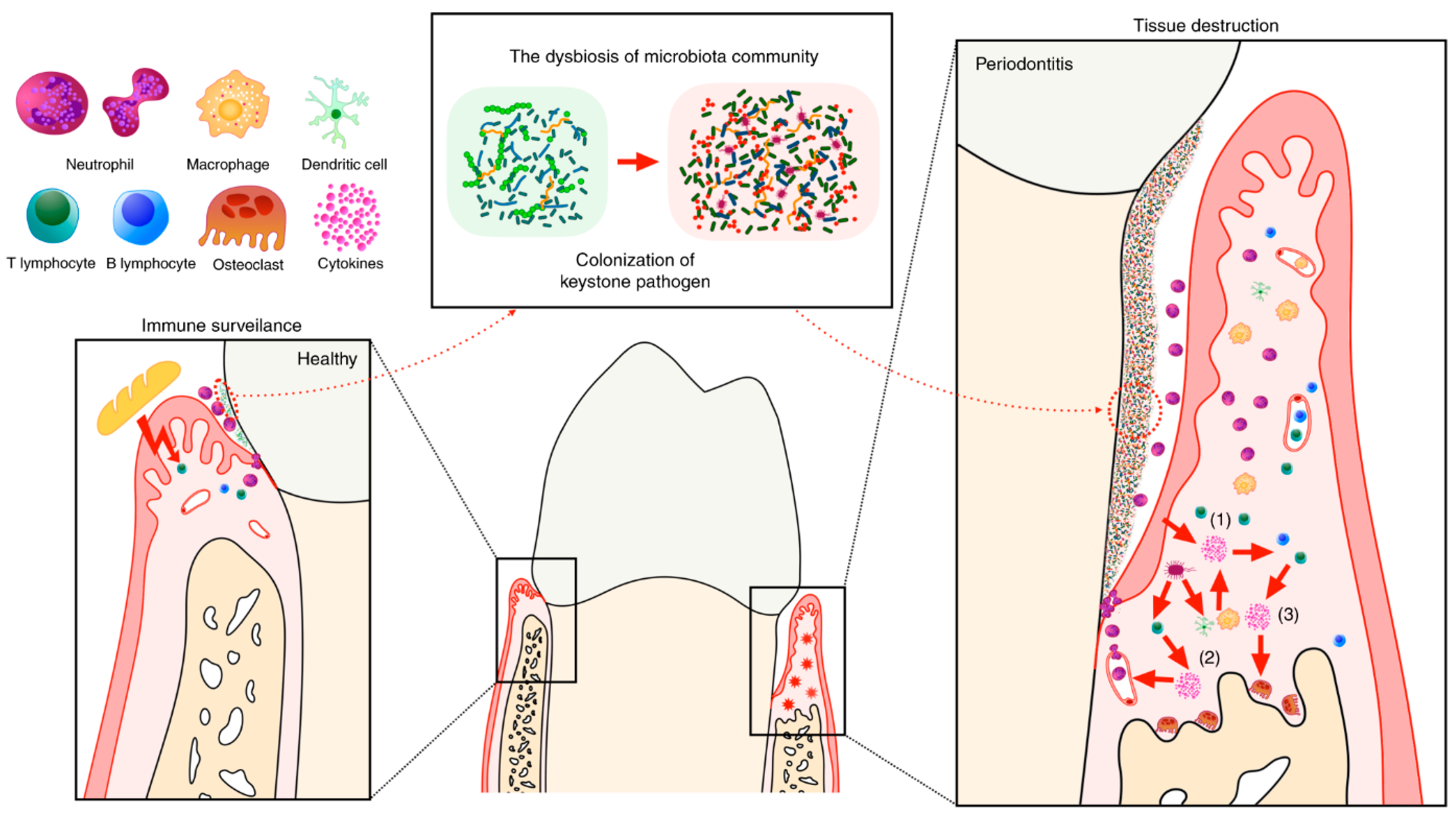
In a healthy oral microbiome, there is a balance between the oral microbiota and the adjacent host tissues (teeth and gingiva) maintained by the continuous presence of serum, inflammatory cells and mediators within the gingival crevicular fluid (GCF) [13,14]. Poor oral hygiene practices, along with other contributing factors such as diet, smoking, genetic predisposition, and systemic conditions (such as diabetes), can lead to plaque accumulation on tooth surfaces, resulting in a dysbiotic microbiome and dysregulation of the host inflammatory responses [15,16]. The primary etiological factor in periodontal inflammation is the invasion of pathogenic polymicrobial biofilm (also known as plaque) on the teeth surfaces and into the gingival crevices (Figure 1) [17]. Pathogenic bacteria exert their tissue destructive effects via direct and indirect mechanisms—triggering a cascade of chronic inflammation.
Direct damages are incurred when bacteria release a flurry of toxins, exoenzymes and waste products into the host tissues, cells and intercellular matrix. Additional indirect damage is caused by the release of bacterial cell wall components and other structures (such as fimbriae and flagella), membrane proteins, vesicles, nucleic acids and exopolysaccharides which create an inflammatory microenvironment to initiate host tissue [17,18]. These direct and indirect mechanisms activate the host immune response which attempts to eliminate the invading pathogens but instead exacerbates the inflammatory cascade and subsequent tissue destruction.
The host immune system recognizes these bacterial components as pathogen-associated-molecular-patterns (PAMPs) by pattern recognition receptors (PRRs), such as Toll-like receptors (TLRs), which are expressed on gingival epithelial cells, fibroblasts, dendritic cells, and macrophages. Activation of TLRs initiates downstream signalling through pathways like NF-κB and MAPK, resulting in the production of proinflammatory cytokines (e.g., IL-1β, TNF-α, IL-6) and chemokines. These mediators promote the recruitment of neutrophils and other immune cells to the site of infection. Although these mediators and immune cells are essential for early microbial defence, excessive activation contributes to tissue damage through the release of reactive oxygen species (ROS), matrix metalloproteinases (MMPs), and neutrophil extracellular traps (NETs) [15,17,19].
This immune response, if unresolved, perpetuates inflammation, leads to degradation of connective tissue, and stimulates osteoclastogenesis via Receptor Activator of NF-κB Ligand (RANKL) expression, driving the characteristic bone loss observed in periodontitis [17,20]. The periodontal tissues become loosely attached to the tooth, followed by inflammation and swelling of the tissues [20]. The periodontal pocket deepens due to the destruction of the attaching connective tissues which leads to clinical attachment loss. Intensifying this process, the inflammatory response ensuing gingival tissue destruction consequentially provides nutrients to the microbes which leads to an increase in pathogenic anaerobic Gram-negative bacteria load within the gingival sulcus, such as P. gingivalis, A. actinomycetemcomitans and Fusobacterium nucleatum [14,21].
Figure 1. The homeostasis of periodontal tissue, pathogenesis of chronic periodontitis and roles of the involved cytokines. In a healthy state, local challenge and a mild host immune response are balanced. Both the commensal microbiota and mechanical stimulation caused by mastication participate in the training of local mucosal immunity. In this state, there is an appropriate number of infiltrating neutrophils in the gingival sulcus, as well as some resident immune cells in the gingival tissue, including Th17 cells and innate lymphoid cells. However, if the immune pathogenicity of the local microbiota is elevated by the colonization of keystone pathogens, which over-activate the host immune response, tissue destruction is initiated. The interaction between the microbiota and all host cells leads to the first wave of cytokine secretion (1), which mainly participates in the amplification of the proinflammatory cytokine cascade and the recruitment, activation and differentiation of specific immune cells. In addition, a group of cytokines (2) closely related to the differentiation of a specific subset of lymphocytes are secreted by MNPs and APCs after stimulation by the microbiome. Each of these cell subsets secretes a certain pattern of cytokines, which might act as the positive-feedback factor or direct effector (3), eventually leading to tissue destruction. Reproduced from [22]; Creative Commons Attribution 4.0 International CC BY 4.0 Licence.
Jnt 06 00031 g001

2.3. Potential Biomarkers as Diagnostic and Therapeutic Targets in Periodontitis

Identifying reliable biomarkers is pivotal for both the diagnosis and targeted treatment of periodontitis. Since bacterial invasion and chronic inflammation function together in the pathogenesis of the disease, culminating in tissue destruction, it necessitates the need for multiple-targeting strategies using a combination of bacterial and inflammatory biomarkers, inflammatory mediators (cytokines, ROS, MMPs) and key immunomodulation pathways (TLR, RANKL and neutrophil activation pathways) for managing periodontitis. Salivary and gingival crevicular fluids, dental plaque, and exhaled breath condensate (EBC) are non-invasive easily accessible host biological analytes that contain an assortment of bacterial and inflammatory constituents that serve as reliable biomarkers for monitoring the onset and progression of periodontitis for clinical and point-of-care applications [23,24].

2.3.1. Salivary Fluid

Saliva has emerged as a particularly promising diagnostic medium due to its ease of collection and its reflection of both systemic and local oral health. Recent studies have confirmed the diagnostic value of inflammatory cytokines such as interleukin-1β (IL-1β), tumour necrosis factor-alpha (TNF-α), and matrix metalloproteinase-8 (MMP-8), which are elevated in individuals with active periodontitis [24]. Cathepsin K, B and S, lysosomal cysteine proteases, have also been detected in higher concentrations in the saliva of periodontitis patients and is associated with alveolar bone resorption and loss of attachment [25]. In addition to these proteolytic enzymes, aspartate aminotransferase (AST), alkaline phosphatase (ACP), alkaline phosphatase (ALP), and lactate dehydrogenase (LDH) have gained attention as markers of cellular injury and bone metabolism, respectively [26,27,28]. These biomarkers are believed to mirror the extent of inflammation and tissue damage in the periodontium, making them valuable clinical indicators for monitoring patients following periodontal treatment [24,26,29]. These enzymes, individually and in combination, provide an accurate biochemical profile of periodontitis activity. Moreover, salivary microRNAs (miRNAs), including miR-146a and miR-155, have been identified as novel biomarkers that correlate with disease severity and therapeutic outcomes [30,31,32].

2.3.2. Gingival Crevicular Fluid

Gingival crevicular fluid (GCF), a serum-derived fluid found in the gingival sulcus, provides a localized snapshot of periodontal inflammation. It contains a concentrated mix of host-derived enzymes, cytokines, and bone metabolism markers. Similarly to saliva, it contains several potential biomarkers to detect microbial infection (such as bacterial myeloperoxidase) and tissue destruction (such as AST, LDH, cathepsins, lysosomes, lactoferrin, and several matrix metalloproteinases) [33]. Recent proteomic analyses have identified osteoprotegerin (OPG), RANKL, and interleukin-6 (IL-6) as key indicators of alveolar bone resorption and disease activity in immunodiagnostic and prognostic applications for the various stages of periodontitis [34,35]. These biomarkers not only reflect current disease status but can also offer predictive value for future tissue breakdown, making GCF a valuable tool for both diagnosis and monitoring of treatment efficacy.

2.3.3. Dental Plaque

Dental plaque (also known as dental biofilm), particularly subgingival plaque, remains a critical source of microbial biomarkers indicating site-specific dysbiosis in periodontitis [36]. In contrast to host-derived biomarkers, which predominantly reflect the host’s immunoinflammatory response to periodontal pathogens, microbiome-derived biomarkers provide direct insights into the compositional and functional roles of the microbial community in periodontitis pathogenesis [37]. Advances in metagenomics and metatranscriptomics have enabled the identification of specific bacterial species and gene expression profiles associated with disease progression, particularly the presence of red complex bacteria (such as P. gingivalis, T. forsythia, and Treponema denticola) which continues to be strongly associated with periodontitis [38,39]. The identification of specific bacterial species implicated in periodontal microbiota dysbiosis is critical not only for elucidating the molecular mechanisms underlying the initiation and progression of periodontitis but also for guiding the development of targeted, pathogen-specific therapeutic interventions [38]. Additionally, bacterial RNA transcripts and virulence factors such as gingipains and lipopolysaccharides are being explored as dynamic biomarkers that reflect microbial activity and host–pathogen interactions in real time [40,41].

2.3.4. Exhaled Breath Condensate

Exhaled breath condensate (EBC) has emerged as a promising non-invasive diagnostic medium, capable of capturing volatile organic compounds (VOCs) generated through oral and systemic metabolic processes. It is obtained by condensing warm, humid exhaled air onto a cooled surface, allowing the collection of aerosolized droplets derived from the airway lining fluids [42]. This bioanalyte is commonly used for detecting IL-1β, IL-6 and TNF-α-common inflammatory cytokines characteristic of periodontitis. In periodontitis, elevated levels of hydrogen sulfide, methyl mercaptan, methanethiol, dimethyl disulfide and other sulfur-containing VOCs have been linked to the metabolic activity of anaerobic bacteria and inflammatory activity in periodontal pockets [43,44]. Therefore, this association deserves further investigation to validate the potential use of EBC and VOCs as a source of early biomarkers in periodontitis diagnosis and management [44,45].
The integration of multi-omics platforms (including genomics, proteomics, metabolomics, and microbiomics) is revolutionizing the identification of comprehensive biomarker panels for personalized periodontal care [46]. The incorporation of these molecular insights into smart drug delivery systems and biomaterial-based therapeutics will support the development of responsive, site-specific interventions to further enhance the precision and personalization of periodontal diagnostics and therapy.

2.4. The Necessity for Dual Targeting of Chronic Inflammation and Microbial Invasion

Given the synergistic pathology of microbial persistence and exaggerated inflammation, there is a compelling need for dual-targeting therapeutic strategies (Figure 2). Conventional antimicrobial agents are frequently inadequate due to their limited penetration into the dense extracellular matrix of biofilms, while systemic anti-inflammatory therapies are often insufficient to eliminate the microbial burden. Nanotheranostic platforms offer a promising alternative by integrating diagnostic and therapeutic functionalities within a single system. Functionalized nanoparticles, for instance, may incorporate antimicrobial peptides or quorum sensing inhibitors to disrupt biofilm architecture, while simultaneously delivering encapsulated anti-inflammatory agents for localized and sustained immunomodulation [47,48].
Furthermore, the intrinsic responsiveness of nanomaterials to pathological stimuli (such as acidic pH or elevated ROS) within inflamed periodontal pockets enables spatial and temporal precision in drug release [19]. This targeted release paradigm enhances therapeutic efficacy while minimizing systemic exposure and off-target effects. As such, the dual-focused nanotheranostic approach represents a significant advancement in periodontal therapy, aligning with emerging principles of precision medicine and offering a tailored strategy for the management of chronic periodontitis.

3. Current Limitations in the Diagnosis and Treatment of Periodontitis

Despite notable advancements in periodontal research and clinical practice, the diagnosis and management of periodontitis remain constrained by several critical limitations that impede early detection, individualized treatment planning, and sustained disease control.

3.1. Diagnostic Limitations

Current diagnostic approaches for periodontitis remain predominantly clinical and retrospective in nature, relying on parameters such as probing pocket depth, clinical attachment loss, bleeding on probing, and radiographic assessment of alveolar bone loss [49]. While these measures are well-established, they primarily reflect cumulative tissue destruction and fail to capture active or early-stage disease processes. As a result, subclinical inflammation and sudden shifts in disease activity often go undetected. Moreover, these assessments are inherently operator-dependent, subject to inter-examiner variability, and lack the sensitivity and specificity required for accurate disease staging or real-time monitoring of therapeutic outcomes.
Emerging biomarker-based diagnostics which utilize biological fluids such as saliva, gingival crevicular fluid (GCF), or peripheral blood, offer promise for detecting molecular signatures of disease activity. However, their integration into routine clinical practice remains limited due to challenges in standardization, cost-effectiveness, and accessibility. Additionally, many currently proposed biomarker panels exhibit limited specificity and reproducibility across heterogeneous patient populations, thereby constraining their translational potential and widespread clinical adoption [41,46].

3.2. Therapeutic Limitations

Conventional periodontal therapy primarily comprises mechanical debridement through scaling and root planing, adjunctive antibiotic administration, and, in more advanced cases, surgical intervention [50,51]. While these strategies are effective in reducing microbial burden and attenuating inflammation, they are associated with several notable limitations that hinder long-term therapeutic efficacy and personalization of care [51].
One major drawback is the non-specific nature of systemic drug delivery. Systemic antibiotics often result in subtherapeutic concentrations at the local disease site, increasing the risk of antibiotic resistance and systemic adverse effects [51]. Furthermore, the dense and structured architecture of subgingival biofilms poses a significant barrier to drug penetration, limiting the effectiveness of many conventional antimicrobials and anti-inflammatory agents, thus necessitating the elimination of such biofilms via biomarker targeted nanotechnological strategies [52,53].
Topically applied therapeutics, although site-directed, frequently suffer from poor retention within the periodontal pocket due to salivary dilution, enzymatic degradation, and mechanical disruption from tongue movements, necessitating frequent reapplication. Conventional formulations such as antimicrobial suspensions or rinses offer only transient therapeutic effects and are unable to sustain adequate drug concentrations at the target site for prolonged periods [53]. Additionally, current treatment regimens are largely standardized and fail to account for interindividual variability in microbial composition, host immune response, and other non-microbial factors such as demographics, lifestyle effects, underlying systemic disease, and genetic susceptibility–factors that are increasingly recognized as critical determinants of periodontitis progression and treatment response [18].
Conventional therapies can halt disease progression but fail to fully restore lost periodontal structures, highlighting the need for effective regenerative strategies. Advances in tissue engineering (such as using stem cells, bioactive molecules, and biomimetic scaffolds) show promise for developing biomimetic scaffolds, yet challenges re-main in replicating the native tissue environment, achieving stable vascularization, and ensuring seamless integration with existing structures [54]. The absence of predictable regenerative approaches continues to limit long-term clinical success of periodontitis, underscoring the need for innovative, interdisciplinary solutions [54,55].

3.3. Challenges in Treatment Monitoring and Follow-Ups

A critical limitation in current periodontal management is the absence of real-time monitoring tools capable of assessing therapeutic response or detecting early signs of disease recurrence. Clinicians predominantly rely on conventional clinical parameters (such as probing pocket depth, bleeding on probing, and radiographic evaluation) which are typically assessed during follow-up visits scheduled weeks or months post-treatment and are reliant on subjective interpretations [49]. These retrospective measurements provide delayed insight into disease progression and are often insufficient to detect subtle or early-stage alterations within the periodontal microenvironment. This time lag can result in missed opportunities for timely intervention, particularly in cases where subclinical inflammation or residual biofilm persists despite initial therapy. In the absence of dynamic feedback mechanisms, clinicians are constrained in their ability to personalize treatment regimens, adjust therapeutic strategies in real time, or promptly address therapeutic failure–thus, ultimately compromising disease control and increasing the risk of preventable tissue destruction. These diagnostic modalities are poorly equipped to capture molecular or cellular-level changes, such as fluctuations in proinflammatory cytokines, oxidative stress markers, or enzymatic activity (e.g., matrix metalloproteinases), which may serve as early indicators of treatment efficacy or impending relapse. The lack of routine implementation of such biomarkers in clinical settings limits the evolution of responsive and adaptive treatment protocols.
Nanotheranostic platforms present a compelling solution to this gap by enabling real-time, non-invasive monitoring of disease activity through integrated diagnostic modalities. Smart nanomaterials can be engineered to respond to local biochemical cues, including pH shifts, ROS, and enzyme activity, providing dynamic data that inform clinicians of drug release profiles, inflammation status, or residual microbial presence. Such capabilities pave the way for precision-guided, adaptive periodontal therapy, transforming the traditional model of delayed assessment into one of proactive, data-driven intervention.

4. Nanomaterials for Periodontal Applications

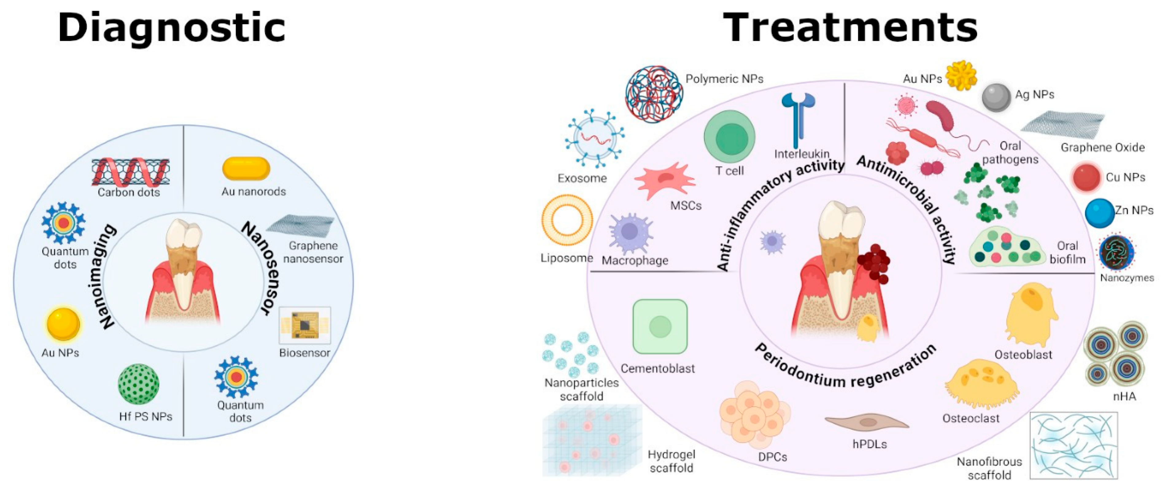
Nanomaterials have garnered significant attention as multifunctional agents in theranostic applications, integrating both diagnostic and therapeutic functionalities for the effective management of periodontitis. Incorporating nanotechnology into periodontal therapy has enabled the development of advanced platforms for precise diagnostics, real-time disease monitoring, and targeted therapeutic delivery. These nanosystems are capable of simultaneously transporting bioactive agents while facilitating diagnostic imaging or biosensing to assess treatment response and disease progression. Broadly, nanomaterials are categorized as organic (polymeric and lipid based) or inorganic, with each class exhibiting distinctive physicochemical attributes that align with the complex demands of periodontal treatment (Figure 3). Their high surface-to-volume ratio, tunable size, modifiable surface chemistry, and responsive behaviour to pathological stimuli render them ideal candidates for localized drug delivery and real-time feedback mechanisms. In periodontitis, nanomaterials are specifically investigated for their capacity to penetrate bacterial biofilms, localize within inflamed periodontal tissues, achieve stimulus-responsive drug release, and promote periodontal bone regeneration, thereby offering a synergistic approach to both therapeutic intervention and diagnostic precision [8].
Nanomaterials display higher chemical reactivity and have better optical, magnetic, biological, electrical, or mechanical properties compared to their macroscopic or microscopic counterparts [57]. While nanoparticles represent the most extensively studied nanotheranostic systems, a broad range of alternative nanomaterials and nanostructures have been investigated for theranostic applications in periodontitis. These include, but are not limited to, nanofibers, nanoshells, nanoclays, quantum dots, dendrimers, and carbon-based nanostructures–each offering unique advantages in terms of drug loading capacity, tissue specificity, imaging modalities, and stimulus-responsiveness. Such materials extend beyond conventional nanoparticulate carriers to provide multifunctional platforms capable of simultaneous diagnostic and therapeutic functions. Their physicochemical diversity enables tailored interactions with periodontal tissues and biofilms, thereby enhancing the precision, efficacy, and monitoring of periodontal treatment strategies.
Although nanomaterials offer various advantages, there are several disadvantages associated with it, such as high production costs, short-half lives, the presence of cytotoxicity in metallic nanoparticles at high concentrations and the leakage and fusion of the encapsulated drug [57,58]. However, the advantages that come along with the nanoparticle use outweigh the disadvantages, which is why nanoparticles are still highly favourable in drug delivery systems and diagnostic applications today. This section discusses the principal types of nanomaterials used in periodontitis applications as well as those with promising appeal, their functionalization strategies, and their roles in overcoming biological barriers associated with chronic inflammation and microbial infection in the periodontium. Table 1 summarizes the key properties and diagnostic and therapeutic functions of the various nanomaterials.

4.1. Polymeric Nanomaterials

Polymeric nanomaterials have reigned as the predominantly versatile and multifunctional nanocarriers for the management of periodontitis, offering the dual benefits of localized drug delivery and integrated diagnostic capability [59]. Their physicochemical tunability, biocompatibility, and potential for controlled release make them particularly well-suited to address the multifactorial pathology of periodontal disease. These nanomaterials can be shaped into a variety of morphologies such as nanoparticles, nanofibres, nanocapsules, and nanostructured hydrogels to deliver therapeutic bioactives, antimicrobial agents, reduce inflammation, and facilitate tissue repair in periodontal disease. These systems are primarily synthesized from two broad classes of polymers—polyesters (synthetic polymers) and polysaccharides (natural polymers)—each contributing distinct structural, functional, and biological attributes conducive to periodontal therapy and diagnosis.
Synthetic polyester-based nanomaterials, particularly those fabricated from poly(lactic-co-glycolic acid) (PLGA), polylactic acid (PLA), and polycaprolactone (PCL), have garnered extensive attention over several years owing to their favourable biocompatibility and biodegradability profiles, mechanical properties, and regulatory approval for biomedical use [7,8]. These materials degrade into non-toxic byproducts (such as lactic and glycolic acid), which are naturally metabolized by the body, making them safe for intra-pocket administration [60]. Their mechanical properties and degradation profiles can be tailored to match the therapeutic needs of periodontal tissues, allowing for sustained and localized drug release. This is especially beneficial in managing the chronic inflammatory environment in periodontitis, where prolonged exposure to antimicrobials or anti-inflammatory agents is required [77]. Their compatibility with a wide range of therapeutic agents, including antibiotics, growth factors, and anti-inflammatory drugs, makes them versatile carriers for both treatment and regenerative applications in periodontal therapy.
PLGA and PCL nanomaterials (such as nanoparticles and nanofibers, respectively) have been commonly employed for the sustained intra-pocket delivery of antimicrobial agents such as amoxicillin, doxycycline and metronidazole, achieving high drug loading efficiency, enhanced bacterial eradication with reduced systemic drug exposure whilst also promoting cell proliferation and differentiation [78,79,80,81]. PCL nanomaterials, notable for its hydrophobicity and compatibility, have demonstrated efficacy in delivering lipophilic bioactive molecules (such as doxycycline and tetracycline) and thus may be promising for delivering other bioactives such as curcumin and regenerative or antimicorbial peptides, thereby supporting concurrent antimicrobial activity and tissue regeneration [82].
In parallel, natural polysaccharide-based polymeric nanomaterials offer mucoadhesion, structural flexibility, excellent biocompatibility, biodegradability, intrinsic bioactivity, and superior safety profiles for periodontal applications [7]. These materials, notably chitosan, hyaluronic acid, alginate, and dextran, exhibit structural and biochemical similarities to the extracellular matrix, thereby supporting cellular adhesion, proliferation, and tissue regeneration [83,84]. Fabricated into diverse formats such as nanoparticles, nanogels, and electrospun nanofibers, polysaccharide-based systems offer a versatile platform for localized, sustained, and responsive therapeutic periodontal delivery. Chitosan, a cationic polysaccharide, is particularly advantageous in periodontal therapy due to its mucoadhesive nature and intrinsic antimicrobial activity. Its electrostatic interaction with negatively charged biofilms and mucosal surfaces enhances retention within periodontal pockets and facilitates biofilm disruption [83]. Chitosan nanomaterials have been extensively investigated for the localized delivery of a broad range of therapeutic agents, including antibiotics, anti-inflammatory compounds, and regenerative peptides to the periodontal pocket [8,85].
Hyaluronic acid, an anionic glycosaminoglycan, plays a pivotal role in wound healing, angiogenesis, and extracellular matrix remodelling. Its hydrophilic nature supports tissue hydration and cellular migration, making it well-suited for regenerative periodontal applications [83]. Hyaluronic acid-based nanomaterials function not only as drug carriers but also as bioactive agents that promote soft tissue repair. Alginate, characterized by its anionic gel-forming capacity and mild encapsulation conditions, enables the efficient bioactive loading and sustained release of labile therapeutic molecules such as growth factors and anti-inflammatory agents.
Alginate-based nanoparticle composites are particularly suitable for injectable formulations that conform to the irregular architecture of periodontal pockets, enhancing their clinical applicability and tissue regeneration capability [86,87]. Dextran, a naturally occurring branched polysaccharide, presents a chemically versatile and biocompatible platform for periodontal nanotherapeutics. Its biodegradability and favourable safety profile, coupled with its capacity for extensive chemical modification, render it highly suitable for multifunctional delivery systems. Engineered into cationic derivatives, dextran exhibits enhanced biofilm-disruptive activity, with recent studies demonstrating its ability to penetrate the dense extracellular matrix of mature oral biofilms and facilitate targeted access to embedded periodontal pathogens.
Dextran, a naturally occurring branched polysaccharide, offers a versatile and chemically modifiable platform ideal for periodontal applications. Its inherent biocompatibility and biodegradability are complemented by potent biofilm-controlling properties, particularly when engineered into cationic derivatives [88]. Moreover, dextran nanocarriers can be engineered to exhibit stimuli-responsive behaviour triggered by environmental cues such as acidic pH, redox gradients, or enzymatic activity to enable controlled, site-specific release within inflamed periodontal pockets [60]. This bioresponsive delivery capability, combined with its structural adaptability, positions dextran as a promising platform for next-generation, integrated therapeutic, and diagnostic strategies in periodontal care.
Polymeric nanomaterials are primarily employed in periodontitis therapy for the localized and sustained delivery of therapeutic agents, including antimicrobials, anti-inflammatory drugs, bioactive molecules, and growth factors. Their tunable degradation profiles, biocompatibility, and ability to encapsulate a wide range of therapeutics make them ideal for targeting the periodontal pocket, enhancing treatment efficacy while minimizing systemic exposure—thus supporting infection control and tissue regeneration requirements. While their primary application remains therapeutic, polymeric nanomaterials hold promise for exploration as nanotheranostic platforms. By embedding diagnostic agents such as fluorescent dyes, magnetic iron oxide cores, or radiocontrast materials and via functionalization with molecules that bind to host or microbial biomarkers (such as antibodies or and peptides) within the polymer matrix, these nanomaterials can enable non-invasive imaging through modalities like fluorescence microscopy, magnetic resonance imaging (MRI), or computed tomography (CT) [6,60]. This allows for real-time monitoring of nanoparticle distribution, biofilm disruption, and therapeutic response within the periodontal microenvironment.
Although polymeric nanomaterials are not yet widely used in biosensing or diagnostic applications in periodontitis, recent research is beginning to explore their potential in this domain. Studies have investigated their use in detecting inflammatory biomarkers such as IL-1β and alkaline phosphatase in saliva or gingival crevicular fluid, especially when combined with smart nanocomposite systems or hybrid platforms [6]. These developments suggest that polymeric nanoparticles could evolve into dual-function systems, capable of delivering therapeutics while simultaneously providing diagnostic feedback.

4.2. Lipid-Based Nanomaterials

Lipid-based nanomaterials have emerged as promising platforms for theranostic interventions in periodontitis, attributed to their excellent biocompatibility, structural versatility, and capacity to encapsulate both hydrophilic and hydrophobic therapeutic agents. These systems facilitate enhanced drug delivery within the complex periodontal microenvironment and can be tailored for diagnostic imaging or stimuli-responsive release. Among the most extensively studied lipid-based systems, solid lipid nanoparticles (SLNs), nanostructured lipid carriers (NLCs), and liposomes have demonstrated considerable potential in delivering antimicrobial agents, anti-inflammatory compounds, and regenerative bioactives [61]. Solid lipid nanoparticles, composed entirely of solid lipids, offer high drug stability, controlled release, and protection of labile compounds, although they may suffer from limited drug loading and potential expulsion during storage, particularly of hydrophilic drugs [62,89]. Nanostructured lipid carriers, composed of a mixture of both solid and liquid lipids, exhibit enhanced drug-loading efficiency, physicochemical stability, and reduced drug expulsion compared to SLNs [90,91,92]. Liposomes, composed of phospholipid bilayers enclosing aqueous cores, are particularly suited for the encapsulation of hydrophilic molecules and can be engineered for triggered drug release in response to local pH changes, enzymatic activity, or ROS accumulation characteristic microenvironment features of inflamed periodontal pockets [61,62].
In addition to these established platforms, newer lipid-based nanostructures, such as lipid nanocapsules (LNCs), nanoemulsions, lipid micelles, cubosomes, and hexosomes, are gaining traction as advanced drug delivery systems that could expand to encompass theranostic applications [63,93,94]. Lipid nanocapsules integrate structural features of both liposomes and polymeric nanoparticles, offering high colloidal stability, sustained release kinetics, and the capacity for dual drug-diagnostic agent loading, such as near-infrared fluorophores, superparamagnetic iron oxide nanoparticles or photothermal polymers for advanced theranostic approaches as inspired by cancer theranostics [95]. Nanoemulsions as intra-pocket delivery platforms, comprise thermodynamically stable oil-in-water or water-in-oil dispersions, and are particularly advantageous for mucosal permeation, biofilm disruption, and solubilization of poorly water-soluble drugs [96,97]. Lipid nanomicelles, formed from self-assembling amphiphilic lipid molecules, have shown efficacy in delivering hydrophobic antimicrobials and anti-inflammatories with targeted action against oral biofilms [98].
Emerging attention has also been directed toward liquid crystalline lipid nanoparticles, namely cubosomes and hexosomes, which are characterized by their unique internal bicontinuous cubic and hexagonal liquid crystalline phases, respectively [63,64,99]. These nanocarriers provide a high internal surface area conducive to sustained drug release and demonstrate notable bioadhesiveness and deep tissue penetration within periodontal lesions, as demonstrated by Tang and co-workers who used cubosomes as a bioactive depot for periodontitis therapy [100]. Moreover, their structural organization could permit co-encapsulation of contrast agents, extending their functionality toward non-invasive imaging applications such as fluorescence microscopy or magnetic resonance imaging (MRI) with integration of a therapeutic component [63,64].
The therapeutic precision of these lipid-based nanosystems can be further enhanced through surface functionalization with mucoadhesive polymers, pathogen-specific ligands (e.g., lectins, antibodies), or stimuli-sensitive moieties that respond to pathological cues such as acidic pH, elevated enzymatic activity (e.g., matrix metalloproteinases), or oxidative stress [60].

4.3. Inorganic Nanomaterials

Inorganic nanomaterials constitute a diverse and functionally versatile class of platforms for theranostic applications in periodontitis, offering the integration of site-directed therapeutic action with advanced diagnostic imaging capabilities. Their unique physicochemical attributes such as, high surface reactivity, tunable particle size, and intrinsic structural stability, offer specific suitability for biofilm penetration, and localized drug delivery to periodontal lesions. Beyond therapy, inorganic nanomaterials offer considerable advantages in diagnostics, particularly for real-time, non-invasive detection and longitudinal disease monitoring. Their optical, magnetic, and electrochemical properties support the development of sensitive, miniaturized biosensors. A wide range of inorganic nanoparticles, comprising metallic nanoparticles (such as gold and silver), metal oxides (such as ZnO, CeO2, TiO2), mesoporous silica nanoparticles (MSNs), and calcium phosphate-based nanostructures, have demonstrated significant therapeutic and diagnostic utility in periodontal applications [8,28].
Gold nanoparticles (AuNPs) have emerged as promising antibacterial agents, owing to their unique physicochemical properties and biocompatibility. Recent studies have increasingly focused on elucidating their antimicrobial mechanisms and evaluating their efficacy against a broad spectrum of pathogenic bacteria using photothermal therapy, for periodontal infections [66].
Silver nanoparticles (AgNPs) remain among the most potent broad-spectrum antimicrobial agents, exerting their effects by disrupting microbial membranes, inhibiting biofilm formation, and potentiating antimicrobial efficacy against periodontal pathogens such as P. gingivalis either alone or combined with other antimicrobials such as chlorhexidine [65,101,102]. Moreover, recent research has demonstrated AgNPs for surface-enhanced Raman spectroscopy for rapid, label-free detection of P. gingivalis in saliva [103]. Their strong plasmonic properties create electromagnetic “hot spots” that amplify bacterial Raman signals, enabling sensitive and specific identification without complex processing. In addition to their diagnostic utility, the inherent antimicrobial activity of AgNPs highlight their dual role in nanotheranostic applications for periodontitis.
Zinc oxide nanoparticles (ZnO NPs) exhibit strong intrinsic bactericidal activity, often enhanced via photodynamic or photocatalytic activation to penetrate deep into periodontal lesions and eliminate dental biofilms and bacteria under light irradiation without inducing antibiotic resistance [74,104,105]. Zeolitic imidazolate framework 8 (ZIF-8) release zinc ions during degradation which plays a critical homeostasis role in periodontal tissue remodelling and regeneration in addition to its antibacterial and osteogenic effects [106].
Similarly, light-activated titanium dioxide (TiO2) nanoparticles generate ROS, disrupting established biofilms and enabling site-specific antibacterial phototherapy [66,107,108].
Iron oxide nanoparticles, especially ferromagnetic Fe3O4, offer strong therapeutic potential in periodontitis by disrupting biofilms and enabling magnetically guided drug delivery. Minocycline-loaded Fe3O4 nanoparticles achieved deep biofilm penetration and reduced inflammation under magnetic guidance [68]. In addition, iron oxide nanoparticles such as Fe2O3, owing to their superparamagnetic behavior, are used for magnetic biosensing of bacterial virulence factors like gingipains [28]. Coupled with nanopore sensing technologies, these systems enable ultrasensitive quantification of salivary or gingival crevicular biomarkers at picogram levels, supporting rapid, point-of-care diagnostics [109]. As reviewed by Wang et al. (2024), these nanoparticles support multimodal treatment which combines antibacterial, anti-inflammatory, and regenerative effects, thus making them valuable tools in nanotheranostic periodontal therapy [8].
Metal and metal oxide nanomaterials can also be engineered to detect volatile sulfur compounds such as hydrogen sulfide and methyl mercaptan are early indicators of periodontal pathology. Nanofilms composed of ZnO and Au NPs, developed as exhaled breath sensors for periodontitis, demonstrated sensitivity at clinically relevant thresholds (<10 ppb), thus enabling early-stage disease detection [67].
Mesoporous silica nanoparticles offer a multifunctional platform for periodontal therapy, combining antimicrobial, anti-inflammatory, and regenerative properties. Metal-doped MSNs, particularly those incorporating ions like silver, zinc, or strontium, exhibit potent antibacterial activity against P. gingivalis and promote osteoblast differentiation and alveolar bone regeneration [69,70]. Drug-loading of MSNs, such as tetracycline-loaded MSNs (MSN-TC), can further enhance therapeutic outcomes by enabling controlled antibiotic release and reducing bone resorption through suppression of osteoclastogenesis [106]. Their tunable porosity and high drug-loading capacity support sustained intra-pocket delivery of therapeutic agents, making MSNs ideal candidates for targeted, site-specific treatment of periodontitis [71].
Upconversion nanoparticles (UCNPs) represent a versatile, light-responsive platform for the treatment of periodontitis, offering synergistic therapeutic benefits through photodynamic activation, gas delivery, and tissue regeneration [110]. By converting near-infrared (NIR) light to visible light, UCNPs activate photosensitizers to generate ROS for biofilm disruption [111]. They can also trigger the release of therapeutic gases like nitric oxide and carbon monoxide, which reduce inflammation and promote tissue regeneration [112]. This light-triggered platform enables simultaneous infection control, immune modulation, and periodontal repair. Using the same principle, UCNPs have demonstrated exceptional biosensing capabilities for point-of-care diagnostics in periodontitis. One study employed UCNPs as fluorescent probes in a lateral flow immunoassay designed to detect three key periodontal biomarkers (MMP-8, IL-1β, and P. gingivalis lipopolysaccharide) directly from gingival crevicular fluid [113]. The platform achieved high sensitivity with detection limits in the low ng/mL range and delivered results within 30 min, thus paving the way for real-time, personalized disease monitoring and intervention in periodontal care.
Other promising inorganic nanomaterials include selenium nanoparticles (SeNPs), with dual antimicrobial and antioxidant properties [114,115]. Magnesium oxide nanoparticles (MgO NPs), supresses periodontal pathogens and supports bone regeneration via inhibiting the expression and secretion of lipopolysaccharide-induced IL-1β [116,117]. In addition, several metal oxide nanoparticles function as dynamic redox modulators. For example, cerium oxide nanoparticles (CeO2 NPs), through reversible Ce3+/Ce4+ cycling, scavenge excess ROS and suppress proinflammatory cytokine production, thereby reducing oxidative stress and inflammation in periodontal lesions [118,119,120].
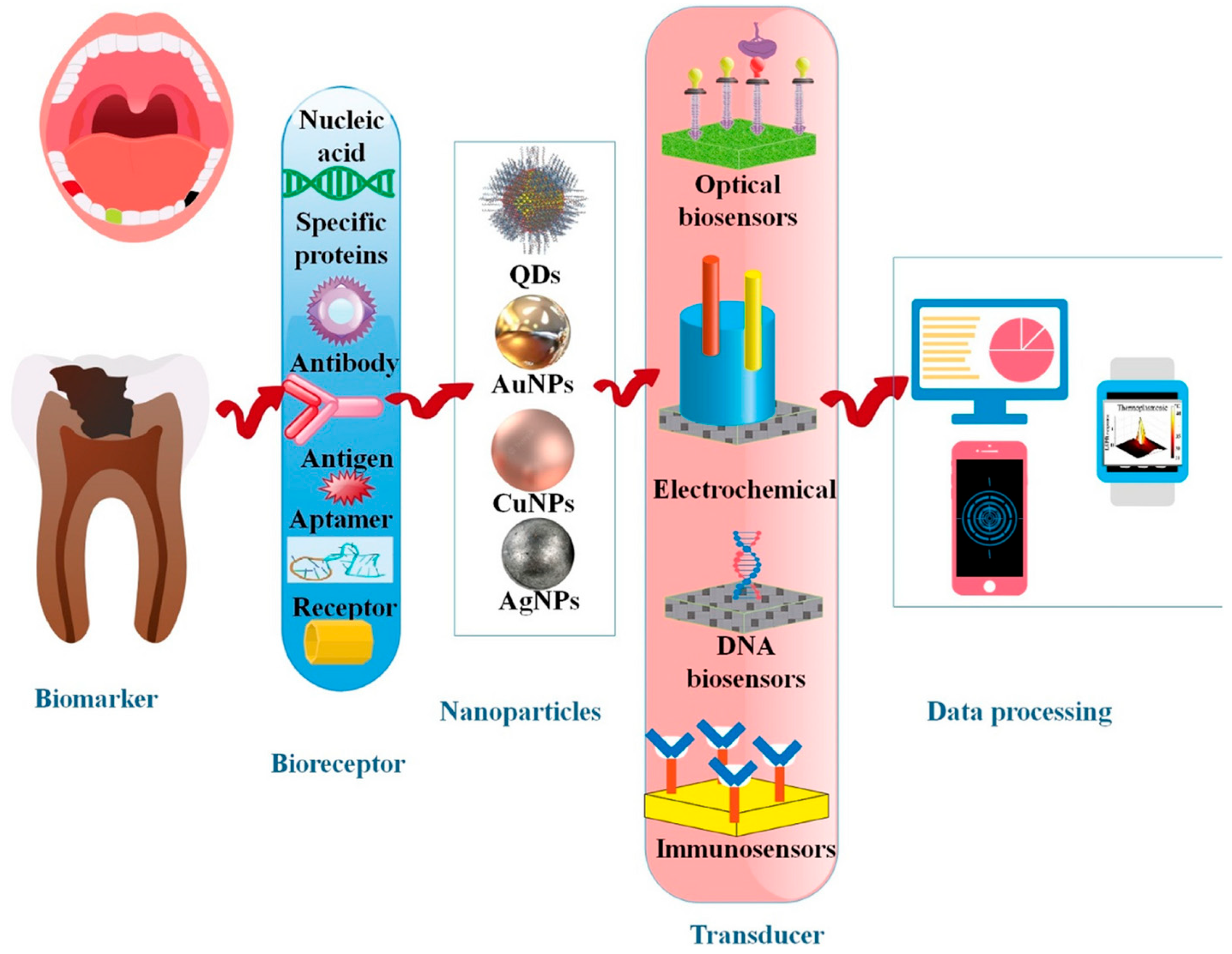
Inorganic nanomaterials offer significant potential for periodontitis nanotheranostics by combining powerful therapeutic and diagnostic functions within a single nanoscale platform. Therapeutically, these systems enable a multi-pronged approach that simultaneously targets microbial biofilms, modulates inflammation, reduces oxidative stress, and promotes tissue regeneration. On the diagnostic front, inorganic nanomaterial-based sensors provide rapid, chairside-compatible detection of key biomarkers and pathogens, facilitating timely disease monitoring (Figure 4). Crucially, these diagnostic technologies form the foundation for integrated theranostic systems, where real-time sensing can be coupled with on-demand therapeutic release, enabling responsive, personalized treatment strategies tailored to the dynamic periodontal microenvironment.

4.4. Carbon-Based Nanomaterials

Carbon-based nanomaterials have emerged as versatile theranostic agents for periodontitis due to their unique combination of structural tunability, physicochemical functionality, and bioactivity. Their high surface area, electrical conductivity, biocompatibility, and capacity for surface modification enable integration into multifunctional nanoplatforms designed for microbial targeting, immunomodulation, tissue regeneration, and diagnostic imaging within the periodontal microenvironment [6,28]. As such, carbon-based nanomaterials offer a compelling foundation for integrating simultaneous diagnosis and therapy within the inflamed periodontal microenvironment.
Graphene and its derivatives, including graphene oxide (GO), reduced graphene oxide (rGO), and graphene quantum dots (GQDs), are among the most extensively investigated. GO and rGO exhibit potent antibacterial properties through physical membrane disruption, oxidative stress induction, and superoxide generation. These nanosheets have been functionalized with antibiotics, peptides, or growth factors, enabling localized delivery within periodontal pockets and promoting both antimicrobial effects and tissue repair [73,121]. Their mechanical reinforcement capacity also supports incorporation into scaffolds or membranes for guided tissue regeneration. Meanwhile, GQDs offer dual diagnostic and therapeutic functionality: Their intrinsic fluorescence enables imaging of inflamed sites, while their tunable surface chemistry supports targeted delivery of osteogenic or anti-inflammatory agents repair [73,121].
Carbon dots (CDs), particularly those derived from pharmacologically active molecules like artesunate, have shown promise as ROS scavengers counteracting oxidative damage and preserving osteoblast viability in periodontitis [19,122]. Their mild antibacterial action, low cytotoxicity, and excellent dispersibility further enhance their suitability for periodontal applications, especially in biofilm-rich environments. Artesunate-derived carbon dots, for instance, reduce inflammation and promote alveolar bone regeneration via AMPK signalling [122].
Carbon-coated magnetic nanoparticles (Fe3O4@C) represent another innovation in this class. These nanostructures exhibit strong biocompatibility with gingival fibroblasts and epithelial cells and have demonstrated potential for magnetically targeted delivery, biofilm disruption, and integration into smart delivery systems guided by external fields [123].
Electrochemical and optical biosensing applications have also leveraged the conductive properties of carbon-based nanomaterials. Graphene derivatives and carbon nanotubes (CNTs) are widely used in sensor electrodes for the detection of salivary biomarkers such as myeloperoxidase, IL-1β, ALP, and LDH [28,124]. These platforms enable rapid, real-time disease monitoring with high sensitivity and signal-to-noise ratios. Similarly, carbon-based quantum dots and nanozymes provide multiplexed fluorescence and enzyme-mimetic activity, respectively, to assess oxidative stress markers like hydrogen peroxide or myeloperoxidase critical for tracking inflammation severity [6,28].
Of important realization, hybrid systems that combine carbon nanomaterials with polymers or inorganic agents (e.g., metals) allow for enhanced mechanical strength, improved drug release control, and deeper biofilm penetration. These configurations are particularly advantageous for addressing the complex interplay of infection, inflammation, and tissue degradation in periodontitis. Altogether, the modularity and multifunctionality of carbon-based nanomaterials make them promising components of next-generation periodontal nanotheranostics. By integrating antimicrobial action, antioxidant activity, regenerative support, and real-time biosensing into a single nanoscale platform, these systems align with the goals of precision, responsiveness, and minimal invasiveness in modern periodontal therapy.

4.5. Smart Nanocomposite Hybrid Systems for Periodontal Applications

Smart nanocomposites and hybrid nanoplatforms represent a leading-edge approach in periodontal theranostics, offering multifunctional systems that integrate the therapeutic and diagnostic strengths of diverse nanomaterial classes. These advanced constructs are rationally engineered to address the multifactorial pathogenesis of periodontitis by incorporating polymeric, lipid-based, inorganic, and carbon-based nanostructures into a single, synergistic platform. Such systems enable targeted and controlled delivery, stimuli-responsive release, deep biofilm penetration, antimicrobial efficacy, and real-time diagnostic imaging capabilities that collectively overcome the limitations of conventional monomaterial strategies. The section explores some recent examples of research that embody a combinatorial approach for the designing periodontal diagnostics and therapeutics.
Hybrid nanocomposites exploit the complementary physicochemical and biological properties of their constituent materials. A biocompatible and biodegradable polymeric nanomaterial can be integrated with carbon-based nanostructures to provide sustained drug release and degradation compatibility, while the carbon-based moieties contribute mechanical reinforcement, ROS generation, antimicrobial activity, and the potential for photodynamic or photothermal therapeutic interventions or biosensing capabilities. For instance, a study by Ramos-López et al. (2025) explored a smart nanocomposite-based dual biosensing platform for the rapid and simultaneous detection of key inflammatory biomarkers in periodontitis. By integrating crystalline nanocellulose and rGO onto screen-printed dual carbon electrodes, the system achieved highly sensitive and selective electrochemical detection of matrix metalloproteinases MMP-9 and MMP-13 which are both implicated in periodontal inflammation [125]. The use of nanocellulose enhances surface area and biomolecule immobilization, improving signal amplification and detection efficiency for multiplex biomarker quantification-offering a clinically translatable diagnostic modality. Its integration with theranostic platforms could facilitate real-time monitoring and timely therapeutic intervention in oral and systemic comorbidities.
Similarly, a study by Ren et al. (2022) showcases a nanocomposite hybrid system for periodontitis therapy by integrating cerium oxide nanoparticles (CeO2 NPs) into electrospun polycaprolactone/gelatin nanofibrous membranes designed for guided tissue regeneration. The resulting bioactive scaffold not only provides structural support but also actively promotes alveolar bone healing by enhancing proliferation and osteogenic differentiation of human periodontal ligament stem cells [119]. In vivo, the composite membrane significantly accelerated bone regeneration in rat cranial defect models, aided by the sustained release of CeO2 NPs. While not directly measured, the known antioxidant and anti-inflammatory properties of CeO2 nanozymes further suggest a dual therapeutic function to modulate oxidative stress and inflammation common in periodontal lesions. Such a system could further harness the biosensing capabilities of nanozymes to provide enzyme-mimetic activity to assess oxidative stress markers for tracking inflammation severity [6,28].
Ramos-López et al. (2024) presented a smart hybrid nanocomposite biosensor that combines carbon-based and inorganic nanomaterials for precise and non-invasive periodontitis diagnosis. The electrochemical immunosensor integrates cerium oxide nanoparticles (CeO2NPs) with multi-walled carbon nanotubes (MWCNTs) to detect salivary myeloperoxidase. Multi-walled carbon nanotubes provide a high surface area, enhance electron conductivity, and prevent nanoparticle aggregation, while CeO2NPs exhibit peroxidase-like activity that amplifies the electrochemical signal [126]. This synergistic design enables highly sensitive and rapid detection using a sandwich-type immunoassay on screen-printed carbon electrodes. Clinically, the sensor effectively distinguishes periodontitis patients from healthy controls using diluted saliva samples, demonstrating strong potential as a point-of-care diagnostic tool. This work exemplifies how carbon-inorganic nanocomposite platforms can serve as smart diagnostic systems for potential within a broader theranostic framework for managing periodontal disease.
A study by Han et al. (2025) investigated a nanocomposite hydrogel system by integrating gold nanocages (Au NCs) with the antioxidant resveratrol into a photothermal-responsive hydrogel (RSV-Au@H)—the platform enables controlled, on-demand drug release triggered by NIR light. Upon NIR irradiation, the Au NCs generate localized heat (~45.8 °C), causing the hydrogel to swell and release resveratrol at the periodontal site to effectively scavenges ROS, restoring mitochondrial function in osteoblasts and promoting periodontal tissue regeneration [127]. Beyond its therapeutic benefits, the hydrogel also maintains a moist, protective microenvironment conducive to healing. Such nanocomposite system utilizing a combination of metal nanomaterials in a stimuli-responsive matrix could be harnessed to integrate diagnostics and therapy in a single platform for targeted, regenerative periodontitis treatment.
Another study designed smart nanocomposite hybrid system that synergistically integrates bioresponsive nanotechnology with probiotic delivery for targeted periodontitis nanotheranostics [76]. Leveraging the natural hypoxia of deep periodontal pockets, the researchers engineered a probiotic-guided delivery system using Bifidobacterium bifidum to transport berberine–indocyanine green-loaded nanoparticles coated with polydopamine. This intelligent platform enables site-specific delivery and NIR-triggered release of berberine for non-antibiotic antibacterial action against P. gingivalis, while simultaneously protecting gingival fibroblasts through NRF2-mediated antiferroptotic effects. The nanocomposite system also modulates immune responses via NF-κB pathway regulation [76]. This multifunctional, low-oxygen-activated nanocomposite platform exemplifies the next generation of periodontitis nanotheranostics combining diagnosis, targeted delivery, and multimodal therapy without relying on conventional antibiotics.
One study leveraged valence-engineered copper silicate nanozymes (CSHSs) for advanced periodontitis biosensing. By modulating the copper oxidation state to increase Cu⁺ content, the researchers significantly enhanced the nanozymes’ peroxidase-like catalytic activity. The optimized CSHSs-Ar nanozymes were incorporated into a colorimetric biosensor platform capable of sensitively detecting hydrogen sulfide—a key biomarker of periodontal infection [128]. The study exemplifies how smart, enzyme-mimicking inorganic nanomaterials can be engineered into hybrid theranostic systems that combine high catalytic efficiency, real-time biomarker sensing, and potential therapeutic integration for precision periodontal healthcare.
Similarly, another study presented an innovative system that integrates advanced materials with artificial intelligence for the early diagnosis of periodontitis. The researchers developed a flexible, ultra-thin dental patch embedded with blue-emitting ZnO quantum dots (QDs) within a PDMS matrix, designed to detect hydrogen sulfide. By engineering surface defects in the ZnO QDs, the patch exhibits enhanced sensitivity, detecting H2S levels as low as 25 μM within 15 min [75]. The fluorescence quenching pattern, corresponding to lesion sites, is processed using AI-based image recognition for accurate, non-invasive localization of hidden periodontal inflammation. By merging responsive nanomaterials with intelligent diagnostics, this approach underscores the emerging role of hybrid nanosystems in precision periodontal care.
Demonstrating a multifunctional strategy, one interesting study presented the concept of a smart nanocomposite hybrid platform by integrating targeted diagnosis and multimodal therapy for periodontitis management. Central to the system was the synthesis of bismuth-doped carbon dots functionalized with anti-inflammatory berberine (BiCD-Ber) and encapsulated within a hyaluronic acid-based hydrogel crosslinked with disulfide bonds, rendering it responsive to P. gingivalis (Pg)-specific enzymes [129]. Upon exposure to the pathogenic microenvironment, the hydrogel degrades, enabling site-specific release of BiCD-Ber. This pathogen-responsive hydrogel-nanodot hybrid exemplifies a smart, multifunctional nanotheranostic strategy that addresses key clinical challenges in periodontitis treatment-antimicrobial control, inflammation attenuation, and prevention of alveolar bone loss.
These advanced hybrid systems allow for the co-delivery of multiple therapeutic modalities (including antibiotics, anti-inflammatory agents, regenerative peptides, and imaging probes) in a temporally synchronized or sequential manner. Such capabilities are particularly advantageous for managing the dual pathologies of microbial infection and host-mediated inflammation in periodontitis. Enhanced biofilm penetration, prolonged residence in periodontal pockets, and integrated imaging functionalities enable precise therapeutic delivery and monitoring, directly addressing the shortcomings of conventional therapeutic approaches. The inherent modularity of smart nanocomposites allows for bespoke design tailored to specific stages of periodontal disease. Systems may be optimized for early-stage gingival inflammation through anti-inflammatory and antimicrobial payloads or adapted for advanced periodontitis with bone-regenerative agents and stem cell recruitment peptides. As the field advances, smart nanocomposite platforms are poised to play a central role in the evolution of precision periodontics-ushering in an era of personalized, responsive, and image-guided interventions. These integrated systems not only enhance therapeutic efficacy and patient compliance but also provide clinicians with tools to assess disease progression and therapeutic outcomes in real time, thereby informing adaptive and evidence-based clinical decision-making in periodontal care.

5. Integrating Diagnostics and Therapeutics: A Promise for Nanotheranostics in Periodontitis

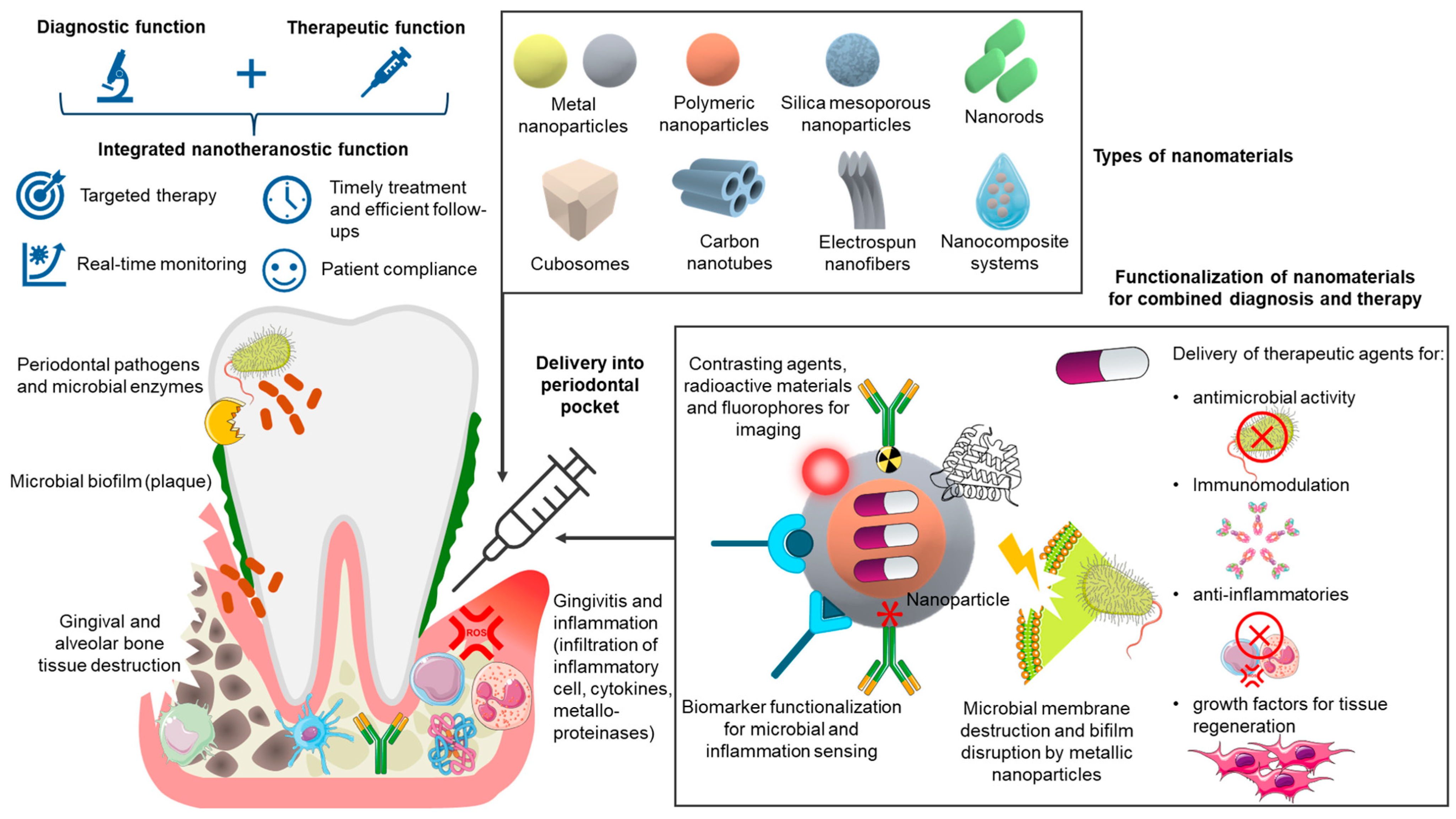
Nanotheranostics in periodontitis refers to the development of single nanoscale platforms that integrate both diagnostic and therapeutic functionalities. This dual-purpose approach enables more precise and responsive management of periodontal disease by facilitating simultaneous detection and treatment within the same system. For example, nanoparticles can be engineered to recognize specific periodontal biomarkers or pathogenic bacteria while concurrently delivering antimicrobial agents, anti-inflammatory drugs, or regenerative bioactives as explored above and shown in Figure 5. This integration not only streamlines the treatment process but also enhances the specificity and efficacy of therapeutic interventions.
A key advantage of nanotheranostic systems is their ability to achieve targeted therapy. Nanoparticles can be functionalized with ligands, such as antibodies, peptides, or lectins, or even genetically engineered with cell membrane to selectively bind to inflamed periodontal tissues or microbial biofilms [130,131]. This targeted interaction ensures that therapeutic agents are delivered directly to diseased sites, thereby maximizing local efficacy while minimizing systemic exposure and associated side effects. Moreover, nanotheranostics hold the unique advantage of enabling real-time monitoring of therapeutic outcomes. By incorporating imaging agents (such as fluorescent dyes, superparamagnetic iron oxide nanoparticles, or radiocontrast materials) into the nanocarrier system, clinicians can visualize nanoparticle distribution, assess biofilm disruption, and track therapeutic response using imaging modalities such as fluorescence microscopy, MRI, or CT. This feedback loop provides a dynamic assessment of treatment success, allowing for early adjustments to therapeutic strategies and promoting personalized, responsive care.
Currently, the approach to periodontitis management follows a modular or sequential approach where diagnostic nanomaterials are first deployed to detect disease activity, followed by administration of tailored therapeutic agents based on those diagnostic findings. Although the diagnostic and therapeutic elements are physically distinct, the system embodies the theranostic paradigm by enabling data-driven, patient-specific intervention. Despite this promising approach, there remains a clear gap in the scientific literature specifically addressing nanotheranostics for periodontitis. While a wealth of research has explored nanoparticles for therapeutic use (focusing on antimicrobial delivery, anti-inflammatory activity, and tissue regeneration) the simultaneous integration of diagnostic functions is still underexplored. Diagnostic applications of nanomaterials (e.g., biosensing of pathogens and inflammatory biomarkers) are being investigated, but often separately from therapeutic delivery. The literature search for the current review identified that most existing studies either target therapy or diagnostics in isolation, suggesting an important and underdeveloped research niche-comprehensive nanotheranostic systems that unify diagnostics and therapeutics. Figure 6 provides a schematic overview of how nanotheranostic platforms can be applied in periodontitis, illustrating the integration of diagnostic functions (biosensing, imaging) with therapeutic actions (antimicrobial activity, biofilm disruption, immunomodulation, and tissue regeneration) to enable more precise and personalized treatment.
The development of nanotheranostics for periodontitis offers a transformative approach by closing the gap between diagnosis and treatment. These systems can significantly improve treatment precision, enhance drug localization, reduce off-target effects, and enable early detection of treatment failure or disease recurrence. Furthermore, the ability to customize these nanosystems based on patient-specific profiles, such as microbial composition, inflammatory status, and tissue characteristics will support the advancement of personalized, minimally invasive periodontal nanotherapies.

6. Translational and Clinical Considerations in Periodontitis Nanotheranostics

Although significant progress has been made in developing nanoscale platforms for diagnostic and therapeutic applications in periodontitis, the clinical translation of nanotheranostics remains constrained by multiple interdependent biological, technological, regulatory, and ethical challenges. While preclinical studies have demonstrated proof-of-concept efficacy, several critical hurdles must be addressed to enable successful implementation in routine dental practice.

6.1. Design Complexity and Functional Compromises

Achieving optimal nanoparticle design involves navigating complex compromises between functionality, stability, and safety. Parameters such as particle size, shape, and surface charge critically influence tissue penetration, cellular uptake, and clearance kinetics. While smaller particles may facilitate biofilm penetration, they may also increase systemic exposure and toxicity. Advanced features such as, pH- or enzyme-responsive designs, shape-switching geometries, and ligand-mediated targeting, offer enhanced periodontal specificity but increase synthetic complexity and cost. Additionally, surface functionalization may alter immunogenicity or disrupt pharmacokinetics, necessitating iterative design refinement [132].

6.2. Biological Complexity and In Vivo Relevance

The complex and dynamic nature of the oral environment presents a major translational barrier. Nanoplatforms that exhibit promising performance in vitro or in small-animal models often face limited efficacy in human settings due to factors such as enzymatic degradation, biofilm-mediated barriers, fluctuating pH, continuous salivary and crevicular fluid flow, and localized immune responses. Effective periodontal nanotherapies must be engineered to navigate mucosal surfaces, resist microbial colonization, and maintain physicochemical stability under variable physiological conditions while retaining their targeting functionality and controlled-release capabilities [133]. There is a pressing need for more sophisticated in vitro and in vivo models that closely mimic the human periodontal microenvironment [134]. These models should enable simultaneous evaluation of therapeutic efficacy and diagnostic responsiveness under conditions of biofilm colonization and chronic inflammation.

6.3. Safety, Immunogenicity, and Long-Term Toxicity

Biocompatibility remains a core concern in the development of nanotheranostic systems. Although many formulations demonstrate short-term safety, their long-term biodistribution, metabolism, and potential for systemic accumulation remain poorly understood. Accumulation in reticuloendothelial organs and unintended interactions with immune or hematopoietic systems may lead to adverse outcomes, including oxidative stress, genotoxicity, endocrine disruption, or neurotoxicity [8,132]. Moreover, not all biodegradable carriers are inherently safe, as some degradation products may elicit inflammatory or toxic responses [131]. Optimization of particle size, surface properties, and degradation profiles remains essential to minimizing these risks.

6.4. Manufacturing, Scalability, and Quality Control

Transitioning from bench-scale synthesis to Good Manufacturing Practice compliant, scalable manufacturing remains a major obstacle. Nanoparticle heterogeneity, including batch-to-batch variation in size, morphology, and functional performance, can compromise therapeutic reproducibility and safety [135]. Standardized, modular fabrication strategies that ensure uniformity and allow for diagnostic-therapeutic integration are critical for clinical readiness. This is particularly relevant in dental applications, where precision in drug release and mechanical stability can significantly impact treatment outcomes [133]. However, achieving such clinical-grade quality requires precise control over parameters such as particle size, drug loading, surface functionalization, and release kinetics which can be further complicated by the inclusion of diagnostic agents. To overcome manufacturing challenges, using FDA-approved excipients, self-assembling systems, and stimuli-responsive polymers may streamline regulatory approval and facilitate scalable production. Robust quality control protocols and real-time analytical tools will also be critical for ensuring batch-to-batch consistency.

6.5. Regulatory and Ethical Considerations

Nanotheranostic platforms occupy a regulatory grey zone as combination products, requiring comprehensive safety data and often lacking clear approval pathways. Current regulatory frameworks do not fully accommodate the complexity of multifunctional nanomedicines, especially in dentistry where validated surrogate endpoints for treatment efficacy are limited and there is no harmonized regulatory pathway for nanotheranostic applications in dentistry. Clinical trials are further complicated by the absence of standardized biomarker panels for disease activity and therapeutic response [8,131]. Delays in regulatory approval, coupled with ethical concerns related to patient data, environmental risk, and long-term exposure, present additional translational bottlenecks. Collaborative networks among academia, industry, and regulatory bodies will be instrumental in establishing evaluation pipelines and defining approval pathways specific to dental nanotheranostics.
As nanotheranostic systems evolve toward precision and personalization, they raise important ethical questions related to informed consent, data privacy (especially with real-time diagnostics), and equitable access [135]. The incorporation of biosensors and imaging agents also introduces considerations around data ownership and interpretation.

6.6. Interdisciplinary Collaboration and Integration into Clinical Workflow

Successful translation requires effective collaboration between material scientists, biomedical engineers, clinicians, and regulatory bodies. For successful clinical adoption, nanotheranostic systems must be seamlessly incorporated into existing periodontal care workflows. Periodontists and dental practitioners play a critical role in defining real-world performance criteria such as application feasibility, patient compliance, and integration into treatment workflows. Without sustained interdisciplinary dialogue, many nanotheranostic systems may remain confined to preclinical settings, lacking the translational refinement necessary for clinical adoption [8]. Additionally, clinicians must be equipped to interpret diagnostic outputs and translate them into actionable treatment decisions [136]. This underscores the need for user-friendly readout systems, automated signal quantification, and decision-support tools, otherwise practitioners may resort to old practices [137]. Systems that simplify procedures, reduce treatment duration, enhance patient comfort, and enable real-time monitoring are likely to see greater clinical acceptance.

6.7. Cost, Accessibility, and Implementation Feasibility

The high cost of nanotheranostic development, regulatory approval, and imaging infrastructure limits accessibility, particularly in public health systems and low-resource settings. Specialized synthesis techniques, biosafety protocols, and the need for advanced imaging technologies contribute to high implementation costs. Ethical considerations, such as long-term safety, environmental sustainability, and equitable access, require transparent risk–benefit health economics evaluations and the inclusion of diverse patient populations in trial design [131,135]. Early integration of cost-effectiveness analyses and patient-centered design approaches will be crucial to ensure that these advanced platforms are both clinically impactful and socially responsible. Strategies to reduce costs include minimizing reliance on high-end imaging platforms, simplifying synthesis protocols, and utilizing scalable, low-cost materials.

7. Conclusions and Future Perspectives

The emergence of nanotheranostic platforms represents a transformative advancement in the diagnosis and treatment of periodontitis, addressing critical limitations of conventional modalities through integrated, multifunctional, and responsive approaches. By uniting diagnostic and therapeutic functions within a single nanosystem, these technologies offer the potential for early disease detection, personalized intervention, and real-time monitoring of therapeutic outcomes. In particular, inorganic nanomaterials, including metallic and metal oxide nanoparticles—have demonstrated exceptional versatility, enabling antimicrobial activity, biofilm disruption, immunomodulation, tissue regeneration, and point-of-care biosensing for microbial and inflammatory biomarkers.
Despite these promising developments, clinical translation remains limited. Key challenges such as the biological complexity of the oral microenvironment, potential nanoparticle toxicity, design and functional optimization, manufacturing scalability, and regulatory ambiguity, must be systematically addressed. Furthermore, the integration of interdisciplinary expertise and patient-centred design considerations are essential to ensure clinical relevance, safety, affordability, and compatibility with routine dental workflows.
Looking forward, several strategies may accelerate clinical impact. These include the development of biodegradable and immuno-evasive nanocarriers, incorporation of stimuli-responsive drug release mechanisms, and coupling of nanotheranostic platforms with mobile health technologies to facilitate chairside diagnostics and teledentistry. Novel materials such as carbon-based nanostructures, exosome-mimetic vesicles, and bioinspired scaffolds further expand the therapeutic design space. Additionally, the integration of artificial intelligence and machine learning for theranostic signal interpretation could enable truly personalized periodontal care, tailored to individual host responses, microbial profiles, and disease phenotypes.
In summary, while significant translational barriers persist, the field of nanotheranostics in periodontitis is rapidly advancing. Sustained investment in interdisciplinary research, regulatory harmonization, and clinical integration will be pivotal in transforming these nanoscale innovations into tangible improvements in patient outcomes. With strategic development, nanotheranostic platforms are well-positioned to redefine the standard of care in periodontology, advancing the broader vision of precision, preventive, and minimally invasive dentistry.

Author Contributions

Conceptualization, P.R.; investigation, P.R. and T.P.K.V.; writing—original draft preparation, P.R. and T.P.K.V.; writing—review and editing, P.R.; project administration, Y.E.C.; supervision, P.R. and Y.E.C. All authors have read and agreed to the published version of the manuscript.

Funding

This research received no external funding.

Conflicts of Interest

The authors declare no conflicts of interest.

Abbreviations

The following abbreviations are used in this manuscript:
GCFGingival crevicular fluid
TLRsToll-like receptors
ROSReactive oxygen species
MMPsMetalloproteinases
NETsNeutrophil extracellular traps
RANKLReceptor activator of NF-κB Ligand
EBCExhaled breath condensate
ASTAspartate aminotransferase
ALPAlkaline phosphatase
LDHLactate dehydrogenase
OPGOsteoprotegerin
VOCsVolatile organic compounds
PLGAPoly(lactic-co-glycolic acid)
PLAPolylactic acid
PCLPolycaprolactone
MRIMagnetic resonance imaging
CTComputed tomography
SLNsSolid lipid nanoparticles
NLCsNanostructured lipid carriers
LNCsLipid nanocapsules
MSNsMesoporous silica nanoparticles
ZIF-8Zeolitic imidazolate framework 8
UCNPsUpconversion nanoparticles
NPsNanoparticles
NIRNear-infrared
GOGraphene oxide
rGOReduced graphene oxide
GQDsGraphene quantum dots
CDsCarbon dots
MWCNTsMulti-walled carbon nanotubes
CSHSsCopper silicate nanozymes
QDsQuantum dots

References

  1. Wang, Y.; Zhuo, L.; Yang, S.; Dong, C.; Shu, P. Burden of Periodontal Diseases in Young Adults. Sci. Rep. 2025, 15, 6653. [Google Scholar] [CrossRef]
  2. Nascimento, G.G.; Alves-Costa, S.; Romandini, M. Burden of Severe Periodontitis and Edentulism in 2021, with Projections up to 2050: The Global Burden of Disease 2021 Study. J. Periodontal Res. 2024, 59, 823–867. [Google Scholar] [CrossRef] [PubMed]
  3. Buduneli, N. Environmental Factors and Periodontal Microbiome. Periodontology 2021, 85, 112–125. [Google Scholar] [CrossRef] [PubMed]
  4. Sedghi, L.; DiMassa, V.; Harrington, A.; Lynch, S.V.; Kapila, Y.L. The Oral Microbiome: Role of Key Organisms and Complex Networks in Oral Health and Disease. Periodontology 2021, 87, 107–131. [Google Scholar] [CrossRef] [PubMed]
  5. Gasner, N.S.; Schure, R.S. Periodontal Disease. In StatPearls; StatPearls Publishing: Treasure Island, FL, USA, 2025. [Google Scholar]
  6. Hooshiar, M.H.; Moghaddam, M.A.; Kiarashi, M.; Al-Hijazi, A.Y.; Hussein, A.F.; Alrikabi, H.; Salari, S.; Esmaelian, S.; Mesgari, H.; Yasamineh, S. Recent Advances in Nanomaterial-Based Biosensor for Periodontitis Detection. J. Biol. Eng. 2024, 18, 28. [Google Scholar] [CrossRef]
  7. Shi, R.; Zhu, Y.; Lu, W.; Zhai, R.; Zhou, M.; Shi, S.; Chen, Y. Nanomaterials: Innovative Approaches for Addressing Key Objectives in Periodontitis Treatment. RSC Adv. 2024, 14, 27904–27927. [Google Scholar] [CrossRef]
  8. Wang, D.; Li, Q.; Xiao, C.; Wang, H.; Dong, S. Nanoparticles in Periodontitis Therapy: A Review of the Current Situation. Int. J. Nanomed. 2024, 19, 6857–6893. [Google Scholar] [CrossRef]
  9. Sunila, B.S.; Shivakumar, G.C.; Abdul, N.S.; Sudhakar, N.; Franco, R.; Ronsivalle, V.; Cicciù, M.; Minervini, G. Conventional Periodontal Probing versus Salivary Biomarkers in Diagnosis and Evaluation of Chronic Periodontitis in Type 2 Diabetes Mellitus. Minerva Dent. Oral Sci. 2025, 74, 187–194. [Google Scholar] [CrossRef]
  10. Yussif, N.; Akarslan, Z. Pathogenesis of Periodontal Disease. In Periodontal Disease-Diagnostic and Adjunctive Non-Surgical Considerations; IntechOpen: Rijeka, Croatia, 2020; ISBN 978-1-78984-460-3. [Google Scholar]
  11. Nanci, A.; Bosshardt, D.D. Structure of Periodontal Tissues in Health and Disease*. Periodontology 2006, 40, 11–28. [Google Scholar] [CrossRef]
  12. Omi, M.; Mishina, Y. Roles of Osteoclasts in Alveolar Bone Remodeling. Genesis 2022, 60, e23490. [Google Scholar] [CrossRef]
  13. Vitkov, L.; Singh, J.; Schauer, C.; Minnich, B.; Krunić, J.; Oberthaler, H.; Gamsjaeger, S.; Herrmann, M.; Knopf, J.; Hannig, M. Breaking the Gingival Barrier in Periodontitis. Int. J. Mol. Sci. 2023, 24, 4544. [Google Scholar] [CrossRef]
  14. Könönen, E.; Gursoy, M.; Gursoy, U. Periodontitis: A Multifaceted Disease of Tooth-Supporting Tissues. J. Clin. Med. 2019, 8, 1135. [Google Scholar] [CrossRef] [PubMed]
  15. Scannapieco, F.A.; Gershovich, E. The Prevention of Periodontal Disease—An Overview. Periodontology 2020, 84, 9–13. [Google Scholar] [CrossRef] [PubMed]
  16. Villoria, G.E.M.; Fischer, R.G.; Tinoco, E.M.B.; Meyle, J.; Loos, B.G. Periodontal Disease: A Systemic Condition. Periodontology 2024, 96, 7–19. [Google Scholar] [CrossRef] [PubMed]
  17. Yekani, M.; Dastgir, M.; Fattahi, S.; Shahi, S.; Maleki Dizaj, S.; Memar, M.Y. Microbiological and Molecular Aspects of Periodontitis Pathogenesis: An Infection-Induced Inflammatory Condition. Front. Cell. Infect. Microbiol. 2025, 15, 1533658. [Google Scholar] [CrossRef]
  18. Ray, R.R. Periodontitis: An Oral Disease with Severe Consequences. Appl. Biochem. Biotechnol. 2023, 195, 17–32. [Google Scholar] [CrossRef]
  19. Chen, E.; Wang, T.; Tu, Y.; Sun, Z.; Ding, Y.; Gu, Z.; Xiao, S. ROS-Scavenging Biomaterials for Periodontitis. J. Mater. Chem. B 2023, 11, 482–499. [Google Scholar] [CrossRef]
  20. Kinane, D.F. Causation and Pathogenesis of Periodontal Disease. Periodontology 2001, 25, 8–20. [Google Scholar] [CrossRef]
  21. Bartold, P.M.; Van Dyke, T.E. An Appraisal of the Role of Specific Bacteria in the Initial Pathogenesis of Periodontitis. J. Clin. Periodontol. 2019, 46, 6–11. [Google Scholar] [CrossRef]
  22. Pan, W.; Wang, Q.; Chen, Q. The Cytokine Network Involved in the Host Immune Response to Periodontitis. Int. J. Oral Sci. 2019, 11, 30. [Google Scholar] [CrossRef]
  23. Ramenzoni, L.L.; Lehner, M.P.; Kaufmann, M.E.; Wiedemeier, D.; Attin, T.; Schmidlin, P.R. Oral Diagnostic Methods for the Detection of Periodontal Disease. Diagnostics 2021, 11, 571. [Google Scholar] [CrossRef] [PubMed]
  24. Ali Alftaikhah, S.A.; Issrani, R.; Alnasser, M.; Almutairi, H.A.; Khattak, O.; Iqbal, A.; Prabhu, N. Salivary Biomarkers in Periodontitis: A Scoping Review. Cureus 2023, 15, e50207. [Google Scholar] [CrossRef] [PubMed]
  25. Jiang, H.; Dong, Z.; Xia, X.; Li, X. Cathepsins in Oral Diseases: Mechanisms and Therapeutic Implications. Front. Immunol. 2023, 14, 1203071. [Google Scholar] [CrossRef] [PubMed]
  26. Nomura, Y.; Shimada, Y.; Hanada, N.; Numabe, Y.; Kamoi, K.; Sato, T.; Gomi, K.; Arai, T.; Inagaki, K.; Fukuda, M.; et al. Salivary Biomarkers for Predicting the Progression of Chronic Periodontitis. Arch. Oral Biol. 2012, 57, 413–420. [Google Scholar] [CrossRef]
  27. Koppolu, P.; Sirisha, S.; Mishra, A.; Deshpande, K.; Lingam, A.S.; Alotaibi, D.H.; Saleh Alwahibi, M.; Penela, S. Alkaline Phosphatase and Acid Phosphatase Levels in Saliva and Serum of Patients with Healthy Periodontium, Gingivitis, and Periodontitis before and after Scaling with Root Planing: A Clinico-Biochemical Study. Saudi J. Biol. Sci. 2021, 28, 380–385. [Google Scholar] [CrossRef]
  28. Taymour, N.; Hassan, M.G.; AlGhamdi, M.A.; Omara, W.S. From Detection to Treatment: Nanomaterial-Based Biosensors Transforming Prosthetic Dentistry and Oral Health Care: A Scoping Review. Prosthesis 2025, 7, 51. [Google Scholar] [CrossRef]
  29. Alsaykhan, K.; Khan, N.S.; Aljumah, M.I.; Albughaylil, A.S. Comparative Evaluation of Salivary Enzyme in Patients With Gingivitis and Periodontitis: A Clinical-Biochemical Study. Cureus 2022, 14, e20991. [Google Scholar] [CrossRef] [PubMed]
  30. Al-Rawi, N.H.; Al-Marzooq, F.; Al-Nuaimi, A.S.; Hachim, M.Y.; Hamoudi, R. Salivary microRNA 155, 146a/b and 203: A Pilot Study for Potentially Non-Invasive Diagnostic Biomarkers of Periodontitis and Diabetes Mellitus. PLoS ONE 2020, 15, e0237004. [Google Scholar] [CrossRef]
  31. Wu, P.; Feng, J.; Wang, W. Expression of miR-155 and miR-146a in the Saliva of Patients with Periodontitis and Its Clinical Value. Am. J. Transl. Res. 2021, 13, 6670–6677. [Google Scholar]
  32. Mata-Monterde, M.; Serrano-Valcarce, A.; Almiñana-Pastor, P.J.; Micó-Martínez, P.; López-Roldán, A. miRNAs as Epigenetic Biomarkers in the Study of the Bidirectional Relationship between Type 2 Diabetes Mellitus and Periodontitis: A Systematic Review. Int. J. Mol. Sci. 2024, 25, 10723. [Google Scholar] [CrossRef]
  33. Subbarao, K.C.; Nattuthurai, G.S.; Sundararajan, S.K.; Sujith, I.; Joseph, J.; Syedshah, Y.P. Gingival Crevicular Fluid: An Overview. J. Pharm. Bioallied Sci. 2019, 11, S135. [Google Scholar] [CrossRef] [PubMed]
  34. Sirisereephap, K.; Maekawa, T.; Tamura, H.; Hiyoshi, T.; Domon, H.; Isono, T.; Terao, Y.; Maeda, T.; Tabeta, K. Osteoimmunology in Periodontitis: Local Proteins and Compounds to Alleviate Periodontitis. Int. J. Mol. Sci. 2022, 23, 5540. [Google Scholar] [CrossRef]
  35. Alwandawi, T.K. Immunodiagnostic Potential of the RANK/RANKL/OPG Ratio in Gingival Crevicular Fluid for Periodontitis. Folia Med. 2025, 67, e144949. [Google Scholar] [CrossRef] [PubMed]
  36. Marsh, P.D. Dental Plaque as a Biofilm and a Microbial Community-Implications for Health and Disease. BMC Oral Health 2006, 6, S14. [Google Scholar] [CrossRef] [PubMed]
  37. Dong, A.; Proctor, G.; Zaric, S. Diagnostic Accuracy of Microbiome-Derived Biomarkers in Periodontitis: Systematic Review and Meta-Analysis. J. Periodontal Res. 2025, 60, 748–761. [Google Scholar] [CrossRef]
  38. Huang, Y.; Zhao, X.; Cui, L.; Huang, S. Metagenomic and Metatranscriptomic Insight Into Oral Biofilms in Periodontitis and Related Systemic Diseases. Front. Microbiol. 2021, 12, 728585. [Google Scholar] [CrossRef]
  39. Kametani, M.; Nagasawa, Y.; Usuda, M.; Kaneki, A.; Ogawa, M.; Shojima, K.; Yamazaki, H.; Tokumoto, K.; Matsuoka, D.; Suehara, K.; et al. Relationship Between the Presence of Red Complex Species and the Distribution of Other Oral Bacteria, Including Major Periodontal Pathogens in Older Japanese Individuals. Int. J. Mol. Sci. 2024, 25, 12243. [Google Scholar] [CrossRef]
  40. Liu, F.; Zhu, B.; An, Y.; Zhou, Z.; Xiong, P.; Li, X.; Mi, Y.; He, T.; Chen, F.; Wu, B. Gingipain from Porphyromonas Gingivalis Causes Insulin Resistance by Degrading Insulin Receptors through Direct Proteolytic Effects. Int. J. Oral Sci. 2024, 16, 53. [Google Scholar] [CrossRef]
  41. Foroughi, M.; Torabinejad, M.; Angelov, N.; Ojcius, D.M.; Parang, K.; Ravnan, M.; Lam, J. Bridging Oral and Systemic Health: Exploring Pathogenesis, Biomarkers, and Diagnostic Innovations in Periodontal Disease. Infection 2025, 1–26. [Google Scholar] [CrossRef]
  42. Ghelli, F.; Panizzolo, M.; Garzaro, G.; Squillacioti, G.; Bellisario, V.; Colombi, N.; Bergamaschi, E.; Guseva Canu, I.; Bono, R. Inflammatory Biomarkers in Exhaled Breath Condensate: A Systematic Review. Int. J. Mol. Sci. 2022, 23, 9820. [Google Scholar] [CrossRef]
  43. Ghasemirad, M.; Tavakol, O.; Etesami, S.; Nikeghbal, K.; Nikeghbal, D.; Diznab, F.A.; Anzabi, R.M. A State of Art Review: Volatile Organic Compounds and Periodontitis: Volatile Organic Compounds and Periodontitis. Galen Med. J. 2024, 13, e3730. [Google Scholar] [CrossRef]
  44. Haiek, M.; Dvoyris, V.; Broza, Y.Y.; Haick, H.; Weiss, E.; Houri-Haddad, Y. Bacterial Volatile Organic Compounds as Potential Caries and Periodontitis Disease Biomarkers. Int. J. Mol. Sci. 2025, 26, 3591. [Google Scholar] [CrossRef] [PubMed]
  45. Lee, Y.-H.; Hong, J.-Y. Oral Microbiome as a Co-Mediator of Halitosis and Periodontitis: A Narrative Review. Front. Oral Health 2023, 4, 1229145. [Google Scholar] [CrossRef] [PubMed]
  46. Constantin, V.; Luchian, I.; Goriuc, A.; Budala, D.G.; Bida, F.C.; Cojocaru, C.; Butnaru, O.-M.; Virvescu, D.I. Salivary Biomarkers Identification: Advances in Standard and Emerging Technologies. Oral 2025, 5, 26. [Google Scholar] [CrossRef]
  47. Campos, J.V.; Pontes, J.T.C.; Canales, C.S.C.; Roque-Borda, C.A.; Pavan, F.R. Advancing Nanotechnology: Targeting Biofilm-Forming Bacteria with Antimicrobial Peptides. BME Front. 2025, 6, 0104. [Google Scholar] [CrossRef]
  48. Ifijen, I.H.; Awoyemi, R.F.; Faderin, E.; Akobundu, U.U.; Ajayi, A.S.; Chukwu, J.U.; Lekan, O.K.; Asiriuwa, O.D.; Maliki, M.; Ikhuoria, E.U. Protein-Based Nanoparticles for Antimicrobial and Cancer Therapy: Implications for Public Health. RSC Adv. 2025, 15, 14966–15016. [Google Scholar] [CrossRef]
  49. Fidyawati, D.; Masulili, S.L.C.; Iskandar, H.B.; Suhartanto, H.; Kiswanjaya, B.; Li, X. Clinical and Radiographic Parameters for Early Periodontitis Diagnosis: A Comparative Study. Dent. J. 2024, 12, 407. [Google Scholar] [CrossRef]
  50. Ng, E.; Tay, J.R.H.; Ong, M.M.A. Minimally Invasive Periodontology: A Treatment Philosophy and Suggested Approach. Int. J. Dent. 2021, 1, 2810264. [Google Scholar] [CrossRef]
  51. de Molon, R.S.; Rodrigues, J.V.S.; Deroide, M.B.; da Silva Barbirato, D.; Garcia, V.G.; Theodoro, L.H. The Efficacy of Topical or Systemic Antibiotics as Adjuvants to Non-Surgical Periodontal Treatment in Diabetic Patients: A Systematic Review and Meta-Analysis of Randomized Clinical Trials. J. Clin. Med. 2024, 13, 4763. [Google Scholar] [CrossRef]
  52. Mirghani, R.; Saba, T.; Khaliq, H.; Mitchell, J.; Do, L.; Chambi, L.; Diaz, K.; Kennedy, T.; Alkassab, K.; Huynh, T.; et al. Biofilms: Formation, Drug Resistance and Alternatives to Conventional Approaches. AIMS Microbiol. 2022, 8, 239–277. [Google Scholar] [CrossRef]
  53. Wu, H.; Li, Y.; Shi, L.; Liu, Y.; Shen, J. New Advances in Periodontal Functional Materials Based on Antibacterial, Anti-Inflammatory, and Tissue Regeneration Strategies. Adv. Healthc. Mater. 2025, 14, 2403206. [Google Scholar] [CrossRef] [PubMed]
  54. Huang, T.-H.; Chen, J.-Y.; Suo, W.-H.; Shao, W.-R.; Huang, C.-Y.; Li, M.-T.; Li, Y.-Y.; Li, Y.-H.; Liang, E.-L.; Chen, Y.-H.; et al. Unlocking the Future of Periodontal Regeneration: An Interdisciplinary Approach to Tissue Engineering and Advanced Therapeutics. Biomedicines 2024, 12, 1090. [Google Scholar] [CrossRef] [PubMed]
  55. Liu, Y.; Guo, L.; Li, X.; Liu, S.; Du, J.; Xu, J.; Hu, J.; Liu, Y. Challenges and Tissue Engineering Strategies of Periodontal-Guided Tissue Regeneration. Tissue Eng. Part C Methods 2022, 28, 405–419. [Google Scholar] [CrossRef] [PubMed]
  56. Budală, D.G.; Luchian, I.; Tatarciuc, M.; Butnaru, O.; Armencia, A.O.; Virvescu, D.I.; Scutariu, M.M.; Rusu, D. Are Local Drug Delivery Systems a Challenge in Clinical Periodontology? J. Clin. Med. 2023, 12, 4137. [Google Scholar] [CrossRef]
  57. Joshi, D.; Garg, T.; Goyal, A.K.; Rath, G. Advanced Drug Delivery Approaches against Periodontitis. Drug Deliv. 2016, 23, 363–377. [Google Scholar] [CrossRef]
  58. Yudaev, P.; Chuev, V.; Klyukin, B.; Kuskov, A.; Mezhuev, Y.; Chistyakov, E. Polymeric Dental Nanomaterials: Antimicrobial Action. Polymers 2022, 14, 864. [Google Scholar] [CrossRef]
  59. Uskoković, V.; Pejčić, A.; Koliqi, R.; Anđelković, Z. Polymeric Nanotechnologies for the Treatment of Periodontitis: A Chronological Review. Int. J. Pharm. 2022, 625, 122065. [Google Scholar] [CrossRef]
  60. D’Amico, E.; Aceto, G.M.; Petrini, M.; Cinquini, C.; D’Ercole, S.; Iezzi, G.; Pierfelice, T.V. How Will Nanomedicine Revolutionize Future Dentistry and Periodontal Therapy? Int. J. Mol. Sci. 2025, 26, 592. [Google Scholar] [CrossRef]
  61. Mutreja, I.; Kumar, D.; Kaushik, A.; Mishra, Y.K. Lipid Nanoparticle-Based Formulations for High-Performance Dentistry Applications. J. Mater. Chem. B 2023, 11, 5990–6023. [Google Scholar] [CrossRef]
  62. Lima de Sousa, T.; Dourado, D.; Rodrigues, J.S.; de Souza Rebouças, J.; Montes, M.A.J.R.; Formiga, F.R. Treatment of Periodontal Disease: Does Drug Delivery Matter? Front. Bioeng. Biotechnol. 2024, 12, 1427758. [Google Scholar] [CrossRef]
  63. Attri, N.; Das, S.; Banerjee, J.; Shamsuddin, S.H.; Dash, S.K.; Pramanik, A. Liposomes to Cubosomes: The Evolution of Lipidic Nanocarriers and Their Cutting-Edge Biomedical Applications. ACS Appl. Bio Mater. 2024, 7, 2677–2694. [Google Scholar] [CrossRef]
  64. Malode, T.D.; Pillare, S.B.; Shahi, A.K.; Lade, S.N.; Taksande, J.B.; Umekar, M.J. Cubosomes as Versatile Nanocarriers: Insights into Composition, Mechanisms, and Therapeutic Applications. Biomed. Mater. Devices 2025, 1–22. [Google Scholar] [CrossRef]
  65. Hedayatipanah, M.; Gholami, L.; Farmany, A.; Alikhani, M.Y.; Hooshyarfard, A.; Hashemiyan, F.S. Green Synthesis of Silver Nanoparticles and Evaluation of Their Effects on the Porphyromonas Gingivalis Bacterial Biofilm Formation. Clin. Exp. Dent. Res. 2024, 10, e887. [Google Scholar] [CrossRef] [PubMed]
  66. Nasiri, K.; Masoumi, S.M.; Amini, S.; Goudarzi, M.; Tafreshi, S.M.; Bagheri, A.; Yasamineh, S.; alwan, M.; Arellano, M.T.C.; Gholizadeh, O. Recent Advances in Metal Nanoparticles to Treat Periodontitis. J. Nanobiotechnol. 2023, 21, 283. [Google Scholar] [CrossRef] [PubMed]
  67. Bae, G.; Kim, M.; Lee, A.; Ji, S.; Jang, M.; Yim, S.; Song, W.; Lee, S.S.; Yoon, D.H.; An, K.-S. Nanometric Lamination of Zinc Oxide Nanofilms with Gold Nanoparticles for Self-Perceived Periodontal Disease Sensors. Compos. Part B Eng. 2022, 230, 109490. [Google Scholar] [CrossRef]
  68. Tong, F.; Wang, P.; Chen, Z.; Liu, Y.; Wang, L.; Guo, J.; Li, Z.; Cai, H.; Wei, J. Combined Ferromagnetic Nanoparticles for Effective Periodontal Biofilm Eradication in Rat Model. Int. J. Nanomed. 2023, 18, 2371–2388. [Google Scholar] [CrossRef]
  69. Nawaz, M.Z.; Alghamdi, H.A.; Zahoor, M.; Rashid, F.; Alshahrani, A.A.; Alghamdi, N.S.; Pugazhendhi, A.; Zhu, D. Synthesis of Novel Metal Silica Nanoparticles Exhibiting Antimicrobial Potential and Applications to Combat Periodontitis. Environ. Res. 2024, 241, 117415. [Google Scholar] [CrossRef]
  70. Naruphontjirakul, P.; Li, M.; Boccaccini, A.R. Strontium and Zinc Co-Doped Mesoporous Bioactive Glass Nanoparticles for Potential Use in Bone Tissue Engineering Applications. Nanomaterials 2024, 14, 575. [Google Scholar] [CrossRef]
  71. Fang, L.; Zhou, H.; Cheng, L.; Wang, Y.; Liu, F.; Wang, S. The Application of Mesoporous Silica Nanoparticles as a Drug Delivery Vehicle in Oral Disease Treatment. Front. Cell. Infect. Microbiol. 2023, 13, 1124411. [Google Scholar] [CrossRef]
  72. Casarrubios, L.; Gómez-Cerezo, N.; Feito, M.J.; Vallet-Regí, M.; Arcos, D.; Portolés, M.T. Ipriflavone-Loaded Mesoporous Nanospheres with Potential Applications for Periodontal Treatment. Nanomaterials 2020, 10, 2573. [Google Scholar] [CrossRef]
  73. Díez-Pascual, A.M. Carbon-Based Nanomaterials. Int. J. Mol. Sci. 2021, 22, 7726. [Google Scholar] [CrossRef]
  74. Liu, C.; Chen, Y.; Bai, H.; Niu, Y.; Wu, Y. Characterization and Application of in Situ Curcumin/ZNP Hydrogels for Periodontitis Treatment. BMC Oral Health 2024, 24, 395. [Google Scholar] [CrossRef]
  75. Liu, Y.; Wang, S.; Qu, X.; Li, Y.; Zhou, C.; Xie, D.; Dong, J.; Ji, L.; Xu, J.; Zhou, J. Highly-Sensitive Ultra-Thin Dental Patches Assisted with Artificial-Intelligence Recognition for Mapping Hidden Periodontitis Lesions. Sens. Actuators B Chem. 2025, 435, 137648. [Google Scholar] [CrossRef]
  76. Li, X.; Huang, R.; Li, P.; Tang, F.K.; He, J.; Sun, H.; Wang, X.; Wang, M.; Lan, X.; Wang, X.; et al. Berberine-Functionalized Bismuth-Doped Carbon Dots in a Pathogen-Responsive Hydrogel System: A Multifaceted Approach to Combating Periodontal Diseases. ACS Nano 2025, 19, 17554–17577. [Google Scholar] [CrossRef]
  77. Di Cristo, F.; Valentino, A.; De Luca, I.; Peluso, G.; Bonadies, I.; Calarco, A.; Di Salle, A. PLA Nanofibers for Microenvironmental-Responsive Quercetin Release in Local Periodontal Treatment. Molecules 2022, 27, 2205. [Google Scholar] [CrossRef]
  78. Milosevic, M.; Stojanovic, D.B.; Simic, V.; Grkovic, M.; Bjelovic, M.; Uskokovic, P.S.; Kojic, M. Preparation and Modeling of Three-layered PCL/PLGA/PCL Fibrous Scaffolds for Prolonged Drug Release. Sci. Rep. 2020, 10, 11126. [Google Scholar] [CrossRef] [PubMed]
  79. Mirzaeei, S.; Mansurian, M.; Asare-Addo, K.; Nokhodchi, A. Metronidazole- and Amoxicillin-Loaded PLGA and PCL Nanofibers as Potential Drug Delivery Systems for the Treatment of Periodontitis: In Vitro and In Vivo Evaluations. Biomedicines 2021, 9, 975. [Google Scholar] [CrossRef] [PubMed]
  80. Shariati, A.; Chegini, Z.; Ghaznavi-Rad, E.; Zare, E.N.; Hosseini, S.M. PLGA-Based Nanoplatforms in Drug Delivery for Inhibition and Destruction of Microbial Biofilm. Front. Cell. Infect. Microbiol. 2022, 12, 926363. [Google Scholar] [CrossRef]
  81. Yürük, G.; Damla Demir, Y.; Vural, Ş.; Seda Kehr, N. Polymeric Biomaterials for Periodontal Tissue Engineering and Periodontitis. RSC Appl. Polym. 2024, 2, 534–556. [Google Scholar] [CrossRef]
  82. Toledano-Osorio, M.; Osorio, R.; Bueno, J.; Vallecillo, C.; Vallecillo-Rivas, M.; Sanz, M. Next-Generation Antibacterial Nanopolymers for Treating Oral Chronic Inflammatory Diseases of Bacterial Origin. Int. Endod. J. 2024, 57, 787–803. [Google Scholar] [CrossRef]
  83. Yang, Z.; Liu, W.; Liu, H.; Li, R.; Chang, L.; Kan, S.; Hao, M.; Wang, D. The Applications of Polysaccharides in Dentistry. Front. Bioeng. Biotechnol. 2022, 10, 970041. [Google Scholar] [CrossRef] [PubMed]
  84. Arif, Z.U. The Role of Polysaccharide-Based Biodegradable Soft Polymers in the Healthcare Sector. Adv. Ind. Eng. Polym. Res. 2025, 8, 132–156. [Google Scholar] [CrossRef]
  85. Sadighi Shamami, M.; Ekhlaspour, M.; Sulaiman, J.M.A.; Abdul Kareem, R.; Mahmood Ahmed Alsultany, N.; Nasiri, K.; Shenasa, N. The Current Advancements in Chitosan Nanoparticles in the Management of Non-Surgical Periodontitis Treatment. Nanotoxicology 2025, 19, 290–324. [Google Scholar] [CrossRef]
  86. Bajpai, D.; Ramamurthy, J. Role of Alginate-Based Scaffolds for Periodontal Regeneration of Intrabony Defects: A Systematic Review. World J. Dent. 2024, 15, 181–187. [Google Scholar] [CrossRef]
  87. Jiang, Y.; Gao, Z.; Fei, T. Innovative Nanomaterials for Periodontal Disease Management: Antibacterial ZnO Nanoparticle-Sodium Alginate-Gelatin Composite. Iran. J. Chem. Chem. Eng. 2025, 44, 29–43. [Google Scholar] [CrossRef]
  88. Li, Y.; Xing, Z.; Wang, S.; Wang, Y.; Wang, Z.; Dong, L. Disruption of Biofilms in Periodontal Disease through the Induction of Phase Transition by Cationic Dextrans. Acta Biomater. 2023, 158, 759–768. [Google Scholar] [CrossRef]
  89. Estanqueiro, M.; Amaral, M.H.; Conceição, J.; Sousa Lobo, J.M. Nanotechnological Carriers for Cancer Chemotherapy: The State of the Art. Colloids Surf. B Biointerfaces 2015, 126, 631–648. [Google Scholar] [CrossRef]
  90. Murgia, D.; Angellotti, G.; D’Agostino, F.; De Caro, V. Bioadhesive Matrix Tablets Loaded with Lipophilic Nanoparticles as Vehicles for Drugs for Periodontitis Treatment: Development and Characterization. Polymers 2019, 11, 1801. [Google Scholar] [CrossRef]
  91. Viegas, C.; Patrício, A.B.; Prata, J.M.; Nadhman, A.; Chintamaneni, P.K.; Fonte, P. Solid Lipid Nanoparticles vs. Nanostructured Lipid Carriers: A Comparative Review. Pharmaceutics 2023, 15, 1593. [Google Scholar] [CrossRef]
  92. Ashfaq, R.; Tóth, N.; Kovács, A.; Berkó, S.; Katona, G.; Ambrus, R.; Polgár, T.F.; Szécsényi, M.; Burián, K.; Budai-Szűcs, M. Hydrogel–Nanolipid Formulations for the Complex Anti-Inflammatory and Antimicrobial Therapy of Periodontitis. Pharmaceutics 2025, 17, 620. [Google Scholar] [CrossRef]
  93. Yaghmur, A.; Mu, H. Recent Advances in Drug Delivery Applications of Cubosomes, Hexosomes, and Solid Lipid Nanoparticles. Acta Pharm. Sin. B 2021, 11, 871–885. [Google Scholar] [CrossRef]
  94. Silveira, G.R.C.; Ganzaroli, V.F.; Toro, L.F.; da Costa, L.L.; Pereira, R.I.L.; da Silva, A.B.; Ferreira, I.R.S.; de Mello-Neto, J.M.; Garcia, V.G.; Theodoro, L.H.; et al. Brazilian Green Propolis Carried in Lipid-Based Nanostructures: A Potent Adjuvant Therapy to Non-Surgical Periodontal Treatment in the Management of Experimental Periodontitis. Biomedicines 2025, 13, 1643. [Google Scholar] [CrossRef]
  95. Wang, M.; Skripka, A.; Zhang, Y.; Cheng, T.; Ng, M.; Wong, S.Y.; Zhao, Y.; Sun, X.; Li, X.; Bhakoo, K.K.; et al. Theranostic Nanocapsules: Heating, Imaging, and Luminescence Nanothermometry. Chem. Mater. 2024, 36, 3285–3295. [Google Scholar] [CrossRef]
  96. Monika; Sharma, S.; Shrivastva, M.; Kumar, S.; Rabbani, S.A.; Garg, A. Novel In-Situ NanoEmulGel (NEG) of Azithromycin with Eugenol for the Treatment of Periodontitis: Formulation Development and Characterization. J. Clust. Sci. 2022, 33, 2589–2600. [Google Scholar] [CrossRef]
  97. Kumari, D.; Karmakar, V.; Sisinthy, S.P.; Pandey, M.; Jain, N.; Gorain, B. Nanoemulsion and Nanoemulgel-Based Carriers as Advanced Delivery Tools for the Treatment of Oral Diseases. Drug Deliv. Transl. Res. 2025, 15, 1139–1155. [Google Scholar] [CrossRef]
  98. Hu, Q.-Y.; Hu, J.; Li, H.; Fang, X.; Sun, Z.-J.; Xu, Z.; Zhang, L. Anti-Inflammatory and Antioxidant Effects of Rhein Loaded Nanomicelles in Periodontitis. Colloids Surf. Physicochem. Eng. Asp. 2022, 654, 130164. [Google Scholar] [CrossRef]
  99. Zheng, J.; Zhang, Y.; Zhang, S. Sustained Release of Azithromycin from Lipid Liquid-Crystalline Nanoparticles Laden in Situ Gel for the Treatment of Periodontitis: In Vitro and Efficacy Study. J. Biomater. Appl. 2022, 37, 482–492. [Google Scholar] [CrossRef]
  100. Tang, M.; Li, J.; Wang, G.; Wang, Y.; Peng, C.; Chang, X.; Tao, Y.; Guo, J.; Gui, S. Cubic Liquid Crystals Containing Propolis Flavonoids as in Situ Thermo-Sensitive Hydrogel Depots for Periodontitis Treatment: Preparation, Pharmacodynamics and Therapeutic Mechanisms. Eur. J. Pharm. Sci. 2024, 196, 106762. [Google Scholar] [CrossRef]
  101. Steckiewicz, K.P.; Cieciórski, P.; Barcińska, E.; Jaśkiewicz, M.; Narajczyk, M.; Bauer, M.; Kamysz, W.; Megiel, E.; Inkielewicz-Stepniak, I. Silver Nanoparticles as Chlorhexidine and Metronidazole Drug Delivery Platforms: Their Potential Use in Treating Periodontitis. Int. J. Nanomed. 2022, 17, 495–517. [Google Scholar] [CrossRef]
  102. Hernández-Venegas, P.A.; Martínez-Martínez, R.E.; Zaragoza-Contreras, E.A.; Domínguez-Pérez, R.A.; Reyes-López, S.Y.; Donohue-Cornejo, A.; Cuevas-González, J.C.; Molina-Frechero, N.; Espinosa-Cristóbal, L.F. Bactericidal Activity of Silver Nanoparticles on Oral Biofilms Related to Patients with and without Periodontal Disease. J. Funct. Biomater. 2023, 14, 311. [Google Scholar] [CrossRef]
  103. Zhang, T.; Xu, Z.; Liu, X.; Liu, L.; Jiang, S.; Zhang, Z.; Li, Y.; Pan, S. Activated Silver Nanoparticle-Based Platform for Specific Capture of Porphyromonas Gingivalis in Human Saliva. Sens. Actuators B Chem. 2024, 403, 135171. [Google Scholar] [CrossRef]
  104. Pushpalatha, C.; Suresh, J.; Gayathri, V.; Sowmya, S.; Augustine, D.; Alamoudi, A.; Zidane, B.; Mohammad Albar, N.H.; Patil, S. Zinc Oxide Nanoparticles: A Review on Its Applications in Dentistry. Front. Bioeng. Biotechnol. 2022, 10, 917990. [Google Scholar] [CrossRef] [PubMed]
  105. Tian, Y.; Song, Y.; Liu, J.; Lan, S.; Chen, B.; Li, Y.; Han, J. Nanoparticle-Mediated Photothermal and Photodynamic Antibacterial Therapy for the Treatment of Periodontitis. Colloids Surf. Physicochem. Eng. Asp. 2025, 708, 135988. [Google Scholar] [CrossRef]
  106. Li, Y.; Xu, C.; Mao, J.; Mao, L.; Li, W.; Liu, Z.; Shin, A.; Wu, J.; Hou, L.; Li, D.; et al. ZIF-8-Based Nanoparticles for Inflammation Treatment and Oxidative Stress Reduction in Periodontitis. ACS Appl. Mater. Interfaces 2024, 16, 36077–36094. [Google Scholar] [CrossRef]
  107. Xin, Y.; Guo, Z.; Ma, A.; Shi, E.; Li, Z.; Liang, Z.; Qian, Z.; Yang, L.; Wang, Y.; Cao, M.; et al. A Robust ROS Generation Nanoplatform Combating Periodontitis via Sonodynamic/Chemodynamic Combination Therapy. Chem. Eng. J. 2023, 451, 138782. [Google Scholar] [CrossRef]
  108. Al-Timimi, Z. Illuminating the Path: The Role of Photodynamic Therapy in Comprehensive Periodontal Treatment. Ir. J. Med. Sci. 1971 2025, 194, 1083–1096. [Google Scholar] [CrossRef]
  109. Palomar, Q.; Svärd, A.; Zeng, S.; Hu, Q.; Liu, F.; Aili, D.; Zhang, Z. Detection of Gingipain Activity Using Solid State Nanopore Sensors. Sens. Actuators B Chem. 2022, 368, 132209. [Google Scholar] [CrossRef]
  110. Zong, B.; Li, X.; Xu, Q.; Wang, D.; Gao, P.; Zhou, Q. Enhanced Eradication of Enterococcus Faecalis Biofilms by Quaternized Chitosan-Coated Upconversion Nanoparticles for Photodynamic Therapy in Persistent Endodontic Infections. Front. Microbiol. 2022, 13, 909492. [Google Scholar] [CrossRef]
  111. Hu, D.; Zhang, C.; Sun, C.; Bai, H.; Xie, J.; Gu, Y.; Li, M.; Jiang, J.; Le, A.; Qiu, J.; et al. Carvacrol Combined with NIR Light-Responsive Nano-Drug Delivery System with Specific Anti-Bacteria, Anti-Inflammation, and Immunomodulation for Periodontitis. Nano Res. 2023, 16, 7199–7215. [Google Scholar] [CrossRef]
  112. Jiang, J.; Xie, J.; Zhou, L.; Han, W.; Ye, J.; Hu, D.; Xie, W.; Qiu, J.; Chen, R.; Wang, X. Near Infrared Responsive Nitric Oxide and Carbon Monoxide Nanoplatform for Synergistic Photodynamic Therapy against Periodontitis. Chem. Eng. J. 2024, 480, 147850. [Google Scholar] [CrossRef]
  113. He, W.; You, M.; Li, Z.; Cao, L.; Xu, F.; Li, F.; Li, A. Upconversion Nanoparticles-Based Lateral Flow Immunoassay for Point-of-Care Diagnosis of Periodontitis. Sens. Actuators B Chem. 2021, 334, 129673. [Google Scholar] [CrossRef]
  114. Hou, J.; Tamura, Y.; Lu, H.-Y.; Takahashi, Y.; Kasugai, S.; Nakata, H.; Kuroda, S. An In Vitro Evaluation of Selenium Nanoparticles on Osteoblastic Differentiation and Antimicrobial Properties against Porphyromonas Gingivalis. Nanomaterials 2022, 12, 1850. [Google Scholar] [CrossRef] [PubMed]
  115. Hamman, N.; Ramburrun, P.; Dube, A. Selenium Nanoparticle Activity against S. Mutans Biofilms as a Potential Treatment Alternative for Periodontitis. Pharmaceutics 2024, 16, 450. [Google Scholar] [CrossRef] [PubMed]
  116. Rodríguez-Hernández, A.-P.; Vega-Jiménez, A.L.; Vázquez-Olmos, A.R.; Ortega-Maldonado, M.; Ximenez-Fyvie, L.-A. Antibacterial Properties In Vitro of Magnesium Oxide Nanoparticles for Dental Applications. Nanomaterials 2023, 13, 502. [Google Scholar] [CrossRef]
  117. Koga, A.; Thongsiri, C.; Kudo, D.; Phuong, D.N.D.; Iwamoto, Y.; Fujii, W.; Nagai-Yoshioka, Y.; Yamasaki, R.; Ariyoshi, W. Mechanisms Underlying the Suppression of IL-1β Expression by Magnesium Hydroxide Nanoparticles. Biomedicines 2023, 11, 1291. [Google Scholar] [CrossRef]
  118. Yu, Y.; Zhao, S.; Gu, D.; Zhu, B.; Liu, H.; Wu, W.; Wu, J.; Wei, H.; Miao, L. Cerium Oxide Nanozyme Attenuates Periodontal Bone Destruction by Inhibiting the ROS–NFκB Pathway. Nanoscale 2022, 14, 2628–2637. [Google Scholar] [CrossRef]
  119. Ren, S.; Zhou, Y.; Zheng, K.; Xu, X.; Yang, J.; Wang, X.; Miao, L.; Wei, H.; Xu, Y. Cerium Oxide Nanoparticles Loaded Nanofibrous Membranes Promote Bone Regeneration for Periodontal Tissue Engineering. Bioact. Mater. 2022, 7, 242–253. [Google Scholar] [CrossRef]
  120. Chatzimentor, I.; Tsamesidis, I.; Ioannou, M.-E.; Pouroutzidou, G.K.; Beketova, A.; Giourieva, V.; Papi, R.; Kontonasaki, E. Study of Biological Behavior and Antimicrobial Properties of Cerium Oxide Nanoparticles. Pharmaceutics 2023, 15, 2509. [Google Scholar] [CrossRef]
  121. Nizami, M.Z.I.; Yin, I.X.; Lung, C.Y.K.; Niu, J.Y.; Mei, M.L.; Chu, C.H. In Vitro Studies of Graphene for Management of Dental Caries and Periodontal Disease: A Concise Review. Pharmaceutics 2022, 14, 1997. [Google Scholar] [CrossRef]
  122. Yu, S.; Liu, J.; Ma, X.; Wei, Z.; Ye, H.; Zou, L.; Liu, D.; Qu, S.; Liu, Z.; Li, D.; et al. Artesunate Carbon Dots for Attenuating Periodontal Inflammation and Promoting Bone Regeneration Activation of AMPK. Colloids Surf. B Biointerfaces 2025, 254, 114873. [Google Scholar] [CrossRef]
  123. Pop, D.; Buzatu, R.; Moacă, E.-A.; Watz, C.G.; Cîntă Pînzaru, S.; Barbu Tudoran, L.; Nekvapil, F.; Avram, Ș.; Dehelean, C.A.; Crețu, M.O.; et al. Development and Characterization of Fe3O4@Carbon Nanoparticles and Their Biological Screening Related to Oral Administration. Materials 2021, 14, 3556. [Google Scholar] [CrossRef]
  124. Valdivieso, M.C.; Ortiz, L.; Castillo, J.J. Myeloperoxidase as a Biomarker in Periodontal Disease: Electrochemical Detection Using Printed Screen Graphene Electrodes. Odontology 2025, 113, 1053–1061. [Google Scholar] [CrossRef] [PubMed]
  125. Ramos-López, C.; García-Rodrigo, L.; Sánchez-Tirado, E.; González-Cortés, A.; Agüí, L.; Yáñez-Sedeño, P.; Pingarrón, J.M. Nanocellulose-Modified Electrodes for Simultaneous Biosensing of Microbiome-Related Oral Diseases Biomarkers. Microchim. Acta 2025, 192, 141. [Google Scholar] [CrossRef]
  126. Ramos-López, C.; García-Rodrigo, L.; Sánchez-Tirado, E.; Agüí, L.; González-Cortés, A.; Yáñez-Sedeño, P.; Pingarrón, J.M. Cerium Dioxide-Based Nanostructures as Signal Nanolabels for Current Detection in the Immunosensing Determination of Salivary Myeloperoxidase. Microchem. J. 2024, 201, 110505. [Google Scholar] [CrossRef]
  127. Han, M.; Tang, K.; Chen, Z. Preparation of NIR Responsive resveratrol@Au Nanocages Using for Regulating Oxidative Stress Microenvironment of Periodontitis. Chem. Eng. J. 2025, 504, 158774. [Google Scholar] [CrossRef]
  128. Liu, X.; Ding, Z.; Xu, C.; Zhang, J.; Liu, Y.; Chen, T.; Dai, S.; Bao, X.; Hu, M.; Liu, Z. In Situ Valence Engineering of Copper Silicate Nanozymes with Enhanced Peroxidase-Like Catalytic Activity for Oral Disease Detection. Adv. Sci. 2025, 12, e03237. [Google Scholar] [CrossRef]
  129. Li, S.; Fan, Z.; Zheng, K.; Wu, Y.; Zhong, G.; Xu, X. Engineered Probiotics with Low Oxygen Targeting Porphyromonas Gingivalis and Gingival Fibroblasts for the Treatment of Periodontitis. ACS Biomater. Sci. Eng. 2025, 11, 2753–2767. [Google Scholar] [CrossRef]
  130. Deng, Y.; Ren, M.; He, P.; Liu, F.; Wang, X.; Zhou, C.; Li, Y.; Yang, S. Genetically Engineered Cell Membrane-Coated Nanoparticles for Antibacterial and Immunoregulatory Dual-Function Treatment of Ligature-Induced Periodontitis. Front. Bioeng. Biotechnol. 2023, 11, 1113367. [Google Scholar] [CrossRef]
  131. Mlachkova, A.; Dosseva-Panova, V.; Maynalovska, H.; Pashova-Tasseva, Z. Nanoparticles as Strategies for Modulating the Host’s Response in Periodontitis Treatment. Nanomaterials 2025, 15, 476. [Google Scholar] [CrossRef]
  132. Zong, C.; Bronckaers, A.; Willems, G.; He, H.; Cadenas De Llano-Pérula, M. Nanomaterials for Periodontal Tissue Regeneration: Progress, Challenges and Future Perspectives. J. Funct. Biomater. 2023, 14, 290. [Google Scholar] [CrossRef]
  133. Younis, M.A.; Tawfeek, H.M.; Abdellatif, A.A.H.; Abdel-Aleem, J.A.; Harashima, H. Clinical Translation of Nanomedicines: Challenges, Opportunities, and Keys. Adv. Drug Deliv. Rev. 2022, 181, 114083. [Google Scholar] [CrossRef]
  134. Wang, C.; Xu, T.; Seneviratne, C.J.; Ong, L.J.Y.; Zhou, Y. Modelling Periodontitis in Vitro: Engineering Strategies and Biofilm Model Development. Front. Biomater. Sci. 2024, 3, 1380153. [Google Scholar] [CrossRef]
  135. Gawne, P.J.; Ferreira, M.; Papaluca, M.; Grimm, J.; Decuzzi, P. New Opportunities and Old Challenges in the Clinical Translation of Nanotheranostics. Nat. Rev. Mater. 2023, 8, 783–798. [Google Scholar] [CrossRef]
  136. Jürgensen, N.; Petersen, P.E.; Ogawa, H.; Matsumoto, S. Translating Science into Action: Periodontal Health through Public Health Approaches. Periodontology 2012, 60, 173–187. [Google Scholar] [CrossRef]
  137. Frantsve-Hawley, J.; Kumar, S.S.; Rindal, D.B.; Weyant, R.J. Implementation Science and Periodontal Practice: Translation of Evidence into Periodontology. Periodontology 2020, 84, 188–201. [Google Scholar] [CrossRef]
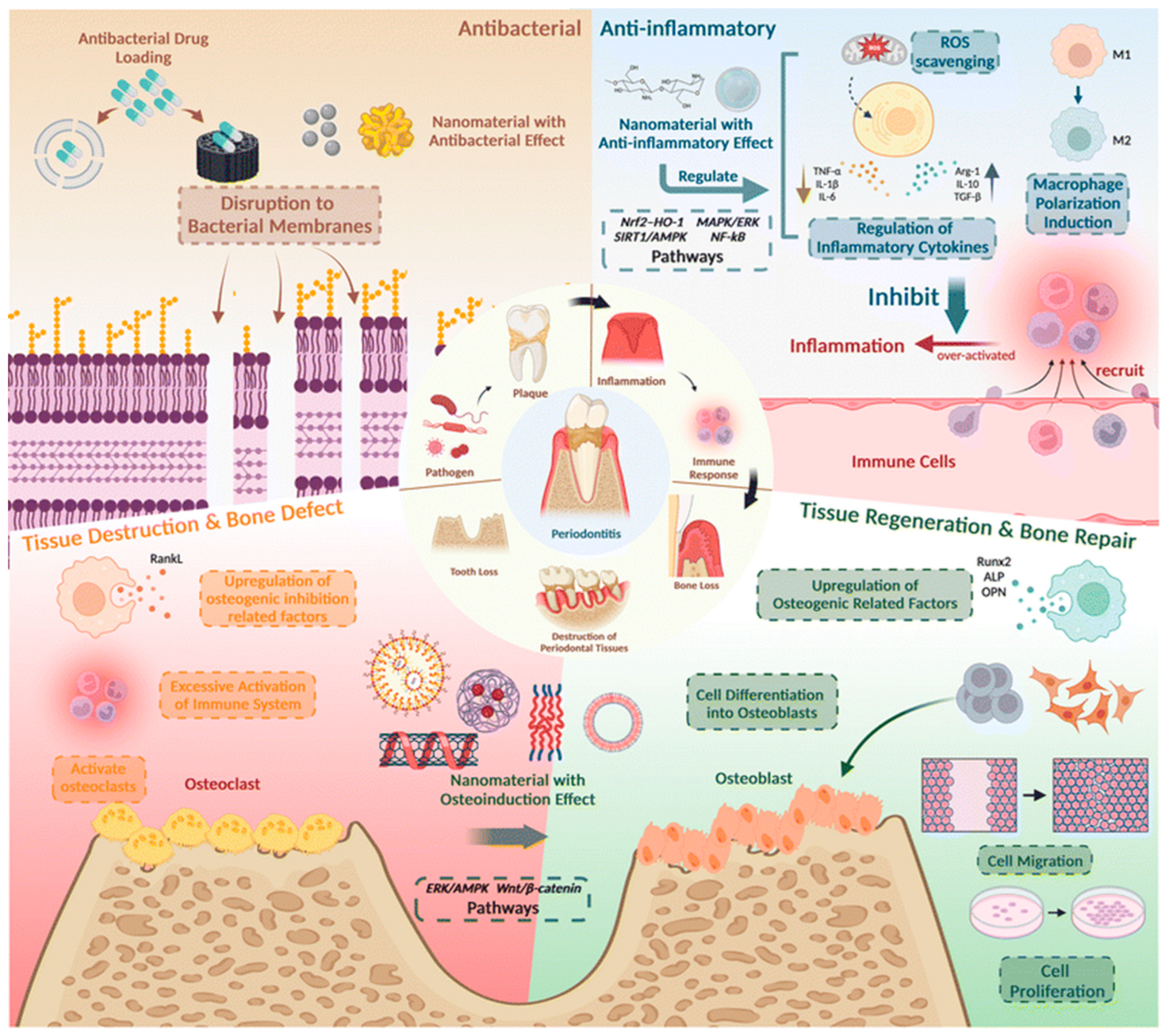
Figure 2. The pathological mechanism and treatment strategies of periodontitis showing the need for targeting chronic inflammation and microbial invasion. Reproduced from [7]; Creative Commons Attribution Non-Commercial 3.0 Unported Licence.
Jnt 06 00031 g002
Figure 3. Broad classification of nanomaterials used in periodontal applications. Reproduced from [56]; Creative Commons Attribution 4.0 International CC BY 4.0 Licence.
Jnt 06 00031 g003
Figure 4. Periodontitis may be detected using various platforms, including those based on electricity, DNA, immunosensors, electrochemistry, and optics. Nanomaterials are often used as transducer materials, playing a crucial role in advancing biosensors. A biosensor has four essential components: A bioreceptor, a transducer, a signal processor for transforming electrical signals into the required form, and an interface for displaying the results. Various metal nanoparticles, including AuNPs and AgNPs, as well as diverse forms of graphene oxide, carbon dots, and quantum dots, are synthesized using easy techniques and serve as distinct nanoplatforms for detecting circulating biomarkers. Reproduced from [6]; Creative Commons Attribution 4.0 International CC BY 4.0 Licence.
Jnt 06 00031 g004
Figure 5. Nanoparticles (NPs) have diverse applications in the diagnosis and treatment of periodontal disease. Diagnostic NPs, such as quantum dots, carbon dots, and gold nanorods, enable highly sensitive and specific detection of periodontal pathogens and biomarkers. Therapeutic NPs, including polymeric NPs, liposomes, metal NPs, graphene oxide, and nanozymes, exhibit antimicrobial, anti-inflammatory, and regenerative properties, supporting tissue healing and preventing disease progression. Reproduced from [60]; Creative Commons Attribution 4.0 International CC BY 4.0 Licence.
Jnt 06 00031 g005
Figure 6. Schematic representation of nanotheranostic applications in periodontitis. Nanomaterials (e.g., metallic, polymeric, carbon-based, and hybrid systems) are designed to integrate diagnostic functions (such as biosensing and imaging) with therapeutic functions (including antimicrobial action, biofilm disruption, immunomodulation, and tissue regeneration). This integrated approach enables real-time monitoring, targeted treatment, and improved clinical outcomes in personalized periodontal therapy.
Jnt 06 00031 g006
Table 1. Key properties and diagnostic and therapeutic functions of common nanomaterials for periodontitis applications.
Nanomaterial TypeKey PropertiesDiagnostic FunctionsTherapeutic FunctionsReferences
Polymeric nanoparticles (e.g., PLGA, chitosan).Biodegradable, tunable size/charge, controlled release.Encapsulation of fluorescent dyes, radiocontrast or antibodies and peptides as diagnostic probes for localized sensing and imaging.Sustained release of antimicrobials, anti-inflammatories, and regenerative growth factors.[6,7,8,59,60]
Lipid-based nanomaterials (e.g., liposomes, solid lipid nanoparticles, nanostructured lipid carriers).Amphiphilic bilayer, biomimetic lipid layers, biocompatible, versatile encapsulation for hydrophilic and lipophilic molecules.Integration of fluorescent/luminescent markers or contrasting agents for imaging.Delivery of antibiotics, anti-inflammatory agents, and regenerative molecules.[61,62,63,64]
Metal/ metal Oxide nanoparticles and nanofilms (e.g., Ag, Au, ZnO, TiO2).High surface reactivity, optical/electrical properties, antimicrobial activity.Biosensing of pathogens and biomarkers (AgNP-based colorimetric detection, AuNP-based plasmonic sensors) and volatile sulfur compounds.Broad-spectrum antimicrobial activity and biofilm inhibiting and penetrating activity, ROS modulation, and anti-inflammatory effects, and periodontal bone regeneration.[6,8,28,65,66,67]
Magnetic Nanoparticles (e.g., Fe3O4).Superparamagnetic, high surface area, easily functionalized.MRI contrast, magnetic biosensing of bacterial virulence factors.Magnetically guided local drug delivery, hyperthermia for bacterial eradication.[8,28,68]
Silica-Based Nanoparticles (e.g., mesoporous silica NPs).High surface area, tunable porosity, modifiable chemistry.Loading of biosensors or imaging probes.Controlled and stimuli-responsive drug release, potential biofilm penetration, periodontal bone regeneration.[69,70,71,72]
Carbon-Based Nanomaterials (e.g., graphene oxide, CNTs, carbon dots).High mechanical strength, conductivity, large surface area.Electrochemical biosensors for bacterial/inflammatory biomarkers.Antimicrobial activity, anti-inflammatory modulation, scaffold reinforcement.[6,28,73]
Nanocomposite systems (e.g., nanoparticle-loaded hydrogels or polymeric scaffolds).Injectable, stimuli-responsive, ECM-mimicking.Embedded carbon-based biosensors for microbial enzyme-responsive activity, and colorimetric detection of volatile sulfur compounds.Localized drug release, tissue regeneration, antimicrobial/anti-inflammatory delivery, sustained release of nanoparticles, and metal oxide nanozymes.[74,75,76]
Disclaimer/Publisher’s Note: The statements, opinions and data contained in all publications are solely those of the individual author(s) and contributor(s) and not of MDPI and/or the editor(s). MDPI and/or the editor(s) disclaim responsibility for any injury to people or property resulting from any ideas, methods, instructions or products referred to in the content.