Review

Theranostic Nanoplatforms in Nuclear Medicine: Current Advances, Emerging Trends, and Perspectives for Personalized Oncology

by
María Jimena Salgueiro
1,2,* and
Marcela Zubillaga
1,2,3
1
Cátedra de Física, Facultad de Farmacia y Bioquímica, Universidad de Buenos Aires, Buenos Aires 1113, Argentina
2
Instituto de Tecnología Farmacéutica y Biofarmacia (InTecFyB), Universidad de Buenos Aires, Buenos Aires 1113, Argentina
3
Consejo Nacional de Investigaciones Científicas y Técnicas (CONICET), Buenos Aires 1113, Argentina
*
Author to whom correspondence should be addressed.
J. Nanotheranostics 2025, 6(4), 27; https://doi.org/10.3390/jnt6040027
Submission received: 15 July 2025 / Revised: 8 September 2025 / Accepted: 25 September 2025 / Published: 3 October 2025

Abstract

The convergence of nanotechnology with nuclear medicine has led to the development of theranostic nanoplatforms that combine targeted imaging and therapy within a single system. This review provides a critical and updated synthesis of the current state of nanoplatform-based theranostics, with a particular focus on their application in oncology. We explore multifunctional nanocarriers that integrate diagnostic radionuclides for SPECT/PET imaging with therapeutic radioisotopes (α-, β-, or Auger emitters), chemotherapeutics, and biological targeting ligands. We highlight advances in nanomaterial engineering—such as hybrid architectures, surface functionalization, and stimuli-responsive designs—that improve tumor targeting, biodistribution, and therapeutic outcomes. Emphasis is placed on translational challenges including pharmacokinetics, toxicity, regulatory pathways, and GMP-compliant manufacturing. The article closes with a forward-looking perspective on how theranostic nanoplatforms could reshape the future of personalized oncology through precision-targeted diagnostics and radiotherapy.

1. Introduction

1.1. Background: From Monotherapy to Integrated Theranostics

The last two decades have witnessed a transformative shift in the conceptualization and clinical management of complex diseases, particularly cancer. Traditional approaches based on monotherapies—whether chemotherapeutic agents or external radiation—have gradually given way to integrated strategies that seek not only to treat but also to understand, monitor, and adapt therapy in real time. At the heart of this transition lies the emergence of theranostics, a hybrid paradigm that integrates diagnostic and therapeutic capabilities within a single platform [1,2].
Theranostics has gained particular relevance in the era of precision medicine, where treatment efficacy depends on individual patient biology and tumor heterogeneity. The underlying principle of theranostics is to deliver molecularly targeted therapy, while simultaneously visualizing its biodistribution, target engagement, and therapeutic outcome. This integrated feedback loop enables patient selection, dose optimization, early assessment of response, and rapid therapeutic adaptation [3].
Among the disciplines that have contributed to the maturation of theranostics, nuclear medicine occupies a uniquely privileged position. This is due to its intrinsic capability to deliver both diagnostic (gamma or positron emitters) and therapeutic (beta or alpha emitters and Auger electrons) radioisotopes to molecular targets via the same or analogous ligands. In this context, theranostics has evolved not merely as a technological innovation but as a defining framework for the field of nuclear medicine, culminating in what is often referred to as nuclear theranostics [4,5].
In nuclear theranostics, a single molecular targeting vector—such as a peptide, antibody fragment, or small molecule—is labeled with a diagnostic radionuclide for molecular imaging (e.g., 68Ga, 18F, 99mTc) and with a therapeutic radionuclide for targeted radionuclide therapy (e.g., 177Lu, 131I, 225Ac). This dual-use concept enables a “see what you treat, treat what you see” approach, ensuring that only patients whose tumors express the appropriate molecular target receive the corresponding therapy [6,7]. Successful clinical examples include the use of radiolabeled somatostatin analogs in neuroendocrine tumors and Prostate Specific Membrane Antigen (PSMA)-targeting ligands in prostate cancer, both of which have revolutionized disease management and opened new avenues for innovation [8].

1.2. The Convergence of Nanotechnology and Nuclear Medicine

The advent of nanotechnology has added further complexity and versatility to the theranostic field. Nanoplatforms such as liposomes, dendrimers, metal–organic frameworks (MOFs), and inorganic nanoparticles offer multifunctional architectures capable of encapsulating radionuclides, chemotherapeutic agents, imaging probes, and targeting ligands within a single construct [9,10]. These platforms not only improve pharmacokinetics and tumor retention via enhanced permeability and retention (EPR) or active targeting, but also enable multimodal imaging and multivalent interactions, potentially overcoming resistance mechanisms and tumor heterogeneity [11].
The convergence of nuclear medicine and nanotechnology represents a frontier in personalized oncology, aiming to enhance the accuracy, efficacy, and safety of cancer care. However, this convergence also brings forth regulatory, radiochemical, and translational challenges that require coordinated interdisciplinary research to ensure clinical translation [12]. The integration of nanotechnology into nuclear medicine has given rise to a powerful synergy that is redefining the frontiers of diagnosis and therapy. Both fields share a molecular-level approach to disease, and their convergence has enabled the creation of highly sophisticated platforms that simultaneously deliver targeted radionuclide therapy, advanced molecular imaging, and even synergistic chemotherapy or immunomodulation [13,14].

1.3. Advances in Nanoplatform Design and Their Pharmacokinetic and Functional Benefits

Nanoplatforms offer a series of advantages for nuclear medicine applications. Their tunable physicochemical properties—including size, shape, surface charge, and functionalization—allow for precise control over pharmacokinetics, biodistribution, and targeting [15]. These characteristics improve tumor accumulation via EPR effect and facilitate active targeting through the conjugation of ligands that bind to tumor-specific markers, such as integrins, human epidermal growth factor receptor 2 (HER2), or PSMA [16]. Importantly, these platforms provide sufficient surface area to accommodate multiple cargoes, such as radionuclides for imaging and therapy (e.g., 68Ga, 89Zr, 177Lu, 225Ac), fluorescent dyes, chemotherapeutic agents, and immune modulators, within a single multifunctional construct [17].
In recent years, there has been a growing interest in the development of radio-nanomedicines, where nanoparticles are radiolabeled to combine the advantages of nuclear imaging (sensitivity, quantification, real-time biodistribution) with the versatility of nanoscale delivery systems [18]. These radio-nanoplatforms enable multimodal imaging (Positron Emission Tomography (PET), Single Photon Emission Computed Tomography (SPECT), Magnetic Resonance Imaging (MRI), optical imaging) and theranostic capabilities, such as image-guided drug delivery or radionuclide-chemotherapy combination therapy. For instance, liposomes and dendrimers labeled with therapeutic radionuclides such as 177Lu or 90Y have been explored for their ability to deliver both radiation and chemotherapeutics deep into solid tumors [19]. Furthermore, emerging inorganic nanomaterials such as gold nanoparticles, silica nanoparticles, and hafnium oxide particles have demonstrated potential in radioenhancement, whereby the local radiation dose is increased due to the interaction of the nanoparticle with ionizing radiation [20]. When combined with radioisotopes, these platforms can act as amplifiers of radiobiological effects, expanding the potential of internal radiotherapy.
Another rapidly expanding area is that of pretargeting strategies, where radiolabeled small molecules are administered after a nanocarrier loaded with a tumor-targeting moiety, improving specificity and reducing off-target exposure [21]. These approaches further demonstrate the flexibility of nanosystems to accommodate complex architectures compatible with radiolabeling techniques, including click chemistry, chelator-based coordination, and intrinsic doping for metallic nanoparticles [22].
Nevertheless, the successful clinical translation of nanotheranostic systems remains challenged by several critical factors, including radiolabeling stability, reproducibility in large-scale synthesis, immunogenicity, and regulatory compliance [11]. Furthermore, the complexity of these hybrid systems requires interdisciplinary collaboration among radiochemists, nanotechnologists, clinicians, and pharmacologists to ensure safety, efficacy, and standardization [23]. Despite these hurdles, the convergence of nanotechnology and nuclear medicine is paving the way for next-generation precision theranostics, offering a route to tailor treatments not only to the molecular profile of the tumor but also to its dynamic evolution during therapy. In this context, nanotheranostics represents not just a research niche but a strategic paradigm for the future of oncologic imaging and therapy.

1.4. Scope of This Review

The rapid expansion of nanotechnology-based platforms and their integration with radionuclide imaging and therapy has transformed the landscape of cancer management, particularly within the emerging paradigm of personalized nanotheranostics. However, the heterogeneity of nanoplatform architectures, radionuclide choices, functionalization strategies, and combinatorial therapeutic approaches calls for a systematic synthesis of current knowledge, translational progress, and persistent challenges.
This review aims to provide a comprehensive, critical overview of the current state, technological advances, and future directions of nanoplatform-based theranostics in nuclear medicine, with a strong emphasis on oncological applications. Our objective is not only to catalog recent developments, but also to highlight the mechanistic rationales that support the design and functional integration of nanocarriers with diagnostic and therapeutic radionuclides. Moreover, we seek to elucidate how these hybrid systems are evolving toward clinically relevant platforms capable of multimodal imaging, image-guided therapy, and combinatorial treatment delivery (e.g., radiotherapy plus chemotherapy or immunotherapy).
This review differs from previous overviews in that it integrates a chemical–functional classification of nanoplatforms with a mechanistic analysis of radionuclide incorporation strategies, radiochemical stability, and biological performance. By explicitly connecting these design principles to translational challenges—including GMP compliance, regulatory pathways, and clinical feasibility—we aim to provide a framework that not only summarizes current knowledge but also guides future development of nanoradiopharmaceuticals. This dual focus on fundamental design rationale and practical clinical translation is intended to support researchers, radiopharmacists, and regulatory scientists in advancing the field toward personalized oncology applications.

2. Theranostic Principles and the Role of Nanoplatforms

2.1. Definition and Conceptual Evolution of Theranostics

The term theranostics—a portmanteau of “therapy” and “diagnostics”—was formally introduced in the early 2000s to describe integrated strategies that combine therapeutic and diagnostic functions within a single system, enabling real-time treatment monitoring and individualized medical interventions [24]. However, the conceptual foundation of theranostics predates its nomenclature and is deeply rooted in the legacy of nuclear medicine. As early as the 1940s and 1950s, radiopharmaceuticals such as 131I were used for both diagnostic imaging and treatment of thyroid diseases, effectively embodying the “see what you treat, and treat what you see” paradigm decades before the field adopted the term “theranostics” [25,26].
Over time, technological and scientific advancements have refined and expanded the scope of theranostic applications. The development of hybrid imaging systems such as PET/Computed Tomography (CT) and PET/MRI has enabled the concurrent acquisition of anatomical and functional data with high resolution and precision [27]. Simultaneously, the rise of nanotechnology has led to the engineering of multifunctional platforms capable of loading multiple payloads—such as radionuclides, chemotherapeutic agents, photosensitizers, and gene-editing components—and delivering them selectively to disease sites [28]. These advances have contributed to a reconceptualization of theranostics as a cornerstone of precision medicine, with applications now extending beyond oncology into cardiology, neurology, and infectious diseases [2,14].
Importantly, the chemical and structural diversity of nanoplatforms—including organic, inorganic, and hybrid systems—has enhanced their adaptability to different biomedical contexts, offering tailored solutions for various disease pathophysiologies. For instance, liposomes and polymeric micelles are often employed for drug delivery and diagnostic imaging in solid tumors, while inorganic structures such as gold and iron oxide nanoparticles have shown promise in treating and monitoring metastatic or hematologic malignancies [29]. Thus, nanoplatforms serve not only as physical carriers but as customizable theranostic architectures capable of integrating molecular targeting, real-time imaging, and controlled therapy.
The evolution of theranostics has also been shaped by a broader shift in healthcare paradigms—from reactive to predictive and personalized medicine. This transition emphasizes biomarker-guided approaches that allow for patient stratification, early intervention, and continuous monitoring of disease progression and response to therapy [30]. In this context, theranostics directly addresses the clinical challenge of biological heterogeneity, enabling clinicians to identify, characterize, treat, and track disease using a unified technological platform. By minimizing the diagnostic–therapeutic gap, such systems not only enhance treatment efficacy but also reduce systemic toxicity and unnecessary exposure to ineffective interventions [31].
While the full potential of nanotheranostics is still being explored, its trajectory builds on decades of innovation and clinical experience. As will be discussed in the following section, the integration of diagnostic and therapeutic functionalities into a single platform reflects a rational, technologically enabled evolution that responds to both the limitations of conventional therapies and the opportunities of precision medicine.

2.2. Main Categories of Nanoplatforms Used in Nuclear Theranostics

Nanoplatforms employed in theranostics are diverse in origin, composition, and functionality. Literature taxonomies often reveal (i) the chemical nature (organic, inorganic, hybrid), (ii) structural characteristics (size, morphology, porosity), and (iii) functionalization potential (targeting ligands, imaging agents, therapeutic payloads, or surface chemistry for radiolabeling). For the purpose of systematization in this article, we propose an analysis from a chemical point of view.

2.3. Organic Nanoplatforms

This group includes polymeric nanoparticles, liposomes, dendrimers, micelles, and lipid nanocarriers. These classical nanoplatforms have long been at the forefront of nanomedicine and have played a foundational role in shaping the current field of nuclear theranostics. Their intrinsic biocompatibility, biodegradability, capacity to encapsulate both hydrophilic and hydrophobic agents and tunable surface properties [32] make them ideal for radiolabeling, multifunctionalization and clinical translation [33,34]. Polymeric nanoparticles, especially those made from Food and Drug Administration (FDA)-approved materials like poly(lactic-co-glycolic acid) (PLGA) or PEGylated polymers, offer controlled and sustained drug release, long circulation times, and low immunogenicity.
Liposomes, spherical vesicles composed of phospholipid bilayers, were among the first nanocarriers used for drug delivery and have since been adapted for theranostic applications by incorporating imaging agents and therapeutic payloads, including radionuclides [35] and they can be functionalized with antibodies or peptides for active targeting. Their aqueous core allows encapsulation of hydrophilic drugs or radionuclides, while lipophilic agents can be integrated into the bilayer. In nuclear imaging, liposomes radiolabeled with technetium-99m (99mTc) or indium-111 (111In) have been used for SPECT/CT applications [36]. A representative example includes 111In-PEGylated immunoliposomes functionalized with monoclonal antibodies and single-chain variable fragments (scFv), enabling specific targeting and high-contrast tumor imaging [37]. Therapeutically, liposomes have been employed to encapsulate β-emitting radionuclides such as 177Lu and α-emitters like 225Ac. For example, thermosensitive liposomes co-loaded with 64Cu and doxorubicin (DOX) have demonstrated PET-guided release and enhanced chemoradiotherapeutic synergy in preclinical tumor models [38]. This design allows for externally triggered drug release in response to mild hyperthermia while simultaneously enabling real-time PET imaging.
Dendrimers are synthetic, branched macromolecules characterized by highly controlled architecture and multivalency. Their size and surface characteristics can be fine-tuned to optimize biodistribution and clearance profiles. Their internal cavities and numerous surface functional groups enable simultaneous loading of radionuclides, targeting moieties, and therapeutic agents. Poly(amidoamine) (PAMAM) dendrimers have been functionalized with chelators like 1,4,7,10-tetraazacyclododecane-1,4,7,10-tetraacetic acid (DOTA) or 1,4,7-Triazacyclononane-1,4,7-triacetic acid (NOTA) to stably bind 68Ga, 177Lu, or 64Cu, enabling their use in both diagnostic and therapeutic applications [39]. 68Ga and 99mTc-labeled PAMAM dendrimers have demonstrated prolonged circulation, high tumor uptake, and favorable pharmacokinetics for PET/SPECT imaging [40]. In addition, 177Lu-conjugated dendrimers targeting HER2 or PSMA have shown promising antitumor effects in xenograft models [7]. Their multivalency also supports the development of multimodal platforms for PET/MRI imaging or radio-chemo-immunotherapy.
Polymeric nanoparticles (PNPs) are solid colloidal systems synthesized from biodegradable and biocompatible polymers such as PLGA, polycaprolactone (PCL), chitosan, and PEGylated copolymers. These systems offer high versatility in terms of size, drug-loading capacity, degradation rate, and compatibility with a wide range of therapeutic and imaging agents. Structurally, they can be formulated as nanospheres, in which the therapeutic or diagnostic agents are uniformly distributed throughout the polymer matrix, or as nanocapsules, where the active compounds are confined within a polymeric shell surrounding a core. In the context of nuclear medicine, PNPs have been extensively explored as vehicles for both diagnostic and therapeutic radionuclides. For example, 177Lu-labeled PLGA nanoparticles have been developed for targeted radionuclide therapy, showing favorable stability and prolonged blood circulation times in preclinical tumor models [41]. When combined with anticancer agents such as paclitaxel or cisplatin, these platforms enable co-delivery strategies that enhance therapeutic efficacy via synergistic mechanisms [42]. Importantly, polymeric particles can be engineered to offer controlled and sustained release profiles, which is critical in maintaining therapeutic levels of radionuclides or drugs within tumors while minimizing systemic exposure. Several studies have also demonstrated the ability of radiolabeled PNPs to improve imaging contrast. For instance, 99mTc-labeled chitosan nanoparticles have been evaluated for sentinel lymph node imaging and inflammation tracking [43]. Similarly, 64Cu- or 89Zr-labeled PLGA nanoparticles have enabled PET imaging of biodistribution and tumor accumulation with high tumor-to-background ratios [44]. Due to their modular design, PNPs can be adapted to carry multiple imaging probes and radionuclides, enabling dual- or multimodal imaging with PET, SPECT, and optical readouts. Polymeric micelles, a subcategory of PNPs, are formed by the self-assembly of amphiphilic block copolymers in aqueous environments. These nanostructures feature a hydrophobic core suitable for encapsulating poorly soluble drugs or lipophilic radionuclide complexes, surrounded by a hydrophilic corona (often PEG), which enhances their solubility and stability in biological fluids. Micelles typically range from 10 to 100 nm in size, favoring passive accumulation in tumors via the EPR effect. Radiolabeled polymeric micelles have shown promising results in theranostic applications. For example, 64Cu-labeled micelles have been employed for PET imaging of tumors, exhibiting high in vivo stability and favorable pharmacokinetics [45]. In therapeutic settings, micelles co-loaded with radionuclides and cytotoxic drugs have demonstrated potent antitumor activity in various xenograft models [14]. Owing to their dynamic assembly and tunable release behavior, micelles offer particular advantages in designing stimuli-responsive systems, which are discussed in later sections. Taken together, polymeric nanoparticles—both solid particles and micellar systems—constitute a robust and adaptable platform for radiotheranostics. Their physicochemical tunability, capacity for multimodal cargo loading, and compatibility with a variety of radionuclides make them ideal candidates for the development of personalized nanomedicines. While many polymer-based systems are currently in preclinical stages, their clinical translation is actively being pursued.

2.4. Inorganic Nanoplatforms

Inorganic nanomaterials have emerged as critical tools in nuclear theranostics due to their structural robustness, tunable physicochemical properties, and capacity for intrinsic imaging. Their high surface-area-to-volume ratio, stability under irradiation, and ability to incorporate or chelate a wide range of radionuclides make them ideal for dual diagnostic and therapeutic use. Among the most studied platforms are gold nanoparticles (AuNPs), quantum dots (QDs), iron oxide nanoparticles (SPIONs), and mesoporous silica nanoparticles (MSNs).
AuNPs exhibit excellent biocompatibility, high atomic number for X-ray attenuation, and facile surface chemistry. They have been radiolabeled with both diagnostic (64Cu, 68Ga) and therapeutic radionuclides (198Au, 177Lu) using chelator-free or chelator-based methods. For example, 64Cu-doped AuNPs have been used for PET imaging with favorable tumor accumulation and pharmacokinetics, while 198Au-labeled AuNPs functionalized with targeting ligands such as Arginylglycylaspartic acid (RGD) peptides demonstrated combined radionuclide therapy and photothermal ablation capabilities in preclinical models [46].
QDs—semiconductor nanocrystals with size-tunable fluorescence—have been functionalized with chelators such as NOTA and labeled with 64Cu or 68Ga for PET-optical imaging applications. A representative example is the use of CdSe/ZnS QDs PEGylated and radiolabeled with 68Ga for imaging U87MG glioma xenografts [47]. Despite concerns over heavy-metal toxicity, strategies such as encapsulation within polymer shells or protein-based carriers (e.g., ferritin) have improved their biocompatibility for in vivo use.
SPIONs are established MRI contrast agents but have also been explored for multimodal nuclear imaging. PEGylated SPIONs radiolabeled with 68Ga or 64Cu have shown dual PET/MRI functionality, enabling combined anatomical and functional imaging [48]. In therapeutic contexts, SPIONs have been loaded with Auger or α-emitting radionuclides, enhancing tumoricidal effects. Furthermore, 177Lu-labeled Fe–gallic acid coordination complexes have demonstrated tumor retention and improved survival in murine models [49].
MSNs offer large pore volumes and tunable surface chemistry, making them versatile for co-loading of chemotherapeutics and radionuclides. Radiolabeled with 64Cu, 89Zr, or 177Lu, MSNs have demonstrated utility in PET imaging and radionuclide therapy. Their mesoporous structure facilitates controlled release and multimodal combinations. For example, 177Lu-labeled MSNs co-loaded with DOX have achieved synergistic antitumor effects and reduced systemic toxicity [50].
Despite their promise, inorganic nanoplatforms face translational hurdles including long-term toxicity, reticuloendothelial system (RES) sequestration, and challenges in radiometal retention. However, their intrinsic imaging properties and stability under radiolabeling continue to position them at the forefront of next-generation radiotheranostic development.

2.5. Hybrid and Multifunctional Nanostructures

Hybrid nanoplatforms combine organic and inorganic components to create multifunctional theranostic agents capable of simultaneous imaging, therapy, targeting, and controlled release. By integrating structural elements such as lipids, polymers, silica, metals, and biomolecules, these platforms offer modularity to fine-tune pharmacokinetics, biodistribution, and payload co-delivery.
These systems often incorporate diagnostic radionuclides (64Cu, 68Ga, 89Zr) and therapeutic isotopes (177Lu, 225Ac), alongside chemotherapeutics or photosensitizers. For instance, 64Cu-doped PdCu@Au nanoparticles functionalized with D-Ala-peptide T-amide (DAPTA) peptides have enabled PET-guided photothermal therapy in CCR5-expressing breast tumors, showcasing synergy between imaging and treatment modalities [51].
Another example is the development of dendrimer–gold nanoparticle hybrids functionalized with folate and bombesin, radiolabeled with 177Lu. These constructs achieved dual-targeting of Gastrin-Releasing Peptide Receptor (GRPr) and Folate Receptor (FR) receptors, enabling combined radionuclide therapy and photothermal ablation with improved tumor specificity [52].
Yolk–shell silica-metal hybrids, such as CuS@MSN, combine the porous framework of MSNs with photothermal metal cores. These systems can be co-loaded with 64Cu and chemotherapeutics for PET-guided chemoradiotherapy. Their responsiveness to acidic tumor environments allows site-specific drug release and enhanced antitumor effects.
Ferritin nanocages (Fn) have also been used to encapsulate CuS nanoparticles, producing CuS-Fn constructs radiolabeled with 64Cu. These particles demonstrated high photothermal efficiency and PET signal in glioblastoma models, highlighting the utility of biomimetic carriers for deep-tissue imaging and treatment [53,54,55].
MOFs are another emerging class of hybrid nanoplatforms. These crystalline porous materials support high loading of drugs and radionuclides, and their structure can be tuned for stability and targeting. Radiolabeled MOFs with 64Cu or 177Lu have been evaluated for PET imaging and combined immunomodulatory therapy [56,57].
Overall, hybrid nanostructures address key limitations of single-component systems by combining high imaging contrast, therapeutic payloads, and targeting specificity. Although their clinical translation is limited by complexity of synthesis, reproducibility, and regulatory demands, early clinical studies (e.g., Cornell dots labeled with 124I) demonstrate feasibility. Continued development hinges on scalable manufacturing and long-term biocompatibility evaluation [11,12,13,14,15,16,17,18,19,20,21,22,23].

2.6. Rationale for Combining Diagnostic and Therapeutic Modalities

While the conceptual and historical foundations of theranostics were outlined above, this section explores the clinical and technological motivations behind the integration of diagnostic and therapeutic functionalities into a single nanoplatform. This convergence is not merely a theoretical ideal, but a practical strategy that addresses several critical limitations of conventional oncology, particularly the need for individualized treatment, real-time therapy monitoring, and reduced systemic toxicity. Figure 1 illustrates the general architecture of theranostic nanoplatforms, highlighting their key components (nanoplatform matrix, targeting moieties, stealth layers, radioisotopes, and therapeutic payloads) and exemplifying representative designs currently explored for combined imaging and therapy in nuclear medicine. Radiolabeled nanoparticles designed for combined therapeutic applications in nuclear medicine, such as imaging-guided therapy, radiosensitization, and drug delivery in conjunction with radionuclide labeling, illustrate how theranostic nanoplatforms integrate diagnostic and combined therapeutic functionalities [42,46,52].
The incorporation of radiodiagnostic agents and therapeutic payloads within a single nanosystem enables image-guided therapy, whereby the biodistribution, tumor uptake, and clearance of the agent can be tracked in vivo. This capability allows for real-time dosimetry and adaptive treatment planning based on patient-specific tumor biology, rather than relying on empirically fixed dosing regimens [58]. For example, PET- or SPECT-labeled nanocarriers enable visualization of drug delivery pathways, quantification of tumor accumulation, and longitudinal tracking of therapeutic efficacy in both preclinical and clinical settings [59]. Moreover, nanoplatforms enhance selective tumor targeting via a combination of passive and active mechanisms. The EPR effect facilitates preferential accumulation of nanoparticles in tumor tissue due to leaky vasculature and impaired lymphatic drainage. This passive accumulation can be further refined through active targeting strategies, where ligands such as monoclonal antibodies, peptides (e.g., RGD, bombesin), or aptamers are conjugated to the nanoparticle surface to recognize and bind specific tumor-associated receptors [2,32].
Another important rationale for nanotheranostics is the reduction in systemic toxicity. By confining the delivery of cytotoxic drugs or radioisotopes to tumor sites, these systems limit exposure to healthy tissues, thereby minimizing adverse effects often associated with conventional chemotherapy or external beam radiotherapy [60]. This is particularly advantageous in pediatric or frail patient populations, where minimizing collateral damage is essential. In addition, nanoplatforms offer a versatile scaffold for multimodal therapeutic integration. Hybrid nanostructures can co-deliver chemotherapeutics and radionuclides or combine radiotherapy with photothermal or photodynamic therapy to enhance cytotoxicity, overcome resistance mechanisms, and improve treatment efficacy [49]. For instance, 177Lu-labeled gold nanoparticles co-loaded with paclitaxel have demonstrated synergistic effects in HER2+ breast cancer models by simultaneously inducing DNA damage and microtubule disruption [43].
Finally, the theranostic paradigm supports biomarker-guided stratification and treatment personalization. By conjugating nanocarriers with ligands targeting molecular markers such as PSMA, Epidermal Growth Factor Receptor (EGFR), or HER2, nanotheranostics can be tailored to individual patient profiles. This not only enhances treatment specificity but also enables predictive imaging that informs therapeutic decisions [61].
As this field progresses, integration with digital health technologies, artificial intelligence, and systems biology is expected to refine patient selection, optimize therapeutic regimens, and predict clinical outcomes more accurately. These developments mark a shift from standardized, protocol-driven oncology toward adaptive, feedback-informed treatment frameworks that capitalize on the full potential of theranostic nanomedicine [62].

3. Radioisotope Selection, Radiolabelling Strategies and Stability Concerns

3.1. Radioisotopes for Diagnostic

In nuclear medicine, the choice of radioisotopes for diagnostic purposes is primarily based on their decay characteristics, which determine the nature and energy of the emitted radiation. Diagnostic radiopharmaceuticals typically incorporate radioisotopes that undergo electromagnetic decay, emitting gamma (γ) photons or positrons (β+). Gamma-emitting isotopes are used in planar scintigraphy and SPECT imaging, as the emitted photons exit the body and are detected by gamma cameras. Table 1 summarizes commonly used γ-emitting radionuclides for scintigraphic imaging. In contrast, positron-emitting isotopes, employed in PET imaging, decay via β+ emission. These positrons travel a short distance in tissue before annihilating with electrons, resulting in the emission of two 511 keV annihilation photons in opposite directions, which are detected in coincidence by PET scanners. The selection of appropriate diagnostic radioisotopes must therefore consider not only the decay mode and photon energy, but also their compatibility with radiolabelling strategies and the physicochemical stability of the resulting radioconjugates. [63].

3.2. Radioisotopes for Therapy

For therapeutic applications, the radioisotopes of choice are those that emit charged particles—either heavy particles such as alpha (α) particles or lighter ones such as beta minus (β) particles—which deposit their energy directly into the surrounding tissue through ionization events or Auger electrons, which deposit their energy in proximity to DNA due to their high linear energy transfer (LET) and short range. The specific ionization capacity of these particles, which depends on their mass and charge, results in limited penetration depths: typically in the micrometer range for α-particles and a few millimeters for β-particles. Unlike photons, these emissions constitute directly ionizing radiation and are capable of inducing localized cytotoxic effects. Within a restricted radius from the decay site, determined by the particle’s energy, they can cause significant DNA damage in tumor cells, leading to impaired replication and ultimately cell death. Table 2 summarizes several of the most commonly used therapeutic radioisotopes in nuclear medicine [63].

3.3. Radioisotope Pairing Strategies

The clinical success of theranostic radiopharmaceuticals relies not only on the biological specificity of the targeting vector but also on the chemical properties of the radioisotopes and their coordination with appropriate chelators. In nanotheragnosis, where radiometals are incorporated into nanoparticle systems, the stability and versatility of these chemical complexes are critical to ensure effective in vivo performance, minimize off-target radiation, and enable matched diagnostic and therapeutic functionalities.
Among the most widely used theranostic pairs is the combination of 68Ga and 177Lu coordinated via DOTA. DOTA forms highly stable octadentate complexes with trivalent radiometals, making it ideal for both diagnostic (68Ga, positron emitter) and therapeutic (177Lu, β emitter) applications. This chemical similarity allows the development of chemically identical radioconjugates, such as [68Ga/177Lu]Ga/Lu-DOTA-TATE, used in the management of somatostatin receptor-expressing neuroendocrine tumors. The DOTA chelator can also be conjugated to nanocarriers (e.g., liposomes, micelles, polymeric nanoparticles), preserving radiolabeling efficiency and in vivo stability under physiological conditions [64,65].
Similarly, the PSMA-targeting theranostic pair 68Ga-PSMA-11/177Lu-PSMA-617 employs DOTA-based or similar macrocyclic chelators (e.g., N, N’-bis(2-hydroxybenzyl)ethylenediamine-N,N’-diacetic acid (HBED) or 2-[1,4,7,10-Tetraazacyclododecane-4,7,10-tris(t-butyl acetate)]-pentanedioic acid-1t-butyl ester (DOTAGA)), selected based on the coordination preferences of each metal ion. These structures not only ensure high thermodynamic and kinetic stability but also allow site-specific conjugation to PSMA ligands or nanostructures, facilitating targeted delivery and controlled biodistribution. The availability of long-lived therapeutic isotopes such as 177Lu and emerging alpha-emitters like 225Ac further extends the potential of these systems for nanotheragnostic applications [66].
The metal–chelator pairing is central to theranostic design: radiometals such as 64Cu, 89Zr, 90Y, and 67Ga offer diverse decay properties and coordination chemistries that can be matched with appropriate chelators (e.g., NOTA, Deferoxamine (DFO), diethylenetriaminepentaacetic acid (DTPA)), enabling tailored pharmacokinetics and optimized imaging or therapeutic windows. Importantly, many of these chelators can be functionalized for covalent attachment to nanoparticles, providing multivalency, enhanced circulation time, and improved tumor accumulation via the EPR effect [67,68].
These chemical considerations—metal ion coordination geometry, charge, oxidation state, and chelator denticity—are especially relevant in the context of nanotheragnosis, where radiolabeling conditions must preserve the structural and functional integrity of the nanoplatform. The use of matched diagnostic/therapeutic radiometals that bind to a common chelator scaffold not only streamlines the synthesis of theranostic agents but also facilitates regulatory translation and clinical implementation.
Table 3 summarizes selected clinically established and emerging theranostic pairs used in nuclear medicine, highlighting their radiophysical properties, commonly used chelators, and suitability for integration into nanotheragnostic platforms. The choice of chelator is critical to ensure radiolabeling efficiency and in vivo stability. DOTA and its derivatives are particularly favored for their capacity to form kinetically inert complexes with a variety of diagnostic and therapeutic radiometals. Nanotheragnostic compatibility is assessed based on the ability to conjugate or encapsulate the radiolabeled compound within nanocarriers without compromising its stability or bioactivity.

3.4. Radiolabeling Strategies

Radiolabeling of nanoplatforms is a critical step in designing nuclear theranostic agents, directly influencing their in vivo behavior, imaging signal, therapeutic efficacy, and biocompatibility. The choice of radiolabeling strategy must account for the physicochemical characteristics of both the radionuclide and the nanoparticle, the intended biological application, and the stability requirements of the final construct [43,69].
Radiolabeling methods may be categorized into three main strategies: chelator-based, chelator-free, and encapsulation or surface sorption [70]. Each approach offers distinct advantages and is suited for different classes of radionuclides and nanomaterials. Importantly, although 99mTc remains a cornerstone of nuclear medicine and has been successfully radiolabeled via all three of these approaches [71,72,73,74,75,76], the present section emphasizes strategies applicable to emerging radionuclides—such as 64Cu, 89Zr, 68Ga, and 177Lu—that are increasingly used in next-generation nuclear theranostics [77].
Chelator-based strategies remain the gold standard for radiolabeling metallic radionuclides, offering excellent radiochemical yields and in vivo stability. These approaches involve the conjugation of bifunctional chelating agents (BFCAs)—such as DOTA, NOTA, 2-[4,7-bis(carboxymethyl)-1,4,7-triazonan-1-yl]-5-(2,5-dioxopyrrolidin-1-yl)oxy-5-oxopentanoic acid (NODAGA), or DTPA—to the nanoparticle surface via reactive functional groups (e.g., –NH2, –SH, –COOH). The chelators tightly bind radiometals such as 64Cu, 68Ga, 89Zr, and 177Lu, forming thermodynamically and kinetically stable complexes [41,78]. For instance, DOTA-conjugated polymeric micelles and lipid-based nanoparticles have been radiolabeled with 177Lu under mild aqueous conditions (pH 5–6, 30–90 °C), achieving >95% radiochemical yield [41]. Likewise, NOTA and NODAGA provide high labeling efficiency with 64Cu, enabling PET imaging of tumor biodistribution and clearance [70]. Chelators can also be pre-attached to targeting moieties or embedded within PEGylated nanocarriers, which prolong circulation and enhance tumor uptake, as demonstrated in studies using 68Ga- or 99mTc-labeled liposomes and micelles [78]. Chelator-based methods allow modular nanoparticle design and are compatible with a wide range of imaging and therapeutic applications. Nonetheless, the choice of chelator must be carefully matched to the coordination chemistry of the radionuclide, as instability may result in transchelation or loss of signal [79,80,81,82].
Chelator-free strategies offer an attractive alternative for radiolabeling nanoparticles without the use of exogenous chelating agents. These methods rely on the direct chemical interaction between the radionuclide and specific functional groups or structural features of the nanomaterial. They simplify synthesis, reduce potential immunogenicity, and often preserve the native physicochemical characteristics of the platform [69]. A widely employed mechanism in this category is doping, where radionuclides are embedded within the crystal lattice of inorganic nanomaterials during or after synthesis. For instance, 64Cu can be doped into copper sulfide (CuS) or palladium-copper-gold (PdCu@Au) nanoparticles under mildly basic aqueous conditions (pH~9, 65–90 °C), achieving high radiochemical yields (>98%) and excellent in vivo stability [43,51,83]. This approach not only ensures robust labeling but also endows the particles with photothermal properties for synergistic therapy. Another chelator-free approach involves surface bonding or sorption. Nanoparticles containing thiol (-SH), catechol, or carboxylate functional groups—such as melanin, iron oxide, or carbon-based nanostructures—can coordinate with metallic radionuclides like 64Cu, 89Zr, or 177Lu directly on their surface. For example, melanin nanoparticles labeled with 64Cu via spontaneous metal-phenol interactions have shown strong in vivo retention and effective tumor imaging [84,85]. Neutron activation is a less common but powerful chelator-free technique, where stable isotopes within the nanoparticle are activated via neutron irradiation. This method generates radiolabeled constructs without altering their morphology or surface chemistry. For example, 198Au-labeled gold nanoparticles, prepared via this route, may be used for both SPECT imaging and β-therapy in preclinical tumor models [86]. The adsorption-based method relies on physical sorption of radionuclides onto high-affinity surfaces such as hydroxyapatite, TiO2, or carbon nanotubes [87,88,89]. For example, 223Ra has been successfully sorbed onto calcium-based nanocarriers through ionic interactions, achieving high labeling efficiency and mitigating recoil-induced daughter loss [90,91,92,93]. While chelator-free techniques offer simplicity and versatility, their success depends heavily on the nanoparticle’s surface chemistry and compatibility with the chosen radionuclide. The absence of a defined chelating scaffold means that achieving long-term stability requires careful engineering of surface charge, hydrophilicity, and protective coatings.

3.5. Regulatory Perspectives and GMP Considerations

The regulatory landscape for nuclear theranostic agents—defined as radiolabeled nanostructures for combined diagnostic and therapeutic purposes—presents unique challenges at the intersection of nanomedicine, radiopharmacy, and pharmaceutical regulation. While traditionally categorized under the umbrella of radiopharmaceuticals, nuclear theranostics involve complex multicomponent constructs that may include radionuclides, nanocarriers, targeting ligands, and therapeutic drugs, each subject to distinct regulatory scrutiny. Consequently, the clinical translation of these agents requires a harmonized and risk-based regulatory framework that adequately considers their dual functionality, nanometric nature, and radiation-emitting properties [94].
From a regulatory standpoint, nuclear theranostics are generally classified as investigational medicinal products (IMPs) or radiopharmaceuticals under both U.S. FDA and European Medicines Agency (EMA) guidelines. In Europe, Directive 2001/83/EC and its subsequent adaptations (including Directive 2013/59/Euratom for radiation protection) provide the overarching legal framework [95,96]. These agents must meet the regulatory requirements related to pharmaceutical quality, radioprotection, and preclinical safety, regardless of their intended use in diagnosis or therapy. The heterogeneous definitions and classifications of radiopharmaceuticals across jurisdictions complicate international clinical trials and market authorization. Therefore, initiatives from the International Atomic Energy Agency (IAEA), World Health Organization (WHO) and the EMA have sought to harmonize guidance documents and promote regulatory convergence for these compounds [94]. Non-clinical development of nuclear theranostics involves comprehensive characterization of the nanoplatform′s pharmacokinetics, dosimetry, biodistribution, and toxicological profile. Regulatory guidance such as ICH M3(R2) and ICH S9 provides general frameworks, yet often lacks specificity for radiolabeled nanoparticles [97,98]. In response, the EMA drafted a dedicated guideline addressing non-clinical requirements for radiopharmaceuticals (EMA/CHMP/QWP/306970/2018), which remains applicable to theranostic nanomaterials [94,99]. A notable challenge is that conventional toxicity testing under Good Laboratory Practice (GLP) is difficult for radioactive compounds. Instead, the non-radioactive components (e.g., the nanocarrier or ligand) are evaluated under GLP, while pharmacokinetics and dosimetry are conducted under radiation safety conditions in specialized facilities [94]. Depending on the novelty and pharmacological activity of the non-radioactive component, different regulatory scenarios may apply [94]. For example, if a theranostic nanoplatform uses an established vector and only the radionuclide is changed (e.g., 64Cu instead of 68Ga), limited additional testing may be sufficient. Conversely, entirely new constructs require full toxicological and pharmacokinetic assessment in one or more animal models [94].
Diagnostic nuclear theranostics often fall within the scope of “microdose” regulation, particularly when used at sub-pharmacological mass levels. The FDA and EMA allow streamlined non-clinical testing for such agents, provided that dosimetry and radiation safety are adequately justified [94]. This includes demonstrating that the administered activity and molar quantity remain below thresholds defined by ICH M3(R2) guidelines [94,97]. Therapeutic nuclear theranostics, however, generally require full toxicology packages, including genotoxicity, hematology, histopathology, and repeat-dose studies. In certain cases—such as advanced oncology—ICH S9 guidelines allow for the omission of reproductive or carcinogenicity studies if the clinical context justifies it [94,98].
Compliance with Good Manufacturing Practices (GMP) principles is essential for the clinical production of nuclear theranostics. However, current GMP guidelines, such as those in Annex 3 of the WHO Technical Report Series No. 1025, were primarily developed for small-molecule radiopharmaceuticals and may not sufficiently address nanomaterials [100].
Key GMP considerations include the following:
Radiochemical and pharmaceutical purity: Must be above 95%, verified by radio-Thin Layer Chromatography, High-Performance Liquid Chromatography, or gamma spectrometry.
Sterility and apyrogenicity: Especially critical for parenteral formulations.
Batch reproducibility: Challenging in nanoscale systems, requiring robust standard operating procedures (SOPs).
Molar activity control: Particularly relevant for receptor-saturating theranostic agents.
Documentation and traceability: Extensively detailed records for precursor synthesis, labeling conditions, and quality control testing are mandatory.
The clinical translation of nanoradiopharmaceuticals must address a comprehensive set of regulatory and GMP requirements that integrate the frameworks for both radiopharmaceuticals and nanomedicines. From a manufacturing standpoint, developers must ensure full characterization of the nanoplatform, including particle size distribution, morphology, surface chemistry, radionuclide loading efficiency, and radiochemical purity. GMP-compliant production also demands sterility, apyrogenicity, batch-to-batch reproducibility, and validated stability profiles under defined storage and transport conditions. Process validation should encompass critical quality attributes (CQAs) such as radionuclide incorporation yield, in vitro and in vivo stability, and the absence of free radionuclide over the intended shelf-life. Implementation of a Quality by Design (QbD) strategy facilitates identification and control of critical process parameters (CPPs) affecting product quality, while in-process controls and process analytical technologies (PATs) support real-time verification of specifications. Early and continuous interaction with regulatory agencies is strongly recommended to align on the required preclinical safety and efficacy data, CMC (Chemistry, Manufacturing and Controls) documentation, and clinical study design, thereby reducing approval timelines and ensuring compliance throughout development [101,102].
A comparative overview of regulatory scenarios is presented in Table 4, highlighting how the level of technological innovation and the nature of the radionuclide influence the expected non-clinical studies, regulatory classification, and GMP considerations for nuclear theranostic platforms.
As nuclear theranostics become more sophisticated and widespread, regulatory authorities must evolve to accommodate their unique features. This includes the development of nanotechnology-specific pharmacopoeial monographs, dedicated EMA or FDA guidance for radiolabeled nanomaterials, and training programs for reviewers and inspectors. Moreover, collaborative platforms such as the IAEA′s Radiopharmaceuticals Program and the EANM Dosimetry and Regulatory Committees can foster dialogue and consensus building across regions. A harmonized, science-driven, and risk-based regulatory model is essential to unlock the full potential of nuclear theranostics in personalized medicine.

3.6. Clinical Trials and Challenges in Nanotheranostics Translation

Recent years have seen a gradual increase in the number of nanoplatform-based theranostic agents entering clinical evaluation, although most remain in early-phase trials. Representative examples are shown in Table 5. These examples illustrate the diversity of platforms—ranging from inorganic to lipid-based systems—and demonstrate the feasibility of translating nanotheranostic designs into clinical testing, despite the regulatory and manufacturing challenges still to be addressed.
Long-term biosafety remains a critical challenge for the clinical adoption of theranostic nanoplatforms. Depending on their composition, size, and surface chemistry, nanomaterials may exhibit prolonged retention in organs of the reticuloendothelial system such as the liver and spleen, potentially leading to chronic toxicity or altered organ function. Some inorganic or hybrid structures may degrade slowly, generating byproducts with unknown biological activity, while repeated administrations could trigger immunogenic responses. Current preclinical safety assessments often lack the duration and sensitivity to fully capture these effects, highlighting the need for long-term follow-up in both animal models and clinical settings. Standardized protocols for biodistribution, clearance kinetics, and chronic toxicity evaluation—aligned with regulatory expectations—are essential to ensure that these platforms meet the stringent safety requirements for repeated or sustained clinical use [101,102].

4. Final Remarks and Conclusion

This comprehensive review highlights the diverse classes of radiolabeled nanoplatforms being developed for nuclear theranostic applications, emphasizing their chemical versatility, structural tunability, and capacity for dual diagnostic and therapeutic functionality. Organic systems such as liposomes, dendrimers, and polymeric micelles continue to evolve with increasingly sophisticated designs for radiolabeling and drug co-loading. Inorganic and hybrid nanostructures offer inherent imaging contrast, improved stability, and multifunctional capabilities—though their long-term biocompatibility remains under scrutiny. Radiolabeling strategies have expanded beyond traditional chelator-based approaches to include chelator-free, doping, surface sorption, and encapsulation techniques. These methods improve radiochemical yields, pharmacokinetics, and site-specific delivery, while supporting the integration of both established and emerging radionuclides, including α-emitters. Moreover, the co-delivery of chemotherapeutic agents with radionuclides has demonstrated promising synergistic effects in preclinical models, potentially addressing tumor heterogeneity and therapeutic resistance. Despite these advances, the translational gap remains considerable. Most of the current evidence originates from animal models, and only a limited number of formulations have entered clinical trials. Critical challenges persist in standardizing production, achieving regulatory approval, and demonstrating long-term safety. The lack of robust clinical outcome data, particularly in the last five years, limits our ability to conclude that radiolabeled nanomaterials represent the immediate future of radiopharmacy.
Nuclear theranostics represents a paradigm shift toward personalized medicine, offering tailored treatment regimens guided by real-time molecular imaging. The convergence of nanotechnology and nuclear medicine enables precise tumor targeting, adaptive dosing, and the integration of multimodal therapies within a single platform. Advances in ligand design, radiochemistry, and image-guided drug release are expected to further enhance therapeutic efficacy while minimizing off-target toxicity. In particular, the potential of nanoradiotheranostics lies in their ability to selectively accumulate in diseased tissues, reduce multi-step procedures, and act as individualized precision tools for complex diseases such as prostate cancer, glioblastoma, and neuroendocrine tumors. Integration with artificial intelligence, systems biology, and digital health platforms is poised to further refine patient selection, improve outcome prediction, and support biomarker-driven stratification in clinical practice. However, a comprehensive understanding of radionanotechnology is essential to improve success rates in human applications. Optimizing nanoparticle design for specific tumor microenvironments, controlling in vivo degradation and clearance, and achieving scalable GMP-compliant manufacturing remain high priorities for the field.
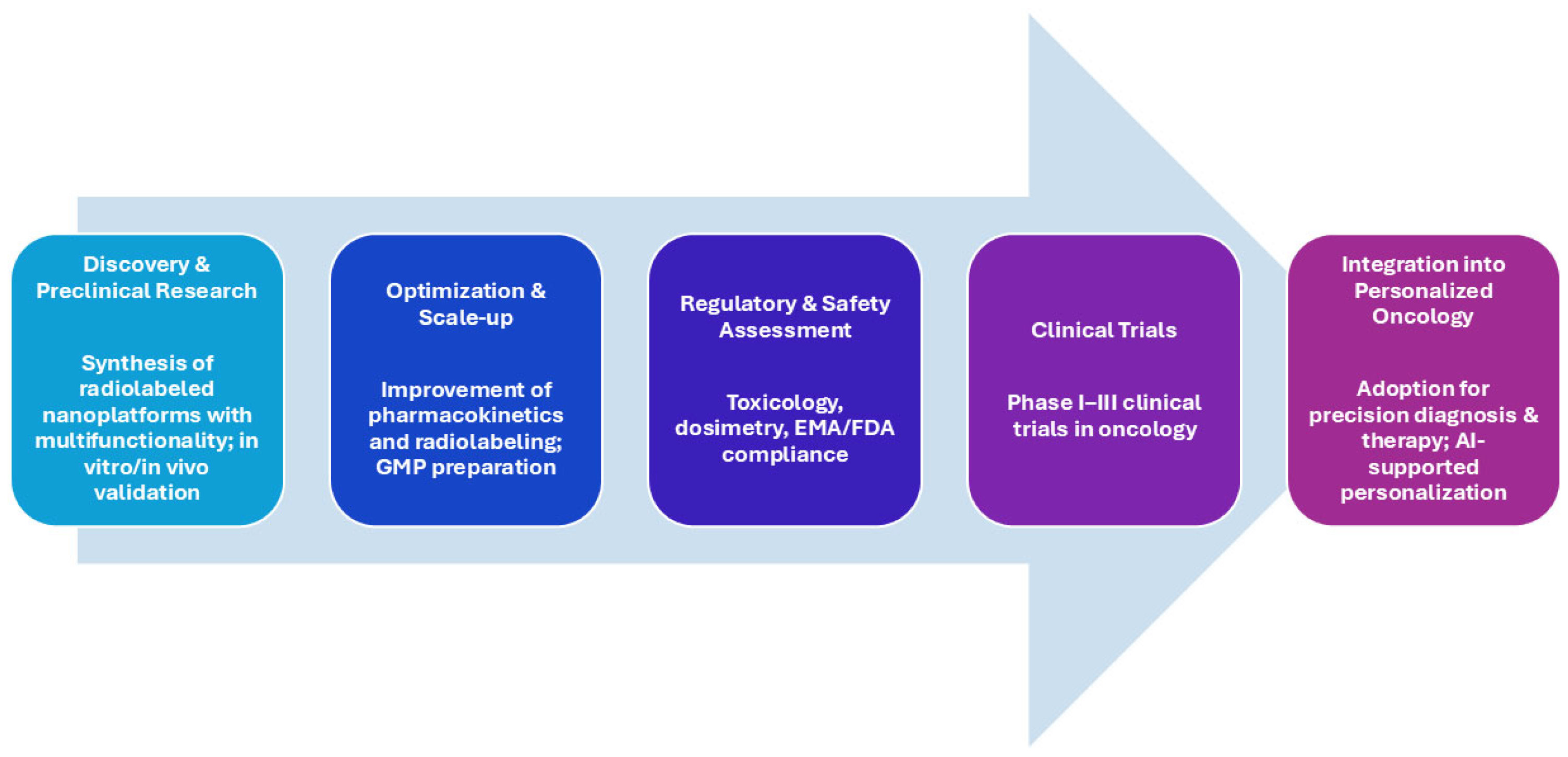
While radiolabeled nanomaterials hold undeniable promise for transforming the landscape of nuclear medicine, the path to clinical adoption is still complex and requires collaborative efforts across disciplines. From engineering and radiochemistry to regulatory science and clinical oncology, the successful translation of nanoradiotheranostics will depend on harmonized regulatory frameworks, robust toxicological assessment, and the generation of clinical evidence that demonstrates safety, efficacy, and cost-effectiveness. Although challenges remain, the future of nuclear theranostics is undeniably bright. As novel formulations continue to mature, and regulatory and technical hurdles are progressively addressed, radiolabeled nanoplatforms are expected to play a central role in the evolution of personalized oncology—ultimately contributing to improved patient outcomes and enhanced quality of life. As illustrated in Figure 2, the successful clinical implementation of nuclear theranostics requires a stepwise progression from robust nanomaterial design to compliance with complex regulatory standards, ultimately enabling their adoption in precision oncology workflows.
In summary, this work provides a comprehensive synthesis that goes beyond cataloging recent advances by establishing clear mechanistic links between the chemical nature of nanoplatforms, their radiolabeling strategies, and their theranostic performance. Our approach highlights how tailored material design can optimize both imaging contrast and therapeutic efficacy while addressing the stringent requirements for clinical translation. By combining scientific insight with regulatory and GMP considerations, we identify key opportunities for innovation, anticipate potential barriers to approval, and outline strategic directions for the scalable, safe, and effective implementation of nanoradiopharmaceuticals in precision oncology.

Funding

This research received no external funding.

Conflicts of Interest

The authors declare no conflicts of interest.

Abbreviations

The following abbreviations are used in this manuscript:
ATMPAdvanced Therapy Medicinal Product
AuNPsGold nanoparticles
BFCAsBifunctional Chelating Agents
CTComputed Tomography
DAPTAD-Ala-peptide T-amide
DFODeferoxamine
DOTA1,4,7,10-tetraazacyclododecane-1,4,7,10-tetraacetic acid
DOTAGA2-[1,4,7,10-Tetraazacyclododecane-4,7,10-tris(t-butyl aceta-te)]-pentanedioic acid-1t-butyl ester
DOXDoxorubicin
DTPAdiethylenetriaminepentaacetic acid
ECElectron Capture
EGFREpidermal Growth Factor Receptor
EMAEuropean Medicines Agency
EPREnhanced Permeability and Retention
FDAFood and Drug Administration
FnFerritin nanocages
FRFolate Receptor
GLPGood Laboratory Practice
GMPGood Manufacturing Practices
GRPRGastrin-Releasing Peptide Receptor
HBEDN,N′-bis(2-hydroxybenzyl)ethylenediamine-N,N′-diacetic acid
HER2Human Epidermal Growth Factor Receptor 2
IAEAInternational Atomic Energy Agency
IMPsInvestigational Medicinal Products
LETLinear energy transfer
MOFsMetal–Organic Frameworks
MRIMagnetic Resonance Imaging
MSNsMesoporous silica nanoparticles
NODAGA2-[4,7-bis(carboxymethyl)-1,4,7-triazonan-1-yl]-5-(2,5-dioxopyrrolidin-1-yl)oxy-5-oxopentanoic acid
NOTA1,4,7-Triazacyclononane-1,4,7-triacetic acid
NPNanoparticles
PAMAMPoly(amidoamine)
PCLPolycaprolactone
PEGPolyethylene glycol
PETPositron Emission Tomography
PLGAPoly(lactic-co-glycolic acid)
PNPsPolymeric nanoparticles
PSMAProstate Specific Membrane Antigen
QDsQuantum Dots
RESReticuloendothelial System
RGDArginylglycylaspartic acid
SPECTSingle Photon Emission Computed Tomography
SPIONsIron oxide nanoparticles
WHOWorld Health Organization

References

  1. James, M.L.; Gambhir, S.S. A Molecular Imaging Primer: Modalities, Imaging Agents, and Applications. Physiol. Rev. 2012, 92, 897–965. [Google Scholar] [CrossRef]
  2. Kelkar, S.S.; Reineke, T.M. Theranostics: Combining Imaging and Therapy. Bioconjugate Chem. 2011, 22, 1879–1903. [Google Scholar] [CrossRef]
  3. Yordanova, A.; Eppard, E.; Kürpig, S.; Bundschuh, R.A.; Schönberger, S.; Gonzalez-Carmona, M.; Feldmann, G.; Ahmadzadehfar, H.; Essler, M. Theranostics in nuclear medicine practice. OncoTargets Ther. 2017, 10, 4821–4828. [Google Scholar] [CrossRef]
  4. Dash, A.; Chakraborty, S.; Pillai, M.R.A.; Knapp, F.F. Peptide Receptor Radionuclide Therapy: An Overview. Cancer Biotherapy Radiopharm. 2015, 30, 47–71. [Google Scholar] [CrossRef]
  5. Baum, R.P.; Kulkarni, H.R. THERANOSTICS: From Molecular Imaging Using Ga-68 Labeled Tracers and PET/CT to Personalized Radionuclide Therapy-The Bad Berka Experience. Theranostics 2012, 2, 437–447. [Google Scholar] [CrossRef]
  6. Zhang, S.; Wang, X.; Gao, X.; Chen, X.; Li, L.; Li, G.; Liu, C.; Miao, Y.; Wang, R.; Hu, K. Radiopharmaceuticals and their applications in medicine. Signal Transduct. Target. Ther. 2025, 10, 1–51. [Google Scholar] [CrossRef]
  7. Fani, M.; André, J.P.; Maecke, H.R. 68Ga-PET: A powerful generator-based alternative to cyclotron-based PET radiopharmaceuticals. Contrast Media Mol. Imaging 2008, 3, 53–63. [Google Scholar] [CrossRef]
  8. Hofman, M.S.; Violet, J.; Hicks, R.J.; Ferdinandus, J.; Thang, S.P.; Akhurst, T.; Iravani, A.; Kong, G.; Kumar, A.R.; Murphy, D.G.; et al. [ 177 Lu]-PSMA-617 radionuclide treatment in patients with metastatic castration-resistant prostate cancer (LuPSMA trial): A single-centre, single-arm, phase 2 study. Lancet Oncol. 2018, 19, 825–833. [Google Scholar] [CrossRef]
  9. Blanco, E.; Shen, H.; Ferrari, M. Principles of nanoparticle design for overcoming biological barriers to drug delivery. Nat. Biotechnol. 2015, 33, 941–951. [Google Scholar] [CrossRef]
  10. Siafaka, P.I.; Okur, N.Ü.; Karantas, I.D.; Okur, M.E.; Gündoğdu, E.A. Current update on nanoplatforms as therapeutic and diagnostic tools: A review for the materials used as nanotheranostics and imaging modalities. Asian, J. Pharm. Health Sci. 2021, 16, 24–46. [Google Scholar] [CrossRef]
  11. Lee, D.-E.; Koo, H.; Sun, I.-C.; Ryu, J.H.; Kim, K.; Kwon, I.C. Multifunctional nanoparticles for multimodal imaging and theragnosis. Chem. Soc. Rev. 2011, 41, 2656–2672. [Google Scholar] [CrossRef]
  12. Hare, J.I.; Lammers, T.; Ashford, M.B.; Puri, S.; Storm, G.; Barry, S.T. Challenges and strategies in anti-cancer nanomedicine development: An industry perspective. Adv. Drug Deliv. Rev. 2017, 108, 25–38. [Google Scholar] [CrossRef]
  13. Lankoff, A.M.; Czerwińska, M.; Kruszewski, M. Advances in Nanotheranostic Systems for Concurrent Cancer Imaging and Therapy: An Overview of the Last 5 Years. Molecules 2024, 29, 5985. [Google Scholar] [CrossRef]
  14. Muthu, M.S.; Leong, D.T.; Mei, L.; Feng, S.-S. Nanotheranostics—Application and Further Development of Nanomedicine Strategies for Advanced Theranostics. Theranostics 2014, 4, 660–677. [Google Scholar] [CrossRef]
  15. Albanese, A.; Tang, P.S.; Chan, W.C. The effect of nanoparticle size, shape, and surface chemistry on biological systems. Annu. Rev. Biomed. Eng. 2012, 14, 1–16. [Google Scholar] [CrossRef]
  16. Yu, B.; Tai, H.C.; Xue, W.; Lee, L.J.; Lee, R.J. Receptor-targeted nanocarriers for therapeutic delivery to cancer. Mol. Membr. Biol. 2010, 27, 286–298. [Google Scholar] [CrossRef]
  17. Lankoff, A.; Czerwińska, M.; Kruszewski, M. Nanoparticle-Based Radioconjugates for Targeted Imaging and Therapy of Prostate Cancer. Molecules 2023, 28, 4122. [Google Scholar] [CrossRef]
  18. Pérez-Medina, C.; Abdel-Atti, D.; Tang, J.; Zhao, Y.; Fayad, Z.A.; Lewis, J.S.; Mulder, W.J.M.; Reiner, T. Nanoreporter PET predicts the efficacy of anti-cancer nanotherapy. Nat. Commun. 2016, 7, 11838. [Google Scholar] [CrossRef]
  19. Zhang, C.; Jugold, M.; Woenne, E.C.; Lammers, T.; Morgenstern, B.; Mueller, M.M.; Zentgraf, H.; Bock, M.; Eisenhut, M.; Semmler, W.; et al. Specific Targeting of Tumor Angiogenesis by RGD-Conjugated Ultrasmall Superparamagnetic Iron Oxide Particles Using a Clinical 1.5-T Magnetic Resonance Scanner. Cancer Res. 2007, 67, 1555–1562. [Google Scholar] [CrossRef]
  20. Kotb, S.; Detappe, A.; Lux, F.; Appaix, F.; Barbier, E.L.; Tran, V.-L.; Plissonneau, M.; Gehan, H.; Lefranc, F.; Rodriguez-Lafrasse, C.; et al. Gadolinium-Based Nanoparticles and Radiation Therapy for Multiple Brain Melanoma Metastases: Proof of Concept before Phase I Trial. Theranostics 2016, 6, 418–427. [Google Scholar] [CrossRef]
  21. Altai, M.; Membreno, R.; Cook, B.; Tolmachev, V.; Zeglis, B.M. Pretargeted Imaging and Therapy. J. Nucl. Med. 2017, 58, 1553–1559. [Google Scholar] [CrossRef]
  22. Boros, E.; Gale, E.M.; Caravan, P. MR imaging probes: Design and applications. Dalton Trans. 2014, 44, 4804–4818. [Google Scholar] [CrossRef]
  23. Van Der Meel, R.; Sulheim, E.; Shi, Y.; Kiessling, F.; Mulder, W.J.M.; Lammers, T. Smart cancer nanomedicine. Nat. Nanotechnol. 2019, 14, 1007–1017. [Google Scholar] [CrossRef]
  24. Xie, J.; Lee, S.; Chen, X. Nanoparticle-based theranostic agents. Adv. Drug Deliv. Rev. 2010, 62, 1064–1079. [Google Scholar] [CrossRef]
  25. Silberstein, E.B. Radioiodine: The Classic Theranostic Agent. Semin. Nucl. Med. 2012, 42, 164–170. [Google Scholar] [CrossRef]
  26. Kassis, A.I.; Korideck, H.; Wang, K.; Pospisil, P.; Adelstein, S.J. Novel Prodrugs for Targeting Diagnostic and Therapeutic Radionuclides to Solid Tumors. Molecules 2008, 13, 391–404. [Google Scholar] [CrossRef]
  27. Srinivasarao, M.; Galliford, C.V.; Low, P.S. Principles in the design of ligand-targeted cancer therapeutics and imaging agents. Nat. Rev. Drug Discov. 2015, 14, 203–219. [Google Scholar] [CrossRef]
  28. Mura, S.; Couvreur, P. Nanotheranostics for personalized medicine. Adv. Drug Deliv. Rev. 2012, 64, 1394–1416. [Google Scholar] [CrossRef]
  29. Lammers, T.; Kiessling, F.; Hennink, W.E.; Storm, G. Drug targeting to tumors: Principles, pitfalls and (pre-) clinical progress. J. Control Release 2012, 161, 175–187. [Google Scholar] [CrossRef]
  30. Louie, A. Multimodality Imaging Probes: Design and Challenges. Chem. Rev. 2010, 110, 3146–3195. [Google Scholar] [CrossRef]
  31. Hernot, S.; Klibanov, A.L. Microbubbles in ultrasound-triggered drug and gene delivery. Adv. Drug Deliv. Rev. 2008, 60, 1153–1166. [Google Scholar] [CrossRef]
  32. Peer, D.; Karp, J.M.; Hong, S.; Farokhzad, O.C.; Margalit, R.; Langer, R. Nanocarriers as an emerging platform for cancer therapy. Nat. Nanotechnol. 2007, 2, 751–760. [Google Scholar] [CrossRef]
  33. Sercombe, L.; Veerati, T.; Moheimani, F.; Wu, S.Y.; Sood, A.K.; Hua, S. Advances and Challenges of Liposome Assisted Drug Delivery. Front. Pharmacol. 2015, 6, 286. [Google Scholar] [CrossRef]
  34. Zhang, Y.; Chan, H.F.; Leong, K.W. Advanced materials and processing for drug delivery: The past and the future. Adv. Drug Deliv. Rev. 2013, 65, 104–120. [Google Scholar] [CrossRef]
  35. Gabizon, A.; Shmeeda, H.; Barenholz, Y. Pharmacokinetics of Pegylated Liposomal Doxorubicin. Clin. Pharmacokinet. 2003, 42, 419–436. [Google Scholar] [CrossRef]
  36. Phillips, W.T.; Goins, B.A.; Bao, A. Radioactive liposomes. WIREs Nanomed. Nanobiotechnology 2008, 1, 69–83. [Google Scholar] [CrossRef]
  37. Torchilin, V.P. Multifunctional, stimuli-sensitive nanoparticulate systems for drug delivery. Nat. Rev. Drug Discov. 2014, 13, 813–827. [Google Scholar] [CrossRef]
  38. Franco, M.S.; Gomes, E.R.; Roque, M.C.; Oliveira, M.C. Triggered Drug Release from Liposomes: Exploiting the Outer and Inner Tumor Environment. Front. Oncol. 2021, 11, 623760. [Google Scholar] [CrossRef]
  39. Konda, S.D.; Aref, M.; Brechbiel, M.; Wiener, E.C. Development of a Tumor-Targeting MR Contrast Agent Using the High-Affinity Folate Receptor. Investig. Radiol. 2000, 35, 50–57. [Google Scholar] [CrossRef]
  40. Patri, A.K.; Majoros, I.J.; Baker, J.R. Dendritic polymer macromolecular carriers for drug delivery. Curr. Opin. Chem. Biol. 2002, 6, 466–471. [Google Scholar] [CrossRef]
  41. Pijeira, M.S.O.; Viltres, H.; Kozempel, J.; Sakmár, M.; Vlk, M.; İlem-Özdemir, D.; Ekinci, M.; Srinivasan, S.; Rajabzadeh, A.R.; Ricci-Junior, E.; et al. Radiolabeled nanomaterials for biomedical applications: Radiopharmacy in the era of nanotechnology. EJNMMI Radiopharm. Chem. 2022, 7, 8. [Google Scholar] [CrossRef]
  42. Janjua, T.I.; Cao, Y.; Yu, C.; Popat, A. Clinical translation of silica nanoparticles. Nat. Rev. Mater. 2021, 6, 1072–1074. [Google Scholar] [CrossRef]
  43. Jayakumar, R.; Prabaharan, M.; Nair, S.; Tamura, H.; Selvamurugan, N. Novel chitin and chitosan nanofibers in biomedical applications. Biotechnol. Adv. 2010, 28, 142–150. [Google Scholar] [CrossRef]
  44. Pérez-Campaña, C.; Gómez-Vallejo, V.; Puigivila, M.; Martín, A.; Calvo-Fernández, T.; Moya, S.E.; Ziolo, R.F.; Reese, T.; Llop, J. Biodistribution of Different Sized Nanoparticles Assessed by Positron Emission Tomography: A General Strategy for Direct Activation of Metal Oxide Particles. ACS Nano 2013, 7, 3498–3505. [Google Scholar] [CrossRef]
  45. Guo, J.; Hong, H.; Chen, G.; Shi, S.; Zheng, Q.; Zhang, Y.; Theuer, C.P.; Barnhart, T.E.; Cai, W.; Gong, S. Image-guided and tumor-targeted drug delivery with radiolabeled unimolecular micelles. Biomaterials 2013, 34, 8323–8332. [Google Scholar] [CrossRef]
  46. Liu, Y.; Yuan, H.; Fales, A.M.; Register, J.K.; Vo-Dinh, T. Multifunctional gold nanostars for molecular imaging and cancer therapy. Front. Chem. 2015, 3, 51. [Google Scholar] [CrossRef]
  47. Michalet, X.; Pinaud, F.F.; Bentolila, L.A.; Tsay, J.M.; Doose, S.; Li, J.J.; Sundaresan, G.; Wu, A.M.; Gambhir, S.S.; Weiss, S. Quantum Dots for Live Cells, in Vivo Imaging, and Diagnostics. Science 2005, 307, 538–544. [Google Scholar] [CrossRef]
  48. Pellico, J.; Ruiz-Cabello, J.; Herranz, F. Radiolabeled Iron Oxide Nanomaterials for Multimodal Nuclear Imaging and Positive Contrast Magnetic Resonance Imaging (MRI): A Review. ACS Appl. Nano Mater. 2023, 6, 20523–20538. [Google Scholar] [CrossRef]
  49. Salvanou, E.-A.; Kolokithas-Ntoukas, A.; Prokopiou, D.; Theodosiou, M.; Efthimiadou, E.; Koźmiński, P.; Xanthopoulos, S.; Avgoustakis, K.; Bouziotis, P. 177Lu-Labeled Iron Oxide Nanoparticles Functionalized with Doxorubicin and Bevacizumab as Nanobrachytherapy Agents against Breast Cancer. Molecules 2024, 29, 1030. [Google Scholar] [CrossRef]
  50. Zhao, Y.; Trewyn, B.G.; Slowing, I.I.; Lin, V.S.-Y. Mesoporous Silica Nanoparticle-Based Double Drug Delivery System for Glucose-Responsive Controlled Release of Insulin and Cyclic AMP. J. Am. Chem. Soc. 2009, 131, 8398–8400. [Google Scholar] [CrossRef]
  51. Pang, B.; Zhao, Y.; Luehmann, H.; Yang, X.; Detering, L.; You, M.; Zhang, C.; Zhang, L.; Li, Z.-Y.; Ren, Q.; et al. 64Cu-Doped PdCu@Au Tripods: A Multifunctional Nanomaterial for Positron Emission Tomography and Image-Guided Photothermal Cancer Treatment. ACS Nano 2016, 10, 3121–3131. [Google Scholar] [CrossRef]
  52. Mendoza-Nava, H.; Ferro-Flores, G.; Ramírez, F.d.M.; Ocampo-García, B.; Santos-Cuevas, C.; Azorín-Vega, E.; Jiménez-Mancilla, N.; Luna-Gutiérrez, M.; Isaac-Olivé, K. Fluorescent, Plasmonic, and Radiotherapeutic Properties of the 177Lu–Dendrimer-AuNP–Folate–Bombesin Nanoprobe Located Inside Cancer Cells. Mol. Imaging 2017, 16, 1536012117704768. [Google Scholar] [CrossRef]
  53. Tian, Q.; Hu, J.; Zhu, Y.; Zou, R.; Chen, Z.; Yang, S.; Li, R.; Su, Q.; Han, Y.; Liu, X. Sub-10 nm Fe3O4@Cu2–xS Core–Shell Nanoparticles for Dual-Modal Imaging and Photothermal Therapy. J. Am. Chem. Soc. 2013, 135, 8571–8577. [Google Scholar] [CrossRef]
  54. Wu, Q.; Hou, Y.; Han, G.; Liu, X.; Tang, X.; Li, H.; Song, X.; Zhang, G. Mixed Shell Mesoporous Silica Nanoparticles for Controlled Drug Encapsulation and Delivery. Nanomedicine 2017, 12, 2699–2711. [Google Scholar] [CrossRef]
  55. Shi, Z.; Qing, S.; Luo, Z.; Liu, Y.; Li, J. Thermal physical and magnetic properties of water-based yolk-shell Fe3O4@C nanofluids. Inorg. Chem. Commun. 2023, 151, 110562. [Google Scholar] [CrossRef]
  56. Liang, R.; Li, F.; Chen, X.; Tan, F.; Lan, T.; Yang, J.; Liao, J.; Yang, Y.; Liu, N. Multimodal Imaging-Guided Strategy for Developing 177Lu-Labeled Metal–Organic Framework Nanomedicine with Potential in Cancer Therapy. ACS Appl. Mater. Interfaces 2023, 15, 45713–45724. [Google Scholar] [CrossRef]
  57. Krasnovskaya, O.O.; Abramchuck, D.; Erofeev, A.; Gorelkin, P.; Kuznetsov, A.; Shemukhin, A.; Beloglazkina, E.K. Recent Advances in 64Cu/67Cu-Based Radiopharmaceuticals. Int. J. Mol. Sci. 2023, 24, 9154. [Google Scholar] [CrossRef]
  58. Ferreira, C.A.; Ni, D.; Rosenkrans, Z.T.; Cai, W. Radionuclide-Activated Nanomaterials and Their Biomedical Applications. Angew. Chem. 2019, 58, 13232–13252. [Google Scholar] [CrossRef]
  59. Lee, H.; Shields, A.F.; Siegel, B.A.; Miller, K.D.; Krop, I.; Ma, C.X.; LoRusso, P.M.; Munster, P.N.; Campbell, K.; Gaddy, D.F.; et al. 64Cu-MM-302 Positron Emission Tomography Quantifies Variability of Enhanced Permeability and Retention of Nanoparticles in Relation to Treatment Response in Patients with Metastatic Breast Cancer. Clin. Cancer Res. 2017, 23, 4190–4202. [Google Scholar] [CrossRef]
  60. Bertrand, N.; Wu, J.; Xu, X.; Kamaly, N.; Farokhzad, O.C. Cancer nanotechnology: The impact of passive and active targeting in the era of modern cancer biology. Adv. Drug Deliv. Rev. 2014, 66, 2–25. [Google Scholar] [CrossRef]
  61. Wang, A.Z.; Langer, R.; Farokhzad, O.C. Nanoparticle Delivery of Cancer Drugs. Annu. Rev. Med. 2012, 63, 185–198. [Google Scholar] [CrossRef]
  62. Topol, E.J. High-performance medicine: The convergence of human and artificial intelligence. Nat. Med. 2019, 25, 44–56. [Google Scholar] [CrossRef]
  63. Cañellas, C.O.; Salgueiro, M.J.; Zubillaga, M. Radiofármacos: Del Laboratorio al Paciente, 1st ed.; CJP Ediciones: Ciudad Autó-noma de Buenos Aires, Argentina, 2017. [Google Scholar]
  64. Roy, I.; Krishnan, S.; Kabashin, A.V.; Zavestovskaya, I.N.; Prasad, P.N. Transforming Nuclear Medicine with Nanoradiopharmaceuticals. ACS Nano 2022, 16, 5036–5061. [Google Scholar] [CrossRef]
  65. Maecke, H.R. Radiolabeled Peptides in Nuclear Oncology: Influence of Peptide Structure and Labeling Strategy on Pharmacology. In Molecular Imaging: An Essential Tool in Preclinical Research, Diagnostic Imaging, and Therapy; Springer: Berlin, Germany, 2005; pp. 43–72. [Google Scholar]
  66. Krutzek, F.; Donat, C.K.; Stadlbauer, S. Chelator impact: Investigating the pharmacokinetic behavior of copper-64 labeled PD-L1 radioligands. EJNMMI Radiopharm. Chem. 2024, 9, 14. [Google Scholar] [CrossRef]
  67. Xu, J.; Cai, F.; Luo, Z.; Fan, W.; Dai, J.; Cui, J.; Li, S.; Geng, C.; Zheng, Q.; Wang, Z.; et al. Design, synthesis, and preclinical evaluation of a novel bifunctional macrocyclic chelator for theranostics of cancers. Eur. J. Nucl. Med. 2022, 49, 2618–2633. [Google Scholar] [CrossRef]
  68. Von Witting, E.; Garousi, J.; Lindbo, S.; Vorobyeva, A.; Altai, M.; Oroujeni, M.; Mitran, B.; Orlova, A.; Hober, S.; Tolmachev, V. Selection of the optimal macrocyclic chelators for labeling with 111In and 68Ga improves contrast of HER2 imaging using engineered scaffold protein ADAPT6. Eur. J. Pharm. Biopharm. 2019, 140, 109–120. [Google Scholar] [CrossRef]
  69. Phua, V.J.X.; Yang, C.-T.; Xia, B.; Yan, S.X.; Liu, J.; Aw, S.E.; He, T.; Ng, D.C.E. Nanomaterial Probes for Nuclear Imaging. Nanomaterials 2022, 12, 582. [Google Scholar] [CrossRef]
  70. Goel, S.; Chen, F.; Ehlerding, E.B.; Cai, W. Intrinsically Radiolabeled Nanoparticles: An Emerging Paradigm. Small 2014, 10, 3825–3830. [Google Scholar] [CrossRef]
  71. Garcés, M.; Magnani, N.D.; Pecorelli, A.; Calabró, V.; Marchini, T.; Cáceres, L.; Pambianchi, E.; Galdoporpora, J.; Vico, T.; Salgueiro, J.; et al. Alterations in oxygen metabolism are associated to lung toxicity triggered by silver nanoparticles exposure. Free. Radic. Biol. Med. 2021, 166, 324–336. [Google Scholar] [CrossRef]
  72. Garcés, M.; Marchini, T.; Cáceres, L.; Calabró, V.; Mebert, A.M.; Tuttolomondo, M.V.; Vico, T.; Vanasco, V.; Tesan, F.; Salgueiro, J.; et al. Oxidative metabolism in the cardiorespiratory system after an acute exposure to nickel-doped nanoparticles in mice. Toxicology 2021, 464, 153020. [Google Scholar] [CrossRef]
  73. Fuentes, P.; Bernabeu, E.; Bertera, F.; Garces, M.; Oppezzo, J.; Zubillaga, M.; Evelson, P.; Salgueiro, M.J.; Moretton, M.A.; Höcht, C.; et al. Dual strategy to improve the oral bioavailability of efavirenz employing nanomicelles and curcumin as a bio-enhancer. Int. J. Pharm. 2023, 651, 123734. [Google Scholar] [CrossRef]
  74. Galdopórpora, J.M.; Martinena, C.; Bernabeu, E.; Riedel, J.; Palmas, L.; Castangia, I.; Manca, M.L.; Garcés, M.; Lázaro-Martinez, J.; Salgueiro, M.J.; et al. Inhalable Mannosylated Rifampicin–Curcumin Co-Loaded Nanomicelles with Enhanced In Vitro Antimicrobial Efficacy for an Optimized Pulmonary Tuberculosis Therapy. Pharmaceutics 2022, 14, 959. [Google Scholar] [CrossRef]
  75. Grotz, E.; Tateosian, N.L.; Salgueiro, J.; Bernabeu, E.; Gonzalez, L.; Manca, M.L.; Amiano, N.; Valenti, D.; Manconi, M.; García, V.; et al. Pulmonary delivery of rifampicin-loaded soluplus micelles against Mycobacterium tuberculosis. J. Drug Deliv. Sci. Technol. 2019, 53, 101170. [Google Scholar] [CrossRef]
  76. Giménez, V.M.M.; Moretton, M.A.; Chiappetta, D.A.; Salgueiro, M.J.; Fornés, M.W.; Manucha, W. Polymeric Nanomicelles Loaded with Anandamide and Their Renal Effects as a Therapeutic Alternative for Hypertension Treatment by Passive Targeting. Pharmaceutics 2023, 15, 176. [Google Scholar] [CrossRef]
  77. Chakravarty, R.; Goel, S.; Dash, A.; Cai, W. Radiolabeled inorganic nanoparticles for positron emission tomography imaging of cancer: An overview. Q. J. Nucl. Med. Mol. Imaging 2017, 61, 181–204. [Google Scholar] [CrossRef]
  78. Ni, D.; Jiang, D.; Ehlerding, E.B.; Huang, P.; Cai, W. Radiolabeling Silica-Based Nanoparticles via Coordination Chemistry: Basic Principles, Strategies, and Applications. Accounts Chem. Res. 2018, 51, 778–788. [Google Scholar] [CrossRef]
  79. Cheng, L.; Shen, S.; Jiang, D.; Jin, Q.; Ellison, P.A.; Ehlerding, E.B.; Goel, S.; Song, G.; Huang, P.; Barnhart, T.E.; et al. Chelator-Free Labeling of Metal Oxide Nanostructures with Zirconium-89 for Positron Emission Tomography Imaging. ACS Nano 2017, 11, 12193–12201. [Google Scholar] [CrossRef]
  80. Shaffer, T.M.; Wall, M.A.; Harmsen, S.; Longo, V.A.; Drain, C.M.; Kircher, M.F.; Grimm, J. Silica Nanoparticles as Substrates for Chelator-free Labeling of Oxophilic Radioisotopes. Nano Lett. 2015, 15, 864–868. [Google Scholar] [CrossRef]
  81. Shi, S.; Xu, C.; Yang, K.; Goel, S.; Valdovinos, H.F.; Luo, H.; Ehlerding, E.B.; England, C.G.; Cheng, L.; Chen, F.; et al. Chelator-Free Radiolabeling of Nanographene: Breaking the Stereotype of Chelation. Angew. Chem. Int. Ed. Engl. 2017, 56, 2889–2892. [Google Scholar] [CrossRef]
  82. Tian, L.; Yi, X.; Dong, Z.; Xu, J.; Liang, C.; Chao, Y.; Wang, Y.; Yang, K.; Liu, Z. Calcium Bisphosphonate Nanoparticles with Chelator-Free Radiolabeling to Deplete Tumor-Associated Macrophages for Enhanced Cancer Radioisotope Therapy. ACS Nano 2018, 12, 11541–11551. [Google Scholar] [CrossRef]
  83. Cui, L.; Xiong, C.; Zhou, M.; Shi, S.; Chow, D.S.-L.; Li, C. Integrin αvβ3-Targeted [64Cu]CuS Nanoparticles for PET/CT Imaging and Photothermal Ablation Therapy. Bioconjugate Chem. 2018, 29, 4062–4071. [Google Scholar] [CrossRef]
  84. Mavridi-Printezi, A.; Guernelli, M.; Menichetti, A.; Montalti, M. Bio-Applications of Multifunctional Melanin Nanoparticles: From Nanomedicine to Nanocosmetics. Nanomaterials 2020, 10, 2276. [Google Scholar] [CrossRef]
  85. Fan, Q.; Cheng, K.; Hu, X.; Ma, X.; Zhang, R.; Yang, M.; Lu, X.; Xing, L.; Huang, W.; Gambhir, S.S.; et al. Transferring Biomarker into Molecular Probe: Melanin Nanoparticle as a Naturally Active Platform for Multimodality Imaging. J. Am. Chem. Soc. 2014, 136, 15185–15194. [Google Scholar] [CrossRef]
  86. Khorshidi, A. Gold nanoparticles production using reactor and cyclotron based methods in assessment of 196,198Au production yields by 197Au neutron absorption for therapeutic purposes. Mater. Sci. Eng. C 2016, 68, 449–454. [Google Scholar] [CrossRef]
  87. Zyuzin, M.V.; Antuganov, D.; Tarakanchikova, Y.V.; Karpov, T.E.; Mashel, T.V.; Gerasimova, E.N.; Peltek, O.O.; Alexandre, N.; Bruyere, S.; Kondratenko, Y.A.; et al. Radiolabeling Strategies of Micron- and Submicron-Sized Core–Shell Carriers for In Vivo Studies. ACS Appl. Mater. Interfaces 2020, 12, 31137–31147. [Google Scholar] [CrossRef]
  88. Zhai, D.; Wang, Y.; Yu, S.; Zhou, J.; Song, J.; Hao, S.; Chen, X. Design and evaluation of 32 P-labeled hydroxyapatite nanoparticles for bone tumor therapy. Drug Deliv. 2023, 30, 2168791. [Google Scholar] [CrossRef]
  89. Ghosh, S.; Banerjee, D.; Guleria, A.; Chakravarty, R. Production, purification and formulation of nanoradiopharmaceutical with 211At: An emerging candidate for targeted alpha therapy. Nucl. Med. Biol. 2024, 138–139, 108947. [Google Scholar] [CrossRef]
  90. Kukleva, E.; Suchánková, P.; Štamberg, K.; Vlk, M.; Šlouf, M.; Kozempel, J. Surface protolytic property characterization of hydroxyapatite and titanium dioxide nanoparticles. RSC Adv. 2019, 9, 21989–21995. [Google Scholar] [CrossRef]
  91. Suchánková, P.; Kukleva, E.; Nykl, E.; Nykl, P.; Sakmár, M.; Vlk, M.; Kozempel, J. Hydroxyapatite and Titanium Dioxide Nanoparticles: Radiolabelling and In Vitro Stability of Prospective Theranostic Nanocarriers for 223Ra and 99mTc. Nanomaterials 2020, 10, 1632. [Google Scholar] [CrossRef]
  92. Suchánková, P.; Kukleva, E.; Štamberg, K.; Nykl, P.; Sakmár, M.; Vlk, M.; Kozempel, J. Determination, Modeling and Evaluation of Kinetics of 223Ra Sorption on Hydroxyapatite and Titanium Dioxide Nanoparticles. Materials 2020, 13, 1915. [Google Scholar] [CrossRef]
  93. Li, R.G.; Napoli, E.; Jorstad, I.S.; Bønsdorff, T.B.; Juzeniene, A.; Bruland, Ø.S.; Larsen, R.H.; Westrøm, S. Calcium Carbonate Microparticles as Carriers of 224 Ra: Impact of Specific Activity in Mice with Intraperitoneal Ovarian Cancer. Curr. Radiopharm. 2021, 14, 145–153. [Google Scholar] [CrossRef]
  94. Korde, A.; Mikolajczak, R.; Kolenc, P.; Bouziotis, P.; Westin, H.; Lauritzen, M.; Koole, M.; Herth, M.M.; Bardiès, M.; Martins, A.F.; et al. Practical considerations for navigating the regulatory landscape of non-clinical studies for clinical translation of radiopharmaceuticals. EJNMMI Radiopharm. Chem. 2022, 7, 18. [Google Scholar] [CrossRef]
  95. Directive 2001/83/EC of the European Parliament and of the Council of 6 November 2001 on the Community code relating to medicinal products for human use. Official Journal L 311, 28/11/2001 P. 0067-0128; OPOCE. 2001. Available online: https://eur-lex.europa.eu/LexUriServ/LexUriServ.do?uri=CELEX:32001L0083:EN:HTML (accessed on 13 July 2025).
  96. Directive 2013/59/Euratom: BSS for protection against the dangers arising from exposure to ionising radia-tionEUR-Lex-32013L0059-EN-EUR-Lex. 2013. Available online: https://eur-lex.europa.eu/eli/dir/2013/59/oj (accessed on 13 July 2025).
  97. Anonymous. ICH M3 (R2) Non-clinical safety studies for the conduct of human clinical trials pharmaceuticals. EMA/CPMP/ICH/286/1995, European Medicines Agency. 2009. Available online: https://www.ema.europa.eu/en/ich-m3-r2-non-clinical-safety-studies-conduct-human-clinical-trials-pharmaceuticals (accessed on 13 July 2025).
  98. Anonymous. ICH S9 Non-clinical evaluation for anticancer pharmaceuticals. EMA/CHMP/ICH/646107/2008, European Medicines Agency. 2010. Available online: https://www.ema.europa.eu/en/ich-s9-nonclinical-evaluation-anticancer-pharmaceuticals (accessed on 13 July 2025).
  99. EMA. Radiopharmaceuticals. European Medicines Agency. 2018. Available online: https://www.ema.europa.eu/en/radiopharmaceuticals (accessed on 13 July 2025).
  100. World Health Organization. Annex 2 International Atomic Energy Agency and World Health Organization Guideline on Good Manufacturing Practices for Radiopharmaceutical Product. WHO Technical Report Series, No. 1025. 2020. Available online: https://cdn.who.int/media/docs/default-source/medicines/who-technical-report-series-who-expert-committee-on-specifications-for-pharmaceutical-preparations/trs1025-annex2.pdf?sfvrsn=7aceb0c1_6 (accessed on 13 July 2025).
  101. U.S. Department of Health and Human Services; Food and Drug Administration; Center for Drug Evaluation and Research; Center for Biologics Evaluation and Research. Drug Products, Including Biological Products, that Contain Nanomaterials: Guidance for Industry. 2020. Available online: https://www.gmp-compliance.org/files/guidemgr/19640025_FNL.pdf (accessed on 15 August 2025).
  102. U.S. Department of Health and Human Services; Food and Drug Administration; Center for Drug Evaluation and Research. Liposome Drug Products Chemistry, Manufacturing, and Controls; Human Pharmacokinetics and Bioavailability and Labeling Documentation: Guidance for Industry. U.S. Food and Drug Administration. 2018. Available online: https://www.fda.gov/media/70837/download (accessed on 12 August 2025).
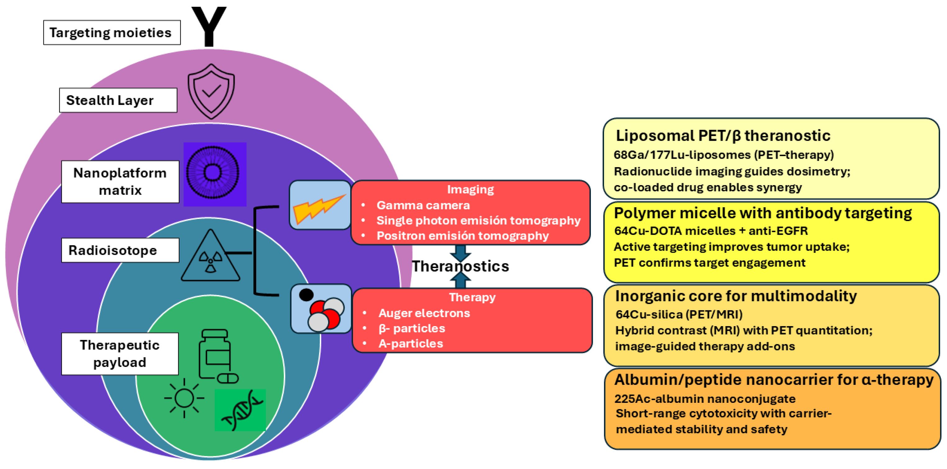
Figure 1. Modular theranostic nanoplatforms in nuclear medicine. Left: Conceptual core–shell schematic shows how nanocarrier layers (matrix, stealth, targeting) enable the following: Center: imaging, therapy, or dual theranostic functions. Right: Representative research exemplars.
Jnt 06 00027 g001
Figure 2. Roadmap to Clinical Implementation of Nuclear Theranostics. Schematic representation of the translational steps required for the clinical adoption of nuclear theranostic nanoplatforms. The pathway includes (1) discovery and preclinical validation, (2) optimization and scale-up under GMP-compatible conditions, (3) regulatory and safety assessment under EMA/FDA/IAEA guidelines, (4) clinical trial execution, and (5) final integration into personalized oncology practice. Color intensity increases with regulatory complexity and translational maturity.
Jnt 06 00027 g002
Table 1. Physical characteristics of candidate radioisotopes currently used in diagnostics.
RadioisotopeDecay ModeT1/2Energy of the Main Photon in keV (Abundance%)Diagnostic Method
99mTcγ6 h140 (89)SPECT
131Iβ8 d364 (81)SPECT
123IEC13.2 h159 (83)SPECT
67GaEC78.3 h93 (37); 185 (20); 300 (17); 395 (5)SPECT
111InEC2.8 d171 (90); 245 (94)SPECT
11CΒ+20 min511 PET
18Fβ+110 min511PET
68Gaβ+68 min511PET
EC: Electron Capture.
Table 2. Physical characteristics of particle-emitting radionuclides currently used for therapy in Nuclear Medicine.
RadioisotopeT1/2Emitted Particle (Energy MeV)Max Range in Soft Tissue
131I8 dβ (0.606)2.3 mm
223Ra11.43 d4α 2β (5.64, 5.715)<100 µm
90Y64.1 hβ (2.27)11.3 mm
177Lu6.65 dβ γ (0.497)1.8 mm
188Re0.7 dβ (2.12)10 mm
225Ac10 d4α 2β (6.83)47-85 μm
Table 3. Theranostic pairs in nuclear medicine: chemical and clinical characteristics relevant to nanotheragnosis.
Diagnostic/Therapeutic PairChelatorNanotheragnostic SuitabilityClinical Application
[68Ga]/[177Lu]-DOTA-TATEDOTAHigh: stable coordination, mild labeling conditionsNETs (neuroendocrine tumors)
[68Ga]/[177Lu]-PSMA-617DOTA/DOTAGAHigh: widely adapted to nanocarriersProstate cancer
[64Cu]/[67Cu]-ChelateNOTA/SarArModerate–High: versatile chelation, redox sensitivity requires stabilizationExperimental-solid
tumors
[89Zr]/[90Y]-ChelateDFO (for 89Zr),
DOTA (for 90Y)
Moderate: DFO less stable long-term, 90Y well adaptedAntibody labeling/
solid tumors
[123I]/[131I]-MIBGDirect iodinationLow for nanoplatforms-instability in vivo without encapsulationNeuroblastoma,
pheochromocytoma
[68Ga]/[225Ac]-PSMADOTA/MacropaHigh: α-emitter integration into nanoparticles for targeted deliverymCRPC, α-therapy
under investigation
Table 4. Regulatory Scenarios for Nuclear Theranostics.
Technological
Innovation Level
Radionuclide TypeRegulatory ClassificationNon-Clinical
Requirements
GMP Considerations
Incremental (e.g., liposomes + 99mTc)Conventional (99mTc, 111In, 131I)IMP/RadiopharmaceuticalReduced studies if vector knownStandard GMP processes apply
Intermediate (e.g., new polymers + 177Lu)Emerging therapeutic (177Lu, 90Y)RadiotherapeuticToxicology (S9), biodistribution, dosimetryProcess validation, radiochemical stability
Disruptive (e.g., hybrid NP + 225Ac)High-risk a-emitter (225Ac, 213Bi, 223Ra)Advanced Radiotherapeutic/ATMPFull toxicology, genotoxicity, organ dosimetryCustom GMP: shielding, purity, retention
Increasing Regulatory ComplexityGreater innovation and risk demand more stringent regulatory oversight and tailored GMP solutions.
ATMP = Advanced Therapy Medicinal Product (according to EMA).
Table 5. Nanoplatforms in Clinical Trials for Cancer Therapy and Imaging. Clinical trial data were extracted from the ClinicalTrials.gov website.
Nanoplatform (Trade/Code)Composition/RadionuclideIndicationClinical PhaseTrial ID/Reference
Cornell dotsSilica NP + PEG, cRGDY peptide/124IImaging of metastatic melanomaPhase INCT01266096
AGuIXGadolinium-based NP/radiosensitizerBrain metastasesPhase INCT03818386
177Lu-PSMA-I&T NPPolymer-based NP/177LuProstate cancerPhase I/IINCT04647526
188Re-BMEDA-liposomeLiposome + BMEDA/188ReAdvanced or metastatic solid tumorsPhase INCT02271516
64Cu SPIONSuperparamagnetic iron oxide nanoparticles/64CuRefractory myeloma with extramedullary diseasePhase INCT05666700
64Cu-NOTA-PSMAi-PEG-Cy5.5-C’ dotsUltrasmall silica C’ dots/64CuProstate cancer (diagnostics and guided surgery)Phase INCT04167969
89Zr-DFO-PSMAi-PEG-Cy5.5-C’ dotsUltrasmall silica C’ dots/89ZrProstate cancer (diagnostics and guided surgery)Phase INCT04167969
Disclaimer/Publisher’s Note: The statements, opinions and data contained in all publications are solely those of the individual author(s) and contributor(s) and not of MDPI and/or the editor(s). MDPI and/or the editor(s) disclaim responsibility for any injury to people or property resulting from any ideas, methods, instructions or products referred to in the content.