

Evolution of Theranostic Nanoparticles Through the Lens of Patents

 , , and
Abstract
1. Introduction
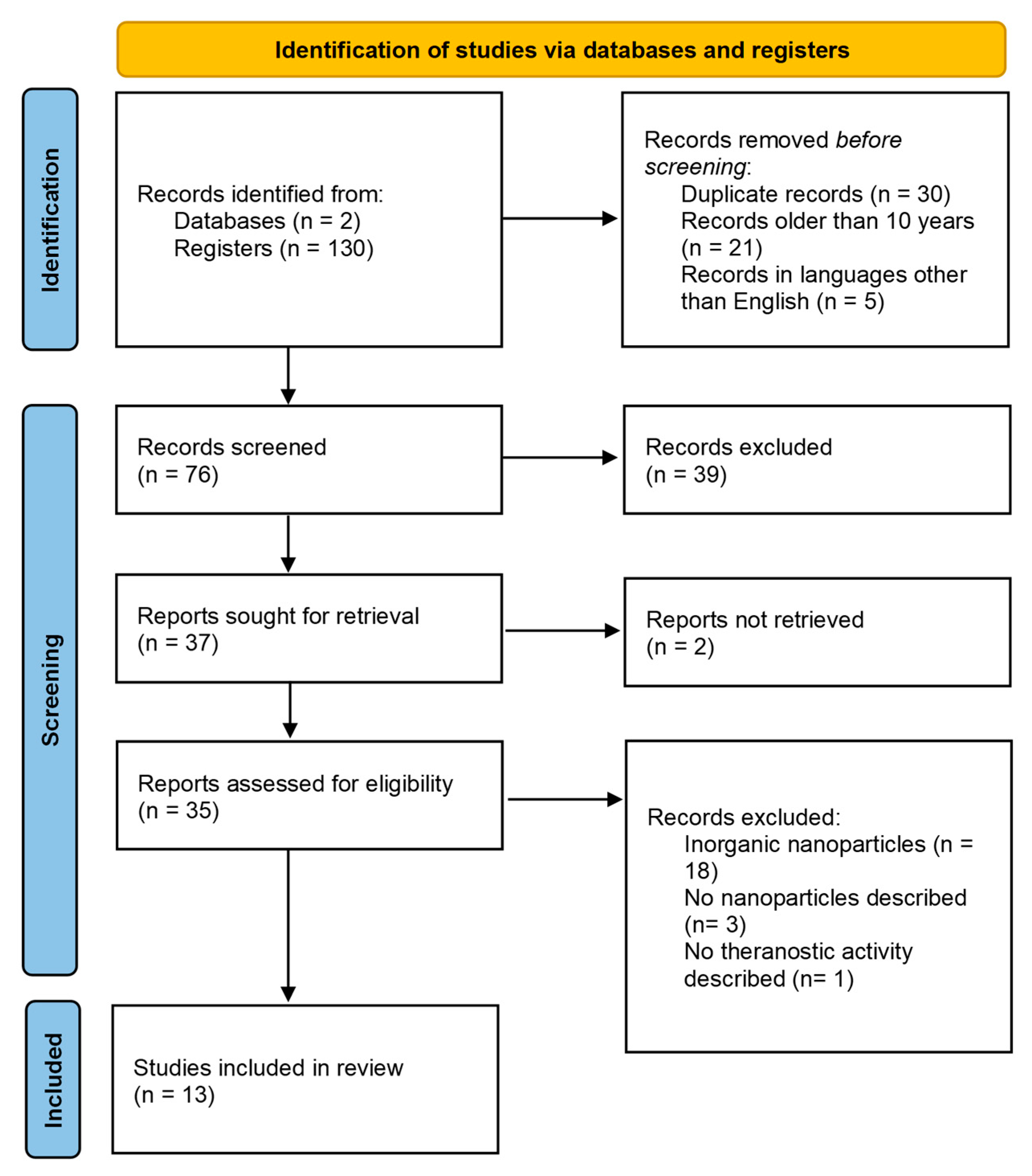
2. Methods
3. Results and Discussion
3.1. General Information About the Patents
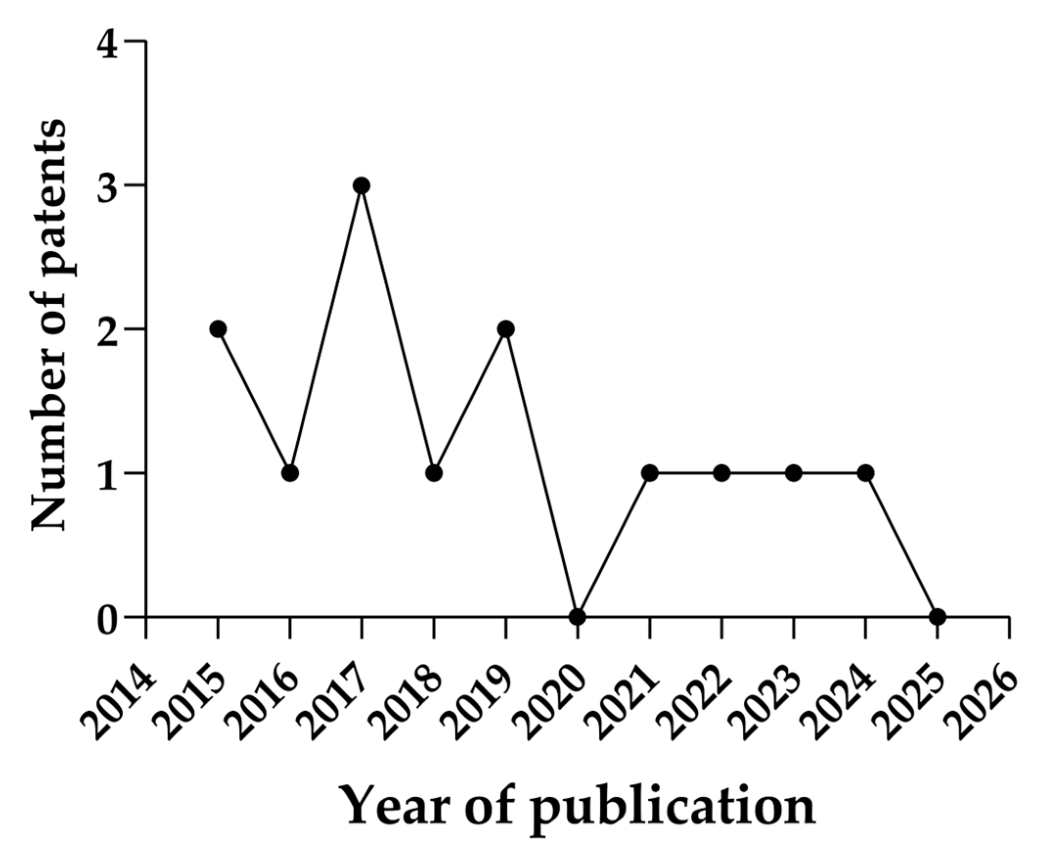
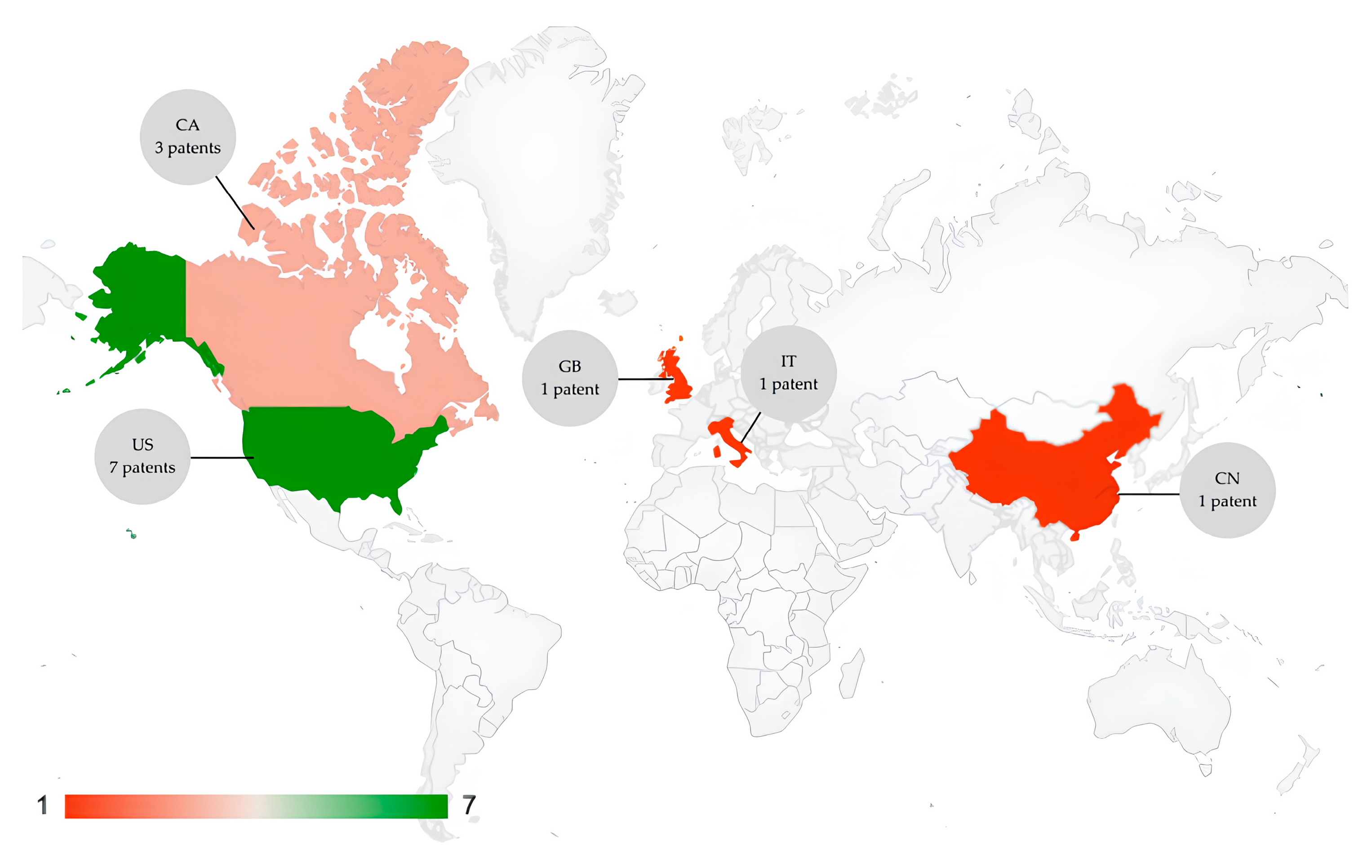
3.2. Annual Evaluation and Country Distribution of Patents
3.3. Polymeric Nanoparticles
3.4. Lipid Nanoparticles and Liposomes
3.5. Hybrid Lipid Nanoparticles
4. Challenges in Clinical Translation of Organic-Based Nanotheranostics
5. Conclusions
Author Contributions
Funding
Conflicts of Interest
References
- Blasco-Navarro, C.; Alonso-Moreno, C.; Bravo, I. From Traditional Nanoparticles to Cluster-Triggered Emission Polymers for the Generation of Smart Nanotheranostics in Cancer Treatment. J. Nanotheranostics 2025, 6, 3. [Google Scholar] [CrossRef]
- Puccetti, M.; Pariano, M.; Schoubben, A.; Giovagnoli, S.; Ricci, M. Biologics, theranostics, and personalized medicine in drug delivery systems. Pharmacol. Res. 2024, 201, 107086. [Google Scholar] [CrossRef] [PubMed]
- Gajbhiye, K.R.; Salve, R.; Narwade, M.; Sheikh, A.; Kesharwani, P.; Gajbhiye, V. Lipid polymer hybrid nanoparticles: A custom-tailored next-generation approach for cancer therapeutics. Mol. Cancer 2023, 22, 160. [Google Scholar] [CrossRef] [PubMed]
- Gong, Z.; Peng, S.; Cao, J.; Tan, H.; Zhao, H.; Bai, J. Advances in the variations and biomedical applications of stimuli-responsive nanodrug delivery systems. Nanotechnology 2024, 35, 132001. [Google Scholar] [CrossRef]
- Uredat, S.; Gujare, A.; Runge, J.; Truzzolillo, D.; Oberdisse, J.; Hellweg, T. A review of stimuli-responsive polymer-based gating membranes. Phys. Chem. Chem. Phys. 2024, 26, 2732–2744. [Google Scholar] [CrossRef]
- Shetty, A.; Chandra, S. Inorganic hybrid nanoparticles in cancer theranostics: Understanding their combinations for better clinical translation. Mater. Today Chem. 2020, 18, 100381. [Google Scholar] [CrossRef]
- Kurul, F.; Turkmen, H.; Cetin, A.E.; Topkaya, S.N. Nanomedicine: How nanomaterials are transforming drug delivery, bio-imaging, and diagnosis. Next Nanotechnol. 2025, 7, 100129. [Google Scholar] [CrossRef]
- Kashyap, B.K.; Singh, V.V.; Solanki, M.K.; Kumar, A.; Ruokolainen, J.; Kesari, K.K. Smart Nanomaterials in Cancer Theranostics: Challenges and Opportunities. ACS Omega 2023, 8, 14290–14320. [Google Scholar] [CrossRef]
- Setia, A.; Challa, R.R.; Vallamkonda, B.; Satti, P.; Mehata, A.K.; Priya, V.; Kumar, S.; Muthu, M.S. Nanomedicine And Nanotheranostics: Special Focus on Imaging of Anticancer Drugs Induced Cardiac Toxicity. Nanotheranostics 2024, 8, 473–496. [Google Scholar] [CrossRef]
- Hosseini, S.M.; Mohammadnejad, J.; Salamat, S.; Beiram Zadeh, Z.; Tanhaei, M.; Ramakrishna, S. Theranostic polymeric nanoparticles as a new approach in cancer therapy and diagnosis: A review. Mater. Today Chem. 2023, 29, 101400. [Google Scholar] [CrossRef]
- Malik, S.; Muhammad, K.; Waheed, Y. Emerging Applications of Nanotechnology in Healthcare and Medicine. Molecules 2023, 28, 6624. [Google Scholar] [CrossRef] [PubMed]
- Page, M.J.; McKenzie, J.E.; Bossuyt, P.M.; Boutron, I.; Hoffmann, T.C.; Mulrow, C.D.; Shamseer, L.; Tetzlaff, J.M.; Akl, E.A.; Brennan, S.E.; et al. The PRISMA 2020 statement: An updated guideline for reporting systematic reviews. BMJ 2021, 372, n71. [Google Scholar] [CrossRef] [PubMed]
- Nascimento, J.A.C., Jr.; Santos, A.M.; Oliveira, A.M.S.; Santos, A.B.; Araújo, A.A.d.S.; Frank, L.A.; Serafini, M.R. Use of nanotechnology applied to sunscreens: Technological prospection based on patents. J. Drug Deliv. Sci. Technol. 2024, 91, 105245. [Google Scholar] [CrossRef]
- Cezar, S.V.S.; Santos, A.B.; Santos, A.M.; Brito, J.R.L.R.; Menezes, P.d.P.; Serafini, M.R. Patents on the move: The therapeutic future of liquid crystals in cancer. J. Drug Deliv. Sci. Technol. 2024, 97, 105822. [Google Scholar] [CrossRef]
- WIPO. World Intellectual Property Organization; WIPO: Geneva, Switzerland, 2025. [Google Scholar]
- Cullis, P.; Kulkarni, J.; Jigaltsev, I.; Tam, Y.Y.; Uzel, A.; Kafshgari, M.H.; Meunier, M. Hybrid Lipid Nanoparticle Comprising an Inorganic Particle and an Agent of Interest. WO/2023/150892, 17 August 2023. [Google Scholar]
- Khaled, A.; Figueroa, J.M.P.; Santra, S.; Kaittanis, C.; Santiesteban, O.; Grimm, J.; Sessions, H. Methods and Compositions for Theranostic Nanoparticles. U.S. Patent No. 10,973,925, 13 April 2015. [Google Scholar]
- Barthet, V.; Hinterleitner, C.; Heller, D.A.; Lowe, S. Nanoparticle-Based Theranostic Platform for Diagnosis and Treatment of Senescence-Related Pathologies. WO/2024/229271, 7 November 2024. [Google Scholar]
- Dravid, V.P.; Nandwana, V. Nanoparticle-Lipid Composite Carriers and Uses Thereof. U.S. Patent No. 11,510,872, 29 November 2017. [Google Scholar]
- Thanou, M.; Wright, M.; Centelles, M.; Miller, A.D.; Gedroyc, W. Precision Therapeutics. 2015. WO/2016/198859, 15 December 2016. [Google Scholar]
- Torino, E.; Netti, P. Process for the Preparation of Double Crosslinked Core-Shell Polymeric Nanoparticles for Multimodal Imaging and Theranostic Applications. U.S. Patent No. 11,311,853, 26 April 2017. [Google Scholar]
- Li, Y.; Wu, H.; Lin, T.-Y. Sequential Targeting in Crosslinking Nano-Theranostics for Treating Brain Tumors. U.S. Patent Application No. 17/785,765, 9 March 2019. [Google Scholar]
- Tang, B.; Ding, D.; Qi, J. Theranostic Agents. 2017. U.S. Patent No. 11,389,446, 14 May 2020. [Google Scholar]
- Dinakaran, D.; Moore, R.; Lewis, J.; Narain, R.; Kumar, P.; Usmani, N. Theranostic Radiophotodynamic Therapy Nanoparticles. U.S. Patent Application No. 17/255,424, 2 September 2018. [Google Scholar]
- Perez, J.M.; Santra, S. Synthesis of Hyperbranched Amphiphilic Polyester and Theranostic Nanoparticles Thereof. U.S. Patent 20160256402A1, 2 February 2016. [Google Scholar]
- Karunya, K.K. Nanoparticles/Theranostic Vehicles. U.S. Patent No. 09,950,002, 19 March 2015. [Google Scholar]
- Samuel, M. Multifunctional pO2/pH-Sensitive Theranostic Liposome Nanocarriers and Methods of Using Same. U.S. Patent Application No. 18/357,810, 8 February 2024. [Google Scholar]
- Yu, W.X. Polymeric Nanoparticles Useful in Theranostics. U.S. Patent No. 10,233,277, 19 March 2019. [Google Scholar]
- WIPO. World Intellectual Property Indicators Report: Global Patent Filings Reach; WIPO: Geneva, Switzerland, 2013. [Google Scholar]
- McKibbin, W.; Fernando, R. The global economic impacts of the COVID-19 pandemic. Econ. Model. 2023, 129, 106551. [Google Scholar] [CrossRef]
- Nanotechnology at NIH; NIH: Bethesda, MD, USA, 2016.
- De Souza, M.L.; Oliveira, D.D.; Ribeiro, P.L.L.; de Paula Pereira, N.; Druzian, J.I. Nanoemulsions for Cosmetic Applications: What Innovation Status? Recent. Pat. Nanotechnol. 2018, 12, 101–109. [Google Scholar] [CrossRef]
- Fernandes, D.A. Theranostic Polymeric Nanoparticles for Cancer. BioNanoScience 2023, 13, 1609–1644. [Google Scholar] [CrossRef]
- Zielińska, A.; Carreiró, F.; Oliveira, A.M.; Neves, A.; Pires, B.; Venkatesh, D.N.; Durazzo, A.; Lucarini, M.; Eder, P.; Silva, A.M.; et al. Polymeric Nanoparticles: Production, Characterization, Toxicology and Ecotoxicology. Molecules 2020, 25, 3731. [Google Scholar] [CrossRef]
- Tehrani, S.F.; Bharadwaj, P.; Leblond Chain, J.; Roullin, V.G. Purification processes of polymeric nanoparticles: How to improve their clinical translation? J. Control Release 2023, 360, 591–612. [Google Scholar] [CrossRef]
- Alregeb, F.; Khalili, F.; Sweileh, B.; Ali, D.K. Synthesis and Characterization of Chelating Hyperbranched Polyester Nanoparticles for Cd(II) Ion Removal from Water. Molecules 2022, 27, 3656. [Google Scholar] [CrossRef]
- Özdal, Z.D.; Gültekin, Y.; Vural, İ.; Takka, S. Development and characterization of polymeric nanoparticles containing ondansetron hydrochloride as a hydrophilic drug. J. Drug Dev. Technol. 2022, 74, 103599. [Google Scholar] [CrossRef]
- Lee, M.W.; Bassiouni, R.; Sparrow, N.A.; Iketani, A.; Boohaker, R.J.; Moskowitz, C.; Vishnubhotla, P.; Khaled, A.S.; Oyer, J.; Copik, A.; et al. The CT20 peptide causes detachment and death of metastatic breast cancer cells by promoting mitochondrial aggregation and cytoskeletal disruption. Cell Death Disease 2014, 5, e1249. [Google Scholar] [CrossRef] [PubMed]
- Deng, Z.; Li, B.; Yang, M.; Lu, L.; Shi, X.; Lovell, J.F.; Zeng, X.; Hu, W.; Jin, H. Irradiated microparticles suppress prostate cancer by tumor microenvironment reprogramming and ferroptosis. J. Nanobiotechnology 2024, 22, 225. [Google Scholar] [CrossRef]
- Moorthy, D.N.; Dhinasekaran, D.; Rebecca, P.N.B.; Rajendran, A.R. Optical Biosensors for Detection of Cancer Biomarkers: Current and Future Perspectives. J. Biophotonics 2024, 17, e202400243. [Google Scholar] [CrossRef]
- Torino, E.; Auletta, L.; Vecchione, D.; Orlandella, F.M.; Salvatore, G.; Iaccino, E.; Fiorenza, D.; Grimaldi, A.M.; Sandomenico, A.; Albanese, S.; et al. Multimodal imaging for a theranostic approach in a murine model of B-cell lymphoma with engineered nanoparticles. Nanomed. Nanotechnol. Biol. Med. 2018, 14, 483–491. [Google Scholar] [CrossRef]
- Zhou, Y.; Peng, Z.; Seven, E.S.; Leblanc, R.M. Crossing the blood-brain barrier with nanoparticles. J. Control Release 2018, 270, 290–303. [Google Scholar] [CrossRef]
- Spirescu, V.A.; Chircov, C.; Grumezescu, A.M.; Andronescu, E. Polymeric Nanoparticles for Antimicrobial Therapies: An Up-To-Date Overview. Polymers 2021, 13, 724. [Google Scholar] [CrossRef]
- Qu, H.; Yang, J.; Li, S.; Xu, J.; Zhou, X.; Xue, X.; Zhang, D.; Du, H.; Shen, Y.; Ramachandran, M.; et al. Programmed-response cross-linked nanocarrier for multidrug-resistant ovarian cancer treatment. J. Control Release 2023, 357, 274–286. [Google Scholar] [CrossRef]
- Maitz, M.F. Applications of synthetic polymers in clinical medicine. Biossurf. Biotribol. 2015, 1, 161–176. [Google Scholar] [CrossRef]
- Beach, M.A.; Nayanathara, U.; Gao, Y.; Zhang, C.; Xiong, Y.; Wang, Y.; Such, G.K. Polymeric Nanoparticles for Drug Delivery. Chem. Rev. 2024, 124, 5505–5616. [Google Scholar] [CrossRef]
- Prasad, A.; Bakr, M.M.; ElMeshad, A.N. Surface-functionalised polymeric nanoparticles for breast cancer treatment: Processes and advances. J. Drug Target. 2024, 32, 770–784. [Google Scholar] [CrossRef] [PubMed]
- Elumalai, K.; Srinivasan, S.; Shanmugam, A. Review of the efficacy of nanoparticle-based drug delivery systems for cancer treatment. Biomed. Technol. 2024, 5, 109–122. [Google Scholar] [CrossRef]
- Zhang, W.; Mehta, A.; Tong, Z.; Esser, L.; Voelcker, N.H. Development of Polymeric Nanoparticles for Blood-Brain Barrier Transfer-Strategies and Challenges. Adv. Sci. 2021, 8, 2003937. [Google Scholar] [CrossRef]
- Dhayalan, M.; Wang, W.; Riyaz, S.U.M.; Dinesh, R.A.; Shanmugam, J.; Irudayaraj, S.S.; Stalin, A.; Giri, J.; Mallik, S.; Hu, R. Advances in functional lipid nanoparticles: From drug delivery platforms to clinical applications. 3 Biotech. 2024, 14, 57. [Google Scholar] [CrossRef]
- Sharmiladevi, P.; Girigoswami, K.; Haribabu, V.; Girigoswami, A. Nano-enabled theranostics for cancer. Mater. Adv. 2021, 2, 2876–2891. [Google Scholar] [CrossRef]
- Miranda, J.A.; Cruz, Y.F.d.; Girão, Í.C.; Souza, F.J.J.d.; Oliveira, W.N.d.; Alencar, É.d.N.; Amaral-Machado, L.; Egito, E.S.T.d. Beyond Traditional Sunscreens: A Review of Liposomal-Based Systems for Photoprotection. Pharmaceutics 2024, 16, 661. [Google Scholar] [CrossRef]
- Nsairat, H.; Ibrahim, A.A.; Jaber, A.M.; Abdelghany, S.; Atwan, R.; Shalan, N.; Abdelnabi, H.; Odeh, F.; El-Tanani, M.; Alshaer, W. Liposome bilayer stability: Emphasis on cholesterol and its alternatives. J. Liposome Res. 2024, 34, 178–202. [Google Scholar] [CrossRef]
- Vargas, K.M.; Shon, Y.-S. Hybrid lipid–nanoparticle complexes for biomedical applications. J. Mater. Chem. B 2019, 7, 695–708. [Google Scholar] [CrossRef]
- Cadinoiu, A.N.; Rata, D.M.; Atanase, L.I.; Daraba, O.M.; Gherghel, D.; Vochita, G.; Popa, M. Aptamer-Functionalized Liposomes as a Potential Treatment for Basal Cell Carcinoma. Polymers 2019, 11, 1515. [Google Scholar] [CrossRef]
- Musielak, M.; Potoczny, J.; Boś-Liedke, A.; Kozak, M. The Combination of Liposomes and Metallic Nanoparticles as Multifunctional Nanostructures in the Therapy and Medical Imaging—A Review. Int. J. Mol. Sci. 2021, 22, 6229. [Google Scholar] [CrossRef]
- Zhigaltsev, I.V.; Tam, Y.Y.C.; Kulkarni, J.A.; Cullis, P.R. Synthesis and Characterization of Hybrid Lipid Nanoparticles Containing Gold Nanoparticles and a Weak Base Drug. Langmuir 2022, 38, 7858–7866. [Google Scholar] [CrossRef] [PubMed]
- Darwish, A.; Sándor, N.; Szenti, I.; Marosvölgyi, T.; Juhász, K.; Rónavári, A.; Kachal, E.; Kutus, B.; Kónya, Z.; Balogi, Z. Highly Stable Antitumor Silver-Lipid Nanoparticles Optimized for Targeted Therapy. Int. J. Nanomed. 2025, 20, 1351–1366. [Google Scholar] [CrossRef] [PubMed]
- Virlan, M.J.; Miricescu, D.; Radulescu, R.; Sabliov, C.M.; Totan, A.; Calenic, B.; Greabu, M. Organic Nanomaterials and Their Applications in the Treatment of Oral Diseases. Molecules 2016, 21, 207. [Google Scholar] [CrossRef]
- Dilnawaz, F.; Acharya, S.; Sahoo, S.K. Recent trends of nanomedicinal approaches in clinics. Int. J. Pharm. 2018, 538, 263–278. [Google Scholar] [CrossRef]
- Nikdouz, A.; Namarvari, N.; Ghasemi Shayan, R.; Hosseini, A. Comprehensive comparison of theranostic nanoparticles in breast cancer. Am. J. Clin. Exp. Immunol. 2022, 11, 1–27. [Google Scholar]
- Naziris, N.; Demetzos, C. Lipid Nanoparticles as Platforms for Theranostic Purposes: Recent Advances in the Field. J. Nanotheranostics 2022, 3, 86–101. [Google Scholar] [CrossRef]
- Ragelle, H.; Fabienne, D.; Véronique, P.; Robert, L.; Anderson, D.G. Nanoparticle-based drug delivery systems: A commercial and regulatory outlook as the field matures. Expert. Opin. Drug Deliv. 2017, 14, 851–864. [Google Scholar] [CrossRef]

| Publication Number | Kind Code | Title | Nanoparticle | Year of Publication | Country | Status | Ref. |
|---|---|---|---|---|---|---|---|
| WO2023150892 A1 | A1 | Hybrid lipid nanoparticle comprising an inorganic particle and an agent of interest | Hybrid Lipid nanoparticle | 2022 | CA | Published | [16] |
| US20210228733 | B2 | Methods and compositions for theranostic nanoparticles | Polymeric nanoparticle | 2021 | US | Granted | [17] |
| WO2024229271 | A1 | Nanoparticle-based theranostic platform for diagnosis and treatment of senescence-related pathologies | Polymeric nanoparticle | 2023 | US | Published | [18] |
| US20200170946 | B2 | Nanoparticle-lipid composite carriers and uses thereof | Hybrid Lipid nanoparticle | 2017 | US | Granted | [19] |
| WO2016198859 | A1 | Precision therapeutics | Lipid nanoparticle | 2015 | GB | Published | [20] |
| US20190381471 | B2 | Process for the preparation of double crosslinked core-shell polymeric nanoparticles for multimodal imaging and theranostic applications | Polymeric nanoparticle | 2017 | IT | Granted | [21] |
| US20230076792 | A1 | Sequential targeting in crosslinking nano-theranostics for treating brain tumors | Polymeric nanoparticles | 2019 | US | Published | [22] |
| US20200147078 | B2 | Theranostic agents | Lipid nanoparticles | 2017 | CN | Granted | [23] |
| US20210268129 | A1 | Theranostic radiophotodynamic therapy nanoparticles | Polymeric nanoparticle | 2018 | CA | Published | [24] |
| US20160256402 | B2 | Synthesis of hyperbranched amphiphilic polyester and theranostic nanoparticles thereof | Polymeric nanoparticles | 2016 | US | Granted | [25] |
| US20150078995 | B2 | Nanoparticles/theranostic vehicles | Polymeric nanoparticles | 2024 | US | Granted | [26] |
| US20240041768 | A1 | Multifunctional pO2/pH-sensitive theranostic liposome nanocarriers and methods of using same | Lipid nanoparticles | 2024 | US | Published | [27] |
| US20190233567 | B2 | Polymeric nanoparticles useful in theranostics | Polymeric nanoparticles | 2019 | CA | Granted | [28] |
Disclaimer/Publisher’s Note: The statements, opinions and data contained in all publications are solely those of the individual author(s) and contributor(s) and not of MDPI and/or the editor(s). MDPI and/or the editor(s) disclaim responsibility for any injury to people or property resulting from any ideas, methods, instructions or products referred to in the content. |
© 2025 by the authors. Licensee MDPI, Basel, Switzerland. This article is an open access article distributed under the terms and conditions of the Creative Commons Attribution (CC BY) license (https://creativecommons.org/licenses/by/4.0/).
Share and Cite
Freire, D.T.; Miranda, J.A.; Dourado, D.; Alencar, É.d.N. Evolution of Theranostic Nanoparticles Through the Lens of Patents. J. Nanotheranostics 2025, 6, 11. https://doi.org/10.3390/jnt6020011
Freire DT, Miranda JA, Dourado D, Alencar ÉdN. Evolution of Theranostic Nanoparticles Through the Lens of Patents. Journal of Nanotheranostics. 2025; 6(2):11. https://doi.org/10.3390/jnt6020011
Chicago/Turabian StyleFreire, Danielle Teixeira, Júlio Abreu Miranda, Douglas Dourado, and Éverton do Nascimento Alencar. 2025. "Evolution of Theranostic Nanoparticles Through the Lens of Patents" Journal of Nanotheranostics 6, no. 2: 11. https://doi.org/10.3390/jnt6020011
APA StyleFreire, D. T., Miranda, J. A., Dourado, D., & Alencar, É. d. N. (2025). Evolution of Theranostic Nanoparticles Through the Lens of Patents. Journal of Nanotheranostics, 6(2), 11. https://doi.org/10.3390/jnt6020011