Review

Green Photocatalysis: A Comprehensive Review of Plant-Based Materials for Sustainable Water Purification

1
Materials and Environmental Process Engineering Research Team, GeMaPE, Higher School of Technology, Hassan II University of Casablanca, Casablanca 20000, Morocco
2
Multidisciplinary Research and Innovation Laboratory, Sultan Moulay Slimane University of Beni Mellal, FP Khouribga, BP. 145, Khouribga 25000, Morocco
*
Author to whom correspondence should be addressed.
Reactions 2025, 6(4), 55; https://doi.org/10.3390/reactions6040055 (registering DOI)
Submission received: 19 August 2025 / Revised: 19 September 2025 / Accepted: 30 September 2025 / Published: 5 October 2025

Abstract

Green synthesis represents a sustainable, reliable, and eco-friendly approach for producing various materials and nanomaterials, including metal and metal oxide nanoparticles. This environmentally conscious method has garnered significant attention from materials scientists. In recent years, interest in plant-mediated nanoparticle synthesis has grown markedly, owing to advantages such as enhanced product stability, low synthesis costs, and the use of non-toxic, renewable resources. This review specifically focuses on the green synthesis of metal oxide nanoparticles using plant extracts, highlighting five key oxides: TiO2, ZnO, WO3, CuO, and Fe2O3, which are prepared through various plant-based methods. The release of toxic effluents like synthetic dyes into the environment poses serious threats to aquatic ecosystems and human health. Therefore, the application of biosynthesized nanoparticles in removing such pollutants from industrial wastewater is critically examined. This paper discusses the synthesis routes, characterization techniques, green synthesis methodologies, and evaluates the photocatalytic performance and dye degradation mechanisms of these plant-derived nanoparticles.

1. Introduction

In recent years, nanotechnology has emerged as a transformative discipline in materials science, serving as a powerful tool that has significantly contributed to advancements in various scientific, industrial, and technological domains worldwide. Metal oxide nanoparticles (NPs) show exceptional promise for applications in photocatalysis, environmental remediation, and biomedical fields. The distinctive capability of this technology lies in its ability to enable the fabrication of materials with dimensions ranging from 1 to 100 nm [1,2,3,4]. Nanotechnology relies on two fundamental approaches for the fabrication of nanomaterials: the top-down and bottom-up strategies (Figure 1). The top-down approach involves breaking down bulk materials into nanoscale structures through physical or mechanical processes. Techniques such as scanning lithography, photolithography, nanosphere lithography, soft lithography, imaging probe lithography, dispersion lithography, optical machining, deposition, ion implantation, and diffusion are commonly employed. Despite its usefulness, the top-down method presents several limitations, including high processing costs, extended etching durations, and the potential introduction of defects or imperfections in the resulting nanostructures. In contrast, the bottom-up approach constructs nanoparticles starting from individual atoms, ions, or molecules, allowing for the formation of well-defined nanostructures through controlled nucleation and growth. This approach encompasses a variety of chemical and physicochemical methods such as wet chemical synthesis, the sol–gel process, metal–organic decomposition, atomic or molecular condensation, chemical vapor deposition, spray pyrolysis, laser pyrolysis, and aerosol-based techniques [5,6,7,8,9,10]. In the realm of nanotechnology, the synthesis of nanoparticles (NPs) generally falls into three primary categories: chemical, physical, and biological (commonly referred to as green synthesis). The chemical approach comprises a broad spectrum of techniques, including but not limited to chemical reduction, sol–gel processing, hydrothermal synthesis, ion-exchange processes, co-precipitation, solvothermal methods, template-assisted synthesis, spray pyrolysis, thermal decomposition, the reflux method, chemical vapor deposition, solution combustion, colloidal methods, and polymer-based techniques [11,12,13,14,15,16,17,18,19,20,21,22,23,24]. The physical approach involves methodologies such as vapor-phase synthesis, mechanical milling, sono-chemical synthesis, physical vapor deposition, sputtering, electrochemical methods, laser ablation, laser pyrolysis, microemulsion techniques, microwave-assisted synthesis, arc discharge, evaporation-condensation, the polyol process, and pulsed wire discharge [25,26,27,28,29,30,31,32,33,34,35,36,37,38]. The biological approach, or green synthesis, has gained substantial attention as a sustainable and environmentally friendly alternative to conventional methods. This strategy employs biological entities such as plant extracts [39], bacteria [40], fungi [41], and yeast [42] as natural reducing and stabilizing agents in the fabrication of nanoparticles. Owing to its biocompatibility, simplicity, and reduced ecological impact, green synthesis is increasingly viewed as a promising route for large-scale and low-toxicity nanomaterial production [43]. Plant-mediated synthesis is especially advantageous due to its cost-effectiveness, simplicity, and ability to utilize various plant parts such as leaves, roots, peels, and seeds, thereby enhancing versatility and resource efficiency. Additionally, phytochemicals in these extracts act as capping agents, improving nanoparticle stability and functional performance. A growing body of research demonstrates the application of green-synthesized nanoparticles (TiO2, ZnO, WO3, CuO, Fe2O3) in photocatalysis, particularly for the degradation of organic pollutants under visible or UV light. However, photocatalytic efficiencies vary based on synthesis routes and require systematic evaluation.
This review systematically examines the green synthesis, structural characterization, and photocatalytic performance of TiO2, ZnO, WO3, CuO, and Fe2O3 nanoparticles prepared using plant extracts via different methods, with the aim of highlighting their environmental remediation potential.

2. Mechanism of Photocatalysis

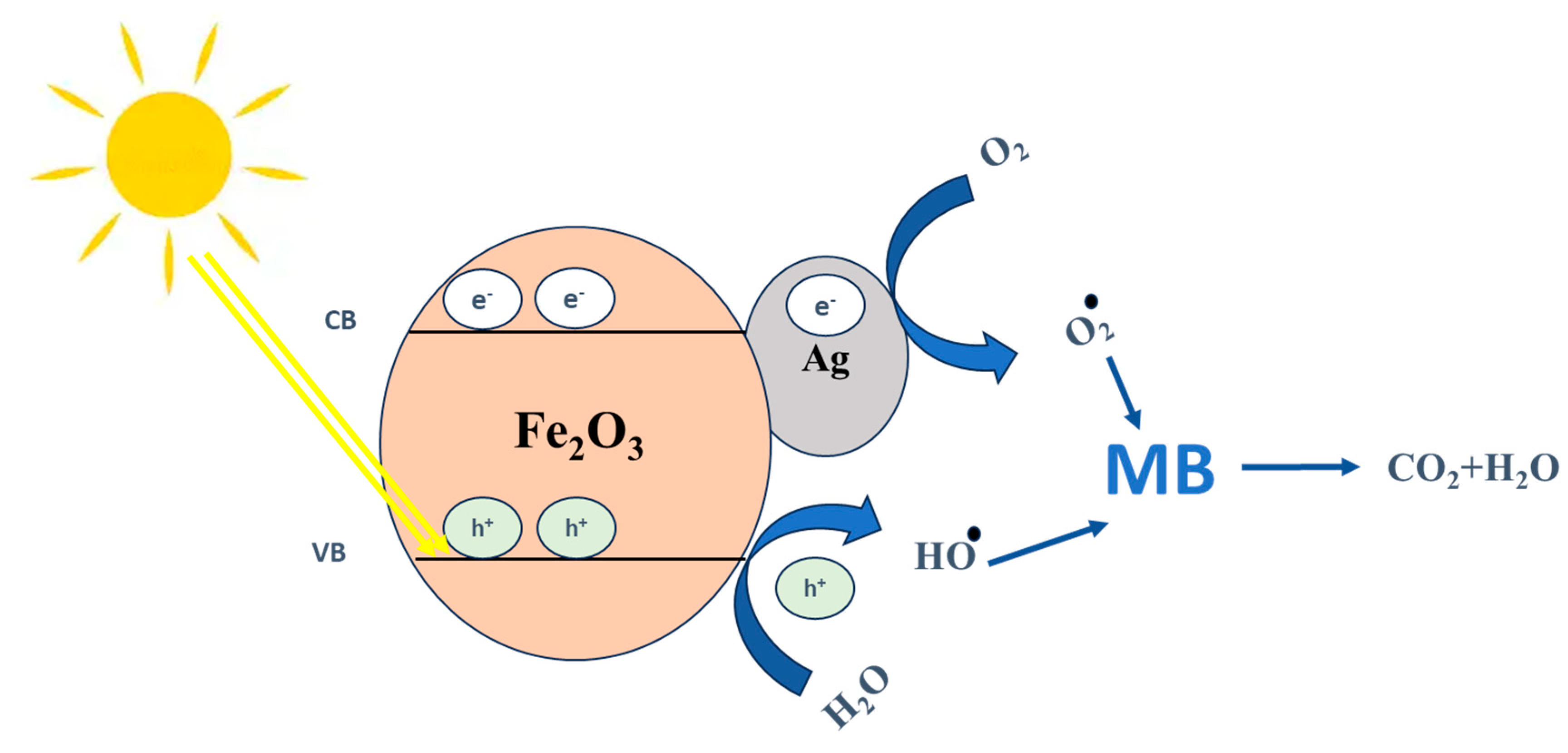
Reactive oxygen species (ROS) are fundamental in understanding the underlying mechanisms of photocatalytic degradation. At the onset of the process, highly reactive ROS such as superoxide anions (O2), hydroxyl radicals ( O H ), sulfate radicals (SO4•−), and singlet oxygen (1O2) are generated [44]. These species possess strong redox potentials, making them effective oxidants capable of breaking down and mineralizing organic pollutants into environmentally benign end-products like carbon dioxide (CO2) and water (H2O). According to Mallah et al. [45], the interaction between ROS and organic contaminants during photocatalysis occurs through three distinct phases: Photoexcitation, Radical formation and pollutant trapping, and Oxidative pollutant degradation. In the Photoexcitation stage, when a photon with energy equal to or greater than the bandgap of the semiconductor strikes the material, it excites an electron from the valence band (VB) to the conduction band (CB). This excitation leaves behind a hole (h+) in the VB while the electron (e) now occupies the CB (Equation (1)). If the electron and hole remain weakly bound by Coulombic attraction, they are collectively referred to as an exciton. The excited electrons typically migrate toward the surface of the photocatalyst, enabling subsequent redox reactions. In the Radical formation and pollutant trapping stage, the photogenerated charge carriers initiate surface reactions leading to the generation of reactive oxygen species (ROS). Electrons in the CB reduce adsorbed oxygen molecules, producing superoxide radicals ( O 2 ), which can subsequently yield hydrogen peroxide and additional hydroxyl radicals ( O H ) (Equation (2)). Concurrently, holes in the VB oxidize surface hydroxyl groups or water molecules, resulting in the formation of highly reactive hydroxyl radicals ( O H ) (Equations (3)–(5)). At the same time, pollutants become adsorbed onto the catalyst surface, a critical step that enhances the likelihood of direct interactions between the generated ROS and the contaminant molecules. Finally, in the Oxidative pollutant degradation stage, the ROS attack and progressively degrade the adsorbed pollutants. Hydroxyl radicals, with their high oxidation potential, indiscriminately cleave C–C, C–H, and C=C bonds, while superoxide radicals act both directly and indirectly by producing secondary oxidizing species. These processes convert complex organic molecules into smaller intermediates, such as alcohols, aldehydes, and carboxylic acids, which are ultimately mineralized into environmentally benign end products, primarily CO2, H2O, and inorganic ions (Equation (6)) [45,46]. Figure 2 schematically illustrates this three-step photocatalytic mechanism. It should be noted that the photocatalytic efficiency is strongly influenced by the catalytic conditions employed during nanoparticle synthesis and photocatalysis. Parameters such as calcination temperature, pH, irradiation source, nanoparticle dosage, and pollutant concentration strongly influence photocatalytic performance. In addition, the type of photocatalyst (e.g., doped photocatalysts, bio-nanocomposites, or clay-based photocatalysts) can alter the morphology, particle size, surface area, and crystallinity of the nanoparticles. These structural features in turn affect the bandgap energy and charge carrier dynamics, including the generation, separation, and lifetime of electron–hole pairs, which are directly linked to ROS formation. For example, smaller particles with higher surface area provide more active sites and facilitate charge separation, enhancing ROS production and pollutant degradation. By considering these experimental factors, the photocatalytic mechanism can be more accurately correlated with the structural and electronic properties of the nanoparticles, providing a comprehensive understanding of their performance [45].
P h o t o c a t a l y s t + h ν h V B + + e C B
O 2 + e C B O 2
H 2 O + h V B + H + + O H
2 h V B + + 2 H 2 O 2 H + + H 2 O 2
H 2 O 2 2 O H
R O S + P o l l u t a n t S i m p l e r   c o m p o u n d + C O 2 + H 2 O
In heterogeneous photocatalysis, a wide range of semiconductors have been utilized as photocatalysts to promote the degradation of dyes into smaller, harmless molecules such as CO2 and H2O. These semiconductor materials exhibit different band gap energies, which influence their light absorption capabilities. For example, TiO2, ZnO, SnO2, and CuO have been extensively investigated, with respective band gap energies of approximately 3.2 eV, 3.4 eV, 3.6 eV, and 1.9 eV [47,48,49,50].

3. Plant-Mediated Green Synthesis of Nanoparticles

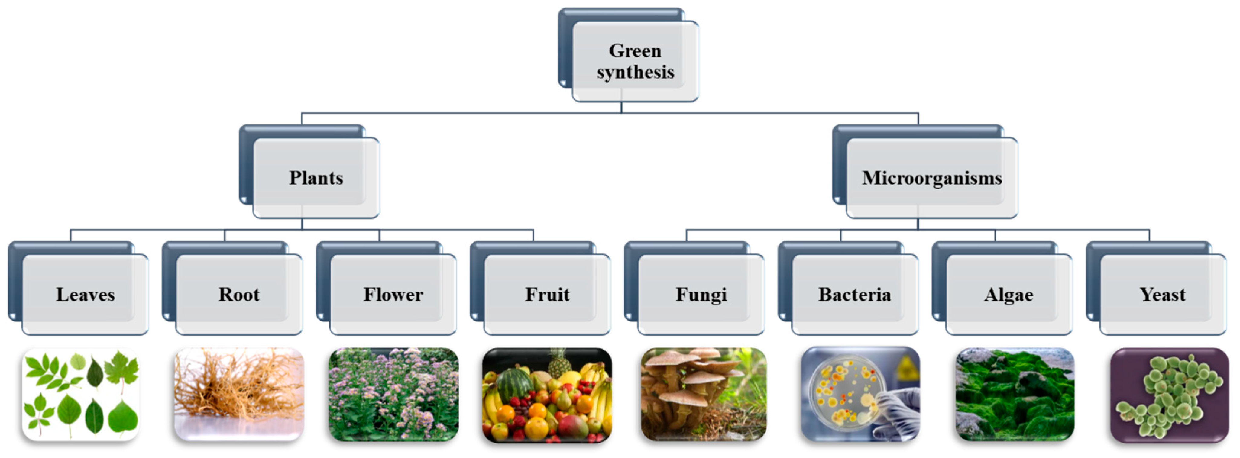
Given the toxic and hazardous effects associated with conventional synthesis methods, green synthesis has emerged as a promising and sustainable alternative [51]. Biological approaches to nanoparticle synthesis typically offer advantages such as mild reaction conditions, the absence of toxic reagents, and the use of eco-friendly, cost-effective resources, as demonstrated in Table 1. A variety of biological systems, including plants, algae, yeast, and fungi, are employed in nanoparticle fabrication [52] (Figure 3). Among these, plant-based or phyto-mediated synthesis stands out due to its rapidity, simplicity, low production cost, and access to a vast and diverse range of natural resources. Notably, phyto-synthesis can utilize different parts of the plant, including fruits, roots, stems, and peels, thus enhancing its versatility and sustainability [53]. One of the notable advantages of green synthesis is its versatility in utilizing nearly all parts of a plant for nanoparticle production, including seeds and seed waste [54], arils [55], fruits [56], flowers [57], roots [58], and others.
The emergence of green technology, particularly through the biological or green synthesis of nanomaterials (NMs), marks the beginning of a new era in safe and sustainable nanotechnology. Consequently, this approach has gained significant attention in recent years due to its environmental and health-related benefits [59].
Table 1. Advantages and disadvantages of Plant-Mediated Photocatalysts [60].
AspectAdvantagesLimitations
Eco-friendlinessUse non-toxic, biodegradable plant extracts; environmentally sustainable.Some processes may still involve non-green conditions (e.g., high temperatures, long synthesis time).
Phytochemical contentRich in natural compounds (e.g., flavonoids, phenolics, tannins) that serve as reducing and stabilizing agents.Phytochemical composition varies by species, plant part, region, and season, affecting reproducibility.
Simplicity and cost-efficiencyRequires no sterile conditions, complex culturing, or expensive equipment.Industrial scalability is limited; plant materials may require storage or preservation.
SafetyNo use of hazardous chemicals; safe for researchers and the environment.Lack of precise control over reaction conditions may result in inconsistent product quality.
Photocatalytic performanceProduces TiO2 NPs with small particle sizes, reduced bandgap, and high dye degradation efficiency (>90%).Some materials show reduced reusability or stability after one photocatalytic cycle.
VersatilityEnables synthesis of various NP shapes (spherical, tubular, etc.) with good surface properties.Irregular particle shapes and low crystallinity may occur, limiting some functional applications.
Mechanistic understandingOffers natural and sustainable synthesis pathways.The exact reaction mechanisms are still not fully understood, limiting optimization strategies.

4. Green Synthesis of Oxide-Based Photocatalysts Using Plant Extracts

Nanomaterials are typically synthesized using two main approaches: top-down and bottom-up methods, each differing in terms of quality, processing speed, and cost-effectiveness. Plant-extract-mediated synthesis processes (PPS) fall under the bottom-up approach of nanomaterial fabrication [61]. In most chemical methods, nanoparticles (NPs) are produced through the interaction of three key components: a metal precursor, a reducing agent, and a capping or stabilizing agent. In the case of PPS synthesis, biomolecules present in plant extracts serve as eco-friendly alternatives to conventional, often toxic, reducing and capping agents such as sodium borohydride and polyvinyl alcohol [62]. The characteristics and properties of the resulting NPs are largely influenced by the chosen synthesis route. Moreover, even the final stages of synthesis, particularly the separation technique (typically centrifugation) and the purification method, play a crucial role in determining the nature and quality of the synthesized NPs [63]. Although the biological synthesis of PPS is chemically assisted, it is distinguished by the substitution of synthetic agents with plant-derived biomolecules. Several methods have been employed for PPS synthesis, and key approaches are outlined below.
In the field of nanoparticle synthesis, several wet-chemical methods have been developed to ensure controlled particle size, morphology, and surface characteristics. Among these, the hydrothermal route was used for the synthesis of tungsten oxide (WO3) NPs using Tamarindus indica leaf extract [64]. The co-precipitation method is widely used, involving the simultaneous precipitation of metal ions, usually as hydroxides in a common aqueous medium. This process includes key steps such as nucleation, particle growth, and agglomeration. Aloe vera and Beetroot extract are used to synthesize TiO2 [65] and ZnO [66], respectively. It offers significant advantages, including low operating temperatures, minimal energy requirements, exclusion of organic solvents, and enhanced control over nanoparticle morphology as demonstrated in biosynthesis of CuO from Euphorbia alata [67] et Bougainvillea [68]. Another frequently employed approach is the sol–gel method, which is based on the hydrolysis and polycondensation of metal alkoxides or salts to form a colloidal sol that gradually evolves into a gel-like network. The sol–gel process enables the synthesis of highly homogeneous and pure nanoparticles, often at relatively low temperatures such as synthesis of TiO2 by Tinospora cordifolia leaf [69] and Mulberry extract [70]. In this review, we provide a comprehensive overview of the green synthesis, physicochemical characterization, and photocatalytic performance evaluation of five widely studied metal oxide nanoparticles: titanium dioxide (TiO2), zinc oxide (ZnO), tungsten trioxide (WO3), cupric oxide (CuO), and hematite (Fe2O3). These metal oxides have been synthesized using a variety of plant-derived extracts, which serve as natural reducing, stabilizing, and capping agents, offering an eco-friendly and sustainable alternative to conventional chemical routes. Different synthesis approaches including co-precipitation, sol–gel, and hydrothermal methods are examined in detail to highlight their influence on the morphological, structural, and optical properties of the resulting nanoparticles. Furthermore, the photocatalytic activity of these bio-synthesized nanomaterials is discussed in the context of organic dye degradation under visible or UV light irradiation, emphasizing their potential for environmental remediation applications.

4.1. Titanium Dioxyde NPs (TiO2)

Titanium dioxide (TiO2) is among the most efficient and widely studied photocatalysts due to its excellent stability, non-toxicity, and strong oxidative power. For this reason, it has been extensively synthesized and investigated, particularly through green synthesis approaches that utilize plant-based extracts as environmentally friendly alternatives to conventional chemical methods. TiO2 nanoparticles were synthesized via a green route by mixing equal volumes of titanium isopropoxide (TTIP) and Tinospora cordifolia extract at 50 °C under stirring. A color change from white to pale yellow indicated the reduction of Ti4+ ions. The formed precipitate was filtered, washed with ethanol, dried, ground into powder, and calcined at 400 °C (Figure 4).
The XRD analysis confirms that the synthesized TiO2 nanoparticles are highly crystalline and predominantly anatase phase, with no detectable impurities or secondary phases (Brookite, Rutil), such as the most intense peak at 2θ = 25.33°, corresponding to the (101) plane, which is a characteristic diffraction peak of anatase-phase TiO2. Other identified anatase peaks are shown in Table 2. Anatase phase is known for its high photocatalytic activity [69].
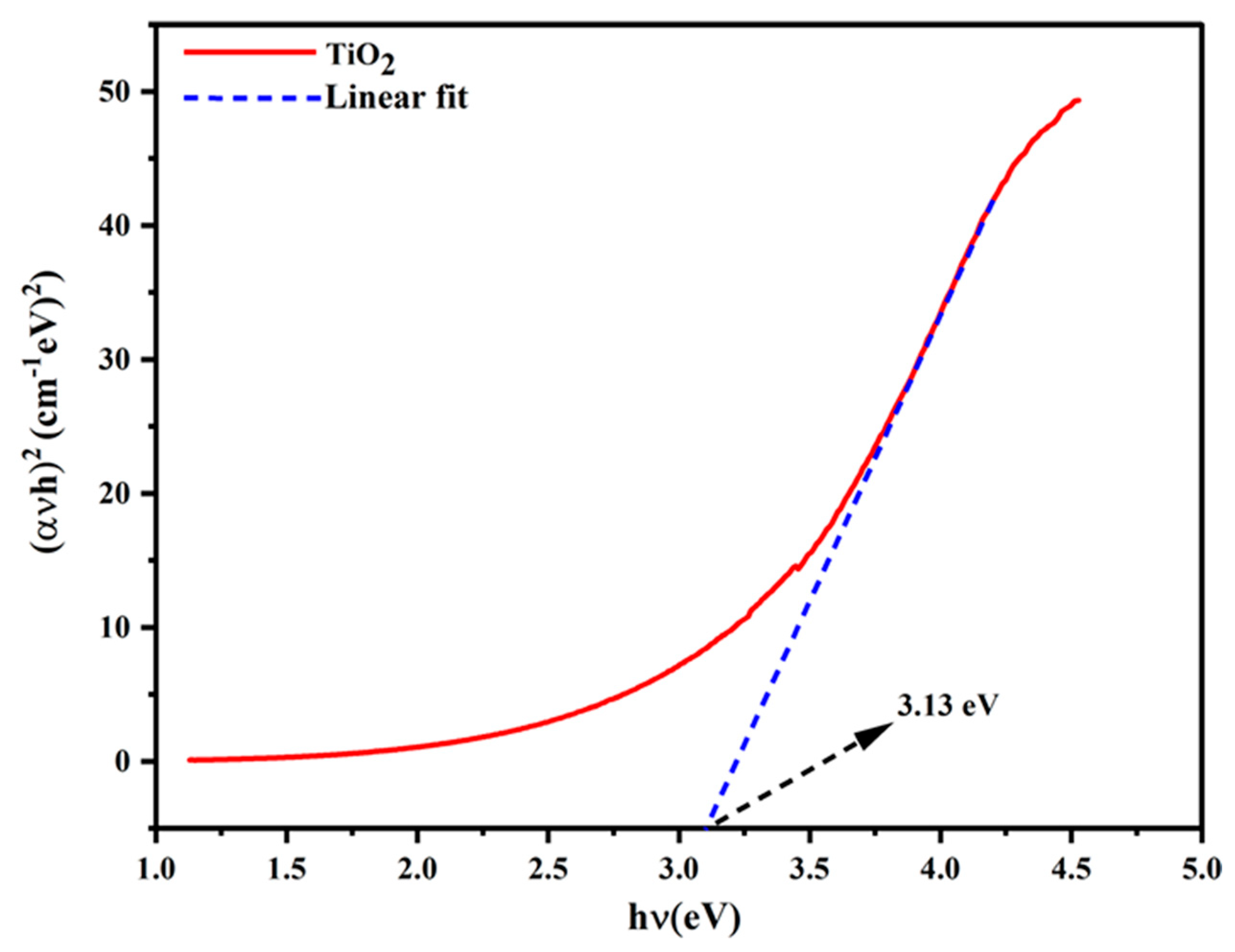
FTIR analysis of Tinospora cordifolia extract and green-synthesized TiO2 nanoparticles confirmed the presence of key functional groups involved in the synthesis process. Characteristic Ti–O stretching at 791 cm−1 and broad bands between 1000–500 cm−1 indicated the anatase phase. The nanoparticles displayed an average crystallite size of 15.02 nm, a slightly reduced band gap of 3.13 eV compared to commercial TiO2 (3.20 eV), and an irregular morphology, suggesting enhanced optical properties through green synthesis Figure 5 [69].
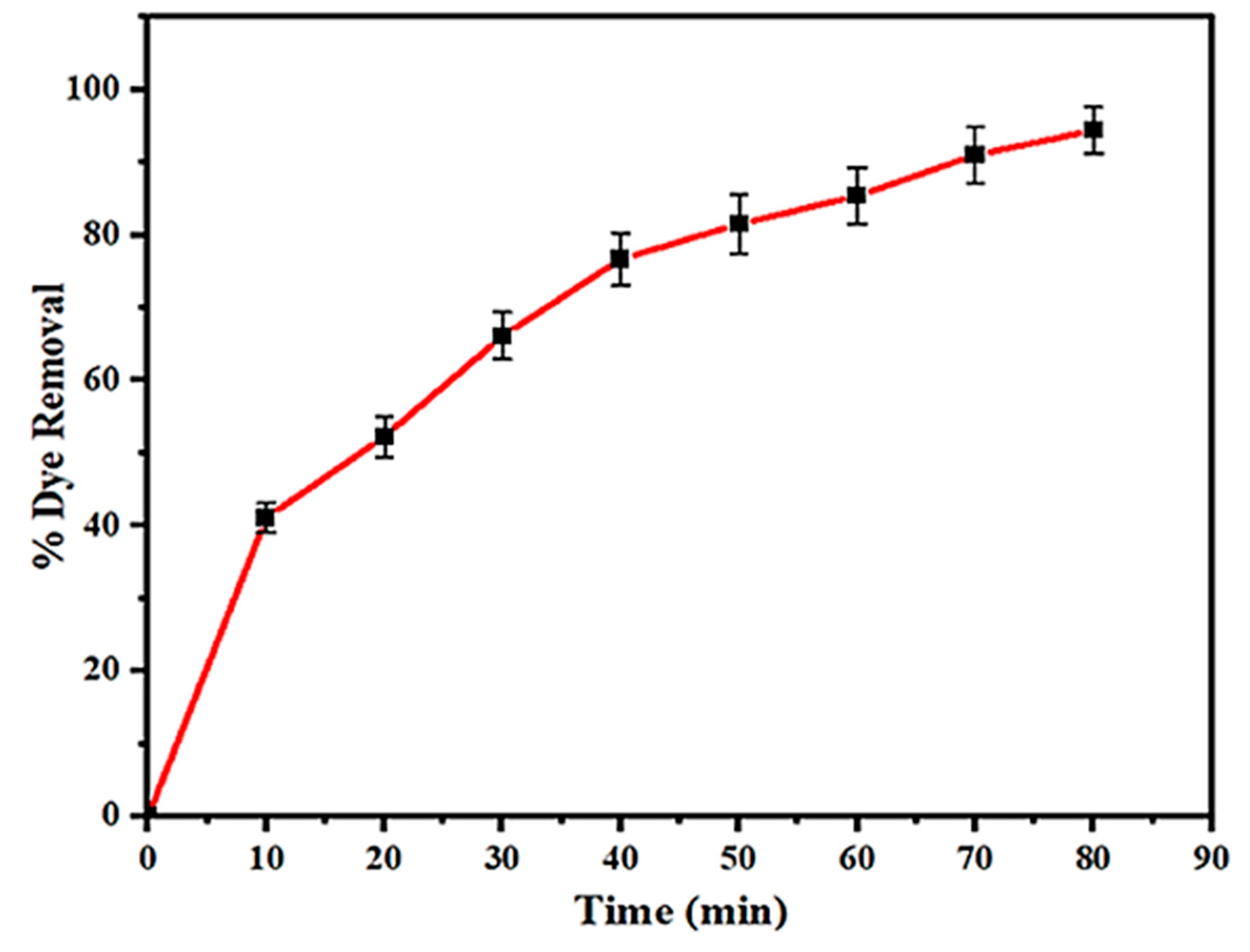
Green-synthesized TiO2 nanoparticles demonstrated excellent photocatalytic activity, achieving 94.43% degradation of Acid Blue 113 dye under UV light within 80 min (Figure 6) [69].
In another preparation by Vanda Wellia et al, using the rind of Aloe vera (L.) Burm extract as a green-capping agent, TiO2 nanoparticles were synthesized by adding 5 mL of TiCl4 (1 M) to deionized water, followed by the dropwise addition of Aloe vera rind extract (4% v/v). The pH was adjusted to 7 with NaOH, and the mixture was stirred for 24 h. The formed precipitate was collected by centrifugation, washed with ethanol and water, dried at 100 °C, and calcined at 500 °C. The product was labeled TOAv4. Similar procedures were followed using 10% and 20% extract concentrations (TOAv10 and TOAv20). A control sample without extract, labeled TO, was also prepared for comparison. Full sample details are provided in Table 3. [65].
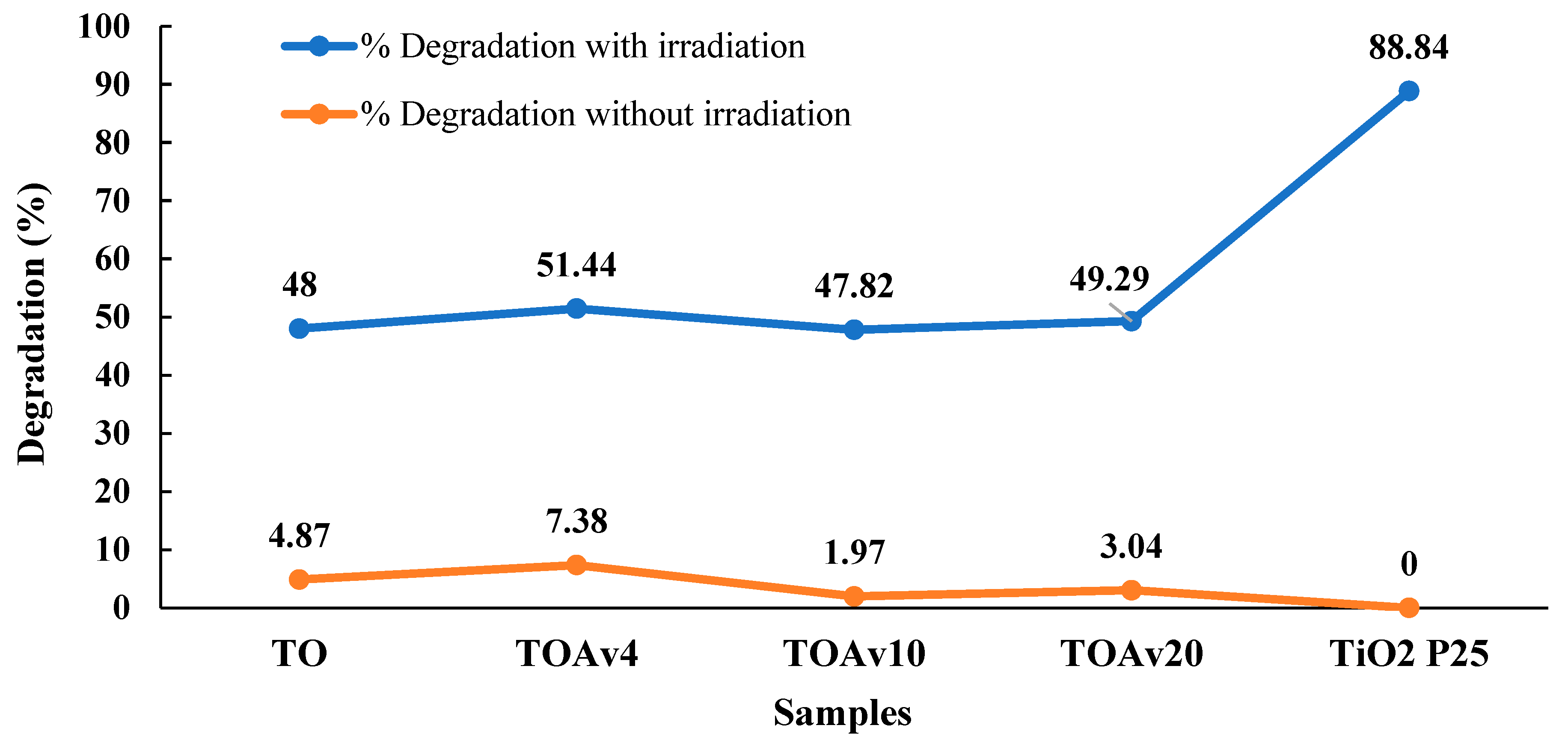
The obtained nanoparticles exhibited the anatase phase with minor NaCl impurities and UV absorption between 326–330 nm. FTIR and EDS analyses confirmed the presence of Ti–O bonds and functional bioactive compounds from the extract. Thermal analysis validated anatase formation between 400–500 °C [71,72,73,74,75]. SEM revealed morphologies ranging from spherical to rod-like, with particle sizes between 14 and 23.4 nm. The TOAv10 sample had the highest surface area, while TOAv4 demonstrated the best photocatalytic efficiency, degrading 51.44% of methylene blue, outperforming other formulations as illustrated in Figure 7 [65]. However, it is important to note that key experimental parameters such as the duration of the photodegradation process, the initial concentration of pollutant, and the amount of TiO2 nanoparticles used were not specified in the study. The absence of these critical details limits the ability to fully assess and compare the photocatalytic efficiency and reaction kinetics. This underlines the necessity of clearly reporting all relevant experimental conditions in photocatalysis research to ensure the reproducibility and scientific validity of the results.
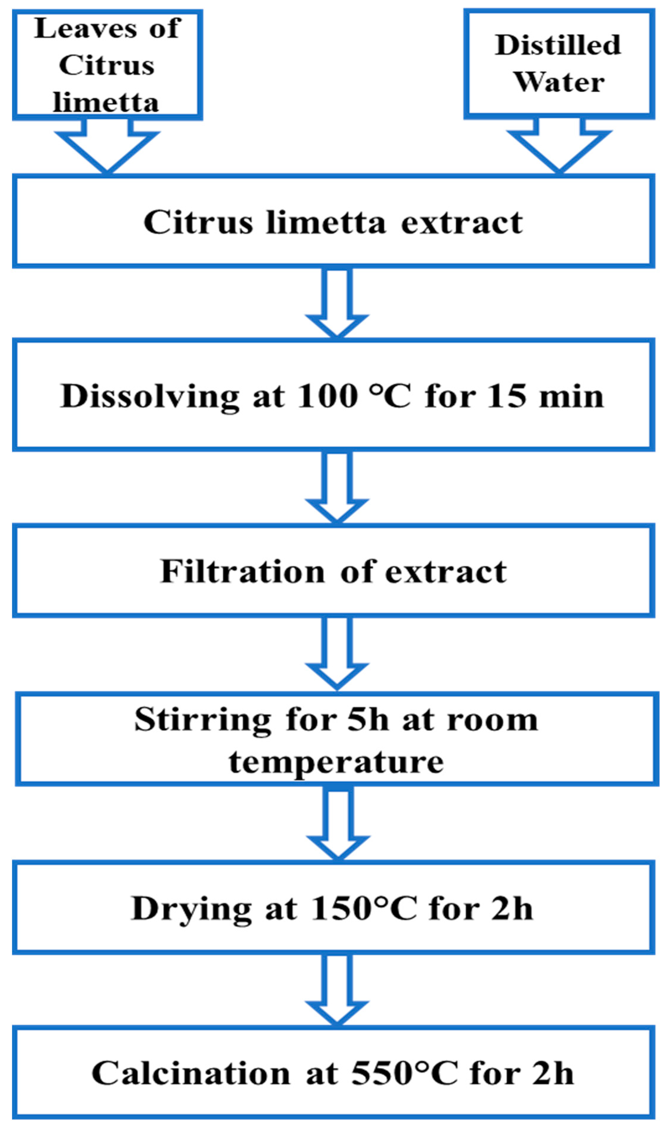
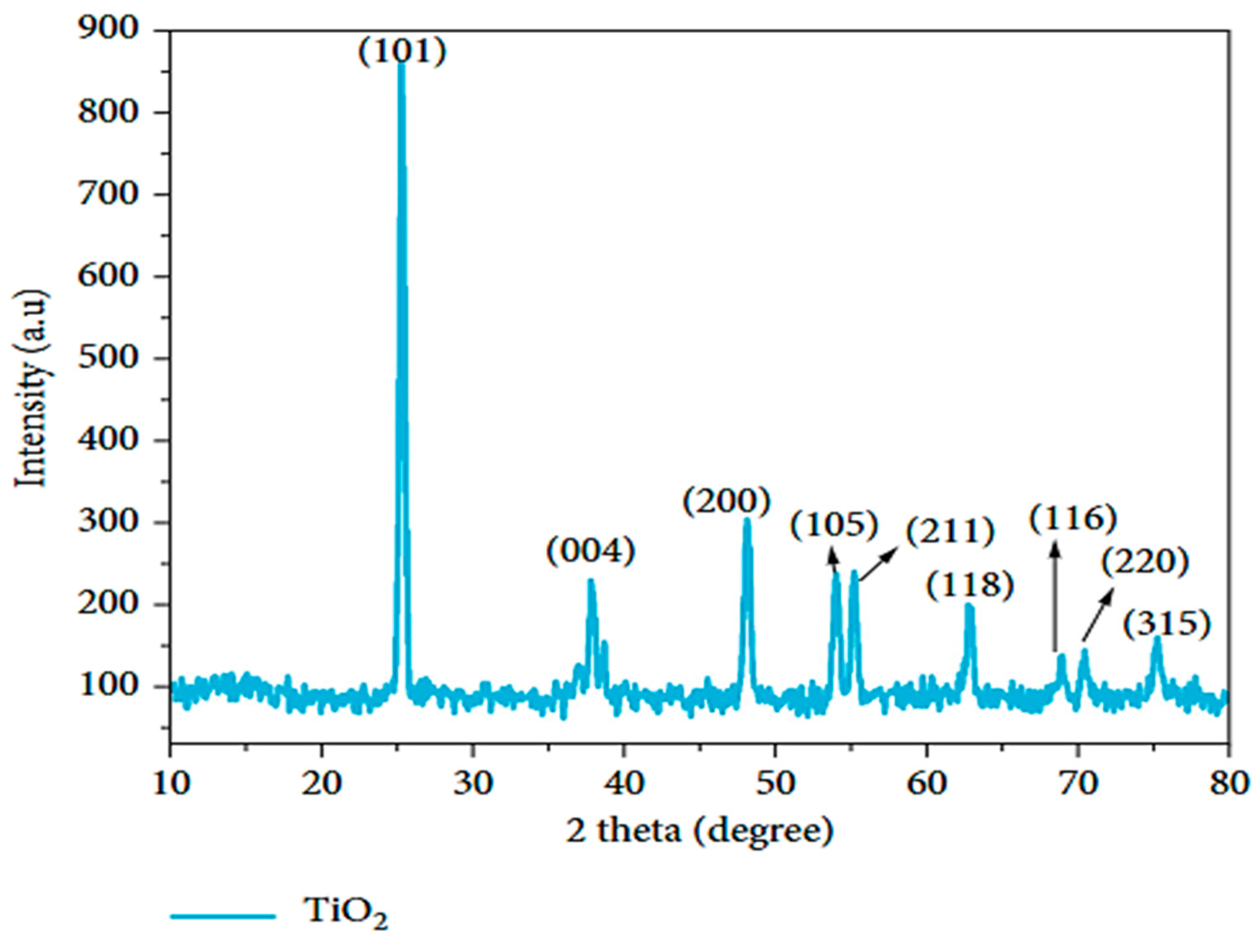
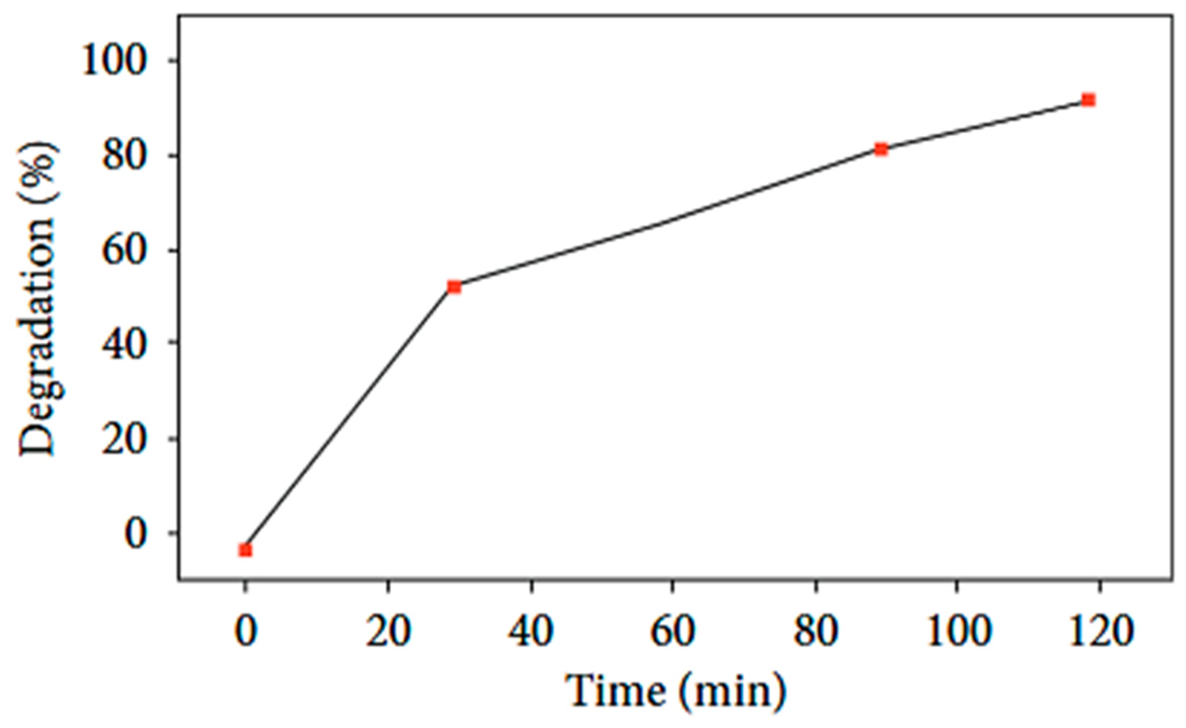
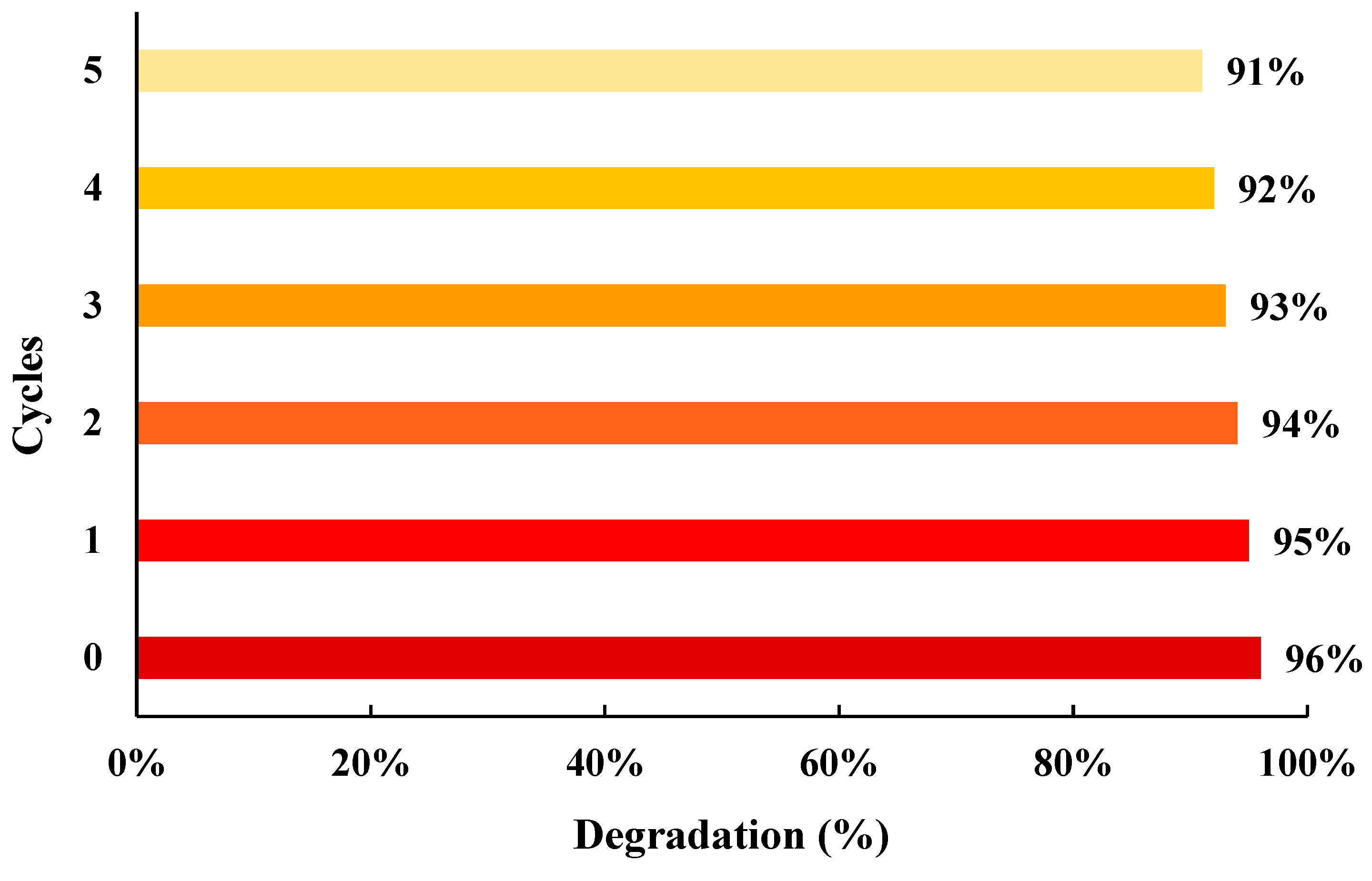
Then, in the research of Ghulam Nabi et al, lemon peel extract was used, and TiO2 NPs were obtained with spherical anatase crystals with the size 80–140 nm (Table 3). TiO2 nanoparticles were prepared by mixing 1.25 g of bulk titania, 2.5 mL of distilled water, and 35 mL of lemon peel extract. A color shift from white to yellow confirmed the reaction. The mixture was stirred for 5 h at room temperature, followed by centrifugation, triple washing, air-drying, and calcination at 500 °C for 2 h. The absorption spectrum confirmed the formation of TiO2 nanoparticles with a band gap of 3.08 eV (Table 3). These synthesized nanoparticles demonstrated over 70% photocatalytic efficiency, outperforming commercial TiO2 [76]. However, the study did not specify the initial concentration of the pollutant or the amount of TiO2 nanoparticles used during the photocatalytic tests. The lack of such critical experimental details hinders a comprehensive evaluation of the reported efficiency and limits the reproducibility and comparability of the results. Titanium dioxide nanoparticles with pure anatase phase were synthesized via a green, cost-effective method using Citrus limetta extract; the synthesis steps are illustrated in Figure 8. Characterization by XRD, SEM, EDS, and UV–Vis confirmed their successful formation with particle sizes between 80–100 nm and a band gap of 3.22 eV (Table 4). The nanoparticles demonstrated high photocatalytic efficiency, degrading over 90% of Rhodamine B within 80 min. This performance is attributed to their small size, clean surface, and high surface-to-volume ratio, highlighting their potential for eco-friendly wastewater treatment applications [77]. In a study conducted by Annin K. Shimi et al., TiO2 nanoparticles were synthesized by using Mulberry plant extract as a natural reducing agent. A 1 M solution of titanium isopropoxide (TTIP) was mixed with 10 mL of Mulberry extract and stirred at 80 °C and 800 rpm. A milky white precipitate formed within 30 min, indicating the formation of nanophase materials. The precipitate was then centrifuged at 15,000 rpm for 10 min, with the process repeated three times. The resulting samples were filtered using Whatman No. 1 filter paper and subsequently dried at 100 °C for 1 h. The dried powders were stored in a desiccator for further characterization. This study highlights an effective and eco-friendly route for producing TiO2 nanoparticles. In this study, TiO2 nanoparticles were successfully synthesized using mulberry plant extract. The obtained nanoparticles exhibited a wide band gap and a pure anatase crystalline phase, confirmed by the presence of the dominant (101) peak at 25.38°, which is known for its superior photocatalytic activity, was observed by the XRD pattern exhibited in Figure 9. Under UV illumination, the TiO2 nanoparticles achieved 96% degradation of methylene blue within 120 min. The photocatalytic process followed pseudo-first-order kinetics with a rate constant of 0.02398 min−1, indicating strong catalytic efficiency Figure 10. Furthermore, the nanoparticles demonstrated good stability and reusability over five consecutive cycles Figure 11. Antibacterial tests showed that TiO2 exhibited significant antibacterial activity, particularly against the Staphylococcus aureus strain [70].
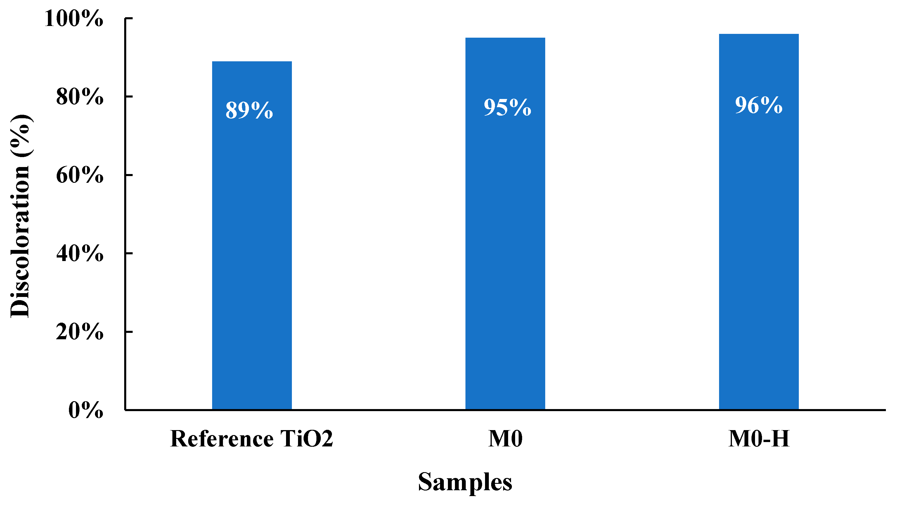
TiO2 nanoparticles were successfully synthesized using Syzygium cumini leaf extract, and their application as photocatalysts for industrial wastewater treatment resulted in significant removal of Pb2+ (82.53%) and COD (75.5%), both following first-order kinetics [78]. In a comparative study on the green synthesis of TiO2 nanoparticles, four different plant leaf extracts, Piper betel, Ocimum tenuiflorum, Moringa oleifera, and Coriandrum sativum, were employed as natural reducing and stabilizing agents. The aim was to evaluate the influence of each extract on the physicochemical properties and photocatalytic performance of the synthesized TiO2. In this green synthesis protocol, 5 mL of leaf extract (Piper betel, Ocimum tenuiflorum, Moringa oleifera, or Coriandrum sativum) was mixed with 5 mL of titanium tetraisopropoxide and 25 mL of double-distilled water. The reaction mixture was stirred continuously using a magnetic stirrer at 70 °C for 3 h. Upon cooling to room temperature (30 °C), a white precipitate was formed and subsequently separated by filtration using 0.5 mm Whatman filter paper. The obtained solid was then subjected to calcination at 400 °C for 3 h in a muffle furnace. The resulting TiO2 nanoparticles were cooled and prepared for further physicochemical characterization. Among the four-leaf extracts tested Piper betel (PB), Ocimum tenuiflorum (OT), Moringa oleifera (MO), and Coriandrum sativum (CS), Moringa oleifera extract proved to be the most effective reducing agent and demonstrated superior photocatalytic performance in the degradation of Malachite Green dye (Table 5) [79]. TiO2 anatase nanoparticles have been prepared via extraction of Mentha spicata in water, a process that combined hydrothermal activation at two different temperatures: M0 (150 °C for 8 h) and M0-H (180 °C for 12 h), followed by heating at 400 °C. The extract of the plant served as a stabilizer and a capping agent that enabled the photocatalytic and antimicrobial performances of the material to be doubled. The sample M0-H showed the highest photocatalytic activity for RB5 dye degradation, which can be attributed to its larger specific surface area, higher pore volume, better dispersion, and smaller particle size (8–12 nm). Moreover, the plant-assisted TiO2 possessed increased hydroxyl content, reduced lattice oxygen, and slightly higher band gap energies (3.22 eV for M0 and 3.25 eV for M0-H) in comparison to the reference. Such improvements in structure and surface properties cause more dye adsorption, better light absorption, as well as less charge recombination, which in turn results in higher photocatalytic efficiency (Figure 12) [80].
Table 5 presents a comparative study of TiO2 nanoparticles synthesized via green methods using various plant extracts. In addition to reporting the photocatalytic efficiency of the synthesized nanoparticles, the table provides detailed experimental conditions used during synthesis, including the type of plant extract, calcination temperature, irradiation source, photocatalysis time, pollutant type and concentration, nanoparticle dosage, and solution pH. The systematic consideration of these parameters is crucial, as they strongly influence the structural, optical, and catalytic properties of the nanoparticles, thereby determining their efficiency and applicability in wastewater treatment. The use of different plant sources not only provides environmentally friendly synthesis routes but also affects key characteristics such as particle size, surface area, crystallinity, and dye degradation efficiency. This study underscores the role of plant-mediated synthesis in tuning the functional properties of TiO2 nanoparticles for enhanced photocatalytic applications. Comparable analyses of ZnO, CuO, and Fe2O3 are also presented in this review, providing an integrated perspective for cross-material comparison and facilitating the identification of both common trends and material-specific behaviors in photocatalytic efficiency.

4.2. Zinc Oxide NPs (ZnO)

This section discusses the preparation of ZnO nanoparticles from various plants such as Beetroot, Myrtus communis, spinach leaves, and Camellia sinensis. ZnO nanoparticles were synthesized using Beetroot extract. The extract was heated to 60 °C, followed by the addition of zinc acetate. The pH was adjusted to 8, and the mixture was stirred at this temperature for 40 min until a pale-yellow solution formed. The resulting product was centrifuged, washed with double-distilled water, and dried at 80 °C. Finally, the dried powder was calcined at 450 °C for 15 min [66]. In the same study Darshan Singh synthesizes calcium-coated ZnO by the coprecipitation method Zn (CH3COO)2. 2H2O and calcium chloride. Zinc acetate was dissolved in the extract at 60 °C and pH ~8, followed by the addition of calcium chloride. The mixture was stirred for one hour, then centrifuged to isolate the nanocomposite. After washing with double-distilled water to remove impurities, the product was dried and calcined at 450 °C for 15 min in a muffle furnace [85]. The structural and morphological characteristics of zinc oxide (ZnO) and calcium-coated zinc oxide (Ca-ZnO) nanoparticles were analyzed using SEM, TEM, EDX, and XRD techniques. SEM and TEM images confirmed a uniform distribution of ZnO nanoparticles with a hexagonal shape and particle sizes ranging from 20 to 100 nm. TEM analysis indicated that calcium coating did not alter the morphology of the ZnO nanoparticles, consistent with previous findings [86]. EDX analysis verified the elemental composition, revealing the presence of zinc and oxygen in ZnO, and calcium, zinc, and oxygen in Ca-ZnO. Chlorine traces were also detected due to calcium salt contamination. XRD patterns showed that the synthesized ZnO nanoparticles crystallized in the hexagonal wurtzite phase, characterized by strong and sharp diffraction peaks. Similar patterns were observed for Ca-ZnO nanocomposites with 1:1 and 1:2 ratios, as shown in Figure 13 [66].
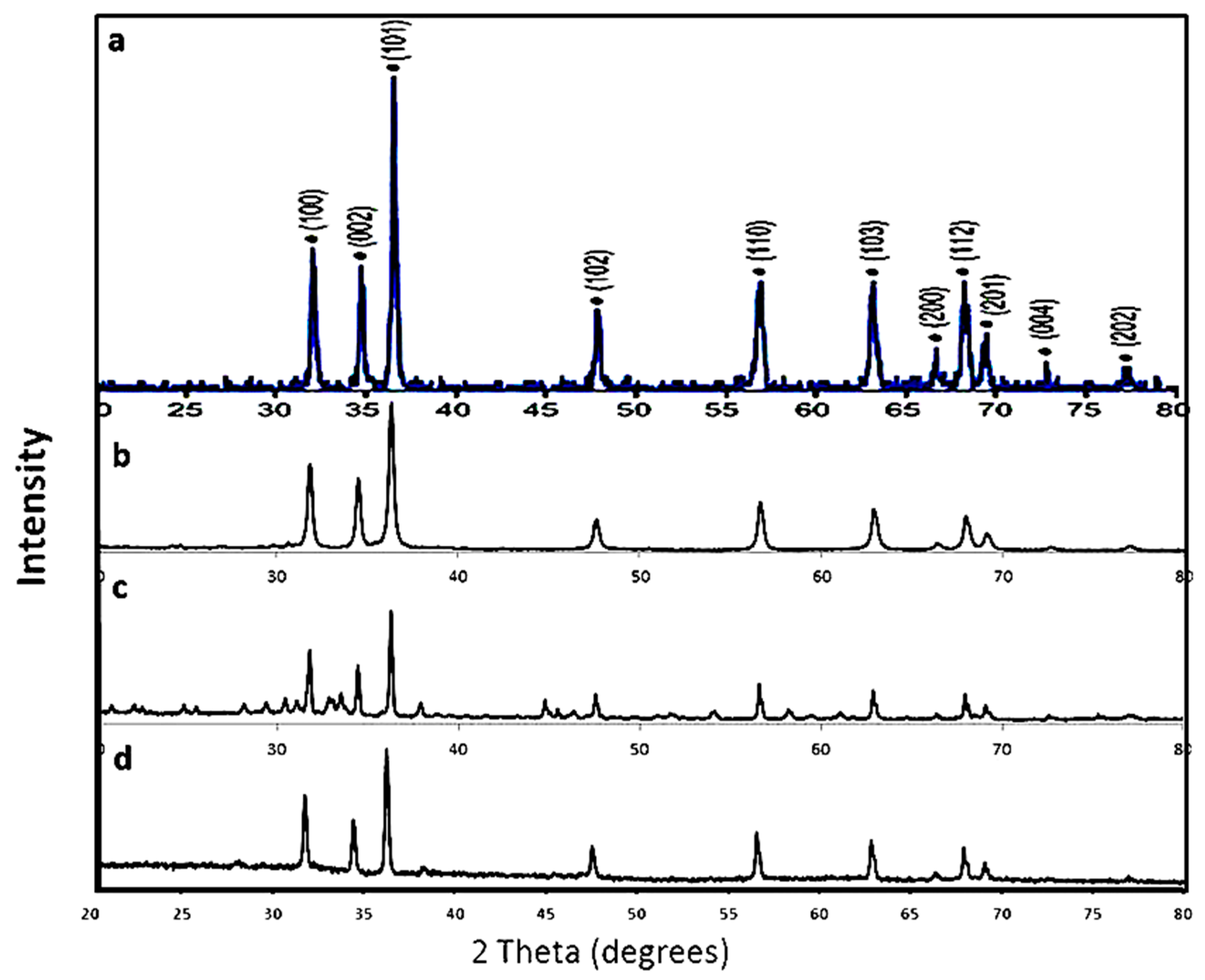
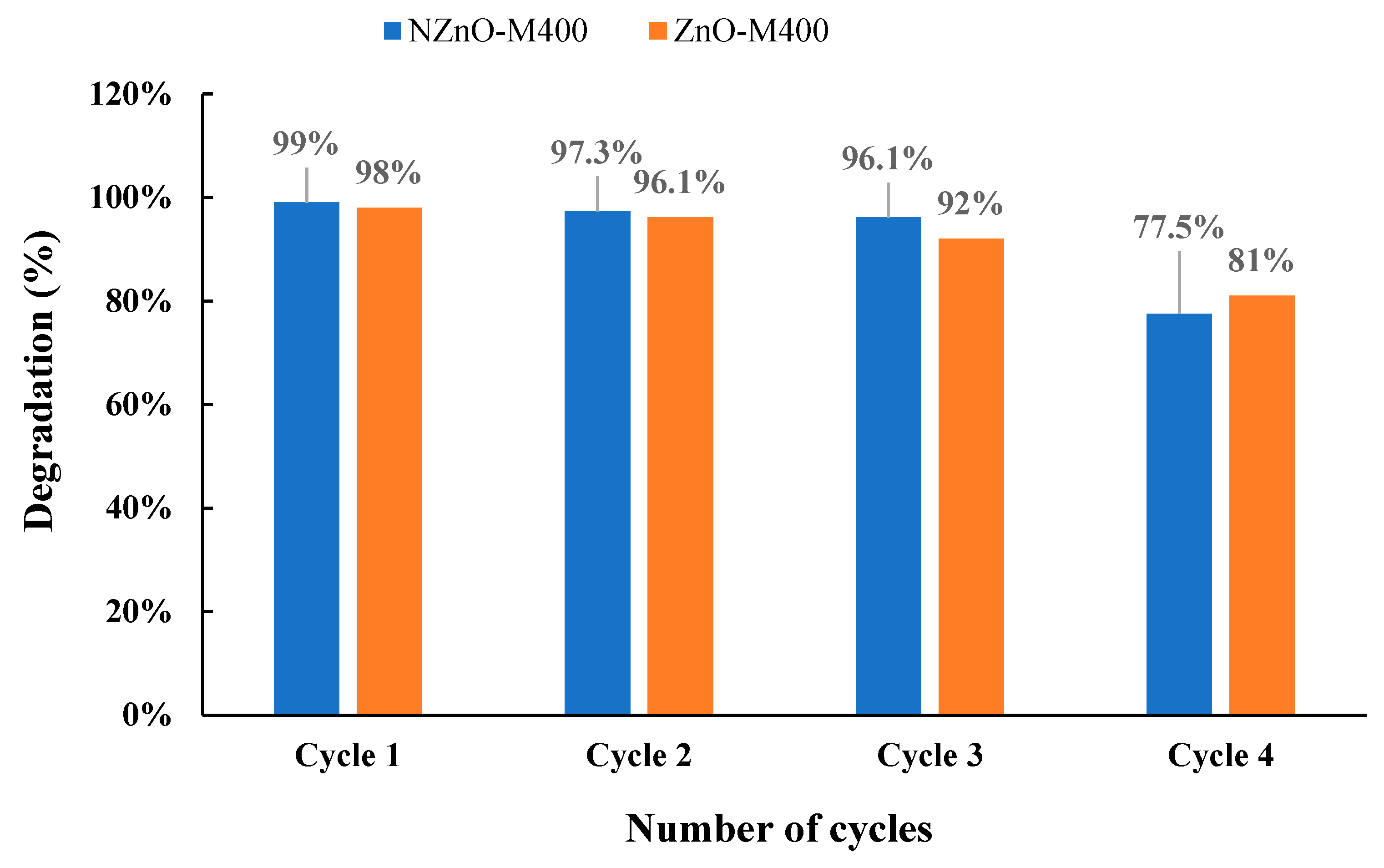
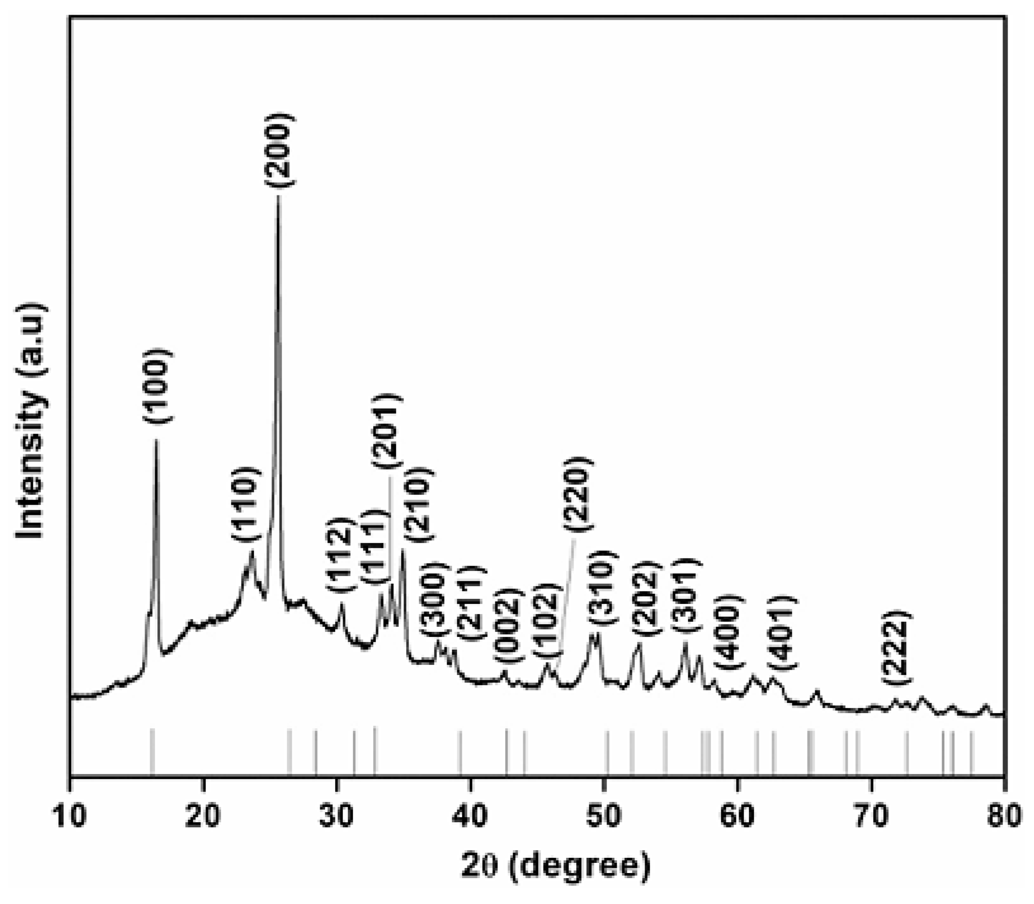
This study demonstrates the photocatalytic efficiency of both uncoated (blank) and calcium-coated ZnO nanoparticles (ratios 1:1 and 1:2) in degrading organic dyes. The uncoated ZnO nanoparticles achieved 100% degradation of methylene blue within 40 min, while the calcium-coated variants showed 80–90% degradation after 60 min. However, calcium coating significantly reduced the photocatalytic performance, particularly in the case of rhodamine B, where the degradation dropped from 55.2% (blank ZnO) to 5.4% and 3.4% for the 1:1 and 1:2 Ca-ZnO ratios, respectively. These findings suggest that while blank ZnO nanoparticles exhibit high photocatalytic activity, especially with methylene blue, the calcium-coated versions result in slower or negligible degradation. Nonetheless, both forms show potential for application in wastewater treatment targeting toxic organic pollutants [66]. A similar analysis of the photocatalytic degradation of methylene blue (MB) using ZnO nanoparticles synthesized with Vitex negundo extract showed nearly 98.50% degradation within 60 min [87]. The same type of analysis on MB dye photocatalytic degradation with ZnO-Myrtus communis NPs also resulted in nearly 94% of ZnO-Acetate and 95% of ZnO-Nitrate degradation in 30 min. Fresh Myrtus communis leaves were dried, ground, and extracted using 70% ethanol at 100 °C for 1 h. The filtered extract was then used to synthesize zinc oxide nanoparticles (ZnO NPs) by adding zinc nitrate or zinc acetate, followed by calcination at different temperatures (400, 600, and 800 °C). The resulting ZnO NPs were characterized using field-emission scanning electron microscopy (FESEM-EDX), X-ray diffraction (XRD), UV-Vis spectroscopy, photoluminescence (PL), and Fourier-transform infrared spectroscopy (FTIR). Samples made with zinc acetate were labeled ZnO-M400, ZnO-M600, and ZnO-M800, while those from zinc nitrate were named NZnO-M400, NZnO-M600, and NZnO-M800. X-ray diffraction (XRD) analysis confirmed that all synthesized ZnO nanoparticles (NPs) exhibit a hexagonal wurtzite crystal structure with a polycrystalline nature and the presence of peaks (100), (002), (101), (102), (110), (103), (200), (112), and (201) in PXRD results in dicates that ZnO nanoparticles were synthesized with high purity [88]. Fourier-transform infrared spectroscopy (FTIR) revealed the presence of active vibrational modes corresponding to functional groups such as hydroxyl (OH), carboxylate (COO), carbon dioxide (CO2), and water (H–OH), indicating the successful formation of ZnO. Ultraviolet-visible (UV–Vis) spectroscopy demonstrated that the optical bandgap values of all ZnO NPs are lower than that of bulk ZnO (Eg = 3.26), suggesting quantum size effects [89]. Photoluminescence (PL) measurements established a correlation between the morphology of the nanomaterials and their defect distribution. Field emission scanning electron microscopy (FESEM) analysis showed that the use of Myrtus communis plant extract enabled the green synthesis of ZnO NPs with well-defined hexagonal structures, including spherical, pyramidal, cylindrical, and rod-like shapes, as well as a variety of particle sizes. The morphology of the synthesized samples was significantly influenced by the choice of precursor and calcination temperature, leading to variations in the hexagonal forms. This study concludes that the size and morphology of green-synthesized ZnO powders are strongly dependent on both the precursor type and the calcination temperature. Notably, smaller particle sizes are associated with larger surface areas, which in turn contribute to enhanced photocatalytic performance, confirming that ZnO NPs preserved their high photocatalytic activity up to four cycles, as supported by numerous previous studies (Figure 14) [88].
ZnO nanoparticles were synthesized via another green method using Spinacia oleracea leaf extract by Velmurugan and al. The process involved mixing the plant extract with zinc acetate and ammonium solution, followed by heating at 90 °C for 3 h. After cooling and filtration, the precipitate was washed with ethanol and deionized water, then dried at 130 °C and calcined at 300 °C [90]. XRD analysis confirmed that the synthesized ZnO nanoparticles possess a hexagonal Wurtzite structure with an average crystallite size of 37.6 nm, which was further supported by SEM and TEM observations. UV–Vis spectroscopy revealed a surface plasmon resonance (SPR) range between 320 and 390 nm, and the Tauc plot indicated a direct bandgap energy of approximately 3.12 eV (Table 6).
FTIR spectra identified characteristic functional groups from biomolecules involved in the synthesis process, with a distinct absorption band at 505 cm−1, verifying the formation of ZnO nanoparticles. Biosynthesized ZnO nanoparticles demonstrated effective photocatalytic activity under simulated sunlight, achieving significant degradation of organic pollutants in gasoline refinery effluent, particularly toluene (84.26%) and xylene (90.36%) at pH 8 after 240 min of exposure (Table 7). In treating paper mill effluents, successive photocatalytic cycles led to a progressive decrease in COD levels, reaching 81.24%, 74.14%, 68.92%, 60.24%, and 53.82%, respectively (Figure 15). Moreover, radical scavenging assays confirmed the strong antioxidant potential of the ZnO NPs, especially under high-concentration conditions [91].

4.3. Tungsten Oxide (WO3)

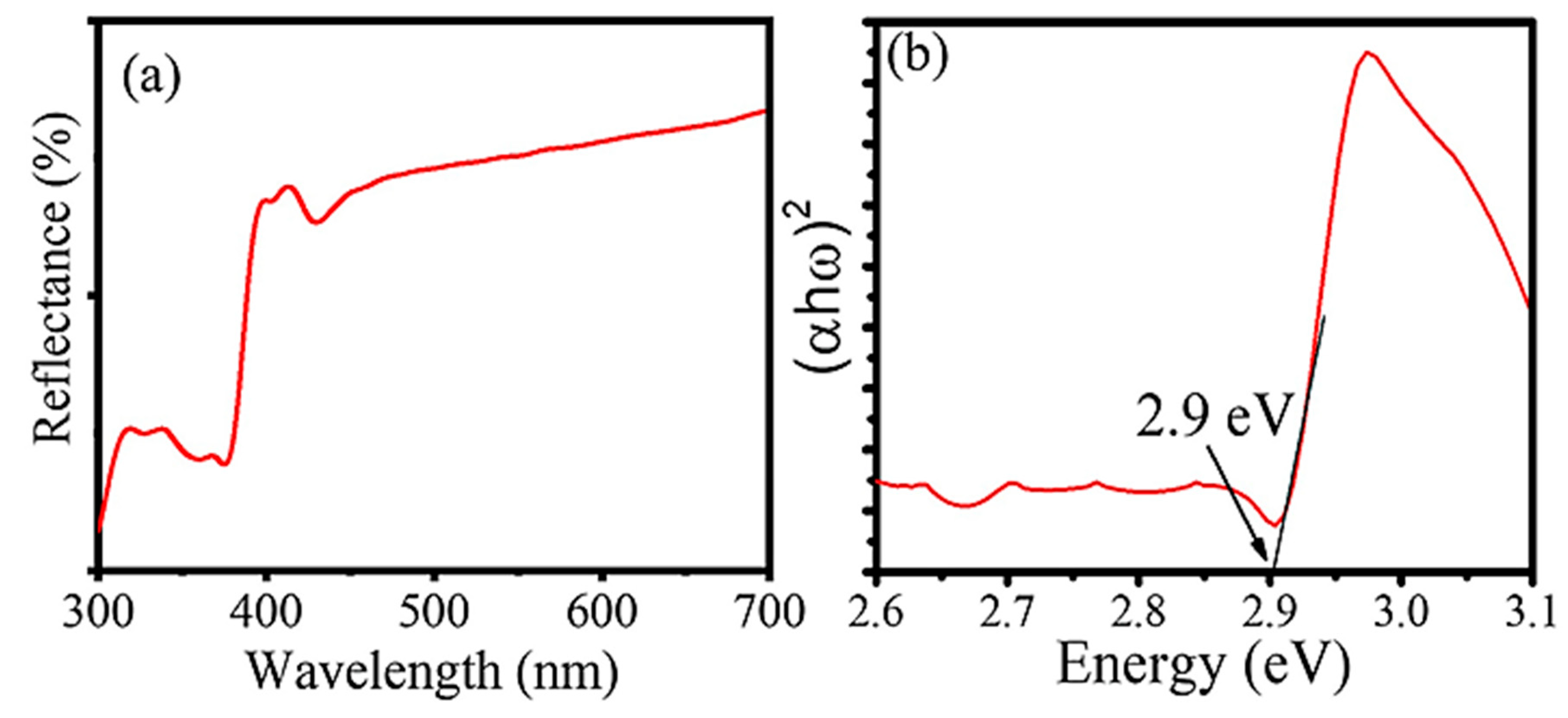
WO3 nanoparticles were green-synthesized by Fadhila Anggraini et al. using Beetroot extract and a 0.1 M Na2WO4·2H2O solution. The influence of pH (4, 7, and 11) was assessed using a phosphate buffer. The extract and precursor were mixed at a 5:1 volume ratio, stirred for 6 h, and aged overnight. The obtained precipitate was dried at 80 °C and sintered at 250 °C or 550 °C for 2 h in air, yielding WO3 nanoparticles. WO3 nanoparticles were successfully synthesized using Beetroot extract. The optimal conditions were found at pH 4 and a sintering temperature of 550 °C. Characterization by XRD, SEM-EDX, and XPS confirmed the formation of a single-phase WO3 with a crystallite size of 26.4 nm and an average particle size of 24 nm. The reflectance spectrum (Figure 16a) depicts the characteristic edge wavelength at around 398 nm. The synthesized nanoparticles exhibited a band gap of 2.9 eV as shown in Figure 16b. Under both UV and visible light, the WO3 NPs showed remarkable photocatalytic activity, achieving 99.67% degradation of Rhodamine B within 30 min of UV exposure. Additionally, the photocatalyst maintained high stability and reusability over five consecutive cycles [98].
In another study, WO3 nanoparticles were successfully synthesized through a green chemistry approach using Hyphaene Thebaica extract as a natural reducing agent. Sodium tungstate was dissolved in the extract, stirred for 24 h, then centrifuged, washed, and dried at 80 °C. The dried product was partially characterized, while the rest was annealed at 400 °C for 4 h to complete the formation of WO3 nanostructures.
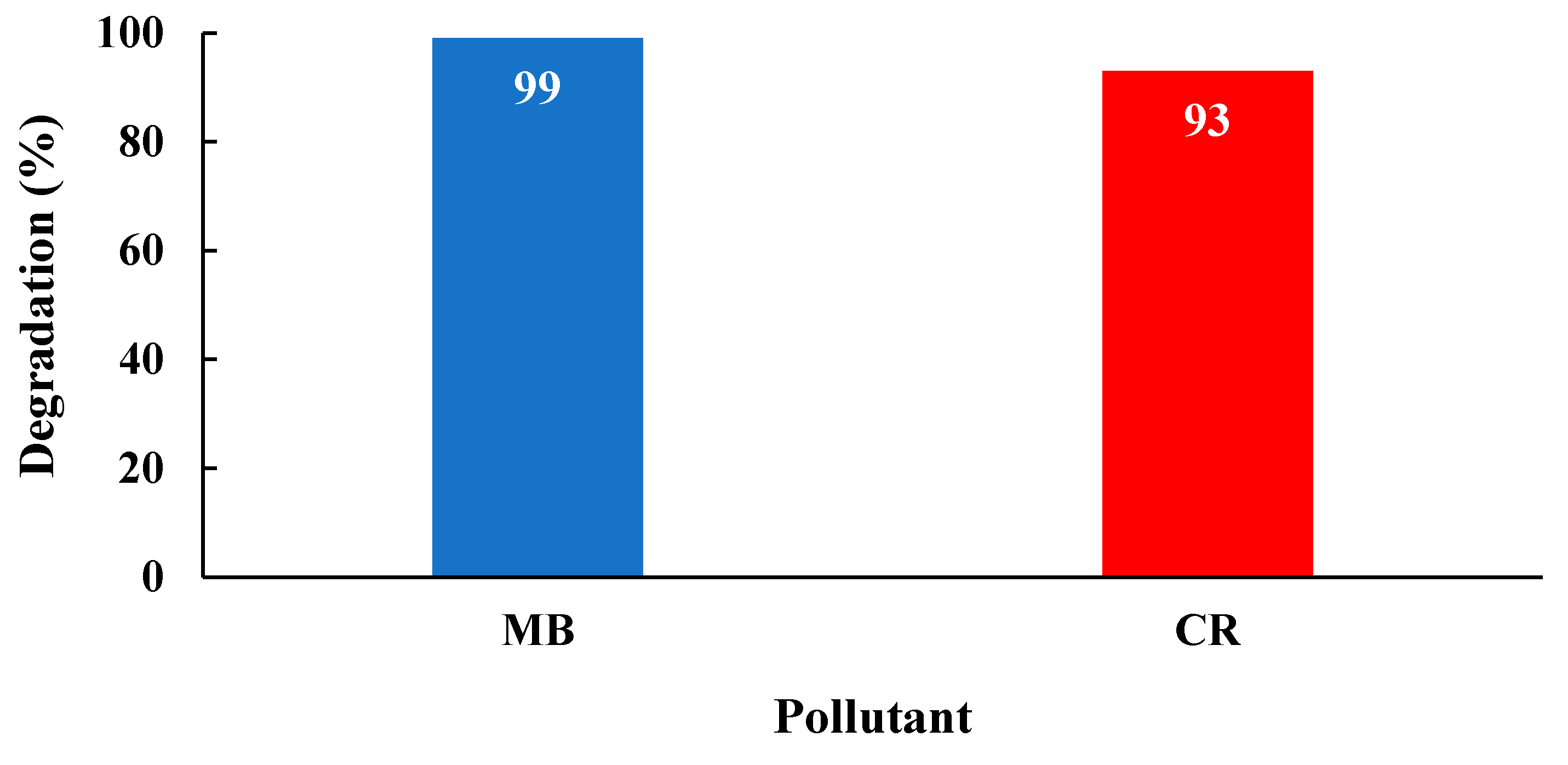
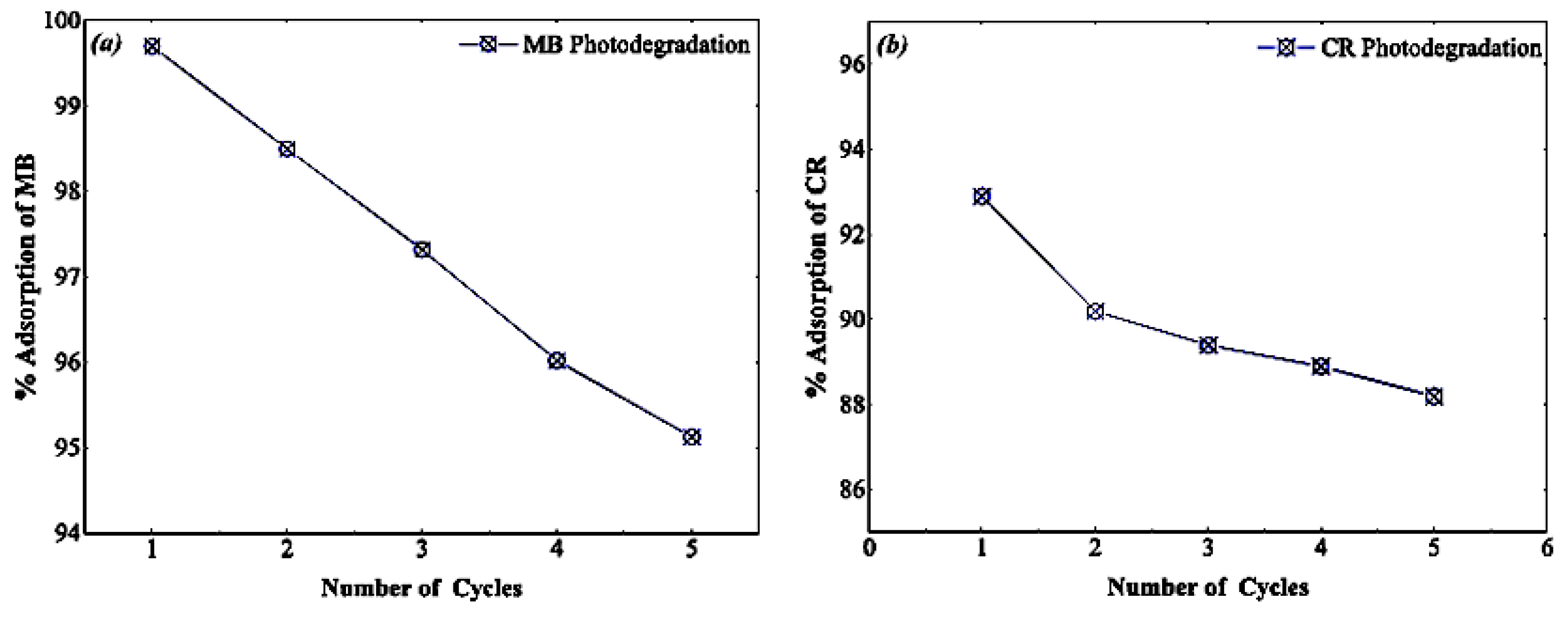
The nanoflakes achieved removal efficiencies of 93% for Congo Red and over 98% for Methylene Blue, underscoring their strong potential for wastewater treatment applications (Figure 17). FTIR analysis confirmed the structural evolution of WO3 nanostructures upon annealing [99]. The as-synthesized WO3 exhibited characteristic bands for adsorbed water (3000–3700 cm−1, 1610 cm−1) and W=O stretching (938 cm−1). A broad band between 400–1000 cm−1 was attributed to W–O vibrations. After annealing at 400 °C, hydroxyl-related bands diminished due to dehydration, and the 938 cm−1 peak merged into a broader 400–1200 cm−1 band, indicating the formation of a well-structured O–W–O lattice in crystalline WO3 [100]. The material exhibited excellent recyclability and structural stability over multiple cycles. Its notable adsorption capacity toward both dyes further supports its application in pollutant removal (Figure 18) [99].
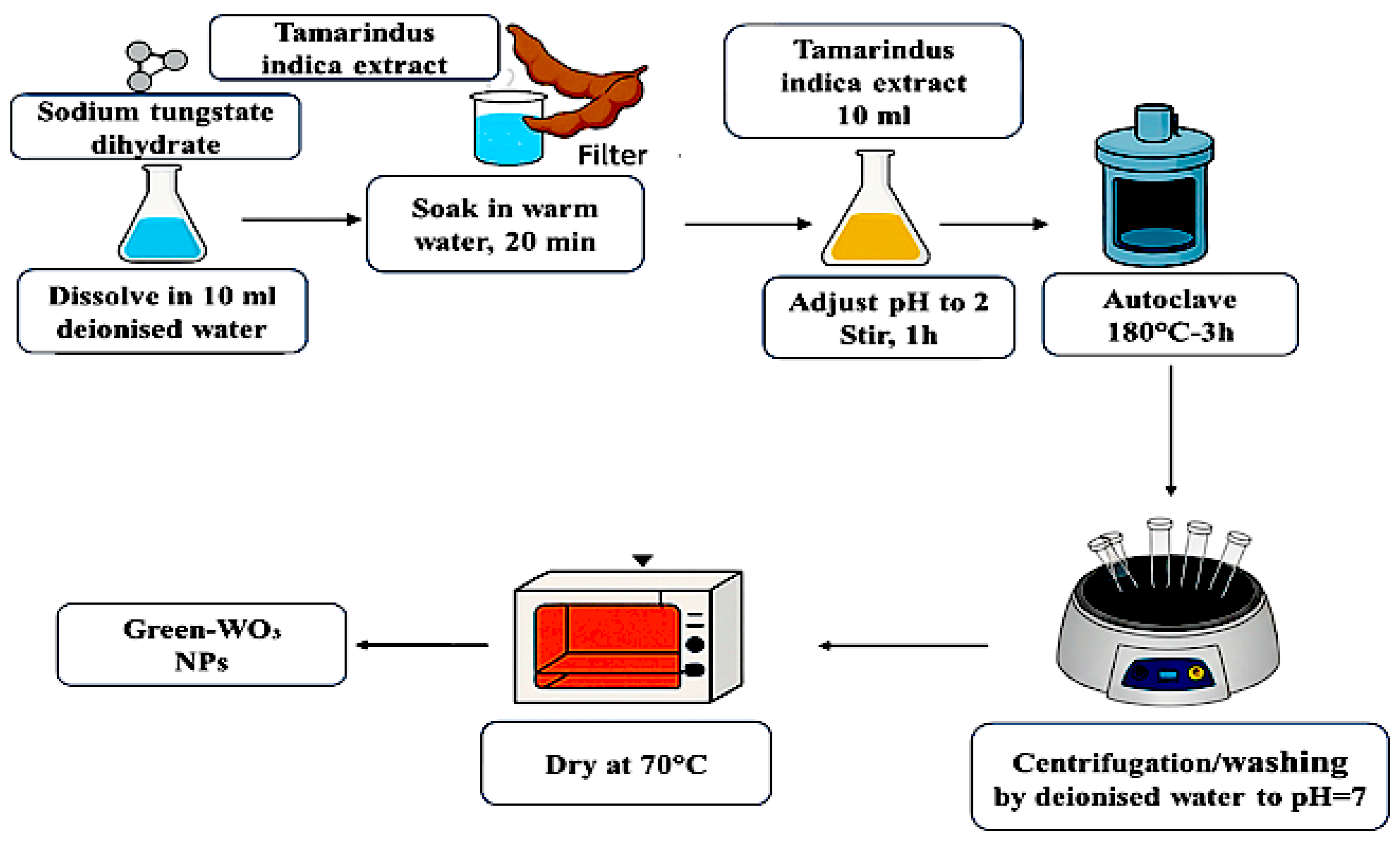
In new research, Jerold Antony tungsten oxide (WO3) nanoparticles were synthesized via a green hydrothermal route using Tamarindus indica extract as a natural reducing and stabilizing agent. The reaction mixture was treated at 180 °C for 3 h, and the product was purified and dried. This method provides an eco-friendly and efficient alternative for WO3 nanoparticle synthesis [64,101]. The experimental procedure for the green synthesis of WO3 nanoparticles is provided in Figure 19.
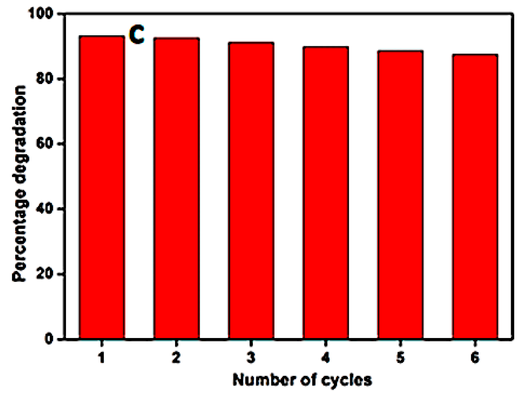
Hexagonal WO3 nanoparticles synthesized via a green method using Tamarindus indica extract demonstrated high photocatalytic efficiency for degrading the antibiotic ofloxacin in wastewater, green WO3 was tested for reusability over six photocatalytic cycles after thorough washing with deionised water, and the final degradation efficiency was recorded (Figure 20) Photodegradation followed pseudo-first-order kinetics, achieving significant removal within 150 min, with hydroxyl radicals playing a key role. The material exhibited a surface area of 19.112 m2·g−1, a pore diameter of 0.09 nm, and a band gap of 2.41 eV. XPS analysis confirmed the presence of tungsten (W) and oxygen (O) without impurities. The O1s peak at 530.09 eV corresponds to W–O bonding (Figure 21). This eco-friendly approach offers a promising and sustainable alternative for pharmaceutical pollutant remediation in wastewater [102].

4.4. Copper Oxide NPs (CuO/Cu2O)

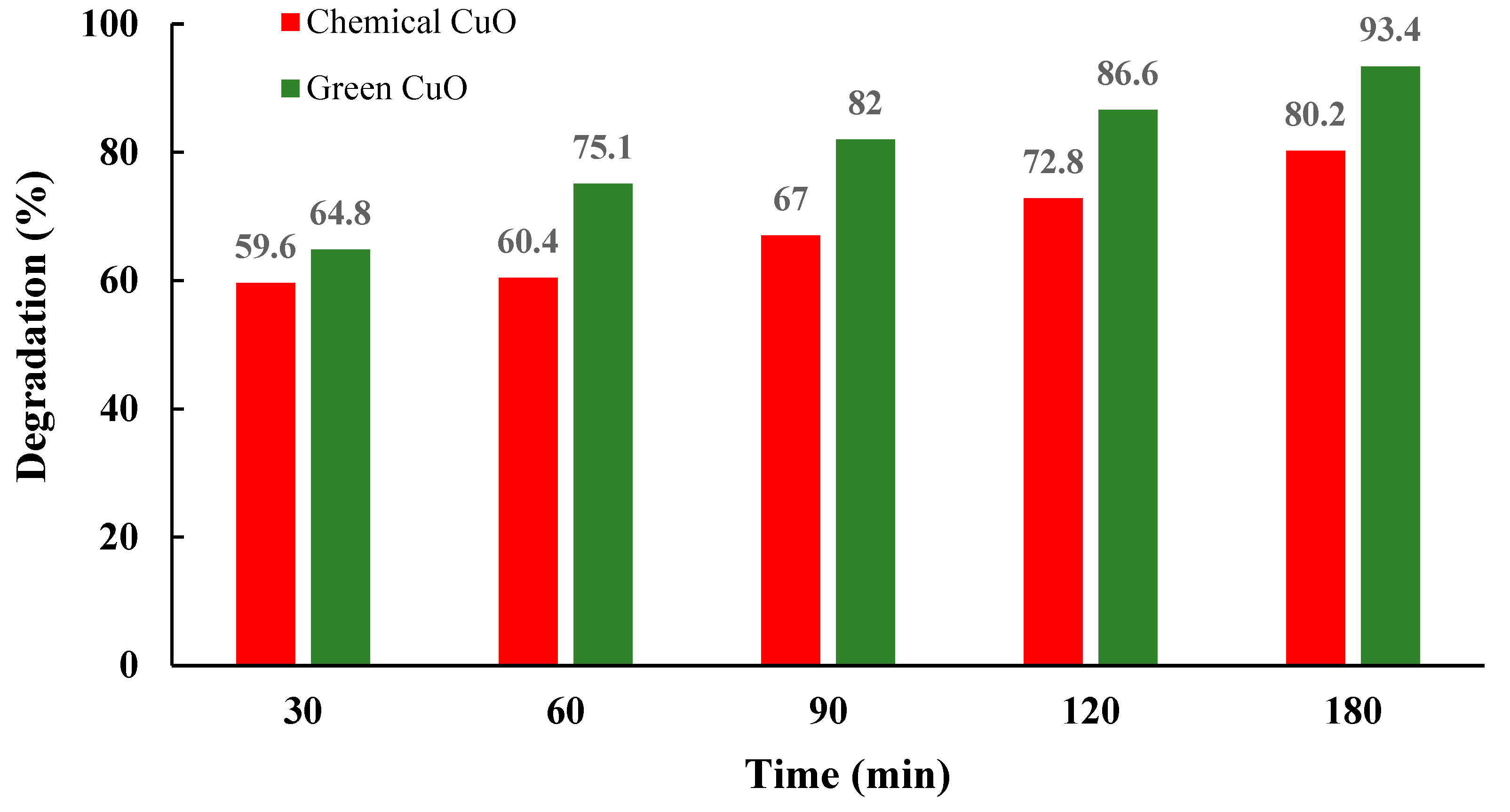
Copper oxide (CuO) nanoparticles were synthesized using a green method based on Euphorbia alata extract. The aerial parts of the plant were dried, ground, and extracted by infusion using distilled water for one week. The resulting extract was heated to 90 °C, and a solution of CuSO4·5H2O was added. The mixture was stirred for 2 h, centrifuged, washed, and then dried at 40 °C. Finally, the product was calcined at 400 °C for 4 h to obtain CuO nanoparticles. Following a green synthesis method, a conventional chemical hydrothermal process was also used to prepare CuO nanoparticles. CuSO4·5H2O was dissolved in deionized water, and KOH was added dropwise until pH 10 was reached. The mixture was stirred at 90 °C for 2 h, forming a dark precipitate, which was then centrifuged, washed, dried at 80 °C, and finally calcined at 400 °C for 4 h to yield CuO nanoparticles [67]. CuO nanoparticles exhibited a monoclinic structure with sizes between 10–16 nm. FTIR confirmed the presence of phenolic and flavonoid groups on biosynthesized CuO-NPs (Figure 22). UV-Vis analysis showed band gap energies of 1.77 eV (chemical) and 1.92 eV (green). Morphological analysis revealed spherical shapes for chemically synthesized CuO-NPs and octahedral clusters for green ones. The green CuO-NPs showed enhanced photocatalytic, antibacterial, and antifungal activities, particularly against S. aureus, B. subtilis, C. albicans, and S. cerevisiae. The E. alata-based synthesis offers high purity, simplicity, low cost, and eco-friendliness, making it suitable for water remediation and biomedical use. The histogram (Figure 23) illustrates that the photocatalytic degradation efficiency of methylene blue (MB) diminishes as the dye concentration increases under sunlight irradiation. Moreover, the biosynthesized CuO-NPs exhibited higher degradation efficiency compared to the chemically synthesized ones [67].
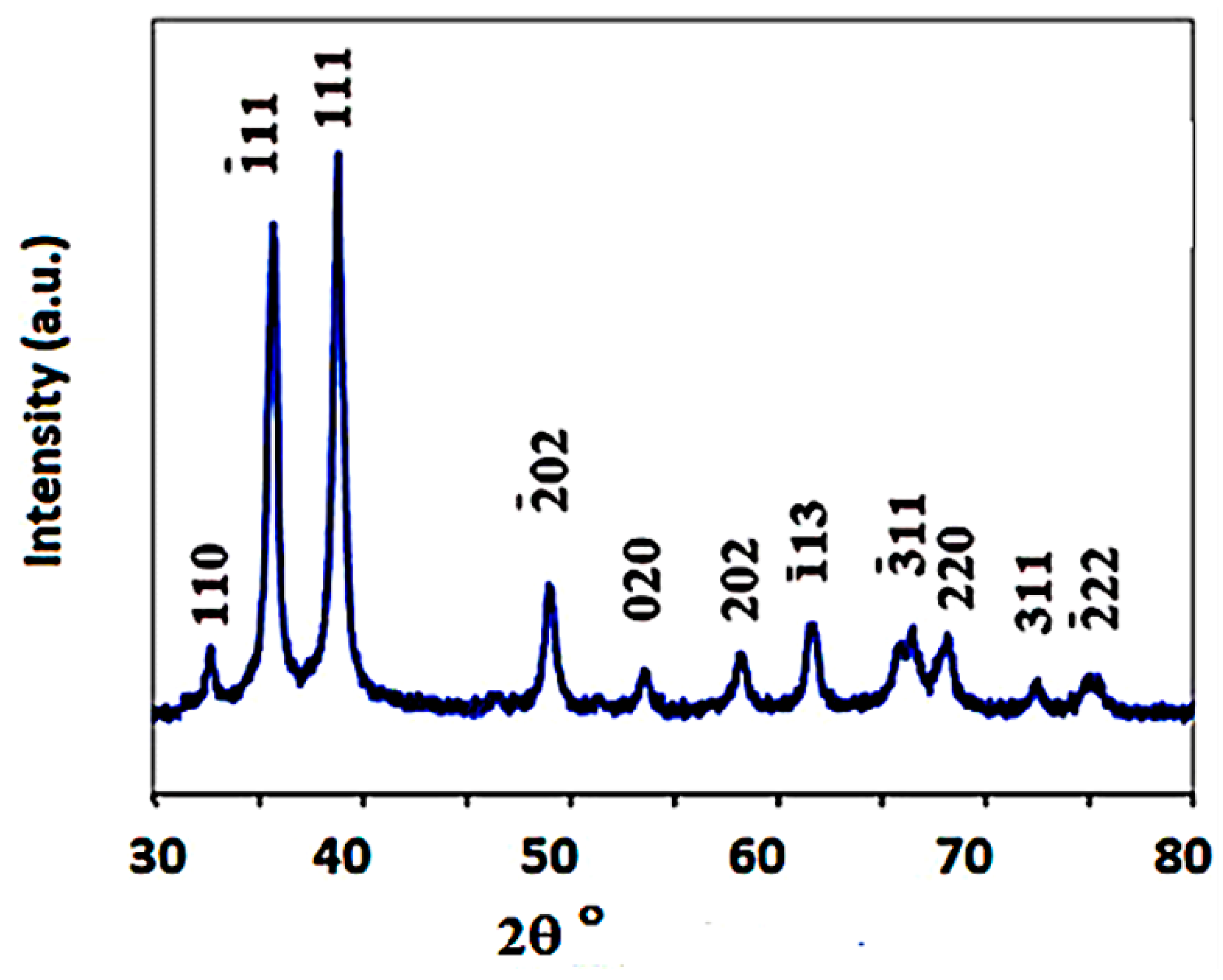
In a continuation of green synthesis approaches, Al-Faouri et al. employed Bougainvillea plant extract as a natural reducing and stabilizing agent for the preparation of copper oxide nanoparticles (CuO-NPs). Bougainvillea leaves were thoroughly washed with tap and deionized water, then air-dried in a shaded, well-ventilated area at room temperature for 10 days. Once fully dried, the leaves were ground into a fine powder using a blender and sieved through a 100 μm mesh to eliminate coarse particles [68]. A total of 10 g of dried Bougainvillea leaf powder was added to 100 mL of deionized water and heated at 80 °C with constant stirring for 20 min, producing a greenish solution. After standing at room temperature for 24 h, the extract was filtered using Whatman No. 1 paper to remove solid residues, yielding a deep honey-colored aqueous extract. This extract was analyzed by FT-IR to identify functional groups and was stored at 277 K for use as a natural reducing and capping agent in nanoparticle synthesis. A 0.14 M solution of cupric acetate (50 g in 1000 mL deionized water) was prepared and stirred, then 100 mL of Bougainvillea extract was added dropwise under continuous stirring. The color transition from blue to emerald green indicated nanoparticle formation. After 4 h of stirring and 48 h of settling, the precipitate was separated via filtration, washed with deionized water and ethanol, and dried at 80 °C for 4 h. The final calcination at 400 °C for 2 h yielded CuO nanoparticles as a fine black powder, which was cooled, ground, and stored for further analysis. Copper oxide nanoparticles (CuO-NPs) synthesized via a green method using Bougainvillea leaf extract demonstrate effective reduction and stabilization through phytochemicals acting as capping agents. These nanoparticles show strong UV absorption, a band gap of 2.74 eV, and favorable optical properties, including notable refractive index and optical conductivity. Structural analyses (XRD) confirm a crystalline monoclinic phase (Figure 24), while SEM and TEM reveal a nearly spherical morphology with minimal agglomeration. FT-IR analysis further confirms the involvement of bioactive compounds in nanoparticle stabilization and formation [68].
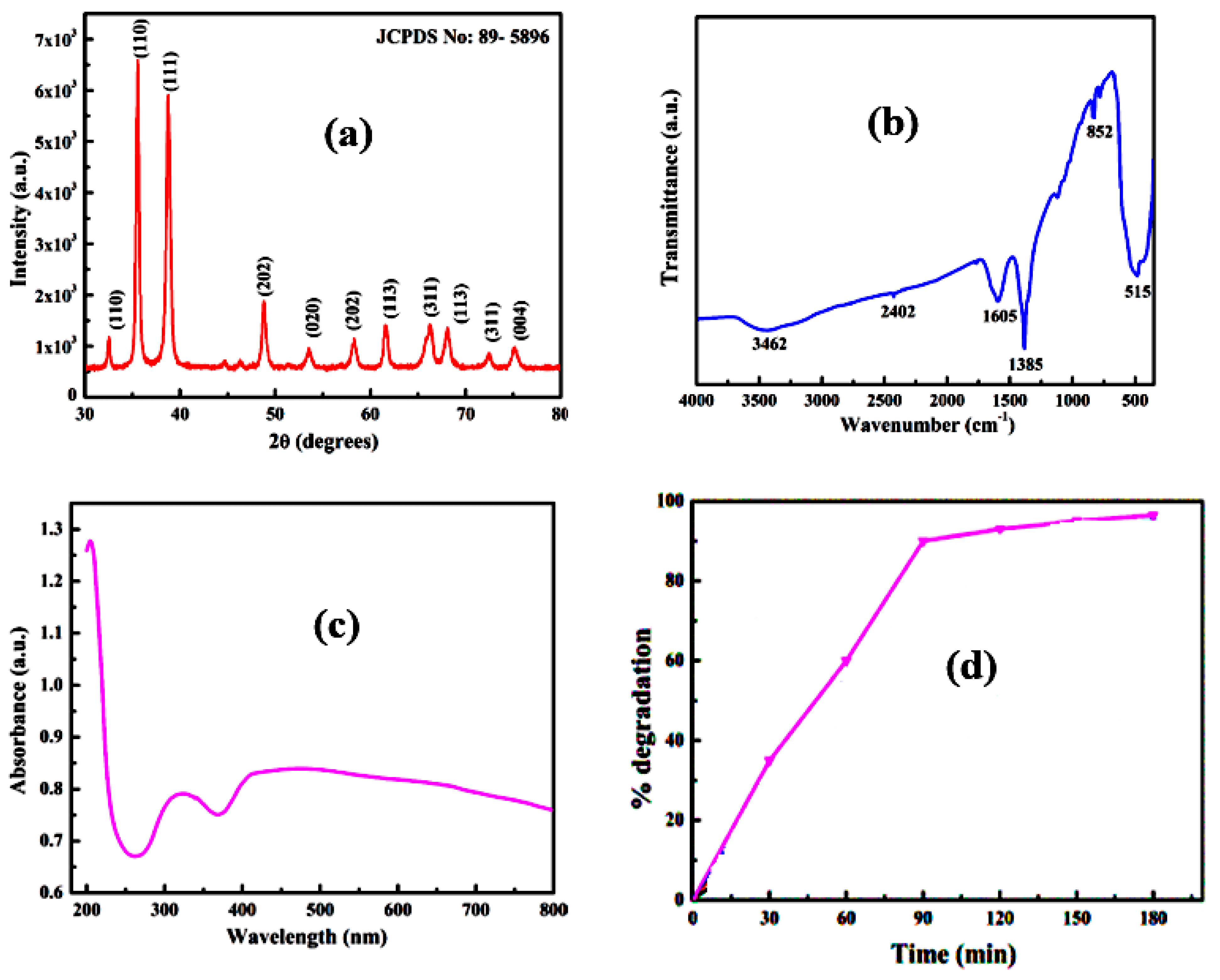
The thorough analysis of the optical and structural characteristics of the synthesized CuO-NPs provides valuable insights into their light–matter interactions and supports their potential deployment across a wide spectrum of practical applications [68]. However, the photocatalytic performance of these nanoparticles remains to be validated through experimental degradation studies, which represents a notable limitation of the current work. In another study conducted by Rupashree et al., copper oxide nanoparticles (CuO-NPs) were synthesized via a green method using cupric nitrate trihydrate and green tea extract in a 1:1 ratio. The components were dissolved in distilled water, stirred magnetically for homogenization, and heated at 120 °C to form a gel. The gel was then calcined at 380 °C for 10 min in a muffle furnace. After cooling to room temperature, phase-pure CuO-NPs were obtained. Copper oxide nanoparticles (CuO-NPs) were successfully synthesized via an environmentally friendly green method using green tea extract as a fuel. Their structural, compositional, and morphological properties were characterized by XRD (Figure 25a), FTIR (Figure 25b), UV-Vis spectroscopy (Figure 25c), EDX, SEM, and TEM. The CuO-NPs demonstrated effective catalytic degradation of methylene blue dye in aqueous solution, achieving complete degradation of a 5 ppm dye concentration within 180 min using 200 mg of catalyst at pH 9. Photocatalytic degradation reached 92% under sunlight and 99% under UV light (Table 10). Figure 25d shows the degradation efficiency of methylene blue (MB) over time using 200 mg of CuO nanoparticles, which represents the optimal mass for maximum degradation. The photocatalytic mechanism was also thoroughly discussed. These features highlight the potential of the fabricated sensor for reliable industrial applications [103].
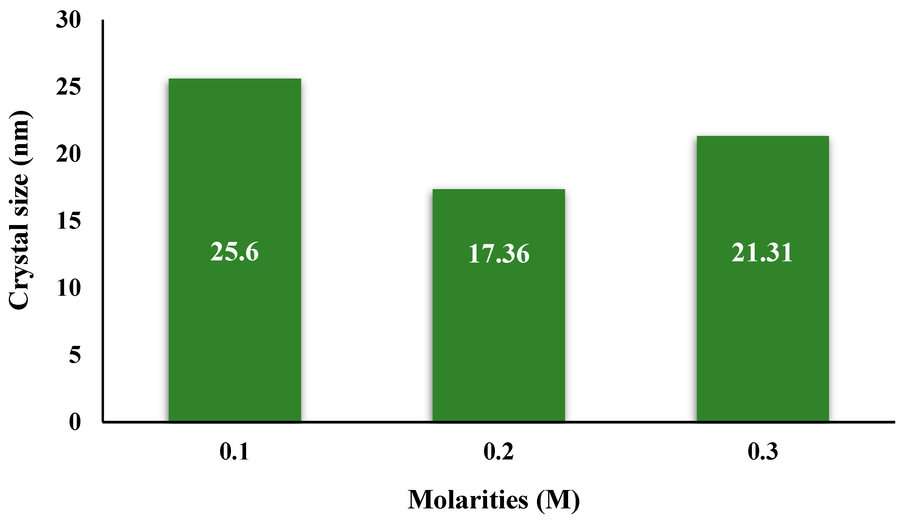
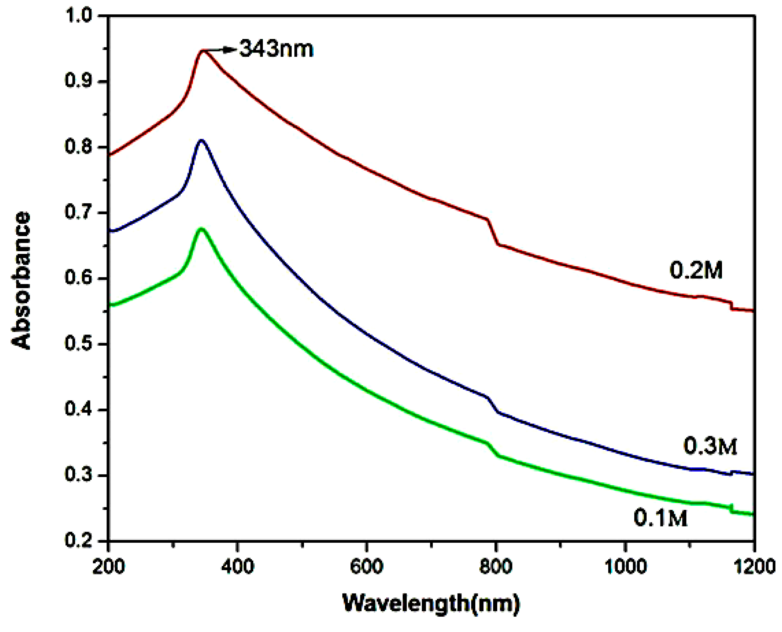
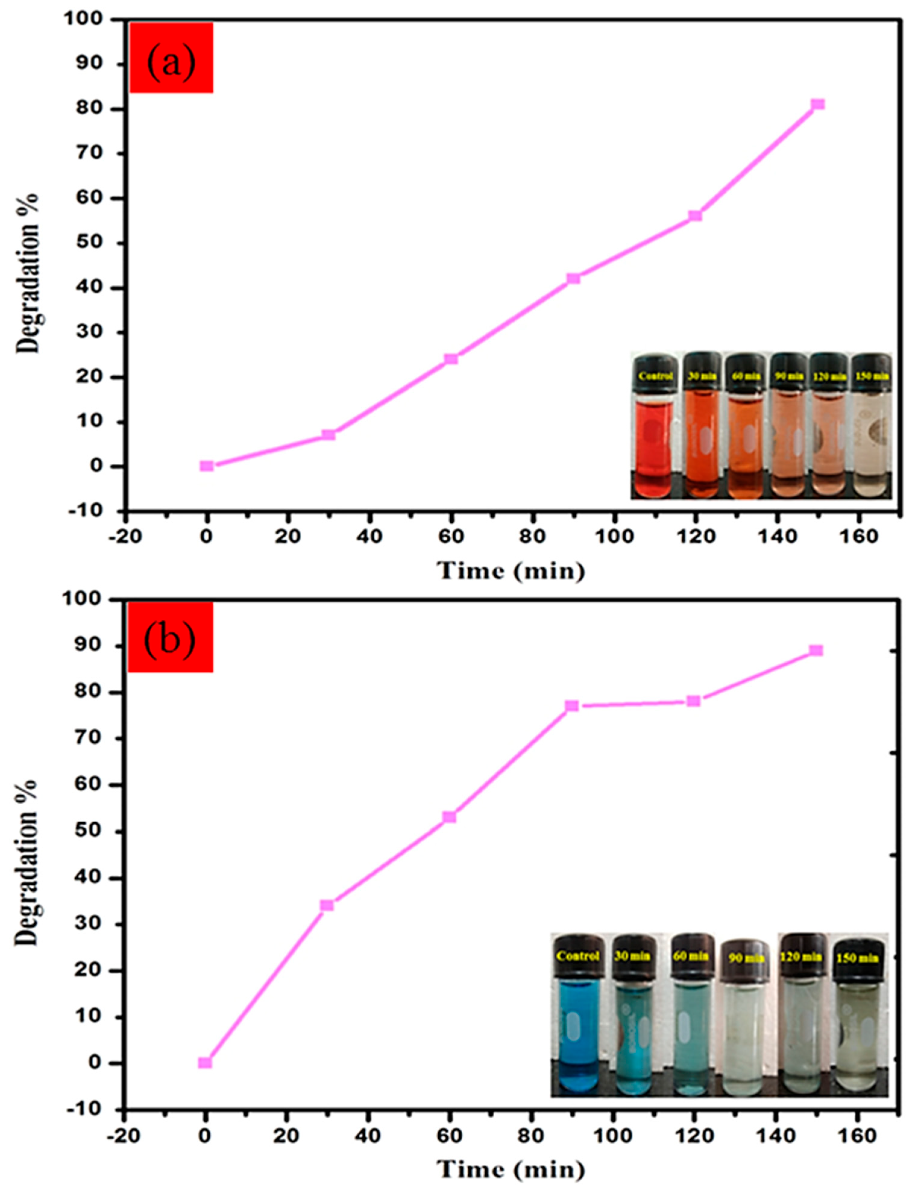
CuO nanopowders were synthesized via a green sol–gel method using copper (II) chloride dihydrate, sodium hydroxide, and Lantana camara leaf extract. A 0.1 M precursor solution was mixed, heated, and combined with plant extract, followed by the dropwise addition of NaOH, leading to a rapid formation of CuO precipitate. The product was washed, dried at 100 °C, and calcined at 500 °C. The synthesis was also carried out at higher precursor concentrations (0.2 M and 0.3 M) to study the effect on nanoparticle formation [104]. XRD pattern shows the two prominent peaks of 2θ and the values 35.5O and 38.7O are assigned to (−111) and (111), which is the evidence of CuO nanoparticles, and revealed that CuO nanoparticles synthesized at 0.2 M molarity exhibited the smallest crystallite size (17 nm) compared to other concentrations (Figure 26) [105]. Optical studies indicated a strong absorption edge and a blue shift at 343 nm, confirming the presence of a distinct optical band gap, is shown in Figure 27 [106]. SEM analysis showed morphological transformation to seed-like spherical particles. FTIR confirmed the presence of functional groups associated with CuO such as peaks at 597,507 and 431 cm−1 corresponded to the CuO deformation vibration. The biosynthesized CuO-NPs demonstrated significant antibacterial activity against four bacterial strains and excellent photocatalytic efficiency, achieving 94.07% degradation of methylene blue within 90 min (Figure 28). The photocatalytic degradation mechanism of MB by CuO nanostructures begins with dye adsorption onto the nanoparticle surface. Upon sunlight exposure, CuO generates electron–hole (e/h+) pairs, which interact with molecular oxygen and hydroxyl groups to form reactive oxygen species (O2 and OH) [107]. These radicals are responsible for breaking down the dye molecules into smaller intermediates, leading to effective degradation [108]. Overall, CuO-NPs synthesized at 0.2 M via green synthesis showed superior properties in terms of size, morphology, and functional performance, particularly in antibacterial and photocatalytic applications. The work offers valuable insights into the synthesis and activity of CuO nanocomposites; however, the absence of reusability studies limits the assessment of its practical potential.
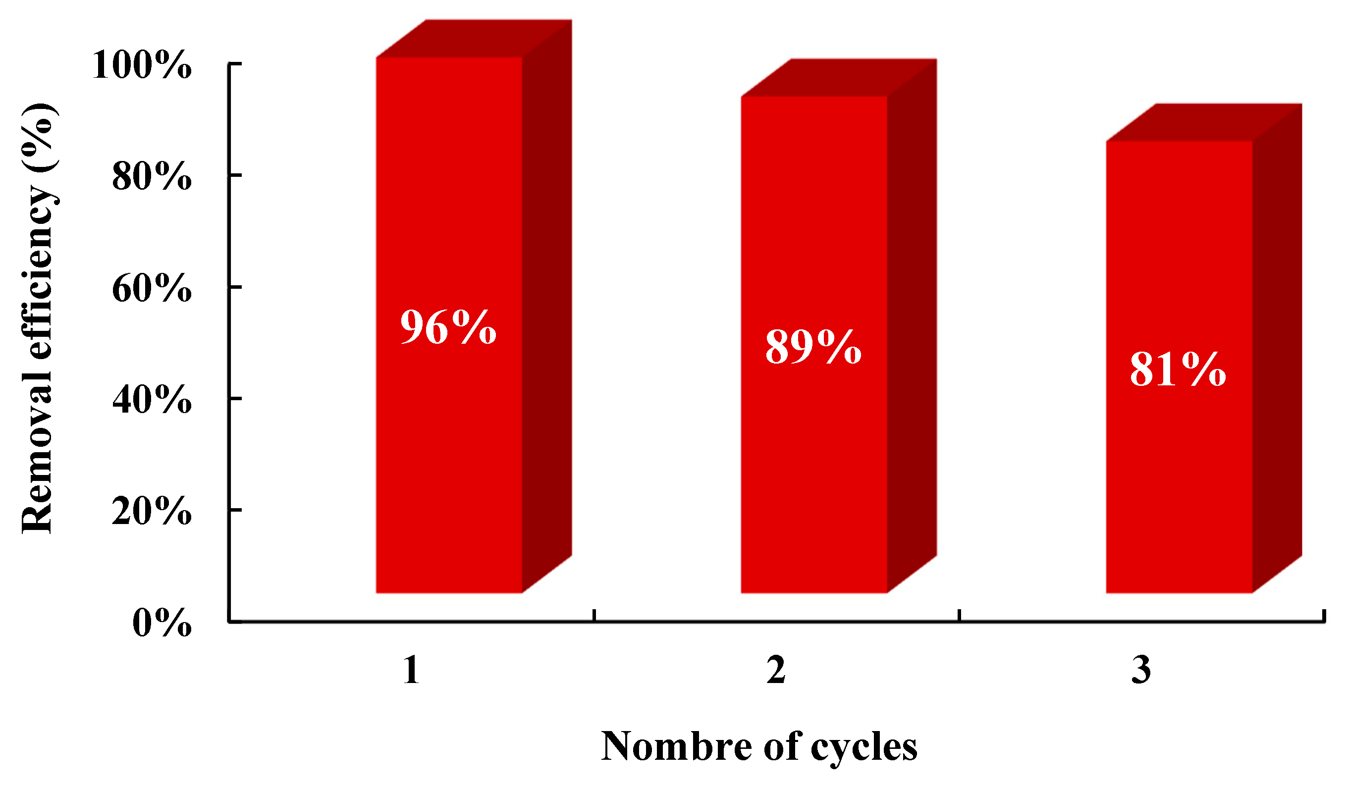
The biosynthesis of copper oxide nanoparticles (CuO NPs) involves using 1 M copper sulfate as a precursor. A 50 mL copper sulfate solution is mixed with 50 mL of Psidium. guajava leaf extract under continuous magnetic stirring at 100 °C. The pH is adjusted using 0.2 M sodium hydroxide (NaOH). A color change from dark green to brownish-black indicates the formation of monodispersed CuO NPs. The resulting precipitate is centrifuged, washed with distilled water to remove impurities, and dried at 90 °C to obtain fine CuO NPs powder for further use [109]. In the present study, CuO nanoparticles (CuO-NPs) were successfully synthesized via a green route using Psidium guajava leaf extract, marking the first reported use of this plant in the green synthesis of CuO-NPs. The eco-friendly approach leverages the phytochemicals present in the extract, which act as natural reducing, capping, and stabilizing agents, resulting in a less toxic and efficient synthesis process. The biosynthesized CuO-NPs were characterized using FT-IR and XRD, confirming the involvement of biomolecules in the formation of crystalline CuO structures, such as the characteristic peaks observed at 624 cm−1, 701 cm−1, and 777 cm−1 in the FT-IR pattern, which are attributed to the formation of biosynthesized CuO metal. Similar findings in the literature have reported absorption peaks at 799 cm−1 and 855 cm−1, which correspond to the stretching and vibrational modes associated with CuO nanoparticles [110]. The XRD spectrum of CuO is characterized by intense, sharp peaks as shown in Table 8 [111]. Morphological analysis by FE-SEM and HR-TEM revealed predominantly spherical particles with sizes ranging from 40 to 150 nm. These morphological features align with previous studies, such as the green synthesis using Tamarix gallica leaf extract, which also produced spherical particles of varying sizes [112], and the neem seed-mediated synthesis that yielded platelet-like structures [113], reported by Revathi and Thambidurai. Elemental composition was confirmed through EDS, while DLS analysis provided the average particle size distribution. Furthermore, photocatalytic studies demonstrated a high degradation efficiency of methylene blue (89%) compared to Congo red (81%) under sunlight irradiation (Figure 29), indicating the strong potential of these biosynthesized CuO-NPs for both biomedical applications and the treatment of industrial wastewater [109]. Consistent with previous studies, effective photodegradation of methylene blue was observed using Mg-doped ZnO nanoparticles under simulated sunlight irradiation [114].
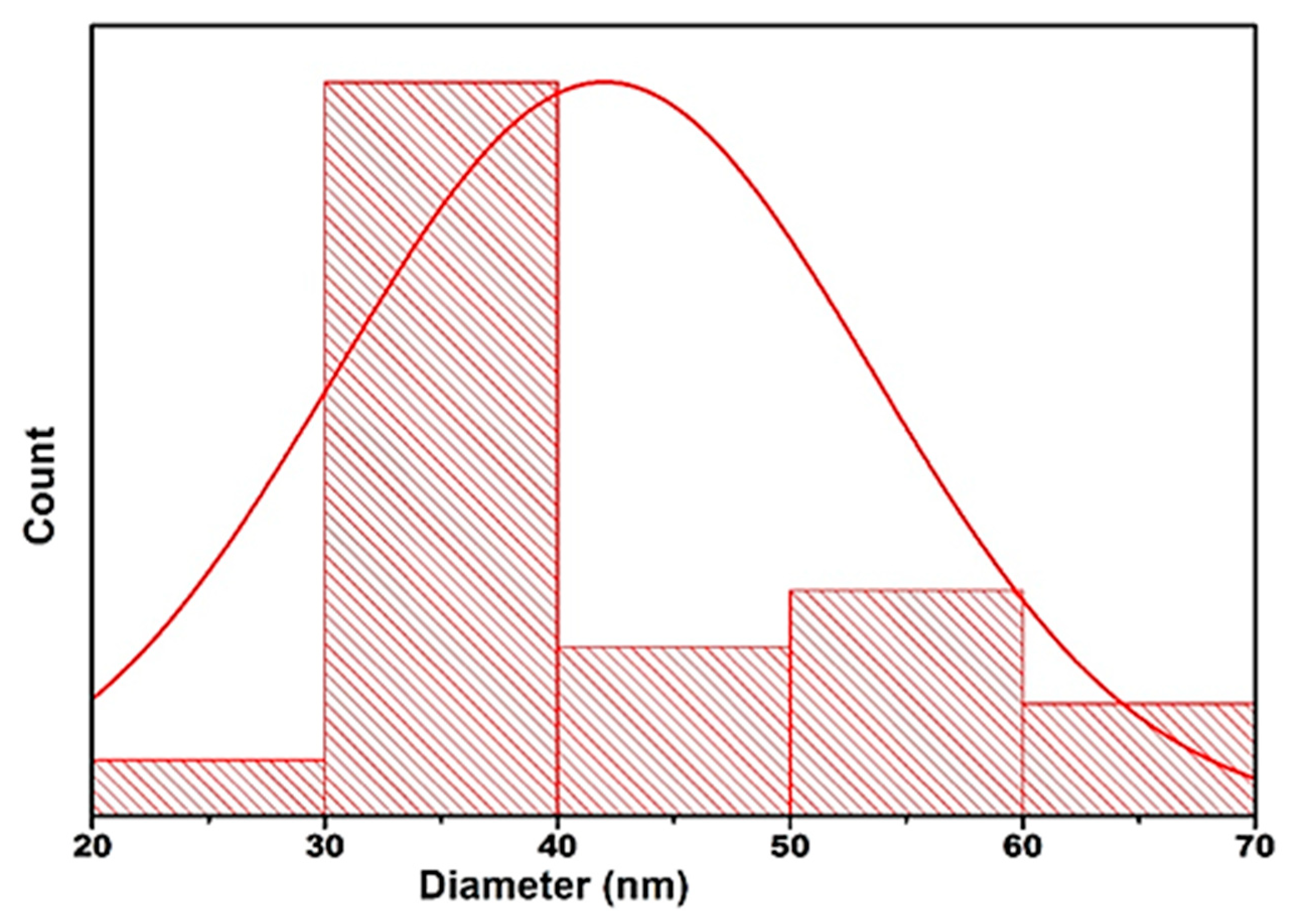
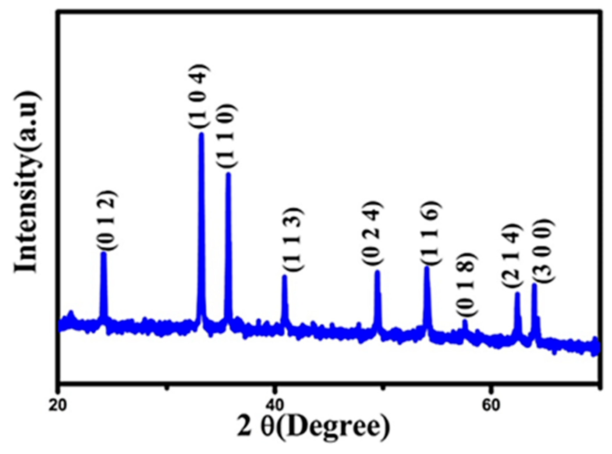
In addition to copper (II) oxide (Cu2O), another important copper oxide is cuprous oxide (Cu2O). While Cu2O contains copper in the +2-oxidation state, Cu2O consists of copper in the +1 state. Both oxides exhibit distinct physical, chemical, and crystallographic properties, and their applications differ accordingly. Cu2O, in particular, possesses a cubic crystal structure and a direct band gap of approximately 2.0 eV, making it suitable for applications in photocatalysis, photovoltaics, and sensing [115]. Cuprous oxide nanoparticles (Cu2O NPs) were synthesized using an eco-friendly method with curcumin serving as both reducing and capping agent. Curcumin was first dissolved in NaOH to prepare a homogeneous solution, which was then mixed with a 0.1 M copper nitrate solution and stirred at 80–90 °C for 20 min. The reaction proceeded for 3 h, during which a color change from blue to reddish brown was observed. The nanoparticles were collected by centrifugation, washed with ethanol, and dried under vacuum. Characterization was performed using XRD, SEM, and UV–Vis analyses. The synthesis was reproducible under identical conditions, confirming the reliability of the method [116]. The six prominent diffraction peaks located at 29.33°, 36.23°, 42.01°, 61.10°, 73.39°, and 77.40° correspond to the crystallographic planes (110), (111), (200), (220), (311), and (222), respectively, which are characteristic of cubic Cu2O, in agreement with the JCPDS reference file No. 78-2076 [117]. These results confirm the successful formation of Cu2O nanoparticles under the applied synthesis conditions. The SEM image reveals that most of the particles exhibit a nearly spherical morphology, with sizes predominantly in the range of 30–40 nm. The corresponding particle size distribution curve is illustrated in Figure 30 [116].
The photocatalytic performance of cuprous oxide nanoparticles can be attributed to their strong UV light absorption capability and their subsequent generation of reactive oxygen species, including superoxide (O2−•) and hydroxyl (OH) radicals. Although no explicit degradation percentage was reported, the qualitative analysis strongly supports the high photocatalytic efficiency of the Cu2O nanoparticles synthesized via curcumin mediation. The structural and electronic properties such as high surface area, optimal charge distribution, and strong interaction between curcumin and the Cu2O surface are key factors known to enhance photocatalytic performance. Density Functional Theory (DFT) calculations further confirmed the electronic charge transfer from curcumin to the Cu2O nanoclusters, facilitating the generation of reactive species under UV light. These qualitative insights suggest that the synthesized Cu2O NPs possess a strong potential for efficient degradation of organic pollutants like methyl orange (Table 9), even though specific degradation rates were not numerically provided [116].

4.5. Iron Oxide NPs (Fe2O3)

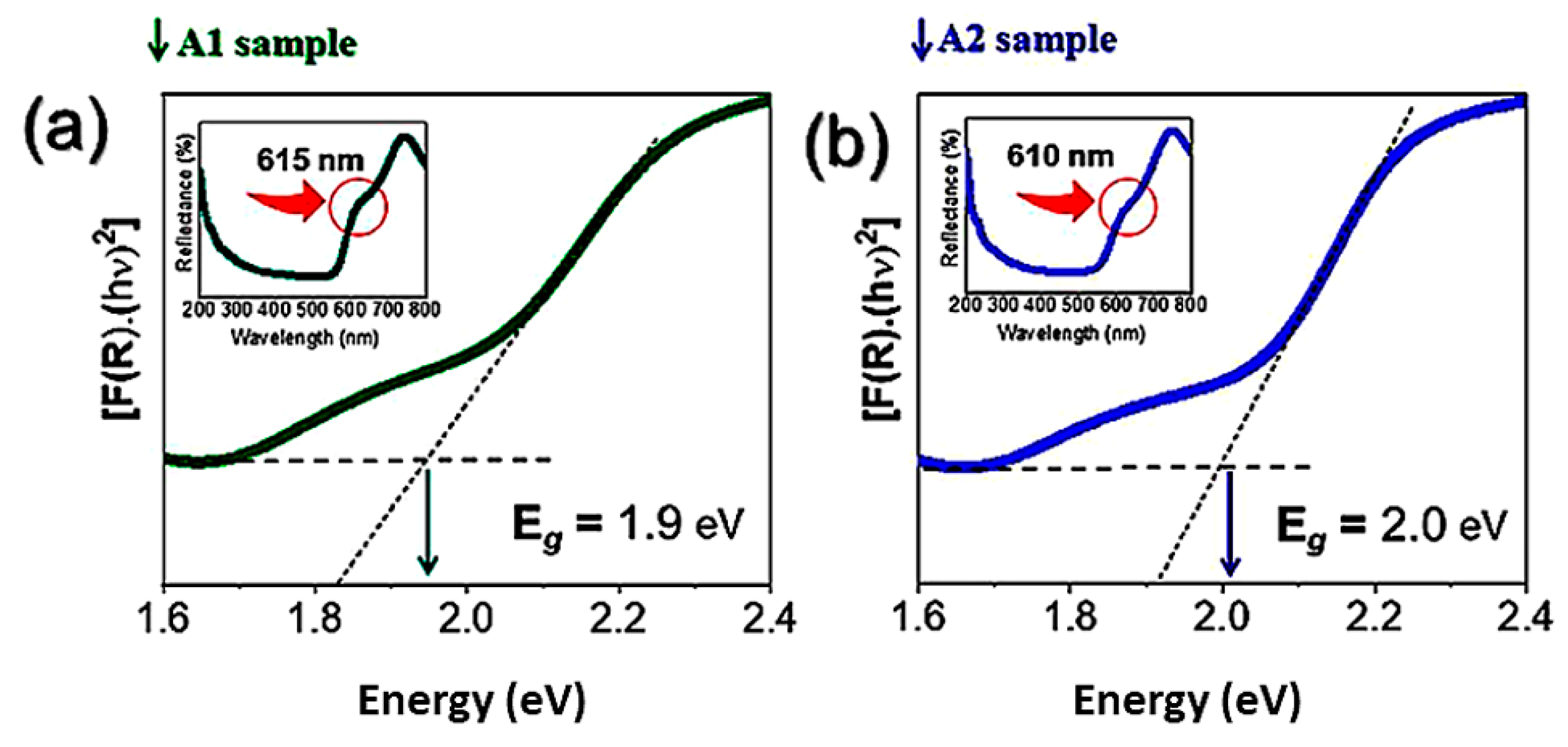
To investigate the effect of Syzygium cumini (L.) Skeels extract concentration on the synthesis of α-Fe2O3 nanoparticles, powders were prepared by dissolving 0.025 mol of Fe(NO3)3·9H2O in 100 mL of plant extract at two concentrations (17.5% and 22.5%). The mixtures were heated and stirred at 60 °C until gel formation, then dried at 100 °C for 24 h. The dried gels were ground and subsequently calcined at 500 °C and 650 °C for 2 h with a heating rate of 10 °C/min. The resulting samples were labeled A1 (17.5%, 500 °C) and A2 (22.5%, 650 °C) [118]. In this study, α-Fe2O3 nanoparticles were successfully synthesized using varying concentrations of Syzygium cumini extract. XRD analysis confirmed the exclusive formation of the α-Fe2O3 crystalline phase. Variations in extract concentration and calcination temperature significantly influenced the morphology of the nanoparticles, particularly in terms of particle size and aggregation. To better understand how the extract acts in the process of nucleation and formation of α-Fe2O3-NPs, it is first necessary to elucidate the phytocompounds present in the extract of the leaves of S. cumini (L.) Skeels [119]. The sample synthesized with a higher extract concentration (A1) exhibited enhanced photocatalytic activity, with a degradation rate of 2.868 × 10² ± 0.019 min−1 toward norfloxacin (NORF), and achieved ~32% mineralization as shown by TOC analysis. On the other hand, the band gap energy obtained from the materials was 1.9 and 2.0 eV (Figure 31). The material also demonstrated excellent stability over three successive photocatalytic cycles (Figure 32) [118]. Scavenger tests indicated that photogenerated holes (h+) play a dominant role in the degradation mechanism, followed by hydroxyl radicals (OH) [120]. These findings highlight that the concentration of S. cumini extract not only facilitates green synthesis of α-Fe2O3 but also modulates surface defect formation, thereby enhancing photocatalytic efficiency. In this context, the structural, morphological, and photocatalytic analyses collectively confirm the effectiveness of S. cumini extract as both a reducing and capping agent. This finding is particularly noteworthy, as it suggests the potential of this extract to be employed in the green synthesis of other functionalized nanoparticles such as ZnO, NiO, CuO [121,122,123], and TiO2 [78] for a wide range of applications.
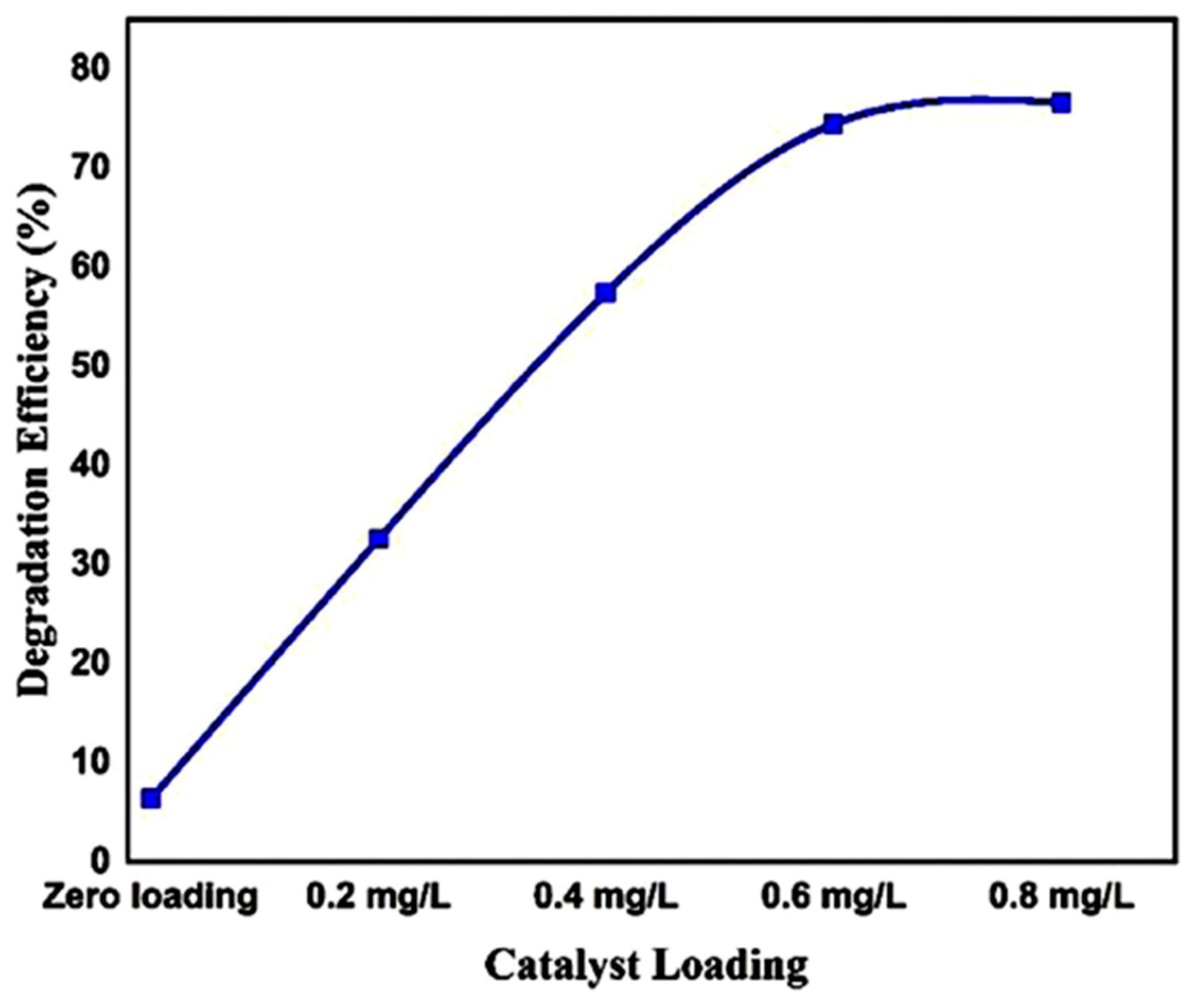
In another eco-friendly and cost-effective study conducted by Awais Ahmad et al., for the synthesis of α-Fe2O3 nanoparticles from a medicinal plant extract of Trachyspermum ammi, thus eliminating the need for hazardous chemicals. The successful formation of α-Fe2O3 was confirmed by XRD and FTIR analyses. The hematite phase of α-Fe2O3 nanoparticles is confirmed by IR bands at 445 and 550 cm−1, indicating metal–oxygen bonding and surface hydroxyl groups that enhance radical formation and charge separation [124]. These features contribute to improved photocatalytic and antibacterial performance. Additionally, the interaction between plant extracts and nitrate precursors favors the formation of highly crystalline, phase-pure α-Fe2O3 with enhanced optical properties (Figure 33). HRTEM and FESEM images revealed spherical nanoparticles with an average diameter of 26 nm, consistent with the crystallite size determined from XRD data [125,126]. The reduced particle size contributed to the improved photocatalytic and antibacterial performance of the material. The green-synthesized α-Fe2O3 demonstrated strong antibacterial activity against Staphylococcus aureus and Escherichia coli. Furthermore, it showed significant photocatalytic efficiency, reaching 91% degradation of methylene blue under visible light within 120 min. These results highlight the potential of T. ammi-derived α-Fe2O3 nanoparticles as promising candidates for environmental remediation and biomedical applications [127]. On the other hand, α-Fe2O3 nanoparticles were successfully synthesized using Carica papaya leaf extract and ferric chloride hexahydrate (FeCl3·6H2O) as the iron precursor, mixed in a 1:1 volume ratio (50 mL each) at room temperature. The pH of the solution was adjusted to 11 using 1 M NaOH, and the mixture was stirred for 30 min, leading to the formation of a dark-colored solution, confirming nanoparticle formation. The resulting nanoparticles were separated by centrifugation at 8000 rpm for 20 min, thoroughly washed with ethanol and distilled water, then dried in a hot-air oven at 80 °C for 3 h and stored for further use. The synthesized α-Fe2O3 nanoparticles exhibited notable photocatalytic activity, achieving approximately 77% degradation of Remazol Yellow RR dye under sunlight irradiation within 6 h at a dosage of 0.8 mg/L (Figure 34) [128].
In a related study, Carica papaya leaf extract was also employed for the green synthesis of silver-doped iron oxide (Ag–Fe2O3) nanoparticles. The presence of phytochemicals in the extract acted as both reducing and stabilizing agents, facilitating the formation of well-dispersed, doped nanoparticles. The incorporation of silver into the Fe2O3 matrix aimed to enhance the material’s photocatalytic and antimicrobial performance by improving charge separation and reducing electron–hole recombination.
Fe2O3 nanoparticles (NPs) and silver-doped iron oxide nanocomposites (Ag/Fe2O3-NCs) were successfully synthesized using a polyphenol-rich plant extract (PPE) through a green approach. Key reaction parameters, including temperature, pH, precursor concentration, and PPE volume, were systematically optimized. The resulting materials were characterized using UV–Vis spectroscopy, X-ray diffraction (XRD), Fourier-transform infrared spectroscopy (FTIR), and scanning electron microscopy (SEM). The Ag/Fe2O3 nanocomposites exhibited excellent photocatalytic activity, achieving complete degradation of methylene blue (MB) within 90 min using only 20 mg of catalyst. Ag/Fe2O3 NCs’ photocatalytic degradation of MB through a potential schematic mechanism is illustrated in Figure 35. Furthermore, the material retained high photocatalytic performance upon reuse, with less than a 10% decrease in degradation efficiency after a second cycle (Figure 36). In addition to their photocatalytic potential, the synthesized Ag/Fe2O3 NPs demonstrated antioxidant properties through radical scavenging activity, and hold promise as multifunctional agents for wastewater disinfection, water purification, and remediation of dye-contaminated water [129].
Table 10 provides a comparative overview of the photocatalytic performance of Fe2O3 nanoparticles tested against various organic dyes. It summarizes key experimental parameters such as dye type, solution volume, catalyst dosage, and observed degradation efficiency. The data highlight the effectiveness of Fe2O3 under different conditions and for different pollutants, with references to the corresponding studies.

5. Quantitative Assessment of Environmental Impact

While the green synthesis of metal oxide nanoparticles using plant extracts offers eco-friendly and cost-effective alternatives to conventional methods, it is crucial to quantitatively evaluate the environmental impact of these processes. Recent studies have employed life cycle analysis (LCA) and other assessment tools to measure parameters such as energy consumption, solvent usage, and waste generation during synthesis. For instance, a study on the green synthesis of zinc oxide nanoparticles from Sida acuta leaves demonstrated a significant reduction in energy consumption and chemical waste compared to traditional chemical methods [134]. Similarly, research on the synthesis of iron oxide nanoparticles highlighted a decrease in solvent usage and improved biodegradability of by-products [135].
These findings underscore the importance of integrating environmental impact assessments into the development of green synthesis protocols. By quantitatively evaluating the sustainability of these processes, researchers can optimize synthesis conditions to minimize environmental footprints, thereby advancing the field of sustainable nanotechnology.

6. Conclusions and Future Perspective

This review has highlighted the recent progress in the green synthesis of TiO2, ZnO, WO3, CuO, and Fe2O3 nanoparticles using various plant extracts. Plant-mediated approaches have been shown to provide an eco-friendly and cost-effective alternative to conventional methods, enabling the fabrication of nanomaterials with controlled morphology, crystallinity, and photocatalytic efficiency. Importantly, these green-synthesized photocatalysts demonstrate remarkable potential for degrading organic dyes and pollutants under both UV and visible light, underscoring their suitability for environmental remediation.
Nevertheless, several challenges remain. Most studies are still confined to laboratory-scale demonstrations, with limited exploration of photocatalyst stability, recyclability, and long-term performance under real environmental conditions. Moreover, the mechanistic role of phytochemicals in tailoring nanoparticle properties is not yet fully understood, restricting the rational design of optimized materials.
Future research should therefore address the following directions:
  • Industrial-scale expansion: Develop scalable synthesis routes that ensure reproducibility, cost-efficiency, and quality control for large-scale production.
  • Advanced material design: Engineer composite and heterostructured catalysts (e.g., doped systems, hybrid nanocomposites, or heterojunctions) to extend light absorption into the visible spectrum and suppress charge recombination.
  • Real-world applicability: Test photocatalysts in actual wastewater systems containing complex pollutant mixtures, variable pH, and competing ions to evaluate their robustness and practical efficiency.
  • Mechanistic insights: Elucidate the molecular role of phytochemicals in nucleation, growth, and surface functionalization to better tune particle size, band structure, and catalytic activity.
  • Catalysis–membrane integration: Explore the development of photocatalytic membranes that combine degradation and separation processes, thereby reducing secondary contamination, improving water quality, and enhancing process sustainability.
  • Multifunctional applications: Explore additional roles such as antimicrobial activity, energy harvesting, or pollutant sensing, thereby broadening the technological relevance of these nanomaterials.
In summary, plant-mediated synthesis of metal oxide nanoparticles represents a promising step toward sustainable nanotechnology. By combining green chemistry principles with innovative material design, catalytic membrane integration, and industrial scaling, future research can bridge the gap between laboratory-scale advances and real-world applications, ultimately contributing to clean water technologies and global environmental sustainability.

Author Contributions

S.M.: Writing—review & editing, Writing—original draft, Visualization, Resources, Methodology, Investigation, Data curation, Conceptualization. M.E.M.: Methodology, Investigation, Conceptualization. S.E.M.: Methodology, Investigation, Conceptualization. H.A.: Methodology, Investigation, Conceptualization. H.E.H.: Methodology, Investigation, Conceptualization. W.B.: Methodology, Investigation, Conceptualization. N.B.: Methodology, Data curation, Conceptualization. A.E.: Writing—review & editing, Writing—original draft, Visualization, Validation, Supervision, Resources, Project administration, Methodology, Data curation, Conceptualization. All authors have read and agreed to the published version of the manuscript.

Funding

This research received no external funding.

Institutional Review Board Statement

Not applicable.

Informed Consent Statement

Not applicable.

Data Availability Statement

No data was used for the research described in the article.

Conflicts of Interest

The authors declare that they have no known competing financial interests or personal relationships that could have appeared to influence the work reported in this paper.

References

  1. Ali, I.; Burakov, A.E.; Melezhik, A.V.; Babkin, A.V.; Burakova, I.V.; Neskomornaya, E.A.; Galunin, E.V.; Tkachev, A.G.; Kuznetsov, D.V. Removal of Copper (II) and Zinc (II) Ions in Water on a Newly Synthesized Polyhydroquinone/Graphene Nanocomposite Material: Kinetics, Thermodynamics and Mechanism. ChemistrySelect 2019, 4, 12708–12718. [Google Scholar] [CrossRef]
  2. Ali, I.; Alharbi, O.M.L.; AlOthman, Z.A.; Alwarthan, A.; Al-Mohaimeed, A.M. Preparation of a Carboxymethylcellulose-Iron Composite for Uptake of Atorvastatin in Water. Int. J. Biol. Macromol. 2019, 132, 244–253. [Google Scholar] [CrossRef]
  3. Ali, I.; Alharbi, O.M.L.; AlOthman, Z.A.; Badjah, A.Y. Kinetics, Thermodynamics, and Modeling of Amido Black Dye Photodegradation in Water Using Co/TiO2 Nanoparticles. Photochem. Photobiol. 2018, 94, 935–941. [Google Scholar] [CrossRef] [PubMed]
  4. Basheer, A.A. Advances in the Smart Materials Applications in the Aerospace Industries. Aircr. Eng. Aero. Technol. 2020, 92, 1027–1035. [Google Scholar] [CrossRef]
  5. Baig, N.; Kammakakam, I.; Falath, W.; Kammakakam, I. Nanomaterials: A Review of Synthesis Methods, Properties, Recent Progress, and Challenges. Mater. Adv. 2021, 2, 1821–1871. [Google Scholar] [CrossRef]
  6. Ivanova, N.; Gugleva, V.; Dobreva, M.; Pehlivanov, I.; Stefanov, S.; Andonova, V. Bottom-Up and Top-Down Approaches for MgO. In Intech, Tourism; IntechOpen: London, UK, 2016; p. 13. [Google Scholar] [CrossRef]
  7. Kumar, S.; Bhushan, P.; Bhattacharya, S. Fabrication of Nanostructures with Bottom-Up Approach and Their Utility in Diagnostics, Therapeutics, and Others. In Nanostructures for the Engineering of Cells, Tissues and Organs; Springer: Singapore, 2018; pp. 167–198. [Google Scholar] [CrossRef]
  8. Sajid, M.; Płotka-Wasylka, J. Nanoparticles: Synthesis, Characteristics, and Applications in Analytical and Other Sciences. Microchem. J. 2020, 154, 104623. [Google Scholar] [CrossRef]
  9. Ganachari, S.V.; Martínez, L.; Kharissova, O.; Kharisov, B. Synthesis Techniques for Preparation of Nanomaterials. In Handbook of Ecomaterials; Martínez, L., Kharissova, O., Kharisov, B., Eds.; Springer: Cham, Switzerland, 2019; Volume 1, pp. 83–103. [Google Scholar] [CrossRef]
  10. Razavi, M.T.L.; Salahinejad, E.; Fahmy, M.; Yazdimamaghani, M.; Vashaee, D. Green Chemical and Biological Synthesis of Nanoparticles and Their Biomedical Applications. In Green Processes for Nanotechnology; Basiuk, V., Basiuk, E., Eds.; Springer: Cham, Switzerland, 2015; pp. 207–235. [Google Scholar] [CrossRef]
  11. Andronic, L.; Enesca, A. Black TiO2 Synthesis by Chemical Reduction Methods for Photocatalysis Applications. Front. Chem. 2020, 8, 565489. [Google Scholar] [CrossRef]
  12. Yahaya, M.Z.; Azam, M.A.; Teridi, M.A.M.; Singh, P.K.; Mohamad, A.A. Recent Characterisation of Sol-Gel Synthesised TiO2 Nanoparticles. In IntechOpen Limited; IntechOpen: London, UK, 2017. [Google Scholar] [CrossRef]
  13. Thapa, R.; Maiti, S.; Rana, T.H.; Maiti, U.N.; Chattopadhyay, K.K. Anatase TiO2 Nanoparticles Synthesis via Simple Hydrothermal Route: Degradation of Orange II, Methyl Orange and Rhodamine B. J. Mol. Catal. A Chem. 2012, 363–364, 223–229. [Google Scholar] [CrossRef]
  14. Wang, M.; Gao, Q.; Duan, H.; Ge, M. Scalable Synthesis of High-Purity TiO2 Whiskers: Via Ion Exchange Method Enables Versatile Applications. RSC Adv. 2019, 9, 23735–23743. [Google Scholar] [CrossRef]
  15. Buraso, W.; Lachom, V.; Siriya, P.; Laokul, P. Synthesis of TiO2 Nanoparticles via a Simple Precipitation Method and Photocatalytic Performance. Mater. Res. Express 2018, 5, 115016. [Google Scholar] [CrossRef]
  16. Aguilar, T.; Carrillo-Berdugo, I.; Gómez-Villarejo, R.; Gallardo, J.J.; Martínez-Merino, P.; Piñero, J.C.; Alcántara, R.; Fernández-Lorenzo, C.; Navas, J. A Solvothermal Synthesis of TiO2 Nanoparticles in a Non-Polar Medium to Prepare Highly Stable Nanofluids with Improved Thermal Properties. Nanomaterials 2018, 8, 816. [Google Scholar] [CrossRef]
  17. Ojeda, M.; Chen, B.; Leung, D.Y.C.; Xuan, J.; Wang, H. A Hydrogel Template Synthesis of TiO2 Nanoparticles for Aluminium-Ion Batteries. Energy Proc. 2017, 105, 3997–4002. [Google Scholar] [CrossRef]
  18. Widiyandari, H.; Purwanto, A.; Gunawan, V.; Widyanto, S.A. Synthesis of Titanium Dioxide (TiO2) Fine Particle by Flame Spray Pyrolysis (FSP) Method Using Liquid Petroleum Gas (LPG) as Fuel. Reaktor 2018, 17, 226–229. [Google Scholar] [CrossRef]
  19. Sofronov, D.; Rucki, M.; Demidov, O.; Doroshenko, A.; Sofronova, E.; Shaposhnyk, A.; Kapustnik, O.; Mateychenko, P.; Kucharczyk, W. Formation of TiO2 Particles during Thermal Decomposition of Ti(NO3)4, TiOF2 and TiOSO4. J. Mater. Res. Technol. 2020, 9, 12201–12212. [Google Scholar] [CrossRef]
  20. Bandgar, A.; Sabale, S.; Pawar, S.H. Studies on Influence of Reflux Time on Synthesis of Nanocrystalline TiO2 Prepared by Peroxotitanate Complex Solutions. Ceram. Int. 2012, 38, 1905–1913. [Google Scholar] [CrossRef]
  21. Reinke, M.; Ponomarev, E.; Kuzminykh, Y.; Hoffmann, P. Combinatorial Characterization of TiO2 Chemical Vapor Deposition Utilizing Titanium Isopropoxide. ACS Comb. Sci. 2015, 17, 413–420. [Google Scholar] [CrossRef]
  22. Kitamura, Y.; Okinaka, N.; Shibayama, T.; Mahaney, O.O.P.; Kusano, D.; Ohtani, B.; Akiyama, T. Combustion Synthesis of TiO2 Nanoparticles as Photocatalyst. Powder Technol. 2007, 176, 93–98. [Google Scholar] [CrossRef]
  23. Moon, J.T.; Lee, S.K.; Joo, J.B. Controllable One-Pot Synthesis of Uniform Colloidal TiO2 Particles in a Mixed Solvent Solution for Photocatalysis. Beilstein J. Nanotechnol. 2018, 9, 1715–1727. [Google Scholar] [CrossRef]
  24. Yasin, S.A.; Zeebaree, S.Y.S.; Zeebaree, A.Y.S.; Zebari, O.I.H.; Saeed, I.A. The Efficient Removal of Methylene Blue Dye Using CuO/PET Nanocomposite in Aqueous Solutions. Catalysts 2021, 11, 241. [Google Scholar] [CrossRef]
  25. Wu, V.K.; Bai, G.-R.; Eastman, J.A.; Zhou, G. Synthesis of TiO2 Nanoparticles Using Chemical Vapor Condensation. Mater. Res. Soc. Symp. Proc. 2005, 879, 712. [Google Scholar] [CrossRef]
  26. Ellouzi, I.; Oualid, H.A. Efficient and Eco-Friendly Mechanical Milling Preparation of Anatase/Rutile TiO2-Glucose Composite with Energy Gap Enhancement. Proceedings 2019, 3, 3. [Google Scholar] [CrossRef]
  27. Guo, J.; Zhu, S.; Chen, Z.; Li, Y.; Yu, Z.; Liu, Q.; Li, J.; Feng, C.; Zhang, D. Sonochemical Synthesis of TiO2 Nanoparticles on Graphene for Use as Photocatalyst. Ultrason. Sonochem. 2011, 18, 1082–1090. [Google Scholar] [CrossRef] [PubMed]
  28. Negrea, D.; Ducu, C.; Moga, S.; Malinovschi, V.; Monty, C.J.A.; Vasile, B.; Dorobantu, D.; Enachescu, M. Solar Physical Vapor Deposition Preparation and Microstructural Characterization of TiO2 Based Nanophases for Dye-Sensitized Solar Cell Applications. J. Nanosci. Nanotechnol. 2012, 12, 8746–8750. [Google Scholar] [CrossRef]
  29. Andriyanti, W.; Nurfiana, F.; Sari, A.N.; Kundari, N.A.; Aziz, I. Synthesis TiO2-Ag Thin Film by DC Sputtering Method for Dye Degradation. J. Phys. Conf. Ser. 2020, 1436, 012008. [Google Scholar] [CrossRef]
  30. Anandgaonker, P.; Kulkarni, G.; Gaikwad, S.; Rajbhoj, A. Synthesis of TiO2 Nanoparticles by Electrochemical Method and Their Antibacterial Application. Arab. J. Chem. 2019, 12, 1815–1822. [Google Scholar] [CrossRef]
  31. Nath, A.; Laha, S.S.; Khare, A. Synthesis of TiO2 Nanoparticles via Laser Ablation at Titanium-Water Interface. Integr. Ferroelectr. Int. J. 2010, 121, 58–64. [Google Scholar] [CrossRef]
  32. Bouhadoun, S.; Guillard, C.; Sorgues, S.; Hérissan, A.; Colbeau-Justin, C.; Dapozze, F.; Habert, A.; Maurel, V.; Herlin-Boime, N. Laser Synthesized TiO2-Based Nanoparticles and Their Efficiency in the Photocatalytic Degradation of Linear Carboxylic Acids. Sci. Technol. Adv. Mater. 2017, 18, 805–815. [Google Scholar] [CrossRef]
  33. Huang, B.; Li, L.; Jiang, Y.P.; Hao, S.X. Preparation of Nano-Titanium Dioxide by Microemulsion Method. Adv. Mater. Res. 2015, 1073–1076, 8–11. [Google Scholar] [CrossRef]
  34. Cabello, G.; Davoglio, R.A.; Pereira, E.C. Microwave-Assisted Synthesis of Anatase TiO2 Nanoparticles with Catalytic Activity in Oxygen Reduction. J. Electroanal. Chem. 2017, 794, 36–42. [Google Scholar] [CrossRef]
  35. Ashkarran, A.A.; Kavianipour, M.; Aghigh, S.M.; Ghoranneviss, M. On the Formation of TiO2 Nanoparticles via Submerged Arc Discharge Technique: Synthesis, Characterization and Photocatalytic Properties. J. Cluster Sci. 2010, 21, 753–766. [Google Scholar] [CrossRef]
  36. Tsai, C.; Hsi, H.; Fan, K. Synthesized Oxygen-Vacant TiO2 Nanopowders with Thermal Plasma Torch Evaporation Condensation Process. World Congr. Eng. Comput. Sci. 2010, II, 20–23. [Google Scholar]
  37. Tripathy, S.K.; Sahoo, T.; Mohapatra, M.; Anand, S.; Yu, Y.T. Polyol-Assisted Synthesis of TiO2 Nanoparticles in a Semi-Aqueous Solvent. J. Phys. Chem. Solids 2009, 70, 147–152. [Google Scholar] [CrossRef]
  38. Tokoi, Y.; Suzuki, T.; Nakayama, T.; Suematsu, H.; Jiang, W.; Niihara, K. Synthesis of TiO2 Nanosized Powder by Pulsed Wire Discharge. Jpn. J. Appl. Phys. 2008, 47, 760–763. [Google Scholar] [CrossRef]
  39. Thakur, B.K.; Kumar, A.; Kumar, D. Green Synthesis of Titanium Dioxide Nanoparticles Using Azadirachta indica Leaf Extract and Evaluation of Their Antibacterial Activity. South Afr. J. Bot. 2019, 124, 223–227. [Google Scholar] [CrossRef]
  40. Landage, K.S.; Arbade, C.J.B.G.; Khanna, P. Biological Approach to Synthesize TiO2 Nanoparticles Using Staphylococcus aureus for Antibacterial and Anti-Biofilm Applications. J. Microbiol. Exp. Res. 2020, 8, 36–43. [Google Scholar] [CrossRef]
  41. Rajakumar, G.; Rahuman, A.A.; Roopan, S.M.; Khanna, V.G.; Elango, G.; Kamaraj, C.; Zahir, A.A.; Velayutham, K. Fungus-Mediated Biosynthesis and Characterization of TiO2 Nanoparticles and Their Activity against Pathogenic Bacteria. Spectrochim. Acta Part A Mol. Biomol. Spectrosc. 2012, 91, 23–29. [Google Scholar] [CrossRef]
  42. Peiris, M.M.K.; Guansekera, T.D.C.P.; Jayaweera, P.M.; Fernando, S.S.N. TiO2 Nanoparticles from Baker’s Yeast: A Potent Antimicrobial. J. Microbiol. Biotechnol. 2018, 28, 1664–1670. [Google Scholar] [CrossRef] [PubMed]
  43. Ijaz, I.; Gilani, E.; Nazir, A.; Bukhari, A. Detail Review on Chemical, Physical and Green Synthesis, Classification, Characterizations and Applications of Nanoparticles. Green Chem. Lett. Rev. 2020, 13, 223–245. [Google Scholar] [CrossRef]
  44. Nosaka, Y.; Nosaka, A.Y. Generation and Detection of Reactive Oxygen Species in Photocatalysis. Chem. Rev. 2017, 117, 11302–11336. [Google Scholar] [CrossRef]
  45. Mallah, S.; El Mchaouri, M.; Boumya, W.; Machrouhi, A.; Elmoubarki, R.; Barka, N.; Elhalil, A. Recent advances and modification strategies of TiO2 semiconductors for the photocatalytic remediation of water-containing organic pollutants. Euro-Mediterr. J. Environ. Integr. 2025, 1–36. [Google Scholar] [CrossRef]
  46. Tan, S.N.; Yuen, M.L.; Ramli, R.A. Photocatalysis of Dyes: Operational Parameters, Mechanisms, and Degradation Pathway. Green Anal. Chem. 2025, 12, 100230. [Google Scholar] [CrossRef]
  47. Nagaraj, G.; Dhayal Raj, A.; Albert Irudayaraj, A.; Josephine, R.L. Tuning the Optical Band Gap of Pure TiO2 via Photon Induced Method. Optik 2019, 179, 889–894. [Google Scholar] [CrossRef]
  48. Jafarova, V.N.; Orudzhev, G.S. Structural and Electronic Properties of ZnO: A First-Principles Density-Functional Theory Study within LDA(GGA) and LDA(GGA)+U Methods. Solid State Commun. 2021, 325, 114166. [Google Scholar] [CrossRef]
  49. Mubeen, A.; Majid, A.; Alkhedher, M.; Tag-ElDin, E.M.; Bulut, N. Structural and Electronic Properties of SnO Downscaled to Monolayer. Materials 2022, 15, 5578. [Google Scholar] [CrossRef]
  50. Oudah, M.H.; Hasan, M.H.; Abd, A.N. Synthesis of Copper Oxide Thin Films by Electrolysis Method Based on Porous Silicon for Solar Cell Applications. IOP Conf. Ser. Mater. Sci. Eng. 2020, 757, 012051. [Google Scholar] [CrossRef]
  51. Gul, A.R.; Shaheen, F.; Rafique, R.; Bal, J. Grass-Mediated Biogenic Synthesis of Silver Nanoparticles and Their Drug Delivery Evaluation: A Biocompatible Anti-Cancer Therapy. Chem. Eng. J. 2020, 407, 127202. [Google Scholar] [CrossRef]
  52. Soni, V.; Raizada, P.; Singh, P.; Cuong, H.N.; S, R.; Saini, A.; Saini, R.V.; Van Le, Q.; Nadda, A.K.; Le, T.-T.; et al. Sustainable and Green Trends in Using Plant Extracts for the Synthesis of Biogenic Metal Nanoparticles toward Environmental and Pharmaceutical Advances: A Review. Environ. Res. 2021, 202, 111622. [Google Scholar] [CrossRef] [PubMed]
  53. Ying, S.; Guan, Z.; Ofoegbu, P.C.; Clubb, P.; Rico, C.; He, F.; Hong, J. Green Synthesis of Nanoparticles: Current Developments and Limitations. Environ. Technol. Innov. 2022, 26, 102336. [Google Scholar] [CrossRef]
  54. He, Y.; Wei, F.; Ma, Z.; Zhang, H.; Yang, Q.; Yao, B.; Huang, Z.; Li, J.; Zeng, C.; Zhang, Q. Green Synthesis of Silver Nanoparticles Using Seed Extract of Alpinia katsumadai, and Their Antioxidant, Cytotoxicity, and Antibacterial Activities. RSC Adv. 2017, 7, 39842–39851. [Google Scholar] [CrossRef]
  55. Rizwana, H.; Bokahri, N.A.; Alkhattaf, F.S.; Albasher, G.; Aldehaish, H.A. Antifungal, Antibacterial, and Cytotoxic Activities of Silver Nanoparticles Synthesized from Aqueous Extracts of Mace-Arils of Myristica fragrans. Molecules 2021, 26, 7709. [Google Scholar] [CrossRef]
  56. Rashidipour, M.; Heydari, R. Biosynthesis of Silver Nanoparticles Using Extract of Olive Leaf: Synthesis and In Vitro Cytotoxic Effect on MCF-7 Cells. J. Nanostruct. Chem. 2014, 4, 112. [Google Scholar] [CrossRef]
  57. Babu, S.A.; Prabu, H.G. Synthesis of AgNPs Using the Extract of Calotropis procera Flower at Room Temperature. Mater. Lett. 2011, 65, 1675–1677. [Google Scholar] [CrossRef]
  58. Behravan, M.; Hossein Panahi, A.; Naghizadeh, A.; Ziaee, M.; Mahdavi, R.; Mirzapour, A. Facile Green Synthesis of Silver Nanoparticles Using Berberis vulgaris Leaf and Root Aqueous Extract and Its Antibacterial Activity. Int. J. Biol. Macromol. 2019, 124, 148–154. [Google Scholar] [CrossRef]
  59. Rani, N.; Singh, P.; Kumar, S.; Kumar, P.; Bhankar, V.; Kumar, K. Plant-Mediated Synthesis of Nanoparticles and Their Applications: A Review. Mater. Res. Bull. 2023, 163, 112233. [Google Scholar] [CrossRef]
  60. Langa, C.; Hintsho-Mbita, N.C. Plant and Bacteria Mediated Synthesis of TiO2 NPs for Dye Degradation in Water: A Review. Chem. Phys. Impact 2023, 7, 100293. [Google Scholar] [CrossRef]
  61. Peralta-Videa, J.R.; Huang, Y.; Parsons, J.G.; Zhao, L.; Lopez-Moreno, L.; Hernandez-Viezcas, J.A.; Gardea-Torresdey, J.L. Plant-Based Green Synthesis of Metallic Nanoparticles: Scientific Curiosity or a Realistic Alternative to Chemical Synthesis? Nanotechnol. Environ. Eng. 2016, 1, 4. [Google Scholar] [CrossRef]
  62. Küünal, S.; Rauwel, P.; Rauwel, E. Plant Extract Mediated Synthesis of Nanoparticles. In Emerging Applications of Nanoparticles and Architectural Nanostructures: Current Prospects and Future Trends; Elsevier Inc.: Amsterdam, The Netherlands, 2018. [Google Scholar] [CrossRef]
  63. Patra, J.K.; Baek, K.H. Green Nanobiotechnology: Factors Affecting Synthesis and Characterization Techniques. J. Nanomater. 2014, 2014, 417305. [Google Scholar] [CrossRef]
  64. Zakharova, G.S.; Natal’ya, V.; Tat’yana, I.G.; Pervova, M.G.; Murzakaev, A.M.; Enyashin, A.N. Morphology-Controlling Hydrothermal Synthesis of h-WO3 for Photocatalytic Degradation of 1,2,4-Trichlorobenzene. J. Alloy. Compd. 2023, 938, 168620. [Google Scholar] [CrossRef]
  65. Wellia, D.V.; Syuadi, A.F.; Rahma, R.M.; Syafawi, A.; Habibillah, M.R.; Arief, S.; Kurnia, K.A.; Saepurahman; Kusumawati, Y.; Saefumillah, A. Rind of Aloe vera (L.) Burm. f Extract for the Synthesis of Titanium Dioxide Nanoparticles: Properties and Application in Model Dye Pollutant Degradation. Case Stud. Chem. Environ. Eng. 2024, 9, 100627. [Google Scholar] [CrossRef]
  66. Singh, D.; Anuradha; Mathur, D.; Kumar, S.; Pani, B.; Kumar, A.; Kanojia, R.; Gupta, R.; Singh, L. Bio-Genic Synthesis of Calcium Coated Zinc Oxide Nanoparticles from Beetroot Extract and Their Photo-Degradation Study on Methylene Blue and Rhodamine B. Plant Nano Biol. 2023, 4, 100031. [Google Scholar] [CrossRef]
  67. Atri, A.; Echabaane, M.; Bouzidi, A.; Harabi, I.; Soucase, B.M.; Ben Chaâbane, R. Green Synthesis of Copper Oxide Nanoparticles Using Ephedra Alata Plant Extract and a Study of Their Antifungal, Antibacterial Activity and Photocatalytic Performance under Sunlight. Heliyon 2023, 9, e13484. [Google Scholar] [CrossRef]
  68. Al-Fa’ouri, A.M.; Abu-Kharma, M.H.; Awwad, A.M.; Abugazleh, M.K. Investigation of Optical and Structural Properties of Copper Oxide Nanoparticles Synthesized via Green Method Using Bougainvillea Leaves Extract. Nano-Struct. Nano-Objects 2023, 36, 101051. [Google Scholar] [CrossRef]
  69. Saini, R.; Kumar, P. Green Synthesis of TiO2 Nanoparticles Using Tinospora cordifolia Plant Extract and Its Potential Application for Photocatalysis and Antibacterial Activity. Inorg. Chem. Commun. 2023, 156, 111221. [Google Scholar] [CrossRef]
  70. Shimi, A.K.; Ahmed, H.M.; Wahab, M.; Katheria, S.; Wabaidur, S.M.; Eldesoky, G.E.; Islam, M.A.; Rane, K.P. Synthesis and Applications of Green Synthesized TiO2 Nanoparticles for Photocatalytic Dye Degradation and Antibacterial Activity. J. Nanomater. 2022, 2022, 7060388. [Google Scholar] [CrossRef]
  71. Mohapatra, D.P.; Brar, S.K.; Daghrir, R.; Tyagi, R.D.; Picard, P.; Surampalli, R.Y.; Drogui, P. Photocatalytic Degradation of Carbamazepine in Wastewater by Using a New Class of Whey-Stabilized Nanocrystalline TiO2 and ZnO. Sci. Total Environ. 2014, 485–486, 263–269. [Google Scholar] [CrossRef] [PubMed]
  72. Bekele, E.T.; Gonfa, B.A.; Zelekew, O.A.; Belay, H.H.; Sabir, F.K. Synthesis of Titanium Oxide Nanoparticles Using Root Extract of Kniphofia foliosa as a Template: Characterization and Its Application on Drug Resistant Bacteria. J. Nanomater. 2020, 2020, 2817037. [Google Scholar] [CrossRef]
  73. Ambika, S.; Sundrarajan, M. [EMIM] BF4 Ionic Liquid-Mediated Synthesis of TiO2 Nanoparticles Using Vitex negundo Linn Extract and Its Antibacterial Activity. J. Mol. Liq. 2016, 221, 986–992. [Google Scholar] [CrossRef]
  74. Rajkumari, J.; Magdalane, C.M.; Siddhardha, B.; Madhavan, J.; Ramalingam, G.; Al-Dhabi, N.A.; Arasu, M.V.; Ghilan, A.; Duraipandiayan, V.; Kaviyarasu, K. Synthesis of Titanium Oxide Nanoparticles Using Aloe barbadensis Mill and Evaluation of Its Antibiofilm Potential against Pseudomonas aeruginosa PAO1. J. Photochem. Photobiol. B 2019, 201, 111667. [Google Scholar] [CrossRef]
  75. Abisharani, J.M.; Devikala, S.; Dinesh Kumar, R.; Arthanareeswari, M.; Kamaraj, P. Green Synthesis of TiO2 Nanoparticles Using Cucurbita pepo Seeds Extract. Mater. Today Proc. 2019, 14, 302–307. [Google Scholar] [CrossRef]
  76. Nabi, G.; Ain, Q.-U.; Tahir, M.B.; Riaz, K.N.; Iqbal, T.; Rafique, M.; Hussain, S.; Raza, W.; Aslam, I.; Rizwan, M. Green synthesis of TiO2 nanoparticles using lemon peel extract: Their optical and photocatalytic properties. J. Mater. Sci. 2020, 55, 12345–12358. [Google Scholar] [CrossRef]
  77. Nabi, G.; Majid, A.; Riaz, A.; Alharbi, T.; Kamran, M.A.; Al-Habardi, M. Green synthesis of spherical TiO2 nanoparticles using Citrus limetta extract: Excellent photocatalytic water decontamination agent for RhB dye. Inorg. Chem. Commun. 2021, 129, 108618. [Google Scholar] [CrossRef]
  78. Sethy, N.K.; Arif, Z.; Mishra, P.K.; Kumar, P. Green synthesis of TiO2 nanoparticles from Syzygium cumini extract for photo-catalytic removal of lead (Pb) in explosive industrial wastewater. Green Process. Synth. 2020, 9, 171–181. [Google Scholar] [CrossRef]
  79. Pushpamalini, T.; Keerthana, M.; Sangavi, R.; Nagaraj, A.; Kamaraj, P. Comparative analysis of green synthesis of TiO2 nanoparticles using four different leaf extracts. Mater. Today Proc. 2021, 40, S180–S184. [Google Scholar] [CrossRef]
  80. Dimitrov, O.; Zaharieva, K.; Stambolova, I.; Eneva, R.; Engibarov, S.; Lazarkevich, I.; Gocheva, Y.; Stoyanova, D.; Shipochka, M.; Markov, P.; et al. Photocatalytic and antimicrobial activity of TiO2 particles, obtained by Mentha spicata leaves–mediated synthesis. Catal. Today 2025, 460, 115466. [Google Scholar] [CrossRef]
  81. Rajendhiran, R.; Deivasigamani, V.; Palanisamy, J.; Masan, S.; Pitchaiya, S. Terminalia catappa and Carissa carandas assisted synthesis of TiO2 nanoparticles—A green synthesis approach. Mater. Today Proc. 2021, 45, 2232–2238. [Google Scholar] [CrossRef]
  82. Subhapriya, S.; Gomathipriya, P. Green synthesis of titanium dioxide (TiO2) nanoparticles by Trigonella foenum-graecum extract and its antimicrobial properties. Microb. Pathog. 2018, 116, 215–220. [Google Scholar] [CrossRef]
  83. Ngoepe, N.M.; Mathipa, M.M.; Hintsho-Mbita, N.C. Biosynthesis of titanium dioxide nanoparticles for the photodegradation of dyes and removal of bacteria. Optik 2020, 224, 165728. [Google Scholar] [CrossRef]
  84. Goutam, S.P.; Saxena, G.; Singh, V.; Yadav, A.K.; Bharagava, R.N.; Thapa, K.B. Green synthesis of TiO2 nanoparticles using leaf extract of Jatropha curcas L. for photocatalytic degradation of tannery wastewater. Chem. Eng. J. 2018, 336, 386–396. [Google Scholar] [CrossRef]
  85. Singh, D.; Anuradha; Mathur, D.; Kumar, A.; Kumar, S.; Pani, B.; Kanojia, R.; Ratan, J. Photocatalytic properties of biologically synthesized uncoated and calcium coated ZnO nanoparticles using cucumber juice. Rasayan J. Chem. 2022, 95–102. [Google Scholar] [CrossRef]
  86. Pham, T.A.T.; Tran, V.A.; Le, V.D.; Nguyen, M.V.; Troung, D.D.; Do, X.T.; Vu, A.-T. Facile Preparation of ZnO Nanoparticles and Ag/ZnO Nanocomposite and Their Photocatalytic Activities under Visible Light. Int. J. Photoenergy 2020, 2020, 8897667. [Google Scholar] [CrossRef]
  87. Venkatesan, S.; Suresh, S.; Arumugam, J.; Ramu, P.; Pugazhenthiran, N.; Jothilakshmi, R.; Prabu, K.M. Sunlight assisted degradation of methylene blue dye by zinc oxide nanoparticles green synthesized using Vitex negundo plant leaf extract. Results Chem. 2024, 7, 101315. [Google Scholar] [CrossRef]
  88. Sedefoglu, N. Green synthesis of ZnO nanoparticles by Myrtus communis plant extract with investigation of effect of precursor, calcination temperature and study of photocatalytic performance. Ceram. Int. 2024, 50, 9884–9895. [Google Scholar] [CrossRef]
  89. Debanath, M.K.; Karmakar, S. Study of Blueshift of Optical Band Gap in Zinc Oxide (ZnO) Nanoparticles Prepared by Low-Temperature Wet Chemical Method. Mater. Lett. 2013, 111, 116–119. [Google Scholar] [CrossRef]
  90. Sarkar, J.; Ghosh, M.; Mukherjee, A.; Chattopadhyay, D.; Acharya, K. Biosynthesis and Safety Evaluation of ZnO Nanoparticles. Bioprocess Biosyst. Eng. 2014, 37, 165–171. [Google Scholar] [CrossRef] [PubMed]
  91. Velmurugan, G.; Chohan, J.S.; Paramasivam, P.; Maranan, R.; Nagaraj, M. Green marvel: Harnessing spinach leaves’ power for enhanced photodegradation of various effluents with biogenic ZnO nanoparticles. Desal. Water Treat. 2024, 319, 100566. [Google Scholar] [CrossRef]
  92. Deenadayalan, J.; Chen, S.-S.; Das, B.; Pasawan, M. Mineralization and toxicity reduction of chlortetracycline using an environmentally friendly ZnO photocatalyst prepared from Camellia sinensis leaf extract. J. Environ. Chem. Eng. 2025, 13, 115345. [Google Scholar] [CrossRef]
  93. Nachimuthu, S.; Thangavel, S.; Kannan, K.; Selvakumar, V.; Muthusamy, K.; Siddiqui, M.R.; Wabaidur, S.M.; Parvathiraja, C. Lawsonia inermis mediated synthesis of ZnO/Fe2O3 nanorods for photocatalysis—Biological treatment for the enhanced effluent treatment, antibacterial and antioxidant activities. Chem. Phys. Lett. 2022, 804, 139907. [Google Scholar] [CrossRef]
  94. Venkatesan, S.; Suresh, S.; Ramu, P.; Arumugam, J.; Thambidurai, S.; Pugazhenthiran, N. Methylene Blue Dye Degradation Potential of Zinc Oxide Nanoparticles Bioreduced Using Solanum trilobatum Leaf Extract. Results Chem. 2022, 4, 100637. [Google Scholar] [CrossRef]
  95. Supin, K.K.; Namboothiri, P.M.P.; Vasundhara, M. Enhanced Photocatalytic Activity in ZnO Nanoparticles Developed Using Novel Lepidagathis ananthapuramensis Leaf Extract. RSC Adv. 2023, 13, 1497–1515. [Google Scholar] [CrossRef]
  96. Venkatesan, S.; Suresh, S.; Ramu, P.; Kandasamy, M.; Arumugam, J.; Thambidurai, S.; Prabu, K.M.; Pugazhenthiran, N. Biosynthesis of Zinc Oxide Nanoparticles Using Euphorbia milii Leaf Constituents: Characterization and Improved Photocatalytic Degradation of Methylene Blue Dye under Natural Sunlight. J. Indian Chem. Soc. 2022, 99, 100436. [Google Scholar] [CrossRef]
  97. Aldeen, T.S.; Mohamed, H.E.A.; Maaza, M. ZnO Nanoparticles Prepared via a Green Synthesis Approach: Physical Properties, Photocatalytic and Antibacterial Activity. J. Phys. Chem. Solids 2022, 160, 110313. [Google Scholar] [CrossRef]
  98. Anggraini, F.; Fatimah, I.; Ramanda, G.D.; Nurlaela, N.; Wijayanti, H.K.; Sagadevan, S.; Oh, W.-C.; Doong, R.-A. Unveiling the green synthesis of WO3 nanoparticles by using beetroot (Beta vulgaris) extract for photocatalytic oxidation of rhodamine B. Chemosphere 2025, 370, 143890. [Google Scholar] [CrossRef] [PubMed]
  99. Hkiri, K.; Mohamed, H.E.A.; Harrisankar, N.; Gibaud, A.; van Steen, E.; Maaza, M. Environmental water treatment with green synthesized WO3 nanoflakes for cationic and anionic dyes removal: Photocatalytic studies. Catal. Commun. 2024, 187, 106851. [Google Scholar] [CrossRef]
  100. Ke, J.; Zhou, H.; Liu, J.; Duan, X.; Zhang, H.; Liu, S.; Wang, S. Crystal transformation of 2D tungstic acid H2WO4 to WO3 for enhanced photocatalytic water oxidation. J. Colloid Interface Sci. 2018, 514, 576–583. [Google Scholar] [CrossRef]
  101. Jayaprakash, N.; Vijaya, J.J.; Kaviyarasu, K.; Kombaiah, K.; Kennedy, L.J.; Ramalingam, R.J.; Munusamy, M.A.; Al-Lohedan, H.A. Green Synthesis of Ag Nanoparticles Using Tamarind Fruit Extract for the Antibacterial Studies. J. Photochem. Photobiol. B Biol. 2017, 169, 178–185. [Google Scholar] [CrossRef]
  102. Antony, A.J.; Bennie, R.B.; Joel, C.; Basavegowda, N.; Hatshan, M.R.; Kumar, Y.A. Green synthesis of metastable h-WO3 nanoparticles for photocatalytic degradation of ofloxacin in waste water. J. Alloys Compd. 2025, 1019, 179305. [Google Scholar] [CrossRef]
  103. Rupashree, M.P.; Soppin, K.; Pratibha, S.; Chethan, B. Cost Effective Photocatalytic and Humidity Sensing Performance of Green Tea Mediated Copper Oxide Nanoparticles. Inorg. Chem. Commun. 2021, 134, 108974. [Google Scholar] [CrossRef]
  104. Arunkumar, B.; Johnson Jeyakumar, S.; Jothibas, M. A Sol-Gel Approach to the Synthesis of CuO Nanoparticles Using Lantana camara Leaf Extract and Their Photocatalytic Activity. Optik 2019, 183, 698–705. [Google Scholar] [CrossRef]
  105. Malka, E.; Perelshtein, I.; Lipovsky, A.; Shalom, Y.; Naparstek, L.; Perkas, N.; Patick, T.; Lubart, R.; Nitzan, Y.; Banin, E.; et al. Eradication of Multi-Drug Resistant Bacteria by a Novel Zn-Doped CuO Nanocomposite. Small 2013, 9, 4069–4076. [Google Scholar] [CrossRef]
  106. Udayabhanu, P.C.; Nethravathi, M.A.; Pavan Kumar, D.; Suresh, K.; Lingaraju, H.; Rajanaika, H.; Nagabhushan, S.C.; Sharma, S.C. Tinospora cordifolia Mediated Facile Green Synthesis of Cupric Oxide Nanoparticles and Their Photocatalytic, Antioxidant and Antibacterial Properties. Mater. Sci. Semicond. Process. 2015, 33, 81–88. [Google Scholar] [CrossRef]
  107. Allaa, S.K.; Verma, A.D.; Kumar, V.; Mandal, R.K.; Sinha, I.; Prasad, N.K. Solvothermal Synthesis of CuO-MgO Nanocomposite Particles and Their Catalytic Applications. RSC Adv. 2016, 6, 61927–61933. [Google Scholar] [CrossRef]
  108. Novikova, A.A.; Moiseeva, D.Y.; Karyukov, E.V.; Kalinichenko, A.A. Facile Preparation Photocatalytically Active CuO Plate-Like Nanoparticles from Brochantite. Mater. Lett. 2016, 167, 165–169. [Google Scholar] [CrossRef]
  109. Sathiyavimal, S.; Vasantharaj, S.; Veeramani, V.; Saravanan, M.; Rajalakshmi, G.; Kaliannan, T.; Al-Misned, F.A.; Pugazhendhi, A. Green Chemistry Route of Biosynthesized Copper Oxide Nanoparticles Using Psidium guajava Leaf Extract and Their Antibacterial Activity and Effective Removal of Industrial Dyes. J. Environ. Chem. Eng. 2021, 9, 105033. [Google Scholar] [CrossRef]
  110. Padil, V.V.T.; Černík, M. Green Synthesis of Copper Oxide Nanoparticles Using Gum Karaya as a Biotemplate and Their Antibacterial Application. Int. J. Nanomed. 2013, 8, 889–898. [Google Scholar] [CrossRef]
  111. Varaprasad, K.; Pariguana, M.; Raghavendra, G.M.; Jayaramudu, T.; Sadiku, E.R. Development of Biodegradable Metal Oxide/Polymer Nanocomposite Films Based on Poly-ε-Caprolactone and Terephthalic Acid. Mater. Sci. Eng. C 2017, 70, 85–93. [Google Scholar] [CrossRef] [PubMed]
  112. Nasrollahzadeh, M.; Sajadi, S.M.; Maham, M. Tamarix gallica Leaf Extract Mediated Novel Route for Green Synthesis of CuO Nanoparticles and Their Application for N-Arylation of Nitrogen-Containing Heterocycles under Ligand-Free Conditions. RSC Adv. 2015, 5, 40628–40635. [Google Scholar] [CrossRef]
  113. Revathi, T.; Thambidurai, S. Cytotoxic, Antioxidant and Antibacterial Activities of Copper Oxide Incorporated Chitosan-Neem Seed Biocomposites. Int. J. Biol. Macromol. 2019, 139, 867–878. [Google Scholar] [CrossRef] [PubMed]
  114. Adam, R.E.; Alnoor, H.; Pozina, G.; Liu, X.; Willander, M.; Nur, O. Synthesis of Mg-Doped ZnO NPs via a Chemical Low-Temperature Method and Investigation of the Efficient Photocatalytic Activity for the Degradation of Dyes under Solar Light. Solid State Sci. 2020, 99, 106053. [Google Scholar] [CrossRef]
  115. Su, Q.; Zuo, C.; Liu, M.; Tai, X. A Review on Cu2O-Based Composites in Photocatalysis: Synthesis, Modification, and Applications. Molecules 2023, 28, 5576. [Google Scholar] [CrossRef]
  116. Ranjan, R.; Shukla, M. Curcumin-Mediated Synthesis of Cuprous Oxide Nanoparticles and Its Photocatalytic Application. Next Mater. 2025, 6, 100481. [Google Scholar] [CrossRef]
  117. Shukla, M.; Pal, S.; Sinha, I. Ionic Liquid Functionalized Cu2O Nanoparticles. J. Mol. Struct. 2022, 1262, 132961. [Google Scholar] [CrossRef]
  118. Ferreira, D.S.; Marques, G.N.; Ferreira, A.M.P.; Oliveira, M.M.; da Rocha, C.Q.; Moreira, A.J.; Fernandes, C.H.M.; Lanza, M.R.; Bernardi, M.I.B.; Mascaro, L.H.; et al. Synthesis of hematite (α-Fe2O3) nanoparticles using Syzygium cumini extract: Photodegradation of norfloxacin and enhanced recyclability. Mater. Chem. Phys. 2025, 332, 130281. [Google Scholar] [CrossRef]
  119. Matinise, N.; Fuku, X.G.; Kaviyarasu, K.; Mayedwa, N.; Maaza, M. ZnO Nanoparticles via Moringa oleifera Green Synthesis: Physical Properties & Mechanism of Formation. Appl. Surf. Sci. 2017, 406, 339–347. [Google Scholar] [CrossRef]
  120. Garg, S.; Yuan, Y.; Mortazavi, M.; Waite, T.D. Caveats in the Use of Tertiary Butyl Alcohol as a Probe for Hydroxyl Radical Involvement in Conventional Ozonation and Catalytic Ozonation Processes. ACS EST Eng. 2022, 2, 1665–1676. [Google Scholar] [CrossRef]
  121. Yadav, A.; Kumar, H.; Kumar, P.; Rani, G.; Maken, S. Syzygium cumini leaf extract mediated green synthesis of ZnO nanoparticles: A sustained release for anticancer, antimicrobial, antioxidant, and anti-corrosive applications. J. Mol. Struct. 2025, 1325, 141017. [Google Scholar] [CrossRef]
  122. Riaz, T.; Munnwar, A.; Shahzadi, T.; Zaib, M.; Shahid, S.; Javed, M.; Iqbal, S.; Rizwan, K.; Waqas, M.; Khalid, B.; et al. Phyto-mediated synthesis of nickel oxide (NiO) nanoparticles using leaves’ extract of Syzygium cumini for antioxidant and dyes removal studies from wastewater. Inorg. Chem. Commun. 2022, 142, 109656. [Google Scholar] [CrossRef]
  123. Din, M.I.; Akbar, R.; Hussain, Z.; Khalid, R.; Arshad, M.; Hussain, T.; Khan, S.A. Biosynthesis of monodispersed stable copper nanoparticles using Syzygium cumini: Characterization and potential applications. Desalination Water Treat. 2023, 281, 130–136. [Google Scholar] [CrossRef]
  124. Attia, N.F.; Abd El-Monaem, E.M.; El-Aqapa, H.G.; Elashery, S.E.; Eltaweil, A.S.; El Kady, M.; El-Seedi, H.R. Iron Oxide Nanoparticles and Their Pharmaceutical Applications. Appl. Surf. Sci. Adv. 2022, 11, 100284. [Google Scholar] [CrossRef]
  125. Sunny, N.E.; Mathew, S.S.; Kumar, S.V.; Saravanan, P.; Rajeshkannan, R.; Rajasimman, M.; Vasseghian, Y. Effect of Green Synthesized Nano-Titanium Synthesized from Trachyspermum ammi Extract on Seed Germination of Vigna radiata. Chemosphere 2022, 300, 134600. [Google Scholar] [CrossRef]
  126. Ali, M.; Wang, X.; Haroon, U.; Chaudhary, H.J.; Kamal, A.; Ali, Q.; Munis, M.F.H. Antifungal Activity of Zinc Nitrate Derived Nano ZnO Fungicide Synthesized from Trachyspermum ammi to Control Fruit Rot Disease of Grapefruit. Ecotoxicol. Environ. Saf. 2022, 233, 113311. [Google Scholar] [CrossRef]
  127. Ahmad, A.; Khan, M.; Javed, M.S.; Hassan, A.M.; Choi, D.; Khawar, M.R.; Waqas, M.; Ayub, A.; Alothman, A.A.; Almuhous, N.A. Eco-benign synthesis of α-Fe2O3 mediated Trachyspermum ammi: A new insight to photocatalytic and bio-medical applications. J. Photochem. Photobiol. A Chem. 2024, 449, 115423. [Google Scholar] [CrossRef]
  128. Bhuiyan, M.S.H.; Miah, M.Y.; Paul, S.C.; Das Aka, T.; Saha, O.; Rahaman, M.; Sharif, J.I.; Habiba, O.; Ashaduzzaman. Green Synthesis of Iron Oxide Nanoparticle Using Carica papaya Leaf Extract: Application for Photocatalytic Degradation of Remazol Yellow RR Dye and Antibacterial Activity. Heliyon 2020, 6, e04603. [Google Scholar] [CrossRef]
  129. Endres, T.H.; Yimer, A.A.; Beyene, T.T.; Muleta, G.G. An efficient green synthesis of Ag/Fe2O3 nanocomposite using Carica papaya peel extract for enhanced photocatalytic, antioxidant, and antibacterial activities. Results Chem. 2025, 15, 102184. [Google Scholar] [CrossRef]
  130. Bibi, I.; Nazar, N.; Ata, S.; Sultan, M.; Ali, A.; Abbas, A.; Jilani, K.; Kamal, S.; Sarim, F.M.; Khan, M.I.; et al. Green Synthesis of Iron Oxide Nanoparticles Using Pomegranate Seeds Extract and Photocatalytic Activity Evaluation for the Degradation of Textile Dye. J. Mater. Res. Technol. 2019, 8, 6115–6124. [Google Scholar] [CrossRef]
  131. Bishnoi, S.; Kumar, A.; Selvaraj, R. Facile Synthesis of Magnetic Iron Oxide Nanoparticles Using Inedible Cynometra ramiflora Fruit Extract Waste and Their Photocatalytic Degradation of Methylene Blue Dye. Mater. Res. Bull. 2018, 97, 121–127. [Google Scholar] [CrossRef]
  132. Sudhakar, C.; Poonkothai, M.; Selvankumar, T.; Selvam, K. Facile Synthesis of Iron Oxide Nanoparticles Using Cassia auriculata Flower Extract and Accessing Their Photocatalytic Degradation and Larvicidal Effect. J. Mater. Sci. Mater. Electron. 2022, 33, 11434–11445. [Google Scholar] [CrossRef]
  133. Qasim, S.; Zafar, A.; Saif, M.S.; Ali, Z.; Nazar, M.; Waqas, M.; Haq, A.U.; Tariq, T.; Hassan, S.G.; Iqbal, F.; et al. Green Synthesis of Iron Oxide Nanorods Using Withania coagulans Extract Improved Photocatalytic Degradation and Antimicrobial Activity. J. Photochem. Photobiol. B Biol. 2020, 204, 111784. [Google Scholar] [CrossRef]
  134. Du, J.; AL-Huqail, A.; Cao, Y.; Yao, H.; Sun, Y.; Garaleh, M.; Massoud, E.E.S.; Ali, E.; Assilzadeh, H.; Escorcia-Gutierrez, J. Green synthesis of zinc oxide nanoparticles from Sida acuta leaf extract for antibacterial and antioxidant applications, and catalytic degradation of dye through the use of convolutional neural network. Environ. Res. 2024, 258, 119204. [Google Scholar] [CrossRef] [PubMed]
  135. Gaur, R. Environmental Impact and Life Cycle Analysis of Green Nanomaterials. In Green Functionalized Nanomaterials for Environmental Applications; Micro and Nano Technologies; Elsevier: Amsterdam, The Netherlands, 2022; pp. 513–539. [Google Scholar] [CrossRef]
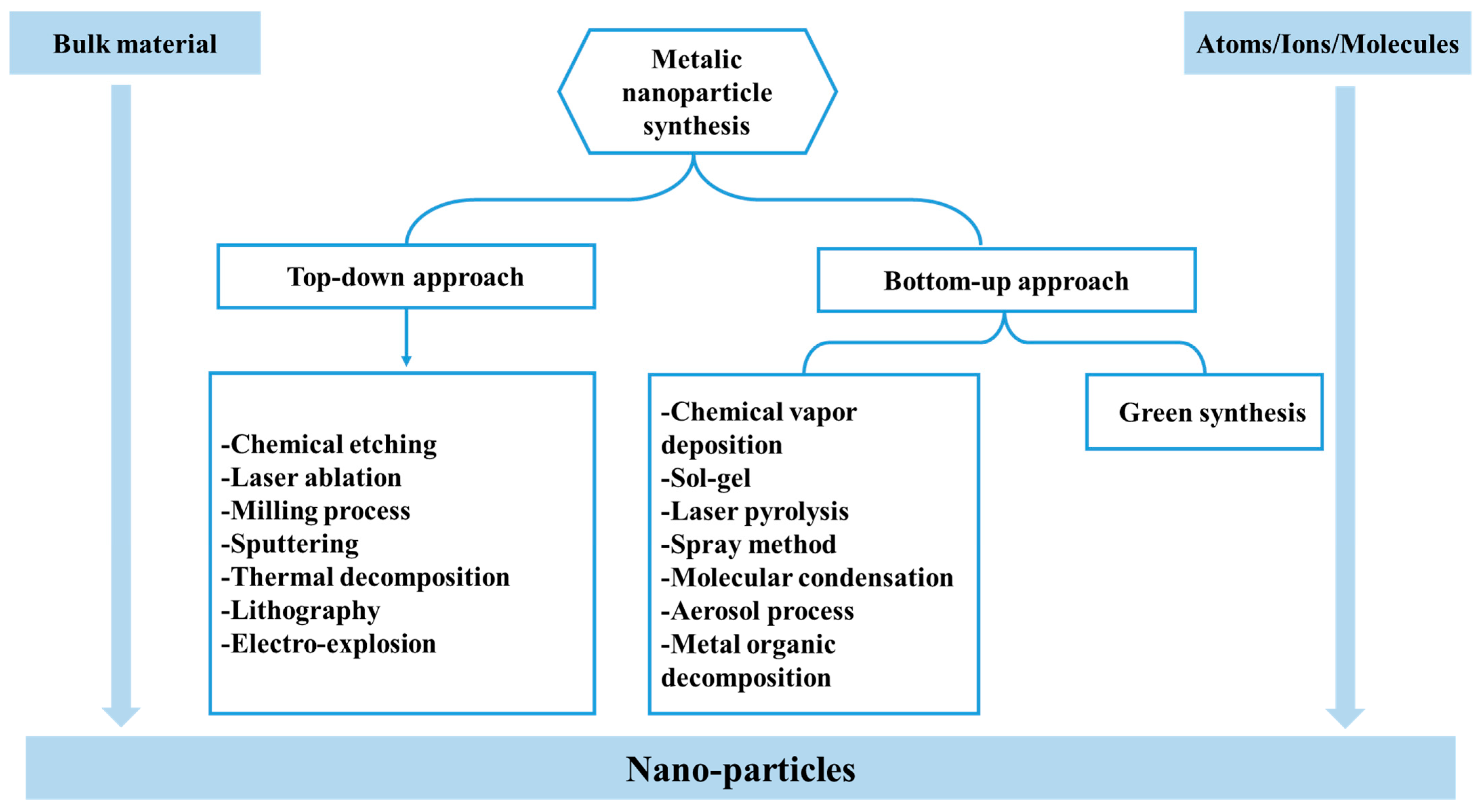
Figure 1. Different approaches for metallic nanoparticle synthesis.
Reactions 06 00055 g001
Figure 2. Photocatalysis mechanism.
Reactions 06 00055 g002
Figure 3. Illustration of various bio-synthetic routes of NPs.
Reactions 06 00055 g003
Figure 4. Green Synthesis Method of TiO2 NPs from Tinospora cordifolia.
Reactions 06 00055 g004
Figure 5. An illustration of Tauc’s plot for the green synthesised TiO2 NPs used for the calculation of their energy band gap through linear extrapolation [69].
Reactions 06 00055 g005
Figure 6. Effect of UV irradiation duration on Acid blue 113 removal [69].
Reactions 06 00055 g006
Figure 7. Percentage of degradation ability for methylene blue of synthesized TiO2 samples with/without visible light irradiation.
Reactions 06 00055 g007
Figure 8. Schematic diagram for the synthesis TiO2 NPs by Citrus limetta extract.
Reactions 06 00055 g008
Figure 9. XRD pattern of synthesized TiO2 nanoparticles [70].
Reactions 06 00055 g009
Figure 10. Degradation efficiency of green TiO2 [70].
Reactions 06 00055 g010
Figure 11. Recycle study of the synthesized TiO2 nanoparticles.
Reactions 06 00055 g011
Figure 12. Discoloration percentage of RB5 at 90 min.
Reactions 06 00055 g012
Figure 13. XRD-Spectra of reported ZnO nanoparticles (a), Synthesized zinc oxide nanoparticles (b) and (c,d) for calcium-coated zinc oxide nanoparticles (1:1,1:2) [66].
Reactions 06 00055 g013
Figure 14. Percentage of photocatalytic degradation of MB across different reuse cycles.
Reactions 06 00055 g014
Figure 15. The potential for reuse of ZnO NPs in terms of their efficacy in removing COD after 5 cycles.
Reactions 06 00055 g015
Figure 16. (a) Reflectance spectrum and (b) Tauc’s plot of WO3NPs [98].
Reactions 06 00055 g016
Figure 17. Degradation rate of MB (20 ppm) and CR (40 ppm) dye under visible light irradiation.
Reactions 06 00055 g017
Figure 18. Effects of various scavengers on the photocatalytic efficiency of WO3 sheets towards (a) MB at initial concentrations of 20 ppm, and (b) CR at initial concentrations of 40 ppm under visible light irradiation [99].
Reactions 06 00055 g018
Figure 19. Experimental procedure for the green synthesis of WO3 NPs with Tamarindus indica extract.
Reactions 06 00055 g019
Figure 20. Recyclability for the photocatalytic degradation of ofloxacin by WO3 nanoparticles [102].
Reactions 06 00055 g020
Figure 21. XRD pattern of WO3 nanoparticles [102].
Reactions 06 00055 g021
Figure 22. FTIR spectra of E. alata extract plant, green synthesized CuO-NPs and chemical CuO-NPs [67].
Reactions 06 00055 g022
Figure 23. Percentage degradation rate of MB dye compared to the chem CuO-NPs and biosynthesized CuO-NPs.
Reactions 06 00055 g023
Figure 24. XRD of green synthesized CuO-NPs [68].
Reactions 06 00055 g024
Figure 25. (a) XRD pattern of CuO, (b) FTIR spectrum of CuO, (c) UV absorbance spectra of CuO, (d) Degradation of (MB) by 200 mg of CuO nanoparticles [103].
Reactions 06 00055 g025
Figure 26. Crystal size of CuO NPs in different molarities.
Reactions 06 00055 g026
Figure 27. UV Absorbance of CuO nanoparticle in Different Molarities [104].
Reactions 06 00055 g027
Figure 28. Photocatalytic efficiency (a) in different molarities. (b) of CuO NPs (0.2 M).
Reactions 06 00055 g028
Figure 29. Degradation percentage of Congo red (a) and methylene blue (b), and insert color images were visualized degradation of Congo red (a) and methylene blue (b) dyes in aqueous solution to treated biosynthesized CuO NPs [109].
Reactions 06 00055 g029
Figure 30. Particle size distribution curve of Cu2O NPs [116].
Reactions 06 00055 g030
Figure 31. Band gap and insertion of the DRS diffuse reflectance spectrum (a) A1, (b) A2 samples [118].
Reactions 06 00055 g031
Figure 32. NORF removal efficiency after repeated test cycles.
Reactions 06 00055 g032
Figure 33. FTIR spectrum of α-Fe2O3 nanoparticles [127].
Reactions 06 00055 g033
Figure 34. Influence of catalyst loading on degradation of dye [128].
Reactions 06 00055 g034
Figure 35. Schematic diagram of a possible mechanism for photocatalytic degradation of MB dye by Ag/Fe2O3.
Reactions 06 00055 g035
Figure 36. Reusability study of Fe2O3 NPs and Ag/Fe2O3 NCs [129].
Reactions 06 00055 g036
Table 2. XRD Peak positions and corresponding crystal planes of green TiO2.
25.33°37.90°47.89°53.90°54.94°62.74°70.18°74.96°
Crystal plane(101)(004)(200)(105)(211)(204)(220)(215)
Table 3. Variation in precursor and Aloe vera (L.) Burm. f. rind extracts volume ratio [65].
Sample Code Percent Extact (v/v) Volume (mL) Ratio Precursor/Extract
TO0 5:0
TOAv44% 5:0.2
TOAv1010% 5:0.5
TOAv2020% 5:1
Table 4. Different plant extracts and precursors were used to synthesize TiO2 NPs.
Plant ExtractMetal PrecursorSize (nm)BG (eV)Ref.
Aloe veraTiO214–243.15–3.18[65]
Tinospora cordifoliaTiO215.023.13 [69]
MulberryTiO2243.16[70]
Lemon peelTiO280–1403.08[76]
Citrus limettaTiO280–1003.22 [77]
Syzygium cuminiTiO2103.48[78]
Piper betelTiO26.6-[79]
Ocimum tenuiflorumTiO27.0-[79]
Moringa oleiferaTiO26.6-[79]
Mentha spicataTiO2113.22–3.25[80]
Terminalia catappa and carissa carandasTiO210–213.21[81]
Trigonella foenum-graecumTiO240–60-[82]
Coriandrum sativumTiO26.8-[79]
Monsonia burkeanaTiO28.933.53[83]
Jatropha curcasTiO2133.28[84]
Table 5. Comparative studies of green-TiO2 on the photocatalytic activities.
PhotocatalystPlant UsedPollutantExperimental Conditions% DegradationRef.
TiO2Aloe veraMB-Calcination temperature: T = 500 °C (1 h)
-Irradiation source: UV light
48%, 51.44%, 47.82%,
49.29% (without plant extract, with 4%, 10%, 20%, respectively)
[65]
TiO2Tinospora cordifoliaAcid Blue
113 dye
-Calcination temperature: T = 400 °C; -Irradiation source: UV light
-Time of photocatalysis: 80 min; -Pollutant concentration: 50 mg/L
-Amount of NPs: 2 g/L; -pH = 4
94.43%[69]
TiO2MulberryMB-Irradiation source: UV light; -Time of photocatalysis: 120 min
-Pollutant concentration: 10 ppm (10 mL); -Mass of NPs: 10 mg
96%[70]
TiO2Lemon peelRhB-Calcination temperature: T = 500 °C (2 h); -Irradiation source: UV light
-Time of photocatalysis: 120 min
>70%[76]
TiO2Citrus limetta -Calcination temperature: 550 °C (2 h); -Irradiation source: UV light; -Time of photocatalysis: 80 min; -Pollutant concentration: 10 mg/L (50 mL); -Mass of NPs: 0.7 g.>90%[77]
TiO2Syzygium cuminiPb-Calcination temperature: 570 °C (3 h); -Irradiation source: UV light; -Time of photocatalysis: 17 h; -pollutant concentration: 8.6 ppm (500 mL); -Mass of NPs: 0.3 g.75.5%[78]
COD-Calcination temperature: 570 °C (3 h); -Irradiation source: UV light; -Time of photocatalysis: 17 h; -Pollutant concentration: 8450 mg/L (500 mL); -Mass of NPs: 0.3 g.82.53%
TiO2Piper betelMalachite
green dye.
-Calcination temperature: T = 400 °C (3 h); -Irradiation source: Solar light; -Time of photocatalysis: 30 min; -pollutant concentration: 100 ppm (50 mL); -Mass of NPs: 100 mg
~50%[79]
Ocimum tenuiflorum~70%
Moringa oleifera~100%
Coriandrum sativum~60%
TiO2Mentha spicataReactive
Black 5
-Calcination temperature: T = 400 °C; -Irradiation source: UV light
-Time of photocatalysis: 90 min; -Pollutant concentration: 5 ppm (75 mL)
-Mass of NPs: 0.075 g
96%[80]
TiO2Monsonia burkeanaMB-Calcination temperature: T = 500 °C (1 h); -Irradiation source: UV light
-Time of photocatalysis: 120 min; -Pollutant concentration: 20 mg/L
-Mass of NPs: 60 mg; -pH = 10
85.5%[83]
TiO2Jatropha curcasCOD-Calcination temperature: T = 450 °C (3 h); -Irradiation source: Solar light.
-Time of photocatalysis: 5 h; -Pollutant concentration: 1428 mg/L.
82.26%[84]
Table 6. Different plant extracts and precursors were used to synthesize ZnO NPs.
Plant ExtractMetal PrecursorSize (nm)BG (eV)Ref.
BeetrootCa-ZnO20–50-[66]
Vitex negundoZnO193.16[87]
Myrtus communisAcetate-ZnO1003.21[88]
Nitrate-ZnO1003.24
Spinacia oleraceaZnO35–403.12[91]
Camellia sinensisZnO12.23.15[92]
Lawsonia inermisZnO223.37[93]
ZnO/Fe2O3392.8
Table 7. Comparative studies of green-ZnO NPs on the photocatalytic activities.
PhotocatalystPlant UsedPollutantExperimental ConditionsDegradation (%)Ref.
ZnOBeetrootMB -Calcination temperature: T = 450 °C (15 min); -Time of photocatalysis: 40 min; -Irradiation source: sunlight; -Pollutant concentration: 10 mg/L (100 mL); -Masse of NPs: 0.01 g100%[66]
Ca-ZnO-Calcination temperature: T = 450 °C (15 min); -Time of photocatalysis: 60 min; -Irradiation source: sunlight; -Pollutant concentration: 10 mg/L (100 mL); -Masse of NPs: 0.01 g90%
ZnO-acetateMyrtus communisMB-Calcination temperature: T = 400 °C; -Irradiation source: UV light.; Time photocatalysis: 50 min; -Pollutant concentration: 10 ppm (100 mL); -Masse of NPs: 0.1 g.98%[88]
ZnO-Nitrate-Calcination temperature: T = 400 °C; -Irradiation source: UV light.; -Time of photocatalysis: 60 min; -Pollutant concentration: 10 ppm (100 mL); -Masse of NPs: 0.1 g99%
ZnOSpinach leavesToluene-Calcination temperature: T = 300 °C (120 min); -Irradiation source: sunlight; Time photocatalysis: 240 min; Pollutant concentration: 0.5 g/L (150 mL); -Masse of NPs: 0.05 g; pH = 884.26%[91]
Xylene-Calcination temperature: T = 300 °C (120 min); -Irradiation source: sunlight; Time photocatalysis: 240 min; Pollutant concentration: 0.5 g/L (150 mL); -Masse of NPs: 0.05 g;
pH = 8
90.36%
COD-Calcination temperature: T = 300 °C (120 min); -Irradiation source: sunlight; -Time photocatalysis: 240 min; Pollutant concentration: 0.5 g/L (150 mL); -Masse of NPs: 0.05 g; pH = 881.24%
ZnOCamellia sinensischlortetracycline-Calcination temperature: T 400 °C (3 h); -Irradiation source: UV-light; -Time photocatalysis: 60 min; -Pollutant concentration: 10 mg/L (50 mL); -Masse of NPs: 50 mg; pH = 3.594.70%[92]
ZnO/H2O2Lawsonia inermisEffluent dye mixture-Irradiation source: Solar light; -Time photocatalysis: 3 h; -Pollutant (50 mL); -Masse of NPs: 1 mg; pH = 7 70.50%[93]
ZnO/Fe2O3/H2O2-Irradiation source: Solar light; -Time photocatalysis: 3 h; -Pollutant (50 mL); -Masse of NPs: 1 mg; pH = 7 90.40%
ZnOSolanum trilobatumMB-Irradiation source: Sunlight; -Time photocatalysis: 90 min; -Pollutant concentration: 10 μm; -Catalyst Amount: 0.6 g/L 94.07%[94]
ZnOLepidagathis ananthapuramensisMB-Irradiation source: Sunlight; -Time photocatalysis: 120 min; -Pollutant concentration: 10 ppm; -Catalyst Amount: 5 mg98.50%[95]
ZnOEuphorbia miliiMB-Irradiation source: Sunlight; -Time photocatalysis: 50 min; -Pollutant concentration: 10 μm
-Catalyst Amount: 0.6 g/L
98.17%[96]
ZnOPhoenix roebeleniiMB-Irradiation source: UV lamp; -Time photocatalysis: 105 min; -Pollutant concentration: 10 ppm; -Masse of NPs: 0.2 g98%[97]
Table 8. XRD Peak Positions and corresponding crystal Planes of green CuO.
25.01°28.91°32.21°33.21°33.85°35.5°37.7°44.96°65.11°
Crystal plane(202)(400)(110)(200)(002)(111)(−142)(112)(113)
Table 9. Comparative studies of green-CuO NPs on the photocatalytic activities.
PhotocatalystPlant UsedPollutantExperimental ConditionsDegradation EfficiencyRef.
CuOEphedra AlataMB-Calcination temperature: T = 400 °C (4 h); -Irradiation source: Sunlight
-Time of photocatalysis 3 h; -Pollutant concentration: 10 mg/L; -Mass of NPs: 20 mg
93.4%[67]
CuOGreen tea extractMB-Calcination temperature: T = 380 °C (10 min)
-Irradiation source: Sunlight (850 Wm−2); -Time of photocatalysis: 3 h
-Pollutant concentration: (5–20 ppm); -Mass of NPs: 50–200 mg
92%[103]
-Calcination temperature: T = 380 °C (10 min)
-Irradiation source: UV-light (125 Wm−2); -Time of photocatalysis: 3 h
-Pollutant concentration: (5–20 ppm); -Mass of NPs: 50–200 mg
99%
CuOLantana camaraMB-Calcination temperature: T = 500 °C (4 h); -Irradiation source: Sunlight;
-Time of photocatalysis: 90 min; -Molarity of NPs: 0.2 M
94%[104]
CuOPsidium guajavaMB-Irradiation source: sunlight; -Time of photocatalysis: 2 h;
-Pollutant concentration: 30 mg/L; -Mass of NPs: 20 mg
89%[109]
Congo Red-Irradiation source: sunlight; -Time of photocatalysis: 2 h;
-Pollutant concentration: 30 mg/L; -Mass of NPs: 20 mg
81%
Cu2OCurcuminMO-Irradiation source: UV-light; -Time of photocatalysis: 2 h
-Pollutant concentration: 150 μm; -Mass of NPs: 50 mg; pH = 3
94.5%[116]
Table 10. Photocatalytic performance of α-Fe2O3 nanoparticles against various dyes.
SamplePlant ExtractPollutant/DyeVolumePollutant DoseCatalyst DoseLight IrradiationContact TimeBG (eV)Crystallite Size (nm)Removal (%)Ref.
α-Fe2O3Syzygium cuminiNorfloxacin50 mL50 mg/L 50 mgUV90 min1.9–2.0 28.51–37.6496 [118]
Fe2O3Trachyspermum ammiMB100 mL10 ppm10 mg Visible120 min-2691.0[127]
α-Fe2O3Carica papayaRemazol Yellow100 mL50 ppm0.8 g/LSunlight250 min-21.5975[128]
Ag/Fe2O3Carica papayaMB-20 mg/L0.2 g/LVisible90 min2.9129.1792.97[129]
Fe2O3Pomegranate seedsReactive Blue100 mL20 mg/L15 mgUV56 min-25–5595.08 [130]
Fe2O3Cynometra ramiflora MB50 mL20 ppm30 mgSunlight4 h-68.1794.0[131]
Fe2O3Cassia auriculataMalachite Green 100 mL-20 mgVisible150 min--91.2[132]
Fe2O3Withania coagulansSafranin100 mL10 ppm0.5 mgSunlight180 min4.2016 ± 268.8[133]
Disclaimer/Publisher’s Note: The statements, opinions and data contained in all publications are solely those of the individual author(s) and contributor(s) and not of MDPI and/or the editor(s). MDPI and/or the editor(s) disclaim responsibility for any injury to people or property resulting from any ideas, methods, instructions or products referred to in the content.