Article

The Second Protonation in the Bio-Catalytic Cycles of the Enzymes Cytochrome P450 and Superoxide Reductase

Department of Chemistry, Columbia University, New York, NY 10027, USA
*
Author to whom correspondence should be addressed.
Reactions 2024, 5(4), 778-788; https://doi.org/10.3390/reactions5040039
Submission received: 4 September 2024 / Revised: 4 October 2024 / Accepted: 11 October 2024 / Published: 14 October 2024

Abstract

:
The enzymes Cytochrome P450 and Superoxide Reductase, which have a similar coordination center [FeN4S], begin their biochemical cycles similarly. They absorb an oxygen molecule, add two electrons, and link a hydrogen atom to the distal oxygen atom of the product obtained, creating the so-called Compound 0 in the case of the first enzyme. However, the bio-catalytic processes of these two enzymes continue in different ways. In the bio-catalytic cycle of Cytochrome P450, the enzyme binds another proton to the distal oxygen atom, producing a water molecule and Compound 1. In contrast, in the bio-catalytic cycle of the Superoxide Reductase, the enzyme binds a proton to the proximal oxygen atom, producing a hydrogen peroxide molecule, which later decomposes into oxygen and water. The MCSCF method in the CASSCF form was used to study the difference in Cytochrome P450 and Superoxide Reductase’s bio-catalytic cycles. The results of these enzymes’ hydroperoxo adduct models’ geometric optimization showed that, in fact, all their properties, including their spin states, the wave functions in their active zones, and the Fe-N, Fe-S, and Fe-O bond lengths, are different. The Fe-N, Fe-S, and Fe-O chemical bond lengths are much longer in the case of the second enzyme compared to the chemical bond lengths in the case of the first enzyme, reflecting a spin value equal to 5/2 in the second case and a spin value equal to 1/2 in the first. A decisive role in the difference in their bio-catalytic cycles is played by the fact that the first bonded hydrogen atom is linked to the distal oxygen atom in the side position in the case of Compound 0 and the up position in the case of the hydroperoxo adduct of the enzyme Superoxide Reductase, protecting the distal oxygen atom from possible interaction with the substrate. The second protonation to Compound 0 at the distal oxygen atom in the case of Cytochrome P450’s bio-catalytic cycle and the second protonation at the proximal oxygen atom in the case of the hydroperoxo adduct of Superoxide Reductase’s bio-catalytic cycle depend on the proton transfer through the Asp251 channel in the first case and on the transferal of H+ from the substrate to the water molecule and the proximal oxygen in the second case.

1. Introduction

The biochemistries of the enzymes Superoxide Reductase and Cytochrome P450 have attracted the attention of an impressive number of researchers [1,2,3]. The iron centers of the active sites of both of these enzymes are coordinated with four very similar structures: Superoxide Reductase has four separate imidazole-like-structure histidine molecules, while Cytochrome P450 has four pyrrole-like subunits connected by carbon bonds and forming a corrin ring, and there is a cysteine ion in the axial ligand position in both cases, forming a similar coordinating center (Figure 1). Superoxide Reductase’s enzymatic activity in anaerobic bacteria leads to the annihilation of toxic peroxide radicals by reducing them into oxygen peroxide, degrading further into water and oxygen [4,5,6,7,8]. Cytochrome P450 is an enzyme family that has some functions in living organisms and humans, such as participation in the synthesis of hormones necessary for the production of androgens; the oxidation of steroids, fatty acids, and xenobiotics; and the detoxification of organisms from various organic compounds and drugs [9,10].
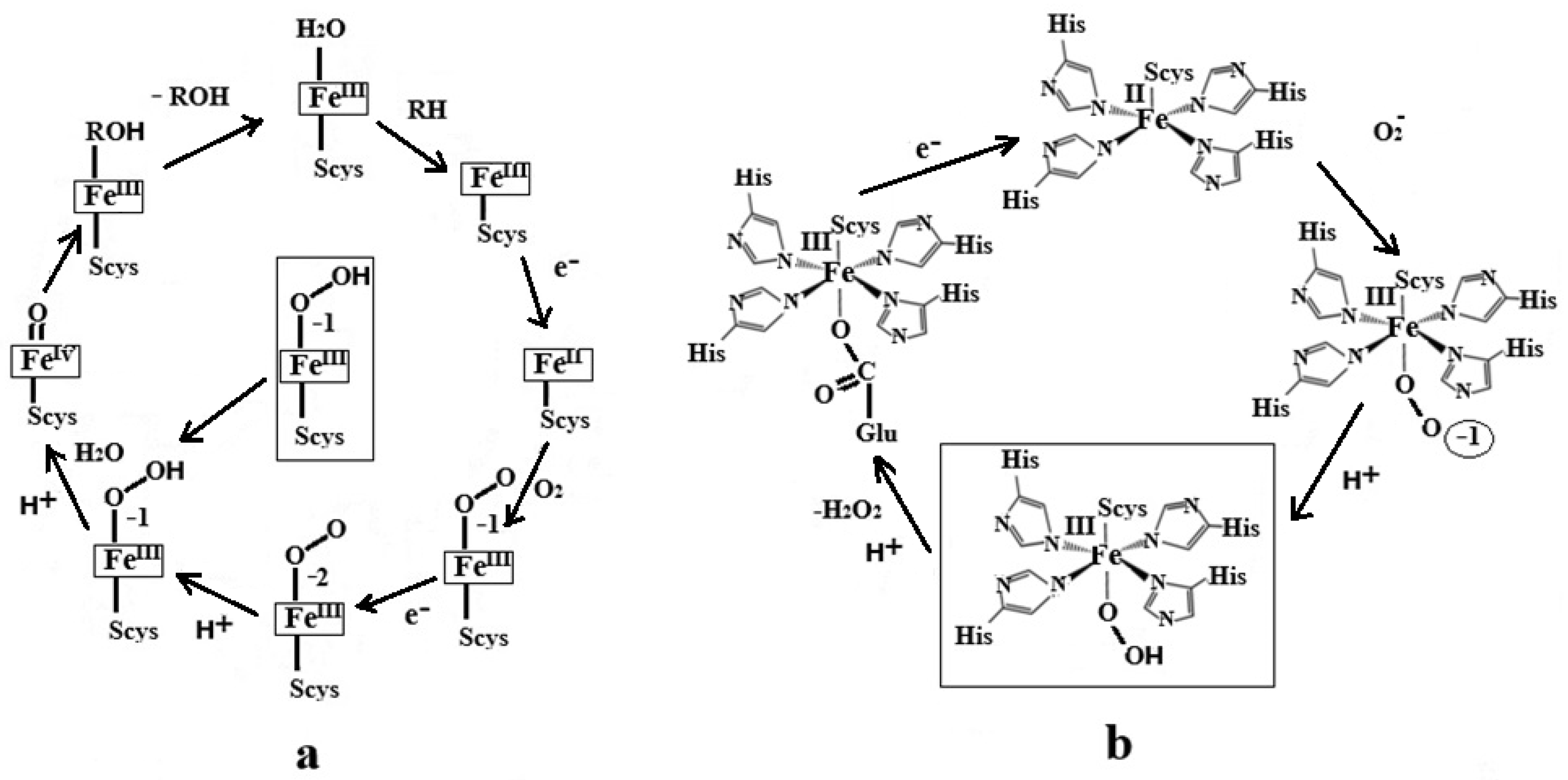
The research community has generally established Superoxide Reductase and Cytochrome P450’s bio-cycles’ catalytic processes (Scheme 1) [1,2,3,4,5]. As shown in Scheme 1b of the Superoxide Reductase enzyme processes, the active site, the iron coordinating center, is reduced by one electron. It coordinates the radical O2, a process that, in principle, would be similar to a reduction by one more electron and the coordination of an oxygen molecule. The enzyme processes occurring at the beginning of the cycle of the bio-catalytic transformations of the active site of Cytochrome P450 are very similar [1,2,3,11,12,13]. In this case, there are also two reductions with one electron and the coordination of the O2 molecule. The active sites of both enzymes continue to behave similarly in the next step; in both cases, the oxygen molecule of the active site binds a proton to the end of the oxygen molecule called the distal oxygen atom. As can be seen from Scheme 1, the subsequent behavior of the active site of Superoxide Reductase radically differs from that of the active site of Cytochrome P450. The active site of Superoxide Reductase acidifies another proton. It produces a hydrogen peroxide molecule, which splits into oxygen and water. In turn, the active site of Cytochrome P450 acidifies another proton, producing a water molecule and Compound 1, of which the latter, being very active, subsequently deactivates toxic organic compounds in humans and animals.
Even though Cytochrome P450 and Superoxide Reductase transfer the second proton to the coordinated oxygen molecule, the conditions for this transfer differ strongly. The active site of Superoxide Reductase is under the direct control of a water molecule or molecules from the substrate complex [11]. In contrast, for Cytochrome P450’s active site, several channels [13] through which the coordinated oxygen molecule can be protonated are available throughout the in vivo structures. Three coordinated oxygen protonation channels are considered in this regard: Asp251 [14,15,16], Glu366 [17,18], and Arg299 [19].
Density functional theory (DFT) and quantum mechanics/molecular mechanics (QM/MM) methods have been used in Cytochrome P450 enzyme biochemistry compound I production studies. Thus, it has been determined that the binding of the first hydrogen to the distal oxygen is energetically more convenient than the binding of the first hydrogen to the proximal oxygen, and the binding of two hydrogens to the distal oxygen automatically leads to the breaking of O=O bonds and the formation of compound I [20,21]. The distal oxygen’s protonation mechanism through the Asp251, Glu366, and Arg299 in vivo channels has been studied [19,20,21,22,23,24,25]. However, priority has been given to the protonation mechanism of distal oxygen through the Asp251 channel [13]. In one study, the Arg299 channel was analyzed using molecular dynamics modeling and a site-directed mutagenesis study; as a result of this activity, the channel was blocked by the substrate [26]. On the other hand, analysis of the Glu366 channel showed that it did not stretch to the surface [14]. As expected, the DFT calculations of mono-protonated and di-protonated Superoxide Reductase peroxo compounds showed that the protonation of proximal oxygen is a way of obtaining hydrogen peroxide in its bio-catalytic circuit [27]. It was shown that Fe-O bond cleavage in all spin states occurs more easily than O=O bond cleavage [28]. Intensive DFT, complete active-space self-consistent field (CASSCF), and UV-vis research was carried out to establish the nature of the chemical bond and the spectra of the active site of Superoxide Reductase [29]. The structures of Superoxide Reductase from the Hyperthermophilic archaea Ignicoccus hospitalis and Nanoarchaeum equitans were determined using the X-ray method, and it was demonstrated that the Fe-N bond distances in Superoxide Reductase’s active site are quite large, indirectly confirming its high-spin state [30]. Another interesting fact is that one or more water molecules can interact directly with the central iron atom [30]. Comparative analyses of the electronic structures of Cytochrome P450 and Superoxide Reductase hydroperoxo-compounds and their cyanides using ENDOR spectroscopy and the DFT method have also been conducted [31,32].
One of the differences between the active sites of the enzymes Cytochrome P450 and Superoxide Reductase is their spin state. Generally, the former is a low-spin compound, while the second has a high-spin state. Considering that the biochemical behavior and spin states of Cytochrome P450 and Superoxide Reductase hydroperoxo compounds in vivo are difficult to determine experimentally, studying them by determining their electronic structures is a priority. On the other hand, to the best of our knowledge, it has not yet been determined why, after adding the first and second protons, Cytochrome P450 produces water and Compound I; in contrast, Superoxide Reductase produces hydrogen peroxide. The multi-configurational self-consistent field (MCSCF) method builds its wave function as a superposition of many determinants with various spin states able to determine the calculated compound’s exact spin state and the Jahn–Teller effect or the pseudo-Jahn–Teller effect’s role in its biochemical behavior. In this study, we used the MCSCF method to determine the behavior of Cytochrome P450 and Superoxide Reductase intermediate hydroperoxo compounds. In the proposed mechanisms, in Compound 0, the proton transferred through the Asp251 channel reaches the W901 water molecule, creating a hydronium ion, which instantly transmits the proton to the distal oxygen, breaking the O=O bond and releasing a water molecule and Compound 1. In turn, the Superoxide Reductase hydroperoxo compound (SORHPC) instantly binds a proton to the proximal oxygen from a water molecule transformed into a hydronium ion by transferring a proton from the substrate, breaking the Fe-O bond instantly and releasing a peroxide hydrogen molecule, which is converted into water and oxygen.

2. Computational Details

The structures of the active sites of Superoxide Reductase and Cytochrome 450, with an oxygen molecule attached to the central iron atom and a proton linked to the distal oxygen, were determined via CASSCF geometry optimization, with an active zone containing 13 electrons and 13 orbitals. Such an active area was successfully applied in MCSCF calculations of vitamin B12 cofactors [33,34,35,36,37]. The proton bound to the distal oxygen was placed in developing structures in various positions, including the up and down ones (Figure S1). The obtained models were recalculated using an active zone of 15 electrons and 14 orbitals. Then, based on the obtained structures, two geometric models were constructed for a similar CASSCF geometric optimization. The first one included the Asp251 channel, in which, along with aspartate-251 and Thr252 models, a trapped molecule of water 901 was used, in addition to the Compound 0 model (Figure S1). The second one included a water molecule placed in different positions toward the -O-O-H group plus the SORHPC model. Other structures were determined: in the case of the model containing the active site of Cytochrome P450, a proton is transferred from a water molecule to the distal oxygen atom, and in the case of the model containing the active site of Superoxide Reductase, a proton is transferred from a water molecule to the proximal oxygen atom. The Def2-TZVP basis set was used for the iron and sulfur atoms, and the 6-31G** basis set was used for the rest of the atoms. Hydrogens replaced the side chains. Adding dynamic factors to calculate the models with the CASPT2 approximation would have been desirable. Still, the cost of calculations for such large systems would have been beyond our current computational possibilities. All calculations were performed by using the NwChem code [38].

3. Discussion

As shown above, after the binding of the oxygen molecule to the central iron atom; two-electron reductions, each with an electron; and the transfer of a proton to the distal oxygen atom, the processes related to the enzymes Cytochrome P450 and Superoxide Reductase continue differently. Compound 0 transfers another proton to the distal oxygen. It produces Compound 1 and a water molecule. The SORHPC transfers another proton to the proximal oxygen atom, producing a hydrogen peroxide molecule that splits into water and oxygen. The mechanism of the second proton’s transfer either to the distal oxygen, in the case of Compound 0, or to the proximal oxygen, as in the case of the SORHPC, depends on these two compounds’ geometric and electronic structures. The previous DFT calculations showed that the proton bound to the distal oxygen would be in the down position [12,13]. Our DFT calculations showed the same thing in both cases.
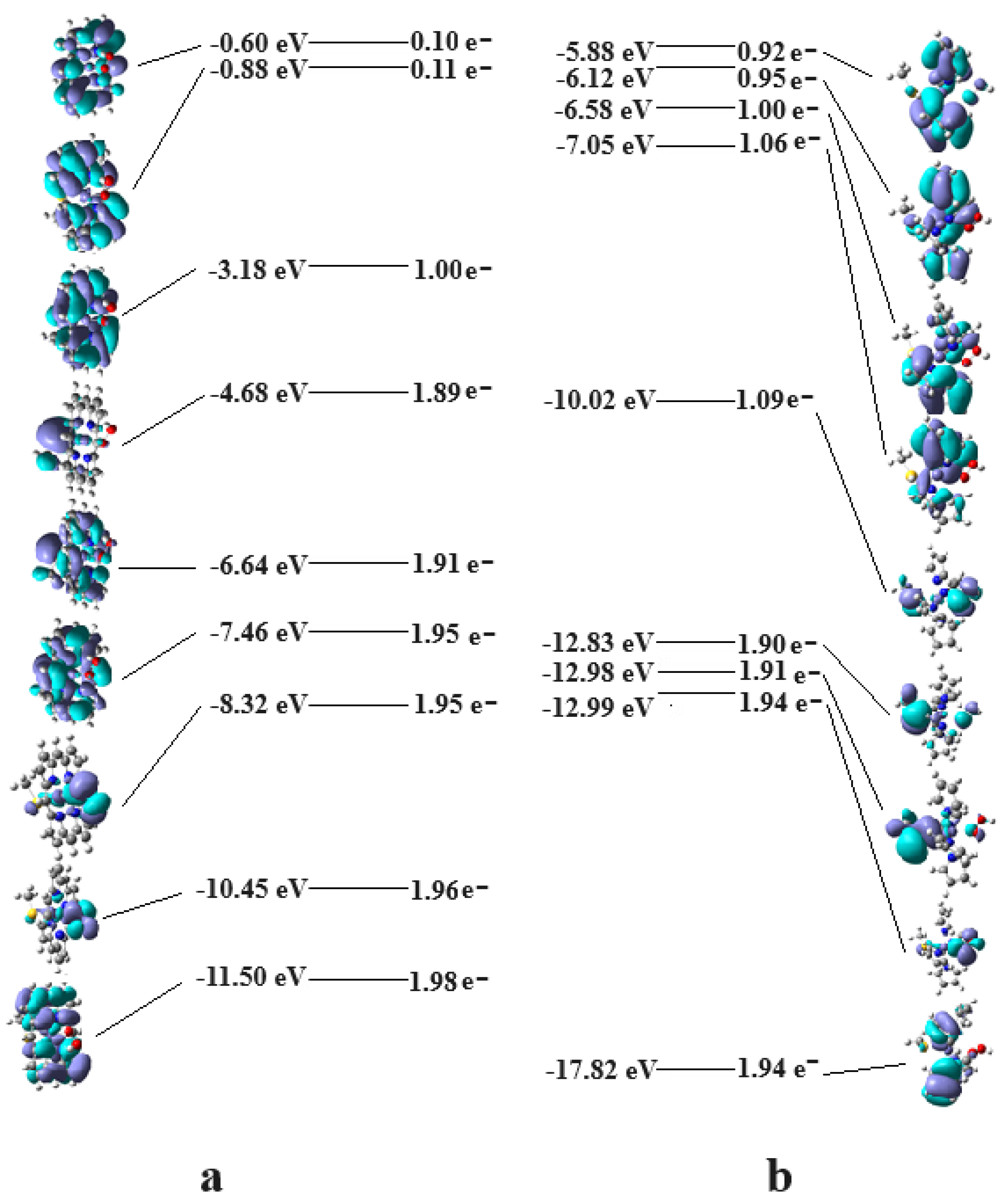
It is known that the DFT method does not consider orbital mixing [39,40]. Therefore, it was interesting to determine the hydroperoxo compounds’ electronic structures using the CASSCF method, which considers the pseudo-Jahn–Teller effect or, in other words, orbital mixing [41]. As shown above, structural models were initially used, in which the proton bound to the distal oxygen is in different positions, including up and down (Figure S1). The results of CASSCF geometry optimizations show that in Compound 0, the proton bound to the distal oxygen is in a quasi-parallel position and slightly bent towards the corrin ring. In the case of the SORHPC, there are two minima of energy, both in the up and down positions. The total energy of the compound with the proton in the down position is at least 1.3 eV higher than that of the compound with the proton in the up position. This result is supported by the structural experimental data obtained, which show no hydrogen bonds in the down position in the crystals of the compound [42]. Therefore, the models used further in the calculations contain Compound 0 with the hydrogen bonded to the distal oxygen in the side position and the SORHPC with the hydrogen bonded to the distal oxygen in the up position. Without a doubt, the most interesting difference between Compound 0 and SORHPC relates to their spin states; in the first case, the spin state is equal to 1/2, while in the second case, it is equal to 5/2, even if the formal structures of the chemical bonds to the central atom of both compounds [FeN4SO] are similar. In fact, as shown in Figure 2, the electron structures of the two compounds are totally different. In the case of Compound 0, the electrons are located on the lowest-possible molecular orbitals, forming only one molecular orbital populated with one electron due to the odd number of electrons in the system. In contrast, in the SORHPC, the mixing of the orbitals leads to the development of a population of electronic densities on the molecular orbitals in the active zone, which almost ideally coincides with the spin state of 5/2. This is a natural and expected result considering that the wave function, in the case of the CASSCF method, is a combination of multiplicities, in which the mixture of them produces real–natural multiplicity. Another interesting novelty is the molecular orbitals (populated with one electron), most of which are π-type, in which the atomic orbitals of the central atom do not necessarily make the biggest contribution in the case of the SORHPC. It should be mentioned that the Fe-N, Fe-S, and Fe-O chemical bond lengths are significantly longer in the case of the SORHPC than in the case of the Compound 0, reflecting the high-spin 5/2 state in the first case and the low-spin 1/2 state in the second case. The bond distances obtained in our CASSCF geometry optimizations agree with the experimental structural data [30]. The results of the CASSCF geometry optimizations, which will be considered in the following electronic structure calculation models, are presented in Tables S1 and S2 of the Supporting Information.
As mentioned above, for the CASSCF calculations, we chose to model the Asp251 channel in which, apart from the Compound 0 model, an aspartate-251, a threonine, Thr252 models, and a trapped molecule of water 901 were used (Figure 3). Given that water molecules influence the SORHPC, we chose to build a calculation model in which, along with its structure model, a water molecule is present, which was placed in various positions toward the -O-O-H group (Figure S1).
As shown above, Cytochrome P450 begins its biochemical cycle with a two-electron reduction, the oxygen molecule’s coordination at the central iron atom, and the transfer of a proton to the distal oxygen atom, forming Compound 0. In the next step, Cytochrome P450 transfers a second proton to the same distal atom of the oxygen molecule coordinated to the iron atom, producing a water molecule and Compound 1. Substitution of Thr252 with serine, valine, alanine, or glycine or structural modification of the Thr252 residue leads to the contamination of the production mechanism of the water molecule and Compound 1, giving a percentage of hydrogen peroxide instead [13]. This proves that both oxygen atoms of Compound 0 can be protonated if the structural construction allows it from a geometrical point of view. Thus, it can be concluded that Thr242 plays a structural role. The role of proton transfer lies in the Asp251 channel, with the water molecule as the last transmission factor, given that the pKa of water would stimulate this process rather than alcohol.
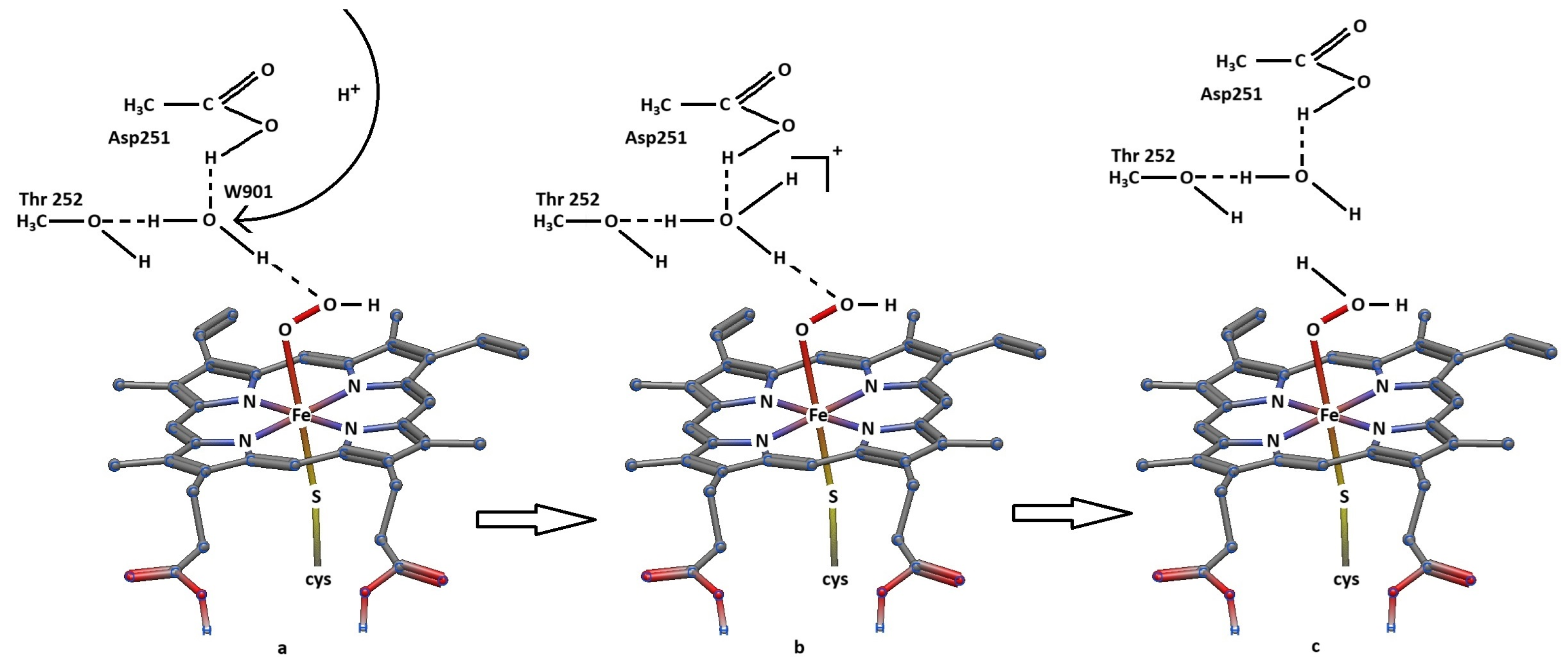
The geometry optimization of Compound 0 together with the neighboring substrates led to three hydrogen bonds between one hydrogen of W901 and the distal oxygen, the length of which is equal to 1.69 Å; between the hydrogen of the OH group of Thr252 and the oxygen of water 901, for which the length is equal to 2.00 Å; and between the hydrogen of the -COOH group of Asp251 and water 901, with a length equal to 1.81 Å (Figure 3). This suggests that there are two ways in which Compound 0 can be protonated: a transfer of a hydrogen atom directly from water 901 to the distal oxygen or the transfer of a proton through the Asp251 channel to water 901, forming a hydronium ion, which, in turn, transfers a proton to the distal oxygen atom. Our calculations show that the transfer of a hydrogen atom from water 901 to the distal oxygen causes an increase in the total energy of the calculated system by at least 1.00 eV. According to Boltzmann’s distribution, the population of this complex is practically equal to zero when compared to the population of the initial structures before the proton transfer. The transfer of a proton from water 901 to the distal oxygen through this mechanism appears improbable. On the other hand, the geometry optimization of the structure, in which water 901 is converted into a hydronium ion, leads to the instantaneous transfer of a proton from the hydronium ion to the distal oxygen and the breaking of the O=O bond in Compound 0, leading to a new water molecule and Compound 1. The proposed mechanism is shown in Figure 4.
The substrate in the case of the SORHPC is different from that around Compound 0. In this case, the proton transfer channels are missing; instead, the SORHPC is under the influence of one or more water molecules [11,30]. Therefore, the CASSCF geometry optimization model includes, in addition to the SORHPC model, a water molecule, which was placed in various relationships with the atoms of the oxygen peroxide group. CASSCF geometry optimizations in which a molecule was placed in various positions with respect to the -O-O-H group showed that the water molecule, with one exception, moved away from this hydroperoxo group without interacting with it. Only in one case did the water molecule form a hydrogen bond with the proximal oxygen atom (Figure 3). A decisive role in this location was played by the hydrogen atom connected to the distal oxygen, which was in the up position, on the one hand preventing water molecules from approaching the distal oxygen and, on the other hand, orienting them such that their negative charge sides face this hydrogen (and distal oxygen atoms) via the positive charge of the latter, making it unlikely for there to be any hydrogen bonds between the water molecules and distal oxygen. Unlike the behavior of the water molecules around the distal oxygen, the single water molecule around the proximal oxygen approached this oxygen during the geometry optimization process, forming a permanent hydrogen bond with it (Figure 3). CASSCF calculations (13,13) showed that the length of this hydrogen bond is equal to 2.13 A.
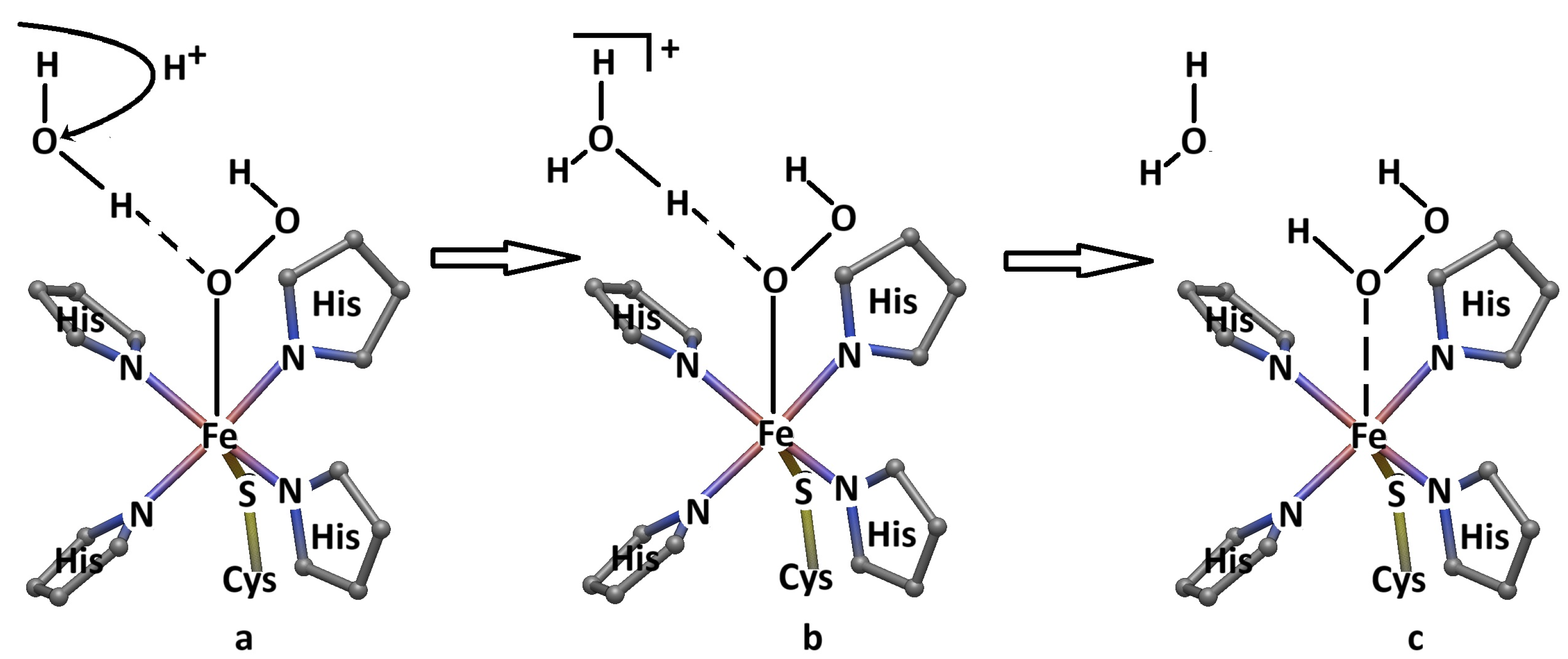
As shown above, the distal oxygen of the SORHPC does not create a chemical bond with a water molecule in its vicinity. It must be said that any potential hydronium ions from the aquatic environment near the SORHPC also cannot form hydrogen bonds with it due to the shielding of the distal oxygen by the positively charged hydrogen atom bound in the up position. Therefore, there are no proton transfer bridges with the distal oxygen of the SORHPC. In contrast, there are two ways to protonate the proximal oxygen of the SORHPC molecule. Indeed, the protonation of the proximal oxygen of the studied system is possible through the transfer of a proton linked by the hydrogen bond between the water molecule and the proximal oxygen or the transfer of a proton from the substrate system to this molecule, creating a hydronium ion close to the proximal oxygen atom, with the possibility of a proton transferring from it to the proximal oxygen. Our calculations show that the transfer of a proton from the water molecule, linked to the proximal oxygen of the studied system, to this oxygen atom leads to an increase in the total energy by approximately 1.00 eV, the population of which in the Boltzmann distribution is practically equal to zero, making this mechanism improbable. On the other hand, our calculations show that the CASSCF geometric optimization of the SORHPC plus a hydronium molecule causes this molecule to approach the proximal oxygen, with the instantaneous transfer of a proton from it to this oxygen atom and with the instantaneous breaking of the Fe-O bond, releasing an oxygen peroxide molecule. In conclusion, the most probable mechanism of SORHPC second protonation and the release of a hydrogen peroxide molecule may take place through the transfer of a proton from the substrate environment to a water molecule, which is linked to the proximal oxygen, instantly transferring it to the proximal oxygen, breaking the Fe-O bond and releasing a hydrogen peroxide molecule. This mechanism is shown in Figure 5.

4. Conclusions

Although Cytochrome P450 and Superoxide Reductase’s hydroperoxo compounds have similar coordinating nuclei [FeN4SO], their structural, spin state, and biochemical properties differ. Our CASSF geometry optimizations show that the Fe-N, Fe-S, and Fe-O bond lengths are much larger in the case of SORHPC than in the case of Compound 0, reflecting the fact that in the first case, we have a compound with high spin, equal to 5/2, and in the case of the second compound, we have low spin, equal to 1/2. Likewise, the wave functions in the active zones of Cytochrome P450 and Superoxide Reductase’s hydroperoxo compounds are different. CASSCF geometry optimization of the Superoxide Reductase and Cytochrome P450 hydroperoxo compounds shows that in the first case, the hydrogen is linked to the distal oxygen in the up position and the side position in the case of the second compound. This fact dramatically influences and distinguishes the mechanism of adding the second proton to the -O-O-H residue of the studied hydroperoxo compounds. Transferring a proton from a water molecule to the -O-O-H group of Compound 0 or SORHPC is practically impossible due to the excessively high total energy of the obtained products. On the other hand, the formation of a hydronium ion by transferring a proton through the Asp251 channel to the water molecule linked by the hydrogen bond with the -O-O-H group of Compound 0 leads to the instantaneous transfer of a proton to the distal oxygen atom and the instantaneous breaking of the O=O bond, creating a water molecule and Compound 1. It looks like Thr252 plays the stereo-structural role of directing the water molecule W901 toward the distal oxygen atom of Compound 0. In the case of the SORHPC, the positively charged hydrogen atom, which is linked to the distal oxygen atom in the up position, rejects and directs the water molecules around the -O-O-H group in such a way that their interaction with the distal oxygen atom becomes improbable. Therefore, interaction with water molecules and the formation of hydrogen bonds only occur in the case of the proximal oxygen atom. This water molecule can accept a proton from the substrate complex to form a hydronium ion. The instantaneous transfer of a proton from the formed hydronium ion to the proximal oxygen atom breaks the Fe-O bond. This process forms a hydrogen peroxide molecule, which decomposes into oxygen and water. In conclusion, the release reactions of Compound 1 and a water molecule in the case of the Cytochrome P450 biocycle depend on proton transfer through the Asp251 channel. In the case of the Superoxide Reductase biocycle, the release of a hydrogen peroxide molecule depends on the transfer of a proton from the substrate environment to the water molecule linked by a hydrogen bond with the proximal oxygen atom.

Supplementary Materials

The following supporting information can be downloaded at: https://www.mdpi.com/article/10.3390/reactions5040039/s1, Figure S1: The initial models of the hydroxo-compounds used in CASSCF geometry optimizations are a. proton-dawn hydroxo Cytochrome P450; b. proton-up hydroxo Cytochrome P450; c. proton-dawn hydroxo Superoxide Reductase; and d. proton-up hydroxo Superoxide Reductase; Table S1: The geometry optimization converged CASSCF coordinates of the Cytochrome P450 cofactor hydroperoxo compound; Table S2: The geometry optimization converged CASSCF coordinates of the Superoxide Reductase cofactor hydroperoxo compound; Table S3: The geometry optimization converged CASSCF coordinates of the Cytochrome P450 cofactor hydroperoxo compound and substrates common model; Table S4: The geometry optimization converged CASSCF coordinates of the Superoxide Reductase hydroperoxo compound and water common model.

Author Contributions

Conceptualization, T.S.; methodology, T.S.; writing draft, review, and edition, T.S.; data curating and analysis, T.S., visualization, T.S., L.M.D., A.M. and M.M.; validation, T.S., L.M.D., A.M. and M.M. All authors have read and agreed to the published version of the manuscript.

Funding

This research was supported, in part, by the National Science Foundation through TeraGrid resources provided by the TeraGrid Science Gateways program under grants CHE090082 and CHE140071.

Data Availability Statement

The data are available at Frontera supercomputer upon an agreement with the corresponding coauthor.

Acknowledgments

The authors thank the Texas Advanced Computing Center team for support during the calculations.

Conflicts of Interest

The authors declare no conflict of interest.

References

  1. Kovacs, J.A. Synthetic Analogues of Cysteinate-Ligated Non-Heme Iron and Non-Corrinoid Cobalt Enzymes. Chem. Rev. 2004, 104, 825–848. [Google Scholar] [CrossRef] [PubMed]
  2. Makris, T.M.; Davydov, R.; Denisov, I.G.; Hoffman, B.M.; Sligar, S.G. Mechanistic Enzymology of Oxygen Activation by the Cytochrome P450. Drug Metab. Rev. 2002, 34, 691–708. [Google Scholar] [CrossRef] [PubMed]
  3. Shaik, S.; Cohen, S.; Wang, Y.; Chen, H.; Kumar, D.; Thiel, W. P450 Enzymes: Their Structure, Reactivity, and Selectivity-Modeled by QM/MM Calculations. Chem. Rev. 2010, 110, 949–1017. [Google Scholar] [CrossRef]
  4. Kovacs, J.A.; Brines, L.M. Understanding How the Thiolate Sulfur Contributes to the Function of the Non-Heme Iron Enzyme Superoxide Reductase. Acc. Chem. Res. 2007, 40, 501–509. [Google Scholar] [CrossRef]
  5. Kovacs, J.A. How Iron Activates O2. Science 2003, 299, 1024–1025. [Google Scholar] [CrossRef]
  6. Jenney, F.E.; Verhagen, M.F.J.M., Jr.; Cui, X.; Adams, M.W.W. Anaerobic Microbes: Oxygen Detoxification without Superoxide Dismutase. Science 1999, 286, 306–309. [Google Scholar] [CrossRef] [PubMed]
  7. Mathé, C.; Mattioli, T.A.; Horner, O.; Lombard, M.; Latour, J.-M.; Fontecave, M.; Nivière, V. Identification of Iron (III) Peroxo Species in the Active Site of the Superoxide Reductase SOR from Desulfoarculus baarsii. J. Am. Chem. Soc. 2002, 124, 4966–4967. [Google Scholar] [CrossRef] [PubMed]
  8. Kurtz, D., Jr. Microbial Detoxification of Superoxide: The Non-Heme Iron Reductive Paradigm for Combating Oxidative Stress. Acc. Chem. Res. 2004, 37, 902–908. [Google Scholar] [CrossRef]
  9. Ogliaro, F.; de Visser, S.P.; Cohen, S.; Sharma, P.K.; Shaik, S. Searching for the Second Oxidant in the Catalytic Cycle of Cytochrome P450: A Theoretical Investigation of the Iron(III)-Hydroperoxo Species and Its Epoxidation Pathways. J. Am. Chem. Soc. 2002, 124, 2806–2817. [Google Scholar] [CrossRef]
  10. Liu, Y.; Denisov, I.G.; Grinkova, Y.V.; Sligar, S.G.; Kincaid, J.R. P450 CYP17A1 Variant with a Disordered Proton Shuttle Assembly Retains Peroxo-Mediated Lyase Efficiency. Chem. A Eur. J. 2020, 26, 16846–16852. [Google Scholar] [CrossRef]
  11. Yeh, A.P.; Hu, Y.; Jenney, F.E., Jr.; Adams, M.W.W.; Rees, D.C. Structures of the Superoxide Reductase from Pyrococcus furiosus in the Oxidized and Reduced States. Biochemistry 2000, 39, 2499–2508. [Google Scholar] [CrossRef] [PubMed]
  12. Surawatanawong, P.; Tye, J.W.; Hall, M.B. Density Functional Theory Applied to a Difference in Pathways Taken by the Enzymes Cytochrome P450 and Superoxide Reductase: Spin States of Ferric Hydroperoxo Intermediates and Hydrogen Bonds from Water. Inorg. Chem. 2010, 49, 188–198. [Google Scholar] [CrossRef] [PubMed]
  13. Altarsha, M.; Benighaus, T.; Kumar, D.; Thiel, W. How is the Reactivity of Cytochrome P450 can be Affected by Thr252X Mutation? A QM/MM Study for X = Serine, Valine, Alanine, Glycine. J. Am. Chem. Soc. 2009, 131, 4755–4763. [Google Scholar] [CrossRef] [PubMed]
  14. Vidakovic, M.; Sligar, S.G.; Li, T.H.; Poulos, T.L. Understanding the role of the essential Asp251 in cytochrome p450cam using site-directed mutagenesis, crystallography, and kinetic solvent isotope effect. Biochemistry 1998, 37, 9211–9219. [Google Scholar] [CrossRef] [PubMed]
  15. Taraphder, S.; Hummer, G. Protein Side-Chain Motion and Hydration in Proton-Transfer Pathways. Results for Cytochrome P450cam. J. Am. Chem. Soc. 2003, 125, 3931–3940. [Google Scholar] [CrossRef]
  16. Kamachi, T.; Yoshizawa, K. A Theoretical Study on the Mechanism of Camphor Hydroxylation by Compound I of Cytochrome P450. J. Am. Chem. Soc. 2003, 125, 4652–4661. [Google Scholar] [CrossRef]
  17. Schlichting, I.; Berendzen, J.; Chu, K.; Stock, A.M.; Shelley, A.; Maves, S.; Benson, S.D.; Sweet, R.M.; Ringe, D.; Petsko, G.A.; et al. The Catalytic Pathway of Cytochrome P450cam at Atomic Resolution. Science 2000, 287, 1615–1622. [Google Scholar] [CrossRef]
  18. Guallar, V.; Friesner, R.A. Cytochrome P450CAM Enzymatic Catalysis Cycle: A Quantum Mechanics/Molecular Mechanics Study. J. Am. Chem. Soc. 2004, 126, 8501–8508. [Google Scholar] [CrossRef]
  19. Oprea, T.I.; Hummer, G.; Garcia, A.E. Identification of a Functional Water Channel in Cytochrome P450 Enzymes. Proc. Nat. Acad. Sci. USA 1997, 94, 2133–2138. [Google Scholar] [CrossRef]
  20. Shaik, S.; Kumar, D.; de Visser, S.P.; Altun, A.; Thiel, W. Theoretical Perspective on the Structure and Mechanism of Cytochrome P450 Enzymes. Chem. Rev. 2005, 105, 2279–2328. [Google Scholar] [CrossRef]
  21. Zheng, J.; Wang, D.; Thiel, W.; Sason Shaik, S. QM/MM Study of Mechanisms for Compound I Formation in the Catalytic Cycle of Cytochrome P450cam. J. Am. Chem. Soc. 2006, 128, 13204–13215. [Google Scholar] [CrossRef]
  22. Harris, D.L.; Loew, G.L. Theoretical Investigation of the Proton Assisted Pathway to Formation of Cytochrome P450 Compound I. J. Am. Chem. Soc. 1998, 120, 8941–8948. [Google Scholar] [CrossRef]
  23. Hata, M.; Hirano, Y.; Hoshino, T.; Nishida, R.; Tsuda, M. Theoretical Study on Compound I Formation in Monooxygenation Mechanism by Cytochrome P450. J. Phys. Chem. B 2004, 108, 11189–11195. [Google Scholar] [CrossRef]
  24. Bach, R.D.; Dmitrenko, O. The “Somersault” Mechanism for the P-450 Hydroxylation of Hydrocarbons. The Intervention of Transient Inverted Metastable Hydroperoxides. J. Am. Chem. Soc. 2006, 128, 1474–1488. [Google Scholar] [CrossRef]
  25. Altarsha, M.; Benighaus, T.; Kumar, D.; Thiel, W. Coupling and uncoupling mechanisms in the methoxythreonine mutant of cytochrome P450cam: A quantum echanical/molecular mechanical study. Biol. Inorg. Chem. 2010, 15, 361–372. [Google Scholar] [CrossRef]
  26. Loida, P.J.; Sligar, S.G.; Paulsen, M.D.; Arnoldn, G.E.; Ornstein, R.L. Stereoselective Hydroxylation of Norcamphor by Cytochrome P450cam: Experimental verification of molecular dynamics simulations. J. Biol. Chem. 1995, 270, 5326–5330. [Google Scholar] [CrossRef]
  27. Silaghi-Dumitrescu, R.; Silaghi-Dumitrescu, I.; Coulter, E.D.; Kurtz, D.M., Jr. Computational study of the non-heme iron active site in superoxide reductase and its reaction with superoxide. Inorg. Chem. 2003, 42, 446–456. [Google Scholar] [CrossRef]
  28. Ali Attia, A.A.A.; Cioloboc, D.; Lupan, A.; Silaghi-Dumitrescu, R. Fe-O versus O-O bond cleavage in reactive iron peroxide intermediates of superoxide reductase. J. Biol. Inorg. Chem. 2013, 18, 95–101. [Google Scholar] [CrossRef]
  29. Ali Attia, A.A.A.; Cioloboc, D.; Lupan, A.; Silaghi-Dumitrescu, R. Multiconfigurational and DFT analyses of the electromeric formulation and UV–vis absorption spectra of the superoxide adduct of ferrous superoxide reductase. J. Inorg. Biochem. 2016, 165, 49–53. [Google Scholar] [CrossRef]
  30. Romão, C.V.; Matias, P.M.; Sousa, C.M.; Pinho, F.G.; Pinto, A.F.; Teixeira, M.; Bandeiras, T.M. Insights into the Structures of Superoxide Reductases from the Symbionts Ignicoccus hospitalis and Nanoarchaeum equitans. Biochemistry 2018, 57, 5271–5281. [Google Scholar] [CrossRef]
  31. Yang, T.-C.; McNaughton, R.L.; Clay, M.D.; Francis, E.; Jenney, J.; Krishnan, R.; Kurtz, D.M.; Adams, M.W.W.; Johnson, M.K.; Hoffman, B.M. Comparing the electronic properties of the low-spin cyano-ferric [Fe(N4)(Cys)] active sites of superoxide reductase and p450cam using ENDOR spectroscopy and DFT calculations. J. Am. Chem. Soc. 2006, 128, 16566–16578. [Google Scholar] [CrossRef]
  32. Silaghi-Dumitrescu, R.; Cioloboc, D. Comparative computational characterization of ferric Cytochrome P450 and Superoxide Reductase binding to cyanide. Stud. UBB Chem. 2016, 61, 45–54. [Google Scholar]
  33. Spataru, T. The Miracle of Vitamin B12 Biochemistry. Reactions 2024, 5, 20–76. [Google Scholar] [CrossRef]
  34. Spataru, T. The complete electronic structure and mechanism of the methionine synthase process as determined by the MCSCF method. J. Organomet. Chem. 2021, 942, 181211–181221. [Google Scholar] [CrossRef]
  35. Spataru, T. The electronic structure and mechanism of the adenosylcobalamin-dependent bio-processes as determined by the MCSCF method. J. Med. Chem. 2021, 11, 595–606. [Google Scholar]
  36. Spataru, T. The First Step and the Cob(II)alamin Cofactor Inactive Particles Reactivation in the Updated Mechanism of the Methionine Synthase Process. Reactions 2023, 4, 274–285. [Google Scholar] [CrossRef]
  37. Spataru, T. The Co-N bond cleavage in the adenosylcobalamin cofactor in advance to Glutamete Mutase and Methylmalonyl-Co-A Mutase processes. Chem. J. Mold. 2023, 18, 96–104. [Google Scholar] [CrossRef]
  38. Valiev, M.; Bylaska, E.J.; Govind, N.; Kowalski, K.; Straatsma, T.P.; van Dam, H.J.J.; Wang, D.; Nieplocha, D.; Apra, E.; Windus, T.L.; et al. “NwChem”: A comprehensive and scalable open-source solution for large scale molecular simulations. Comput. Phys. Commun. 2010, 181, 1477. [Google Scholar] [CrossRef]
  39. Bersuker, I.B. Limitations of Density Functional Theory in Application to the Degenerate States. J. Comp. Chem. 1997, 2, 260–267. [Google Scholar] [CrossRef]
  40. Spataru, T.; Birke, R.L. Carbon-Cobalt Bond Distance and Bond Cleavage in OneElectron Reduced Methylcobalamin: A Failure of the Conventional DFT Method. J. Phys. Chem. A 2006, 110, 8599–8604. [Google Scholar] [CrossRef] [PubMed]
  41. Bearpark, M.J.; Blancafort, L.; Robb, M.A. The pseudo-Jahn—Teller effect: A CASSCF diagnostic. Mol. Phys. 2002, 100, 1735–1739. [Google Scholar] [CrossRef]
  42. Katona, G.; Carpentier, P.; Niviere, V.; Amara, P.; Adam, V.; Ohana, J.; Tsanov, N.; Bourgeois, D. Raman-assisted crystallography reveals end-on peroxide intermediates in a non-heme iron enzyme. Science 2007, 316, 449–453. [Google Scholar] [CrossRef] [PubMed]
Figure 1. The simplified penta-coordinate active site structures of Cytochrome p450 (a) and Superoxide Reductase (b): 1. Cys-cysteine radical group; 2. His-histidine radical group.
Reactions 05 00039 g001
Scheme 1. Schematic representation of the bio-catalytic processes of Cytochrome P450 (a) and Superoxide Reductase (b): Cys—cysteine radical group; His = histidine radical group; Glu—glutamate radical group. The small rectangles denote the substitution of the corrin ring with side chains; the hydroperoxo structures under study are shown in big rectangles.
Reactions 05 00039 sch001
Figure 2. The surface, energy, and electronic populations of the most relevant molecular orbitals of the CASSCF 15/14-geometry-optimized Compound 0 (a) and SORHPC (b) models: red balls—oxygen atoms; blue balls—nitrogen atoms; yellow balls—sulfur atoms; black balls—carbon atoms; grey balls—hydrogen atoms.
Reactions 05 00039 g002
Figure 3. Schematic of the locations of the molecules, modeling the substrates plus Compound 0 (a) and SORHPC (b) yielded as a result of the CASSCF geometric optimization process: red balls—oxygen atoms; blue balls—nitrogen atoms; yellow balls—sulfur atoms; black balls—carbon atoms; grey balls—hydrogen atoms.
Reactions 05 00039 g003
Figure 4. The schematic mechanism of the protonation of Compound 0: (a) proton transfer through Asp251 channel [13]; (b) formation of the hydronium ion; (c) products.
Reactions 05 00039 g004
Figure 5. A schematic of the mechanism of SORHPC protonation: (a) proton transfer; (b) formation of the hydronium ion; (c) products. To maintain simplicity, the histidine radicals are not shown.
Reactions 05 00039 g005
Disclaimer/Publisher’s Note: The statements, opinions and data contained in all publications are solely those of the individual author(s) and contributor(s) and not of MDPI and/or the editor(s). MDPI and/or the editor(s) disclaim responsibility for any injury to people or property resulting from any ideas, methods, instructions or products referred to in the content.