Abstract
Plant diseases of an infectious nature are the reason for major economic losses in agriculture throughout the world. The early, rapid and non-invasive detection of diseases and pathogens is critical for effective control. Optical diagnostic methods have a high speed of analysis and non-invasiveness. The review provides a general description of such methods and also discusses in more detail methods based on the scattering and absorption of light in the UV, Vis, IR and terahertz ranges, Raman scattering and LiDAR technologies. The application of optical methods to all parts of plants, to a large number of groups of pathogens, under various data collection conditions is considered. The review reveals the diversity and achievements of modern optical methods in detecting infectious plant diseases, their development trends and their future potential.
Keywords:
optical methods; plant pathogens; plant diseases; spectroscopy; fluorescence; LiDAR; Raman; FTIR; LIBS 1. Introduction
Plant diseases are considered a risk due to the complexity of the detection and identification of specific features at the early stages [1]. Phytopathogens can affect both plants and harvested crops. Timely diagnostics can lead to the mitigation of more than half of the total crop production losses around the globe, as well as reducing the use of pesticides, through the forward isolation of infected plants and, as a result, preventing disease dissemination. Early diagnosis is more preferable than increasing the area of agricultural land, the use of pesticides and the introduction of disease-resistant GMO crops [2]. The second most important point for diagnosis is the monitoring of fruit diseases at the post-harvest stage, which is important for the consumer and for the health of the future crop in the case of cereal seeds [3].
Traditionally, plant diseases are detected by visual inspection. It is inherently a manual method, time-consuming and inaccurate, and subsequent laboratory evaluation is used to increase the accuracy, which is often expensive and complicated. Today, the following laboratory methods are used: enzyme-linked immunosorbent assay (ELISA), polymerase chain reaction (PCR), in situ hybridization (FISH), immunofluorescent analysis, chromatography, aptamer-based methods, fluorescence and flow cytometry. A large number of serological and microbiological methods are also applied. All these methods are widely used for the detection of phytopathogens and make it possible to uniquely identify a pathogen. Various reviews are devoted to a detailed description of these methods—for example, [4]. Currently used technologies for plant disease detection and identification require personnel to have skills and knowledge in the fields of plant physiology and pathology, the usage of reagents and equipment and sample preparation. In this case, significant time costs are required, and an important role can be played by the human factor, which leads to errors in diagnosis. The obstacles to the use of conventional techniques are the reason that innovative technologies are continually being developed, such as machine vision and neuronets, remote sensing (ground, air, satellite), “electronic noses”, some X-ray methods, potentiometry, thermal image analysis methods, radar technologies and, finally, optical methods. The recent promotion of optical methods in the field of phytopathology is overviewed and evaluated within this review article. Optical methods are distinguished by their speed, ease of use, the possibility of screening large areas, low price and a small number of attendants. Table 1 compares the advantages and disadvantages of optical and classical laboratory methods. The development of technical means to obtain optical information, monitoring systems and computer data processing also contribute to the active introduction of optical methods in the field of detection of phytopathogens.

Table 1.
Comparison of advantages and disadvantages of optical and classical laboratory methods.
To date, optical methods are extensively applied not only in diverse aspects of biology and medicine [5,6] and the food industry [7,8], but also in crop production. Optical methods are used to determine the quality of fruits [3,9,10], maturity [11], the presence of defects [12], the content of valuable substances [13,14], in production processing [15], to determine the biomass of green cover and also for the diagnosis of plant diseases associated with both environmental factors (humidity, availability of nutrients, light, temperature, wind, etc.) and biological agents (rodents, birds, insects [16,17,18], nematodes [19], fungi, bacteria, viruses). The focus of this review is recent achievements in the development of various spectroscopy and imaging techniques for the detection and recognition of infectious plant diseases.
Losses of agricultural crops from pathogens account for approximately 50% of all losses caused by biogenic factors [20]. To date, more than 660,000 papers have been published on optical methods for the diagnosis of plant diseases. The publication patterns in this area over the past 10 years are shown in Figure 1. During these years, interest in the topic has been steadily growing. The main advantages of optical diagnostic methods are non-invasiveness, non-destructiveness, the lack of sample preparation and fast data acquisition. Another important advantage of optical methods is the possibility of remote sensing (using satellite imagery, various aircraft, ground moving equipment) and use in stream measurements (conveyors and sorting lines). Regarding the disadvantages, it is necessary to note the complexity and sometimes ambiguity of the interpretation of the data obtained, the influence of external factors—e.g., illumination parameters (intensity, angle of incidence of light [21]) and contamination—and the heterogeneity of the samples under study.
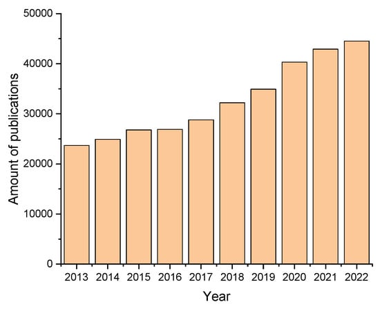
Figure 1.
Publication activity regarding optical methods for plant disease diagnostics (in English) from 2013 to 2022 (search query optical methods, plant diseases). The data were obtained using the Google Scholar search engine.
2. Fundamentals of Optical Methods’ Utilization for Plant Pathogen Diagnosis
Most of the plant pathogen diagnosis methods are based on spectral analysis (Figure 2) to bind or unbind to spatial coordinates. In fact, spectroscopic methods—absorption (transmission) spectroscopy and reflective spectroscopy (more often in the visible or near-IR bands), Raman spectroscopy and fluorescence spectroscopy—are methods that do not bind spectral features to coordinates. Spectroscopic methods are often readily available, inexpensive and use simple and compact instruments based on an illumination source and a photodetector. The main challenge in spectroscopy is to find the ranges of wavelengths that have the maximum transmittance, absorption or reflection rates for the specific sample—in the visible spectral range, this corresponds to the sample tinge. Optical methods where the optical parameters of the objects under study are linked to their spatial coordinates are called spectral imaging methods [22]; often, these are hyperspectral or multispectral imaging and spatially resolved spectroscopy. In such methods, each pixel of a spectral image contains information about the radiation intensities in the selected spectral ranges. The combination of spectral imaging methods with UAVs, etc., allows us to use this concept for the monitoring of large agricultural areas. Visualization, including combined (with various mutually complementing techniques), is the most popular method, due to the high discernability of the results for the operator.
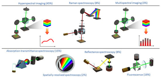
Figure 2.
Optical schemes and frequency of application of the main optical methods used to detect plant diseases.
The foundations of the implemented methods lie in the phenomena of the absorption, transmission, reflection, scattering or fluorescence of electromagnetic waves. The extracted information also depends on the light source wavelength and the analyzed spectral range. It is believed that the NIR region will have higher accuracy in plant disease detection in comparison with the MIR and vis–NIR regions [23]. The majority of ordinary radiation sources are broadband light sources, mainly natural light (solar energy). Techniques using sunlight are sometimes referred to as passive scanning techniques. For active scanning, pulsed laser sources (fluorescence methods, Raman spectroscopy, LiDAR) are used more often. Less often, lasers are used in a continuous emission mode. Data analysis makes it possible to link the identified spectral features with the presence of disease in a plant, to identify, differentiate and determine the severity of the disease. The most widespread mathematical algorithms to identify spectral regions suitable for class division are linear discriminant analysis (LDA) and principal component analysis (PCA) [24], partial least square discriminant analysis (PLS-DA) [25], quadratic discriminant analysis (QDA) and analysis of variance (ANOVA) [26]. The choice of mathematical algorithm in this case often depends on the method of data acquisition. For image analysis, machine learning methods are more often used. The most frequently used are support vector machines (SVM) [27] (which are noise-tolerant and high-precision, but not suitable for large data and bear a low processing rate) and neural networks. These include artificial neural networks (ANNs) [28], which reliable, convenient and suitable for processing noisy data and solving complex problems but require large number of training samples and processing time; convolutional neural networks (CNNs) [29]; backpropagation neural networks (BPNNs); and random forest (RF) with its related representative, the decision tree. Special indexes, discriminative analysis, extreme learning machine (ELM), joint sparse representation-based classification (JSRC); pixel-wise (PW); and self-organizing maps (SOM). The successive projection algorithm (SPA), regression-based models and spectral unmixing algorithms, etc. (Figure 3), are used much less often. In the case of methods that provide a large amount of data as a measurement result, data pre-processing may be required: baseline correction, normalization, noise removal (smoothing), the Savitsky–Golay first derivative [30], a wavelet transform [30], the removal of extrema, differentiation, use of the least squares method, PCA transformation and accounting for background fluorescence in Raman spectroscopy [31].
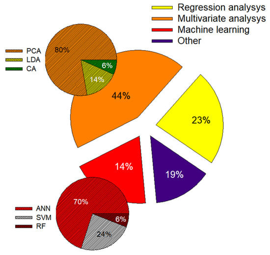
Figure 3.
Frequency of application of mathematical algorithms for analysis of data obtained using optical methods. Linear discriminant analysis (LDA), principal component analysis (PCA), cluster analysis (CA), regression analysis, artificial neural network (ANN), support vector machine (SVM), random forest (RF).
The choice of diagnostic method relies on the pathogen localization, the analyzed (subjected to inquiry, target) part of the plant, the type of pathogen being studied and the diagnostic conditions. Diagnostic methods can be based, on the one hand, on the search for the optical properties of characteristic pathogens—usually the appearance of characteristic (secondary) metabolites or protective compounds. On the other hand, they may be based on alterations in the optical response of the plant itself: changes in the content of chlorophyll and other pigments, or changes in photosynthetic activity, structure and the composition of the cell wall [32]. Fine remote diagnostics can be used; for example, by applying LiDAR, problems can be detected via the appearance and morphology of plant parts or the content of CO2, and root diseases can be determined from the aerial parts. Researchers should take into account that it is not always possible to associate changes in plant parameters with specific pathological changes: diagnosis in such cases is based mainly on statistical patterns [33]. The study can be carried out both in the field and in the laboratory. A feature of the field conditions, as a rule, is the presence of natural light, and the principal requirement is the possibility of remote sensing. In the controlled conditions of laboratories [34] and post-harvest sorting lines [10], any conditions can be created, although there are also limiting factors: in laboratories, it is often the size of the premises; in line conditions, it is dust and soil residues. To scan and diagnose large areas, instruments for remote sensing from ground platforms, aircraft and satellites are used [35]. The majority of optical methods are non-destructive and non-invasive, but, for certain purposes, there is a need for and benefit of invasive monitoring methods. Surface-enhanced Raman spectroscopy of mycotoxins [36] provides a good example of a damaging method. The studied part of the plant—in the field—includes green parts, shoots or the whole plant. On sorting lines, it may include the fruits, root crops and root tubers [37]. The whole plant or the green parts of plants are examined in approximately a quarter of cases. This is due to the easy accessibility for monitoring, especially for remote observation, and to the importance of assessing the state of the plant as a whole for the early diagnosis of diseases. For the green parts of plants, more often than for fruits, fluorescence methods are used, sometimes in combination with other optical techniques. For the photosynthetic parts of the plant, the most common methods are based on chlorophyll fluorescence. The seeds are the most frequently studied part of the plant (nearly half of all cases), due to the importance of cereals to humans. The study of mycotoxins using Raman spectroscopy [36] should be mentioned as a specific point. The fruit is examined in approximately one tenth of cases and roots in approximately one in five studies (Figure 4).
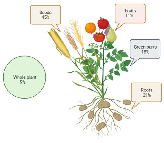
Figure 4.
Frequency of application of optical diagnostic methods to different parts of plants.
If classified according to the objects of study, then most of the work is devoted to the study of pathogenic fungi, and fungi from the Fusarium genus are dominant (Figure 5). This group of pathogen express a natural prevalence as an infectious agent of agricultural crops. Every type of pathogen causes changes in the host organism, and the choice of diagnostic method may be associated with the nature of these changes. For example, viruses often cause discoloration, mosaics and chlorosis. Bacteria cause much more heterogeneous changes—for example, the appearance of rot may begin, as well as spotting, burns, withering, the development of bacterial cancer or gum disease [34]. It is preferable to identify the disease before the appearance of visible symptoms; often, optical methods can achieve this. A study of the literature allows us to conclude that the choice of agricultural crop for the study and application of optical methods correlates with the importance of its culture for humans—for example, the vast majority of publications are devoted to cereals.
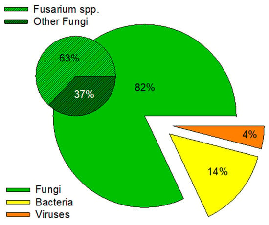
Figure 5.
Frequency of application of optical methods for studied pathogens from different taxonomic kingdoms.
As reference methods, laboratory methods for the determination of phytopathogens are usually used (microbiological, real-time PCR [24,25], immunochromatographic, ELISA [38], as well as artificially controlled infection (in laboratory studies, in most cases), visual control, electron microscopy). To supplement this and obtain a broader overall picture, combinations of multiple optical methods are often used. Approaches that combine various optical methods, such as hyperspectral and fluorescent imaging [39,40], as well as optical and other methods, such as visible and thermal imaging [41,42], hyperspectral, fluorescent and thermal imaging, have also become widespread [43]. The photochemical infection index, which is an integrating effect of viral infection on the kinetic parameters of fluorescence and reflection spectra of pepper leaves, was used by the authors of [44]. In [45], ATR-FTIR and Raman and fluorescence spectroscopy were used to detect Fusarium seed blight. The accuracy of pathogen detection with a method that uses the joint processing of data from two methods becomes higher than when using a single method.
3. Spectral Imaging
The most extensive class of methods is based on the detection of light that is reflected, rarely absorbed or passed through the object. This group of methods is represented by both visualization-related methods and methods of direct spectral imaging. The latter involve obtaining images of an object in which each pixel carries spectral information, by increasing the number of spectral bands and reducing their width: RGB imaging and multispectral (MSI) and hyperspectral imaging (HSI). Most often, such shooting is carried out using narrow-band filters, forming a large number of images, or multi-band filters where a smaller number of images is used.
RGB images are sequences of frames taken consecutively after red, green and blue filters and coincide with the intensity of the radiation reflection for the corresponding colors (RGB) by plant chromophores. For this purpose, both special cameras and smartphones are used [46].
Hyperspectral imaging (HSI) is an advanced, intricate technique that uses reflectance data collected in a wide spectral band, typically between 350 and 2500 nm, to reconstruct a spatial image of a plant through sensitive techniques of special image processing. HSI is a combination of spectroscopy and digital photography and the images form an image cube [47,48]. Since this method provides a large amount of information, it allows us to detect a wider range of diseases than RGB visualization. Among imaging modalities, HSI has the greatest potential (Table 2). At the same time, HSI has disadvantages: high costs, the complexity of sensors and optical components, long data acquisition times and the complexity of processing and analysis. These shortcomings of HSI limit the practical application of the method, mostly in situations where a short response interval or large-area surveys are required [49]. The development of efficient methods to process HSI data remains an unfulfilled objective due to the presence of more than three dimensions, resulting in an excess of information and noisiness. Additional difficulties include the dependence on the observation angle [50], which refers to the dependence on the orientation of the samples [28]. The HSI instruments themselves are very expensive to install on farms and now are mainly used for satellite and aerial imaging [51].

Table 2.
Hyperspectral devices utilized for plant pathogen diagnosis.
The value and nature of the information obtained from the spectral data depend on the wavelength range. The visible range corresponds to human vision; however, a large amount of valuable data on the plant tissue composition and structure is contained in the infrared part of the spectrum. The source of illumination for HSI is natural light, and the analyzed spectral range is determined by the parameters of the detector.
To classify the states of plants, unique spectral markers and vegetation indices [50] are often used, which comprise different ratios of intensities at certain wavelengths, usually in the visible and near-IR ranges. A plant in an intact state contains more chlorophyll and reflects more light energy in the 670–760 nm spectral band. Plant disease causes the destruction of chlorophyll (range < 700 nm); changes in the concentrations of carotenoids, anthocyanins and xanthophylls; and the destruction of cell structures (range >700 nm), which can be reflected in vegetation indices [62,63,64]. Vegetation indices can describe the general condition of a plant—the normalized difference vegetation index (NDVI) (NIR—Red/(NIR + Red)) reveals a decrease in carotenoid content, critical plant rational features such as the status of the chlorophyll amount and leaf area index, changes in photosynthesis rate (photochemical reflectance index) and chlorophyll degradation (normalized phaeophytinization index). Most of the widely used vegetation indices are mentioned and described, for example, in [65]. In addition to identifying plant stress associated with chlorophyll content, these indices can assess biomass and vegetation cover density [66].
4. Spectroscopy (General Characteristics)
Absorption–transmission spectroscopy provides information similar to reflection spectra but is less popular due to the peculiarities of taking spectra in the natural environment (in the field) (Table 3). Infrared and Raman spectroscopy are vibrational spectroscopy methods. Unlike UV and visible spectroscopy, which reflect the electronic transitions of molecules, infrared and Raman spectroscopy describe the vibrational states of molecules and molecular vibrations of a certain frequency, coinciding with the transition energy of a bond or group. Raman and IR spectroscopy are methods that are similar in terms of the information obtained, but they are based on different phenomena. The incidence of inelastic light scattering serves as a basis for Raman spectroscopy: a laser interacts with molecular vibrations and rotations and causes a change in molecular polarizability. Thus, Raman spectroscopy relies on changes in the polarizability of a chemical entity, while IR spectroscopy takes into account dipole momentum changes. It is necessary to calculate the relative frequencies at which the sample scatters radiation to perform Raman spectroscopy. IR spectroscopy evaluates the overall frequencies of radiation absorption for the particular sample. Raman spectroscopy is responsive to homonuclear molecular bonds; it is able to distinguish between single, double and triple bonds between carbon atoms. IR spectroscopy is sensitive to vibrations of heteronuclear functional groups and polar bonds.
5. IR Spectroscopy
IR spectra are usually roughly divided into three parts: near-IR (NIR) (780–2500 nm), mid-IR (MIR) (2500–25,000 nm) and far-IR (FIR) (25,000–300,000 nm). NIR and MIR spectroscopy allow us to extract information about chemical constituents and generate so-called “fingerprints”. MIR spectra are useful to explore fundamental vibrations and associated rotational vibration patterns, while NIR spectra promote overtones or harmonic vibrations’ easy activation. In complex mixtures, NIR spectroscopy has the advantage of analyzing the spectral characteristics of chemical bonds such as the “stretching” and bending of the C-H, N-H, O-H and S-H interconnections. Spectroscopy in the MIR range provides more overall spectrum information concerning the chemical functional groups on the sample surface. Usually, the MIR spectrum is split into four sub-ranges, namely the fingerprint (1500–400 cm−1), the double bond (2000–1500 cm−1) and the triple bond (2500–2000 cm−1) ranges, and the elongation of X-H bonds (4000–2500 cm−1). Fingerprint bands in absorption spectra are “neither obvious nor sharp” [67]. For complex substances, these bands cannot be directly tied with specific compounds, and they cannot be considered indicators of internal substance changes. Moreover, the vibrational groups of water molecules may easily conceal the peak absorption values of plants [68].

Table 3.
Application of spectroscopic methods for plant pathogen and disease diagnosis.
Table 3.
Application of spectroscopic methods for plant pathogen and disease diagnosis.
| Spectroscopy Type | Wavelength | Object | Pathogen/Disease | Analysis | Ref. |
|---|---|---|---|---|---|
| Vis–NIR spectroscopy | 250–1000 nm | Pumpkin leaves | Pathogen type not determined/Powdery mildew | Vegetative indices | [69] |
| 450–1100 nm | Cucumber leaves | Podosphaera xanthii/Powdery mildew | Vegetative indices and SIMCA | [70] | |
| NIR spectroscopy | 900–2600 nm | Potato | Candidatus Liberibacter solanacearum/Zebra chip disease | Stepwise regression in conjunction with canonical DA | [71] |
| 1100–2300 nm | Chestnuts | Mold | A genetic algorithm for feature selection in combination with image analysis grading and LDA, QDA and k-nearest neighbors (kNN) for classification; a Savitzky–Golay first derivative spectral pretreatment | [72] | |
| NIR reflectance spectroscopy | 800–2500 nm | Honeycrisp apple fruits | Pathogen type not determined/Bitter pit | QDA, SVM | [73] |
| 1175–2170 nm | Korean hulled barley | Fusarium sp./Fusarium | PLS-DA; multiple mathematical pretreatments | [74] | |
| 1000–2500 nm | Intact garlic cloves | Fusarium proliferatum/Fusarium | PLSR | [75] | |
| Vis–NIR reflectance spectroscopy | 350–2500 nm (1887, 1872 nm) | Wheat kernels | Fusarium sp./Fusarium | New spectral classification index (NSCI) method | [76] |
| 800–2500 nm | Almond kernels | Pathogen type not determined/Fungal | Canonical DA | [77] | |
| MIR spectroscopy | 4000–400 cm−1 | Oilseed rape leaves | Sclerotinia sclerotiorum/Sclerotinia stem rot | PLSDA, SVM and extreme learning machine | [78] |
| 400–4000 cm−1 | Rice leaves | Magnaporthe grisea/Rice blast Rhizoctonia solani/Rice sheath Xanthomonas oryzae pv. oryzae/Rice leaf blight | Raw data fusion, feature fusion and decision fusion; PCA and autoencoder to extract features; regression and CNN models; identification using SVM | [54] | |
| FTIR spectroscopy | 833–2500 nm | Wheat leaves | Puccnia striiformis f. sp./Stripe rust | Quantitative partial least squares (QPLS), support vector regression (SVR) and QPLS + SVR | [79] |
| 1000–2500 nm | Paddy rice | Fusarium sp./Fusarium | PLS; calibration models based on the partial least squares regression method | [80] | |
| 650–4000 cm−1 | Oil palm tree | Ganoderma boninense/Basal stem rot disease | Descriptive | [81] | |
| 4000–800 cm−1 | Powderized oil palm Ramet: roots, stems and leaves | Ganoderma boninense/Basal stem rot disease | Relative lignin/carbohydrate ratio | [32] | |
| 4000–400 cm−1 | Chilli plants | PYLCV (Pepper yellow leaf curl virus)/Leaf curl | Savitzky–Golay first derivative pre-processing method, DWT; cluster analysis, multilayer perceptron NN, SVM, LDA | [30] | |
| FTIR spectroscopy+ thermal imaging | 2977, 1544, 1050 cm−1 | Cucumber leaf powder | Pseudoperonospora cubensis/Downy mildew | Cluster analysis | [82] |
| ATR-FTIR spectroscopy | 1530–700 cm−1 | Sweet orange tree leaves | Xylella fastidiosa/Citrus variegated chlorosis | PLSR | [83] |
| 1800–900 cm−1 | Whole tomato fruit | Geotrichum candidum/Sour rot | PCA-LDA | [84] | |
| 650–4000 cm−1 | Perennial ryegrass | Neotyphodium sp./Choke disease | Multidimensional factor analysis, hierarchical cluster analysis | [85] | |
| 1000–2500 nm | Papaya leaves | Begomovirus/Leaf distortion | Multivariate exploratory methods: PCA, PLS-DA | [86] |
CNN—convolutional neural network; DA—discriminant analysis; DWT—discrete wavelet transform; LDA—linear discriminant analysis; NN—neural network; PCA—principal component analysis; PLSDA—partial least squares discriminant analysis; PLSR—partial least squares regression; SIMCA—soft independent modeling of class analogy classification; SVM—support vector machine; QDA—quadratic discriminant analysis.
In general, methods based on IR spectroscopy can be considered partly invasive or destructive. This classification is due to the sample preparation process. The process often requires desiccation and grinding to implement IR spectroscopy. For example, a virus was detected in chili plants [30], as well as downy mildew infection on cucumber leaves [82]. A modern modification of the IR spectroscopy method, ATR-FTIR, allows the rapid acquisition of a transmission-like spectrum. The method uses an optically dense crystal, usually a diamond, in contact with the object of study. ATR-FTIR allows for the non-invasive measurement of the sample and a high spatial resolution, as well as the very precise determination of the amount of light entering the sample. This technique was employed to identify rot in tomato fruits [84] and also to assess citrus diseases [80]. An application of FTIR to microscopy, FTIR microscopy, was used to identify potato phytopathogens in [87].
6. Raman Spectroscopy
The main benefit of Raman spectroscopy is the omission of the water effect on the spectrum, while the disadvantages are the possible fluorescence of the sample, which can conceal the spectrum, as well as possible sample destruction by the intense laser radiation of the source. The use of NIR lasers eliminates sample fluorescence. Two thirds of the surveyed studies employed IR lasers, and one third used green lasers. Raman shift detection is usually performed, on average, in the range of 700–1800 cm−1. The intensity and shifts of lines are analyzed, and sometimes the width of the peaks [88], corresponding to compounds characteristic of plant organisms: chlorophyll, anthocyanins [89], carotenoids (1000–1525 cm−1), polyphenols (1247 cm−1) [90], lignin (1601–1630 cm−1), xylan (approximately 1220 cm−1) [27], cellulose (1000–1200 cm−1) and pectin (approximately 800 cm−1) [91], as well as carbon (900–1300 cm−1), amino acids and proteins (800–1500 cm−1), lipids (approximately 1740 cm−1) and biotic stress hormones (1720 cm−1) [92]. Shifts in the spectrum lines are associated with changes in molecules: pectins—esterification, carotenoids—their degradation [93]. The possible use of Raman spectroscopy combined with Fourier transform provides a high spectral resolution [94]. Raman imaging is also used [31,95]. Examples of Raman spectroscopy’s application to the diagnosis of differently originated infections are listed in Table 4.
Raman spectroscopy is characterized by low signal intensity. Striving to increase the receptiveness, precision and applicability of the method, an add-on technique has been developed to amplify the signal by applying a sample to a nanostructured surface. Infection is often accompanied by the emergence and accumulation of toxins. The detection of alternariol and deoxynivalenol mycotoxins was demonstrated by the SERS method using silver nanoparticles modified with pyridine [96,97]. The use of signal amplification with nanoparticles is also applied in other spectroscopic methods—for example, in vis–NIR spectroscopy [98].

Table 4.
Raman spectroscopy instruments in the diagnosis of plant pathogens and diseases.
Table 4.
Raman spectroscopy instruments in the diagnosis of plant pathogens and diseases.
| Method | Object | Pathogen/Disease | Laser | Analysis | Ref. |
|---|---|---|---|---|---|
| Raman spectroscopy | Winter wheat seeds | Fusarium sp./Fusarium | 785 nm | t-test + FDR test | [36] |
| Tomato plants | Sardinia virus/Tomato yellow leaf curl | 780 nm | ASCA, PLS-DA | [99] | |
| Paddy rice leaves | Gibberella fujikuroi/Paddy rice blast | 532 nm | Spectral | [88] | |
| Paddy rice leaves | Magnaporthe grisea/Blast disease | 532 nm | BP-ANN | [100] | |
| Rose | Rose rosette virus (RRV)/Plant deformation | 830 nm | PLS-DA | [17] | |
| Raman spectroscopy hand-held | Wheat and sorghum grain | Claviceps sp./Ergot | 1064 nm | PLS-DA | [93] |
| Maize (Zea mays) kernels | A. flavus, A. niger and Diplodia spp. | 1064 nm | OPLS-DA | [101] | |
| Orange trees | Liberibacter spp./Huanglongbing | 831 nm | OPLS-DA | [25] | |
| Leaves of orange and grapefruit | Liberibacter spp./Huanglongbing | 831 nm | OPLS-DA | [45] | |
| Tomatoes | Liberibacter solanacearum/Liberibacter disease | 780 nm | PLS-DA | [3] | |
| Abutilon | Abutilon mosaic virus (AbMV)/Bright yellow mosaic | 1064 nm | ANOVA, optimal band ratio | [26] | |
| Raman spectroscopy portable | Sweet orange, Persian lime, Mexican lime trees | Candidatus Liberibacter asiaticus/Huanglongbing | 632.8, 785 nm | PCA, LDA | [24] |
| Intact rape leaves | Sclerotinia sclerotiorum/Sclerotinia disease | 514.5 nm | PCA, LS-SVM | [102] | |
| Confocal Raman spectroscopy | Citrus leaves | Liberibacter spp./Huanglongbing | 785 nm | PCA, PLS-DA, BP-ANN | [90] |
| Pear fruit | Alternaria alternate/Leaf spot, rot | 532 nm | PCA | [103] | |
| Raman hyperspectral imaging | Watermelon seeds | Acidovorax citrull/Bacterial fruit spot disease | 785 nm | ANOVA, optimal band ratio | [31] |
| Single maize kernel | Pathogen type not determined/Mildew | PLSR detection model | [95] | ||
| SERS | Maize | Fusarium sp./Fusarium | 785 nm | Classification: KNN and LDA Quantification: MLR, PCR, PLSR | [104] |
| Chinese cabbage plants | Turnip yellow mosaic virus (TYMV)/Leaf mosaic | 785 nm | PCA, LDA | [105] | |
| Fresh sweet corns, kidney beans and oats | Fusarium sp./Fusarium | 785 nm | Linear correlation of SERS peak intensity with the concentration of deoxynivalenol | [97] | |
| Banana pseudostems | Fusarium oxysporum f. sp./Fusarium wilt, different developmental stages | 785 nm | Comparisons of the SERS patterns | [106] |
ANOVA—analysis of variance; ASCA—ANOVA simultaneous component analysis; BP-ANN—backpropagation artificial neural network; FDR—false discovery rate; KNN—k-nearest neighbors; LDA—linear discriminant analysis; LS-SVM—least squares support vector machine; MLR—multiple linear regression; OPLS-DA—orthogonal partial least squares discriminant analysis; PCA—principal component analysis; PCR—principal component regression; PLSR—partial least squares regression.
7. Fluorescent Methods
Fluorescence is the phenomenon of the re-emission of absorbed light with some loss of energy due to the structural features of the substance. In contrast to reflection spectroscopy, fluorescent methods are 2–5 times more sensitive [107]. The concentrations and chemical modifications of molecules make it possible to judge the functional state of the plant; therefore, the measurement of fluorescence is often associated with biochemical and physiological changes induced by pathogens and/or other factors [108]. For example, the R/NIR ratio (e.g., F690/F730 or F685/F740) makes it possible to assess the amount of chlorophyll and thus the stress state of the plant. The F450/F690 ratio is frequently chosen to disclose the plant stress degree and is often used in remote sensing. Similar to optical technologies based on reflection/transmission, there is fluorescence spectroscopy as well as fluorescence imaging. Xenon lamps in spectrofluorometers are used as radiation sources in laboratory studies [109], and LEDs are used in applied work [110]. The method of laser-induced fluorescence stands somewhat apart. With the income generated by the usage of laser light sources with high intensity, it became possible to study whole plants even with remote sensing instruments, to discover the development of stress caused by herbicides [35] and pathogens [111,112]. Natural lighting is often a hindrance to fluorescence methods. Often, this problem is solved by pulsed laser operation. Another means to remove such interference is to correct the obtained spectrum for the background spectrum with the laser turned off [35]. Solar radiation is not always a hindrance to measurement; for example, there is an interesting method to detect sunlight-induced chlorophyll fluorescence—a method based on the isolation of Fraunhofer lines, 687 and 760 nm, overlapping with the red and far-red bands of the chlorophyll spectrum used for remote sensing [113]. The intensity of chlorophyll fluorescence in plants usually decreases under stress conditions [114]. Examples of the use of fluorescence methods for the detection of plant pathogens and diseases are presented in Table 5.
There are two types of fluorescence: blue–green, with a maximum of approximately 440 nm (F440) and a shoulder of approximately 520 nm (F520)—mainly due to the presence of phenolic compounds (flavonoids)—and red, with maxima of approximately 680 nm (F680) and 740 nm (F740), due to the presence of chlorophyll. The intensity of fluorescence in the spectrum of the blue–green range increases with stress. For example, it has been shown that in the emission spectra of leaves infected with downy mildew, a maximum appears at a wavelength of 390 nm. This maximum was 4–14 times more intense than the maximum at 450 nm in control leaves. A biochemical study showed that the appearance of this fluorescence maximum was associated with the accumulation of stilbene, which allowed the researchers to deduce that stilbene fluorescence in leaves can be an important advanced marker of pathogen attacks [115]. Moreover, the blue–green range of the fluorescence spectrum was used in another work to distinguish leaves infected with powdery mildew of the vine [116]. Using a multicolor fluorescent imaging system, changes in the intensity of UV-induced blue (F440) and green (F520) fluorescence in pathogen-infected leaves were recorded. It was found that these changes were associated with an increase in the concentrations of phenolic compounds involved in plant protection (scopoletin, chlorogenic and ferulic acids) [24]. In general, phenolic compounds are of particular importance in phytopathology and are mentioned in many other works [117,118].

Table 5.
Application of fluorescence methods for the diagnosis of plant pathogens and diseases.
Table 5.
Application of fluorescence methods for the diagnosis of plant pathogens and diseases.
| Light Source | Excitation/Emission | Object | Pathogen/Disease | Ref. |
|---|---|---|---|---|
| Laser | 532 nm/547–850 nm | Citrus leaves | Xanthomonas citricitrus canker | [112] |
| 405 nm/420–900 nm | Citrus leaves | Liberibacter spp./Huanglongbing | [119] | |
| 442 nm/450–850 nm | Orange trees | Xanthomonas axonopodis pv. citri/Citrus canker | [120] | |
| 532 nm/550–850 nm | Citrus limonia plants | Xanthomonas axonopodis pv. citri/Citrus canker | [121] | |
| 405 nm/500–700 nm | Grapefruit plants | Xanthomonas axonopodis pv. citri/Citrus canker | [122] | |
| 337 nm/370-800 nm | Winter wheat leaves | Blumeria Graminis F. sp. Tritici/Powdery mildew | [123] | |
| 338 nm/410–620 nm | Winter wheat leaves | Blumeria Graminis F. sp. Tritici/Powdery mildew | [123] | |
| 405 nm/430–800 nm 527 nm/660–720 nm | Apple fruit and potato tuber | Pathogen type not determined/Rot | [124] | |
| LED | 650 nm/670–850 nm | Olive tree orchards | Verticillium dahliae/Verticillium wilt | [43] |
| 655 nm/670–850 nm | Olive tree orchards | Verticillium dahliae/Verticillium wilt | [42] | |
| 635 nm/670–850 nm | Grapevine leaves | Grapevine leafroll-associated virus 3/Leaf curling | [38] | |
| 460 nm/500–540 nm | Tobacco leaves | Potato virus X/Leaf mosaic and mottling | [125] | |
| 650 nm/670–850 nm | Tobacco leaves | Cucumber mosaic virus/Leaf mosaic | [34] | |
| 405 nm/560, 580, 690 nm | Grapevine leaves | Erysiphe necator/Powdery mildew | [116] | |
| 470 nm/530, 550, 690 nm | Sweet orange tree leaves | Pathogen type not determined/Huanglongbing | [126] | |
| 650 nm/670–850 nm | Ginseng leaves | Pythium irregulare Buisman/Water molds | [127] | |
| Wide-range light sources | 280–390 nm/390–600 nm | Grapevine leaves | Plasmopara viticola/Downy mildew | [115] |
| 200–400 nm/220–400 nm | Grapevine leaves | Venturia inaequalis/Scab and rot | [128] | |
| 232, 362, 424, 485, 528 nm/235–700 nm | Wheat, oat and barley seeds | Fusarium sp./Fusarium | [129] | |
| 200–400 nm/220–380 nm | Winter wheat seeds | Fusarium sp./Fusarium | [130] | |
| 200–3000 nm/747, 762, 780 nm | Olive tree orchards | Verticillium dahliae/Verticillium wilt | [43] | |
| 200–400 nm/440, 520, 680, 740 nm | Melon leaves | Dickeya dadantii/Soft rot | [131] | |
| 200–3000 nm/687, 759.5, 684, 757.5 nm | Cassava leaves | Cassava mosaic virus/Cassava mosaic virus disease | [113] | |
| 350–420 nm/550, 690 nm | Winter wheat leaves | Puccinia striiformis/Yellow rust | [132] |
Chlorophyll is a green pigment, the major indicator of the state, strength and health of a plant [133]. A specific method to explore the concentration and state of chlorophyll—its fluorescence—is founded on the study of the kinetic dependences of chlorophyll fluorescence by the method of pulse-amplitude modulation. Chlorophyll fluorescence is a non-invasive method for the measurement of the activity of photosystem II, and it is widely used in plant physiology [5,134,135,136,137,138]. It is known that the kinetics of chlorophyll fluorescence induction are associated with photochemical processes and processes involved in energy dissipation. The kinetics of chlorophyll fluorescence induction are good indicators of stress, which also makes it possible to observe pathological processes that begin in the green parts of plants. The most extensively assigned chlorophyll fluorescence parameters for the detection of disorders include the maximum quantum efficiency of photosystem II (Fv/Fm). Another parameter that is frequently changed is the index characterizing non-photochemical fluorescence quenching (NPQ), which is a marker to trace the absorbed light energy thermal dissemination. Pathologic processes usually cause a decrease in the electron flow in the chloroplast’s electron transport chain; this weakening immediately results in an Fv/Fm index decrease [24], as well as in increased values of the NPQ index [125,139]. Interestingly, there is a study that shows multidirectional effects, including an infection-induced change in Fv/Fm and NPQ at some stages of the development of the disease [139]. It has been established that disease spread and host–pathogen interactions can be studied both in space and time at the leaf level, also using NPQ and Fq′/Fm′ (photosystem II quantum efficiency in light) [140].
It should be noted that not all studies focus only on the blue or red bands of the fluorescence spectrum. Multispectral fluorescence imaging, for example, originates from the fluorescence of four characteristic regions near the intensity maxima at 440 nm, 520 nm, 690 nm and 740 nm [38]. Less important information is obtained by changing the wavelengths of excitation or emission. Another type of fluorescence-based method that has found application in phytopathology is time-resolved fluorescence spectroscopy [141]. For example, an increase in the fluorescence span in the region of 410–620 nm was shown in the presymptomatic diagnosis of wheat powdery mildew by laser-induced fluorescence with a nanosecond resolution using a gated integrator [123].
8. Laser-Induced Breakdown Spectroscopy (LIBS) Method
The method of laser-induced breakdown spectroscopy (LIBS) involves the spectra of laser breakdown plasma, which are used to analyze samples [142,143,144]. A laser beam with sufficient energy is wrapped up in the surface layer of the sample and is used to ionize it [145]. At the end of laser exposure, the excited particles enter the ground state, generating optical radiation, the spectral maxima of which correspond to certain chemical elements. In [146], LIBS was used in association with SIMCA to directly measure, without any preventive procedures, the profiles of organic and inorganic signals from citrus leaves infected with the Candidatus Liberibacter asiaticus (CLas) bacterium. The same entities (greening disease and citrus canker) were studied in [147]. The use of LIBS for the diagnosis of TMV in tobacco has been demonstrated [148]. In general, the method of laser-spark emission spectrometry is rarely used for plant pathogen detection. A brief overview of the secondary methods used for the detection of phytopathogens is given in Table 6.

Table 6.
Application of minor methods for the diagnosis of plant pathogens and diseases.
9. Photoacoustic Spectroscopy (PAS)
PAS is method of measuring the optical, pulsed or intensity-modulated radiation absorbed by a substance using an acoustic sensor (microphone or piezoelectric transducer). The method is based on the thermos-optical mechanism of the excitation of pressure waves upon the absorption of laser radiation. This method is insensitive to scattering and reflection losses, requires no for sample provision and can be used for the detection and diagnosis of various plant diseases as a valuable supplementary method [149]. The use of the PAS method in the MIR range for the detection of raspberry powdery mildew [154] and rice blast (rotten neck) [150] was demonstrated.
10. Terahertz Time-Domain Spectroscopy
Terahertz time-domain spectroscopy is a technique used to explore the properties of matter under the influence of short, picosecond, terahertz radiation pulses. When terahertz radiation interacts with the object of study, a phase shift occurs, information about which is extracted from direct measurements of the electric field of the pulse. The THz-TDS method makes it possible to demonstrate the presence or absence of pathogens in plants; however, it requires the time-consuming preparation of samples before measurement. It has been shown that the THz-TDS method makes it possible to assess the degree of Fusarium infection in cereal seeds (oats, wheat and barley) and late blight in potato tubers. Changes are observed in refractive indices, absorption coefficients and dielectric functions in healthy and infected plants [155]. Terahertz imaging has also been used to identify fungal infections in chestnut trees [156], powdery mildew in tomato leaves [157] and wheat mold [158]. In general, a rise in the number of papers devoted to plant pathogen detection based on the generation and detection of terahertz radiation can be expected in the near future [159].
11. LiDAR Sensing
Light detection and ranging (LiDAR) is a technology that implies remote sensing by active source of laser radiation [160]. LiDAR converts the measurement of the time lapse between an emitted and registered laser pulse into the distance to target objects [161]. The method makes it possible to study the morphology of plants by measuring angles and distances with great accuracy, to “see” the lower layers of vegetation due to the ability of laser radiation to penetrate through the gaps in the foliage. The major part of the method consists of measuring the gap between the emitted and received pulsed laser beam reflected from an object or tested surface. There are two modes of LiDAR operation: (1) LiDAR “time of flight” measurements of the time taken for a generated laser pulse to travel back and forth amid the sensor and the target; (2) phase-shift-based LiDAR, which measures the laser beam’s phase alteration between oncoming and reflected signals. LiDAR uses different light sources: UV, vis or NIR. It should be noted that for agricultural and forestry applications, an NIR light source is preferred.
The advantages of LiDAR are the high measurement speed, high accuracy, 3D visualization, small size and relatively low price. At the same time, the application of the method may be limited in the case of dust and high humidity; there are errors associated with the position of the LiDAR (orientation angle, distance from the center of the tree); and the vast volume of data saved in log-files also makes the method less convenient [162]. LiDAR can be used to detect phytopathogens. Thus, in [51], an indirect method for the detection of strawberry powdery mildew by monitoring the CO2 concentration is presented. The characteristic of the laser beam received by the detector gives information about the medium through which the beam has passed. In infected plants, damage to the green parts occurs, resulting in a reduction in the plant’s ability to absorb CO2. In general, the developed bistatic LiDAR system monitors CO2 concentration anomalies associated with photosynthesis disorders, which are often associated with green mass diseases. Another paper also presents an indirect method of detection via the development and size of the crown of the basal stem rot in maize palm [152]. Existing types of LiDAR sensing, such as Raman and fluorescent types of LiDAR, are also used in phytopathology—for example, to detect damaged potato tubers [153]. In general, LiDAR sensing is a technique with underutilized application potential.
12. Conclusions
The experience in using optical methods for predominantly the early diagnosis of plant diseases is considered. Optical methods often favorably differ from others in their non-invasiveness and the speed of obtaining results. At present, optical methods are increasingly being used in the study of the state of plants, their changes due to internal (natural changes during plant ontogenesis, ripening and aging of fruits) and external factors. At present, an impressive database has been accumulated regarding the monitoring of a variety of diseases in various plant objects using various optical methods.
The presented methods are extremely diverse. These methods are based on different optical phenomena (reflection, absorption, fluorescence, scattering) and various radiation sources (sunlight, broad-spectrum lamps, narrow-band laser sources) operating in different modes (continuous and pulsed). The analyzed objects differ in scale by many orders of magnitude (from fields to individual grains). The methods and techniques described in the research are mostly non-destructive. In laboratory diagnostics, fluorescence methods are more often used, while field studies are based on light reflection. In essence, these are spectroscopic methods: methods of vibrational spectroscopy or reflective spectroscopy based on the analysis of characteristic frequencies, with the subsequent formation of the corresponding indices and ratios. Hyperspectral imaging provides a huge amount of information (in spectral, spatial and temporal coordinates), which is both an advantage and disadvantage, as there are difficulties in data processing and analysis and measurement errors can accumulate. In our opinion, methods for the analysis of kinetic parameters, such as the fluorescence lifetime and characteristics of photosynthetic processes, are underestimated. LiDAR technologies are somewhat separate, used for the analysis of the shape, the composition of the air around plants and the fluorescent characteristics of the vegetation cover. The variety of optical methods allows the researcher to choose the most appropriate one, depending on the scale of the monitored area, the required resolution, external conditions and the object of study. The most universal methods are those based on reflectance spectroscopy; the fluorescence methods and vibrational spectroscopy methods are more specific regarding the nature of the object, and these methods are more unreliable when used in the field. In general, the variety of methods corresponds to the variety of objects, tasks and possibilities of research.
Future directions for the development of this area include improving the methods of processing and analyzing data, taking into account environmental factors, which, first of all, may be associated with the involvement of artificial intelligence. To increase the reliability of the analysis, the use of a combination of several detection methods can be an effective technique, since changes that occur in plants as result of disease can manifest themselves in changes in the optical properties of tissues of various nature. An important requirement for devices is portability and a light weight for installation on unmanned aerial vehicles. Moreover, a necessary requirement for mass use for diagnostic systems of this types is a low price and rapidity in obtaining results. Another important area of activity is the search for new relations among the optical parameters of plants and their morphology, physiology, biochemistry and molecular biology, which is also required for the design of new optical methods for the detection of pathogens and diseases. At present, the use of optical methods is of an empirical, experimental and exploratory nature. The mechanism underlying the optical signs of the disease is not always understood, and there are not sufficient works explaining how to predict these optical changes. Optical methods have great potential in the future mass diagnostics of plant pathogens and diseases, although they need further improvement, for both instrumental and analytical parts.
Author Contributions
Conceptualization, S.V.G. and. T.A.M.; validation, R.M.S. and A.V.S.; formal analysis, S.V.G. and. T.A.M.; resources, M.N.M.; writing—original draft preparation, S.V.G. and T.A.M.; visualization, T.A.M. and E.V.S.; supervision, S.V.G.; project administration, A.S.D.; funding acquisition, A.Y.I. All authors have read and agreed to the published version of the manuscript.
Funding
This work was supported by a grant from the Ministry of Science and Higher Education of the Russian Federation for large scientific projects in priority areas of scientific and technological development (grant number 075-15-2020-774).
Data Availability Statement
Not applicable.
Conflicts of Interest
The authors declare no conflict of interest.
References
- Ali, M.M.; Bachik, N.A.; Muhadi, N.A.; Yusof, T.N.T.; Gomes, C. Non-destructive techniques of detecting plant diseases: A review. Physiol. Mol. Plant Pathol. 2019, 108, 101426. [Google Scholar] [CrossRef]
- Farber, C.; Mahnke, M.; Sanchez, L.; Kurouski, D. Advanced spectroscopic techniques for plant disease diagnostics. A review. TrAC Trends Anal. Chem. 2019, 118, 43–49. [Google Scholar] [CrossRef]
- Sanchez, P.D.C.; Hashim, N.; Shamsudin, R.; Nor, M.Z.M. Applications of imaging and spectroscopy techniques for non-destructive quality evaluation of potatoes and sweet potatoes: A review. Trends Food Sci. Technol. 2020, 96, 208–221. [Google Scholar] [CrossRef]
- Donoso, A.; Valenzuela, S. In-field molecular diagnosis of plant pathogens: Recent trends and future perspectives. Plant Pathol. 2018, 67, 1451–1461. [Google Scholar] [CrossRef]
- Burmistrov, D.E.; Yanykin, D.V.; Simakin, A.V.; Paskhin, M.O.; Ivanyuk, V.V.; Kuznetsov, S.V.; Ermakova, J.A.; Alexandrov, A.A.; Gudkov, S.V. Cultivation of Solanum lycopersicum under Glass Coated with Nanosized Upconversion Luminophore. Appl. Sci. 2021, 11, 10726. [Google Scholar] [CrossRef]
- Gudkov, S.; Andreev, S.; Barmina, E.; Bunkin, N.; Kartabaeva, B.; Nesvat, A.; Stepanov, E.; Taranda, N.; Khramov, R.; Glinushkin, A. Effect of visible light on biological objects: Physiological and pathophysiological aspects. Phys. Wave Phenom. 2017, 25, 207–213. [Google Scholar] [CrossRef]
- Karlo, J.; Prasad, R.; Singh, S.P. Biophotonics in food technology: Quo vadis? J. Agric. Food Res. 2022, 11, 100482. [Google Scholar] [CrossRef]
- Hamdy, O.; Mohammed, H.S. Post-heating Fluorescence-based Alteration and Adulteration Detection of Extra Virgin Olive Oil. J. Fluoresc. 2023, 33, 1631–1639. [Google Scholar] [CrossRef]
- Li, L.; Peng, Y.; Yang, C.; Li, Y. Optical sensing system for detection of the internal and external quality attributes of apples. Postharvest Biol. Technol. 2020, 162, 111101. [Google Scholar] [CrossRef]
- Zhu, H.; Yang, L.; Fei, J.; Zhao, L.; Han, Z. Recognition of carrot appearance quality based on deep feature and support vector machine. Comput. Electron. Agric. 2021, 186, 106185. [Google Scholar] [CrossRef]
- Rizzo, M.; Marcuzzo, M.; Zangari, A.; Gasparetto, A.; Albarelli, A. Fruit ripeness classification: A survey. Artif. Intell. Agric. 2023, 7, 44–57. [Google Scholar] [CrossRef]
- Ji, Y.; Sun, L.; Li, Y.; Ye, D. Detection of bruised potatoes using hyperspectral imaging technique based on discrete wavelet transform. Infrared Phys. Technol. 2019, 103, 103054. [Google Scholar] [CrossRef]
- Rady, A.M.; Guyer, D.E.; Watson, N.J. Near-infrared spectroscopy and hyperspectral imaging for sugar content evaluation in potatoes over multiple growing seasons. Food Anal. Methods 2021, 14, 581–595. [Google Scholar] [CrossRef]
- Shao, Y.; Liu, Y.; Xuan, G.; Wang, Y.; Gao, Z.; Hu, Z.; Han, X.; Gao, C.; Wang, K. Application of hyperspectral imaging for spatial prediction of soluble solid content in sweet potato. RSC Adv. 2020, 10, 33148–33154. [Google Scholar] [CrossRef] [PubMed]
- Khazaee, Y.; Kheiralipour, K.; Hosainpour, A.; Javadikia, H.; Paliwal, J. Development of a novel image analysis and classification algorithms to separate tubers from clods and stones. Potato Res. 2022, 65, 707–728. [Google Scholar] [CrossRef]
- Saranwong, S.; Thanapase, W.; Haff, R.; Kawano, S. Detection of fruit fly eggs and larvae in intact mango by near infrared spectroscopy and imaging. NIR News 2013, 24, 6–8. [Google Scholar] [CrossRef]
- Sanchez, L.; Farber, C.; Lei, J.; Zhu-Salzman, K.; Kurouski, D. Noninvasive and nondestructive detection of cowpea bruchid within cowpea seeds with a hand-held Raman spectrometer. Anal. Chem. 2019, 91, 1733–1737. [Google Scholar] [CrossRef]
- Abdullah, H.M.; Mohana, N.T.; Khan, B.M.; Ahmed, S.M.; Hossain, M.; Islam, K.S.; Redoy, M.H.; Ferdush, J.; Bhuiyan, M.; Hossain, M.M. Present and future scopes and challenges of plant pest and disease (P&D) monitoring: Remote sensing, image processing, and artificial intelligence perspectives. Remote Sens. Appl. Soc. Environ. 2023, 32, 100996. [Google Scholar]
- Senesi, G.S.; De Pascale, O.; Marangoni, B.S.; Caires, A.R.L.; Nicolodelli, G.; Pantaleo, V.; Leonetti, P. Chlorophyll fluorescence imaging (CFI) and laser-induced breakdown spectroscopy (LIBS) applied to investigate tomato plants infected by the root knot nematode (RKN) Meloidogyne incognita and tobacco plants infected by Cymbidium ringspot virus. Photonics 2022, 9, 627. [Google Scholar] [CrossRef]
- Skolik, P.; McAinsh, M.R.; Martin, F.L. Chapter Two—Biospectroscopy for Plant and Crop Science. In Comprehensive Analytical Chemistry; Lopes, J., Sousa, C., Eds.; Elsevier: Amsterdam, The Netherlands, 2018; Volume 80, pp. 15–49. [Google Scholar]
- Oberti, R.; Marchi, M.; Tirelli, P.; Calcante, A.; Iriti, M.; Borghese, A.N. Automatic detection of powdery mildew on grapevine leaves by image analysis: Optimal view-angle range to increase the sensitivity. Comput. Electron. Agric. 2014, 104, 1–8. [Google Scholar] [CrossRef]
- Kim, Y.-K.; Baek, I.; Lee, K.-M.; Qin, J.; Kim, G.; Shin, B.K.; Chan, D.E.; Herrman, T.J.; Cho, S.-k.; Kim, M.S. Investigation of reflectance, fluorescence, and Raman hyperspectral imaging techniques for rapid detection of aflatoxins in ground maize. Food Control 2022, 132, 108479. [Google Scholar] [CrossRef]
- Mohd Hilmi Tan, M.I.S.; Jamlos, M.F.; Omar, A.F.; Dzaharudin, F.; Chalermwisutkul, S.; Akkaraekthalin, P. Ganoderma boninense Disease Detection by Near-Infrared Spectroscopy Classification: A Review. Sensors 2021, 21, 3052. [Google Scholar] [CrossRef] [PubMed]
- Pérez-Bueno, M.L.; Granum, E.; Pineda, M.; Flors, V.; Rodriguez-Palenzuela, P.; López-Solanilla, E.; Barón, M. Temporal and spatial resolution of activated plant defense responses in leaves of Nicotiana benthamiana infected with Dickeya dadantii. Front. Plant Sci. 2016, 6, 1209. [Google Scholar] [CrossRef] [PubMed]
- Sanchez, L.; Pant, S.; Irey, M.; Mandadi, K.; Kurouski, D. Detection and identification of canker and blight on orange trees using a hand-held Raman spectrometer. J. Raman Spectrosc. 2019, 50, 1875–1880. [Google Scholar] [CrossRef]
- Yeturu, S.; Jentzsch, P.V.; Ciobotă, V.; Guerrero, R.; Garrido, P.; Ramos, L.A. Handheld Raman spectroscopy for the early detection of plant diseases: Abutilon mosaic virus infecting Abutilon sp. Anal. Methods 2016, 8, 3450–3457. [Google Scholar] [CrossRef]
- Sanchez, L.; Pant, S.; Xing, Z.; Mandadi, K.; Kurouski, D. Rapid and noninvasive diagnostics of Huanglongbing and nutrient deficits on citrus trees with a handheld Raman spectrometer. Anal. Bioanal. Chem. 2019, 411, 3125–3133. [Google Scholar] [CrossRef] [PubMed]
- Mansuri, S.M.; Chakraborty, S.K.; Mahanti, N.K.; Pandiselvam, R. Effect of germ orientation during Vis-NIR hyperspectral imaging for the detection of fungal contamination in maize kernel using PLS-DA, ANN and 1D-CNN modelling. Food Control 2022, 139, 109077. [Google Scholar] [CrossRef]
- Cui, R.; Li, J.; Wang, Y.; Fang, S.; Yu, K.; Zhao, Y. Hyperspectral imaging coupled with Dual-channel convolutional neural network for early detection of apple valsa canker. Comput. Electron. Agric. 2022, 202, 107411. [Google Scholar] [CrossRef]
- Agustika, D.K.; Mercuriani, I.; Purnomo, C.W.; Hartono, S.; Triyana, K.; Iliescu, D.D.; Leeson, M.S. Fourier transform infrared spectrum pre-processing technique selection for detecting PYLCV-infected chilli plants. Spectrochim. Acta Part A Mol. Biomol. Spectrosc. 2022, 278, 121339. [Google Scholar] [CrossRef]
- Lee, H.; Kim, M.S.; Qin, J.; Park, E.; Song, Y.-R.; Oh, C.-S.; Cho, B.-K. Raman hyperspectral imaging for detection of watermelon seeds infected with Acidovorax citrulli. Sensors 2017, 17, 2188. [Google Scholar] [CrossRef] [PubMed]
- Chow, Y.Y.; Ting, A.S.Y. Influence of fungal infection on plant tissues: FTIR detects compositional changes to plant cell walls. Fungal Ecol. 2019, 37, 38–47. [Google Scholar] [CrossRef]
- Astashev, M.E.; Serov, D.A.; Gudkov, S.V. Application of Spectral Methods of Analysis for Description of Ultradian Biorhythms at the Levels of Physiological Systems, Cells and Molecules (Review). Mathematics 2023, 11, 3307. [Google Scholar] [CrossRef]
- Lei, R.; Jiang, H.; Hu, F.; Yan, J.; Zhu, S. Chlorophyll fluorescence lifetime imaging provides new insight into the chlorosis induced by plant virus infection. Plant Cell Rep. 2017, 36, 327–341. [Google Scholar] [CrossRef]
- Lednev, V.N.; Grishin, M.Y.; Sdvizhenskii, P.A.; Kurbanov, R.K.; Litvinov, M.A.; Gudkov, S.V.; Pershin, S.M. Fluorescence Mapping of Agricultural Fields Utilizing Drone-Based LIDAR. Photonics 2022, 9, 963. [Google Scholar] [CrossRef]
- Moskovskiy, M.N.; Sibirev, A.V.; Gulyaev, A.A.; Gerasimenko, S.A.; Borzenko, S.I.; Godyaeva, M.M.; Noy, O.V.; Nagaev, E.I.; Matveeva, T.A.; Sarimov, R.M.; et al. Raman Spectroscopy Enables Non-Invasive Identification of Mycotoxins p. Fusarium of Winter Wheat Seeds. Photonics 2021, 8, 587. [Google Scholar] [CrossRef]
- Dorokhov, A.; Aksenov, A.; Sibirev, A.; Hort, D.; Mosyakov, M.; Sazonov, N.; Godyaeva, M. Development of an Optical System with an Orientation Module to Detect Surface Damage to Potato Tubers. Agriculture 2023, 13, 1188. [Google Scholar] [CrossRef]
- Montero, R.; Pérez-Bueno, M.L.; Barón, M.; Florez-Sarasa, I.; Tohge, T.; Fernie, A.R.; Ouad, H.E.A.; Flexas, J.; Bota, J. Alterations in primary and secondary metabolism in Vitis vinifera ‘Malvasía de Banyalbufar’upon infection with Grapevine leafroll-associated virus 3. Physiol. Plant. 2016, 157, 442–452. [Google Scholar] [CrossRef] [PubMed]
- Bauriegel, E.; Herppich, W.B. Hyperspectral and chlorophyll fluorescence imaging for early detection of plant diseases, with special reference to Fusarium spec. infections on wheat. Agriculture 2014, 4, 32–57. [Google Scholar] [CrossRef]
- Moshou, D.; Bravo, C.; Oberti, R.; West, J.; Bodria, L.; McCartney, A.; Ramon, H. Plant disease detection based on data fusion of hyper-spectral and multi-spectral fluorescence imaging using Kohonen maps. Real-Time Imaging 2005, 11, 75–83. [Google Scholar] [CrossRef]
- Raza, S.-e.-A.; Prince, G.; Clarkson, J.P.; Rajpoot, N.M. Automatic detection of diseased tomato plants using thermal and stereo visible light images. PLoS ONE 2015, 10, e0123262. [Google Scholar] [CrossRef]
- Berdugo, C.; Zito, R.; Paulus, S.; Mahlein, A.K. Fusion of sensor data for the detection and differentiation of plant diseases in cucumber. Plant Pathol. 2014, 63, 1344–1356. [Google Scholar] [CrossRef]
- Calderón, R.; Navas-Cortés, J.A.; Lucena, C.; Zarco-Tejada, P.J. High-resolution airborne hyperspectral and thermal imagery for early detection of Verticillium wilt of olive using fluorescence, temperature and narrow-band spectral indices. Remote Sens. Environ. 2013, 139, 231–245. [Google Scholar] [CrossRef]
- Tseliou, E.; Chondrogiannis, C.; Kalachanis, D.; Goudoudaki, S.; Manoussopoulos, Y.; Grammatikopoulos, G. Integration of biophysical photosynthetic parameters into one photochemical index for early detection of Tobacco Mosaic Virus infection in pepper plants. J. Plant Physiol. 2021, 267, 153542. [Google Scholar] [CrossRef] [PubMed]
- Pankin, D.; Povolotckaia, A.; Kalinichev, A.; Povolotskiy, A.; Borisov, E.; Moskovskiy, M.; Gulyaev, A.; Lavrov, A.; Izmailov, A. Complex Spectroscopic Study for Fusarium Genus Fungi Infection Diagnostics of “Zalp” Cultivar Oat. Agronomy 2021, 11, 2402. [Google Scholar] [CrossRef]
- Pandey, A.; Jain, K. A robust deep attention dense convolutional neural network for plant leaf disease identification and classification from smart phone captured real world images. Ecol. Inform. 2022, 70, 101725. [Google Scholar] [CrossRef]
- Mahlein, A.-K.; Oerke, E.-C.; Steiner, U.; Dehne, H.-W. Recent advances in sensing plant diseases for precision crop protection. Eur. J. Plant Pathol. 2012, 133, 197–209. [Google Scholar] [CrossRef]
- Terentev, A.; Dolzhenko, V.; Fedotov, A.; Eremenko, D. Current state of hyperspectral remote sensing for early plant disease detection: A review. Sensors 2022, 22, 757. [Google Scholar] [CrossRef]
- Gowen, A.A.; O’Donnell, C.P.; Cullen, P.J.; Downey, G.; Frias, J.M. Hyperspectral imaging–an emerging process analytical tool for food quality and safety control. Trends Food Sci. Technol. 2007, 18, 590–598. [Google Scholar] [CrossRef]
- Song, L.; Wang, L.; Yang, Z.; He, L.; Feng, Z.; Duan, J.; Feng, W.; Guo, T. Comparison of algorithms for monitoring wheat powdery mildew using multi-angular remote sensing data. Crop J. 2022, 10, 1312–1322. [Google Scholar] [CrossRef]
- Pham, H.; Lim, Y.; Gardi, A.; Sabatini, R.; Pang, E. A novel bistatic lidar system for early-detection of plant diseases from unmanned aircraft. In Proceedings of the 31th Congress of the International Council of the Aeronautical Sciences (ICAS 2018), Belo Horizonte, Brazil, 9–14 September 2018; pp. 9–14. [Google Scholar]
- Su, W.-H.; Yang, C.; Dong, Y.; Johnson, R.; Page, R.; Szinyei, T.; Hirsch, C.D.; Steffenson, B.J. Hyperspectral imaging and improved feature variable selection for automated determination of deoxynivalenol in various genetic lines of barley kernels for resistance screening. Food Chem. 2021, 343, 128507. [Google Scholar] [CrossRef]
- Furlanetto, R.H.; Nanni, M.R.; Mizuno, M.S.; Crusiol, L.G.T.; da Silva, C.R. Identification and classification of Asian soybean rust using leaf-based hyperspectral reflectance. Int. J. Remote Sens. 2021, 42, 4177–4198. [Google Scholar] [CrossRef]
- Feng, L.; Wu, B.; Zhu, S.; Wang, J.; Su, Z.; Liu, F.; He, Y.; Zhang, C. Investigation on data fusion of multisource spectral data for rice leaf diseases identification using machine learning methods. Front. Plant Sci. 2020, 11, 577063. [Google Scholar] [CrossRef] [PubMed]
- Nguyen, C.; Sagan, V.; Maimaitiyiming, M.; Maimaitijiang, M.; Bhadra, S.; Kwasniewski, M.T. Early detection of plant viral disease using hyperspectral imaging and deep learning. Sensors 2021, 21, 742. [Google Scholar] [CrossRef] [PubMed]
- Gorretta, N.; Nouri, M.; Herrero, A.; Gowen, A.; Roger, J.-M. Early detection of the fungal disease” apple scab” using SWIR hyperspectral imaging. In Proceedings of the 2019 10th Workshop on Hyperspectral Imaging and Signal Processing: Evolution in Remote Sensing (WHISPERS), Amsterdam, The Netherlands, 24–26 September 2019; pp. 1–4. [Google Scholar]
- Song, H.; Yoon, S.-R.; Dang, Y.-M.; Yang, J.-S.; Hwang, I.M.; Ha, J.-H. Nondestructive classification of soft rot disease in napa cabbage using hyperspectral imaging analysis. Sci. Rep. 2022, 12, 14707. [Google Scholar] [CrossRef]
- Sun, Y.; Lu, R.; Pan, L.; Wang, X.; Tu, K. Assessment of the optical properties of peaches with fungal infection using spatially-resolved diffuse reflectance technique and their relationships with tissue structural and biochemical properties. Food Chem. 2020, 321, 126704. [Google Scholar] [CrossRef] [PubMed]
- Huang, Y.; Wang, D.; Liu, Y.; Zhou, H.; Sun, Y. Measurement of early disease blueberries based on vis/nir hyperspectral imaging system. Sensors 2020, 20, 5783. [Google Scholar] [CrossRef]
- Barbedo, J.G.; Tibola, C.S.; Fernandes, J.M. Detecting Fusarium head blight in wheat kernels using hyperspectral imaging. Biosyst. Eng. 2015, 131, 65–76. [Google Scholar] [CrossRef]
- Ashourloo, D.; Mobasheri, M.R.; Huete, A. Developing Two Spectral Disease Indices for Detection of Wheat Leaf Rust (Pucciniatriticina). Remote Sens. 2014, 6, 4723–4740. [Google Scholar] [CrossRef]
- Devadas, R.; Lamb, D.; Simpfendorfer, S.; Backhouse, D. Evaluating ten spectral vegetation indices for identifying rust infection in individual wheat leaves. Precis. Agric. 2009, 10, 459–470. [Google Scholar] [CrossRef]
- Gitelson, A.A.; Merzlyak, M.N.; Chivkunova, O.B. Optical properties and nondestructive estimation of anthocyanin content in plant leaves. Photochem. Photobiol. 2001, 74, 38–45. [Google Scholar] [CrossRef]
- Young, A.; Britton, G. Carotenoids and stress. In Stress Responses in Plants: Adaptation, Acclimation Mechanisms; Cumming, J.R., Ed.; Wiley: New York, NY, USA, 1990; pp. 87–112. [Google Scholar]
- Zhang, J.-C.; Pu, R.-l.; Wang, J.-h.; Huang, W.-j.; Yuan, L.; Luo, J.-h. Detecting powdery mildew of winter wheat using leaf level hyperspectral measurements. Comput. Electron. Agric. 2012, 85, 13–23. [Google Scholar] [CrossRef]
- Kross, A.; McNairn, H.; Lapen, D.; Sunohara, M.; Champagne, C. Assessment of RapidEye vegetation indices for estimation of leaf area index and biomass in corn and soybean crops. Int. J. Appl. Earth Obs. Geoinf. 2015, 34, 235–248. [Google Scholar] [CrossRef]
- Abbas, O.; Pissard, A.; Baeten, V. Near-infrared, mid-infrared, and Raman spectroscopy. In Chemical Analysis of Food; Elsevier: Amsterdam, The Netherlands, 2020; pp. 77–134. [Google Scholar]
- Weng, S.; Hu, X.; Wang, J.; Tang, L.; Li, P.; Zheng, S.; Zheng, L.; Huang, L.; Xin, Z. Advanced Application of Raman Spectroscopy and Surface-Enhanced Raman Spectroscopy in Plant Disease Diagnostics: A Review. J. Agric. Food Chem. 2021, 69, 2950–2964. [Google Scholar] [CrossRef] [PubMed]
- Rivera-Romero, C.A.; Palacios-Hernández, E.R.; Trejo-Durán, M.; Rodríguez-Liñán, M.d.C.; Olivera-Reyna, R.; Morales-Saldaña, J.A. Visible and near-infrared spectroscopy for detection of powdery mildew in Cucurbita pepo L. leaves. J. Appl. Remote Sens. 2020, 14, 044515. [Google Scholar] [CrossRef]
- Atanassova, S.; Nikolov, P.; Valchev, N.; Masheva, S.; Yorgov, D. Early detection of powdery mildew (Podosphaera xanthii) on cucumber leaves based on visible and near-infrared spectroscopy. AIP Conf. Proc. 2019, 2075, 160014. [Google Scholar]
- Liang, P.-S.; Haff, R.P.; Hua, S.-S.T.; Munyaneza, J.E.; Mustafa, T.; Sarreal, S.B.L. Nondestructive detection of zebra chip disease in potatoes using near-infrared spectroscopy. Biosyst. Eng. 2018, 166, 161–169. [Google Scholar] [CrossRef]
- Moscetti, R.; Monarca, D.; Cecchini, M.; Haff, R.P.; Contini, M.; Massantini, R. Detection of mold-damaged chestnuts by near-infrared spectroscopy. Postharvest Biol. Technol. 2014, 93, 83–90. [Google Scholar] [CrossRef]
- Kafle, G.K.; Khot, L.R.; Jarolmasjed, S.; Yongsheng, S.; Lewis, K. Robustness of near infrared spectroscopy based spectral features for non-destructive bitter pit detection in honeycrisp apples. Postharvest Biol. Technol. 2016, 120, 188–192. [Google Scholar] [CrossRef]
- Lim, J.; Kim, G.; Mo, C.; Oh, K.; Yoo, H.; Ham, H.; Kim, M.S. Classification of Fusarium-infected Korean hulled barley using near-infrared reflectance spectroscopy and partial least squares discriminant analysis. Sensors 2017, 17, 2258. [Google Scholar] [CrossRef]
- Tamburini, E.; Mamolini, E.; De Bastiani, M.; Marchetti, M.G. Quantitative determination of Fusarium proliferatum concentration in intact garlic cloves using near-infrared spectroscopy. Sensors 2016, 16, 1099. [Google Scholar] [CrossRef]
- Zhang, D.; Wang, Q.; Lin, F.; Weng, S.; Lei, Y.; Chen, G.; Gu, C.; Zheng, L. New spectral classification index for rapid identification of Fusarium infection in wheat kernel. Food Anal. Methods 2020, 13, 2165–2175. [Google Scholar] [CrossRef]
- Liang, P.-S.; Slaughter, D.C.; Ortega-Beltran, A.; Michailides, T.J. Detection of fungal infection in almond kernels using near-infrared reflectance spectroscopy. Biosyst. Eng. 2015, 137, 64–72. [Google Scholar] [CrossRef]
- Zhang, C.; Feng, X.; Wang, J.; Liu, F.; He, Y.; Zhou, W. Mid-infrared spectroscopy combined with chemometrics to detect Sclerotinia stem rot on oilseed rape (Brassica napus L.) leaves. Plant Methods 2017, 13, 39. [Google Scholar] [CrossRef] [PubMed]
- Zhao, Y.; Gu, Y.; Qin, F.; Li, X.; Ma, Z.; Zhao, L.; Li, J.; Cheng, P.; Pan, Y.; Wang, H. Application of near-infrared spectroscopy to quantitatively determine relative content of Puccnia striiformis f. sp. tritici DNA in wheat leaves in incubation period. J. Spectrosc. 2017, 2017, 9740295. [Google Scholar] [CrossRef]
- Qiang, Z.; Fuguo, J.; Chenghai, L.; Jingkun, S.; Xianzhe, Z. Rapid detection of aflatoxin B1 in paddy rice as analytical quality assessment by near infrared spectroscopy. Int. J. Agric. Biol. Eng. 2014, 7, 127–133. [Google Scholar]
- Dayou, J.; Alexander, A.; Sipaut, C.S.; Phin, C.K.; Chin, L.P. On the possibility of using FTIR for detection of Ganoderma boninense in infected oil palm tree. Int. J. Adv. Agric. Environ. Eng. 2014, 1, 161–163. [Google Scholar]
- Wen, D.-M.; Chen, M.-X.; Zhao, L.; Ji, T.; Li, M.; Yang, X.-T. Use of thermal imaging and Fourier transform infrared spectroscopy for the pre-symptomatic detection of cucumber downy mildew. Eur. J. Plant Pathol. 2019, 155, 405–416. [Google Scholar] [CrossRef]
- do Brasil Cardinali, M.C.; Boas, P.R.V.; Milori, D.M.B.P.; Ferreira, E.J.; e Silva, M.F.; Machado, M.A.; Bellete, B.S. Infrared spectroscopy: A potential tool in huanglongbing and citrus variegated chlorosis diagnosis. Talanta 2012, 91, 1–6. [Google Scholar] [CrossRef]
- Skolik, P.; McAinsh, M.R.; Martin, F.L. ATR-FTIR spectroscopy non-destructively detects damage-induced sour rot infection in whole tomato fruit. Planta 2019, 249, 925–939. [Google Scholar] [CrossRef]
- Brandl, H. Detection of fungal infection in Lolium perenne by Fourier transform infrared spectroscopy. J. Plant Ecol. 2013, 6, 265–269. [Google Scholar] [CrossRef][Green Version]
- Haq, Q.M.; Mabood, F.; Naureen, Z.; Al-Harrasi, A.; Gilani, S.A.; Hussain, J.; Jabeen, F.; Khan, A.; Al-Sabari, R.S.; Al-Khanbashi, F.H. Application of reflectance spectroscopies (FTIR-ATR & FT-NIR) coupled with multivariate methods for robust in vivo detection of begomovirus infection in papaya leaves. Spectrochim. Acta Part A Mol. Biomol. Spectrosc. 2018, 198, 27–32. [Google Scholar]
- Erukhimovitch, V.; Hazanovsky, M.; Huleihel, M. Direct identification of potato’s fungal phyto-pathogens by Fourier-transform infrared (FTIR) microscopy. Spectroscopy 2010, 24, 609–619. [Google Scholar] [CrossRef]
- Yue, X.; Tan, Y.; Fan, W.; Song, S.; Ji, H.; Li, B. Raman spectroscopic analysis of paddy rice infected by three pests and diseases common in Northeast Asia. J. Phys. Conf. Ser. 2019, 1324, 012050. [Google Scholar] [CrossRef]
- Sharma, S.; Baran, C.; Tripathi, A.; Awasthi, A.; Tiwari, A.; Sharma, S.; Jaiswal, A.; Uttam, R.; Tandon, P.; Singh, R.; et al. Non-Destructive Monitoring of the Ripening of Plums Using Confocal Micro-Raman and Laser Induced Fluorescence Spectroscopy. Anal. Lett. 2023, 1–18. [Google Scholar] [CrossRef]
- Wang, K.; Liao, Y.; Meng, Y.; Jiao, X.; Huang, W.; Liu, T.C.-y. The early, rapid, and non-destructive detection of citrus Huanglongbing (HLB) based on microscopic confocal Raman. Food Anal. Methods 2019, 12, 2500–2508. [Google Scholar] [CrossRef]
- Sanchez, L.; Ermolenkov, A.; Tang, X.-T.; Tamborindeguy, C.; Kurouski, D. Non-invasive diagnostics of Liberibacter disease on tomatoes using a hand-held Raman spectrometer. Planta 2020, 251, 64. [Google Scholar] [CrossRef]
- Farber, C.; Shires, M.; Ong, K.; Byrne, D.; Kurouski, D. Raman spectroscopy as an early detection tool for rose rosette infection. Planta 2019, 250, 1247–1254. [Google Scholar] [CrossRef]
- Egging, V.; Nguyen, J.; Kurouski, D. Detection and identification of fungal infections in intact wheat and sorghum grain using a hand-held Raman spectrometer. Anal. Chem. 2018, 90, 8616–8621. [Google Scholar] [CrossRef]
- Baranski, R.; Baranska, M.; Schulz, H. Changes in carotenoid content and distribution in living plant tissue can be observed and mapped in situ using NIR-FT-Raman spectroscopy. Planta 2005, 222, 448–457. [Google Scholar] [CrossRef]
- Long, Y.; Huang, W.; Wang, Q.; Fan, S.; Tian, X. Integration of textural and spectral features of Raman hyperspectral imaging for quantitative determination of a single maize kernel mildew coupled with chemometrics. Food Chem. 2022, 372, 131246. [Google Scholar] [CrossRef]
- Pan, T.-t.; Sun, D.-W.; Pu, H.; Wei, Q. Simple approach for the rapid detection of alternariol in pear fruit by surface-enhanced Raman scattering with pyridine-modified silver nanoparticles. J. Agric. Food Chem. 2018, 66, 2180–2187. [Google Scholar] [CrossRef]
- Yuan, J.; Sun, C.; Guo, X.; Yang, T.; Wang, H.; Fu, S.; Li, C.; Yang, H. A rapid Raman detection of deoxynivalenol in agricultural products. Food Chem. 2017, 221, 797–802. [Google Scholar] [CrossRef]
- Kang, W.; Duan, Y.; Lin, H.; Ahmad, W.; Chen, Q.; Li, H. Enhancing count of Aspergillus colony in wheat based on nanoparticles modified chemo-responsive dyes combined with visible/near-infrared spectroscopy. Sens. Actuators B Chem. 2022, 363, 131816. [Google Scholar] [CrossRef]
- Mandrile, L.; Rotunno, S.; Miozzi, L.; Vaira, A.M.; Giovannozzi, A.M.; Rossi, A.M.; Noris, E. Nondestructive Raman spectroscopy as a tool for early detection and discrimination of the infection of tomato plants by two economically important viruses. Anal. Chem. 2019, 91, 9025–9031. [Google Scholar] [CrossRef] [PubMed]
- Tan, F.; Cai, Q.; Sun, X.; Ma, Z.; Hou, Z. Analyzing plant characteristics of rice suffering leaf blast in cold area based on Raman spectrum. Trans. Chin. Soc. Agric. Eng. 2015, 31, 191–196. [Google Scholar]
- Farber, C.; Kurouski, D. Detection and identification of plant pathogens on maize kernels with a hand-held Raman spectrometer. Anal. Chem. 2018, 90, 3009–3012. [Google Scholar] [CrossRef]
- Zhao, Y.; Yu, K.; Li, X.; He, Y. Application of Raman spectroscopy for early detection of rape sclerotinia on rapeseed leaves. Trans. Chin. Soc. Agric. Eng. 2017, 33, 206–211. [Google Scholar]
- Pan, T.-T.; Pu, H.; Sun, D.-W. Insights into the changes in chemical compositions of the cell wall of pear fruit infected by Alternaria alternata with confocal Raman microspectroscopy. Postharvest Biol. Technol. 2017, 132, 119–129. [Google Scholar] [CrossRef]
- Lee, K.-M.; Herrman, T.J.; Bisrat, Y.; Murray, S.C. Feasibility of surface-enhanced Raman spectroscopy for rapid detection of aflatoxins in maize. J. Agric. Food Chem. 2014, 62, 4466–4474. [Google Scholar] [CrossRef] [PubMed]
- Kim, S.; Lee, S.; Chi, H.-Y.; Kim, M.-K.; Kim, J.-S.; Lee, S.-H.; Chung, H. Feasibility study for detection of Turnip yellow mosaic virus (TYMV) Infection of Chinese Cabbage Plants Using Raman Spectroscopy. Plant Pathol. J. 2013, 29, 105. [Google Scholar] [CrossRef][Green Version]
- Lin, Y.-J.; Lin, H.-K.; Lin, Y.-H. Construction of Raman spectroscopic fingerprints for the detection of Fusarium wilt of banana in Taiwan. PLoS ONE 2020, 15, e0230330. [Google Scholar] [CrossRef] [PubMed]
- Luo, J.; Forsberg, E.; Fu, S.; He, S. High-precision four-dimensional hyperspectral imager integrating fluorescence spectral detection and 3D surface shape measurement. Appl. Opt. 2022, 61, 2542–2551. [Google Scholar] [CrossRef]
- Shanmugam, M.; Ramasamy, A. Multi-crop Chlorophyll Meter System Design for Effective Fertilization. Res. J. Appl. Sci. Eng. Technol. 2015, 9, 98–105. [Google Scholar] [CrossRef]
- Dorokhov, A.; Moskovskiy, M.; Belyakov, M.; Lavrov, A.; Khamuev, V. Detection of Fusarium infected seeds of cereal plants by the fluorescence method. PLoS ONE 2022, 17, e0267912. [Google Scholar] [CrossRef]
- Moskovskiy, M.N.; Belyakov, M.V.; Dorokhov, A.S.; Boyko, A.A.; Belousov, S.V.; Noy, O.V.; Gulyaev, A.A.; Akulov, S.I.; Povolotskaya, A.; Efremenkov, I.Y. Design of Device for Optical Luminescent Diagnostic of the Seeds Infected by Fusarium. Agriculture 2023, 13, 619. [Google Scholar] [CrossRef]
- Nilsson, H. Remote sensing and image analysis in plant pathology. Annu. Rev. Phytopathol. 1995, 33, 489–528. [Google Scholar] [CrossRef] [PubMed]
- Lins, E.C.; Belasque, J.; Marcassa, L.G. Detection of citrus canker in citrus plants using laser induced fluorescence spectroscopy. Precis. Agric. 2009, 10, 319–330. [Google Scholar] [CrossRef]
- Raji, S.N.; Subhash, N.; Ravi, V.; Saravanan, R.; Mohanan, C.N.; Nita, S.; Kumar, T.M. Detection of mosaic virus disease in cassava plants by sunlight-induced fluorescence imaging: A pilot study for proximal sensing. Int. J. Remote Sens. 2015, 36, 2880–2897. [Google Scholar] [CrossRef]
- Kim, M.S.; Chen, Y.; Mehl, P. Hyperspectral reflectance and fluorescence imaging system for food quality and safety. Trans. ASAE 2001, 44, 721. [Google Scholar]
- Poutaraud, A.; Latouche, G.; Martins, S.; Meyer, S.; Merdinoglu, D.; Cerovic, Z.G. Fast and local assessment of stilbene content in grapevine leaf by in vivo fluorometry. J. Agric. Food Chem. 2007, 55, 4913–4920. [Google Scholar] [CrossRef] [PubMed]
- Bélanger, M.C.; Roger, J.M.; Cartolaro, P.; Viau, A.; Bellon-Maurel, V. Detection of powdery mildew in grapevine using remotely sensed UV-induced fluorescence. Int. J. Remote Sens. 2008, 29, 1707–1724. [Google Scholar] [CrossRef]
- Sambangi, P. Phenolic Compounds in the Plant Development and Defense: An Overview. In Plant Stress Physiology; Mirza, H., Kamran, N., Eds.; IntechOpen: Rijeka, Croatia, 2022; p. Ch. 7. [Google Scholar] [CrossRef]
- Vibhakar, C.; Sheena, A.; Rohan, V.P.; Jigna, G.T. Physiological Function of Phenolic Compounds in Plant Defense System. In Phenolic Compounds; Farid, A.B., Ed.; IntechOpen: Rijeka, Croatia, 2021; p. Ch. 10. [Google Scholar] [CrossRef]
- Ranulfi, A.C.; Cardinali, M.C.; Kubota, T.M.; Freitas-Astua, J.; Ferreira, E.J.; Bellete, B.S.; da Silva, M.F.G.; Boas, P.R.V.; Magalhaes, A.B.; Milori, D.M. Laser-induced fluorescence spectroscopy applied to early diagnosis of citrus Huanglongbing. Biosyst. Eng. 2016, 144, 133–144. [Google Scholar] [CrossRef]
- Marcassa, L.G.; Gasparoto, M.; Belasque, J.; Lins, E.; Dias Nunes, F.; Bagnato, V.S. Fluorescence spectroscopy applied to orange trees. Laser Phys. 2006, 16, 884–888. [Google Scholar] [CrossRef]
- Belasque Jr, J.; Gasparoto, M.; Marcassa, L.G. Detection of mechanical and disease stresses in citrus plants by fluorescence spectroscopy. Appl. Opt. 2008, 47, 1922–1926. [Google Scholar] [CrossRef] [PubMed]
- Saleem, M.; Atta, B.M.; Ali, Z.; Bilal, M. Laser-induced fluorescence spectroscopy for early disease detection in grapefruit plants. Photochem. Photobiol. Sci. 2020, 19, 713–721. [Google Scholar] [CrossRef] [PubMed]
- Bürling, K.; Hunsche, M.; Noga, G. Presymptomatic detection of powdery mildew infection in winter wheat cultivars by laser-induced fluorescence. Appl. Spectrosc. 2012, 66, 1411–1419. [Google Scholar] [CrossRef] [PubMed]
- Matveyeva, T.A.; Sarimov, R.M.; Simakin, A.V.; Astashev, M.E.; Burmistrov, D.E.; Lednev, V.N.; Sdvizhenskii, P.A.; Grishin, M.Y.; Pershin, S.M.; Chilingaryan, N.O. Using fluorescence spectroscopy to detect rot in fruit and vegetable crops. Appl. Sci. 2022, 12, 3391. [Google Scholar] [CrossRef]
- Grishina, A.; Sherstneva, O.; Grinberg, M.; Zdobnova, T.; Ageyeva, M.; Khlopkov, A.; Sukhov, V.; Brilkina, A.; Vodeneev, V. Pre-symptomatic detection of viral infection in tobacco leaves using pam fluorometry. Plants 2021, 10, 2782. [Google Scholar] [CrossRef]
- Wetterich, C.B.; de Oliveira Neves, R.F.; Belasque, J.; Ehsani, R.; Marcassa, L.G. Detection of Huanglongbing in Florida using fluorescence imaging spectroscopy and machine-learning methods. Appl. Opt. 2017, 56, 15–23. [Google Scholar] [CrossRef]
- Ivanov, D.A.; Bernards, M.A. Chlorophyll fluorescence imaging as a tool to monitor the progress of a root pathogen in a perennial plant. Planta 2016, 243, 263–279. [Google Scholar] [CrossRef]
- Sarimov, R.M.; Lednev, V.N.; Sibirev, A.V.; Gudkov, S.V. The use of fluorescence spectra for the detection of scab and rot in fruit and vegetable crops. Front. Phys. 2021, 8, 640887. [Google Scholar] [CrossRef]
- Belyakov, M.V.; Moskovskiy, M.N.; Litvinov, M.A.; Lavrov, A.V.; Khamuev, V.G.; Efremenkov, I.Y.; Gerasimenko, S.A. Method of Optical Diagnostics of Grain Seeds Infected with Fusarium. Appl. Sci. 2022, 12, 4824. [Google Scholar] [CrossRef]
- Bashilov, A.M.; Efremenkov, I.Y.; Belyakov, M.V.; Lavrov, A.V.; Gulyaev, A.A.; Gerasimenko, S.A.; Borzenko, S.I.; Boyko, A.A. Determination of Main spectral and luminescent characteristics of winter wheat seeds infected with pathogenic microflora. Photonics 2021, 8, 494. [Google Scholar] [CrossRef]
- Pineda, M.; Pérez-Bueno, M.L.; Barón, M. Detection of bacterial infection in melon plants by classification methods based on imaging data. Front. Plant Sci. 2018, 9, 164. [Google Scholar] [CrossRef]
- Bravo, C.; Moshou, D.; Oberti, R.; West, J.; McCartney, A.; Bodria, L.; Ramon, H. Foliar disease detection in the field using optical sensor fusion. E-JOURNAL-CIGR 2004, 6, 1–14. [Google Scholar]
- Lichtenthaler, H.K.; Rinderle, U. The role of chlorophyll fluorescence in the detection of stress conditions in plants. CRC Crit. Rev. Anal. Chem. 1988, 19, S29–S85. [Google Scholar] [CrossRef]
- Yanykin, D.V.; Burmistrov, D.E.; Simakin, A.V.; Ermakova, J.A.; Gudkov, S.V. Effect of up-converting luminescent nanoparticles with increased quantum yield incorporated into the fluoropolymer matrix on Solanum lycopersicum growth. Agronomy 2022, 12, 108. [Google Scholar] [CrossRef]
- Paskhin, M.O.; Yanykin, D.V.; Gudkov, S.V. Current Approaches to Light Conversion for Controlled Environment Agricultural Applications: A Review. Horticulturae 2022, 8, 885. [Google Scholar] [CrossRef]
- Grinberg, M.; Gromova, E.; Grishina, A.; Berezina, E.; Ladeynova, M.; Simakin, A.V.; Sukhov, V.; Gudkov, S.V.; Vodeneev, V. Effect of Photoconversion Coatings for Greenhouses on Electrical Signal-Induced Resistance to Heat Stress of Tomato Plants. Plants 2022, 11, 229. [Google Scholar] [CrossRef]
- Gudkov, S.V.; Simakin, A.V.; Bunkin, N.F.; Shafeev, G.A.; Astashev, M.E.; Glinushkin, A.P.; Grinberg, M.A.; Vodeneev, V.A. Development and application of photoconversion fluoropolymer films for greenhouses located at high or polar latitudes. J. Photochem. Photobiol. B Biol. 2020, 213, 112056. [Google Scholar] [CrossRef]
- Murchie, E.H.; Lawson, T. Chlorophyll fluorescence analysis: A guide to good practice and understanding some new applications. J. Exp. Bot. 2013, 64, 3983–3998. [Google Scholar] [CrossRef]
- Rys, M.; Juhász, C.; Surówka, E.; Janeczko, A.; Saja, D.; Tóbiás, I.; Skoczowski, A.; Barna, B.; Gullner, G. Comparison of a compatible and an incompatible pepper-tobamovirus interaction by biochemical and non-invasive techniques: Chlorophyll a fluorescence, isothermal calorimetry and FT-Raman spectroscopy. Plant Physiol. Biochem. 2014, 83, 267–278. [Google Scholar] [CrossRef]
- Scholes, J.D.; Rolfe, S.A. Chlorophyll fluorescence imaging as tool for understanding the impact of fungal diseases on plant performance: A phenomics perspective. Funct. Plant Biol. 2009, 36, 880–892. [Google Scholar] [CrossRef] [PubMed]
- Ma, T.; Inagaki, T.; Tsuchikawa, S. Development of a sensitivity-enhanced chlorophyll fluorescence lifetime spectroscopic method for nondestructive monitoring of fruit ripening and postharvest decay. Postharvest Biol. Technol. 2023, 198, 112231. [Google Scholar] [CrossRef]
- Lednev, V.N.; Sdvizhenskii, P.A.; Grishin, M.Y.; Nikitin, E.A.; Gudkov, S.V.; Pershin, S.M. Improving calibration strategy for LIBS heavy metals analysis in agriculture applications. Photonics 2021, 8, 563. [Google Scholar] [CrossRef]
- Lednev, V.; Sdvizhenskii, P.; Grishin, M.Y.; Gudkov, S.; Dorokhov, A.; Bunkin, A.; Pershin, S. Improving the LIBS analysis of heavy metals in heterogeneous agricultural samples utilizing large laser spotting. J. Anal. At. Spectrom. 2022, 37, 2563–2572. [Google Scholar] [CrossRef]
- Lednev, V.; Sdvizhenskii, P.; Dorohov, A.; Gudkov, S.; Pershin, S. Improving LIBS analysis of non-flat heterogeneous samples by signals mapping. Appl. Opt. 2023, 62, 2030–2038. [Google Scholar] [CrossRef] [PubMed]
- Lednev, V.; Sdvizhenskii, P.; Grishin, M.Y.; Stavertiy, A.Y.; Tretyakov, R.; Asyutin, R.; Pershin, S. Laser welding spot diagnostics by laser-induced breakdown spectrometry. Phys. Wave Phenom. 2021, 29, 221–228. [Google Scholar] [CrossRef]
- Pereira, F.M.V.; Milori, D.M.B.P.; Venâncio, A.L.; Russo, M.d.S.T.; Martins, P.K.; Freitas-Astúa, J. Evaluation of the effects of Candidatus Liberibacter asiaticus on inoculated citrus plants using laser-induced breakdown spectroscopy (LIBS) and chemometrics tools. Talanta 2010, 83, 351–356. [Google Scholar] [CrossRef]
- Sankaran, S.; Ehsani, R.; Morganc, K.T. Detection of Anomalies in Citrus Leaves Using Laser-Induced Breakdown Spectroscopy (LIBS). Appl. Spectrosc. 2015, 69, 913–919. [Google Scholar] [CrossRef] [PubMed]
- Peng, J.; Song, K.; Zhu, H.; Kong, W.; Liu, F.; Shen, T.; He, Y. Fast detection of tobacco mosaic virus infected tobacco using laser-induced breakdown spectroscopy. Sci. Rep. 2017, 7, 44551. [Google Scholar] [CrossRef]
- Rai, N.K.; Singh, J.P.; Rai, A.K. Chapter 23—Photoacoustic spectroscopy: A novel optical characterization technique in agricultural science. In Photoacoustic and Photothermal Spectroscopy; Thakur, S.N., Rai, V.N., Singh, J.P., Eds.; Elsevier: Amsterdam, The Netherlands, 2023; pp. 491–509. [Google Scholar]
- Gaoqiang, L.; Changwen, D.; Fei, M.; Yazhen, S.; Jianmin, Z. Responses of leaf cuticles to rice blast: Detection and identification using depth-profiling fourier transform mid-infrared photoacoustic Spectroscopy. Plant Dis. 2020, 104, 847–852. [Google Scholar] [CrossRef] [PubMed]
- Pieczywek, P.; Cybulska, J.; Szymańska-Chargot, M.; Siedliska, A.; Zdunek, A.; Nosalewicz, A.; Baranowski, P.; Kurenda, A. Early detection of fungal infection of stored apple fruit with optical sensors–Comparison of biospeckle, hyperspectral imaging and chlorophyll fluorescence. Food Control 2018, 85, 327–338. [Google Scholar] [CrossRef]
- Husin, N.A.; Khairunniza-Bejo, S.; Abdullah, A.F.; Kassim, M.S.; Ahmad, D.; Azmi, A.N. Application of ground-based LiDAR for analysing oil palm canopy properties on the occurrence of basal stem rot (BSR) disease. Sci. Rep. 2020, 10, 6464. [Google Scholar] [CrossRef] [PubMed]
- Lobachevsky, Y.; Dorokhov, A.; Aksenov, A.; Sibirev, A.; Moskovskiy, M.; Mosyakov, M.; Sazonov, N.; Godyaeva, M. RAMAN and Fluorimetric Scattering Lidar Facilitated to Detect Damaged Potatoes by Determination of Spectra. Appl. Sci. 2022, 12, 5391. [Google Scholar] [CrossRef]
- Du, C.; Zhou, J. Fourier Transform Mid-Infrared Photoacoustic Spectroscopy for Presymptomatic Detection of Powdery Mildew Infection in Rubus corchorifolius L. Spectrosc. Lett. 2015, 48, 610–615. [Google Scholar] [CrossRef]
- Penkov, N.V.; Goltyaev, M.V.; Astashev, M.E.; Serov, D.A.; Moskovskiy, M.N.; Khort, D.O.; Gudkov, S.V. The application of terahertz time-domain spectroscopy to identification of potato late blight and fusariosis. Pathogens 2021, 10, 1336. [Google Scholar] [CrossRef]
- Di Girolamo, F.; Pagano, M.; Tredicucci, A.; Bitossi, M.; Paoletti, R.; Barzanti, G.; Benvenuti, C.; Roversi, P.; Toncelli, A. Detection of fungal infections in chestnuts: A terahertz imaging-based approach. Food Control 2021, 123, 107700. [Google Scholar] [CrossRef]
- Zhang, X.; Wang, Y.; Zhou, Z.; Zhang, Y.; Wang, X. Detection Method for Tomato Leaf Mildew Based on Hyperspectral Fusion Terahertz Technology. Foods 2023, 12, 535. [Google Scholar] [CrossRef]
- Hongyi, G.; Fei, W.; Yuying, J.; Li, L.; Yuan, Z.; Keke, J. Identification of wheat mold using terahertz images based on Broad Learning System. Chin. J. Quantum Electron. 2023, 40, 360. [Google Scholar]
- Ge, H.; Lv, M.; Lu, X.; Jiang, Y.; Wu, G.; Li, G.; Li, L.; Li, Z.; Zhang, Y. Applications of THz spectral imaging in the detection of agricultural products. Photonics 2021, 8, 518. [Google Scholar] [CrossRef]
- Myasnikov, A.; Pershin, S.; Grishin, M.Y.; Zavozin, V.; Makarov, V.; Ushakov, A. Estimation of the influence of meteorological factors on the aerosol lidar signal in tunnels above the Elbrus Volcano Chamber. Phys. Wave Phenom. 2022, 30, 119–127. [Google Scholar] [CrossRef]
- Lednev, V.; Grishin, M.Y.; Sdvizhenskii, P.; Zavozin, V.; Bunkin, A.; Kurbanov, R.; Litvinov, M.; Tretinnikov, O.; Pershin, S. Drone Based Fluorescence LIDAR for Agriculture Fields in Situ Diagnostics. Bull. Lebedev Phys. Inst. 2023, 50, 103–107. [Google Scholar] [CrossRef]
- Rosell, J.; Sanz, R. A review of methods and applications of the geometric characterization of tree crops in agricultural activities. Comput. Electron. Agric. 2012, 81, 124–141. [Google Scholar] [CrossRef]
Disclaimer/Publisher’s Note: The statements, opinions and data contained in all publications are solely those of the individual author(s) and contributor(s) and not of MDPI and/or the editor(s). MDPI and/or the editor(s) disclaim responsibility for any injury to people or property resulting from any ideas, methods, instructions or products referred to in the content. |
© 2023 by the authors. Licensee MDPI, Basel, Switzerland. This article is an open access article distributed under the terms and conditions of the Creative Commons Attribution (CC BY) license (https://creativecommons.org/licenses/by/4.0/).