Article

The Dual Impact of Mobile Social Media Breaks on Cortical Arousal and Neurophysiological Recovery: A Spectral Analysis of EEG Signals

1
School of Allied Health Sciences, Walailak University, Nakhonsithammarat 80160, Thailand
2
Center of Excellence in Innovation on Essential oil, Walailak University, Nakhonsithammarat 80160, Thailand
3
School of Informatics, Walailak University, Nakhonsithammarat 80160, Thailand
4
School of Medicine, Walailak University, Nakhonsithammarat 80160, Thailand
5
Walailak University Hospital, Walailak University, Nakhonsithammarat 80160, Thailand
*
Author to whom correspondence should be addressed.
Signals 2026, 7(4), 68; https://doi.org/10.3390/signals7040068
Submission received: 29 April 2026 / Revised: 24 June 2026 / Accepted: 8 July 2026 / Published: 10 July 2026

Abstract

Arousal is essential for cognitive awareness, and it typically declines during prolonged tasks. Currently, mobile devices are frequently used during breaks to relax and counteract this decline. However, their impact on neurophysiological recovery remains poorly understood. Therefore, this study compared the effects of traditional quiet rest versus a mobile task break on cortical arousal using spectral analysis of electroencephalography (EEG) signals in twenty healthy young females (20–25 years). Raw EEG data were transformed using Fast Fourier Transform (FFT) to determine power spectral densities, with a state of physiological underarousal first induced via a prolonged eyes-closed condition. Results revealed that this state was characterized by reduced alpha/beta power and delta/theta synchronization starting at the 4th minute. Although traditional quiet rest suppressed delta/theta synchronization, it failed to sustain cortical arousal, with alpha and beta powers declining by the 8th minute. In contrast, passive social media browsing acted as a potent neurocognitive stimulant, not only sustaining arousal but markedly increasing high-frequency beta power by the 16th minute. Furthermore, preliminary network-level connectivity analysis using Phase Locking Value (PLV) revealed that mobile tasks induced widespread beta-band synchronization across frontal-midline regions, suggesting enhanced functional coupling within the executive control network. In conclusion, in healthy young females, while mobile tasks strategically counteract low arousal, they fail to facilitate the neurophysiological disengagement necessary for true recovery. These findings underscore the importance of digital hygiene, highlighting a distinction between alertness-boosting activities and recovery-focused rest. The results suggest that mobile tasks may create a subjective perception of rest despite objective signs of sustained cortical activation, implying that such activities may not facilitate genuine neurophysiological recovery within the context of short-term neurophysiological modulation.

1. Introduction

In today’s digitally driven society, short breaks from work or study are often spent using mobile devices. This common behavior raises a critical question regarding its practical application: does this form of digital rest truly facilitate neurophysiological recovery by restoring attentional resources, or does it merely sustain a state of arousal while appearing to be a break? To investigate this, it is essential to decode and understand the brain’s underlying state of arousal. By employing electroencephalography (EEG) for real-time neurodynamic monitoring, this study aims to determine if digital engagement prevents genuine neural restoration. We hypothesize that such interaction sustains high levels of mental arousal through continuous signal activation, leaving the brain in a state of cognitive demand despite the perceived rest. This will be confirmed by analyzing EEG data to provide an objective measure of the brain’s actual arousal levels.
In a state of wakeful rest, a particular brain state promotes cortical activation and processing; this state, which contains no specific information itself, allows for the awareness of external stimuli [1]. The energetic states of the brain are described using terms like arousal, alertness, vigilance, and attention, which have partially different uses among psychologists, cognitive neuroscientists, animal behavior scientists, and clinical neurophysiologists [2]. Arousal is a foundational state of physiological activation along the sleep–wake axis. It represents a generalized energetic state of the brain that promotes cortical processing and awareness of external stimuli, underlying all motivated cognitive functions, emotional responses, behaviors, and personal characteristics [3] during specific arousal states [4]. This state is dynamically regulated by bottom–up (stimulus-driven) and top–down (goal-driven) neuronal pathways. Bottom–up regulation begins when external stimuli from the sensory systems (visual, olfactory, etc.) activate the ascending reticular activating system in the brainstem, which then sends information to cortical areas [5]. Top–down pathways use the cortico-thalamo-cortical feedback loop to integrate cerebral activity with vigilance states [6]. While arousal is a general state, it is distinct from more focused cognitive states like attention (enhanced information processing), alertness (a state of readiness for cognitive processing or phasic alertness) [7], and vigilance (sustained attention or tonic alertness and alertness) [8].
Physiologically, arousal levels can be assessed using various techniques, including electroencephalography (EEG), electrodermal activity, and autonomic nervous system activity measures [9]. Although EEG provides direct insight into brain dynamics, the complexity of neural signals requires advanced spectral analysis to distinguish subtle psychophysiological states. Using the Fast Fourier Transform (FFT), raw signals can be decomposed into power spectral densities (PSDs), linking high arousal to desynchronized high-frequency oscillations (beta and gamma) and low arousal to synchronized slow waves (alpha, theta, and delta) [10]. These objective features, such as the arousal index, are essential for monitoring the brain’s non-stationary dynamics and validating cognitive demand. The beta-to-alpha ratio was utilized in this study as a quantitative arousal index. While the theta-to-beta ratio is frequently employed in ADHD and vigilance studies, the beta-to-alpha ratio is specifically recognized in the recent literature as an effective marker for high-frequency cortical activation and the monitoring of fatigue-related alertness decay [11]. This metric is particularly relevant when evaluating the neurodynamic shifts caused by modern digital environments, where algorithmic platforms frequently induce structural divided attention and global overstimulation [12].
From a functional perspective, the relationship between arousal and cognitive performance is defined by the Yerkes–Dodson Law [13]. This law posits an inverted-U relationship where performance peaks at an optimal point of arousal and declines if arousal becomes excessive or remains insufficient. This framework is crucial for understanding how break activities modulate arousal to sustain cognitive efficacy. Arousal regulation presents a critical dichotomy: neurocognitive recovery requires a reduction in arousal, yet prolonged cognitive tasks often induce suboptimal low arousal (physiological underarousal). Under such underaroused states, the brain must exert significant top–down compensatory effort to maintain performance, a process characterized by a marked elevation in fronto-central high-frequency beta activity [14]. Conversely, a crucial finding in this domain is that arousal naturally declines during extended periods of rest without a task (non-task resting states), as evidenced by decreases in both skin conductance and spectral power signatures (EEG activity) [1]. The primary electrophysiological difference between eyes-closed and eyes-open resting conditions is the suppression of alpha wave activity, which occurs when the eyes are open and the visual cortex is engaged. While maintaining tonic arousal is essential for prolonged tasks, few studies have utilized advanced signal processing to systematically compare the neurocognitive effects of different modern rest-break activities. This inquiry directly intersects with the Attention Restoration Theory (ART); while viewing natural environments systematically enhances focus by restoring voluntary attention, modern digital breaks often involve an influx of multimedia stimuli that compete for cognitive resources [15]. Although interactive mobile frameworks can systematically modulate user engagement and enhance the focused attention dimension during structured activities [16], passive social media interactions during rest periods may yield different neurophysiological costs. Recent evidence suggests that while hedonic social media microbreaks provide temporary psychological detachment, they fundamentally fall short in delivering full resource recovery, particularly regarding fatigue mitigation, when compared to nature-based rests [15].
Therefore, the present study evaluates how different rest interventions modulate neural oscillations. We first induced a baseline low-arousal state through a prolonged eyes-closed period. We then employed advanced spectral analysis of EEG signals to investigate two distinct recovery interventions for a traditional rest (involving tandem eyes-closed and eyes-open phases) and a digital rest (integrated social media mobile tasks). By utilizing FFT-based feature extraction, we continuously monitored the signal dynamics of cortical arousal. This work provides a signal-based characterization of the neurocognitive costs of digital breaks, aiming to distinguish between alertness-boosting stimulants and true neurophysiological recovery for long-term cognitive sustainability. The knowledge obtained from this study can be used to understand the neurocognitive cost of digital breaks and to mitigate confounding effects associated with lowered frontal lobe functioning in applied settings.

2. Materials and Methods

2.1. Participants

To exclude gender, age, and circadian variation, twenty right-handed, non-clinical, healthy female volunteer subjects aged from 20 to 25 years old (21.70 ± 0.47) were recruited for this study. The participants’ BMIs ranged from 15.42 to 30.83 kg/m2 (21.00 ± 3.86). All participants had adequate sleep prior to the experimental day at approximately 6 h. Alcohol, smoking, tea, and coffee were forbidden for a week before the experiment and all experiments were conducted in the same period. The protocols of this study were approved by the Institutional Review Board, Walailak University. The approval number is WUEC-20-035-01. In addition, all participants were informed and signed consent according to the standard of ethical practice in research involving humans. To minimize the potential confounding effects of hormonal fluctuations on neural activity, individuals who were in their menstrual cycle phase at the time of recruitment were excluded from the study.

2.2. Experimental Design

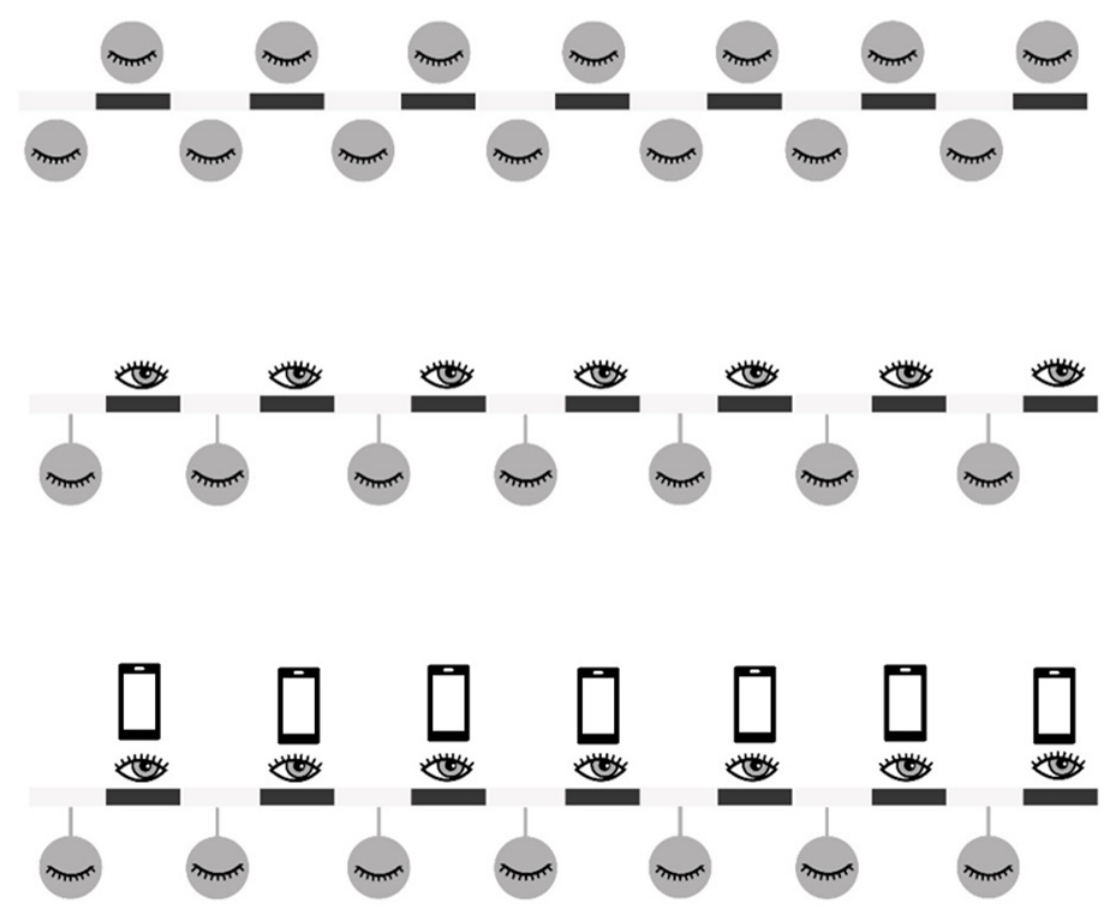
Three experimental conditions were used in this study (Figure 1). Prolonged eyes-closed (EC) conditions were carried out for 7 cycles of 4 min each. For tandem eyes-closed and eyes-open (ECEO) conditions, 7 cycles of ECEO were performed. Participants were asked to perform both rest conditions and mobile task conditions in separate sessions. To control for potential order effects, the sequence of experimental conditions was counterbalanced across all participants. The time intervals for eyes-closed and eyes-open were 2 min, and participants were instructed to sit quietly without any task during the eyes-open periods. For the last experiment, a mobile task was performed. Participants were allowed to use a mobile phone to watch only any passive content of interest on the Facebook or Instagram platform during eyes-open periods. To ensure experimental consistency, the smartphones were set to a standardized auto-brightness level appropriate for the controlled laboratory lighting environment. Participants were instructed to maintain a natural viewing distance of approximately 30–40 cm from the screen throughout the task. Furthermore, to strictly isolate the visual and interactive components of social media scrolling, all device audio was muted during the mobile task segments. Chat boxes via both applications were not allowed to be used during eyes-open periods. Three epochs of 10 s intervals during each eyes-closed segment were obtained for further processing.

2.3. Electroencephalography

The brainwaves were recorded as previously described [17]. The electroencephalography (EEG) was recorded using a SynAmps RT 64-channel amplifier (Compumedics Limited, Abbotsford, Australia). A Quikcap electrode cap (Compumedics Limited, Abbotsford, Australia) was used, and nineteen channels were loaded with Quikgel to reduce the impedance. FP1, FP2, F3, F1, FZ, F4, F8, T7, C3, CZ, C6, T8, P7, P3, PZ, P4, P8, O1, and O2 electrodes were used. The sampling rate was 1 kHz. M1 and M2 were used as the reference electrodes. Electrode impedance below 5 kΩ was accepted. The raw data during the eyes-closed segments were then analyzed in triplicate with a 10 s epoch duration. The raw data were then processed, filtered, and artifact-reduced using Curry 7 software version 7.0.12 (Compumedics Limited, Abbotsford, Australia). A notch filter at 50 Hz was used. High-pass and low-pass filters were set to 1 Hz and 30 Hz, respectively, to obtain delta, theta, alpha, and beta. Three of the 10 s epochs of the eyes-closed period were chosen to be analyzed per interval. Eye-blinking artifacts were removed using an electrooculographic electrode (VEO) as a probe. The frequency-domain data were transformed from time-domain data using a Fast Fourier Transform. Power spectral analysis was adjusted to clearly display the differences among frequencies. The total brain response was processed and displayed.

2.4. Functional Connectivity Analysis

To evaluate the network-level cortical interactions, we conducted a preliminary functional connectivity analysis using the Phase Locking Value (PLV). Based on our hypothesis that the digital task demands sustained executive control and cognitive arousal, we defined a Region of Interest (ROI) specifically targeting the frontal and midline areas (Fp1, Fp2, F3, Fz, F4, Cz, and Pz). Continuous EEG data were segmented into 10 s non-overlapping epochs. The PLV was computed in the beta frequency band (13–30 Hz) utilizing the multitaper method implemented via the mne-connectivity package in Python (version 3.10). Grand average PLV matrices were constructed for both the resting baseline and the mobile task conditions to quantify the degree of phase synchronization between the selected electrode pairs.

2.5. Statistical Analysis

The distribution of EEG spectral power data was assessed using the Shapiro–Wilk test, which revealed a non-normal distribution. Consequently, the Kruskal–Wallis test, followed by Dunn’s multiple comparisons test, was selected as a robust non-parametric approach to analyze differences among the experimental conditions. This choice ensures a conservative estimation given the nature of the dataset. The brainwave powers are shown as the median with the 95% CI. Statistical differences were considered at p-values less than 0.05. EEG brainwave activities were processed by power spectral analysis of each sub-band, including slow alpha (8–11 Hz), fast alpha (11–13 Hz), low beta (13–15 Hz), mid beta (15–20 Hz), and high beta (20–30 Hz). The beta-to-alpha ratio was calculated and used as an arousal index. All the statistics and graphs were produced using GraphPad Prism 9 (GraphPad Software, San Diego, CA, USA).

3. Results

3.1. Effects of Non-Task Resting-State ECEO Intervention and Prolonged EC Condition on Brainwaves

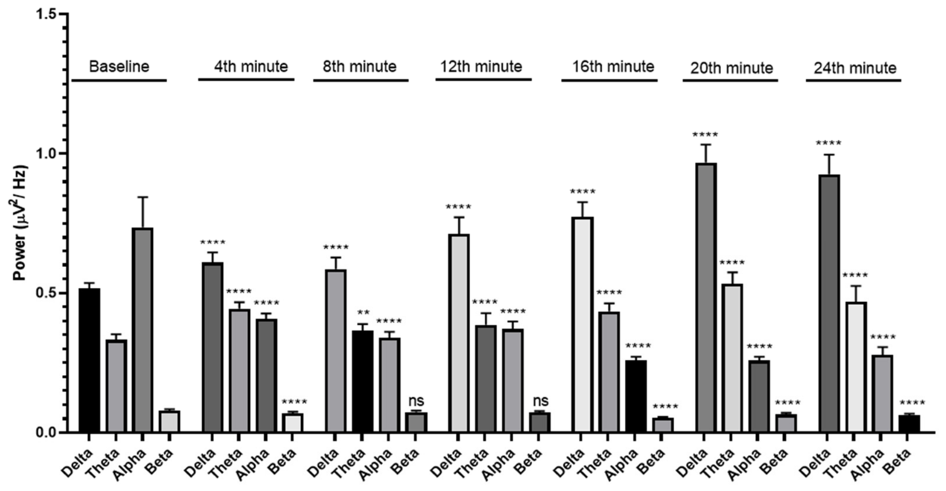
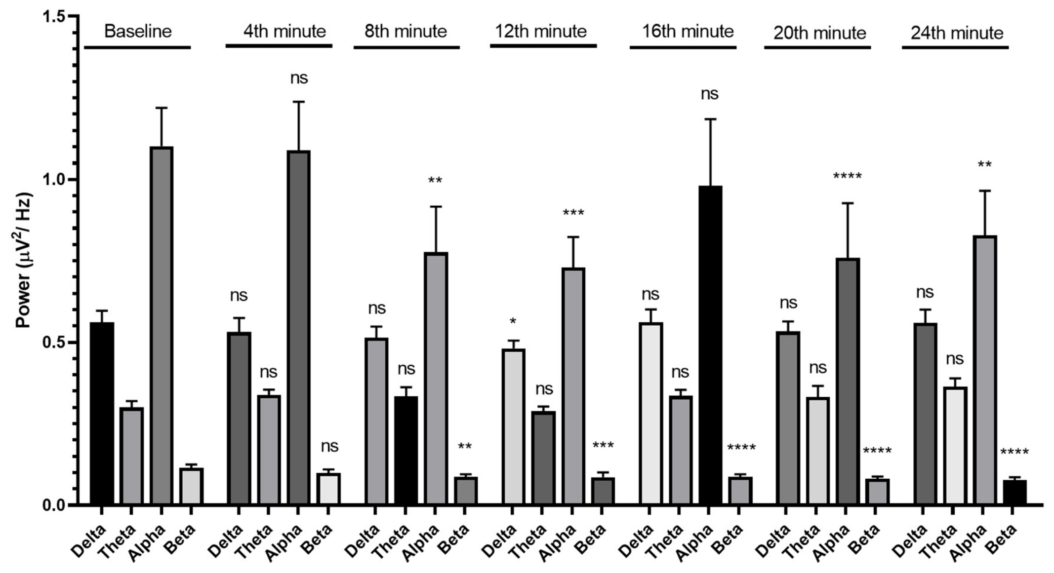
During the prolonged EC condition in the non-task resting state, alpha and beta wave powers were reduced compared to baseline from the 4th minute onwards. Meanwhile, the delta and theta wave powers were significantly increased in a time-dependent manner compared to baseline (Figure 2). These results indicate the effect of prolonged EC conditions on the induction of theta and delta synchronization in the brain. On the other hand, during the EC condition in the non-task resting state with the ECEO intervention, the reductions in alpha and beta wave powers were observed at the 8th minute, which were slower than in the prolonged EC condition. However, the alpha wave powers in the prolonged EC condition were more significantly reduced than in the ECEO condition compared with their baselines. Meanwhile, the delta and theta wave powers did not significantly change from baseline (Figure 3).

3.2. Effects of Non-Task Resting-State ECEO Intervention and Prolonged EC Condition on Arousal and Their Sub-Bands

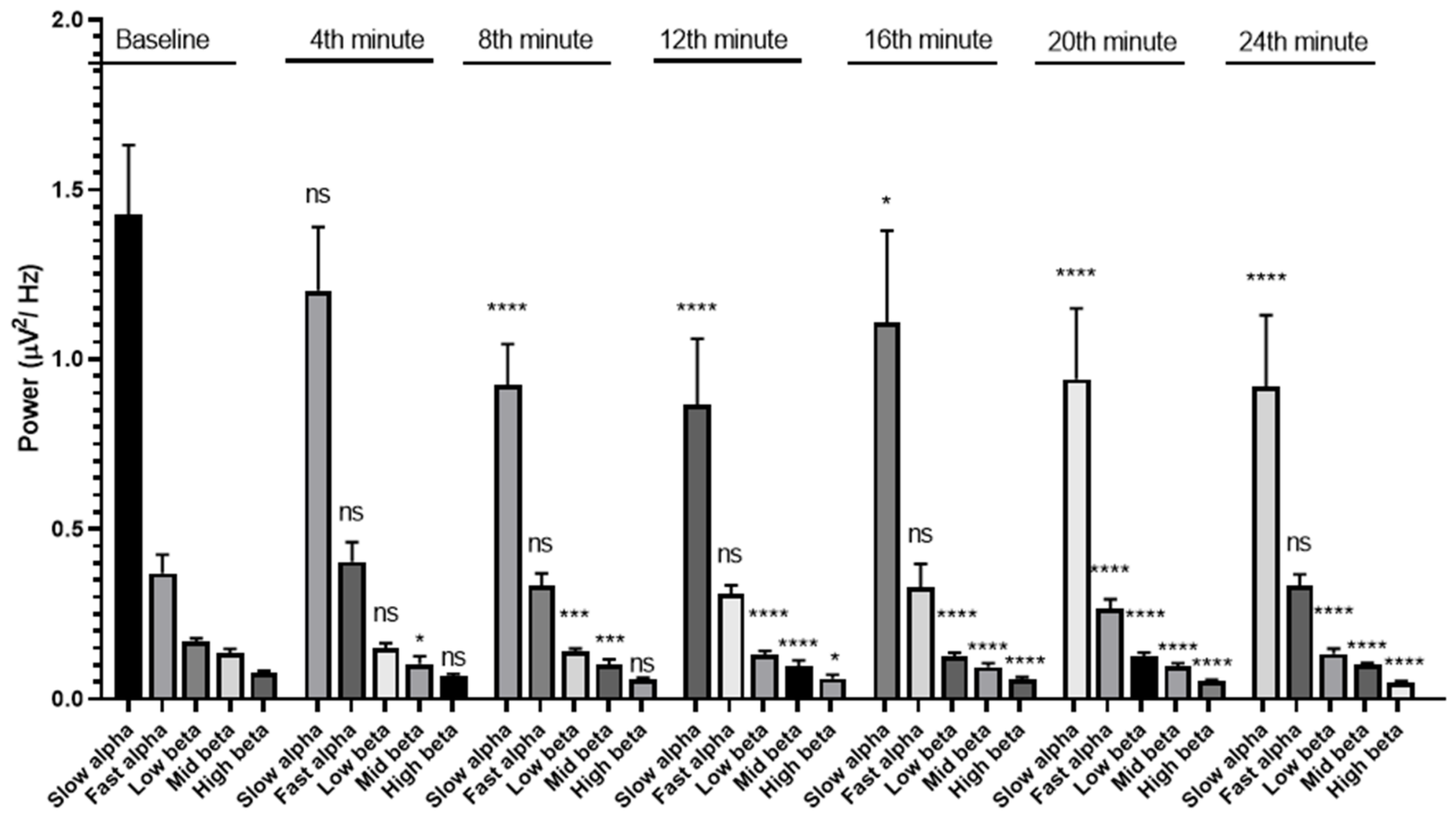
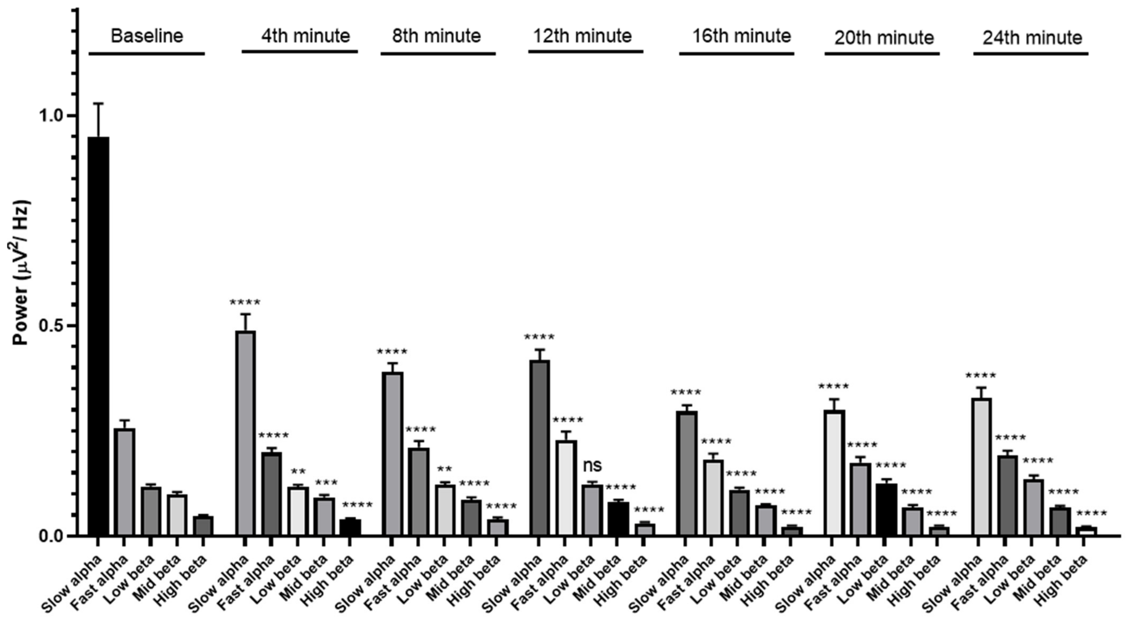
Alpha and beta brainwaves are the most common type of brainwave found during the awake state. To evaluate the effect of the ECEO intervention and prolonged EC condition in this manner, the EEG traces were analyzed at the slow alpha (9 Hz), fast alpha (12 Hz), low beta (14 Hz), mid beta (17 Hz), and high beta frequencies (25 Hz). The prolonged EC condition produced a reduction in the slow alpha, fast alpha, mid beta, and high beta wave powers, while the low beta wave powers were increased in this condition (Figure 4). The ECEO intervention showed a decrease in slow alpha wave powers, while there was no significant change in fast alpha. The low beta, mid beta, and high beta wave powers were also decreased in this condition. There was no significant change in the low beta wave powers at the 4th minute and high beta wave powers at the 4th and 8th minutes (Figure 5).

3.3. Effects of Non-Task Resting-State ECEO and Mobile Task Resting-State Interventions on Brainwaves and Arousal

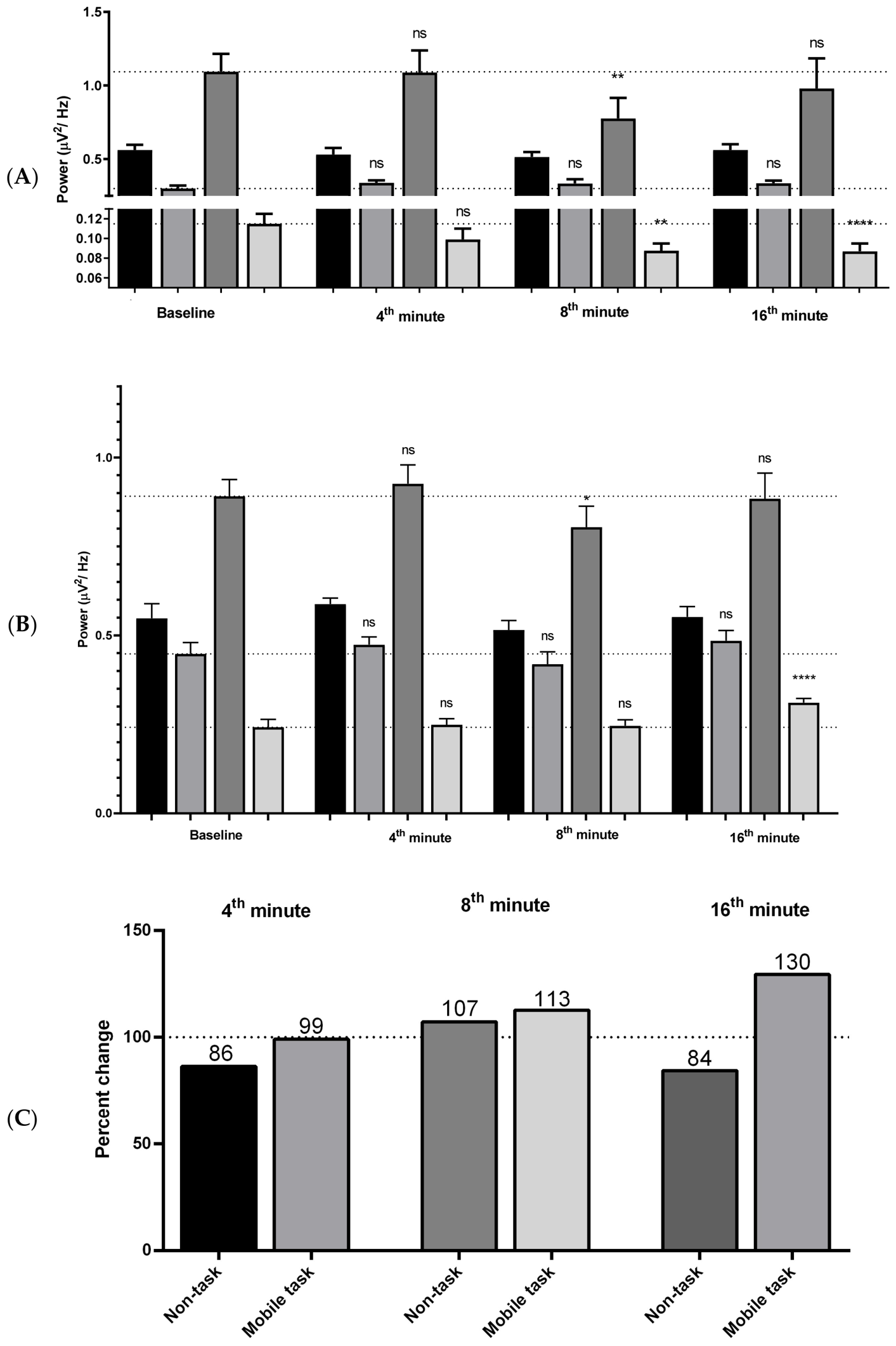
In the non-task resting-state ECEO condition, the reduction in the alpha and beta wave powers decreased from the 8th minute onwards, as shown in Figure 6A. During the mobile task, the overall brainwaves did not significantly change compared with baseline. Interestingly, marked increases in beta wave powers were observed at the 16th minute (Figure 6B). These results may indicate the effect of mobile tasks on arousal maintenance. In order to better understand the influence of non-task resting-state and mobile task conditions on arousal, we displayed the EEG power in the ratio of beta to alpha as the indicator of arousal (arousal index). To provide a comprehensive overview of the neurodynamic shifts during the interventions, we calculated an arousal index represented by the beta-to-alpha ratio. It is important to note that these values are presented as intra-individual percentage changes relative to each participant’s baseline, an approach adopted to normalize the substantial inter-individual variance inherent in EEG spectral data. As such, the indices shown in Figure 6C serve primarily as descriptive indicators of the temporal trajectory of cortical arousal throughout the task duration, rather than as absolute values intended for separate point-by-point statistical comparisons between conditions at each time interval.

3.4. Frontal-Midline Beta Functional Connectivity

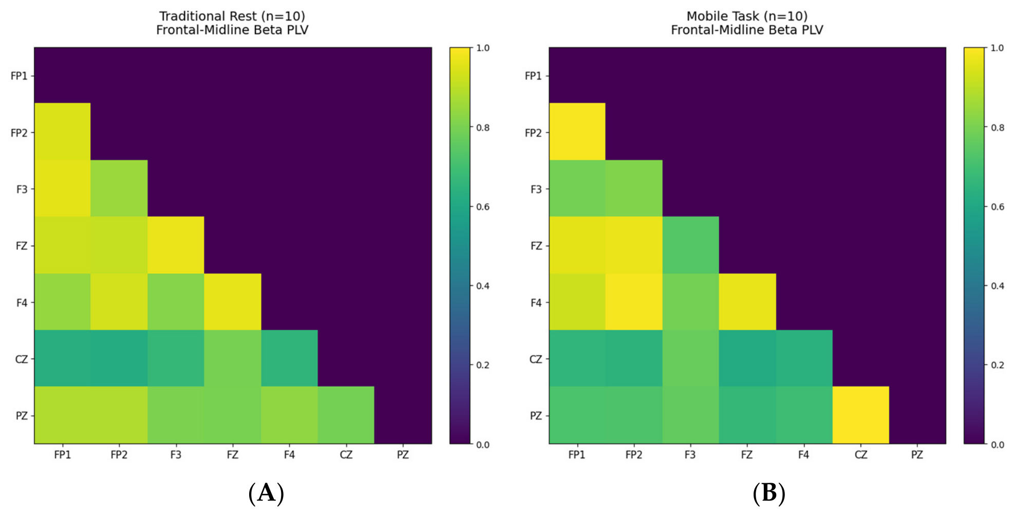
The PLV analysis revealed distinct patterns of functional connectivity within the frontal-midline network when comparing the baseline and mobile task conditions (Figure 7). During the traditional rest, the grand average PLV matrix exhibited relatively weak phase synchronization across the targeted regions. In contrast, engagement in the mobile task induced a pronounced increase in beta-band connectivity, particularly among the prefrontal and central regions. This widespread beta synchronization indicates enhanced functional coupling within the executive control network, reflecting the sustained cognitive arousal and high attentional demands required during social media interaction.

4. Discussion

Our findings present a clear narrative contrasting three distinct neurocognitive states characterized by their spectral power signatures. Non-task resting conditions are used in several psychophysiological studies, such as the psychophysics of odorant or sound stimuli. Not only are sedative or stimulating effects of stimuli observed, but also self-synchronization may be unintentionally measured in prolonged studies. The appropriate study duration should be carefully determined to reduce non-specific arousal reductions. However, our results emphasize that self-synchronization of brainwaves may unintentionally occur during prolonged studies. In this study, the approximately 24 min EC condition induced significant low-frequency synchronization of delta and theta activities, alongside a decrease in alpha and beta powers commencing at the 4th minute. This established a baseline of physiological underarousal, demonstrating the brain’s internal tendency to enter a less alert state over time.
In contrast, the traditional ECEO rest condition, which separated EC segments with 2 min EO intervals, successfully suppressed delta and theta synchronizations. Alpha and beta wave powers were slightly reduced at the 8th minute, which was slower than the EC condition. This traditional ECEO rest condition established a baseline of physiological drowsiness, where cortical arousal (alpha and beta powers) significantly decreased after 8 min, demonstrating the brain’s normal tendency to enter a less alert state over time. The most striking observation was during the mobile task intervention, where no reduction in cortical arousal was detected even at the 16th minute. This intervention acted as a powerful signal modulator, completely halting the natural decay of arousal and maintaining a steady neurodynamic state. This highlights a fundamental difference between a traditional break and a mobile task break. It should be noted that the eyes-closed resting state is not purely restorative; it may involve varying degrees of physiological underarousal or even the onset of an incipient hypnagogic state, particularly during prolonged sessions. Our EEG data, characterized by increased delta/theta synchronization and suppressed beta activity, align with markers of reduced cortical arousal. While this confirms the state of underarousal, future studies should incorporate concurrent polysomnographic or psychomotor vigilance assessments to more precisely differentiate between restorative resting states and the onset of hypnagogic transition.
A reduction in arousal during a prolonged non-task resting state was reported by Barry et al. [1]; we extend the knowledge using a mobile task to sustain the arousal level. EO induces desynchronized brainwaves from the visual input [18]. While the present study directly observed changes in EEG spectral power and beta-band connectivity, the precise underlying neurobiological processes remain beyond the scope of direct measurement. Our EEG findings suggest a state of sustained cortical activation that may theoretically be driven by bottom–up pathways, where visual input stimulates the ascending reticular activating system in the brainstem. Similarly, it is plausible to hypothesize, based on the established literature, that orexin neurons subsequently activate noradrenergic, histaminergic, and dopaminergic neurons, as well as brainstem cholinergic neurons. Furthermore, the modulation of beta-band oscillations may potentially involve VIP–SST–glutamatergic principal cell disinhibition. It is important to emphasize that these mechanisms are proposed as theoretical underpinnings for the spectral changes observed in our participants; these specific pathways were not directly imaged in the current study [19,20]. The mobile task in this study provided light from the mobile phone and selective content, which activate the bottom–up and top–down pathways, respectively. For the top–down mechanism, the interesting topic chosen by the participants will cause selective attention. Vasoactive intestinal peptide (VIP)–somatostatin (SST)–glutamatergic principal cell (PC) disinhibition, SST–parvalbumin (PV)–PC disinhibition, VIP-negative-based disinhibition, PC gain modulation, and SST-mediated desynchronization are the essential drivers for cognitive arousal which increase beta and gamma powers and reduce low-frequency synchronization [21]. In a rodent study, blue light could induce arousal responses and sleep delay [22]. Social media use also increased cognitive arousal and was associated with poor sleep quality in humans [23]. This continuous brain activation aligns with the concept of digital overstimulation detailed by Fayize in 2025, which highlights that continuous content updates demand rapid processing shifts, resulting in brain clutter and a lack of true neurocognitive disengagement [12]. The top–down processing and arousal state may interact with each other and increase global attention [21]. Top–down selective attention is impaired in neuropsychiatric diseases such as schizophrenia and attention deficit hyperactivity disorder [6,24]. These findings have significant practical implications for digital well-being within the specific demographic tested, such as young adult students in educational environments, and should be carefully extrapolated before applying to broader workplace settings. Our results challenge the common perception of using social media as a form of restorative break. This behavior often results in a subjective perception of rest, where an individual may feel they are taking a break; however, our objective EEG findings suggest that the brain remains in a state of heightened cortical arousal during these mobile interactions. This neurodynamic pattern provides a clear neurophysiological mechanism that explains recent behavioral data by Grobelny et al. [15], which revealed that brief, hedonic social media breaks fail to yield full resource replenishment and fall short in alleviating fatigue. Although users experience a superficial sense of psychological detachment, the ongoing cortical activation observed in our high-frequency beta parameters demonstrates why true recovery is obstructed [15]. These findings suggest that such digital breaks might have potential implications for cumulative cognitive fatigue, reduced productivity, and sleep onset if taken habitually or prior to bedtime; however, these remain speculative observations that warrant future longitudinal investigation [23,25]. Our study provides neurophysiological evidence supporting the need for more mindful break-taking strategies that allow for genuine cortical disengagement and restoration of attentional resources. It is important to clarify that while the mobile task effectively counters physiological underarousal, it does not replenish depleted cognitive resources.
However, our findings do not suggest that mobile devices are uniformly detrimental. Instead, this sustained arousal effect can be viewed as a double-edged sword, highly dependent on the user’s context and goal. This duality is best explained by the Yerkes–Dodson Law, which posits that optimal performance requires a moderate, optimal level of arousal. On one edge (the negative side), when the goal is genuine rest or recovery from stress, the mobile task break is counterproductive. As our study shows, it sustains (or even potentiates) arousal far beyond the optimal level needed for rest, preventing neurocognitive recovery. On the other edge (the positive side), in a context of low arousal such as boredom or fatigue during a long lecture, this same mechanism becomes a strategic tool. The stimulation from a mobile task can effectively boost a non-optimal low-arousal state back up toward the optimal level, thereby sustaining attention and engagement for learning or work.
Electroencephalograms can be analyzed in several ways. Frequency analysis using a Fast Fourier Transform (FFT) is frequently used to quantify these states with precision. Delta, theta, alpha, beta, and gamma are characterized by frequencies with a difference in amplitude and shape. High-frequency desynchronizations of beta and gamma are correlated with high-arousal states. Low-frequency synchronizations of theta and delta are correlated with low arousal states [21] and are utilized in the sleep staging of non-rapid eye movement sleep [26]. Eye closure increases alpha activity in the occipital area, and eye opening increases beta activity. Barry et al. recommended using eyes-closed periods in analysis [1]. We analyzed only eyes-closed segments to reduce eye-blinking and eye movement artifacts. The arousal index is calculated by the ratio of beta to alpha wave powers. Low arousal indicates calmness or drowsiness, which may be induced by chemicals such as essential oils. Lavender [27] and tangerine essential oils [28] showed sedative effects or low arousal induction which finally shortened the sleep latency. High-arousal states showed a relationship with cognitive processing [29], memory [30], delayed sleep latency [25], and motor performance [31].
This positive application aligns with previous findings demonstrating that smartphones, tablets, or laptops can be strategically utilized to raise alertness, arousal, and attention for lecture-related contents [32]. Specifically, this is consistent with recent mobile-learning evaluations by Herpich et al., confirming that interactive digital frameworks on mobile devices significantly stimulate the focused attention dimension of user engagement [16]. Thus, the key is not the tool itself, but the context of its use.
However, several limitations must be considered when interpreting these findings. First, our participant pool was restricted to a modest sample of healthy females aged 20–25 years (n = 20). While this homogeneity was vital to systematically minimize inter-individual variance and suppress confounding gender-driven electrical variations—as empirical evidence by Rajendran et al. [11] underscores distinct spectral fluctuations between male and female students—it inherently limits the broader generalizability of our results. Future investigations involving larger, representative mixed-gender cohorts across diverse age groups are necessary to determine whether these digital rest dynamics are sex- or age-dependent.
Second, to preserve the ecological validity of a naturalistic social media break, we allowed participants to freely browse their feeds. Consequently, the uncontrolled visual complexity, emotional valence, individual cognitive load, and ambient light intensity acted as significant confounding variables. Therefore, the observed high-frequency beta surges represent a generalized neurophysiological response to mixed digital stimuli. Future studies should employ strictly controlled visual mock-ups and standardized screen brightness to accurately dissect the specific neurophysiological contributions of cognitive engagement versus light-induced arousal.
Third, our paradigm of inducing fatigue via prolonged eyes-closed rest may involve a complex interplay between restoration and underarousal. Future research would benefit from utilizing standardized cognitive tasks to induce fatigue. Furthermore, while our selection of 10 s artifact-free epochs ensured high-quality signal analysis at specific time points, exploring continuous signal processing could better capture the full temporal dynamics of neurophysiological recovery.
Finally, while the current study utilizes aggregate spectral power to characterize cortical arousal, we acknowledge that this approach provides a fragmented view of complex cognitive states and regional dynamics. As demonstrated by our preliminary Phase Locking Value (PLV) network-level analysis, mobile tasks induce widespread beta-band synchronization, highlighting the value of functional connectivity. We recognize that detailed topographical mapping, such as source localization and advanced connectivity modeling, is essential for spatial precision. We identify these advanced analytical techniques as a key priority for our future longitudinal studies to expand upon the current spectral power framework and provide a more holistic understanding of brain-wide network dynamics.


5. Conclusions

In summary, this study confirms that prolonged eye closure successfully induces a state of physiological underarousal, characterized by a significant natural reduction in alpha and beta spectral powers. While traditional quiet rest (ECEO) offers only a temporary delay in arousal reduction, passive social media interaction acts as a powerful signal modulator that maintains and increases cortical arousal. The marked rise in high-frequency beta power and the enhanced frontal-midline beta synchronization indicate that mobile breaks create an illusion of rest while actively demanding sustained executive control. Consequently, these digital breaks function as a double-edged sword: they hinder genuine neurophysiological disengagement and recovery, but they can be utilized strategically as a tool to boost alertness when arousal levels are suboptimally low. These findings highlight the critical role of advanced EEG monitoring in uncovering the hidden neurocognitive costs of modern digital habits among young adults.

Author Contributions

Funding acquisition, methodology, analysis, writing—original draft, and writing—review and editing, A.S.; methodology, formal analysis, and writing—original draft, K.K., W.A., M.N. and P.P.; conceptualization, formal analysis, funding acquisition, methodology, project administration, writing—original draft, and writing—review and editing, P.K. All authors have read and agreed to the published version of the manuscript.

Funding

This project is funded by the National Research Council of Thailand (NRCT) and Walailak University, grant number N41A640166.

Institutional Review Board Statement

The study was conducted in accordance with the Declaration of Helsinki, and approved by the Institutional Review Board of Walailak University (protocol code WUEC-20-035-01 and date of approval 25 February 2020).

Informed Consent Statement

Informed consent was obtained from all subjects involved in the study.

Data Availability Statement

The presented data are available on reasonable request to the corresponding author.

Acknowledgments

The authors thank the Center for Scientific and Technological Equipment and the Research Institute for Health Sciences, Walailak University, for providing research support. The authors used a GenAI tool (Gemini 3.1 Pro) solely for English language editing and grammar refinement to improve the clarity and readability of the manuscript. The authors confirm that all authors have carefully reviewed, edited, and verified the output produced by the AI tool. Furthermore, all authors take full responsibility for the content, accuracy, and integrity of this publication.

Conflicts of Interest

The authors declare no conflicts of interest.

Abbreviations

The following abbreviations are used in this manuscript:
ARTAttention Restoration Theory
BMIBody Mass Index
EEGElectroencephalography
ECEyes-Closed
ECEOEyes-Closed and Eyes-Open
nsNot Significant
PLVPhase Locking Value

References

  1. Barry, R.J.; Clarke, A.R.; Johnstone, S.J.; Magee, C.A.; Rushby, J.A. EEG differences between eyes-closed and eyes-open resting conditions. Clin. Neurophysiol. 2007, 118, 2765–2773. [Google Scholar] [CrossRef] [PubMed]
  2. Oken, B.S.; Salinsky, M.C.; Elsas, S. Vigilance, alertness, or sustained attention: Physiological basis and measurement. Clin. Neurophysiol. 2006, 117, 1885–1901. [Google Scholar] [CrossRef] [PubMed]
  3. Thompson, R.F. Foundations of Physiological Psychology; Harper & Row: New York, NY, USA, 1967. [Google Scholar]
  4. Pfaff, D.; Ribeiro, A.; Matthews, J.; Kow, L.M. Concepts and mechanisms of generalized central nervous system arousal. Ann. N. Y. Acad. Sci. 2008, 1129, 11–25. [Google Scholar] [CrossRef] [PubMed]
  5. Calderon, D.; Kilinc, M.; Maritan, A.; Banavar, J.; Pfaff, D. Generalized CNS arousal: An elementary force within the vertebrate nervous system. Neurosci. Biobehav. Rev. 2016, 68, 167–176. [Google Scholar] [CrossRef] [PubMed]
  6. Krone, L.; Frase, L.; Piosczyk, H.; Selhausen, P.; Zittel, S.; Jahn, F.; Kuhn, M.; Feige, B.; Mainberger, F.; Klöppel, S. Top-down control of arousal and sleep: Fundamentals and clinical implications. Sleep Med. Rev. 2017, 31, 17–24. [Google Scholar] [CrossRef] [PubMed]
  7. Posner, M.I.; Petersen, S.E. The attention system of the human brain. Annu. Rev. Neurosci. 1990, 13, 25–42. [Google Scholar] [CrossRef] [PubMed]
  8. Nebes, R.D.; Brady, C.B. Phasic and tonic alertness in Alzheimer’s disease. Cortex 1993, 29, 77–90. [Google Scholar] [CrossRef] [PubMed]
  9. Kreibig, S.D. Autonomic nervous system activity in emotion: A review. Biol. Psychol. 2010, 84, 394–421. [Google Scholar] [CrossRef] [PubMed]
  10. Vinck, M.; Batista-Brito, R.; Knoblich, U.; Cardin, J.A. Arousal and locomotion make distinct contributions to cortical activity patterns and visual encoding. Neuron 2015, 86, 740–754. [Google Scholar] [CrossRef] [PubMed]
  11. Rajendran, V.; Jayalalitha, S.; Adalarasu, K. EEG based evaluation of examination stress and test anxiety among college students. Irbm 2022, 43, 349–361. [Google Scholar] [CrossRef]
  12. Fayize, P.V. The Impact of Social Media on Cognitive Function and Brain Health. Acta Neurophysiol. 2025, 6, 180107. [Google Scholar]
  13. Yerkes, R.M.; Dodson, J.D. The relation of strength of stimulus to rapidity of habit-formation. J. Comp. Neurol. Psychol. 1908, 18, 459–482. [Google Scholar] [CrossRef]
  14. Fischer, T.; Langner, R.; Birbaumer, N.; Brocke, B. Arousal and attention: Self-chosen stimulation optimizes cortical excitability and minimizes compensatory effort. J. Cogn. Neurosci. 2008, 20, 1443–1453. [Google Scholar] [CrossRef] [PubMed]
  15. Grobelny, J.; Glinka, M.; Chirkowska-Smolak, T. The impact of hedonic social media use during microbreaks on employee resources recovery. Sci. Rep. 2024, 14, 21603. [Google Scholar] [CrossRef] [PubMed]
  16. Herpich, F.; Guarese, R.L.; Cassola, A.T.; Tarouco, L.M. Mobile augmented reality impact in student engagement: An analysis of the focused attention dimension. In Proceedings of the 2018 International Conference on Computational Science and Computational Intelligence (CSCI); IEEE: Piscataway, NJ, USA, 2018; pp. 562–567. [Google Scholar]
  17. Koomhin, P.; Sattayakhom, A.; Chandharakool, S.; Sinlapasorn, J.; Suanjan, S.; Palipoch, S.; Na-ek, P.; Punsawad, C.; Matan, N. Michelia essential oil inhalation increases fast alpha wave activity. Sci. Pharm. 2020, 88, 23. [Google Scholar] [CrossRef]
  18. Barry, R.J.; De Blasio, F.M. EEG differences between eyes-closed and eyes-open resting remain in healthy ageing. Biol. Psychol. 2017, 129, 293–304. [Google Scholar] [CrossRef] [PubMed]
  19. Wan, L.; Huang, H.; Schwab, N.; Tanner, J.; Rajan, A.; Lam, N.B.; Zaborszky, L.; Li, C.s.R.; Price, C.C.; Ding, M. From eyes-closed to eyes-open: Role of cholinergic projections in EC-to-EO alpha reactivity revealed by combining EEG and MRI. Hum. Brain Mapp. 2019, 40, 566–577. [Google Scholar] [PubMed]
  20. Chemelli, R.M.; Willie, J.T.; Sinton, C.M.; Elmquist, J.K.; Scammell, T.; Lee, C.; Richardson, J.A.; Williams, S.C.; Xiong, Y.; Kisanuki, Y. Narcolepsy in orexin knockout mice: Molecular genetics of sleep regulation. Cell 1999, 98, 437–451. [Google Scholar] [CrossRef] [PubMed]
  21. Batista-Brito, R.; Zagha, E.; Ratliff, J.M.; Vinck, M. Modulation of cortical circuits by top-down processing and arousal state in health and disease. Curr. Opin. Neurobiol. 2018, 52, 172–181. [Google Scholar] [CrossRef] [PubMed]
  22. Wu, F.; Wu, S.; Gui, Q.; Tang, K.; Xu, Q.; Tao, Y.; Chen, M.; Cheng, J.; Wang, L.; Zhang, L. Blue light insertion at night is involved in sleep and arousal-promoting response delays and depressive-like emotion in mice. Biosci. Rep. 2021, 41, BSR20204114. [Google Scholar] [CrossRef]
  23. Alonzo, R.; Hussain, J.; Stranges, S.; Anderson, K.K. Interplay between social media use, sleep quality, and mental health in youth: A systematic review. Sleep Med. Rev. 2021, 56, 101414. [Google Scholar] [CrossRef] [PubMed]
  24. Sander, C.; Hensch, T.; Wittekind, D.A.; Böttger, D.; Hegerl, U. Assessment of wakefulness and brain arousal regulation in psychiatric research. Neuropsychobiology 2015, 72, 195–205. [Google Scholar] [CrossRef] [PubMed]
  25. He, J.-w.; Tu, Z.-h.; Xiao, L.; Su, T.; Tang, Y.-x. Effect of restricting bedtime mobile phone use on sleep, arousal, mood, and working memory: A randomized pilot trial. PLoS ONE 2020, 15, e0228756. [Google Scholar] [CrossRef] [PubMed]
  26. Cluydts, R.; De Valck, E.; Verstraeten, E.; Theys, P. Daytime sleepiness and its evaluation. Sleep Med. Rev. 2002, 6, 83–96. [Google Scholar] [CrossRef] [PubMed]
  27. Koulivand, P.H.; Khaleghi Ghadiri, M.; Gorji, A. Lavender and the nervous system. Evid.-Based Complement. Altern. Med. 2013, 2013, 681304. [Google Scholar] [CrossRef]
  28. Chandharakool, S.; Koomhin, P.; Sinlapasorn, J.; Suanjan, S.; Phungsai, J.; Suttipromma, N.; Songsamoe, S.; Matan, N.; Sattayakhom, A. Effects of tangerine essential oil on brain waves, moods, and sleep onset latency. Molecules 2020, 25, 4865. [Google Scholar] [CrossRef] [PubMed]
  29. Clarke, A.R.; Barry, R.J.; Karamacoska, D.; Johnstone, S.J. The EEG theta/beta ratio: A marker of arousal or cognitive processing capacity? Appl. Psychophysiol. Biofeedback 2019, 44, 123–129. [Google Scholar] [CrossRef] [PubMed]
  30. Cahill, L.; McGaugh, J.L. Mechanisms of emotional arousal and lasting declarative memory. Trends Neurosci. 1998, 21, 294–299. [Google Scholar] [CrossRef] [PubMed]
  31. Landers, D.M. The arousal-performance relationship revisited. Res. Q. Exerc. Sport 2007, 78, A-12. [Google Scholar]
  32. Gehlen-Baum, V.; Weinberger, A.; Pohl, A.; Bry, F. Technology use in lectures to enhance students’ attention. In Proceedings of the European Conference on Technology Enhanced Learning, Graz, Austria, 16–19 September 2014; pp. 125–137. [Google Scholar]
Figure 1. Experimental design of the prolonged eyes-closed condition, tandem eyes-closed and eyes-open condition, as well as the mobile task with social media content.
Signals 07 00068 g001
Figure 2. EEG signal of human brainwaves during eyes-closed in eyes-closed (EC) condition characterized by frequencies consisting of delta, theta, alpha, and beta wave powers. The signal was analyzed at 0 min (baseline), and at the 4th, 8th, 12th, 16th, 20th, and 24th minutes. ns, not significant; ** and **** represent no statistical differences at p < 0.01 and p < 0.0001, respectively.
Signals 07 00068 g002
Figure 3. EEG signal of human brainwaves during eyes-closed in eyes-closed–eyes-open (ECEO) condition characterized by frequencies consisting of delta, theta, alpha, and beta wave powers. The signal was analyzed at 0 min (baseline), and at the 4th, 8th, 12th, 16th, 20th, and 24th minutes. ns, not significant; *, **, ***, and **** represent no statistical differences at p < 0.05, p < 0.01, p < 0.001, and p < 0.0001, respectively.
Signals 07 00068 g003
Figure 4. EEG signal of alpha and beta brainwaves during eyes-closed in eyes-closed (EC) condition characterized by frequencies consisting of slow alpha (9 Hz), fast alpha (12 Hz), low beta (14 Hz), mid beta (17 Hz), and high beta (25 Hz). The signal was analyzed at 0 min (baseline), and at the 4th, 8th, 12th, 16th, 20th, and 24th minutes. ns, not significant; **, ***, and **** represent no statistical differences at p < 0.01, p < 0.001, and p < 0.0001, respectively.
Signals 07 00068 g004
Figure 5. EEG signal of alpha and beta brainwaves during eyes-closed in eyes-closed–eyes-open (ECEO) condition characterized by frequencies consisting of slow alpha (9 Hz), fast alpha (12 Hz), low beta (14 Hz), mid beta (17 Hz), and high beta (25 Hz). The signal was analyzed at 0 min (baseline), and at the 4th, 8th, 12th, 16th, 20th, and 24th minutes. ns, not significant; *, ***, and **** represent no statistical differences at p < 0.05, p < 0.001, and p < 0.0001, respectively.
Signals 07 00068 g005
Figure 6. EEG signal and arousal state during eyes-closed in non-task resting-state ECEO condition (A) and mobile task resting-state condition (B). Brainwaves were characterized by frequencies consisting of delta, theta, alpha, and beta wave powers. The signal was analyzed at 0 min (baseline), and at the 4th, 8th, and 16th minutes. The arousal level was characterized by the percent change in the beta-to-alpha ratio compared with baseline (C). ns, not significant; ** and **** represent no statistical differences at p < 0.01 and p < 0.0001, respectively.
Signals 07 00068 g006
Figure 7. Grand average Phase Locking Value (PLV) matrices in the beta band (13–30 Hz) across the frontal-midline network (Fp1, Fp2, F3, Fz, F4, Cz, and Pz). (A) Resting baseline condition showing lower connectivity. (B) Mobile task condition displaying enhanced beta synchronization. The color scale represents the strength of phase synchronization, ranging from 0 (dark blue/purple, indicating no synchronization) to 1.0 (yellow, indicating perfect phase synchronization).
Signals 07 00068 g007
Disclaimer/Publisher’s Note: The statements, opinions and data contained in all publications are solely those of the individual author(s) and contributor(s) and not of MDPI and/or the editor(s). MDPI and/or the editor(s) disclaim responsibility for any injury to people or property resulting from any ideas, methods, instructions or products referred to in the content.