Case Report

A Pyogenic Liver Abscess Masquerading as Advanced Hepatocellular Carcinoma: Diagnostic Pitfalls of LI-RADS 5 in a Non-Cirrhotic Patient and Vascular Complications of Percutaneous Drainage


1
Subspecialty Program in Gastroenterology-Hepatology Division, Department of Internal Medicine, Dr. Soetomo General Academic Hospital, Surabaya 60286, Indonesia
2
Subspecialty Program in Gastroenterology-Hepatology Division, Department of Internal Medicine, Faculty of Medicine, Universitas Airlangga, Surabaya 60132, Indonesia
3
Division of Gastroenterohepatology, Department of Internal Medicine, Dr. Soetomo General Academic Hospital, Surabaya 60286, Indonesia
4
Division of Gastroenterohepatology, Department of Internal Medicine, Faculty of Medicine, Universitas Airlangga, Surabaya 60132, Indonesia
*
Author to whom correspondence should be addressed.
Gastrointest. Disord. 2026, 8(3), 53; https://doi.org/10.3390/gidisord8030053
Submission received: 28 April 2026 / Revised: 28 May 2026 / Accepted: 28 May 2026 / Published: 9 September 2026

Abstract

Background: Liver abscesses represent uncommon yet potentially life-threatening infections of the hepatic parenchyma. Their imaging appearances can vary widely, and—in rare instances—may closely resemble hepatocellular carcinoma (HCC) on multimodality imaging. We report a case of pyogenic liver abscess initially misclassified as advanced HCC based on imaging characteristics meeting LI-RADS 5 criteria. Case Presentation: A 45-year-old male without known cirrhosis, viral hepatitis, or diabetes presented with right upper quadrant pain, abdominal rigidity, nausea, and vomiting. He denied alcohol use, recent dental procedures, and travel to endemic areas. Contrast-enhanced computed tomography and magnetic resonance imaging demonstrated multiple exophytic hepatic masses with arterial-phase hyperenhancement and venous/delayed washout, classified as LI-RADS 5 and initially interpreted as advanced HCC (BCLC stage C). Alpha-fetoprotein (AFP) was not elevated. Hepatitis B surface antigen and anti-HIV were non-reactive. Ultrasound-guided fine-needle aspiration biopsy yielded purulent material; histopathology confirmed chronic suppurative inflammation consistent with an abscess, with negative acid-fast bacilli staining. Cultures were sterile. The patient was treated with doripenem and metronidazole. A post-drainage complication of middle hepatic artery bleeding required urgent transcatheter embolization. The patient was subsequently discharged in stable condition. Discussion: This case illustrates a recognized but uncommon diagnostic pitfall: pyogenic liver abscesses fulfil imaging criteria for HCC. LI-RADS 5 classification carries an estimated false-positive rate of approximately 5%, and its application is technically restricted to patients with established HCC risk factors (e.g., cirrhosis, chronic hepatitis B). In non-cirrhotic patients presenting with fever, leukocytosis, and an atypical or rapidly enlarging hepatic mass, tissue confirmation is essential before committing to an oncological diagnosis. Conclusions: When a hepatic mass displays imaging features fulfilling LI-RADS 5 criteria in a non-cirrhotic patient with clinical signs of infection—including fever, leukocytosis, and elevated inflammatory markers—the possibility of a liver abscess must be actively excluded. Biopsy and percutaneous drainage are essential to avoid misdiagnosis and to enable timely, appropriate treatment.

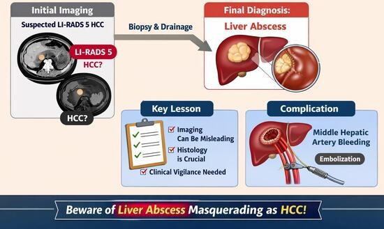
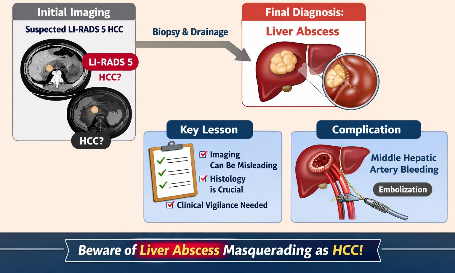
Graphical Abstract

1. Introduction

A liver abscess is defined as a collection of suppurative material enclosed within the hepatic parenchyma [1]. Although relatively uncommon, liver abscesses are potentially life-threatening infections attributable to bacterial, fungal, protozoal, or helminthic organisms [2]. The two predominant etiologies are pyogenic liver abscess (PLA) and amoebic liver abscess (ALA); less frequently, causative agents include fungi, mycobacteria, and other atypical organisms [3]. Reported incidence ranges from 2.3 cases per 100,000 hospitalizations in North America to 275.4 per 100,000 in Taiwan. Historically carrying a mortality rate of 75–80%, improvements in antibiotic therapy and interventional drainage have reduced this figure to 10–40% in contemporary series [1].
The clinical course of liver abscess is frequently insidious, with nonspecific symptoms including fever, right upper quadrant pain, and constitutional decline. Diagnosis is often confirmed by imaging, particularly ultrasonography and computed tomography (CT), which carry sensitivities of 96–100% [1]. While most abscesses are recognized by characteristic imaging features (hypoechoic or hypodense lesions with rim enhancement), a subset may exhibit heterogeneous enhancement patterns that closely mimic hepatic malignancy—most notably hepatocellular carcinoma (HCC) [4].
HCC is the most common primary liver malignancy and a leading cause of cancer-related mortality worldwide. Its diagnosis by imaging is standardized through the Liver Imaging Reporting and Data System (LI-RADS), which stratifies hepatic observations in at-risk patients from LR-1 (definitively benign) to LR-5 (definitively HCC). LR-5 classification requires the presence of major imaging features, including arterial-phase hyperenhancement, venous/delayed washout, and an enhancing capsule, and is associated with a positive predictive value exceeding 95% in appropriately selected at-risk populations [5,6]. However, this system is explicitly designed for patients with established HCC risk factors, particularly cirrhosis or chronic hepatitis B, and its application in non-cirrhotic patients may result in false-positive classifications [7].
We herein report a case of a 45-year-old male without cirrhosis or known hepatitis, whose multimodality imaging findings—including multiple exophytic hepatic masses with arterial hyperenhancement and washout—fulfilled LI-RADS 5 criteria and led to an initial diagnosis of advanced HCC (BCLC stage C). Tissue biopsy and percutaneous drainage ultimately established pyogenic liver abscess as the correct diagnosis, and the clinical course was further complicated by middle hepatic artery hemorrhage requiring emergency embolization. This case underscores the importance of tissue confirmation in atypical hepatic masses and highlights clinical and imaging red flags that should prompt consideration of infectious etiologies before an oncological diagnosis is finalized.

2. Case Presentation

2.1. Clinical Background and Risk Factors

A 45-year-old male presented with right upper quadrant pain, abdominal rigidity, nausea, and vomiting for 10 days prior to admission. He had no prior history of chronic liver disease, viral hepatitis, alcohol consumption, diabetes mellitus, or immunocompromising conditions. He denied recent dental procedures, abdominal trauma, or travel to areas endemic for Entamoeba histolytica. There was no known family history of liver disease or malignancy. These details are relevant, as the absence of classical HCC predisposing factors and the presence of constitutional symptoms should have raised early suspicion for an infectious process.

2.2. Initial Evaluation and Imaging

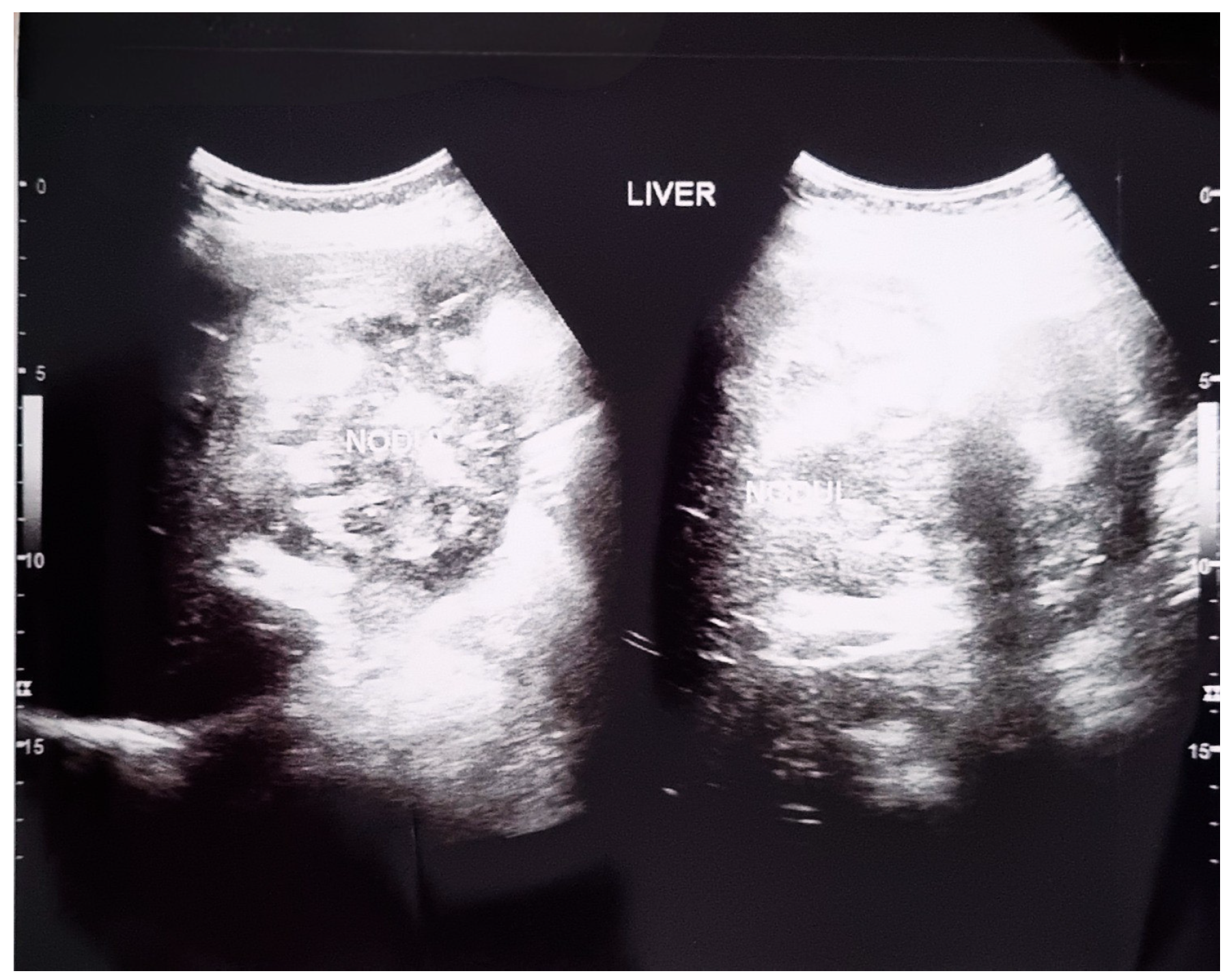
The patient was initially evaluated at a private hospital, where abdominal ultrasonography (GE LOGIQ E9; GE Healthcare, Chicago, IL, USA) revealed hepatomegaly with multiple solid masses in the right hepatic lobe—the largest measuring 11.3 cm × 7.7 cm—causing inferior compression of the right kidney, interpreted as hepatoma (Figure 1). Contrast-enhanced abdominal CT demonstrated hepatomegaly with multiple solid, lobulated, exophytic masses in hepatic segments 2, 3, 4a, and 4b, the largest measuring 9.8 cm × 12.7 cm × 9.2 cm, with arterial-phase hyperenhancement and venous/delayed-phase washout. A peripheral capsule was identified. These findings were classified as LI-RADS 5, consistent with HCC. Splenomegaly and left pleural effusion were additionally noted (Figure 2). The patient was then referred to a tertiary center for further management.

2.3. Admission Assessment

On admission to the referral hospital, the patient appeared moderately ill. Body weight was 61 kg and height was 165 cm (BMI 22.4 kg/m2). Vital signs: blood pressure 136/77 mmHg; heart rate 107 beats/min; respiratory rate 18 breaths/min; oxygen saturation 96% on room air; and axillary temperature 37.8 °C. The presence of tachycardia and low-grade fever alongside marked leukocytosis (described below) was noted but not initially attributed to infection given the prior imaging diagnosis.
Laboratory evaluation revealed hemoglobin 9.3 g/dL, hematocrit 27.8%, leukocyte count of 18,660/μL with differential showing 91.4% neutrophils (consistent with neutrophilia), platelet count of 403,000/μL, and an erythrocyte sedimentation rate of 99 mm/h. Coagulation studies showed prothrombin time 11.1 s, aPTT 34.8 s, and INR 1.2. Liver chemistry: direct bilirubin 4.78 mg/dL, total bilirubin 6.67 mg/dL, AST 41.5 U/L, ALT 28.6 U/L, alkaline phosphatase 306 U/L, GGT 309.5 U/L, albumin 2.36 g/dL, and total protein 6.19 g/dL. Alpha-fetoprotein (AFP), the primary tumor marker for HCC, was not elevated. Carcinoembryonic antigen (CEA) was 0.9 ng/mL (normal). Chest radiography demonstrated cardiomegaly.
The working diagnosis remained HCC BCLC stage C. Empirical therapy was initiated with intravenous ondansetron 8 mg three times daily, omeprazole 40 mg twice daily, ceftriaxone 1 g twice daily, N-acetylcysteine 5 g once daily, and paracetamol 1 g three times daily, alongside oral probiotics, nutritional supplementation, silybin–phospholipid, ambroxol, and ursodeoxycholic acid.

2.4. Inpatient Course and Further Investigations

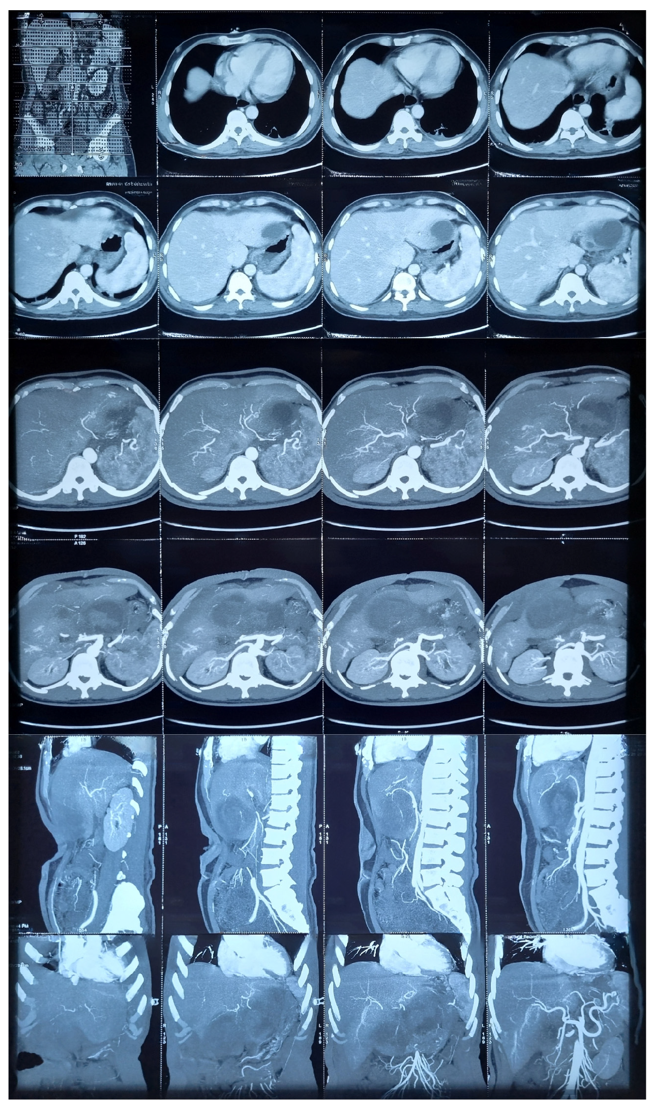
Repeat contrast-enhanced abdominal CT at the referral hospital confirmed hepatomegaly with multiple exophytic, lobulated, solid masses in segments 2, 3, 4a, and 4b, associated with left pleural effusion and splenomegaly (Figure 3). Laboratory follow-up on hospital day 5 showed leukocyte count 17,830/μL, hemoglobin 7.3 g/dL, direct bilirubin 3.34 mg/dL, total bilirubin 4.84 mg/dL, and albumin 2.62 g/dL. Hyponatremia (sodium 132 mmol/L) was also noted. Doripenem 500 mg three times daily was added to the antibiotic regimen.
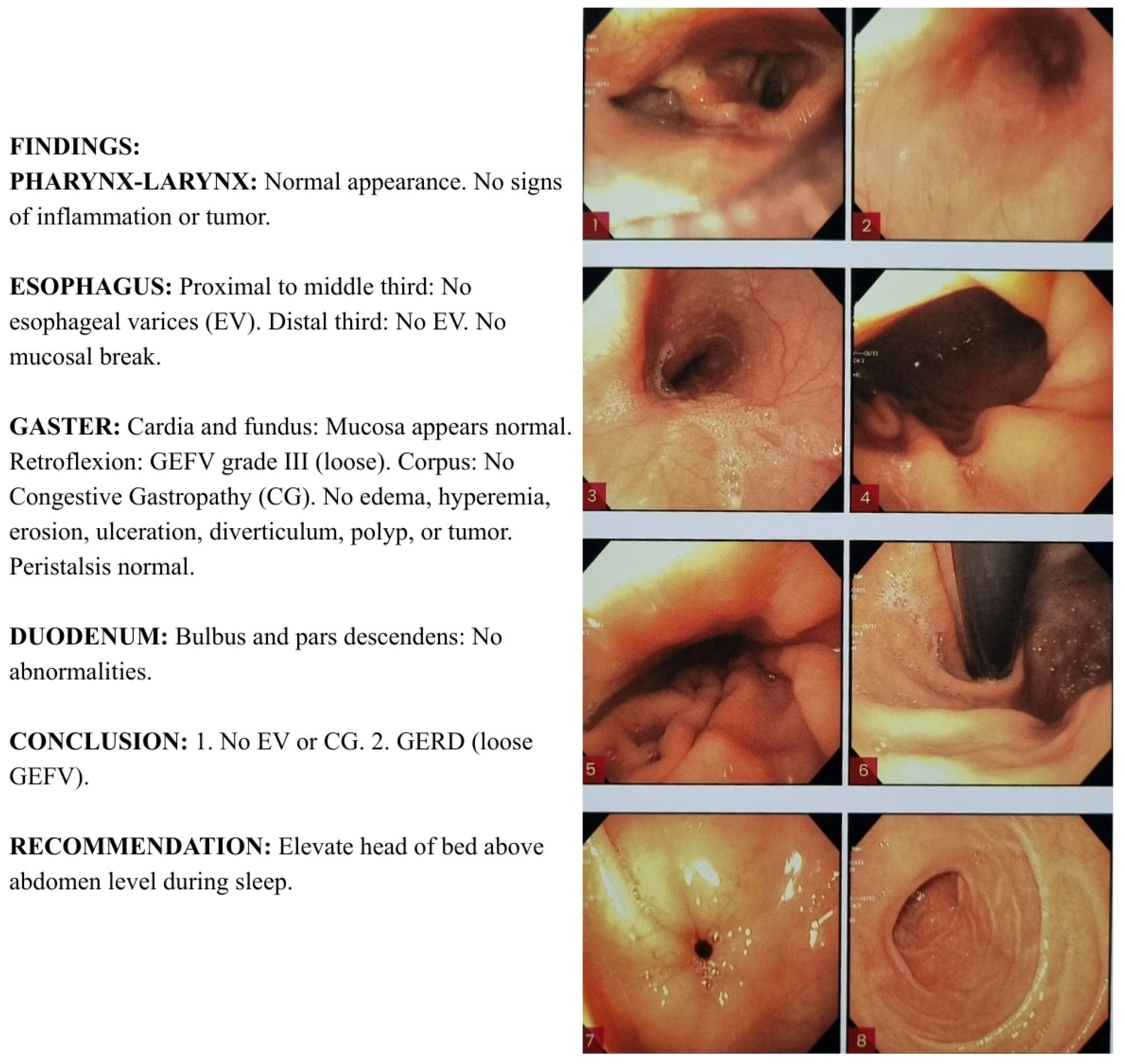
On hospital day 8, hepatitis B surface antigen (HBsAg) and anti-HIV were non-reactive, effectively excluding viral hepatitis B and HIV-associated immunocompromise as underlying predisposing factors. Esophagogastroduodenoscopy demonstrated no esophageal varices, portal hypertensive gastropathy, or endoscopic stigmata of cirrhosis; findings were consistent with gastroesophageal reflux disease with a loose gastroesophageal flap valve (Figure 4). Echocardiography was unremarkable.
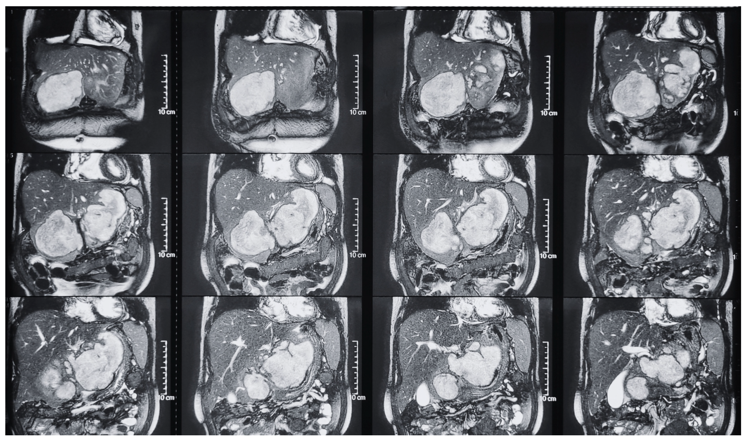
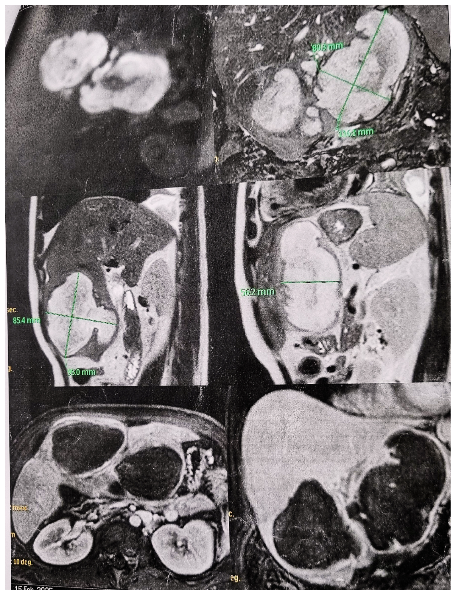
On hospital day 12, abdominal MRI demonstrated a multilobulated hepatic mass in segments 2, 3, and 4, measuring 11.6 cm × 8.0 cm × 5.6 cm, with diffusion restriction on diffusion-weighted imaging (DWI) and heterogeneous T2 hyperintensity. Dynamic contrast imaging revealed thick peripheral irregular enhancement, raising suspicion for hepatic lymphoma or atypical HCC with cystic/necrotic degeneration. The mass was adjacent to the intrahepatic bile duct with mild distal biliary dilatation. Minimal bilateral pleural effusion and a Bosniak II cyst in the lower pole of the right kidney were noted (Figure 5). Biopsy was recommended.

2.5. Biopsy, Drainage, and Revised Diagnosis

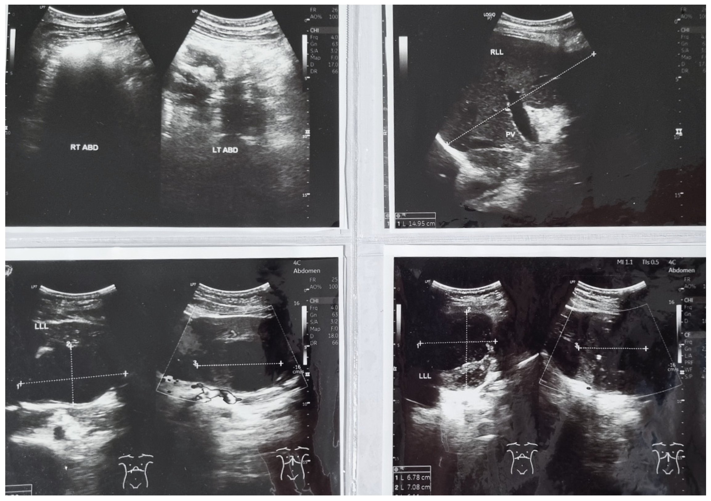
As the palpable abdominal mass decreased in size and liver chemistry improved, percutaneous drainage and liver biopsy were performed. Drainage yielded 130 mL of purulent material, with immediate symptomatic relief. A percutaneous drain was left in place. Pus cultures showed no bacterial growth; acid-fast bacilli staining by Ziehl–Neelsen was negative. Ultrasound-guided fine-needle aspiration biopsy confirmed chronic suppurative inflammation consistent with abscess (Figure 6). The working diagnosis was formally revised to pyogenic liver abscess, and the antibiotic regimen was adjusted to intravenous doripenem 500 mg three times daily and intravenous metronidazole 500 mg three times daily.

2.6. Complication: Middle Hepatic Artery Hemorrhage

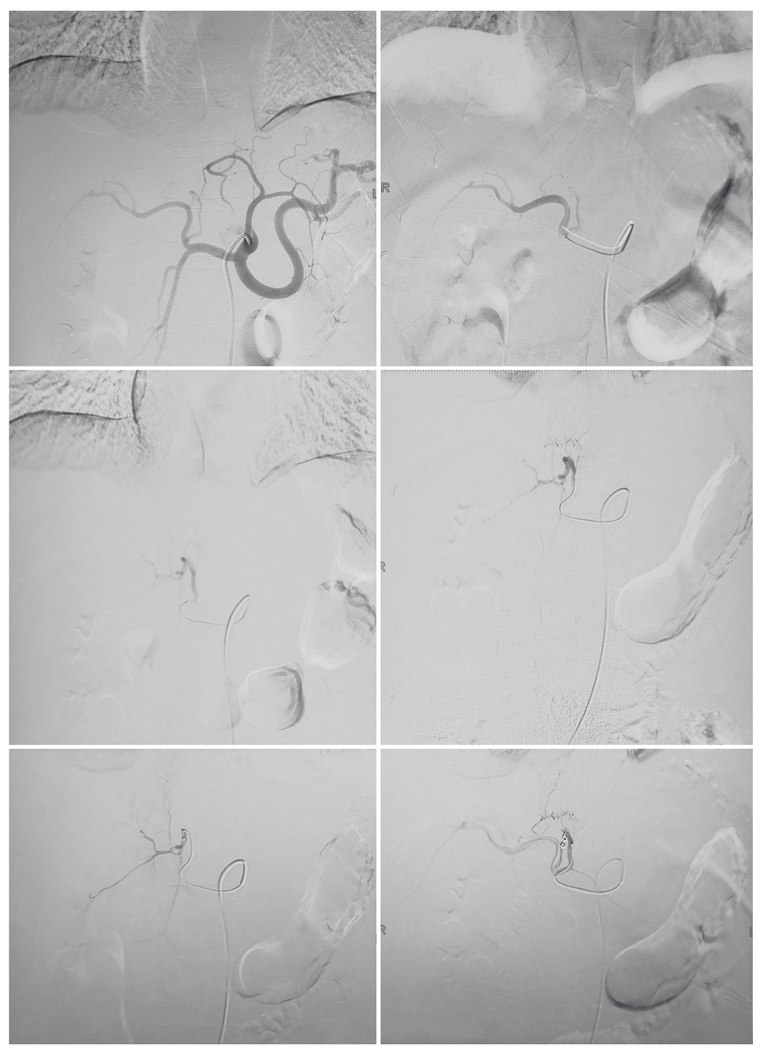
Following drain removal, acute arterial hemorrhage was observed, manifesting as hematemesis and hemodynamic instability. CT angiography was urgently performed, revealing lobulated cystic lesions in hepatic segments 1, 2, and 4 (measuring 11.9 cm × 6.7 cm × 8.8 cm) containing hematoma. Prominent branches of the middle hepatic artery were identified as supplying the cystic lesion in segment 4 during the arterial phase, consistent with an infected hemorrhagic abscess complicated by arterial pseudoaneurysm or direct arterial erosion. The lesion also compressed the posterior main portal vein, with reduced contrast filling of the superior mesenteric vein near its confluent junction. Urgent transcatheter middle hepatic artery embolization was successfully performed (Figure 7).

2.7. Post-Embolization Course and Discharge

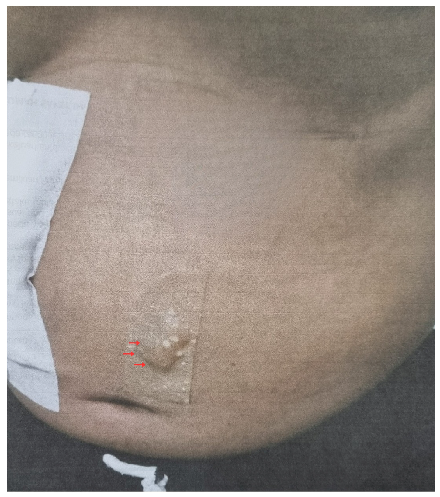
Following embolization, the patient reported progressive pain reduction. Vital signs stabilized. Laboratory values normalized, with hemoglobin recovering to 12.2 g/dL, leukocyte count 4510/μL, albumin 3.44 g/dL, and direct bilirubin 1.02 mg/dL. The inguinal puncture site and abdominal drain site showed progressive healing (Figure 8). The patient was discharged in stable condition with oral cefixime 200 mg twice daily, metronidazole 750 mg three times daily, and ondansetron 8 mg three times daily (Table 1 summarizes the clinical timeline).

3. Discussion

This case illustrates a diagnostically challenging scenario in which a pyogenic liver abscess fulfilled multiple major imaging criteria for HCC under LI-RADS, leading to a protracted misdiagnosis. Four distinct and interrelated issues merit discussion: (1) the predisposing factors for pyogenic liver abscess; (2) the imaging mimicry of HCC and the limitations of LI-RADS in non-cirrhotic patients; (3) the rationale for the antimicrobial regimen chosen; and (4) the procedural risk and management of arterial hemorrhage as a complication of drainage in hypervascular-appearing lesions.

3.1. Predisposing Factors for Pyogenic Liver Abscess

Established risk factors for PLAs include diabetes mellitus, underlying malignancy, cirrhosis, immunosuppression (including chemotherapy, prolonged corticosteroid use, or inherited/acquired immunodeficiency), prolonged proton pump inhibitor use, biliary tract disease, and prior abdominal surgery [1,2]. In the present case, the patient denied diabetes, alcohol consumption, and recent invasive procedures. HBsAg and anti-HIV were non-reactive. Endoscopy confirmed the absence of cirrhotic stigmata. No biliary etiology was identified, and cultures were sterile.
Notably, the patient had marked leukocytosis (18,660/μL) with neutrophilia (91.4%), an elevated erythrocyte sedimentation rate (99 mm/h), low-grade fever, and hypoalbuminemia on admission—findings that, in retrospect, are more consistent with an infectious process than with HCC. The absence of identifiable predisposing factors does not exclude PLA; cryptogenic PLAs account for a significant minority of cases and are increasingly recognized, particularly in the absence of classical risk factors. C-reactive protein (CRP) was not measured in this patient, representing a gap in the initial evaluation; CRP is among the most sensitive inflammatory markers in PLA and, if markedly elevated, could have heightened early suspicion for an infectious etiology [1].

3.2. Cognitive Bias and Diagnostic Delay

A critical and underappreciated dimension of this case is the 14-day interval between the detection of early inflammatory signals and the performance of diagnostic drainage and biopsy (Day 1 to Day 15). On admission, the clinical picture was already discordant with HCC: the patient had marked leukocytosis (18,660/μL) with neutrophilia (91.4%), low-grade fever (37.8 °C), and an elevated erythrocyte sedimentation rate (99 mm/h). These findings are not characteristic of hepatocellular carcinoma but are classic features of systemic infectious or inflammatory processes. Despite this, the working diagnosis remained HCC BCLC stage C for nearly two weeks. This pattern is consistent with anchoring bias—a well-described cognitive error in clinical reasoning in which clinicians anchor disproportionately on an initial diagnosis, discounting subsequent contradictory information [1]. In the present case, the LI-RADS 5 classification assigned at the referring hospital served as a powerful cognitive anchor that persisted through the tertiary admission, influencing the interpretation of clinical and laboratory findings that should have prompted earlier reconsideration. Explicit recognition of anchoring bias as a contributing factor in this diagnostic delay is important not only for its educational value, but because it suggests a systemic vulnerability: when a definitive imaging category such as LI-RADS 5 is assigned—particularly in the absence of verified risk factors—it may disproportionately suppress subsequent diagnostic inquiry even when clinical evidence evolves in a contradictory direction.
This case also highlights a systemic issue in the radiological reporting workflow: the LI-RADS 5 classification was assigned at the referring hospital without documented verification of the patient’s underlying hepatic risk status. LI-RADS v2018 explicitly restricts its use to patients with established HCC risk factors; its application in a patient without confirmed cirrhosis or chronic viral hepatitis constitutes a technical misuse of the system. Radiologists must either be provided with, or must actively seek, the patient’s clinical context—including hepatitis serology, alcohol history, and hepatic fibrosis status—before formally assigning a LI-RADS category. Assigning LI-RADS 5 in a clinical vacuum carries the risk of triggering diagnostic inertia downstream: referring clinicians, oncologists, and interventionalists may treat a radiological LR-5 designation as a near-definitive HCC diagnosis, particularly when the imaging features are compelling. A formal clinical risk factor checklist integrated into radiology reporting templates for liver lesions in non-cirrhotic patients may reduce such errors [5].

3.3. Imaging Mimicry of HCC and LI-RADS Limitations

The LI-RADS classification system, established by the American College of Radiology, standardizes the interpretation of liver CT and MRI observations in patients at high risk for HCC. Observations are stratified from LR-1 (definitely benign) to LR-5 (definitely HCC), incorporating major features (arterial-phase hyperenhancement, washout, enhancing capsule, growth ≥50% in <6 months) and ancillary features [6,8]. Critically, LI-RADS v2018 explicitly restricts its application to patients with recognized HCC risk factors—primarily cirrhosis or chronic hepatitis B infection—and its use in the general population or in patients without such risk factors is not validated [5].
Despite its high specificity (reported as 85–100% in appropriate at-risk cohorts), LI-RADS 5 carries a recognized false-positive rate of approximately 5%. Chen et al. demonstrated, using LI-RADS v2018 criteria on extracellular contrast-enhanced MRI, two false-positive LR-5 results per reader in a cohort evaluating small HCCs, corresponding to histologically confirmed benign entities [7]. Alhasan et al. reported a specificity of 97.3% but a sensitivity of only 53.7% for LI-RADS 5 in HCC diagnosis [9]. In the present case—where the patient had no cirrhosis, no chronic viral hepatitis, and no other established HCC risk factors—the application of LI-RADS 5 was technically outside the validated scope of the system, rendering the false-positive result more foreseeable.
The imaging characteristics of PLAs can range from well-circumscribed cystic lesions with rim enhancement to heterogeneously enhancing, mass-like lesions that are difficult to distinguish from hepatic neoplasms [4]. Arterial-phase hyperenhancement in liver abscesses can arise from reactive hyperemia in the surrounding parenchyma, while apparent capsule-like peripheral enhancement may represent the abscess wall rather than a neoplastic capsule. In the present case, these features collectively mimicked the LI-RADS 5 appearance of HCC [10,11].
Several imaging features may have been helpful in raising the differential diagnosis of abscess earlier. On MRI, diffusion-weighted imaging (DWI) demonstrated restricted diffusion, which, while present in both abscesses and HCC, is particularly pronounced in the dependent purulent core of an abscess [1]. The irregular, thick peripheral enhancement with a heterogeneous internal signal on dynamic MRI is more consistent with an abscess wall than with the thin enhancing pseudocapsule typically observed in HCC. Contrast-enhanced ultrasound (CEUS) could have provided additional information, as abscesses typically demonstrate a distinctive peripheral enhancement pattern without arterial-phase globular fill-in, which differs from the hyperenhancement pattern of HCC. The absence of pre-existing cirrhosis and the presence of diffuse leukocytosis with marked neutrophilia should have prompted re-evaluation of the LI-RADS 5 classification in this clinical context [12].
Beyond the HCC versus PLA differential, the multilobulated, necrotic-appearing morphology of the present lesion raises several additional diagnoses that should be considered in non-cirrhotic patients with large hepatic masses. Infected hepatic cysts—including congenital simple cysts or biliary cystadenomas complicated by secondary bacterial infection—can produce imaging features resembling those of PLA, including rim enhancement, internal debris, and peripheral vascularity. Peripheral intrahepatic cholangiocarcinoma (iCCA) may present as a mass-forming lesion with heterogeneous enhancement, delayed peripheral washout, and associated biliary dilatation; in the present case, the MRI finding of mild distal biliary dilatation adjacent to the lesion could have been interpreted as supporting an iCCA diagnosis. Hepatic lymphoma—explicitly raised as a differential on the MRI report in this case—typically presents as hypoechoic masses with minimal enhancement and may mimic both PLAs and HCC on cross-sectional imaging. Awareness of this differential spectrum is important because each entity carries a distinct treatment strategy, and misclassification as HCC—as occurred here—may result in inappropriate oncological management [12].
The frequency with which a pyogenic liver abscess mimics HCC on cross-sectional imaging is not well-quantified in the literature, but existing case series and reports indicate it is a recognized—if uncommon—diagnostic pitfall. Kim et al. described a hepatic abscess mimicking HCC in the setting of alcoholic liver disease, and Chou et al. reported a Salmonella-related liver abscess fulfilling HCC imaging criteria in a diabetic and cirrhotic patient. In a broader context, any hypervascular hepatic mass with apparent washout may fulfill LR-5 criteria in the absence of clinical verification, and the reported false-positive rate of LI-RADS 5 of approximately 5% translates to a clinically significant number of incorrect classifications at the population level, particularly in regions where LI-RADS is applied liberally outside its validated scope. Two additional early discriminators deserve emphasis: (1) C-reactive protein (CRP), if measured on admission, typically exceeds 100 mg/L in PLA and is not elevated in HCC—making it a rapid, low-cost serological discriminator and (2) contrast-enhanced ultrasound (CEUS), which can differentiate abscesses from HCC by demonstrating peripheral nodular enhancement without central fill-in in abscesses, contrasting with the rapid arterial-phase global hyperenhancement and portal-phase washout characteristic of HCC [12].

3.4. Antimicrobial Regimen and Etiological Classification

Pus cultures were sterile and Ziehl–Neelsen staining for acid-fast bacilli was negative. Sterile cultures in PLA are a well-recognized phenomenon, reported in 20–60% of cases, attributed to prior antibiotic administration, fastidious organisms, or inadequate microbiological processing. The absence of growth does not preclude a pyogenic etiology; in this patient, prior treatment with ceftriaxone likely suppressed bacterial growth in culture [1].
Serology for Entamoeba histolytica was not documented in this case; however, the absence of travel history, the purulent (rather than anchovy-paste) macroscopic appearance of the drained material, and the histopathological finding of chronic suppurative inflammation are consistent with a pyogenic rather than amoebic etiology. Amoebiasis was empirically excluded based on clinical and microbiological grounds. Metronidazole was nevertheless included in the regimen, which is appropriate as it provides antimicrobial cover against anaerobes (present in a substantial proportion of PLAs) and retains activity against amoebic infection in the event of diagnostic uncertainty. The combination of doripenem and metronidazole provides broad-spectrum coverage appropriate for sterile-culture PLA, as recommended guidelines advocate third-generation cephalosporins or carbapenems combined with metronidazole or piperacillin–tazobactam [1].

3.5. Procedural Risk of Drainage in Hypervascular-Appearing Lesions

The patient developed arterial hemorrhage following drain removal on hospital day 23, ultimately requiring emergency transcatheter embolization of the middle hepatic artery. This complication merits careful analysis in the context of the preceding misdiagnosis. The imaging appearance of the lesion—marked arterial-phase hyperenhancement and apparent vascularity—was one of the primary features used to justify the LI-RADS 5 classification. In retrospect, this hypervascularity reflected reactive arterial supply to the abscess wall rather than the neovascularity of malignancy [12].
Vascular complications following percutaneous drainage of liver abscesses, including arterial pseudoaneurysm formation and hemobilia, are recognized but uncommon, with reported rates of 1–4%. Proximity of the abscess to major intrahepatic arterial branches—as demonstrated by the prominent middle hepatic artery branches identified on CT angiography—increases procedural risk. When a hepatic lesion demonstrates florid arterial enhancement on cross-sectional imaging, regardless of the underlying diagnosis, the interventional team should be prepared for potential arterial complications and have facilities for emergency embolization available [1]. This case illustrates that the hypervascularity of an abscess can increase procedural risk comparably to that of a hypervascular malignancy, and that pre-procedural vascular mapping may be prudent in such cases.

4. Conclusions

This case demonstrates that pyogenic liver abscesses can fulfill all major LI-RADS 5 imaging criteria in a non-cirrhotic patient, leading to an initial—and potentially harmful—misdiagnosis of advanced HCC. Clinicians should question an LI-RADS 5 classification in the absence of established HCC risk factors when the following red flags are present: (1) absence of cirrhosis or chronic viral hepatitis; (2) fever or temperature above 37.5 °C; (3) leukocytosis (≥10,000/μL) with neutrophilia; (4) markedly elevated inflammatory markers (ESR, CRP); (5) rapidly changing lesion morphology over days to weeks; and (6) hypoalbuminemia disproportionate to hepatic synthetic function. In such cases, tissue sampling via image-guided biopsy and diagnostic/therapeutic drainage should be performed before committing to an oncological diagnosis and treatment pathway.
Furthermore, the vascular complication encountered in this case—middle hepatic artery hemorrhage following drain removal—underscores that apparent hypervascularity of a hepatic lesion, regardless of etiology, confers procedural risk and mandates pre-procedural vascular mapping and immediate access to interventional radiology facilities.

Author Contributions

Conceptualization, F.S. and U.M.; methodology, F.S. and U.M.; investigation, F.S.; data curation, F.S.; writing—original draft preparation, F.S.; writing—review and editing, U.M.; supervision, U.M. All authors have read and agreed to the published version of the manuscript.

Funding

This research received no external funding.

Institutional Review Board Statement

Ethical review and approval were waived for this study in accordance with the regulations of the Health Research Ethics Committee of Dr. Soetomo General Academic Hospital, Surabaya, Indonesia (Komite Etik Penelitian Kesehatan RSUD Dr. Soetomo), which exempts single case reports based on routine clinical care from formal ethical review, pursuant to Indonesian national health research ethics guidelines (Peraturan Menteri Kesehatan Republik Indonesia No. 11 Tahun 2017). The study was conducted in accordance with the Declaration of Helsinki.

Informed Consent Statement

Written informed consent for publication of this case report, including all clinical data and anonymized images, was obtained from the patient prior to submission. A blank version of the informed consent form has been provided to the Editorial Office. Written informed consent has been obtained from the patient to publish this paper.

Data Availability Statement

The data supporting the findings of this study are available from the corresponding author upon reasonable request, subject to privacy and ethical restrictions related to patient confidentiality.

Acknowledgments

The authors would like to thank the clinical staff of Dr. Soetomo General Academic Hospital involved in the diagnosis and management of this patient.

Conflicts of Interest

The authors declare no conflicts of interest.

References

  1. Mavilia, M.G.; Molina, M.; Wu, G.Y. The Evolving Nature of Hepatic Abscess: A Review. J. Clin. Transl. Hepatol. 2016, 4, 158–168. [Google Scholar] [CrossRef] [Scilit] [PubMed]
  2. Maria Kozielewicz, D.; Sikorska, K.; Stalke, P. Liver abscesses—From diagnosis to treatment. Clin. Exp. Hepatol. 2021, 7, 329–336. [Google Scholar] [CrossRef] [Scilit]
  3. Sharma, S.; Ahuja, V. Liver Abscess: Complications and Treatment. Clin. Liver Dis. 2021, 18, 122–126. [Google Scholar] [CrossRef] [Scilit] [PubMed]
  4. Kim, J.W.; Shin, S.S.; Heo, S.H.; Lim, H.S.; Hur, Y.H.; Kim, J.H. Hepatic abscess mimicking hepatocellular carcinoma in a patient with alcoholic liver disease. Clin. Mol. Hepatol. 2013, 19, 431. [Google Scholar] [CrossRef] [Scilit] [PubMed]
  5. Ronot, M.; Fouque, O.; Esvan, M.; Lebigot, J.; Aubé, C.; Vilgrain, V. Comparison of the accuracy of AASLD and LI-RADS criteria for the non-invasive diagnosis of HCC smaller than 3 cm. J. Hepatol. 2018, 68, 715–723. [Google Scholar] [CrossRef] [Scilit] [PubMed]
  6. Bukeirat, F.A.; Rose, L. The Clinical Spectrum of Liver Masses: Know Before You Judge. J. Gastroenterol. Hepatol. Rep. 2024, 5, 1–5. [Google Scholar] [CrossRef] [Scilit]
  7. Chen, J.; Kuang, S.; Zhang, Y.; Tang, W.; Xie, S.; Zhang, L.; Rong, D.; He, B.; Deng, Y.; Xiao, Y.; et al. Increasing the sensitivity of LI-RADS v2018 for diagnosis of small (10–19 mm) HCC on extracellular contrast-enhanced MRI. Abdom. Radiol. 2021, 46, 1530–1542. [Google Scholar] [CrossRef] [Scilit] [PubMed]
  8. Yu, Y.; Guo, L.; Hu, C.; Chen, K. Spectral CT imaging in the differential diagnosis of necrotic hepatocellular carcinoma and hepatic abscess. Clin. Radiol. 2014, 69, e517–e524. [Google Scholar] [CrossRef] [Scilit] [PubMed]
  9. Alhasan, A.; Cerny, M.; Olivié, D.; Billiard, J.-S.; Bergeron, C.; Brown, K.; Bodson-Clermont, P.; Castel, H.; Turcotte, S.; Perreault, P.; et al. LI-RADS for CT diagnosis of hepatocellular carcinoma: Performance of major and ancillary features. Abdom. Radiol. 2019, 44, 517–528. [Google Scholar] [CrossRef] [Scilit] [PubMed]
  10. Brown, K.T.; Gandhi, R.T.; Covey, A.M.; Brody, L.A.; Getrajdman, G.I. Pylephlebitis and liver abscess mimicking hepatocellular carcinoma. Hepatobiliary Pancreat. Dis. Int. 2003, 2, 221–225. [Google Scholar] [PubMed]
  11. Chou, Y.; Changchien, C.; Chiu, K.; Kuo, C.; Kuo, F.; Kuo, C. Salmonellosis with liver abscess mimicking hepatocellular carcinoma in a diabetic and cirrhotic patient: A case report and review of the literature. Liver Int. 2006, 26, 498–501. [Google Scholar] [CrossRef] [Scilit] [PubMed]
  12. Chernyak, V.; Santillan, C.S.; Papadatos, D.; Sirlin, C.B. LI-RADS® algorithm: CT and MRI. Abdom. Radiol. 2018, 43, 111–126. [Google Scholar] [CrossRef] [Scilit] [PubMed]
Figure 1. Abdominal ultrasonography at the referring hospital showing hepatomegaly with multiple solid masses in the right hepatic lobe, the largest measuring 11.3 cm × 7.7 cm, with inferior compression of the right kidney, interpreted as hepatoma.
Gastrointestdisord 08 00053 g001
Figure 2. Contrast-enhanced abdominal CT at the referring hospital (1 February 2025) demonstrating multiple lobulated, exophytic hepatic masses with arterial-phase hyperenhancement and portal/delayed-phase washout, classified as LI-RADS 5.
Gastrointestdisord 08 00053 g002
Figure 3. Contrast-enhanced abdominal CT at the referral hospital (hospital day 3), confirming hepatomegaly with multiple exophytic lobulated masses in hepatic segments 2, 3, 4a, and 4b, with associated left pleural effusion and splenomegaly.
Gastrointestdisord 08 00053 g003
Figure 4. Esophagogastroduodenoscopy at the referral hospital (hospital day 8) demonstrating no esophageal varices or portal hypertensive gastropathy, with findings consistent with gastroesophageal reflux disease. Panels 1–8 represent sequential endoscopic images corresponding to the pharynx-larynx, esophagus (proximal, middle, distal), gastric cardia/fundus, gastric corpus, and duodenal bulb, respectively.
Gastrointestdisord 08 00053 g004
Figure 5. Abdominal MRI at the referral hospital (hospital day 12) demonstrating a multilobulated liver mass with diffusion restriction on DWI and heterogeneous T2 hyperintensity, raising differential of hepatic lymphoma versus atypical HCC with necrotic degeneration.
Gastrointestdisord 08 00053 g005
Figure 6. Follow-up abdominal ultrasonography at the referral hospital (hospital day 20) demonstrating left lobe liver abscesses measuring 4.8 cm × 8.3 cm × 7.2 cm and 7.0 cm × 6.7 cm × 6.1 cm, with the percutaneous drainage tube in situ. Dotted lines indicate ultrasound caliper measurements of the residual abscess collections.
Gastrointestdisord 08 00053 g006
Figure 7. CT angiography at the referral hospital (hospital day 23) demonstrating a cystic hepatic lesion with associated hematoma and arterial supply from prominent middle hepatic artery branches in segment 4, followed by successful embolization.
Gastrointestdisord 08 00053 g007
Figure 8. Post-drainage wound site following embolization and clinical stabilization, demonstrating progressive healing. Arrow indicates the healing percutaneous drain insertion site.
Gastrointestdisord 08 00053 g008
Table 1. Clinical timeline of key events during hospitalization.
Hospital DayEventKey Finding/Action
Referral (pre-admission)Referred to hospital imagingUSG + CECT: multiple hepatic masses, arterial enhancement and washout → LI-RADS 5, HCC BCLC C
Day 1 (Admission)Tertiary center admissionLeukocytosis 18,660/μL; neutrophilia 91.4%; AFP not elevated; HBsAg negative; anti-HIV negative; empiric broad-spectrum antibiotics initiated
Day 3Repeat CT abdomenConfirms multi-segment hepatic masses; doripenem added
Day 8EGD + serologyNo varices, no cirrhotic stigmata; HBsAg and anti-HIV non-reactive
Day 12Abdominal MRIMultilobulated mass, T2 hyperintense, diffusion restriction; thick peripheral enhancement → differential: lymphoma vs. atypical HCC; biopsy recommended
Day 15 (approx.)Percutaneous drainage + biopsy130 mL pus drained; revised diagnosis: pyogenic liver abscess; metronidazole added
Day 20Follow-up USGResidual abscesses 4.8 cm × 8.3 cm and 7.0 cm × 6.7 cm; drain in situ
Day 23Complication + CTADrain removal → arterial bleeding; CTA: middle hepatic artery hemorrhage; urgent embolization performed
Day 25 (discharge)DischargeStable; oral cefixime + metronidazole + ondansetron
Disclaimer/Publisher’s Note: The statements, opinions and data contained in all publications are solely those of the individual author(s) and contributor(s) and not of MDPI and/or the editor(s). MDPI and/or the editor(s) disclaim responsibility for any injury to people or property resulting from any ideas, methods, instructions or products referred to in the content.