1. Introduction
Achalasia is a rare primary esophageal motility disorder characterized by impaired relaxation of the lower esophageal sphincter and absence of effective esophageal peristalsis [
1]. Patients typically present with progressive dysphagia, regurgitation, chest pain, and weight loss, with symptoms that significantly affect quality of life [
2,
3,
4]. Although pharmacological and endoscopic therapies may provide temporary relief, definitive treatment aims to disrupt the hypertonic lower esophageal sphincter [
1,
5,
6]. Surgical myotomy remains one of the most effective therapeutic options, particularly in patients with durable symptoms or those unsuitable for endoscopic interventions [
7].
Minimally invasive Heller myotomy (HM), usually combined with a partial Dor fundoplication, has become the standard surgical treatment for achalasia [
2,
8]. Both laparoscopic and robotic approaches have demonstrated excellent functional outcomes and durable symptom control [
2,
8,
9,
10]. Nevertheless, the procedure requires precise dissection of the esophageal muscular layers while preserving the underlying mucosa [
10,
11,
12]. Two technical challenges are particularly relevant during the operation: ensuring the completeness of the myotomy and identifying any mucosal perforation [
13,
14]. Incomplete division of muscle fibers may lead to persistent obstruction and symptom recurrence, whereas mucosal injury represents one of the most feared intraoperative complications [
14,
15].
Traditionally, surgeons rely on intraoperative endoscopy, air leak testing, or dye instillation to confirm mucosal integrity and verify adequate myotomy [
13,
16]. Although effective, these adjuncts may require additional equipment, specialized personnel, and operative time [
11,
12,
13,
17]. Intraoperative endoscopy also introduces logistical challenges in some surgical settings and may interrupt the surgical workflow [
3,
10,
11]. Accordingly, alternative techniques capable of providing real time visual confirmation of myotomy completeness and mucosal integrity have been explored.
Indocyanine Green fluorescence (ICG) has been widely adopted in minimally invasive surgery for multiple applications, including tissue perfusion assessment, biliary anatomy visualization, and lymphatic mapping [
14,
15,
18,
19]. Its role in esophageal surgery continues to evolve. In the context of HM, fluorescence imaging has been proposed as a method to enhance visualization of the mucosal layer and residual muscle fibers, potentially facilitating a more accurate and safer myotomy [
20,
21,
22]. Different technical approaches have been described, including intravenous administration of ICG to highlight tissue perfusion and intraluminal instillation to directly illuminate the esophageal mucosa [
9,
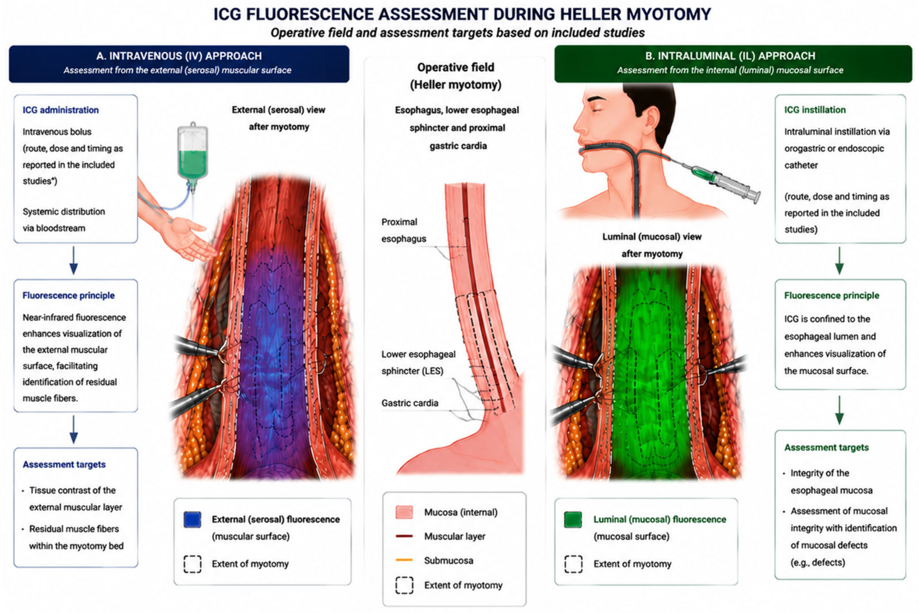
22]. These techniques are based on different optical principles and provide complementary intraoperative information. Intravenous ICG enhances the contrast between vascularized tissues and residual muscle fibers, whereas intraluminal ICG directly outlines the esophageal mucosa, facilitating assessment of myotomy completeness and detection of mucosal perforation.
Despite growing interest in fluorescence guided surgery, evidence regarding the role of ICG during HM remains limited and fragmented. The available evidence consists mainly of small case series and technical descriptions, with considerable heterogeneity in administration techniques and clinical applications. A comprehensive synthesis of the available evidence is therefore needed to clarify how this technology has been used and what potential advantages it may offer.
This scoping review aimed not only to map the current literature on the intraoperative use of ICG during minimally invasive Heller myotomy for achalasia, but also to identify methodological heterogeneity, summarize the currently available technical approaches, and define the principal evidence gaps that should inform future clinical research and protocol standardization. To our knowledge, this is the first review specifically dedicated to the role of ICG fluorescence in Heller myotomy.
2. Methods
2.1. Review Questions and Objectives
The primary objective of this scoping review was to map the available evidence on the intraoperative use of indocyanine green fluorescence during minimally invasive HM for achalasia. Specifically, the review aimed to identify how indocyanine green has been applied intraoperatively, to describe the different technical approaches used, and to summarize the reported clinical applications and outcomes.
The review was guided by the following questions: how is ICG administered during HM, what intraoperative information does fluorescence imaging provide, and what are the reported benefits and limitations of its use in this setting.
2.2. Eligibility Criteria
Studies were considered eligible if they met the following criteria: clinical studies involving patients diagnosed with esophageal achalasia undergoing HM, intraoperative use of ICG imaging, and minimally invasive surgical approach, including laparoscopic or robotic procedures.
No restrictions on study design were applied to capture all available clinical evidence. Only studies published in English and involving human subjects were included. Conference abstracts without sufficient clinical data were excluded. Exclusion criteria were non clinical reports, review articles, editorials, purely technical descriptions without patient data, and duplicate publications reporting overlapping cohorts.
2.3. Information Sources and Search Strategy
A systematic literature search was conducted in three electronic databases: PubMed, Scopus, and Web of Science. The search strategy combined controlled vocabulary and free text terms related to achalasia, HM, and fluorescence imaging. No publication date restrictions were applied. The review protocol was registered in the Open Science Framework (OSF; DOI: 10.17605/OSF.IO/D2MGB), with registration completed on 15 March 2026, at 4:01 PM.
The search strategy was developed to maximize sensitivity because of the expected limited literature on this topic. Database-specific combinations of Medical Subject Headings (MeSH) and free-text terms were used. The PubMed search strategy was (“achalasia” OR “esophageal achalasia”) AND (“Heller myotomy” OR “Heller-Dor” OR “cardiomyotomy”) AND (“indocyanine green” OR “ICG” OR “fluorescence imaging” OR “near infrared” OR “NIR”). The Scopus search strategy was TITLE-ABS-KEY (“achalasia” AND (“Heller myotomy” OR “cardiomyotomy” OR “Heller-Dor”) AND (“indocyanine green” OR “ICG” OR “fluorescence” OR “near infrared”)). The Web of Science strategy was TS=(“achalasia” AND (“Heller myotomy” OR “cardiomyotomy” OR “Heller-Dor”) AND (“indocyanine green” OR “ICG” OR “fluorescence imaging” OR “near infrared imaging”)). No publication date restrictions were applied. In addition, reference lists of included articles were manually screened to identify potentially relevant studies not captured during the primary database search.
The final search was performed before manuscript completion, and reference lists of eligible articles were manually screened to identify additional relevant studies.
2.4. Study Selection
All records identified through the database search were imported into reference management software and duplicates were removed. Titles and abstracts were screened to identify potentially relevant studies. Full texts of eligible articles were subsequently assessed according to the predefined inclusion and exclusion criteria. Title and abstract screening, as well as full-text eligibility assessment, were independently performed by two reviewers (A.F. and M.S.). Any disagreements regarding study eligibility were resolved through discussion and consensus.
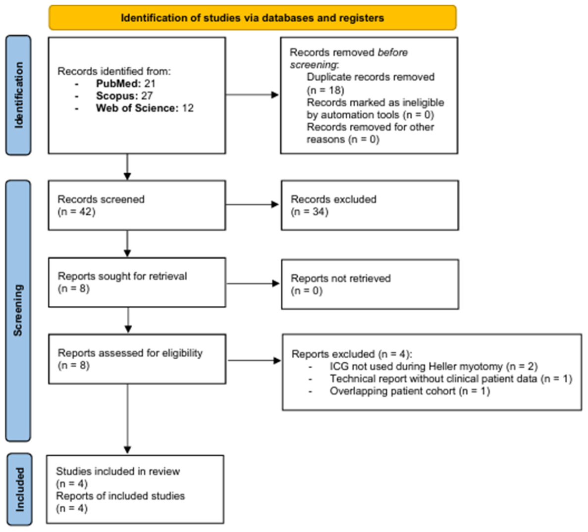
The study selection process followed the PRISMA-ScR framework (
Figure 1). A total of 60 records were identified, of which 18 were duplicates. Forty two studies underwent screening, and eight full text articles were assessed for eligibility. Four studies met the inclusion criteria and were included in the final review.
2.5. Data Extraction and Charting
Data were extracted from the included studies and organized into structured tables. The following variables were collected: publication year, study design, surgical approach, number of patients, technique of indocyanine green administration, intraoperative purpose of fluorescence imaging, and reported outcomes.
Particular attention was given to technical details, including route of administration, dose, timing of indocyanine green use, and imaging modality. Data extraction was performed systematically to ensure consistency across studies.
2.6. Critical Appraisal of Included Studies
Although formal risk-of-bias assessment is not mandatory for scoping reviews, a structured critical appraisal was performed to contextualize the strength of the available evidence. Each included study underwent a structured methodological appraisal based on predefined descriptive domains, including study design, sample size, presence of a comparator group, clarity of the ICG protocol, consistency of outcome reporting, and duration of postoperative follow-up. The appraisal was performed to contextualize the methodological characteristics of the available evidence rather than to formally rate study quality or exclude studies. The appraisal findings are summarized in
Table 1. Consistent with the objectives of a scoping review and current PRISMA-ScR guidance, a formal risk-of-bias assessment was not undertaken.
2.7. Data Synthesis
Given the exploratory nature of the review and the limited number of available studies, a quantitative synthesis was not performed. A formal meta-analysis was considered inappropriate because of the limited number of studies, heterogeneous fluorescence protocols, differences in surgical platforms, and inconsistent outcome reporting. Instead, findings were summarized descriptively using a narrative approach.
The synthesis focused on identifying patterns in the intraoperative use of ICG, including its role in confirming myotomy completeness, identifying residual muscle fibers, and detecting mucosal perforation. Differences between intravenous and intraluminal administration techniques were analyzed to highlight variations in practice and potential implications for clinical use.
4. Discussion
This review highlights an emerging area within fluorescence-guided foregut surgery for which current evidence remains limited but progressively expanding. Beyond summarizing the available literature, the present review provides a structured framework for interpreting the current evidence by distinguishing the different fluorescence strategies, identifying methodological inconsistencies among published studies, and outlining priorities for future research. The available literature suggests that ICG may have a practical role during minimally invasive HM, not merely as another technological adjunct, but as an intraoperative tool directed at two of the most consequential technical endpoints of the operation: adequacy of muscular division and preservation of mucosal integrity. This is clinically relevant because the success of HM depends on achieving a sufficiently extended and complete myotomy while avoiding mucosal injury [
8,
16,
23]. Failure to achieve either objective has direct consequences. Residual fibers may leave the patient with persistent outflow obstruction and recurrent dysphagia, whereas an unrecognized mucosal perforation can result in leak related morbidity, prolonged hospitalization, and, in more severe cases, mediastinal or pleural sepsis [
3,
4,
8].
For this reason, surgeons have traditionally relied on adjunctive methods to verify the endpoint of the myotomy [
4,
8,
16]. Intraoperative endoscopy remains the most widely accepted reference method because it allows direct luminal assessment, confirmation of esophagogastric junction opening, and leak testing [
2,
3,
4,
8]. However, endoscopy has practical limitations. It requires additional instrumentation, staff availability, and procedural coordination, and may interrupt the operative sequence at a critical stage [
24,
25,
26]. This consideration is particularly relevant in units where advanced minimally invasive surgery is routine but intraoperative endoscopy is not consistently integrated into theater workflow [
27]. The principal advantage of ICG in the reviewed studies lies in its ability to provide immediate optical feedback directly within the surgical field, under the control of the operating team, without requiring changes to the operative setup [
9,
22]. However, this practical advantage should not be confused with diagnostic equivalence. Intraoperative endoscopy remains the most established method for direct luminal assessment, and the current evidence does not justify abandoning it in routine practice.
A central finding of this review is that the published experience has evolved along two conceptually distinct technical pathways. Romanzi et al. used intravenous ICG during robotic HDP, exploiting differential tissue perfusion and fluorescence contrast to improve delineation of the mucosal plane and identify residual muscle fibers [
9]. In their retrospective comparative series, fluorescence helped detect residual muscle fibers in three patients and was associated with a significantly shorter operative time than intraoperative endoscopy (105 vs. 126 min;
p = 0.026), while no mucosal perforation occurred in the fluorescence group [
9]. This observation, however, derives from a single retrospective study including only 17 patients who underwent fluorescence-guided surgery and therefore requires confirmation in larger prospective comparative studies. By contrast, Shah, Gadiyaram, and Patel et al. described an intraluminal approach in which ICG was instilled via a nasogastric tube after completion of the myotomy, producing direct fluorescence of the exposed mucosal tube under near-infrared imaging [
21,
22]. These are not merely technical variations of the same test. They are based on different optical principles and answer slightly different intraoperative questions. Importantly, the currently available evidence does not permit disentangling the effects of the route of ICG administration from those of the surgical platform, since intravenous fluorescence has only been investigated in robotic surgery, whereas intraluminal fluorescence has exclusively been reported during laparoscopic procedures.
The intravenous approach is essentially a contrast-enhancement strategy. It seems particularly suited to a robotic platform, where stable magnified visualization and integrated near-infrared imaging may maximize subtle differences between vascularized tissue planes. In Romanzi’s experience, residual muscle fibers appeared less fluorescent than the underlying layer, thereby facilitating completion of the myotomy under direct vision [
9]. From a conceptual standpoint, this technique is less a leak test than a dissection aid and endpoint refinement tool. It may therefore be especially useful in technically demanding cases, including revisional surgery or patients with fibrosis after prior endoscopic treatment, where tissue planes are less distinct and the margin between adequate myotomy and mucosal injury becomes narrower.
The intraluminal technique serves a different function. Here, the mucosa itself becomes the fluorescent target. A continuous, contained fluorescent signal indicates that the mucosal tube is intact and fully exposed, whereas interruption of the pattern by dark horizontal bands suggests residual muscle fibers, and leakage of fluorescence outside the lumen indicates a mucosal defect. This logic is consistently reflected across the laparoscopic reports. Shah et al. described residual muscle fibers visualized as black bands and subsequently divided, with no leak observed and findings confirmed by intraoperative endoscopy. Gadiyaram et al. reported uniform fluorescence along the myotomy line in four patients, which was interpreted as indirect confirmation of complete myotomy and preserved mucosal integrity. Unlike Romanzi et al. and Shah et al., however, the authors did not specifically report visualization or targeted division of residual muscle fibers. Patel et al., in the largest intraluminal series, extended this concept to fifteen patients and documented one intraoperative mucosal perforation detected by leakage of ICG, which was repaired without subsequent adverse sequelae [
22]. This is probably the most clinically persuasive aspect of the available evidence, because it shows that fluorescence can function not only as a confirmatory tool when the myotomy is complete, but also as an alarm signal when it is not safely completed.
At the same time, the current literature makes clear that fluorescence guidance should not yet be interpreted as a validated replacement for intraoperative endoscopy. The evidence remains too limited for such a conclusion. Only one comparative study is available, and even that study was retrospective, single center, and small in size [
9]. The laparoscopic reports are essentially feasibility studies or small case series. In some of them, fluorescence findings were corroborated by endoscopy or leak testing, which strengthens internal consistency but does not establish non inferiority. The real question is not whether ICG can be used during HM, because clearly it can. The question is whether it performs with sufficient sensitivity and negative predictive value to safely replace established adjuncts in routine practice. None of the currently available studies can answer that question definitively. At present, the available literature should be interpreted as proof of technical feasibility rather than proof of clinical efficacy.
Another key finding emerging from this review is the lack of standardization. The studies differ substantially in route of administration, dose, dilution, timing, and operative interpretation. Romanzi et al. administered 6.4 mg intravenously after completion of the myotomy in robotic surgery [
9]. Shah et al. used 4 mg diluted in 100 mL of normal saline intraluminally [
21]. Gadiyaram et al. instilled 10 mg in 50 mL of saline and, in one patient undergoing Dor fundoplication, mixed the solution with egg white to improve retention and fluorescence intensity in the absence of effective distal occlusion [
20]. Patel et al. compared their earlier pilot experience with 4 mg in 100 mL to 10 mg in 100 mL and concluded that the higher concentration provided better intraoperative contrast, subsequently adopting that dose in the extended series [
22]. This heterogeneity is clinically relevant because it means that the published literature is not yet describing a reproducible standard technique. Rather, it reflects an innovation phase in which individual teams are refining local protocols. That is appropriate for an emerging technology, but it also means that between study comparisons must be interpreted cautiously. An additional source of methodological heterogeneity is the incomplete reporting of technical details. None of the included studies explicitly specified whether fluorescence assessment was performed exclusively by the operating surgeon or involved an independent observer, limiting the reproducibility and external validity of the available evidence. Future studies should report this aspect to facilitate comparison across institutions and improve methodological transparency. Several technical and disease-related factors may also influence fluorescence quality. Prior pneumatic dilation, previous POEM, submucosal fibrosis, severe esophageal dilatation, incomplete luminal occlusion, and differences among near-infrared imaging platforms may all affect signal intensity and interpretation. Although these variables have not been systematically evaluated in the currently available clinical studies, they should be considered when designing future prospective investigations. These variables were not systematically addressed in the available studies and should be incorporated into future protocols.
From a surgical perspective, one of the more interesting signals in the literature is the recurring observation that ICG may help identify residual fibers that are not as easily appreciated under white light alone. This point appears in both the robotic intravenous experience and the laparoscopic intraluminal reports. If confirmed in larger series, this could be clinically meaningful. Recurrent symptoms after HM are multifactorial, but inadequate distal or proximal extension of the myotomy remains one of the most important technical causes of failure. A modality that improves visual confidence at the endpoint of muscle division could therefore affect outcomes beyond the immediate intraoperative setting. The current studies do not provide enough long-term data to demonstrate this, but the hypothesis is plausible and worth testing prospectively.
Possible workflow advantages have been suggested, although the available evidence remains limited. In the only retrospective comparative study, Romanzi et al. reported a significantly shorter operative time in the fluorescence-guided group than in the endoscopy group (105 vs. 126 min; p = 0.026). By contrast, the remaining studies were uncontrolled case series. Although Patel et al. reported individual operative times for all 15 patients, no comparator group was included, precluding any conclusions regarding the effect of ICG on operative duration. Accordingly, the current evidence supporting shorter operative times remains limited to a single retrospective comparative study. The laparoscopic reports also suggest that fluorescence imaging can be performed by the operating team using equipment increasingly available in minimally invasive operating rooms, potentially avoiding the need for a separate endoscopic setup. However, these observations should be regarded as preliminary. Formal cost-effectiveness analyses have not been performed, and any potential reduction in operative time must be balanced against the availability and cost of near-infrared imaging systems.
Safety appears reassuring, but the available dataset is too small to support firm conclusions. No study reported adverse reactions attributable to ICG. That said, the absence of observed complications in fewer than sixty reported patients should not be overinterpreted. More importantly, the available studies are underpowered to assess false negative performance. A technique intended to exclude mucosal perforation must be judged less by how often it finds an obvious defect than by how reliably it rules one out. This is particularly relevant because a false negative fluorescence assessment could provide inappropriate reassurance and delay recognition of a clinically significant mucosal injury. Future studies should therefore report not only detected perforations, but also delayed leaks, postoperative imaging findings, reinterventions, and concordance with intraoperative endoscopy or air leak testing. This represents one of the principal limitations of the current evidence. The absence of delayed leak in reported series is encouraging, but it does not substitute for formal diagnostic validation against a reference standard.
The next step for the field should not simply be accumulation of more case reports. What is needed is methodological consolidation. Future studies should clearly distinguish between intravenous and intraluminal applications, standardize dose and timing, define what constitutes a positive or negative fluorescence finding, and compare fluorescence guidance directly with established adjuncts such as intraoperative endoscopy and leak testing. Future studies should also investigate whether fluorescence imaging can detect ischemic changes at the margins of mucosal injuries that are not readily identifiable by conventional intraoperative assessment. At present, however, this potential application remains hypothetical and has not been validated in clinical studies.
Prospective multicenter studies would be particularly valuable, especially if they include clinically meaningful endpoints such as intraoperative detection of residual fibers, mucosal injury rate, postoperative leak, dysphagia recurrence, and need for revisional treatment.
In summary, the current literature does not support considering ICG the new standard for intraoperative assessment during HM. It does, however, support regarding fluorescence as a promising adjunct with a sound technical rationale and encouraging early clinical results. Its most credible present role is as a tool that may enhance visual discrimination at a critical phase of the operation.
Future research should focus on prospective comparative studies with standardized protocols for ICG administration, aiming to define its diagnostic accuracy and clinical impact in comparison with established intraoperative assessment methods.
5. Limitations
This scoping review has several limitations that should be acknowledged. First, the available evidence is extremely limited, comprising only four clinical studies with 58 patients overall, including 41 patients exposed to intraoperative ICG fluorescence assessment. Most included studies were single-center case series with small sample sizes, while only one retrospective comparative study was available. Second, considerable heterogeneity was observed across studies regarding the route of ICG administration, dosage, dilution, timing of administration, imaging platforms, and outcome definitions, precluding quantitative synthesis or meaningful comparisons between studies. Third, postoperative follow-up was inconsistently reported, and none of the included studies was specifically designed to evaluate the diagnostic accuracy of ICG for detecting residual muscle fibers or mucosal perforation.
Another limitation concerns the potential overlap of patient cohorts. Patel et al. explicitly reported that their 2024 series represents an extension of a previously published pilot experience, resulting in partial overlap between publications. Although this does not affect the objective of the present scoping review, which was to map the evolution of the available evidence rather than to perform a quantitative synthesis, it should be considered when interpreting the apparent volume of published clinical experience.
The present review also has methodological limitations. As a scoping review, its objective was to map the existing literature rather than to assess treatment effectiveness or generate pooled estimates. Consequently, no formal meta-analysis was performed. Although a structured critical appraisal of the included studies was undertaken, a formal risk-of-bias assessment was not conducted, in accordance with current PRISMA-ScR recommendations. Finally, the small number of available publications and the possibility of publication bias, whereby positive early experiences are more likely to be reported than negative or inconclusive findings, should be considered when interpreting the results.




 ,
,