Article

The Treatment Effects of Percutaneous Drainage with or without Sclerotherapy for Symptomatic Liver Cysts

1
Department of Gastroenterology and Hepatology, Kusunoki Hospital, Fujioka 375-0024, Japan
2
Department of Clinical Research, NHO Takasaki General Medical Center, Takasaki 370-0829, Japan
3
Department of Gastroenterology, Gunma Saiseikai Maebashi Hospital, Maebashi 371-0821, Japan
4
Department of Internal Medicine, Isesaki Municipal Hospital, Isesaki 372-0817, Japan
5
Department of Internal Medicine, Kiryu Kosei General Hospital, Kiryu 376-0224, Japan
6
Department of Gastroenterology and Hepatology, Gunma University Graduate School of Medicine, Maebashi 371-8511, Japan
7
Department of Gastroenterology, NHO Takasaki General Medical Center, Takasaki 370-0829, Japan
*
Author to whom correspondence should be addressed.
Gastrointest. Disord. 2024, 6(1), 13-25; https://doi.org/10.3390/gidisord6010002
Submission received: 7 October 2023 / Revised: 28 November 2023 / Accepted: 3 January 2024 / Published: 5 January 2024

Abstract

:
Background: While the current guidelines recommend laparoscopic deroofing for symptomatic simple liver cysts, percutaneous drainage may serve as a less invasive alternative method. In this study, the treatment effects of percutaneous drainage with or without sclerotherapy for symptomatic simple liver cysts were evaluated. Methods: Between April 2016 and March 2021, 79 patients who initially required hospitalization due to symptomatic simple liver cysts were enrolled in this multicenter retrospective study. They were treated percutaneously with or without sclerotherapy. The factors associated with symptom recurrence, clinical course and prognosis were investigated. Results: Of the 79 patients treated percutaneously, 11 (13.9%) had symptom recurrence due to liver cysts during the observation period. The maximum diameter of liver cysts at baseline was the only significant factor for the recurrence of these symptoms (p = 0.004). In a receiver operating characteristics analysis, the cut-off of the diameter for symptom recurrence was 16.5 cm. No additional effect of sclerotherapy on drainage was demonstrated in patients with a cyst diameter of <16.5 cm, and in patients with a cyst diameter of ≥16.5 cm, the cumulative recurrence rates of symptoms were significantly lower in the patients treated via sclerotherapy with 5% ethanolamine oleate or with minocycline hydrochloride than in those treated with drainage alone or via sclerotherapy with absolute ethanol. No problematic adverse effects were observed of sclerotherapy. Conclusions: Drainage with sclerotherapy with 5% ethanolamine oleate or minocycline hydrochloride was an effective and safe treatment for patients whose liver cysts had a maximum diameter of ≥16.5 cm. Considering both its efficacy and safety, sclerotherapy with either of these agents is recommended for patients with a maximum liver cyst diameter of ≥16.5 cm.

1. Introduction

Simple liver cysts (SHCs) are the most commonly diagnosed benign liver lesions and are found in 18% of the general population upon abdominal computed tomography (CT) for unrelated pathologies [1,2,3]. Simple liver cysts arise congenitally from aberrant bile duct cells and contain a clear, bile-like fluid [4,5]. Because bile duct epithelium covers the inner lining of a simple cyst, it is hypothesized that SHCs arise during embryogenesis when intrahepatic ductules fail to connect to extrahepatic ducts [6,7].
Although almost all cases with SHCs are asymptomatic and not problematic, larger cases with SHCs sometimes show symptoms such as abdominal fullness and need treatment. While the efficacy of surgical treatments such as open or laparoscopic deroofing is excellent, percutaneous drainage with or without sclerotherapy for symptomatic SHCs has been reported as a minimally invasive method [8]. However, the selection of treatment for symptomatic SHCs differs among facilities, and a standard treatment has not been established. Thus, the treatment policy is usually dependent on the judgment of each institution and the attending physician in such cases. Given the above, we performed the present study to clarify the clinical features of symptomatic SHCs and the difference in the prognosis depending on the treatment method in a real-world setting.

2. Results

2.1. Patient Characteristics upon Initial Treatment

This study enrolled a total of 79 patients from six medical institutions, including 20 males and 59 females, with a median age of 70 (64–80) years. No cases of acquired liver cysts, such as parasitic cysts, hydatid cysts, amebic abscess, pyogenic liver abscess, false cysts (traumatic intrahepatic hemorrhaging, intrahepatic infarction, intrahepatic biloma) and neoplastic cysts, were included in this study.
The baseline characteristics of the enrolled patients are shown in Table 1. The age, the rate of male sex (p = 0.01), the rate of infection of liver cysts (p < 0.001) and ALT levels (p = 0.002) were significantly higher in the patients treated with percutaneous drainage alone than in those treated via percutaneous drainage with sclerotherapy. The prothrombin time activity (p = 0.021) and albumin levels (p = 0.002) were significantly lower in the patients treated with percutaneous drainage alone than in those treated via drainage with sclerotherapy.
In terms of treatment selection, 24 patients underwent drainage alone, while 55 patients underwent drainage with sclerotherapy. The drainage volume of the liver cysts varied based on the size of the cysts. The maximum volume of the discharged fluid sometimes reached nearly 4000 mL, but natural discharge over approximately 1 to 2 h, without forcible negative pressure, did not adversely affect vital signs. Among the 55 patients treated with drainage and sclerotherapy, 29 were treated with MCH, 15 with EO, and 11 with AE (Table 2).
Among the 29 patients treated with MCH, 27 received the agent after the placement of a pigtail catheter in the liver cyst with immediate drainage, aiming for a total cyst volume of nearly 3000 mL. Patients primarily underwent sclerotherapy with 500 mg of MCH dissolved in 10 mL of saline, either administered once on the day of drainage or for three consecutive days starting from the day of drainage. MCH was discarded after a 1 h series of postural changes (supine, prone and left and right lateral positions for 15 min each), and the catheter was removed on the day of the last MCH administration. On the other hand, the two patients in which a pigtail catheter was not used received 500 mg of MCH dissolved in 10 mL of saline immediately after drainage, aiming for the total removal of the liver cyst contents using an 18-gauge percutaneous transhepatic aspiration needle. In both of these cases, neither postural changes nor the drainage of MCH dissolved in saline was performed, and the aspiration needle was removed immediately after administering the sclerosing agent. The drainage time for these procedures was approximately 30 min.
All 15 patients treated with EO received the agent after the placement of a pigtail catheter and immediate drainage, aiming for the complete removal of the liver cyst contents. Among these patients, 10 were treated with 40 mL, 4 with 100 mL, and 1 with 20 mL of EO. Similar to the procedure with MCH, EO was discarded through a pigtail catheter, and the catheter was removed after a 1 h series of postural changes.
Eleven patients treated with AE also received the agent after the placement of a pigtail catheter and immediate drainage, aiming for the complete removal of the liver cyst contents. These patients were treated with 10–60 mL of AE. Thirty minutes later, AE was discarded through a pigtail catheter without postural changes, aimed at minimizing AE absorption and preventing alcohol intoxication. The catheter was then immediately removed.

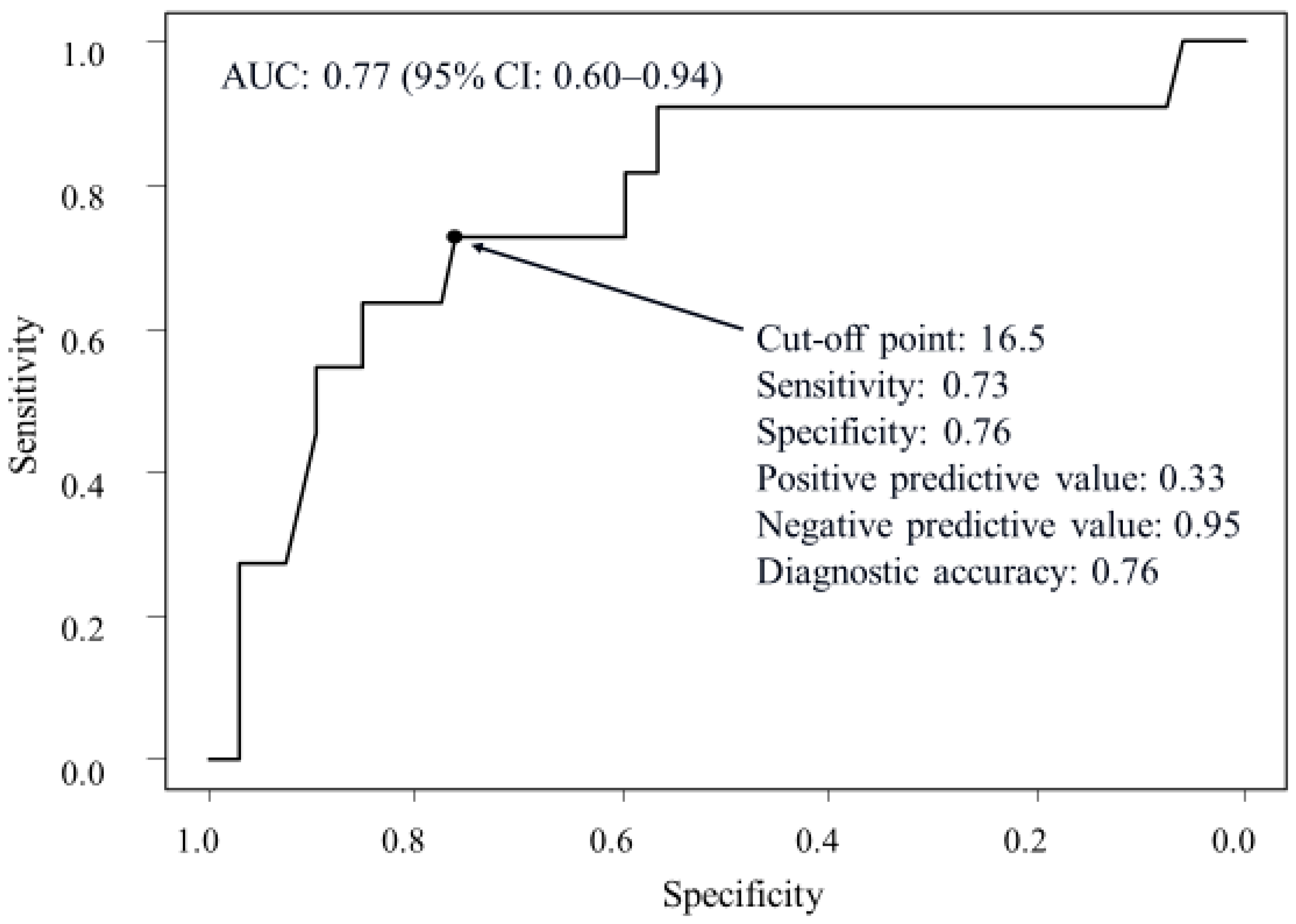
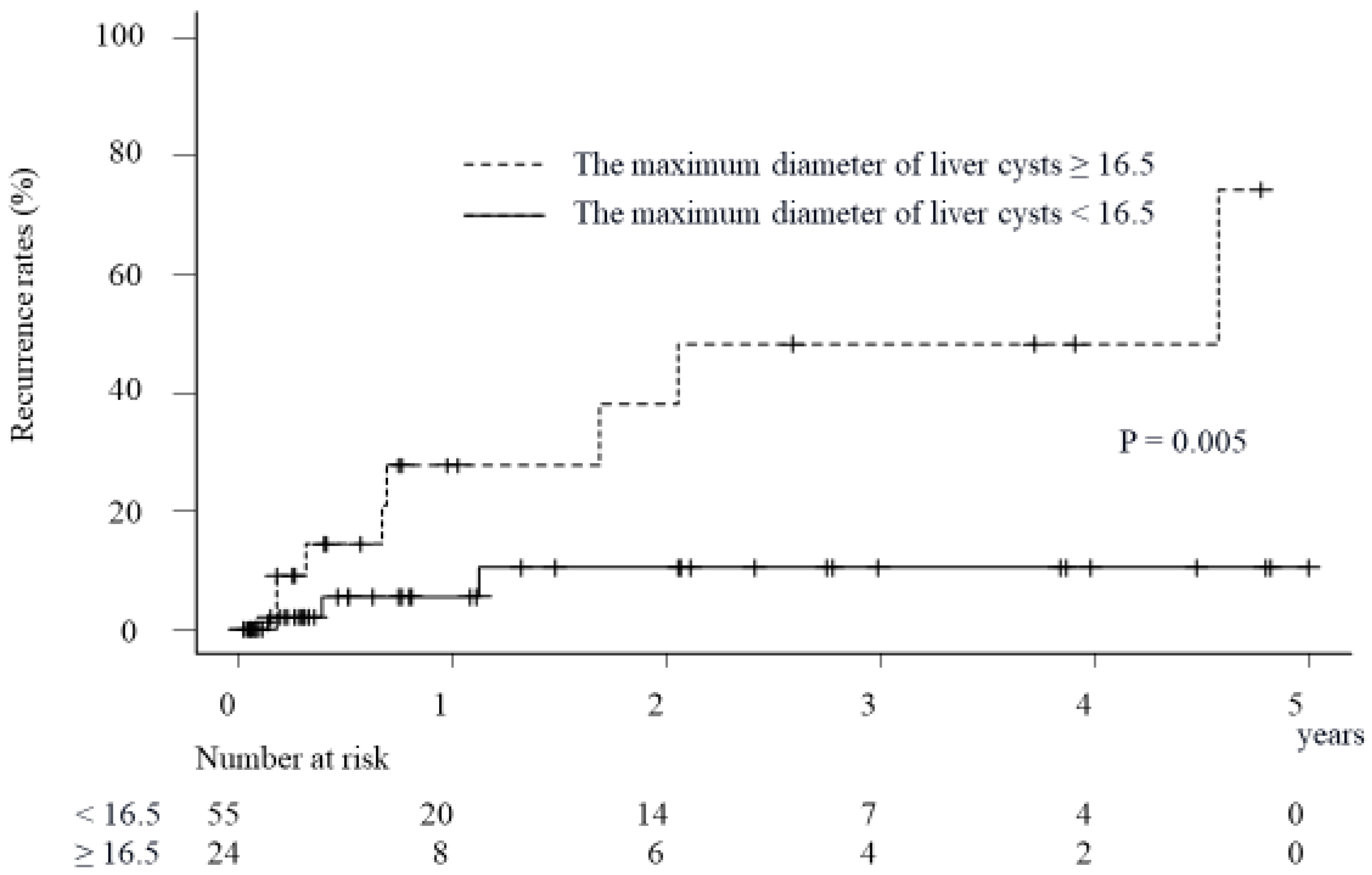
2.2. Factors Associated with Recurrence of Symptoms Due to Liver Cysts in Patients Treated via the Percutaneous Approach

The observation period for the patients was 282 (82–631) days, and 11 patients (13.9%) had symptom recurrence due to the re-progression of liver cysts during that period. In the univariate analysis, only the maximum diameter of liver cysts at the baseline was a significant predictive factor for symptom recurrence (p = 0.004). No significant differences were seen in the age, sex, rate of complication with renal cysts, number of liver cysts, ECOG PS, platelet count, PT activity, symptoms at baseline, or albumin, T-Bil, ALT or Cr levels. In addition, the multivariate analysis identified the maximum diameter of liver cysts as a statistically significant predictive factor for symptom recurrence (odds ratio: 1.21, 95% confidence interval: 1.06–1.38, p = 0.005) (Table 3). In the ROC analysis, the cut-off of the maximum diameter of liver cysts at baseline for symptom recurrence due to liver cyst re-progression was 16.5 cm (sensitivity: 0.73, specificity: 0.76, positive predictive value: 0.33, negative predictive value: 0.95, diagnostic accuracy: 0.76). The area under the curve was 0.77 (95% confidence interval: 0.6–0.94) (Figure 1). In the patients with a maximum liver cyst diameter of ≥16.5 cm, the cumulative recurrence rates at 1, 3 and 4 years were 27.6%, 48.3% and 48.3%, respectively (Figure 2). Furthermore, in the patients with a maximum liver cyst diameter of <16.5 cm, the cumulative recurrence rates at 1, 3 and 4 years were 5.4%, 10.5% and 10.5%, respectively. A significant difference in the cumulative recurrence rates of symptoms was demonstrated between the patients with a maximum liver cyst diameter of ≥16.5 cm versus <16.5 cm (p = 0.005).

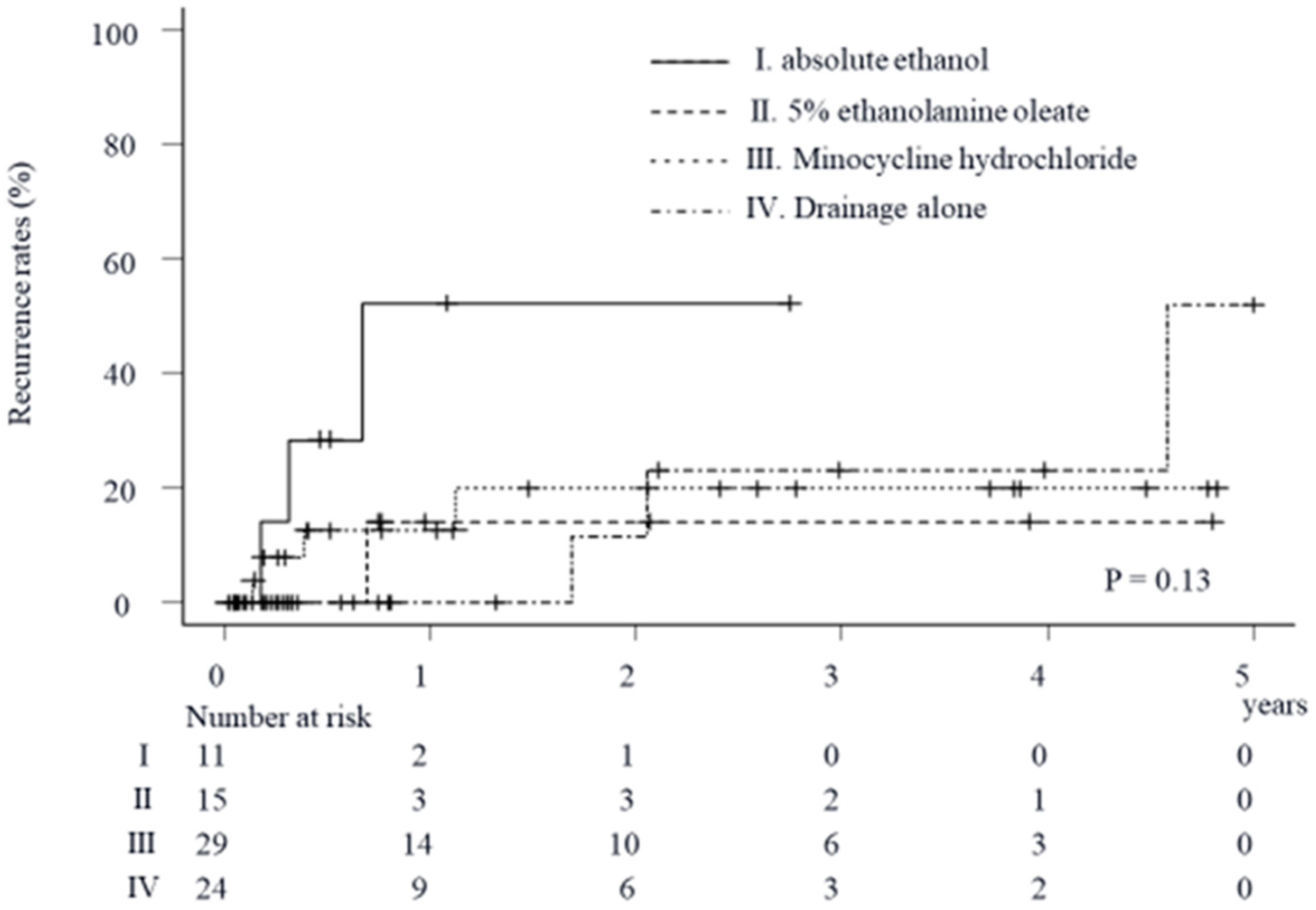
2.3. Impact of Sclerosing Agents on Cumulative Recurrence Rates of Symptoms Due to Liver Cysts in Patients Treated via the Percutaneous Approach

The cumulative recurrence rates of symptoms due to liver cyst re-progression in patients treated with percutaneous cyst drainage alone at 1, 3 and 4 years were 0%, 23.2% and 23.2%, respectively (Figure 3). These rates in patients treated via percutaneous drainage with sclerotherapy using AE at 1 and 2 years were both 52.4%. In addition, these rates in the patient treated via percutaneous drainage with sclerotherapy using EO at 1, 3 and 4 years were all 14.2%. Furthermore, these rates using MCH at 1, 3 and 4 years were 12.7%, 20% and 20%, respectively. Although no significant differences in the cumulative recurrence rates of the symptoms due to liver cysts among the four treatment groups were statistically demonstrated (p = 0.13), the recurrence rates seemed to be higher in the patients treated via drainage with sclerotherapy using AE than in those receiving other percutaneous treatments.

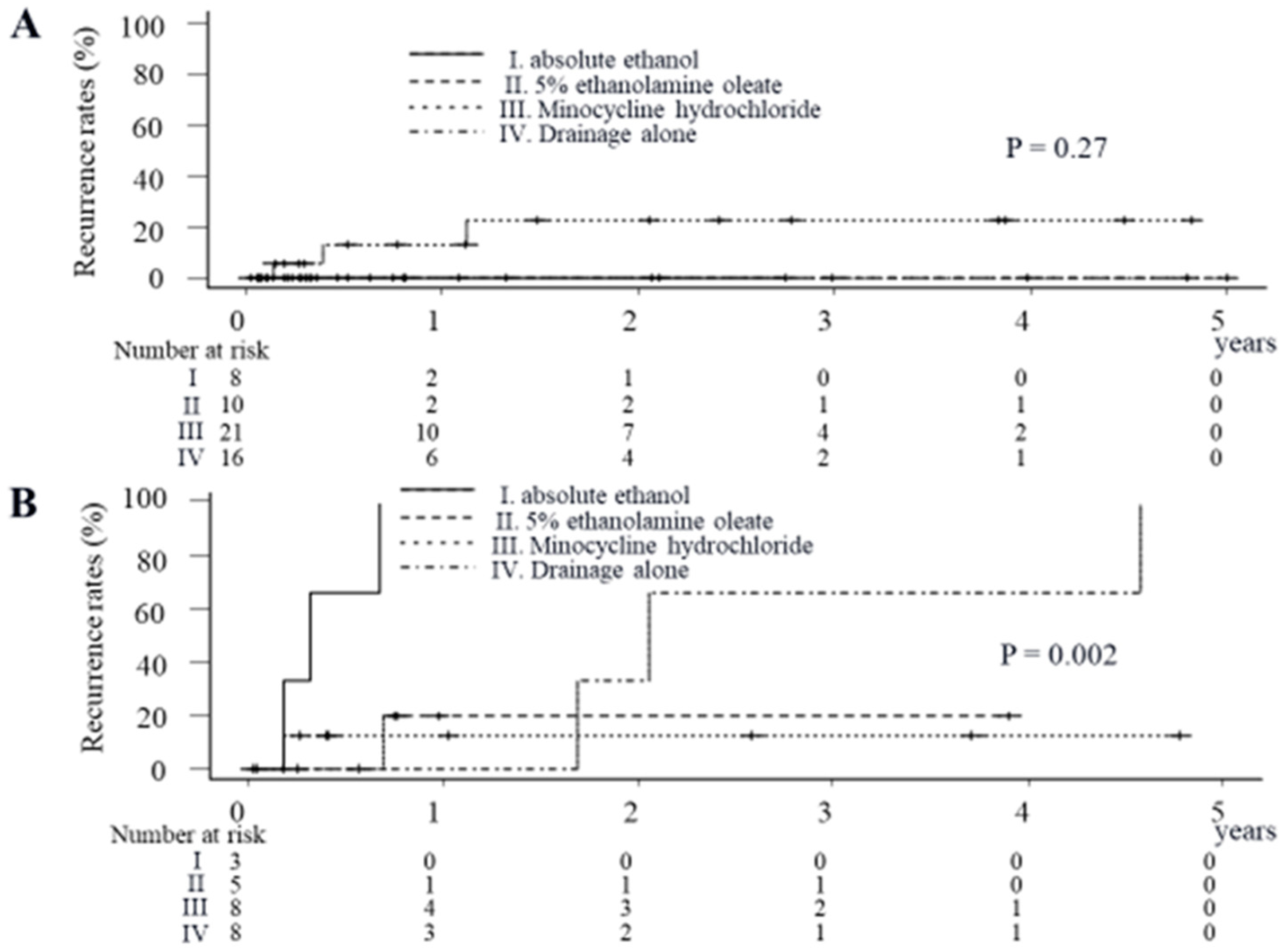
2.4. Impact of Sclerosing Agents on Cumulative Recurrence Rates of Symptoms Due to Liver Cysts in Patients with a Maximum Liver Cyst Diameter of <16.5 cm

Of the 79 patients, 55 had a maximum liver cyst diameter of <16.5 cm at baseline, and the effects of the sclerosing agents were analyzed in these 55 patients. The cumulative recurrence rates of symptoms due to liver cyst re-progression in the patients treated with percutaneous cyst drainage alone at 1, 3 and 4 years, these rates in the patients treated via percutaneous drainage with sclerotherapy using AE at 1 and 2 years and these rates in the patients treated via percutaneous drainage with sclerotherapy using EO at 1, 3 and 4 years were all 0% (Figure 4A). Although these rates in the patients treated via percutaneous drainage with sclerotherapy using MCH at 1, 3 and 4 years were 12.8%, 22.5% and 22.5%, respectively, no significant differences in the cumulative recurrence rates of the symptoms due to liver cysts among these four groups were shown (p = 0.27).

2.5. Impact of Sclerosing Agents on Cumulative Recurrence Rates of Symptoms Due to Liver Cysts in the Patients with a Maximum Liver Cyst Diameter of ≥16.5 cm

Of the 79, the remaining 24 had a maximum liver cyst diameter of ≥16.5 cm at baseline. The cumulative recurrence rates of the symptoms due to liver cyst re-progression in the patients treated with percutaneous cyst drainage alone at 1, 3 and 4 years were 0%, 66.7% and 66.7%, respectively (Figure 4B). These rates in the patients treated via percutaneous drainage with sclerotherapy using AE at 1 year were 100%; these rates in the patients treated via percutaneous drainage with sclerotherapy using EO at 1, 2 and 3 years were all 20%; and these rates in the patients treated via percutaneous drainage with sclerotherapy using MCH at 1, 3 and 4 years were all 12.5%. Significant differences in the cumulative recurrence rates of the symptoms due to liver cysts among these four groups were statistically demonstrated (p = 0.002).

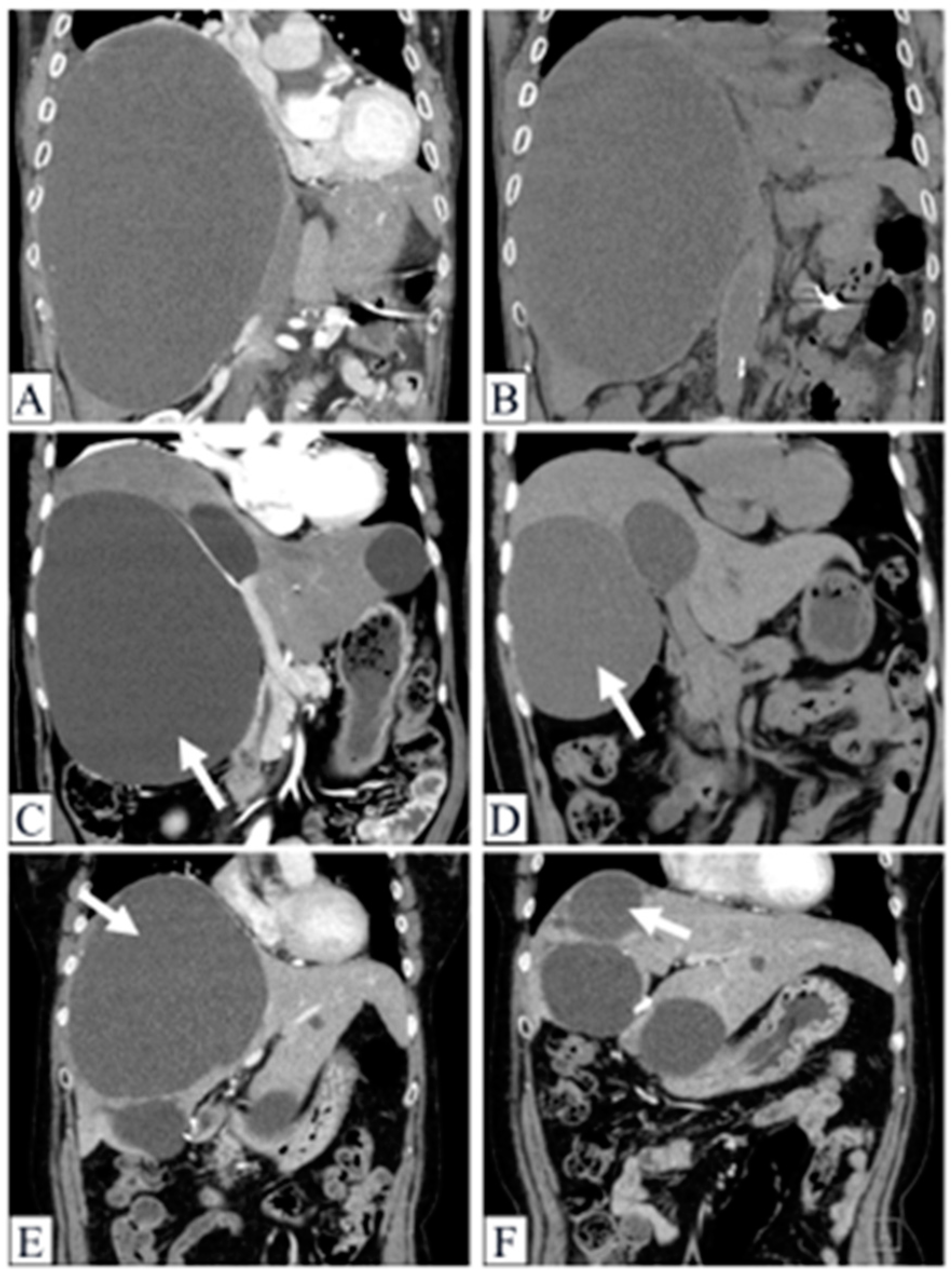
2.6. Demonstrable Cases

2.6.1. Case 1

A male in his early 90s was examined by CT due to abdominal distension and diagnosed with SHCs with a maximum diameter of 26 cm (Figure 5A). Despite treatment with percutaneous drainage with sclerotherapy using 50 mL of AE, his symptoms recurred, and the SHCs redeveloped to approximately the same size only 2 months after the treatment (Figure 5B).

2.6.2. Case 2

A female in her late 60s was examined by CT due to right upper abdominal pain and diagnosed with SHCs with a maximum diameter of 20 cm (Figure 5C). She was treated via percutaneous drainage with sclerotherapy using 100 mL of EO. At one year and three months after the treatment, she had no symptom recurrence. On CT, the maximum diameter of the treated liver cysts was 12 cm and remained smaller than before the treatment (Figure 5D).

2.6.3. Case 3

A female in her late 60s was examined by CT due to abdominal distension and diagnosed with SHCs with a maximum diameter of 17 cm (Figure 5E). She was treated via percutaneous drainage with sclerotherapy using 500 mg of MCH dissolved in 10 mL of saline. At seven months after the treatment, she had no recurrence of the symptoms. On CT, the maximum diameter of the treated liver cyst was 6 cm and remained smaller than before the treatment (Figure 5F).

2.7. Adverse Events and the Morbid Cases in the Present Study

No cases with severe complications associated with the treatments were reported in this study. Although two patients died during the observation period due to pancreatic carcinoma and congestive heart failure (one each), these causes of death were not related to the liver cysts themselves or the treatments thereof.

3. Discussion

This study investigated symptom recurrence due to liver cyst re-progression and demonstrated that the cut-off level of the maximum liver cyst diameter at baseline had excellent negative predictive value for recurrence after percutaneous treatments. In our present study, the age of the patients treated with drainage alone was higher than that of the patients treated via drainage with sclerotherapy. We presumed that many elderly patients had been treated with drainage alone in order to avoid adverse events due to sclerotherapy.
In addition, the albumin level was significantly lower in the patients treated with drainage alone than in those treated via drainage with sclerotherapy. In contrast, the rate of infection of liver cysts and ALT level were significantly higher in the patients treated with drainage alone than in those treated via drainage with sclerotherapy (Table 1). In the patients whose liver cysts were infected, sclerotherapy seemed to be avoided due to concerns about the exacerbation of intra-cystic inflammation. Because the development of liver cyst infection following sclerotherapy has been reported [9,10], the use of sclerosing agents may be avoided by the physician.
The present study revealed that few patients had a single SHC, and 47 of the 79 patients (59.4%) treated with percutaneous treatment had ≥5 liver cysts, while 34 (43.0%) were complicated by renal cysts. Treatment failure in cases with percutaneous liver cyst sclerotherapy may be due to the instillation of an insufficient amount of sclerosing agent or by insufficient exposure of the cyst lining to the sclerosing agent, especially in cases with large liver cysts [11]. In our study, the maximum diameter of the liver cysts at baseline was also the only significant factor predicting symptom recurrence due to liver cysts. Symptoms due to liver cysts may recur after treatment in patients with a maximum liver cyst diameter of ≥16.5 cm. In contrast, most patients with a maximum liver cyst diameter of <16.5 cm did not show recurrence of symptoms due to liver cysts. Given these findings, the cut-off of 16.5 cm for the maximum liver cyst diameter at baseline was considered useful for excluding cases likely to show symptom recurrence after percutaneous treatment.
In the analysis for the therapeutic effect of each percutaneous treatment method in the 79 total patients and the 55 patients with a maximum liver cyst diameter of <16.5 cm, no significant differences were demonstrated in the cumulative recurrence rates of symptoms due to liver cyst re-progression between the patients treated with drainage alone and those with additional sclerotherapy. It was reported that although re-progression of liver cysts occurred in all patients treated with drainage alone, the recurrence of symptoms due to liver cysts did not necessarily occur in all patients [12]. In our study, none of the patients with a maximum liver cyst diameter of <16.5 cm had recurrence of symptoms due to liver cysts, even among those treated with drainage alone. Since treatment success is defined by symptom relief and not by a reduction in the volume of liver cysts [13], the effect of drainage alone was sufficient, and the additional effect of sclerotherapy could not be demonstrated.
In contrast, in the patients with a maximum liver cyst diameter of ≥16.5 cm, the recurrence rates were significantly higher in the patients treated with drainage alone and those treated with sclerotherapy using AE than in those treated with sclerotherapy using EO or MCH. In the patients with a maximum liver cyst diameter of ≥16.5 cm, drainage alone was apparently insufficient, and additional sclerotherapy was necessary. Although drainage with sclerotherapy has been possible with some substances without clear evidence of the superiority of any one agent [13], EO is reportedly more effective than alcohol at destroying the epithelium of liver cysts, and exposure to EO leads to cytolysis followed by thrombogenesis [14]. In addition, EO functions as a cytolytic agent owing to its anionic surfactant properties that bring about a change in cellular permeability [15]. Similarly, MCH has been also reported to have an extremely low pH in solution, which causes the cyst wall cells to react, leading to fibrosis and adhesion of the walls [16]. Although the most frequently used sclerosing agent thus far has probably been AE, this agent has a number of drawbacks [17]. In addition to common complications, such as immediate burning pain and a fever, reports of an increase in blood alcohol concentration are frequent, and the alcohol can filter through the cyst wall and end up in the bloodstream [17]. In some cases, these values are particularly high, with patients suffering from alcohol poisoning [18]. Considering both its efficacy and safety, sclerotherapy using EO or MCH should be recommended for patients with a maximum liver cyst diameter of ≥16.5 cm.
It has been reported that the rate of persistent symptoms due to SHCs is slightly higher after percutaneous drainage with sclerotherapy compared to laparoscopic cyst deroofing (3.5% vs. 2.1%, respectively) [8]. However, this difference is small, and given the suggestion that percutaneous drainage with sclerotherapy may be associated with a lower incidence of complications than laparoscopic or open surgical treatment [19], we believe that drainage with sclerotherapy could be selected as the first-line treatment, especially in high-risk patients such as the elderly. Additionally, future prospective studies may focus on the outcomes of a step-up approach for SHCs, reserving laparoscopic cyst deroofing for patients with symptomatic recurrence of SHCs after one or two percutaneous sclerotherapies [8].
The cumulative recurrence rate of symptoms in our study appeared to be higher compared to the previously mentioned report [8]. Our study included 24 patients (30.4%) with a liver cyst diameter of ≥16.5 cm, which was the cutoff for symptom recurrence, and among them, 12 patients (15.2%) had SHCs with a diameter of ≥20 cm. While the exact reason for this difference is unclear, the inclusion of a significant number of patients with large-sized SHCs may be one contributing factor to the higher recurrence rate in our study.
Several limitations associated with the present study warrant mention. In particular, the retrospective nature of the analysis is considered a primary limitation in our study due to the potential bias introduced by the selection of treatment methods by each attending physician. Our study failed to show any significant difference in efficacy between EO and MCH. The use of sclerosing agents for liver cysts has not been approved by the Japanese insurance system, resulting in this approach being quite expensive to perform, especially for EO. In two of the six affiliated institutions, although EO was selected as the first choice and was administered at a volume of 10% of the liver cyst aspiration contents, as previously suggested [15], in principle, the maximum dosage of this sclerosing agent was determined to be 100 mL at one institution and 40 mL at another. Thus, the EO dosage may have been insufficient in patients with large liver cyst contents. If the optimum dosage of EO could have been administered, the efficacy of EO might have been improved. Furthermore, since this study was limited to patients who had been admitted within the past five years, the analysis of the long-term prognosis of liver cysts was insufficient. Therefore, large-scale, longer-term, prospective studies are needed.

4. Patients and Methods

4.1. Patients

We included patients who required initial hospitalization due to symptomatic simple hepatic cysts (SHCs) and received percutaneous treatments at six affiliated hospitals between April 2016 and March 2021 in this retrospective study. We confirmed that none of them were complicated with malignant diseases, human immunodeficiency virus infection or other liver diseases (e.g., hepatitis B, hepatitis C, alcoholic cirrhosis, liver cirrhosis due to non-alcoholic fatty liver disease, autoimmune hepatitis, primary biliary cholangitis, primary sclerosing cholangitis, congestive cirrhosis, hemochromatosis and Wilson’s disease). The diagnosis of SHCs was based on the findings of abdominal ultrasonography (US), contrast-enhanced CT or magnetic resonance imaging (MRI) and was confirmed by by board-certified radiologists.

4.2. Demographics, Laboratory Data and Study Endpoints

The demographic data upon the first hospitalization, such as age, sex, complication with renal cysts, number and maximum diameter of liver cysts, symptoms due to liver cysts (poor nutritional state with a body mass index (BMI) < 18.5 kg/m2, appetite loss, abdominal distension, ascites, infection of liver cysts), Eastern Cooperative Oncology Group performance status (ECOG PS), and treatment methods (drainage, drainage with sclerotherapy), as well as the laboratory data upon the first hospitalization, including the platelet count, prothrombin (PT) activity, albumin, total bilirubin (T-Bil), alanine aminotransferase (ALT) and creatinine (Cr) levels, were included as baseline data.
We then investigated the date of symptom recurrence due to liver cyst re-progression and that of the last survival confirmation. The recurrence of symptoms due to a liver cyst was diagnosed based on imaging findings, abdominal findings and the relief of symptoms following additional treatment. In addition, the factors associated with symptom recurrence were analyzed in the patients. We also investigated whether or not the cumulative recurrence rates of symptoms differed depending on drainage alone or the use of sclerosants.

4.3. Methods of Administering Each Sclerosing Agent to Liver Cysts

As this study was conducted retrospectively, a precise protocol for each drainage method with a sclerosing agent could not be established. In practice, the selection and dosage of the sclerosing agent were determined by each attending physician, taking into consideration the diameter of the liver cyst and the performance status (PS) of each patient.
Primarily, absolute ethanol (AE) was utilized for patients in good condition due to its potency, while minocycline hydrochloride (MCH) was administered as a milder irritant for those with poor risk factors or older patients. For patients falling between the criteria for receiving AE and MCH, 5% ethanolamine oleate (EO) was employed. It is important to note that this strategy for selecting a sclerosing agent was not universally applicable and was at times flexibly employed.

4.4. Ethical Considerations

This study was approved by the institutional review board at each affiliated hospital, and the need for written informed consent was waived because of the retrospective nature of the study. In addition, this study complied with the Ethical Guidelines for Medical and Health Research Involving Human Subjects in Japan.

4.5. Statistical Analyses

Continuous variables were expressed as the median (interquartile range) and analyzed using the Mann–Whitney U test or Kruskal–Wallis analysis as nonparametric and unpaired analyses. Post hoc analyses for these variables were performed using the Steel-Dwass method. Categorical variables were analyzed using Fisher’s exact test, and post hoc analyses were performed using the Bonferroni method. The cut-off values of continuous variable were determined by a receiver operating characteristic (ROC) analysis. The cumulative incidence rates were determined, and the differences among groups were assessed using the Gray test. A multivariate logistic regression analysis was conducted.
All statistical analyses were performed using EZR version 1.55 (Saitama Medical Center, Jichi Medical University, Saitama, Japan) [9].

5. Conclusions

The maximum liver cyst diameter was the only predictive factor of symptom recurrence due to liver cysts after percutaneous drainage. In patients with a maximum liver cyst diameter of ≥16.5 cm, not only drainage but also sclerotherapy with EO or MCH should be administered. Drainage with sclerotherapy with EO or MCH was an effective and safe treatment for patients whose liver cysts had a maximum diameter of ≥16.5 cm. Considering both its efficacy and safety, sclerotherapy using EO or MCH should be recommended for patients with a maximum liver cyst diameter of ≥16.5 cm.

Author Contributions

Conceptualization, S.T. and H.T. (Hitoshi Takagi); investigation, N.S., T.K. (Tatsuya Kohga), T.U. (Takashi Ueno), T.H., M.N., H.T. (Hiroki Tojima) and T.K. (Takashi Kosone); data curation, A.N.; writing—original draft preparation, S.T.; writing—review and editing, S.K.; supervision, T.U. (Toshio Uraoka) and H.T. (Hitoshi Takagi). All authors have read and agreed to the published version of the manuscript.

Funding

This research received no external funding.

Institutional Review Board Statement

The study was conducted in accordance with the Declaration of Helsinki, and approved by the institutional review board at each affiliated hospital. In addition, this study complied with the Ethical Guidelines for Medical and Health Research Involving Human Subjects in Japan.

Informed Consent Statement

The need for written informed consent was waived because of the retrospective nature of the study.

Data Availability Statement

The datasets generated and/or analyzed during the current study are available from the corresponding author on reasonable request.

Conflicts of Interest

The authors declare no conflicts of interest.

References

  1. Carrim, Z.I.; Murchison, J.T. The prevalence of simple renal and hepatic cysts detected by spiral computed tomography. Clin. Radiol. 2003, 58, 626–629. [Google Scholar] [CrossRef] [PubMed]
  2. European Association for the Study of the Liver (EASL). EASL Clinical Practice Guidelines on the management of benign liver tumours. J. Hepatol. 2016, 65, 386–398. [Google Scholar] [CrossRef] [PubMed]
  3. Lantinga, M.A.; Gevers, T.J.; Drenth, J.P. Evaluation of hepatic cystic lesions. World J. Gastroenterol. 2013, 19, 3543–3554. [Google Scholar] [CrossRef] [PubMed]
  4. Vardakostas, D.; Damaskos, C.; Garmpis, N.; Antoniou, E.A.; Kontzoglou, K.; Kouraklis, G.; Dimitroulis, D. Minimally invasive management of hepatic cysts: Indications and complications. Eur. Rev. Med. Pharmacol. Sci. 2018, 22, 1387–1396. [Google Scholar] [PubMed]
  5. Läuffer, J.M.; Baer, H.U.; Maurer, C.A.; Stoupis, C.; Zimmerman, A.; Büchler, M.W. Biliary cystadenocarcinoma of the liver: The need for complete resection. Eur. J. Cancer 1998, 34, 1845–1851. [Google Scholar] [CrossRef] [PubMed]
  6. Choi, B.Y.; Nguyen, M.H. The diagnosis and management of benign hepatic tumors. J. Clin. Gastroenterol. 2005, 39, 401–412. [Google Scholar] [CrossRef] [PubMed]
  7. Jones, W.L.; Mountain, J.C.; Warren, K.W. Symptomatic non-parasitic cysts of the liver. Br. J. Surg. 1974, 61, 118–123. [Google Scholar] [CrossRef] [PubMed]
  8. Furumaya, A.; van Rosmalen, B.V.; de Graeff, J.J.; Haring, M.P.D.; de Meijer, V.E.; van Gulik, T.M.; Verheij, J.; Besselink, M.G.; van Delden, O.M.; Erdmann, J.I.; et al. Systematic review on percutaneous aspiration and sclerotherapy versus surgery in symptomatic simple hepatic cysts. HPB 2021, 23, 11–24. [Google Scholar] [CrossRef] [PubMed]
  9. Kanda, Y. Investigation of the freely available easy-to-use software ‘EZR’ for medical statistics. Bone Marrow Transplant. 2013, 48, 452–458. [Google Scholar] [CrossRef] [PubMed]
  10. Wijnands, T.F.; Lantinga, M.A.; Drenth, J.P. Hepatic cyst infection following aspiration sclerotherapy: A case series. J. Gastrointestin. Liver Dis. 2014, 23, 441–444. [Google Scholar] [CrossRef] [PubMed]
  11. Erdogan, D.; van Delden, O.M.; Rauws, E.A.; Busch, O.R.; Lameris, J.S.; Gouma, D.J.; van Gulik, T.M. Results of percutaneous sclerotherapy and surgical treatment in patients with symptomatic simple liver cysts and polycystic liver disease. World J. Gastroenterol. 2007, 13, 3095–3100. [Google Scholar] [CrossRef] [PubMed]
  12. Saini, S.; Mueller, P.R.; Ferrucci, J.T., Jr.; Simeone, J.F.; Wittenberg, J.; Butch, R.J. Percutaneous aspiration of hepatic cysts does not provide definitive therapy. AJR Am. J. Roentgenol. 1983, 141, 559–560. [Google Scholar] [CrossRef] [PubMed]
  13. European Association for the Study of the Liver. EASL Clinical Practice Guidelines on the management of cystic liver diseases. J. Hepatol. 2022, 77, 1083–1108. [Google Scholar] [CrossRef] [PubMed]
  14. Nakaoka, R.; Das, K.; Kudo, M.; Chung, H.; Innoue, T. Percutaneous aspiration and ethanolamine oleate sclerotherapy for sustained resolution of symptomatic polycystic liver disease: An initial experience. AJR Am. J. Roentgenol. 2009, 193, 1540–1545. [Google Scholar] [CrossRef] [PubMed]
  15. Yamamoto, K.; Sakaguchi, H.; Anai, H.; Tanaka, T.; Morimoto, K.; Kichikawa, K.; Uchida, H. Sclerotherapy for simple cysts with use of ethanolamine oleate: Preliminary experience. Cardiovasc. Interv. Radiol. 2005, 28, 751–755. [Google Scholar] [CrossRef] [PubMed]
  16. Ohkawa, M.; Tokunaga, S.; Orito, M.; Shimamura, M.; Hirano, S.; Okasho, A.; Kosaka, S. Percutaneous injection sclerotherapy with minocycline hydrochloride for simple renal cysts. Int. Urol. Nephrol. 1993, 25, 37–43. [Google Scholar] [CrossRef] [PubMed]
  17. Danza, F.M.; Falcione, M.; Bordonaro, V.; Infante, A.; Paladini, A.; Bonomo, L. Minocycline hydrochloride as a soft sclerotizing agent for symptomatic simple renal and hepatic cysts. Eur. Rev. Med. Pharmacol. Sci. 2017, 21, 408–415. [Google Scholar] [PubMed]
  18. Skolarikos, A.; Laguna, M.P.; de la Rosette, J.J. Conservative and radiological management of simple renal cysts: A comprehensive review. BJU Int. 2012, 110, 170–178. [Google Scholar] [CrossRef] [PubMed]
  19. Moorthy, K.; Mihssin, N.; Houghton, P.W. The management of simple hepatic cysts: Sclerotherapy or laparoscopic fenestration. Ann. R. Coll. Surg. Engl. 2001, 83, 409–414. [Google Scholar]
Figure 1. Results of receiver operating characteristics analysis regarding the maximum liver cyst diameter at baseline for symptom recurrence due to liver cyst re-progression after percutaneous drainage. According to the Youden index, the best cut-off of that diameter was 16.5 cm (sensitivity: 0.73, specificity: 0.76, positive predictive value: 0.33, negative predictive value: 0.95, diagnostic accuracy: 0.76). The area under the curve was 0.77 (95% confidence interval: 0.6–0.94).
Gastrointestdisord 06 00002 g001
Figure 2. The cumulative recurrence rates of symptoms due to liver cysts after percutaneous drainage with or without sclerotherapy, classified by the cut-off of the maximum liver cyst diameter. A significant difference in these rates was demonstrated between the patients with a maximum liver cyst diameter of ≥16.5 cm and those with a maximum diameter of <16.5 cm (p = 0.005).
Gastrointestdisord 06 00002 g002
Figure 3. The cumulative recurrence rates of the symptoms due to liver cysts after percutaneous drainage with or without sclerotherapy, classified by treatment method, such as drainage alone, drainage with sclerotherapy using absolute ethanol, drainage with sclerotherapy using 5% ethanolamine oleate and drainage with sclerotherapy using minocycline hydrochloride. Although no significant differences in the cumulative recurrence rates of these symptoms among these 4 groups were statistically demonstrated (p = 0.13), the rates were higher in the patients treated via drainage with sclerotherapy using absolute ethanol than in those receiving other percutaneous treatments.
Gastrointestdisord 06 00002 g003
Figure 4. A comparison of the cumulative recurrence rates of symptoms due to liver cysts after percutaneous drainage with or without sclerotherapy, classified by treatment method, in patients with a maximum liver cyst diameter of <16.5 cm (A) and ≥16.5 cm (B). No significant differences in the rates of these symptoms among the 4 groups are statistically demonstrated in (A) (p = 0.27). In contrast, significant differences in the rates of these symptoms among the 4 groups are statistically demonstrated in (B) (p = 0.002).
Gastrointestdisord 06 00002 g004
Figure 5. (A) The findings of computed tomography before and after percutaneous treatments of liver cysts in the demonstrable cases. In case 1, symptomatic liver cysts with a maximum diameter of 26 cm were confirmed. (B) Despite treatment with drainage and sclerotherapy using 50 mL of absolute ethanol, the liver cyst redeveloped to approximately the same size, and the symptoms recurred just 2 months after treatment. (C) In case 2, symptomatic liver cysts with a maximum diameter of 20 cm were confirmed (arrow). (D) At 1 year and 3 months after treatment with drainage with sclerotherapy using 100 mL of 5% ethanolamine oleate, the target liver cyst remained smaller than before the treatment (arrow), and symptoms due to liver cysts did not recur. (E) In case 3, symptomatic liver cysts with a maximum diameter of 17 cm were confirmed (arrow). (F) At 7 months after treatment with drainage with sclerotherapy using 500 mf of minocycline hydrochloride dissolved in 10 mL of saline, the target liver cyst remained smaller than before the treatment (arrow), and symptoms due to liver cysts did not recur.
Gastrointestdisord 06 00002 g005
Table 1. Baseline characteristics of the study patients upon the first hospitalization.
All Cases (n = 79)Drainage Alone (n = 24)Drainage with Sclerotherapy (n = 55)p
Age (years)70 (64–80)75 (67–83)69 (62–79)0.049
Male, n (%)20 (25.3)11 (45.8)9 (16.4)0.01
Complication with renal cysts, n (%)34 (43.0)12 (50)22 (40)0.46
Number of liver cysts, n (%)
  <532 (40.5)8 (33.3)24 (43.6)0.58
  5–1021 (26.6)6 (25)15 (27.3)
  >1026 (32.9)10 (41.7)16 (29.1)
Maximum diameter of liver cysts (cm)13.1 (10.0–17.8)12.6 (8.8–18.1)13.1 (10.3–16.9)0.61
Symptoms, n (%)
  Poor nutrition status with BMI < 18.5 kg/m23 (3.8)0 (0)3 (5.5)0.55
  Appetite loss16 (20.2)6 (25)10 (18.2)0.55
  Abdominal distension35 (44.3)8 (33.3)27 (49.1)0.23
  Ascites8 (10.1)4 (16.7)4 (7.3)0.24
  Infection of liver cysts23 (29.1)15 (62.5)8 (14.5)<0.001
ECOG PS, n (%)
  0/1/2/3/461 (77.2)/9 (11.4)/
5 (6.3)/1 (1.3)/3 (3.8)
15 (62.5)/4 (16.7)/2 (8.3)/
1 (4.2)/2 (8.3)
46 (83.6)/5 (9.1)/3 (5.5)/
0 (0)/1 (1.8)
0.13
Platelet count (×109/L)207 (166–275)243 (171–298)204 (166–242)0.12
PT (%)91.4 (78.3–101.0)83.9 (62.6–94.0)93.0 (82.0–101.0)0.021
Albumin (g/dL)3.8 (3.2–4.2)3.4 (2.3–3.9)3.9 (3.5–4.3)0.002
T-Bil (mg/dL)0.8 (0.7–1.2)0.9 (0.7–1.5)0.8 (0.7–1.1)0.33
ALT (IU/L)20 (15–37)35 (20–64)18 (12–23)0.002
Creatinine (mg/dL)0.7 (0.6–0.8)0.7 (0.5–1.0)0.7 (0.6–0.8)0.79
ALT, alanine aminotransferase; BMI, body mass index; ECOG PS, Eastern Cooperative Oncology Group performance status; PT, prothrombin time; T-Bil, total bilirubin.
Table 2. Baseline characteristics of the patients treated via drainage with sclerotherapy (n = 55).
Minocycline Hydrochloride (n = 29)Ethanolamine Oleate (n = 15)Absolute Ethanol (n = 11)p
Age (years)70 (65–80)64 (60–73)69 (63–80)0.32
Male, n (%)4 (13.8)1 (6.7)4 (36.4)0.14
Complication with renal cysts, n (%)10 (34.5)8 (53.3)4 (36.4)0.47
Number of liver cysts, n (%)
  <513 (44.8)5 (33.3)6 (54.5)0.83
  5–107 (24.1)5 (33.3)3 (27.3)
  >109 (31.0)5 (33.3)2 (18.2)
Maximum diameter of liver cysts (cm)13.1 (11.6–16.5)13.8 (10.8–19.2)12.0 (10.0–18.0)0.9
Symptoms, n (%)
  Poor nutrition status with BMI < 18.5 kg/m22 (6.9)1 (6.7)0 (0)1
  Appetite loss5 (17.2)2 (13.3)3 (27.3)0.72
  Abdominal distension14 (48.3)8 (53.3)5 (45.5)1
  Ascites3 (10.3)1 (6.7)0 (0)0.8
  Infection of liver cysts6 (20.7)0 (0)2 (18.2)0.15
ECOG PS, n (%)
  0/1/2/3/424 (82.8)/2 (6.9)/
2 (6.9)/0 (0)/1 (3.4)
13 (86.7)/2 (13.3)/
0 (0)/0 (0)/0 (0)
9 (81.8)/1 (9.1)/
1 (9.1)/0 (0)/0 (0)
0.92
Platelet count (×109/L)204 (166–229)196 (181–263)208 (158–238)0.91
PT (%)95.5 (78.7–107.5)93 (87.7–100.8)85.6 (80.5–99.4)0.72
Albumin (g/dL)4.0 (3.7–4.4)3.9 (3.5–4.2)3.7 (3.2–4.1)0.14
T-Bil (mg/dL)0.8 (0.7–1.0)0.7 (0.6–0.9)1.1 (0.8–1.4)0.1
ALT (IU/L)17 (12–21)18 (15–22)22 (14–43)0.29
Creatinine (mg/dL)0.7 (0.6–0.8)0.7 (0.6–0.8)0.7 (0.6–0.9)0.9
ALT, alanine aminotransferase; BMI, body mass index; ECOG PS, Eastern Cooperative Oncology Group performance status; PT, prothrombin time; T-Bil, total bilirubin.
Table 3. The factors associated with symptom recurrence due to liver cyst re-progression (n = 79).
Univariate AnalysisMultivariate Analysis
Recurrence (−)
(n = 68)
Recurrence (+)
(n = 11)
pOdds Ratio
(95% Confidence Interval)
p
Age (years)70 (64–79)80 (66–86)0.3
Male, n (%)16 (23.5)4 (36.4)0.46
Complication with renal cysts, n (%)30 (44.1)4 (36.4)0.75
Number of liver cysts, n (%)
  <526 (38.2)6 (54.5)0.42
  5–1020 (29.4)1 (9.1)
  >1022 (32.4)4 (36.4)
Maximum diameter of liver cysts (cm)12.0 (9.8–16.0)19.7 (15.0–21.5)0.0041.21 (1.06–1.38)0.005
Symptoms, n (%)
  Poor nutrition status with BMI < 18.5 kg/m23 (4.4)0 (0)1
  Appetite loss13 (19.1)3 (27.3)0.69
  Abdominal distension29 (42.6)6 (54.5)0.52
  Ascites8 (11.8)0 (0)0.59
  Infection of liver cysts20 (29.4)3 (27.3)1
ECOG PS0 (0–0)0 (0–1)0.29
Treatment methods
  Drainage alone21 (30.9)3 (27.3)0.57
  Drainage and injection of absolute ethanol8 (11.8)3 (27.3)
  Drainage and injection of 5% ethanolamine oleate14 (20.6)1 (9.1)
  Drainage and injection of minocycline hydrochloride25 (36.8)4 (36.4)
Platelet count (×109/L)208 (173–280)146 (134–234)0.099
PT (%)92.0 (81.0–101.0)85.6 (64.8–92.3)0.16
Albumin (g/dL)3.9 (3.4–4.2)3.0 (2.6–3.9)0.12
T-Bil (mg/dL)0.8 (0.6–1.2)1.0 (0.8–1.6)0.11
ALT (IU/L)19 (15–35)23 (14–54)0.44
Creatinine (mg/dL)0.7 (0.6–0.8)0.7 (0.5–0.9)0.42
ALT, alanine aminotransferase; BMI, body mass index; ECOG PS, Eastern Cooperative Oncology Group performance status; PT, prothrombin; T-Bil, total bilirubin.
Disclaimer/Publisher’s Note: The statements, opinions and data contained in all publications are solely those of the individual author(s) and contributor(s) and not of MDPI and/or the editor(s). MDPI and/or the editor(s) disclaim responsibility for any injury to people or property resulting from any ideas, methods, instructions or products referred to in the content.

