Article

Application of Piper betle Leaf Extract as a Bioactive Additive in Eco-Friendly Antifouling Coatings

Coastal Branch of Joint Vietnam-Russia Tropical Science and Technology Research Center, 30 Nguyen Thien Thuat, Nha Trang 57127, Vietnam
*
Author to whom correspondence should be addressed.
Surfaces 2025, 8(4), 72; https://doi.org/10.3390/surfaces8040072 (registering DOI)
Submission received: 20 July 2025 / Revised: 3 October 2025 / Accepted: 8 October 2025 / Published: 11 October 2025

Abstract

The present study aimed to evaluate the antifouling efficacy of Piper betle leaf extracts as a bioactive additive for eco-friendly antifouling coatings. The composition of P. betle extract was determined and analyzed. Phytochemical analysis revealed that the ethanol extract of P. betle contained phenolics, tannins, proteins, carbohydrates, and flavonoids, with total phenolic content reaching 260.3 mg GAE/g dry weight and flavonoid content reaching 52.56 mg QE/g dry weight. The antibacterial test results showed that the ethanol extract of P. betle exhibited maximum antibacterial efficacy against E. coli, B. subtilis, S. aureus, and marine bacteria, with inhibition zone diameters of 28.7 ± 0.5, 27.0 ± 1.6, 22.1 ± 0.6, and 35.1 ± 0.5 mm, respectively. Based on the laboratory test results, the ethanol extract of P. betle was chosen to be added to coatings as an antifouling additive. The content of the extract was 0.5, 1.0, and 1.5 wt.%. A field test conducted in tropical seawater (at Nha Trang Bay) demonstrated that incorporating 1 wt.% of P. betle extract into an acrylic copolymer-based coating significantly enhanced its antifouling performance. After nine months of immersion in seawater, this sample maintained an antifouling efficiency of 74%. These findings highlight the potential of P. betle extract as a sustainable alternative to conventional antifouling agents in marine coatings.

1. Introduction

Biofouling refers to the undesirable accumulation of biological organisms—microorganisms, barnacles, mussels, algae, and other marine invertebrates—on the surfaces of materials continuously immersed in seawater. This phenomenon has several negative consequences, such as increased hydrodynamic drag and structural loads on vessels and offshore structures, reduced navigational efficiency and speed, elevated fuel consumption, material degradation through corrosion, and overall reduction in service life [1].
In the context of expanding maritime operations, developing environmentally benign antifouling coatings is imperative for the protection of marine ecosystems as well as for ensuring the sustainability of aquaculture and marine engineering industries [2,3]. Among the various antifouling strategies, applying antifouling coatings remains the most prevalent approach due to their high efficacy, ease of implementation, and relatively low cost. Copper-based compounds, such as copper flakes and copper oxides, are among the most commonly employed active agents in these coatings [4,5]. In addition, synthetic biocides, including Irgarol 1051, DCOIT, dichlofluanid, and chlorothalonil, are widely incorporated as antifouling agents [6]. Nevertheless, these synthetic and metal-based biocides exhibit varying degrees of ecotoxicity, raising concerns regarding their environmental persistence and impact. As a result, regulatory authorities in several countries have imposed restrictions on the use of copper compounds and synthetic toxicants in antifouling formulations [7]. In response to increasingly stringent environmental standards, developing eco-friendly antifouling coatings—non-toxic or based on naturally derived bioactive agents—has attracted growing scientific attention [8,9,10].
One promising approach involves the use of plant-derived compounds as natural antifouling additives. Extracts may be derived from various plant parts, including roots, leaves, fruits, bark, and seeds. Plant extracts are rich in polyphenolic compounds—more than 8000 polyphenol types have been identified—classified as flavonoids, phenolic acids, polyphenolic amides, and others [11,12]. These compounds possess high antimicrobial and antifouling properties and have been extensively applied in pharmacy, medicine, and in the development of bio-based antifouling coatings. Notable examples include extracts from Zingiber officinale R. (ginger root) [13], Nerium oleander L. (oleander) [14], capsaicin from chili peppers [15,16,17], and extracts from the leaves of Sonneratia lanceolata B. [18].
Piper betle Linn. (P. betle) is a plant species widely distributed throughout South and Southeast Asia. Its leaves contain high concentrations of bioactive compounds with potent antimicrobial properties and are traditionally utilized in food preservation and medicinal applications [19]. Phytochemical investigations have shown that P. betle extracts contain several bioactive compounds, including phenolics, alkaloids, saponins, tannins, and flavonoids [20,21]. Compared to other plant extracts, the P. betle extract showed potent antibacterial activity against Gram-negative and Gram-positive bacterial strains [22,23,24,25]. Studies have also shown that phenolic compounds such as hydroxychavicol and allylpyrocatechol, identified in the P. betle extract, have been reported to exhibit anti-plaque activity by compromising the integrity of the cell wall of Streptococcus sanguinis, a primary bacterium involved in dental plaque formation [26]. In addition, the P. betle extract has high antibacterial activity and anti-biofilm potential against S. pseudintermedius and E. coli [27,28]. This makes the P. betle extract a promising candidate for incorporation into environmentally friendly antifouling coatings. However, there have been no reports on using P. betle extract as an additive in antifouling coatings.
This study investigates the use of P. betle extract as a natural antifouling additive in the formulation of non-toxic, bio-based coatings intended to replace conventional biocide-based systems. The P. betle extract is analyzed to determine the content of certain organic functional groups and then is tested to evaluate their antibacterial activity. The extract is incorporated into an acrylic-based coating and applied onto steel panels. The coated samples are thereafter subjected to an antifouling efficacy investigation. In addition, the morphology and surface properties of the coating are also examined.

2. Materials and Methods

2.1. Preparation of P. betle Extracts

Piper betle Linn. leaves were collected from Nha Trang City, Khanh Hoa Province, Vietnam (12.2385° N, 109.159° E) on 16 September 2024 and were identified by the Research Institute for Biotechnology and Environment, Nong Lam University, Ho Chi Minh city. The voucher specimens 21,002 have been deposited in the Coastal Branch of the Joint Vietnam–Russia Tropical Science and Technology Research Center. The leaves were thoroughly washed and dried at 45 °C for 72 h, then ground into fine powder using a mechanical grinder (PG2500, Spring Green Evolution, Bangkok, Thailand). The powders (500 g) were macerated in various solvents (methanol, ethanol, and ethyl acetate) at 1:10 (w/v) for 24 h at 25 °C. This procedure was repeated three times to maximize the extraction. The mixture was filtered using filter paper, and then the combined filtrates were concentrated using a rotary vacuum evaporator (R215, Buchi, Switzerland) at 40 °C under reduced pressure (175 mbar for ethanol, 337 mbar for methanol, and 240 mbar for ethyl acetate). The concentrated extracts were vacuum-dried at 45 °C using a vacuum oven (SH VDO-125NG, SH Scientific, Sejong-si, Republic of Korea) until the solvent was removed. The crude extracts were stored at 4 °C for subsequent experimental procedures. In the current study, the extraction yielded 70.5 g of the crude extract of P. betle.

2.2. Extracts Characterization

2.2.1. Fourier Transform Infrared (FTIR) Analysis

The extracts were characterized for chemical structure by an FTIR spectrometer (Agilent Cary 630, Agilent Technologies, Malaysia) in the range of 4000–630 cm−1.

2.2.2. Determination of the Presence of P. betle Extract Compounds by Phytochemical Screening

To determine the presence of phytochemical constituents such as phenolics, tannins, proteins, carbohydrates, and flavonoids in crude extract, the test was done according to [29]. The concentrated extracts were dissolved in ethanol to obtain a 100 mg/mL concentration.
Phenolic compound test: 2 mL of the filtered extract was added to a 1% FeCl3 solution to detect phenolic compounds. The appearance of a dark blue or greenish-black coloration indicated the presence of phenolic compounds.
Tannins test: 2 mL of the filtered extract was mixed with a 1% lead acetate [Pb(C2H3O2)2] to test for tannins. The formation of a gelatinous precipitate was taken as evidence of tannin presence.
Protein test: Proteins were detected using the Biuret test. 2 mL of the filtered extract was treated with a drop of 2% CuSO4 solution and 1 mL of 95% ethanol. A small amount of KOH was then added. The formation of a pink-colored precipitate indicated the presence of proteins.
Carbohydrate test: Carbohydrates were detected by gradually treating 2 mL of the filtered extract with a few drops of diluted sulfuric acid (H2SO4). The appearance of a purple precipitate confirmed the presence of carbohydrate compounds.
Flavonoids test: 2 mL of the filtered extract was treated with a small amount of metallic magnesium to assess the presence of flavonoids, followed by a few drops of concentrated hydrochloric acid (HCl). The development of a red coloration suggested the presence of flavonoid compounds.

2.2.3. Determination of the Total Phenolic Content (TPC) and the Total Flavonoid Content (TFC)

The total phenolic content (TPC) was determined following the method described in [30]. According to that, 2.5 mL of 10% (v/v) Folin–Ciocalteu reagent in distilled water was mixed with 0.5 mL of the extract (diluted with ethanol to obtain a concentration of 5 µg/mL). After 6 min, 2 mL of 7.5% (w/v) Na2CO3 solution was added to the mixture, which was then left in the dark for one hour at room temperature. The mixture absorbance was measured at 760 nm using a UV-Vis spectrophotometer (DR3900, Hach, Loveland, CO, USA). Gallic acid was used as a quantification standard, and the TPC results were expressed in milligram gallic acid equivalents (GAE) per gram of dry weight. The total flavonoid content (TFC) was determined using the AlCl3 colorimetric method. Dimethyl sulfoxide (DMSO) was used as the solvent to dilute and prepare a concentration series of the crude extracts. Specifically, 0.5 mL of P. betle extract (at a concentration of 100 µg/mL) was mixed with 1.5 mL of ethanol and allowed to stand for 5 min. Subsequently, 0.1 mL of 10% AlCl3 solution was added, and the mixture was shaken gently. After a 6-min reaction, 0.1 mL of 1 M CH3COOK and 2.8 mL of distilled water were added. The mixture was thoroughly shaken and incubated at room temperature. After 30 min, the absorbance was measured at 415 nm using a UV-Vis spectrophotometer. Quercetin was used as a positive standard. The total flavonoid content in the P. betle extract was calculated in milligrams of quercetin (QE) per gram of dry weight [31].

2.2.4. Antibacterial Activity Test

The antibacterial activity of P. betle extract was evaluated against three bacterial strains: Escherichia coli ATCC 15034, Bacillus subtilis BKM B501, and Staphylococcus aureus ATCC 21027. Bacterial strains are supported at –85 °C in cryotubes in the Collection of Heterotrophic Bacteria, Coastal Branch of the Joint Vietnam–Russia Tropical Science and Technology Research Center. In addition, the extracts were also assessed against marine bacteria. Seawater samples containing native marine microorganisms were collected from the Dam Bay Marine Research and Testing Station (12.197° N, 109.292° E) on 12 December 2024. Marine bacterial cultures were obtained by inoculating 0.1 mL of seawater onto Marine Broth agar plates. The extract samples (at a concentration of 300 µg/mL) were applied to wells (50 µL per well) created in the agar. Penicillin G (at a concentration of 6 µg/mL) was used as a positive control. The agar plates were incubated at 30 °C for 18 h. Five test runs on each sample were carried out. Antibacterial activity was assessed by observing the presence or absence of inhibition zones and measuring the diameter of the inhibition zones (in mm), which reflects the extent of microbial growth suppression.

2.3. Preparation and Characterization of Coatings

2.3.1. Paint Preparation

Paints were prepared by mixing the ingredients using a high-speed disperser (Alligator Machine, AAU Technology Trading Co., Ltd., Ho Chi Minh City, Vietnam). The formulation included 37.5 g of acrylic copolymer (butyl methacrylate, methacrylic acid, methyl methacrylate), 50 g of rosin, 7.5 g of oleic acid, 25 g of CaCO3, 50 g of ZnO, 5 g of bentonite, and 250 mL of solvent (xylene). The ethanol extract of P. betle was added to the matrix at concentrations of 0.5, 1.0, and 1.5 wt.%.

2.3.2. Surface Morphology Analysis

The surface micromorphology of the coatings was examined using scanning electron microscopy (SEM), SNE-ALPHA (SEC Co., Ltd., Suwon, Republic of Korea).

2.3.3. Surface Wettability Analysis

The surface wettability of coatings was determined by advancing contact angle measurement using an optical contact angle measuring and contour analysis system, OCA 15EC (DataPhysics Instruments GmbH, Filderstadt, Germany). For each composition, three samples were tested.

2.3.4. Determination of the Self-Polishing Rate (SPR) of Coatings

The study of the self-polishing rate of the coatings was conducted under laboratory conditions over a 10-day immersion period in natural seawater, following the methodology described in [32]. Coating samples were prepared in square specimens (3 × 3 cm). After drying for 48 h, the initial mass of each specimen was measured using an analytical balance with a precision of 0.0001 g. The samples were then immersed in 150 mL of natural seawater in sealed containers and placed on an orbital shaker (Grant-bio PSU-20i, Grant Instruments Ltd., Royston, UK) with an operating speed of 120 rpm. For each composition, three replicate samples were tested. After 10 days of immersion, the samples were allowed to air-dry at room temperature for 48 h, and then the weight of the specimens was recorded to assess their self-polishing rate. The self-polishing rate was calculated using the following equation:
S P R = W 0 W t , d r y S . t
where W0—initial sample mass before immersion; Wt,dry—sample mass after drying post-immersion; S—surface area of the sample (cm2); t—time of immersion (days).

2.3.5. Field Test of Coatings

To evaluate the antifouling performance of antifouling coatings, a real sea hanging test was conducted at the marine experimental station located at Dam Bay Marine Research and Testing Station (Nha Trang Bay, 12.197° N; 109.292° E). The average seawater parameters were as follows: temperature 29.24 °C, pH 8.27, dissolved oxygen 6.47 mg/L, and salinity 33.4‰. The coating samples were applied to Ct3 carbon steel panels with dimensions of 150 × 100 mm and exposed under full immersion conditions at a depth ranging from 0.5 to 1.5 m below the seawater surface. A coating sample without an antifouling agent was used as the negative control, while a coating containing 20 wt.% Cu2O served as the positive control.
To perform preliminary observations of fouling, the experimental samples were taken out for 5–10 min, and their surfaces were photographed at high magnification. Subsequently, the photos of the samples were examined on a PC at maximum digital magnification. The physical performance of the coating surface was evaluated, and the area of the fouling surface of the samples was calculated using ImageJ 1.48v software (National Institutes of Health, Bethesda, MD, USA). The antifouling efficiency (AE) was assessed taking into account the species of organisms and fouling area according to the formula [33]
AE = 100 − (0.2 × S1 + 0.5 × S2 + 15 × S3)
where AE—antifouling efficiency (%), S1—percent of area covered with slime; S2—percent occupied by non-encrusting organisms; S3—percent occupied by fouling organisms with hard shells larger than 5 mm. The higher the AE, the better the antifouling properties of the coating.
Statistical analysis. All results are presented as means ± standard deviation (SD). Statistically significant differences (p < 0.05) were determined using one-way ANOVA and Tukey’s test.

3. Results and Discussion

To evaluate the composition of organic functional groups in the extracts, Fourier-transform infrared (FTIR) spectroscopy was performed. The results are presented in Figure 1. The spectra exhibit relatively consistent peak positions across the samples, indicating a similar composition of functional groups. Notable absorption bands were observed at the following wavenumbers: a broad peak in the range of 3600–3300 cm−1 corresponding to O–H stretching vibrations in carboxylic or hydroxyl groups; a peak at 2928 cm−1 attributed to the stretching vibration of C–H bonds in the methyl group on the aromatic ring; The presence of an aromatic ring is further confirmed by the presence of peaks at 1600–1400 cm−1. The presence of phenolic hydroxyl groups is confirmed by absorption bands at 1263–1200 cm−1 [34].
As the FTIR spectral analysis revealed similar results across the extracts obtained using different solvents, the ethanol extract of P. betle was selected to analyze specific compound groups. The results of the phytochemical screening are shown in Table 1. Based on the screening, phenolics, tannins, proteins, carbohydrates, and flavonoids were detected. Compared with previous studies, Kaveti et al. [35] also reported the presence of several classes of compounds in P. betle extracts, including alkaloids, tannins, glycosides, and saponins. Meanwhile, P. betle collected in Mauritius contained phenols, flavonoids, and tannins [36]. Samples collected in India contained steroids, tannins, proteins, amino acids, flavonoids, terpenoids, saponins, and carbohydrates [37].
Phenolic and flavonoid compounds present in plant extracts have been widely demonstrated to exhibit potent antibacterial activity [14,38]. Therefore, analyses of total phenolic content (TPC) and total flavonoid content (TFC) were conducted for the ethanol extract of P. betle (Table 2). The results showed that the ethanol extract of P. betle contains a high concentration of phenolics (260.3 ± 5.22 mg GAE/g dry weight) and flavonoids (52.56 ± 1.26 mg QE/g dry weight). Similar results were also reported in the studies of Lam Thi Truc Nguyen et al. [39] on the use of different solvents (methanol, ethanol, ethyl acetate, and hexane) on P. betle, implying that ethanolic extracts had the maximum phenolic and flavonoid content (249.96 ± 6.42 mg GAE/g and 27.82 ± 1.25 RE/g). On the other hand, the P. betle extract collected in Mauritius had a lower TPC (129.43 ± 0.50 mg GAE/g), which may reflect differences in ecological conditions [36]. The chemical composition of P. betle extracts has been previously investigated in the literature. Several phenolic constituents have been identified in P. betle extracts, including allyl pyrocatechol, allylcatechol, methyl eugenol, estragole (methyl chavicol), chavibetol, chavibetol acetate, safrole, 4-allyl-2-methoxy-phenol acetate, and 3-allyl-6-methoxyphenol [40,41].
The results of the antibacterial activity of P. betle extracts obtained using different solvents are presented in Figure 2 and Table 3. The results indicate that the ethanol extract showed high antibacterial activity against all tested bacterial strains. The inhibition zone diameters for E. coli, B. subtilis, S. aureus, and marine bacteria were 28.7 ± 0.5 mm, 27.0 ± 1.6 mm, 22.1 ± 0.6 mm, and 35.1 ± 0.5 mm, respectively. In contrast, the methanol extract demonstrated vigorous antibacterial activity specifically against marine bacteria, with an inhibition zone diameter of 33.0 ± 0.5 mm. The extract obtained using ethyl acetate exhibited the lowest antibacterial activity among all tested extracts against the evaluated bacterial strains. These results are consistent with previous studies. In [35], the authors reported that the ethanol extract of P. betle demonstrated the highest antibacterial efficacy. In addition, ethanol extracts at 50–100 µg/mL concentrations showed the largest inhibition zones against E. coli and P. aeruginosa. Compared to other plants, P. betle showed an outstanding antibacterial activity against the Gram-positive methicillin-resistant Staphylococcus aureus and vancomycin-resistant Enterococcus, with the inhibition zones recorded at 17–33 mm [25]. According to the literature, the abundance of active phenolics and flavonoids may confer strong inhibitory effects of the extract against microorganisms [36,42].
Based on the results of antibacterial activity, the ethanol extract of P. betle was selected for use as an antifouling agent in marine antifouling coatings. The extract was added to the acrylic-based coating matrix at concentrations of 0.5, 1.0, and 1.5 wt.%. The results of contact angle measurement of coatings are shown in Figure 3 and Table 4. It is seen that, as the extract’s concentration increased, the hydrophobicity of the coating increased. At the highest concentration (1.5 wt.%), the contact angle increased to 73.2°, indicating a substantial enhancement compared to the control sample. This may be related to phenolic compounds in P. betle extract. A study by Md Sadek Ali et al. [43] shows that incorporating phenolic compounds from P. betle onto chitosan-based films can improve their hydrophobicity.
The results of the self-polishing rate (Table 4) show that the self-polishing rate of the coating decreased with the addition of the extract at concentrations of 0.5 and 1.0 wt.%, but increased when the extract concentration was raised to 1.5 wt.%. These findings suggest that incorporating a small amount of P. betle extract enhances the coating’s stability, potentially due to interactions between the organic functional groups in the extract and the polymer matrix, which may strengthen internal bonding and reduce the polymer degradation rate. However, at a higher extract concentration (1.5 wt.%), the excessive presence of organic compounds may disrupt the structural integrity of the polymer network, leading to a higher self-polishing rate.
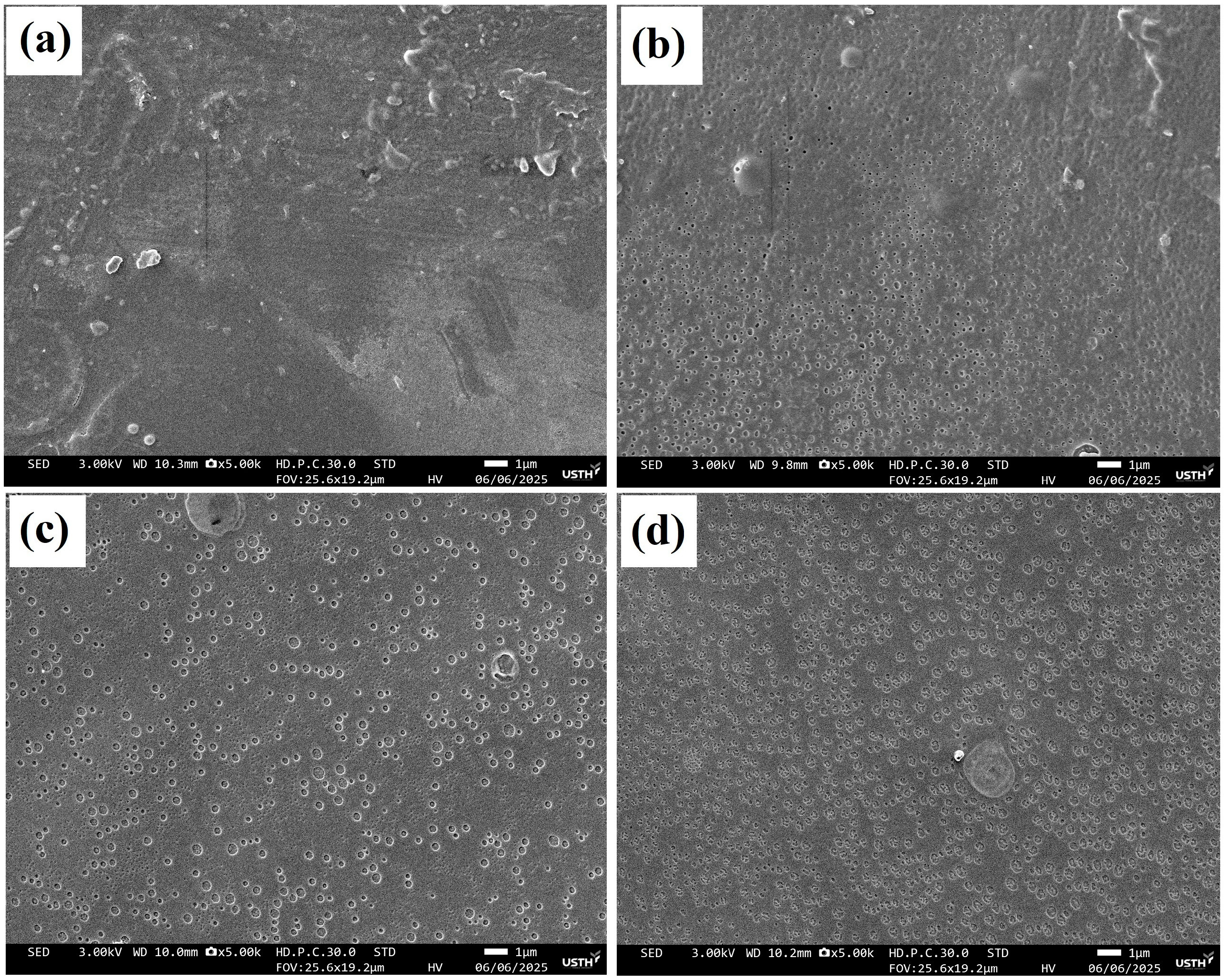
The SEM images of the coating surfaces are presented in Figure 4. The surface of the control sample (without additive) appears relatively smooth but still exhibits some irregular defects and heterogeneities (Figure 4a). Compared to the control, the coatings containing P. betle extract showed a more uniform surface morphology. The extract was relatively well dispersed within the polymer matrix; however, it did not dissolve but instead formed microdroplets with diameters smaller than one micrometer (Figure 4b–d). These microdroplets were gradually released when immersed in seawater, acting as antifouling agents on the coating surface. At an extract concentration of 1.5 wt.%, the surface exhibits a noticeable increase in the number and size of voids or dispersed domains (Figure 4d). This phenomenon indicates possible phase incompatibility or oversaturation of the extract, which may lead to disruption of the polymer matrix. The increased porosity and heterogeneity will likely result in a higher self-polishing rate and reduced structural stability, consistent with previous experimental findings.
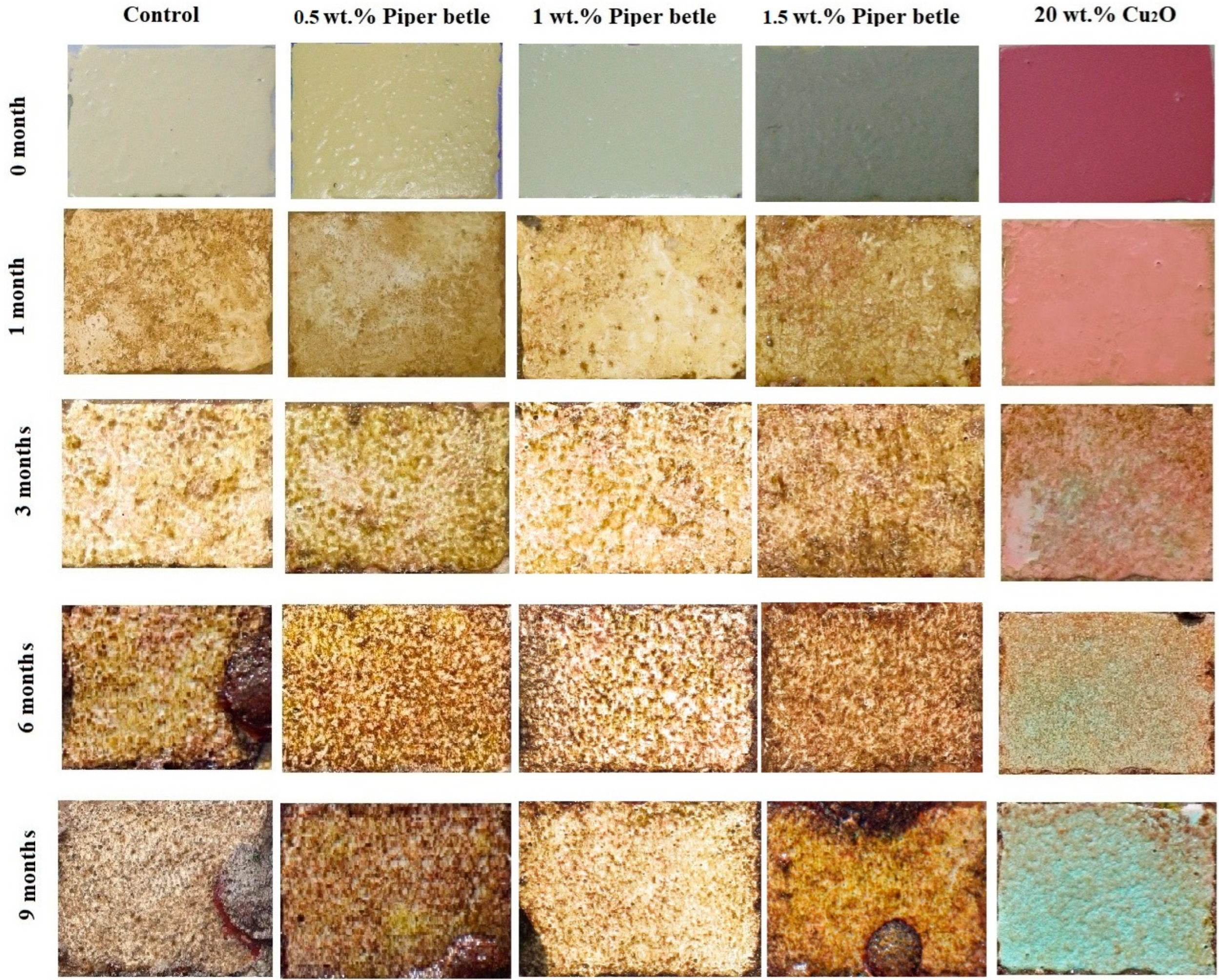
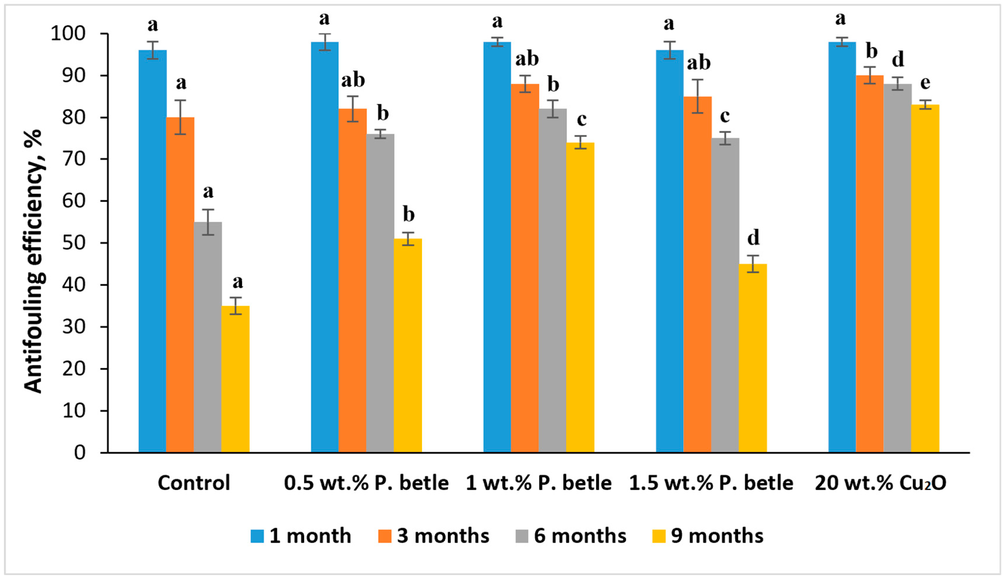
The results of the natural field test are presented in Figure 5 and Figure 6. After 6 months of immersion, the coating sample containing 1.0 wt.% P. betle extract demonstrated a high antifouling efficiency (over 80%), comparable to the Cu-based biocide sample. No visible macrofouling was observed on its surface. In contrast, the control sample (without antifouling additives) exhibited signs of macrofouling, particularly along the sample edges. However, after 9 months of immersion, the antifouling performance of P. betle-based coatings declined significantly compared to the Cu2O-based biocide sample. Only the coating containing 1 wt.% of P. betle extract maintained a relatively high antifouling efficiency (74%). In contrast, the efficiency of coatings containing 0.5 wt.% and 1.5 wt.% extract dropped below 50% (Figure 6), and macrofouling appeared on the surface (Figure 5). For the sample containing 0.5 wt.% extract, the reduced antifouling performance may be attributed to insufficient active compounds remaining in the coating matrix over time. In the case of the 1.5 wt.% sample, the high self-polishing rate may have led to an accelerated release of the active antifouling agents from the coating surface, causing a rapid decline in antifouling effectiveness. No delamination or cracking was observed on the surface of the coatings (Figure 5).
Several review articles have been published in recent years describing natural compounds isolated from various organisms with antifouling properties [44,45,46]. These reviews cover over 700 natural compounds and over 100 synthetic analogues identified and synthesized between 2004 and 2020. However, it should be noted that, to identify effective antifouling compounds, laboratory studies must be complemented by subsequent verification of antifouling performance under natural marine conditions. Thus, only a few of the 700 known compounds with antifouling properties have been tested under complete immersion in seawater when incorporated into coating formulations. Table 5 presents the literature sources that report data on natural seawater tests of the antifouling activity of natural compounds (or extracts) incorporated into coating formulations.
One of the main limitations of antifouling coatings utilizing plant-derived bio-extracts is the difficulty in maintaining long-term antifouling efficacy. This issue is closely related to the inability to control the sustained release rate of the active antifouling agents from the coating surface. Moreover, the compatibility between the plant extract and the polymer matrix plays a crucial role in determining the durability of the coating under seawater immersion. To address this challenge, current research efforts have focused on several strategies, such as encapsulating the extracts into microcapsules [55] or modifying the coating matrix to regulate the release kinetics of the bioactive compounds [56].

4. Conclusions

This study investigated the composition and antibacterial activity of P. betle extract. The extract was incorporated into acrylic copolymer-based antifouling coatings to evaluate its potential as a natural bio-based additive. The results demonstrated that the P. betle extract contains high levels of bioactive compounds such as phenolics and flavonoids, exhibiting strong antibacterial properties. The extract showed significant antibacterial activity against representative bacterial strains and marine isolates. Incorporation of the ethanol extract of P. betle into the coating formulation at a concentration of 1 wt.% enhanced the antifouling performance of the coating under tropical marine conditions. These findings highlight P. betle extract as a sustainable alternative to copper-based agents and justify further work on controlled-release strategies to extend long-term performance.

Author Contributions

Conceptualization, N.D.A. and C.N.L.; methodology, C.N.L. and L.T.M.H.; investigation, N.D.A.; data curation, L.T.M.H. and D.V.K.; writing—original draft preparation, N.D.A.; writing—review and editing, C.N.L.; visualization, D.V.K. All authors have read and agreed to the published version of the manuscript.

Funding

This research was funded by the Coastal Branch of the Joint Vietnam–Russia Tropical Science and Technology Research Center grant number (VB.Đ2.05/24).

Institutional Review Board Statement

Not applicable.

Informed Consent Statement

Not applicable.

Data Availability Statement

The original contributions presented in this study are included in the article. Further inquiries can be directed to the corresponding author.

Acknowledgments

We would like to thank the Coastal Branch of the Joint Vietnam–Russia Tropical Science and Technology Research Center for always providing complete support with equipment and laboratories so the research team can work smoothly.

Conflicts of Interest

The authors declare no conflicts of interest.

References

  1. Shi, X.; Wei, H.; Zhou, W.; Rodriguez, P.E.D.S.; Lin, C.; Wang, L.; Zhang, Z. Advanced strategies for marine antifouling based on nanomaterial-enhanced functional PDMS coatings. Nano Mater. Sci. 2024, 6, 375–395. [Google Scholar] [CrossRef]
  2. Li, Z.; Liu, P.; Chen, S.; Liu, X.; Yu, Y.; Li, T.; Wan, Y.; Tang, N.; Liu, Y.; Gu, Y. Bioinspired marine antifouling coatings: Antifouling mechanisms, design strategies and application feasibility studies. Eur. Polym. J. 2023, 190, 111997. [Google Scholar] [CrossRef]
  3. Zang, X.; Ni, Y.; Wang, Q.; Cheng, Y.; Huang, J.; Cao, X.; Carmalt, C.J.; Lai, Y.; Kim, D.H.; Liu, Y.; et al. Non-toxic evolution: Advances in multifunctional antifouling coatings. Mater. Today 2024, 75, 210–243. [Google Scholar] [CrossRef]
  4. Srinivasan, M.; Swain, G.W. Managing the Use of Copper-Based Antifouling Paints. Environ. Manag. 2007, 39, 423–441. [Google Scholar] [CrossRef]
  5. Ding, T.; Xu, L.; Liu, X.; Ma, L.; Cui, Y.; Li, D.; Sun, X.; Gao, C. A Cu2O-based marine antifouling coating with controlled release of copper ion mediated by amphiphilic PLMA-b-PDMAEMA copolymers. Prog. Org. Coat. 2022, 170, 107003. [Google Scholar] [CrossRef]
  6. Ansanelli, G.; Manzo, S.; Parrella, L.; Massanisso, P.; Chiavarini, S.; Landa, G.D.; Ubaldi, C.; Cannarsa, S.; Cremisini, C. Antifouling biocides (Irgarol, Diuron and dichlofluanid) along the Italian Tyrrhenian coast: Temporal, seasonal and spatial threats. Reg. Stud. Mar. Sci. 2017, 16, 254–266. [Google Scholar] [CrossRef]
  7. Montemor, M.F.B. Smart Composite Coatings and Membranes: Transport, Structural, Environmental and Energy Applications, 1st ed.; Wood. Pub.: Cambridge, UK, 2016; 490p. [Google Scholar]
  8. Chen, L.; Duan, Y.; Cui, M.; Huang, R.; Su, R.; Qi, W.; He, Z. Biomimetic surface coatings for marine antifouling: Natural antifoulants, synthetic polymers and surface microtopography. Sci. Total. Environ. 2021, 766, 144469. [Google Scholar] [CrossRef]
  9. Carteau, D.; Vallée-Réhel, K.; Linossier, I.; Quiniou, F.; Davy, R.; Compère, C.; Delbury, M.; Faÿ, F. Development of environmentally friendly antifouling paints using biodegradable polymer and lower toxic substances. Prog. Org. Coat. 2014, 77, 485–493. [Google Scholar] [CrossRef]
  10. Ma, C.; Zhang, W.; Zhang, G.; Qian, P. Environmentally Friendly Antifouling Coatings Based on Biodegradable Polymer and Natural Antifoulant. Sustain. Chem. Eng. 2017, 5, 6304–6309. [Google Scholar] [CrossRef]
  11. Sultana, H.; Chetia, A.; Saikia, A.; Khan, N. An Updated Review on Extraction, Isolation, and Identification of Bioactive Compounds from Plant Extracts. Sch. Acad. J. Pharm. 2023, 12, 2320–4206. [Google Scholar] [CrossRef]
  12. Awad, A.M.; Kumar, P.; Ismail-Fitry, M.R.; Jusoh, S.; Ab Aziz, M.F.; Sazili, A.Q. Green Extraction of Bioactive Compounds from Plant Biomass and Their Application in Meat as Natural Antioxidant. Antioxidants. 2021, 10, 1465. [Google Scholar] [CrossRef] [PubMed]
  13. Etoh, H.; Kondoh, T.; Noda, R.; Singh, I.P.; Sekiwa, Y.; Morimitsu, K.; Kubota, K. Shogaols from Zingiber ocinale as Promising Antifouling Agents. Biosci. Biotechnol. Biochem. 2002, 66, 1748–1750. [Google Scholar] [CrossRef]
  14. Liu, H.; Chen, S.Y.; Guo, J.Y.; Su, P.; Qiu, Y.K.; Ke, C.H.; Feng, D.Q. Effective natural antifouling compounds from the plant Nerium oleander and testing. Int. Biodeterior. Biodegrad. 2018, 127, 170–177. [Google Scholar] [CrossRef]
  15. Qian, P.Y.; Xu, Y.; Fusetani, N. Natural products as antifouling compounds: Recent progress and future perspectives. Biofouling 2010, 26, 223–234. [Google Scholar] [CrossRef]
  16. An, X.; Yang, X.; Dong, W.; Ni, C.; Jiang, X. Synthesis and fouling resistance of capsaicin derivatives containing amide groups. Chem. Phys. Lett. 2022, 808, 139824. [Google Scholar] [CrossRef]
  17. Qin, Y.; Xue, J.; Wang, S.; Fan, Y.; Wang, L.; Xu, J.; Zhao, J. Capsaicin-based silicone antifouling coating with enhanced interlocking adhesion via SIPN. Colloids Surfaces A Physicochem. Eng. Asp. 2023, 667, 132346. [Google Scholar] [CrossRef]
  18. Mohd Ramzi, M.; Rahman, N.I.A.; Rawi, N.N.; Bhubalan, K.; Ariffin, F.; Mazlan, N.W.; Saidin, J.; Danish-Daniel, M.; Siong, J.Y.F.; Bakar, K.; et al. Antifouling Potential of Diadema setosum and Sonneratia lanceolata Extracts for Marine Applications. J. Mar. Sci. Eng. 2023, 11, 602. [Google Scholar] [CrossRef]
  19. Madhumita, M.; Guha, P.; Nag, A.B. Processing and potential health benefits of betel leaf (Piper betle L.). In Herbal Medicine in India, 2nd ed.; Sen, S., Chakraborty, A., Eds.; Springer: Singapore, 2020; pp. 237–246. [Google Scholar]
  20. Syahidah, A.; Saad, C.R.; Hassan, M.D.; Rukayadi, Y.; Norazian, M.H.; Kamarudin, M.S. Phytochemical analysis, identification and quantification of antibacterial active compounds in betel leaves, piper betle methanolic extract. Pak. J. Biol. Sci. 2017, 20, 70–81. [Google Scholar] [CrossRef] [PubMed]
  21. Biswal, S. Phytochemical analysis and a study on the antiestrogenic antifertility effect of leaves of Piper betel in female albino rat. Anc. Sci. Life. 2014, 34, 16. [Google Scholar] [CrossRef]
  22. Rashida, M.; Islam, I.; Haque, A.; Rahman, A.; Hossain, T.; Hamid, A. Antibacterial Activity of Polyaniline Coated Silver Nanoparticles Synthesized from Piper Betle Leaves Extract. Iran. J. Pharm. Res. 2016, 15, 591–597. [Google Scholar]
  23. Phumat, P.; Khongkhunthian, S.; Wanachantararak, P.; Okonogi, S. Potential of Piper Betle Extracts on Inhibition of Oral Pathogens. Drug Discov. 2017, 11, 307–331. [Google Scholar] [CrossRef]
  24. Sivareddy, B.; Reginald, B.A.; Sireesha, D.; Samatha, M.; Reddy, K.H.; Subrahamanyam, G. Antifungal Activity of Solvent Extracts of Piper Betle and Ocimum Sanctum Linn on Candida Albicans: An in Vitro Comparative Study. J. Oral Maxillofac. Pathol. 2019, 23, 333–337. [Google Scholar] [CrossRef]
  25. Valle, D.L.; Andrade, J.I.; Puzon, J.J.M.; Cabrera, E.C.; Rivera, W.L. Antibacterial Activities of Ethanol Extracts of Philippine Medicinal Plants against Multidrug-Resistant Bacteria. Asian Pac. J. Trop. Biomed. 2015, 5, 532–540. [Google Scholar] [CrossRef]
  26. Kurnia, D.; Hutabarat, G.S.; Windaryanti, D.; Herlina, T.; Herdiyati, Y.; Satari, M.H. Potential Allylpyrocatechol Derivatives as Antibacterial Agent Against Oral Pathogen of S. Sanguinis ATCC 10,556 and as Inhibitor of MurA Enzymes: In Vitro and in Silico Study. Drug Des. Dev. Ther. 2020, 14, 2977–2985. [Google Scholar] [CrossRef] [PubMed]
  27. Pavarish, J.; Varomyalin, T.; Warapond, W.; Nuvee, P.; Monton, V.; Dennapa, S.S. Potential natural antimicrobial and antibiofilm properties of Piper betle L. against Staphylococcus pseudintermedius and methicillin-resistant strains. J. Ethnopharmacol. 2023, 317, 116820. [Google Scholar] [CrossRef] [PubMed]
  28. Kulnanan, P.; Chuprom, J.; Thomrongsuwannakij, T.; Romyasamit, C.; Sangkanu, S.; Manin, N.; Nissapatorn, V.; Pereira, M.d.L.; Wilairatana, P.; Kitpipit, W.; et al. Antibacterial, antibiofilm, and anti-adhesion activities of Piper betle leaf extract against Avian pathogenic Escherichia coli. Arch. Microbiol. 2022, 204, 49. [Google Scholar] [CrossRef]
  29. Jaradat, N.; Hussen, F.; Al Ali, A. Preliminary phytochemical screening, quantitative estimation of total flavonoids, total phenols and antioxidant activity of Ephedra alata Decne. J. Mater. Environ. Sci. 2015, 6, 1771–1778. [Google Scholar]
  30. Singleton, V.L.; Orthofer, R.; Lamuela-Raventós Rosa, M. Analysis of total phenols and other oxidation substrates and antioxidants by means of folin-ciocalteu reagent. Methods Enzymol. 1999, 299, 152–178. [Google Scholar]
  31. Chang, C.C.; Yang, M.H.; Wen, H.M.; Chern, J.-C. Estimation of total flavonoid content in propolis by two complementary colorimetric methods. J. Food Drug Anal. 2002, 10, 178–182. [Google Scholar]
  32. Wang, D.; Xu, J.; Yang, J.; Zhou, S. Preparation and synergistic antifouling effect of self-renewable coatings containing quaternary ammonium-functionalized SiO2 nanoparticles. J. Colloid Interface Sci. 2020, 563, 261–271. [Google Scholar] [CrossRef]
  33. Silva, E.R.; Ferreira, O.; Ramalho, P.A.; Azevedo, N.F.; Bayón, R.; Igartua, A.; Bordado, J.C.; Calhorda, M.J. Eco-friendly non-biocide-release coatings for marine biofouling prevention. Sci. Total. Environ. 2019, 650, 2499–2511. [Google Scholar] [CrossRef]
  34. Asharani, I.V.; Badmapriya, D. Dye degradation studies catalysed by green sythesized Iron oxide nanoparticles. Int. J. ChemTech Res. 2016, 9, 409–416. [Google Scholar]
  35. Kaveti, B.; Tan, L.; Sarnnia, K.T.S.; Baig, M. Antibacterial Activity of Piper Betel Leaves. Int. J. Pharm. Teach. Pract. 2011, 2, 129–132. [Google Scholar]
  36. Taukoorah, U.; Lall, N.; Mahomoodally, F. Piper betle L. (betel quid) shows bacteriostatic, additive, and synergistic antimicrobial action when combined with conventional antibiotics. S. Afr. J. Bot. 2016, 105, 133–140. [Google Scholar] [CrossRef]
  37. Periyanayagam, K.; Jagadeesan, M.; Kavimani, S.; Vetriselvan, T. Pharmacognostical and Phyto-physicochemical profile of the leaves of Piper betle L. var Pachaikodi (Piperaceae)-Valuable assessment of its quality. Asian Pac. J. Trop. Biomed. 2012, 2, 506–510. [Google Scholar] [CrossRef]
  38. Nayaka, N.M.D.M.W.; Sasadara, M.M.V.; Sanjaya, D.A.; Yuda, P.E.S.K.; Dewi, N.L.K.A.A.; Cahyaningsih, E.; Hartati, R. Piper betle L.: Recent review of antibacterial and antifungal properties, safety profiles, and commercial applications. Molecules 2021, 26, 2321. [Google Scholar] [CrossRef]
  39. Nguyen, L.T.T.; Nguyen, T.T.; Nguyen, H.N.; Bui, T.Q.P. Simultaneous determination of active compounds in Piper betle Linn. leaf extract and effect of extracting solvents on bioactivity. Eng. Rep. 2020, 2, e12246. [Google Scholar] [CrossRef]
  40. Prakash, B.; Shukla, R.; Singh, P.; Kumar, A.; Mishra, P.K.; Dubey, N.K. Efficacy of Chemically Characterized Piper betle L. Essential Oil against Fungal and Aflatoxin Contamination of Some Edible Commodities and Its Antioxidant Activity. Int. J. Food Microbiol. 2010, 142, 114–119. [Google Scholar] [CrossRef]
  41. Salehi, B.; Zakaria, Z.A.; Gyawali, R.; Ibrahim, S.A.; Rajkovic, J.; Shinwar, Z.K.; Khan, T.; Sharifi-Rad, J.; Ozleyen, A.; Turkdonmez, E. Piper Species: A Comprehensive Review on Their Phytochemistry, Biological Activities and Applications. Molecules 2019, 24, 1364. [Google Scholar] [CrossRef]
  42. Tan, Y.P.; Chan, E.W.C. Antioxidant, antityrosinase and antibacterial properties of fresh and processed leaves of Anacardium occidentale and Piper betle. Food Biosci. 2014, 6, 17–23. [Google Scholar] [CrossRef]
  43. Md, S.A.; Byung, S.C. Characterization of chitosan-based packaging film incorporating betel leaves phenolic compounds recovered via subcritical water extraction. Food Biosci. 2025, 65, 106125. [Google Scholar] [CrossRef]
  44. Qian, P.Y.; Li, Z.; Xu, Y.; Li, Y.; Fusetani, N. Mini-review: Marine natural products and their synthetic analogs as antifouling compounds: 2009–2014. Biofouling 2015, 31, 101–122. [Google Scholar] [CrossRef]
  45. Wang, K.L.; Wu, Z.H.; Wang, Y.; Wang, C.Y.; Xu, Y. Mini-review: Antifouling natural products from marine microorganisms and their synthetic analogs. Mar. Drugs 2017, 15, 266. [Google Scholar] [CrossRef] [PubMed]
  46. Liu, L.L.; Wu, C.H.; Qian, P.Y. Marine natural products as antifouling molecules—A mini-review (2014–2020). Biofouling 2020, 36, 1210–1226. [Google Scholar] [CrossRef]
  47. Sjögren, M.; Dahlström, M.; Göransson, U.; Jonsson, P.R.; Bohlin, L. Recruitment in the field of Balanus improvisus and Mytilus edulis in response to the antifouling cyclopeptides barettin and 8,9-dihydrobarettin from the marine sponge Geodia barretti. Biofouling 2004, 20, 291–297. [Google Scholar] [CrossRef]
  48. Stupak, M.E.; Garcia, M.T.; Perez, M.C. Non-toxic alternative compounds for marine antifouling paints. Int. Biodeter. Biodegrad. 2003, 52, 49–52. [Google Scholar] [CrossRef]
  49. Zmozinski, A.V.; Peres, R.S.; Brust, F.R.; Macedo, A.J.; Becker, E.M.; Napp, A.P.; de Brito, H.A.; Vainstein, M.H.; Schrank, A.; Ferreira, C.A. The effect of rue (Ruta graveolens) and ginger (Zingiber officinale) extracts as antifouling agents in silicone matrix coatings. J. Coat. Technol. Res. 2021, 18, 1013–1025. [Google Scholar] [CrossRef]
  50. Feng, D.Q.; He, J.; Chen, S.Y.; Su, P.; Ke, C.H.; Wang, W. The plant alkaloid camptothecin as a novel antifouling compound for marine paints: Laboratory bioassays and field trials. Mar. Biotechnol. 2018, 20, 623–638. [Google Scholar] [CrossRef] [PubMed]
  51. Peres, R.S.; Armelin, E.; Alemán, C.; Ferreira, C.A. Modified tannin extracted from black wattle tree as an environmentally friendly antifouling pigment. Ind. Crop. Prod. 2015, 65, 506–514. [Google Scholar] [CrossRef]
  52. Noor, I.M.S.; Ferry, M.; Wan, N.W.B.; Jasnizat, S. Evaluation of tannin from Rhizophora apiculata as natural antifouling agents in epoxy paint for marine application. Prog. Org. Coat. 2015, 81, 125–131. [Google Scholar] [CrossRef]
  53. Pérez, M.; García, M.; Sánchez, M.; Stupak, M.; Mazzuca, M.; Palermo, J.A.; Blustein, G. Effect of secochiliolide acid isolated from the Patagonian shrub Nardophyllum bryoides as active component in antifouling paints. Int. Biodeter. Biodegrad. 2014, 89, 37–44. [Google Scholar] [CrossRef]
  54. Wang, X.; Yu, L.; Liu, Y.; Jiang, X. Synthesis and fouling resistance of capsaicin derivatives containing amide groups. Sci. Total Environ. 2020, 710, 136361. [Google Scholar] [CrossRef]
  55. Li, Y.; Wang, G.; Guo, Z.; Wang, P.; Wang, A. Preparation of Microcapsules Coating and the Study of Their Bionic Anti-Fouling Performance. Materials 2020, 13, 1669. [Google Scholar] [CrossRef] [PubMed]
  56. Lu, Z.; Chen, Z.; Guo, Y.; Ju, Y.; Liu, Y.; Feng, R.; Xiong, C.; Ober, C.K.; Dong, L. Flexible Hydrophobic Antifouling Coating with Oriented Nanotopography and Nonleaking Capsaicin. ACS Appl. Mater. Interfaces 2018, 10, 9718–9726. [Google Scholar] [CrossRef] [PubMed]
Figure 1. FTIR spectra of P. betle extracts obtained using different solvents.
Surfaces 08 00072 g001
Figure 2. Inhibition zone of P. betle extracts against E. coli (a), B. subtilis (b), S. aureus (c), and marine bacteria (d). Symbols ‘+’ and ‘–’ indicate positive and negative controls, respectively.
Surfaces 08 00072 g002
Figure 3. Contact angle of acrylic-based coatings: (a)—control coating without extract, (b)—coating with 0.5 wt.%, (c)—coating with 1 wt.%, and (d)—coating with 1.5 wt.% P. betle extract.
Surfaces 08 00072 g003
Figure 4. SEM images of the surface of acrylic-based coatings: (a)—control coating without extract, (b)—coating with 0.5 wt.%, (c)—coating with 1 wt.%, and (d)—coating with 1.5 wt.% P. betle extract.
Surfaces 08 00072 g004
Figure 5. Visual observation of biofouling accumulation on coating surfaces during marine immersion.
Surfaces 08 00072 g005
Figure 6. Antifouling efficiency of coatings after 9 months of immersion in Nha Trang Bay. Data are presented as mean ± SD (n = 3). Columns labeled with different letters indicate significant differences at p < 0.05.
Surfaces 08 00072 g006
Table 1. Preliminary phytochemical screening of P. betle extract obtained using ethanol.
Phytochemical GroupPresence (+/−) 1
Phenolics+
Tannins+
Proteins+
Carbohydrates+
Flavonoids+
1Note: (+) detected; (−) not detected.
Table 2. Total phenolic and flavonoid contents in the ethanol extract of P. betle. Data are presented as mean ± SD (n = 3).
ComponentResults
Total phenolic content, mg GAE/g dry weight260.3 ± 5.22
Total flavonoid content, mg QE/g dry weight52.56 ± 1.26
Table 3. Antibacterial activity of P. betle extracts against E. coli, B. subtilis, S. aureus, and marine bacteria. Data are presented as mean ± SD (n = 5).
ExtractsDiameter of Inhibition Zone (mm)
E. coliB. subtilisS. aureusMarine bacteria
Positive control15.0 ± 0.511.4 ± 0.240.1 ± 0.642.3 ± 0.4
Ethanol extract28.7 ± 0.527.0 ± 1.622.1 ± 0.635.1 ± 0.5
Methanol extract23.3 ± 0.420.0 ± 0.218.1 ± 1.033.0 ± 0.5
Ethyl acetate extract15.7 ± 1.812.3 ± 0.79.1 ± 1.020.0 ± 1.5
Table 4. Value of contact angle and self-polishing rates of antifouling coatings containing different P. betle ethanol extract concentrations. Data are presented as mean ± SD (n = 3).
Extract Concentration (wt.%)Contact Angle (o)Self-Polishing Rate (mg/cm2·day)
052.8 ± 1.950.470 ± 0.012
0.556.6 ± 1.570.351 ± 0.011
1.065.8 ± 1.030.393 ± 0.015
1.573.2 ± 1.490.560 ± 0.018
Table 5. Some natural products incorporated into antifouling coatings.
Source of Natural CompoundsTest Duration and LocationReferences
Barettein and 8,9-dehydrobarettein were isolated from a marine sponge2 months in seawater (near Sweden)[47]
Tannins from plant extracts (chestnut, mimosa, quebracho)4 months (Atlantic Ocean, near Argentina)[48]
Rue (Ruta graveolens) and ginger (Zingiber officinale) extracts6 months (Atlantic Ocean, near Brazil)[49]
Plant alkaloid camptothecin from Camptotheca acuminata11 months (near Dalipuyu Islet in Xiamen Bay)[50]
Plant extract of Nerium oleander1 month (Lingshui
Bay, China)
[14]
Modified tannin derived from black acacia7 months (Mediterranean Sea, Spain)[51]
Tannins from mangrove trees (Rhizophora apiculata)3 months (Chendering Port, Malaysia)[52]
Plant extract of Nardophyllum bryoides1.5 months (Mar del
Plata harbor, Argentina)
[53]
Capsaicin and its derivatives from Capsicum annuum3 months (Qingdao Bay, China)[54]
Disclaimer/Publisher’s Note: The statements, opinions and data contained in all publications are solely those of the individual author(s) and contributor(s) and not of MDPI and/or the editor(s). MDPI and/or the editor(s) disclaim responsibility for any injury to people or property resulting from any ideas, methods, instructions or products referred to in the content.