Morphology of Dried Drop Patterns of Saliva from a Healthy Individual Depending on the Dynamics of Its Surface Tension
Abstract
1. Introduction
2. Materials and Methods
2.1. Participants
2.2. Collection, Processing and Storage of Saliva Samples
2.3. Crystallization of Saliva Samples
2.4. Surface Tension Measurements
2.5. Biochemical Analysis of Saliva
2.6. Statistical Methods
3. Results
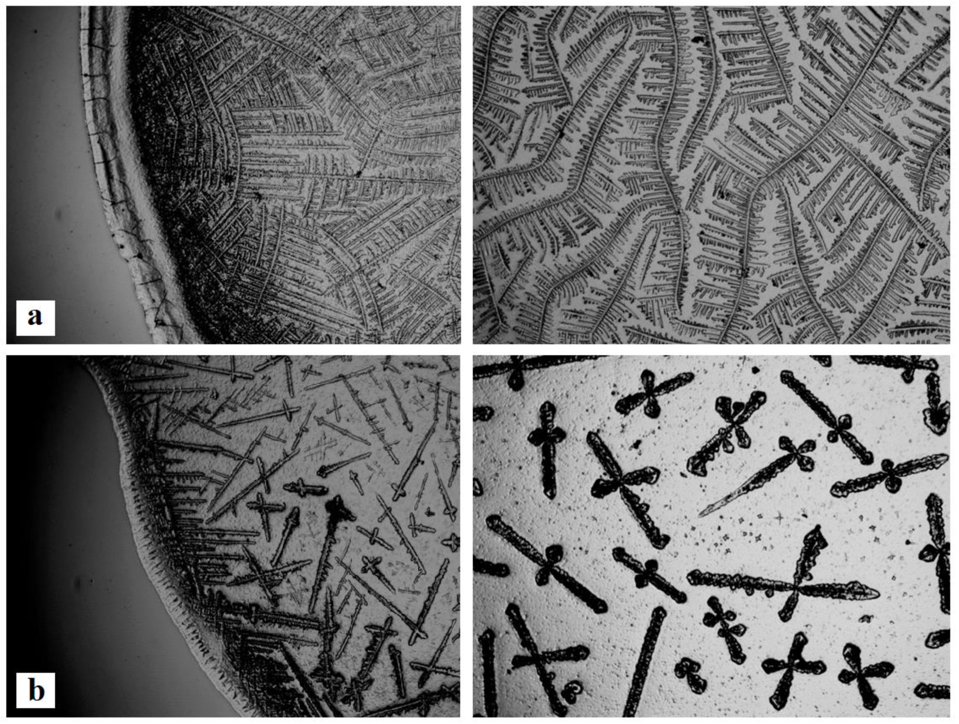
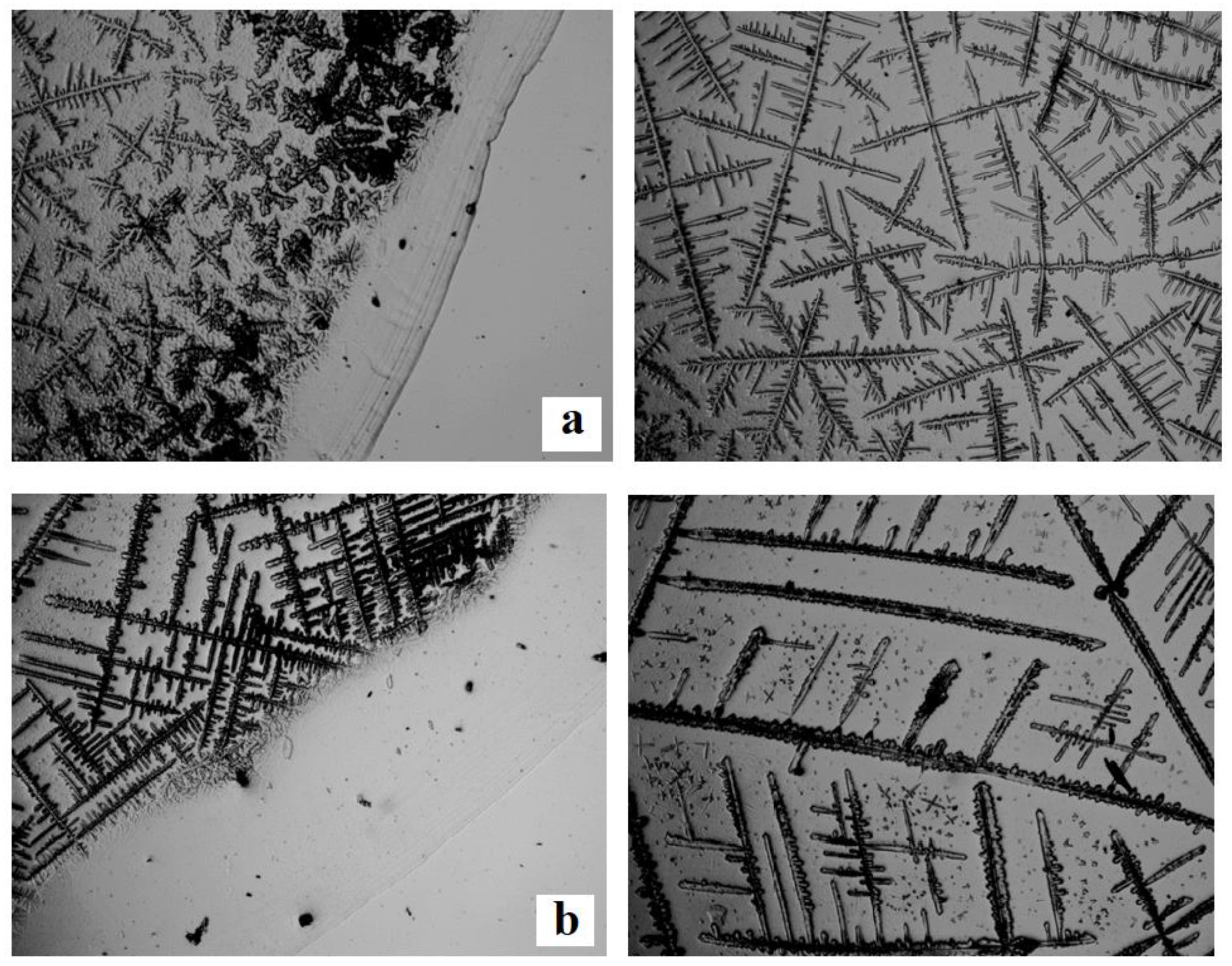
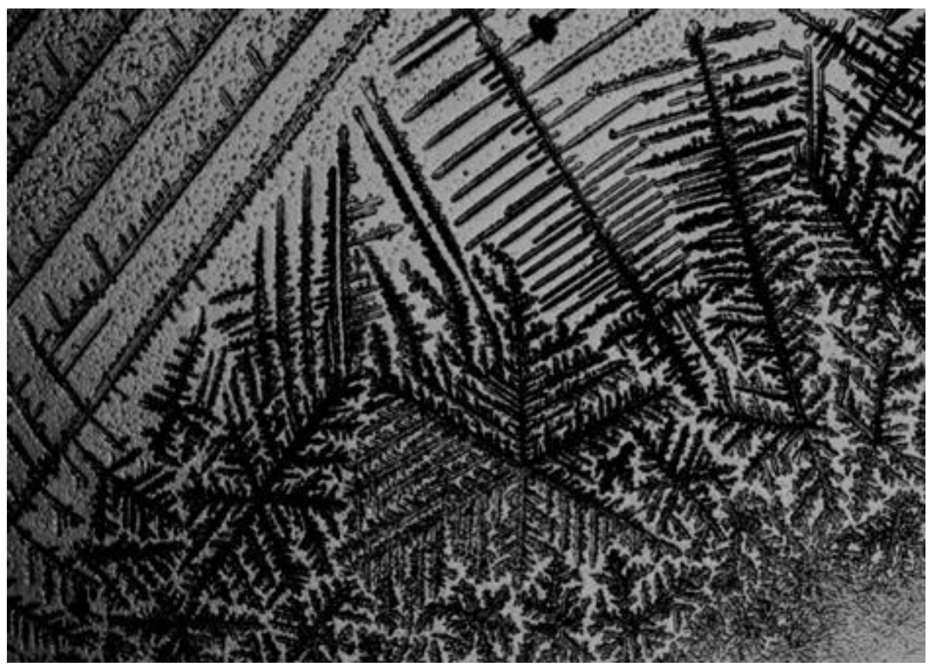
3.1. Zones in the Dried Drop Patterns of Saliva
3.2. The Influence of Gender and Age Characteristics on Morphological Features of Dried Drop Patterns of Saliva
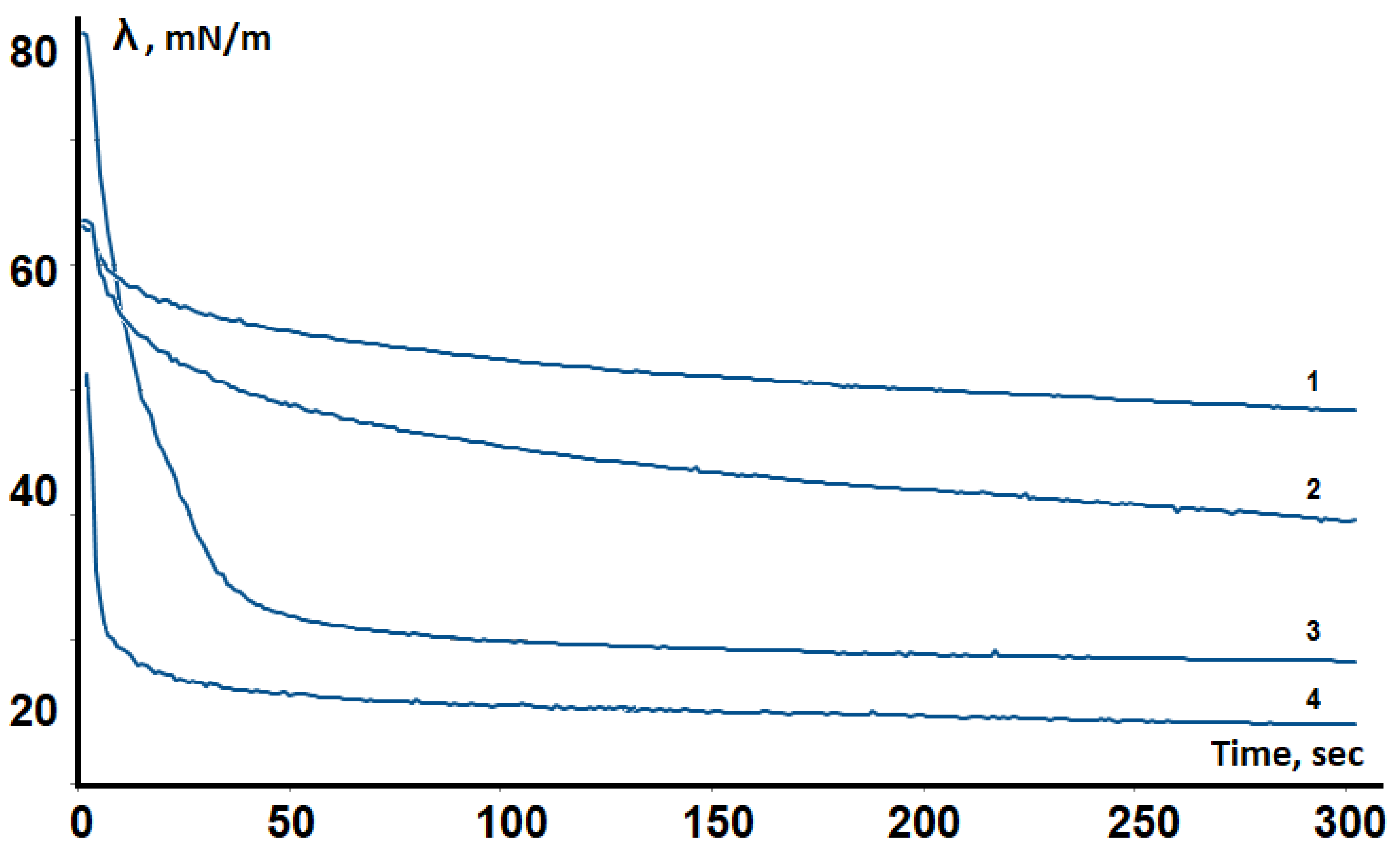
3.3. The Relationship of Surface Tension and Morphological Features of Dried Drop Patterns of Saliva
4. Discussion
5. Conclusions
Author Contributions
Funding
Conflicts of Interest
References
- Bahmani, L.; Neysari, M.; Maleki, M. The study of drying and pattern formation of whole human blood drops and the effect of thalassaemia and neonatal jaundice on the patterns. Colloids Surf. A Phys. Eng. Asp. 2017, 513, 66–75. [Google Scholar] [CrossRef]
- Chen, R.; Zhang, L.; Zang, D.; Shen, W. Blood drop patterns: Formation and applications. Adv. Colloid Interface Sci. 2016, 231, 1–14. [Google Scholar] [CrossRef] [PubMed]
- Kazakov, V.N.; Vozianov, A.F.; Sinyachenko, O.V.; Trukhin, D.V.; Kovalchuk, V.I.; Pison, U. Studies on the application of dynamic surface tensiometry of serum and cerebrospinal liquid for diagnostics and monitoring of treatment in patients who have rheumatic, neurological or oncological diseases. Adv. Colloid Interface Sci. 2000, 86, 1–38. [Google Scholar] [CrossRef]
- Kazakov, V.N.; Udod, A.A.; Zinkovych, I.I.; Fainerman, V.B.; Miller, R. Dynamic surface tension of saliva: General relationships and application in medical diagnostics. Colloids Surf. B Biointerfaces 2009, 74, 457–461. [Google Scholar] [CrossRef] [PubMed]
- Yakhno, T.A.; Kazakov, V.V.; Sanina, O.A.; Sanin, A.G.; Yakhno, V.G. Drops of biological fluids drying on a hard substrate: Variation of the morphology, weight, temperature, and mechanical properties. Tech. Phys. 2010, 55, 929–935. [Google Scholar] [CrossRef]
- Brutin, D.; Sobac, B.; Loquet, B.; Sampol, J. Pattern formation in drying drops of blood. J. Fluid Mech. 2011, 667, 85–95. [Google Scholar] [CrossRef]
- Christy, J.R.E.; Sefiane, K.; Munro, E. A Study of the Velocity Field during Evaporation of Sessile Water and Water/Ethanol Drops. J. Bionic Eng. 2010, 7, 321–328. [Google Scholar] [CrossRef]
- Filik, J.; Stone, N. Analysis of human tear fluid by Raman spectroscopy. Anal. Chim. Acta 2008, 616, 177–184. [Google Scholar] [CrossRef] [PubMed]
- Carpenter, G.H. The Secretion, Components, and Properties of Saliva. Annu. Rev. Food Sci. Technol. 2013, 4, 67–76. [Google Scholar] [CrossRef]
- Lee, Y.H.; Wong, D.T. Saliva: An emerging biofluid for early detection of diseases. Am. J. Dent. 2009, 22, 241–248. [Google Scholar] [PubMed]
- Chojnowska, S.; Baran, T.; Wilińska, I.; Sienicka, P.; Cabaj-Wiater, I.; Knaś, M. Human saliva as a diagnostic material. Adv. Med. Sci. 2018, 63, 185–191. [Google Scholar] [CrossRef]
- Nunes, L.A.; Mussavira, S.; Bindhu, O.S. Clinical and diagnostic utility of saliva as a non-invasive diagnostic fluid: A systematic review. Biochem. Med. 2015, 25, 177–192. [Google Scholar] [CrossRef]
- Kaur, J.; Jacobs, R.; Huang, Y.; Salvo, N.; Politis, C. Salivary biomarkers for oral cancer and pre-cancer screening: A review. Clin. Oral Investig. 2018, 22, 633–640. [Google Scholar] [CrossRef] [PubMed]
- Porto-Mascarenhas, E.C.; Assad, D.X.; Chardin, H.; Gozal, D.; De Luca Canto, G.; Acevedo, A.C.; Guerra, E.N.S. Salivary biomarkers in the diagnosis of breast cancer: A review. Crit. Rev. Oncol. Hematol. 2017, 110, 62–73. [Google Scholar] [CrossRef] [PubMed]
- Sahibzada, H.A.; Khurshid, Z.; Khan, R.S.; Naseem, M.; Siddique, K.M.; Mali, M.; Zafar, M.S. Salivary IL-8, IL-6 and TNF-α as Potential Diagnostic Biomarkers for Oral Cancer. Diagnostics 2017, 7, 21. [Google Scholar] [CrossRef] [PubMed]
- Khan, R.S.; Khurshid, Z.; Asiri, F.Y.I. Advancing Point-of-Care (PoC) Testing Using Human Saliva as Liquid Biopsy. Diagnostics 2017, 7, 39. [Google Scholar] [CrossRef]
- Zhang, Y.; Sun, J.; Lin, C.; Abemayor, E.; Wang, M.B.; Wong, D. The Emerging Landscape of Salivary Diagnostics. Oral Health Dent. Manag. 2014, 13, 200–210. [Google Scholar] [CrossRef] [PubMed]
- Bonne, N.J.; Wong, D.T. Salivary biomarker development using genomic, proteomic and metabolomic approaches. Genome Med. 2012, 4, 82. [Google Scholar] [CrossRef] [PubMed]
- Ghimenti, S.; Lomonaco, T.; Onor, M.; Murgia, L.; Paolicchi; Fuoco, A.R.; Ruocco, L.; Pellegrini, G.; Trivella, M.G.; Di Francesco, F. Measurement of Warfarin in the Oral Fluid of Patients Undergoing Anticoagulant Oral Therapy. PLoS ONE 2011, 6, e28182. [Google Scholar] [CrossRef]
- Lomonaco, T.; Ghimenti, S.; Piga, I.; Biagini, D.; Onor, M.; Fuoco, R.; Paolicchi, A.; Ruocco, L.; Pellegrini, G.; Trivella, M.G.; et al. Monitoring of warfarin therapy: Preliminary results from a longitudinal pilot study. Microchem. J. 2018, 136, 170–176. [Google Scholar] [CrossRef]
- Bellagambi, F.G.; Degano, I.; Ghimenti, S.; Lomonaco, T.; Dini, V.; Romanelli, M.; Mastorci, F.; Gemignani, A.; Salvo, P.; Fuoco, R.; et al. Determination of salivary α-amylase and cortisol in psoriatic subjects undergoing the Trier Social Stress Test. Microchem. J. 2018, 136, 177–184. [Google Scholar] [CrossRef]
- Yakhno, T. Salt-induced protein phase transitions in drying drops. J. Colloid Interface Sci. 2008, 318, 225–230. [Google Scholar] [CrossRef] [PubMed]
- Kubínek, R.; Hozman, J.; Vařenka, J. Dendritic growth in viscous solutions containing organic molecules. J. Cryst. Growth 1998, 193, 174–181. [Google Scholar] [CrossRef]
- Smith, F.R.; Brutin, D. Wetting and spreading of human blood: Recent advances and applications. Curr. Opin. Colloid Interface Sci. 2018, 36, 78–83. [Google Scholar] [CrossRef]
- Sefiane, K. On the Formation of Regular Patterns from Drying Droplets and Their Potential Use for Bio-Medical Applications. J. Bionic Eng. 2010, 7, S82–S93. [Google Scholar] [CrossRef]
- Sefiane, K. Patterns from Drying Drops. Adv. Colloid Interface Sci. 2014, 206, 372–381. [Google Scholar] [CrossRef]
- Gorr, H.M.; Zueger, J.M.; McAdams, D.R.; Barnard, J.A. Salt-induced pattern formation in evaporating droplets of lysozyme solutions. Colloids Surf. B Biointerfaces 2013, 103, 59–66. [Google Scholar] [CrossRef]
- Neyraud, E.; Palicki, O.; Schwartz, C.; Nicklaus, S.; Feron, G. Variability of human saliva composition: Possible relationships with fat perception and liking. Arch. Oral Biol. 2012, 57, 556–566. [Google Scholar] [CrossRef]
- Quintana, M.; Palicki, O.; Lucchi, G.; Ducoroy, P.; Chambon, C.; Salles, C.; Morzel, M. Inter-individual variability of protein patterns in saliva of healthy adults. J. Proteom. 2009, 72, 822–830. [Google Scholar] [CrossRef]
- Lomonaco, T.; Ghimenti, S.; Biagini, D.; Bramanti, E.; Onor, M.; Bellagambi, F.G.; Fuoco, R.; Di Francesco, F. The effect of sampling procedures on the urate and lactate concentration in oral fluid. Microchem. J. 2018, 136, 255–262. [Google Scholar] [CrossRef]
- Lomonaco, T.; Ghimenti, S.; Piga, I.; Biagini, D.; Onor, M.; Fuoco, R.; Di Francesco, F. Influence of Sampling on the Determination of Warfarin and Warfarin Alcohols in Oral Fluid. PLoS ONE 2014, 9, e114430. [Google Scholar] [CrossRef]
- Udod, A.A.; Zinkovich, I.I.; Prokofyeva, T.I. Dynamic tensiometry of oral fluid. Arch. Clin. Exp. Med. 2004, 13, 88–91. [Google Scholar]
- Krishnan, A.; Wilson, A.; Sturgeon, J.; Siedlecki, C.A.; Vogler, E.A. Liquid-vapor interfacial tension of blood plasma, serum and purified protein constituents thereof. Biomaterials 2005, 26, 3445–3453. [Google Scholar] [CrossRef]
- Kazakov, V.N.; Fainerman, V.B.; Kondratenko, P.G.; Elin, A.F.; Sinyachenko, O.V.; Miller, R. Dilational rheology of serum albumin and blood serum solutions as studied by oscillating drop tensiometry. Colloids Surf. B 2008, 62, 77–82. [Google Scholar] [CrossRef] [PubMed]
- Wüstneck, R.; Perez-Gil, J.; Wüstneck, N.; Cruz, A.; Fainerman, V.B.; Pison, U. Interfacial properties of pulmonary surfactant layers. Adv. Colloid Interface Sci 2005, 117, 33–58. [Google Scholar] [CrossRef] [PubMed]
- Fainerman, V.B.; Trukhin, D.V.; Zinkovych, I.I.; Miller, R. Interfacial tensiometry and dilational surface visco-elasticity of biological liquids in medicine. Adv. Colloid Interface Sci. 2018, 255, 34–46. [Google Scholar] [CrossRef] [PubMed]
- Adamczyk, E.; Arnebrant, T.; Glantz, P.O. Time-dependent interfacial tension of whole saliva and saliva-bacteria mixes. Acta Odontol. Scand. 1997, 55, 384–389. [Google Scholar] [CrossRef]
- Bel’skaya, L.V.; Kosenok, V.K.; Sarf, E.A. Chronophysiological features of the normal mineral composition of human saliva. Arch. Oral Biol. 2017, 82, 286–292. [Google Scholar] [CrossRef]
- Dos Santos, D.R.; Souza, R.O.; Dias, L.B.; Ribas, T.B.; Farias de Oliveira, L.C.; Sumida, D.H.; Dornelles, R.C.M.; Nakamune, A.C.S.; Chaves-Neto, A.H. The effects of storage time and temperature on the stability of salivary phosphatases, transaminases and dehydrogenase. Arch. Oral Biol. 2018, 85, 160–165. [Google Scholar] [CrossRef] [PubMed]
- Hu, H.; Larson, R.G. Evaporation of a Sessile Droplet on a Substrate. J. Phys. Chem. B 2002, 106, 1334–1344. [Google Scholar] [CrossRef]
- Brutin, D.; Sobac, B.; Nicloux, C. Influence of Substrate Nature on the Evaporation of a Sessile Drop of Blood. J. Heat Transf. 2012, 134, 061101. [Google Scholar] [CrossRef]
- Garrett, P.R.; Ward, R.D. A reexamination of the measurement of dynamic surface tensions using the maximum bubble pressure method. J. Colloid Interface Sci 1989, 132, 475–490. [Google Scholar] [CrossRef]
- Waterman, H.A.; Blom, C.; Holterman, H.J.; Gravenmade, E.J.; Mellema, J. Rheological properties of human saliva. Arch. Oral Biol. 1988, 33, 589–596. [Google Scholar] [CrossRef]
- Bel’skaya, L.V. Application of capillary electrophoresis for determination of the mineral composition of human saliva. Byulleten Nauki i Praktiki 2017, 15, 132–140. [Google Scholar]
- Dutta, T.; Giri, A.; Choudhury, M.D.; Tarafdar, S. Pattern formation in droplets of starch gels containing NaCl dried on different surfaces. Colloid. Surf. A Phys. Eng. Asp. 2013, 432, 127–131. [Google Scholar] [CrossRef]
- Tarafdar, S.; Tarasevich, Y.Y.; Dutta Choudhury, M.; Dutta, T.; Zang, D. Droplet Drying Patterns on Solid Substrates: From Hydrophilic to Superhydrophobic Contact to Levitating Drops. Adv. Condens. Matter Phys. 2018, 5214924. [Google Scholar] [CrossRef]
- Yakhno, T.A.; Sanin, A.G.; Sanina, O.A.; Yakhno, V.G. Dynamics of mechanical properties of drying drops of biological liquids as a reflection of the features of self-organization of their components from nano- to microlevel. Biophysics 2011, 56, 1005–1010. [Google Scholar] [CrossRef]
- Yakhno, T.A. Sodium chloride crystallization from drying drops of albumin–salt solutions with different albumin concentrations. Tech. Phys. 2015, 60, 1601–1608. [Google Scholar] [CrossRef]
- Milyukov, V.Y.; Zharikova, T.S. Criteria for the formation of age groups of patients in medical research. Klinicheskaya Meditsina 2015, 11, 5–11. [Google Scholar]
- Rykke, M.; Young, A.; Devold, T.; Smistad, G.; Rolla, G. Fractionation of salivary micelle-like structures by gel chromatography. Eur. J. Oral Sci. 1997, 10, 495–501. [Google Scholar] [CrossRef]
- Soares, R.V.; Lin, T.; Siqueira, C.C.; Bruno, L.S.; Li, X. Salivary micelles: Identification of complexes containing MG2, sIgA, lactoferrin, amylase, glycosylated proline-rich protein and lysozyme. Arch. Oral Biol. 2004, 49, 337–343. [Google Scholar] [CrossRef]
- Young, A.; Rykke, M.; Rolla, G. Quantitative and qualitative analyses of human salivary micelle-like globules. Acta Odontol. Scand. 1999, 57, 105–110. [Google Scholar] [CrossRef] [PubMed]
- Inoue, H.; Ono, K.; Masuda, W.; Inagaki, T.; Yokota, M.; Inenaga, K. Rheological properties of human saliva and salivary mucins. J. Oral Biosci. 2008, 50, 134–141. [Google Scholar] [CrossRef]
- Yakhno, T.A.; Kazakov, V.V.; Sanin, A.G.; Shaposhnikova, O.B.; Chernov, A.S. A comparative assessment of mechanical properties of adsorption layers in the human blood serum protein solutions. J. Tech. Phys. 2007, 77, 119–122. [Google Scholar] [CrossRef]
- Kazakov, V.N.; Sinyachenko, O.V.; Fainerman, V.B.; Pison, U.; Miller, R. Dynamic surface tension of biological fluids in healthy people. Arch. Clin. Exp. Med. 1996, 5, 3–6. [Google Scholar]
- Kazakov, V.N.; Sinyachenko, O.V.; Postovaya, M.V. Interphase tensiometry of biological fluids: Theories, methods and perspective of application in medicine. Arch. Clin. Exp. Med. 1998, 7, 5–12. [Google Scholar]
- Fedorova, A.A.; Ulitin, M.V. Surface tension and adsorption of electrolytes in the solution-gas interphase. J. Phys. Chem. 2007, 81, 1279–1281. [Google Scholar]
- Chernysheva, M.G.; Ivanov, R.A.; Soboleva, O.A.; Badun, G.A. Do low surfactants concentrations change lysozyme colloid properties? Colloids Surf. A 2013, 436, 1121–1129. [Google Scholar] [CrossRef]
- Fedorova, A.A.; Ulitin, M.V. Thermodynamic characteristics of the process of hydrogen, potassium and sodium chloride adsorption in the solution-gas interphase. J. Phys. Chem. 2009, 83, 113–118. [Google Scholar]
- Zaitsev, S.Y. Interphase tensiometry method for comparative analysis of model systems and blood as the most important biological fluid. Her. Mosc. State Univ. Ser. 2 Chem. 2016, 57, 198–202. [Google Scholar]


| Parameters | 1 Type of Crystallization n = 82 | 2 Type of Crystallization n = 17 | 3 Type of Crystallization n = 1 |
|---|---|---|---|
| Scr, % | 88.9 [78.2; 92.9] | 54.9 [48.9; 60.9] | 12.4 |
| - | p = 0.0000 | - | |
| Tensiometric parameters | |||
| γ0.01, mN/m | 63.19 [60.01; 68.47] | 65.61 [62.74; 67.44] | 73.56 |
| γ1.0, mN/m | 60.26 [56.27; 64.26] | 62.42 [57.90; 65.39] | 71.77 |
| γmax, mN/m | 54.78 [49.12; 58.23] | 54.49 [50.77; 58.27] | 65.47 |
| γ∞, mN/m | 46.35 [35.22; 50.34] | 42.48 [34.95; 47.39] | 53.83 |
| Biochemical parameters | |||
| pH | 6.61 [6.47; 6.77] | 6.79 [6.51; 6.99] | 6.88 |
| Calcium, mmol/L | 1.11 [0.77; 1.47] | 0.96 [0.75; 1.48] | 0.21 |
| Phosphorus, mmol/L | 4.70 [3.95; 5.61] | 4.32 [4.05; 4.89] | 3.20 |
| Ca/P | 0.237 [0.196; 0.262] | 0.221 [0.185; 0.303] | 0.066 |
| Sodium, mmol/L | 4.2 [2.9; 5.9] | 3.8 [2.4; 6.5] | 5.4 |
| Potassium, mmol/L | 10.2 [7.4; 13.4] | 9.2 [6.3; 12.1] | 8.0 |
| Na/K | 0.42 [0.39; 0.44] | 0.42 [0.39; 0.53] | 0.68 |
| Chlorides, mmol/L | 18.5 [14.6; 24.3] | 13.8 [13.3; 19.8] | 6.61 |
| - | p = 0.0141 | - | |
| Magnesium, mmol/L | 0.229 [0.161; 0.325] | 0.226 [0.195; 0.331] | 0.051 |
| Protein, g/L | 0.72 [0.54; 0.84] | 0.61 [0.45; 0.82] | 0.10 |
| Urea, mmol/L | 7.84 [6.36; 9.59] | 5.70 [4.59; 7.86] | 1.55 |
| - | p = 0.0332 | - | |
| Albumin, g/L | 0.28 [0.20; 0.41] | 0.24 [0.12; 0.38] | 0.04 |
| Parameters | Females n = 60 | Males n = 40 | p-Value |
|---|---|---|---|
| Scr, % | 84.3 [72.3; 92.0] | 82.3 [65.3; 93.5] | 0.8922 |
| γ0.01, mN/m | 62.50 [59.78; 67.44] | 65.66 [61.13; 69.46] | 0.0883 |
| γ1.0, mN/m | 59.95 [56.25; 63.42] | 63.14 [58.07; 66.75] | 0.0271* |
| γmax, mN/m | 54.08 [50.06; 57.62] | 55.68 [48.58; 60.09] | 0.1895 |
| γ∞, mN/m | 45.46 [34.86; 49.86] | 46.37 [35.06; 52.30] | 0.5879 |
| λ0, mN·m−1·s−1/2 | 1.65 [1.29; 3.19] | 1.89 [1.41; 2.97] | 0.4817 |
| λ∞, mN·m−1·s−1/2 | 0.48 [0.39; 0.64] | 0.56 [0.30; 0.76] | 0.3407 |
| Parameters | 30–39 Years (1) | 40–49 Years (2) | 50–59 Years (3) |
|---|---|---|---|
| Males | |||
| Group size | n = 8 | n = 12 | n = 20 |
| Age, years | 33.0 [28.6; 37.4] | 44.6 [42.0; 47.2] | 53.6 [50.9; 56.7] |
| - | p1-2 < 0.0001 | p1-3 < 0.0001; p2-3 < 0.0001 | |
| Scr, % | 74.8 [65.2; 91.1] | 83.8 [75.8; 91.6] | 86.8 [77.2; 92.6] |
| γ0.01, mN/m | 66.77 [64.03; 70.17] | 65.61 [62.77; 69.49] | 65.64 [60.23; 68.71] |
| γ1.0, mN/m | 63.81 [57.83; 67.42] | 64.10 [57.98; 69.11] | 61.85 [58.39; 65.28] |
| γmax, mN/m | 56.63 [48.52; 60.70] | 56.43 [47.28; 60.06] | 55.68 [51.00; 59.60] |
| γ∞, mN/m | 45.28 [35.47; 53.75] | 44.95 [30.28; 52.46] | 46.37 [35.19; 50.32] |
| λ0, mN·m−1·s−1/2 | 1.97 [1.49; 3.55] | 1.75 [1.27; 4.32] | 1.89 [1.50; 2.65] |
| λ∞, mN·m−1·s−1/2 | 0.54 [0.32; 0.76] | 0.41 [0.24; 0.58] | 0.66 [0.48; 0.85] |
| - | - | p2-3 = 0.0027 | |
| Females | |||
| Group size | n = 10 | n = 20 | n = 30 |
| Age, years | 34.9 [30.6; 38.5] | 46.8 [43.4; 48.6] | 54.3 [52.2; 57.5] |
| - | p1-2 < 0.0001 | p1-3 < 0.0001; p2-3 < 0.0001 | |
| Scr, % | 80.8 [62.1; 91.8] | 81.8 [71.9; 94.7] | 78. 6 [64.9; 94.2] |
| γ0.01, mN/m | 67.73 [62.74; 70.43] | 61.38 [56.86; 63.97] | 61.86 [60.26; 65.62] |
| γ1.0, mN/m | 65.31 [58.95; 67.16] | 58.31 [54.28; 61.49] | 59.47 [56.27; 62.30] |
| - | p1-2 = 0.0122 | p1-3 = 0.0244 | |
| γmax, mN/m | 55.44 [53.49; 60.40] | 52.90 [48.19; 56.50] | 53.26 [50.37; 57.62] |
| γ∞, mN/m | 44.04 [34.86; 50.93] | 43.76 [34.87; 49.68] | 45.46 [36.82; 49.20] |
| λ0, mN·m−1·s−1/2 | 3.05 [1.54; 3.84] | 1.51 [1.26; 2.14] | 1.56 [1.29; 2.91] |
| - | - | p1-3 = 0.0049 | |
| λ∞, mN·m−1·s−1/2 | 0.46 [0.39; 0.64] | 0.47 [0.31; 0.66] | 0.49 [0.40; 0.61] |
| Parameters | 30–39 Years (1) | 40–49 Years (2) | 50–59 Years (3) |
|---|---|---|---|
| Males | |||
| pH | 6.62 [6.43; 6.76] | 6.84 [6.52; 7.00] | 6.73 [6.52; 6.85] |
| Calcium, mmol/L | 1.05 [0.75; 1.54] | 1.16 [0.84; 1.54] | 1.13 [0.76; 1.55] |
| Phosphorus, mmol/L | 4.77 [4.42; 5.92] | 5.52 [4.71; 6.14] | 4.52 [3.77; 5.63] |
| Sodium, mmol/L | 2.9 [2.2; 4.2] | 4.4 [2.7; 5.7] | 5.8 [4.1; 8.8] |
| - | - | p1-3 = 0.0168 | |
| Potassium, mmol/L | 7.7 [6.8; 10.7] | 9.4 [8.0; 14.4] | 11.1 [8.3; 13.5] |
| - | - | p1-3 = 0.0433 | |
| Chlorides, mmol/L | 15.9 [15.1; 19.9] | 18.3 [14.6; 21.9] | 19.2 [16.2; 23.6] |
| Magnesium, mmol/L | 0.194 [0.135; 0.288] | 0.180 [0.143; 0.281] | 0.259 [0.198; 0.374] |
| Protein, g/L | 0.66 [0.47; 0.83] | 0.80 [0.68; 0.91] | 0.78 [0.61; 0.90] |
| Urea, mmol/L | 6.96 [6.39; 7.06] | 8.52 [7.25; 9.88] | 9.52 [8.16; 10.99] |
| - | p1-2 = 0.0206 | p1-3 = 0.0110 | |
| Albumin, g/L | 0.29 [0.23; 0.36] | 0.22 [0.18; 0.44] | 0.29 [0.18; 0.44] |
| Females | |||
| pH | 6.67 [6.45; 6.98] | 6.60 [6.51; 6.76] | 6.61 [6.47; 6.71] |
| Calcium, mmol/L | 0.88 [0.68; 1.39] | 1.04 [0.59; 1.24] | 1.20 [0.65; 1.48] |
| Phosphorus, mmol/L | 3.39 [2.67; 4.22] | 4.66 [3.31; 5.63] | 4.41 [4.13; 5.02] |
| - | - | p1-3 = 0.0244 | |
| Sodium, mmol/L | 3.9 [2.6; 8.1] | 3.5 [2.7; 7.6] | 4.1 [2.5; 5.5] |
| Potassium, mmol/L | 8.5 [5.3; 9.4] | 10.4 [6.2; 13.1] | 11.5 [6.6; 14.3] |
| Chlorides, mmol/L | 13.7 [13.0; 21.6] | 17.1 [12.6; 21.4] | 21.6 [15.1; 26.7] |
| - | - | p1-3 = 0.0478 | |
| Magnesium, mmol/L | 0.267 [0.125; 0.351] | 0.193 [0.068; 0.258] | 0.277 [0.226; 0.328] |
| Protein, g/L | 0.47 [0.39; 0.54] | 0.62 [0.43; 0.83] | 0.68 [0.55; 0.84] |
| - | - | p1-3 = 0.0054 | |
| Urea, mmol/L | 5.45 [4.05; 7.19] | 6.80 [6.10; 8.53] | 7.38 [6.04; 9.45] |
| - | - | p1-3 = 0.0498 | |
| Albumin, g/L | 0.20 [0.12; 0.26] | 0.30 [0.18; 0.44] | 0.30 [0.23; 0.47] |
| - | p1-2 = 0.0386 | p1-3 = 0.0110 | |
| Parameters | Cluster 1, n = 39 (1) | Cluster 2, n = 37 (2) | Cluster 3, n = 8 (3) | Cluster 4, n = 15 (4) |
|---|---|---|---|---|
| Scr, % | 86.8 [71.8; 92.0] | 81.0 [62.3; 92.0] | 90.9 [80.8; 95.3] | 81.8 [71.5; 92.6] |
| γ0.01, mN/m | 65.64 [63.19; 69.69] | 60.64 [58.81; 62.76] | 83.59 [75.07; 89.00] | 56.95 [49.61; 67.89] |
| - | p1-2 < 0.0001 | p1-3 = 0.0001; p2-3<0.0001 | p1-4 = 0.0342; p3-4 = 0.0033 | |
| γ1.0, mN/m | 64.05 [61.70; 66.62] | 58.61 [56.59; 60.69] | 74.02 [71.95; 75.48] | 47.07 [41.02; 54.92] |
| - | p1-2 < 0.0001 | p1-3 = 0.0013; p2-3 = 0.0004 | p1-4 < 0.0001; p2-4 < 0.0001; p3-4 = 0.0002 | |
| γmax, mN/m | 59.16 [57.26; 61.30] | 52.71 [50.66; 54.49] | 53.65 [47.74; 59.56] | 36.16 [29.79; 41.99] |
| - | p1-2 < 0.0001 | p1-3 = 0.0203 | p1-4 < 0.0001; p2-4 < 0.0001; p3-4 = 0.0001 | |
| γ∞, mN/m | 51.69 [49.52; 53.88] | 43.51 [37.88; 46.59] | 32.89 [31.02; 37.35] | 28.69 [27.08; 30.49] |
| - | p1-2 < 0.0001 | p1-3 < 0.0001; p2-3 = 0.0007 | p1-4 < 0.0001; p2-4 < 0.0001; p3-4 = 0.0033 | |
| λ0, mN·m−1·s−1/2 | 1.33 [1.20; 1.88] | 1.75 [1.40; 2.78] | 6.02 [5.22; 7.40] | 3.79 [2.64; 5.40] |
| - | p1-2 = 0.0060 | p1-3 < 0.0001; p2-3 < 0.0001 | p1-4 < 0.0001; p2-4 < 0.0001; p3-4 = 0.0090 | |
| λ∞, mN·m−1·s−1/2 | 0.54 [0.48; 0.68] | 0.56 [0.40; 0.78] | 0.60 [0.23; 0.66] | 0.12 [0.10; 0.20] |
| - | - | - | p1-4 < 0.0001; p2-4 < 0.0001; p3-4 = 0.0017 |
| Parameters | Cluster 1, n = 39 (1) | Cluster 2, n = 37 (2) | Cluster 3, n = 8 (3) | Cluster 4, n = 15 (4) |
|---|---|---|---|---|
| Age, years | 49.0 [42.4; 52.8] | 51.9 [48.4; 56.8] | 45.0 [41.8; 48.7] | 47.0 [39.8; 53.4] |
| - | p1-2 = 0.0344 | p2-3 = 0.0206 | - | |
| pH | 6.58 [6.46; 6.79] | 6.65 [6.45; 6.80] | 6.87 [6.51; 7.04] | 6.72 [6.56; 6.82] |
| - | - | p1-3 = 0.0578 | - | |
| Calcium, mmol/L | 1.08 [0.68; 1.36] | 1.05 [0.75; 1.48] | 1.13 [0.86; 1.81] | 1.14 [0.73; 1.47] |
| Phosphorus, mmol/L | 4.53 [3.62; 5.65] | 4.41 [3.79; 5.28] | 4.85 [4.11; 5.83] | 5.08 [4.22; 6.60] |
| Ca/P | 0.212 [0.168; 0.296] | 0.238 [0.163; 0.315] | 0.263 [0.176; 0.376] | 0.193 [0.149; 0.284] |
| Sodium, mmol/L | 4.2 [3.3; 5.8] | 4.7 [3.5; 7.4] | 3.3 [2.4; 4.7] | 2.7 [1.6; 4.5] |
| - | - | - | p1-4 = 0.0083; p2-4 = 0.0035 | |
| Potassium, mmol/L | 10.3 [7.9; 14.1] | 11.4 [9.2; 14.1] | 4.9 [3.8; 8.8] | 7.4 [6.2; 9.8] |
| - | - | p1-3 = 0.0059; p2-3 = 0.0051 | p1-4 = 0.0232; p2-4 = 0.0146 | |
| Na/K | 0.38 [0.29; 0.63] | 0.46 [0.32; 0.72] | 0.58 [0.49; 0.96] | 0.36 [0.17; 0.63] |
| Chlorides, mmol/L | 18.3 [15.1; 24.3] | 22.4 [15.3; 24.3] | 14.4 [11.8; 18.5] | 14.3 [13.5; 18.3] |
| - | - | p1-3 = 0.0559; p2-3 = 0.0174 | p1-4 = 0.0332; p2-4 = 0.0171 | |
| Magnesium, mmol/L | 0.262 [0.196; 0.362] | 0.234 [0.195; 0.311] | 0.131 [0.105; 0.346] | 0.175 [0.091; 0.258] |
| - | - | - | p1-4 = 0.0031; p2-4 = 0.0038 | |
| Protein, g/L | 0.73 [0.53; 0.84] | 0.61 [0.46; 0.80] | 0.51 [0.42; 0.77] | 0.79 [0.61; 0.94] |
| - | - | - | p2-4 = 0.0471 | |
| Urea, mmol/L | 8.47 [6.51; 10.27] | 8.39 [5.70; 10.25] | 6.31 [5.26; 7.71] | 6.65 [5.78; 6.95] |
| - | - | p1-3 = 0.0617 | p1-4 = 0.0386 | |
| Albumin, g/L | 0.30 [0.20; 0.38] | 0.28 [0.22; 0.49] | 0.25 [0.17; 0.37] | 0.19 [0.09; 0.28] |
| - | - | - | p1-4 = 0.0095; p2-4 = 0.0170 | |
| Seromucoids | 0.087 [0.063; 0.143] | 0.098 [0.063; 0.118] | 0.073 [0.028; 0.103] | 0.139 [0.098; 0.169] |
| - | - | - | p1-4 = 0.0436; p2-4 = 0.0348; p3-4 = 0.0139 |
© 2019 by the authors. Licensee MDPI, Basel, Switzerland. This article is an open access article distributed under the terms and conditions of the Creative Commons Attribution (CC BY) license (http://creativecommons.org/licenses/by/4.0/).
Share and Cite
Bel’skaya, L.V.; Sarf, E.A.; Solonenko, A.P. Morphology of Dried Drop Patterns of Saliva from a Healthy Individual Depending on the Dynamics of Its Surface Tension. Surfaces 2019, 2, 395-414. https://doi.org/10.3390/surfaces2020029
Bel’skaya LV, Sarf EA, Solonenko AP. Morphology of Dried Drop Patterns of Saliva from a Healthy Individual Depending on the Dynamics of Its Surface Tension. Surfaces. 2019; 2(2):395-414. https://doi.org/10.3390/surfaces2020029
Chicago/Turabian StyleBel’skaya, Lyudmila V., Elena A. Sarf, and Anna P. Solonenko. 2019. "Morphology of Dried Drop Patterns of Saliva from a Healthy Individual Depending on the Dynamics of Its Surface Tension" Surfaces 2, no. 2: 395-414. https://doi.org/10.3390/surfaces2020029
APA StyleBel’skaya, L. V., Sarf, E. A., & Solonenko, A. P. (2019). Morphology of Dried Drop Patterns of Saliva from a Healthy Individual Depending on the Dynamics of Its Surface Tension. Surfaces, 2(2), 395-414. https://doi.org/10.3390/surfaces2020029


