Article

Scientific Art in Glass: Archaeometric Analysis and Conservation of Blaschka Models

1
Department of Cultural Heritage, Alma Mater Studiorum University of Bologna, Campus of Ravenna, 40121 Ravenna, Italy
2
Lumière Technology, 77310 Saint-Fargeau-Ponthierry, France
3
Department of Physics, Alma Mater Studiorum University of Bologna, 40126 Bologna, Italy
4
Department of Earth and Environmental Sciences, University of Pavia, 27100 Pavia, Italy
*
Author to whom correspondence should be addressed.
Heritage 2025, 8(9), 376; https://doi.org/10.3390/heritage8090376
Submission received: 3 June 2025 / Revised: 10 July 2025 / Accepted: 31 August 2025 / Published: 12 September 2025
(This article belongs to the Special Issue The Conservation of Glass in Heritage Science)

Abstract

Leopold Blaschka (1822–1895) and his son Rudolf (1857–1939) created scientifically accurate glass models of marine invertebrates that reshaped natural history education in the 19th century. Their work overcame the limitations of traditional preservation techniques, allowing for detailed and lifelike representations of soft-bodied sea creatures and botanic species. Today, their models are preserved in prestigious collections worldwide. This paper examines not only the historical and artistic significance of the Blaschka models but also presents the findings of recent material analyses, including computed tomography (CT), scanning electron microscopy combined with energy dispersive X-ray analysis (SEM-EDS), visible ultraviolet fluorescence (UVF), and Fourier-transform infrared spectroscopy (FTIR). The multi-analytical approach allowed for the characterization of the chemical composition of the glass and adhesives used, shedding light on the Blaschkas’ unique manufacturing processes and material choices. Data from this study demonstrate how the combination of a multi-analytical approach with knowledge of historical glassmaking practices can provide a solid foundation for both conservation efforts and further academic investigation into these composite objects. The study underscores the models’ value not only as artistic masterpieces but also as technological artifacts, offering insights into 19th-century scientific craftsmanship at the intersection of art and biology. Furthermore, the study presents a conservation intervention based on scientific evidence and a skilfully tailored solution, chosen piece-by-piece, part-by-part of the intricate glass models.

1. Introduction

Leopold (1822–1895) and his son Rudolf (1857–1939) Blaschka were Bohemian glass artisans renowned for their highly accurate and detailed reproductions of invertebrates. Between 1863 and 1890, their models gained widespread acclaim for their realism, which made them invaluable tools for the study of marine species. Unlike terrestrial animals such as mammals and birds, which were preserved through taxidermy, marine invertebrates could be stored in alcohol or other liquids such as formaline with the disadvantage of losing their colour and shape over time. The Blaschkas’ glass models thus served as essential didactic aids and were acquired by universities and museums, many of which still house these collections [1,2,3,4].
The Blaschkas created their models based on anatomical illustrations by contemporary taxonomists, marine invertebrates stored in alcohol, and on direct observation of living specimens in aquaria and during oceanic voyages. Their technique, known as lampworking, involved manipulating glass over a flame (extraordinarily well reproduced and described in https://whatson.cmog.org/exhibitions-galleries/fragile-legacy-marine-invertebrate-glass-models-leopold-and-rudolf-blaschka (accessed on 6 August 2025). Although predominantly made of glass, their models also incorporated materials such as metals, animal glue, natural resins, pigments, paper, and other media [5,6].
Studies on Blaschka’s production worldwide concern both the botanical models made for Harward [7,8,9,10] and the models of marine invertebrates.
The literature on compositional investigations of Blaschka productions includes analyses of Blaschka models from the Natural History Museum in London (Bertini, 2016) [11], as well as an extensive study of a large number of objects published in Van Giffen (2015) [6], which continues the studies published in Van Giffen (2010) [12] and Van Giffen (2009) [13]. Additional literature has approached the marine invertebrate models from artistic, curatorial, or conservation perspectives [2,4,9,14,15,16,17,18,19,20,21,22,23,24,25,26]. More recently, Fried et al. [27] and Jungck et al. [28] have explored the use of 3D scanning by combining photography with computed tomography.
This paper presents the findings of a multidisciplinary study on five Blaschka glass models from two Italian institutions: the Liceo Giovanni Prati in Trento (Trentino-Alto Adige) and the Liceo Ugo Foscolo in Pavia (Lombardy). Regarding the Trento models, their existence was known [3], but the exact number of models within the institute was unspecified. Thanks to the Blaschka business’s sales register for the years 1872–1887, which reports a sale to the Deutsche Gymnasium in Trient, it can be assumed that the Blaschka models in Trento were sold in September 1886. Concerning the Pavia models, no information is available to indicate the period in which they were made or sold. Thanks to a label with the words “A. Dall’Eco, Firenze”, it can only be assumed that they were sold by the scientific equipment dealer Alberto De Eccher (1842–1925).
This study is of particular interest as it represents the first multi-analytical investigation of Blaschka models in Italy, where relatively few examples are found. The conservation of the models from Trento and Pavia provides an important opportunity to investigate the composition, technical features, and production methods of Blaschka glass, areas where current knowledge remains limited.
Despite the small number of specimens, the study aims to enrich current understanding of the Blaschkas’ production methods through a multidisciplinary approach that brings together expertise from conservation science and restoration practice. The need to preserve the glass models and to make them available for public exhibition led to the restorers planning a scientific intervention, for which they called on a team of experts in diagnostic techniques for cultural heritage. To the authors’ knowledge, this is the first case of a multi-analytical approach that followed the conservation intervention step-by-step. The rationale of the diagnostic plan is to give an overview of the objects by the appropriate imaging techniques, followed by a micro-sampling selection and further chemical characterization. Thanks to this approach, a preliminary autoptic and microscopical observation was followed by CT imaging investigations to define the structural character and the compositional variations of the glass matrix. This led to the choice of selected samples that were subject to SEM-EDS analyses to investigate the elemental chemical composition of the selected parts. In a similar way, the nature of the adhesive—as observed by microscopical inspection—was firstly localized by UVF imaging and subsequently chemically defined by FTIR spectroscopy.

2. Materials

The five Blaschka models examined in this study represent five distinct species: Anthea cereus, Aurelia aurita, Corallium rubrum, Bougainvillia fruticosa, and Hydractinia echinata. The first three pieces originate from the Liceo Ugo Foscolo in Pavia, while the latter two are housed at the Liceo Giovanni Prati in Trento. These objects were the focus of a comprehensive conservation project, which included a thorough historical–artistic analysis and archaeometric investigation.
A comprehensive photographic documentation is provided in Table S1, Supplementary Materials.
Upon examination of all five models, it is evident that their surfaces are coated with an uneven and incoherent layer of dusty deposits (Figures S11, S16, S32 and S40), also originating from the adhesives employed in assembling the individual components, which have undergone significant degradation. In particular, the animal-based adhesive has yellowed with age and lost much of its cohesive strength due to hardening, resulting in increased brittleness (Figures S5, S12, S13, S17, S18, S29, and S35). This deterioration has led to the detachment and loss of several parts from each Blaschka model. Each glass model presents distinct conservation challenges.
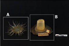
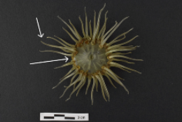
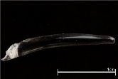
The Anthea cereus model is made of two components: the upper section (Element A), which includes the oral disc and tentacles, and the lower section (Element B), which consists of the cylindrical body, base, and a cardboard disc (Table 1). Adhesive residues at the points of detachment indicate the loss of ten tentacles, of which six are missing (Figure S4). The upper surface of the oral disc retains a thin paint layer exhibiting losses of various sizes (Figure S3). On the underside, adhesive traces suggest where Element A was once bonded to Element B (Figure S5). The tentacles, when viewed at high magnification, display horizontal striations within their glass structure and elongated air bubbles arranged longitudinally (Figure S7). Regarding Element B, the cardboard disc has slightly lifted, revealing the white interior of the base (Figure S1). This, together with observations under a binocular light microscope, supports the hypothesis that the base is composed of a gypsum-like material painted to simulate the rocky substrate to which this species adheres in nature. The glass cylinder with visible adhesive residue on its top surface (Figure S6) is affixed to the cardboard disc. A paint layer persists on the underside of the glass (Figure S2). Although the remaining decorative layer is partially preserved, and only a minimum trace of 3 cm × 0.5 cm remains, comparative analysis with other Blaschka specimens of the same species suggests that the outer surface of the cylinder was originally fully painted. A useful comparison is the Anthea cereus model from the Corning Museum of Glass (https://digital.library.cornell.edu/catalog?f%5Blegacy_value_tesim%5D%5B%5D=Anthea+cereus&search_field=all_fields (accessed on 6 August 2025).
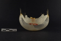
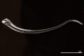
The Aurelia aurita model consists of a double spherical cap formed from a single piece of blown glass, to which several tentacles are attached using an adhesive (Table 1). In addition to a substantial accumulation of surface dust, an inconsistent black deposit is evident along the outer edge (Figure S10). Originally, the model was mounted on a metal rod affixed to a wooden base. It is presumed that at some point in its history the object fell and fragmented. Thanks to the efforts of the staff at the Liceo Foscolo in Pavia, numerous fragments were preserved. The central portion of the underside of the model of Aurelia aurita contains the insertion point of the metal support, within which fibrous material is present (Figure S13). This area is obscured by a yellowed adhesive that overlaps the glass edge. Comparative analysis with intact models, such as the one from the Canterbury Museum of Christchurch, New Zealand (https://collection.canterburymuseum.com/objects/192937/glass-model-invertebrate-aurelia-aurita-adult (accessed on 6 August 2025)) and the presence of incompatible glass fractures on the stem (Figure S14) suggest that four additional glass elements, corresponding to the oral arms (now missing), were originally present in this region.
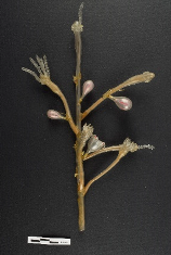
The Bougainvillia fruticosa model features a central stem with six remaining branches out of an original eleven (Table 1). Of these, only four are firmly attached, while two are partially detached. A thin layer of dust covers the model, but the principal conservation concern is the deterioration of the original adhesive (Figure S17). Additionally, several components are missing; their former positions are indicated by adhesive residues and the presence of copper wires, which would have provided structural support (Figure S18). Condensation droplets are present on the interior surfaces of the drop-shaped elements, a characteristic feature of so-called “weeping glass” (Figures S19 and S20). Furthermore, the interior of the stems contains an orange powdery deposit that is probably the original pigment applied by Blaschka (Figure S22) [4,29]. This deposit is also visible—albeit more diffusely—on the lower portion of the central stem.
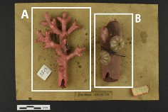
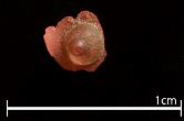
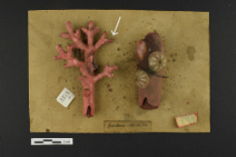
The Corallium rubrum model consists of two distinct components (Elements A and B) mounted on a cardboard base (Table 1). Element A corresponds to the branched object, while element B corresponds to the cylindrical one. The support features a small hole and an imprint indicating the vertical grafting point of element A (Figure S26). This was attached to the board using a nail that was inserted into a piece of wood which, in turn, was inserted into the hollow bottom of the A-piece. Dark stains of varying sizes are distributed across the cardboard paper surface. Both glass components are coated with a cold-applied paint that exhibits significant deterioration, including flaking, cracking, and lifting (Figures S25 and S31). Element A represents a branching structure that originally included six branches, one of which is now missing. The point where the glass broke is evident from the fracture in the glass, accompanied by paint loss and lifting (Figures S25 and S28). Tiny transparent glass elements, representing polyps, are adhered to the painted surface (Figure S30). This design choice has introduced notable conservation issues: as the paint peels, the polyps detach, further compromising the integrity of the decorative layer (Figure S27). Most of the polyps are either loose or already lost, often resulting in additional damage to the underlying paint. Element B is a cylindrical section fastened to the base with a wire. The surface exposed to the air is covered in dust and exhibits a darker area in the upper left quadrant (Figure S31). In contrast, the portion that remained in contact with the cardboard is clean. A pink glass section with a ribbed surface is inserted in the lower part of element B. Element B appears to be an enlarged representation of the terminal branch of Corallium rubrum, created to illustrate the detailed morphology of the polyps, which are now missing. Comparative studies with other Blaschka specimens of Corallium rubrum, such as the one at the University Museum of Utrecht (https://umu.nl/collectie-verhaal/pareltjes-van-glas-in-de-wetenschap (accessed on 6 August 2025) and the one at the Corning Museum of Glass (https://glasscollection.cmog.org/objects/67333/corallium-rubrum?ctx=f3edad7fa172adf72cf10ea5ed1ea283dce5847e&idx=1580 (accessed on 6 August 2025), confirm that two conical elements are incomplete and a third polyp is entirely absent.
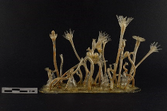
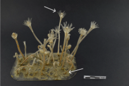
The Hydractinia echinata (Table 1) is in a severely compromised state, primarily due to adhesive degradation (Figure S35). A thick layer of dust further obscures its features. The model is mounted on a flat glass base to which the various elements were originally adhered, most of which are now detached or partially detached. As the base is almost entirely obscured by glass spheres and yellowed adhesive, it is impossible to discern any traces of workmanship that would indicate whether the base was crafted by hand. There are eight discernible attachment points for stems and two for conical elements, all identifiable by residual adhesive marks (Figures S36 and S37). Numerous small glass spheres, approximately 1 mm in diameter, are also present on the base; most are either detached or in the process of detaching (Figure S34). Three of the stems retain drop-shaped elements containing internal condensation—an indication of “weeping glass” (Figure S40).

3. Methods

Analyses of the Blaschkas glass objects described above were carried out to determine the state of conservation, the production technique, and the materials used, with the ultimate aim of carrying out a conservation intervention.

3.1. Visible Fluorescence Analysis (UVF)

Visible fluorescence analysis (UVF) was employed for preliminary detection of any organic materials (i.e., adhesives of animal origin or natural resins) used to assemble the elements of the glass models. The aim was to identify which parts were glued and which were attached by lampworking, when the glass was still hot, without any adhesive. The acquisition of visible fluorescence (UVF) images via ultraviolet radiation was conducted in complete darkness using a Nikon D800 camera (36 MP, Nikon Corporation, Tokyo, Japan) equipped with an AF MICRO Nikkor 60 mm 1:2.8 lens, fitted with a Hama UV&IR CUT filter. The shooting parameters were set to a 10 s exposure time, ISO 100, and an aperture of f/8. As light source, two Mada Tec ultraviolet lamps with an emission peak at 365 nm were employed.

3.2. X-Ray Computed Tomography (CT)

X-ray computed tomography (CT) was performed to detect if any parts of the glass had a different composition, and to study the objects’ internal morphology, and, consequently, how they were assembled. A custom CT system developed at the Department of Physics and Astronomy of Bologna University (Bologna, Italy) was used. This scanner features an indirect conversion flat panel detector (Varian PS2520D, Palo Alto, CA, USA, active area of 19.5 × 24.4 cm, 1536 × 1920 pixels, 127 μm pixel size), a microfocus X-ray tube (Kevex PXS10, Thermo Scientific, Waltham, MA, USA, 130 kVp maximum voltage, 0.5 mA maximum current), a high-precision rotary table by Physik Instrumente S.r.l., Karlsruhe, Germany, and a couple of orthogonal translation axes for the detector to enlarge the Field Of View (FOV) of the CT system up to about 50 × 50 cm2. The acquisition parameters were the following: voltage 80 kV; current 350 µA; frame rate 5 fps; frame AVG 4; projections 900; pixel size 0.127 mm; source–detector distance 735.0 mm; source–object distance 556.0 mm; object–detector distance 179.0 mm; magnification 1.322; voxel size 0.096 mm; scanning time 86 min.

3.3. Scanning Electron Microscope (SEM) with Energy Dispersive X-Ray Spectrometer (EDS)

Scanning electron microscope (SEM) analysis combined with an energy dispersive X-ray spectrometer (EDS) allowed a preliminary identification of different compositions of the glass. The SEM-EDS analyses were performed at the Arvedi Laboratory at the University of Pavia, on samples embedded in Epo Fix (Struers)® (15:2) epoxy resin (Struers, Ballerup, Denmark), polished and carbon-coated. A Tescan FE-SEM (Mira 3XMU series) (TESCAN, Brno, Czech Republic), equipped with the Apollo XL silicon drift detector energy dispersive X-Ray spectrometer-SDD-EDS, was employed. The operating conditions were as follows: 20 kV acceleration voltage, 12 mA beam current, 100 s count collection for EDS analysis, average data from 3 to 5 points of sampling, variable analysis area, depending on the size of the phases to be analysed. The microanalyses were performed using the EDAX Genesis software (version 4.51) and the data obtained were processed using ZAF correction.

3.4. Fourier-Transform Infrared Spectroscopy (FTIR)

Fourier-transform infrared spectroscopy (FTIR) was used to characterise the adhesives. For FTIR analyses, the samples were appropriately processed to obtain homogeneous thin pellets; 10.0 mg of each fragment was ground and subsequently pressed (5 tons for 60 s) with 100.0 mg of potassium bromide (KBr). The analyses were carried out with a Bruker Tensor 27 Spectrometer (Bruker Scientific Instrument, Billerica, MA, USA), controlled by Opus 7.2 software (Bruker). The spectra were acquired in transmission mode, in the MIR range (4000–400 cm−1), integrating 64 scans for each measurement, and with a 4 cm−1 spectral resolution. OMNIC 7.2 software (Thermo Electron Corporation, Waltham, MA, USA) was used for result interpretation and manipulation including baseline correction, slight spectral smoothing, and CO2 band removal.

4. Results and Discussion

4.1. Investigation of Glass Models’ Structure and Semi-Quantitative Composition (CT and SEM-EDS)

Computed tomography (CT) was performed on the glass models for two purposes: to detect areas with varying radiopacity that would indicate the usage of various glass types and to investigate the interior morphological structure of the objects to gather data on the assembly procedure.
With the only exception of Corallium rubrum, the CT images show that two different glass mixtures were used. The Blaschkas used to prepare the components of the models in advance, assembling them when they received an order. This meant that parts of the same model could be made at different times, not necessarily with the same batch of glass [2,11].
CT slices of the Anthea cereus glass model highlight materials that absorb X-rays in a completely different way (Figure 1): the tentacles and the oral disc appear to be slightly radiopaque, while the small milky-looking hemispheres applied to the edge of the oral disc below the tentacles are extremely radiopaque.
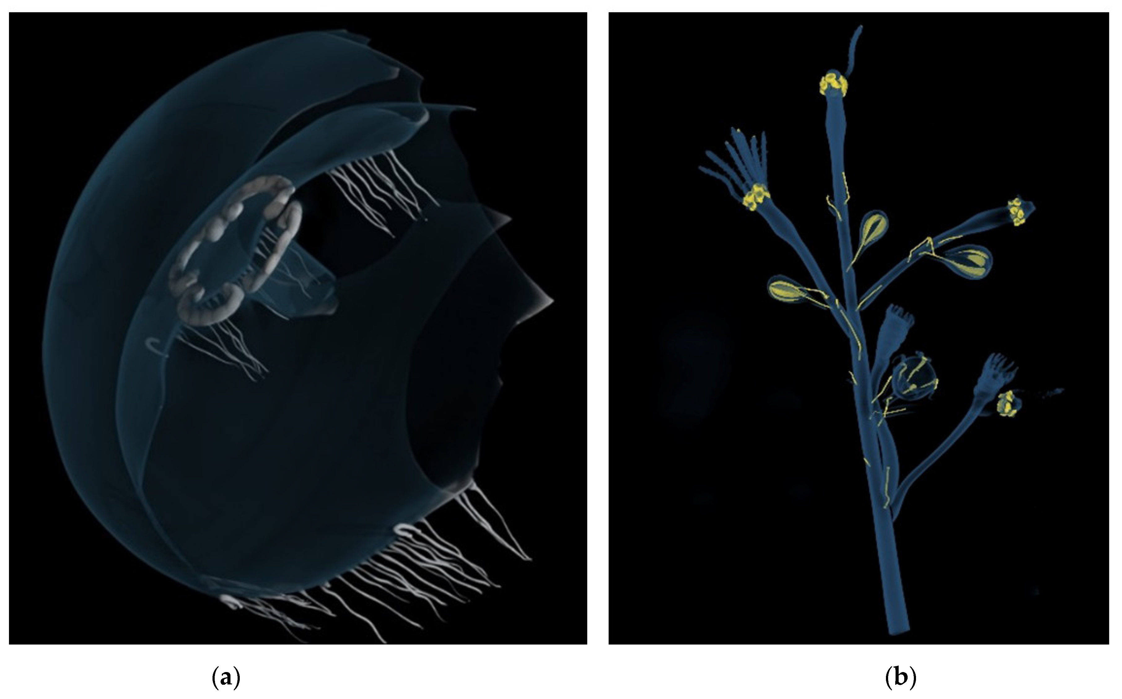
The same features can be observed in the model of Aurelia aurita, where the tentacles (made of colourless glass) and the gonads (made of pink glass) show a higher radiopacity than the bell (made of colourless glass) (Figure 2a). The fact that two transparent pieces of glass used in the same model have different compositions can be attributed to the Blaschkas’ method of preparing the models’ various elements.
The strong radiopacity of the pink glass is also found in Bougainvillia fruticosa (Figure 2b), in which the white filaments in the reproductive structure exhibit the same phenomenon. In this model, the tomographic images also highlight the presence of metal filaments and adhesives showing higher radiopacity.
The investigation into the composition of the pink and white glass used in Blaschka’s naturalistic models was constrained by the need to avoid invasive sampling: the only possible hypothesis about their composition is the presence of elements of high atomic number—such as Pb—because of their radiopacity [6,11].
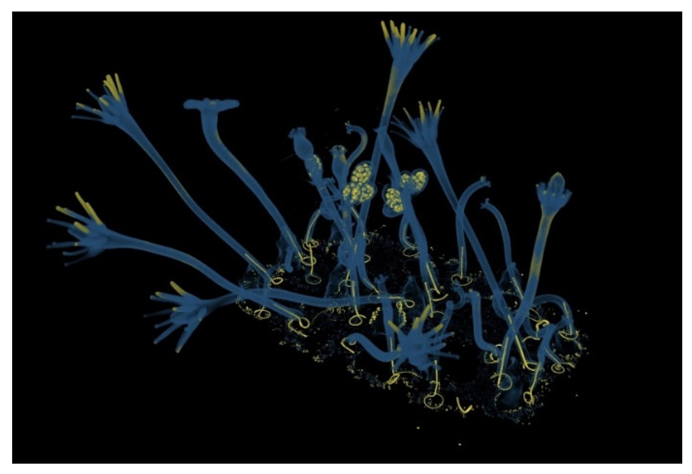
In the Hydractinia echinata model, tomographic imaging reveals notable variations in radiopacity within the glass components, particularly within the gonophores, where the yellow glass sections appear highly radiopaque (Figure 3). As for the coloured glass of the other models, the impossibility of sampling parts of higher radiopacity did not allow the identification of the chromophores, which would, in any case, have been quite a complicated task. In this regard, thorough discussions of Blaschka glasses can be found in previous works [6,11] and—more generally—in the pioneering work of Weyl [30].
The tomographic analysis of Hydractinia echinata also reveals additional radiopaque zones: at the base spheres, tentacle tips, and along the stem. The spheres are likely composed of glass containing higher-atomic-number elements. In contrast, the irregular and inconsistent radiopacity in the tentacles may be due to external material deposits rather than the glass itself. The radiopaque areas along the stems correspond to a powdery substance inside the hollow structure, which could not be analysed further.
Finally, the imaging provides insight into construction techniques: the stems are reinforced with a metal wire inserted a few centimetres into the glass and bent into a circular shape at the base attachment point, likely to ensure greater structural stability.
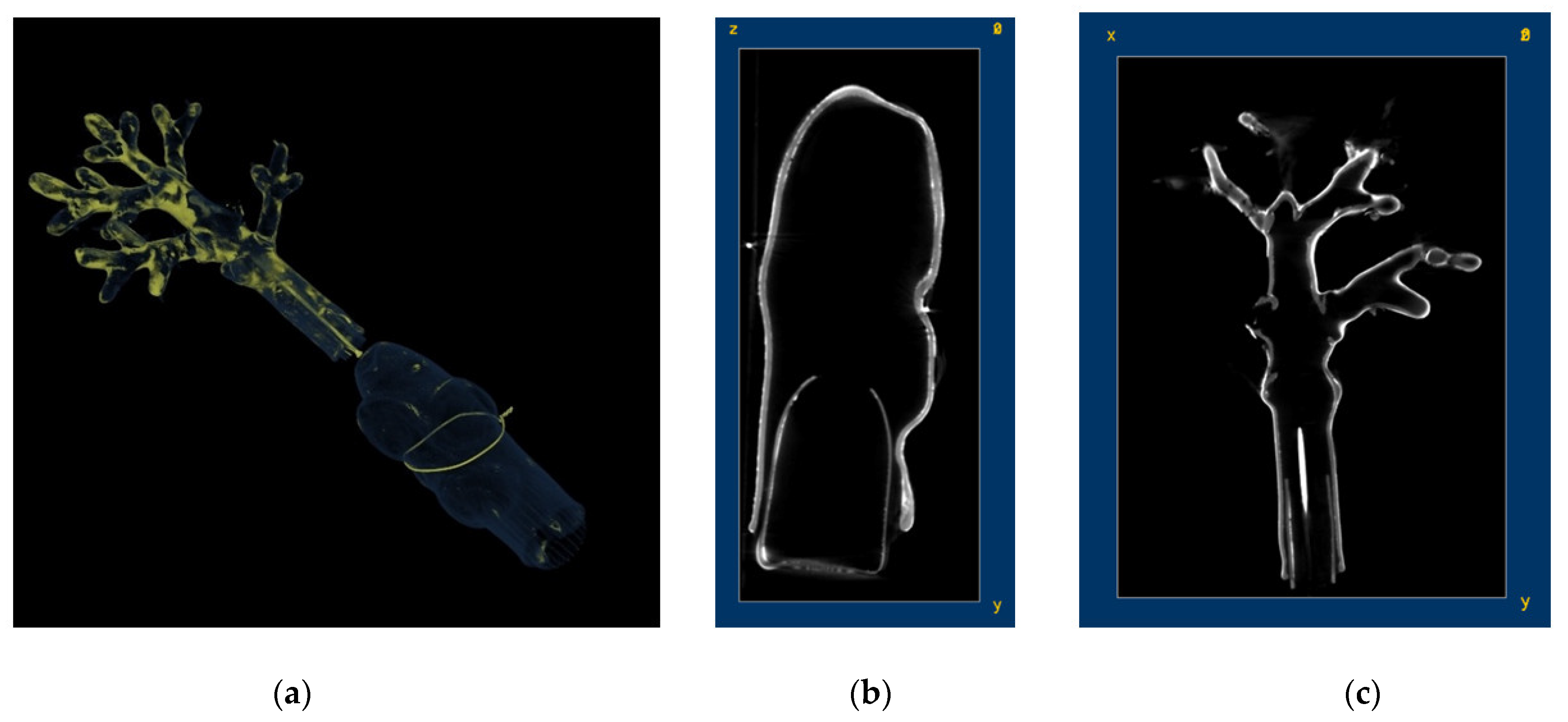
Regarding Corallium rubrum, tomographic images reveal that the object showing a ribbed surface and a hole at the top is inserted inside element B, and is approximately one-third of its total height (Figure 4). Upon examining element A, it is observed that the nail, of which only the head is visible, is about one-third of the height of the entire object, while the ribbed glass cylinder reaches approximately one-quarter of the total height. The painted surface appears more radiopaque than the glass, which could be due to heavy elements present in the pigment used. To verify this hypothesis, further analyses were conducted.
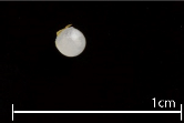
The SEM + EDS samples were chosen after viewing the CT results, which showed that most of the objects are made of two glasses of different composition and consequently different radiopacity. Other selection criteria were the availability of detached fragments that could not be reattached or elements whose absence would not affect object legibility. Table 2 summarises the set of samples and their position.
SEM-EDS semi-quantitative compositional analyses show that mixed alkali glass was mainly used, five samples out of seven being of this type (AA1, AA2, AC2, CR1, HE1), while two samples (AC1 and HE2) are made of lead glass. Table 3 shows semi-quantitative compositions of the samples analysed by SEM-EDS.
In the mixed alkali glass the main flux is sodium (Na2O around 14%), in which K2O is around 4% in samples AA1, AA2, CR1, and HE1, while AC2 has a slightly higher content of K2O, around 7%. Calcium is the more abundant stabilizer in the mixed alkali glass (CaO between ca. 5 and 7%, MgO < 0.2%). As for the accessory components, Al2O3 ranges between 2 and 3%, being slightly lower in AC2; SO3 is less than or around 1%, again with the exception of AC2, in which it is not detected. AC2 also differs in chlorine content, with Cl2O around 1%, while its content is ca. 0.1–0.2% in the other mixed alkali glass samples. Accounting for unavoidable heterogeneity, the mixed alkali glass of the models analysed can be considered of the same type, the only exception being the Anthea cereus sample AC2 (tentacle). A comparison with previous published compositional data allowed us to find a correspondence with the category of mixed alkali colourless glass reported in the analyses of the models from the Natural History Museum [11], particularly with the set of samples from the acquisition of 1899 (the latest), which differ from the others, on average, in their higher K and lower Na contents and in the relatively higher content of Mn. The exception in this study (sample AC2) has a good correspondence with the chemical composition of a sample from the same set representing the model of Raphidiophrys elegans (heliozoan—sample code 25a), confirming both the compositional variability within the same set and the possible time range of production in the late stage of the Blaschkas’ activity.
In the two samples containing lead (AC1 and HE2), the main flux is potassium, being K2O around 10%; among the stabilizers, calcium is more abundant than magnesium (CaO around 2%, MgO < 1%) while Al2O3 is less than 1%, corresponding with the analyses conducted on the material at the Natural History Museum in London 11], which shows a significantly lower aluminium content in the glass containing lead. Another correspondence with the analyses of the lead glass from the Natural History Museum [11] is found in the use of high amounts of As (around 2000 ppm) employed for lead glass clarification (data from LA-ICP-MS preliminary analyses that are not fully reported in this work). Apart from this correspondence, the lead glass samples are quite different from one another. Only the sample of Anthea cereus AC1 can be associated with the category of suckers from the 1883 acquisition of the Natural History Museum [11], thus confirming a possible late production; the sample from the spheres of the Hydractinia echinata (HE2) cannot be associated with any of the previously studied lead-glass samples, with—first of all—its content of Pb being definitely lower than the reported cases.
All samples contain a small amount of manganese (MnO less than 0.5%), which should have been used as a decolourizer. Decolourizers are introduced into the vitreous matrix to balance the chromophore effect of iron that may be present as impurity (unintentionally coloured glass). In the samples analysed, iron is represented by an average of about 0.3–0.5% of Fe2O3. Comparisons with published data account for a deliberate use of manganese, whose content is again comparable to the late models reported in [11], as the most used decolourizer, possibly associated with Sb.
As for compositional similarities between the same models, a comparison with the data from the Natural History Museum does not reveal any one-to-one correspondence, indicating that the production of the models did not rely on a specific type of glass but was possibly determined by the glass’s working properties and by the availability of raw materials. As noted by Bertini and colleagues [11], the nature of the raw materials cannot be established (not to mention the possibility of recycling), but the consistency of the mixed alkali composition of this work, also in relation to data from [11], could be considered proof of the accessibility of the desired (or even customized) raw materials.
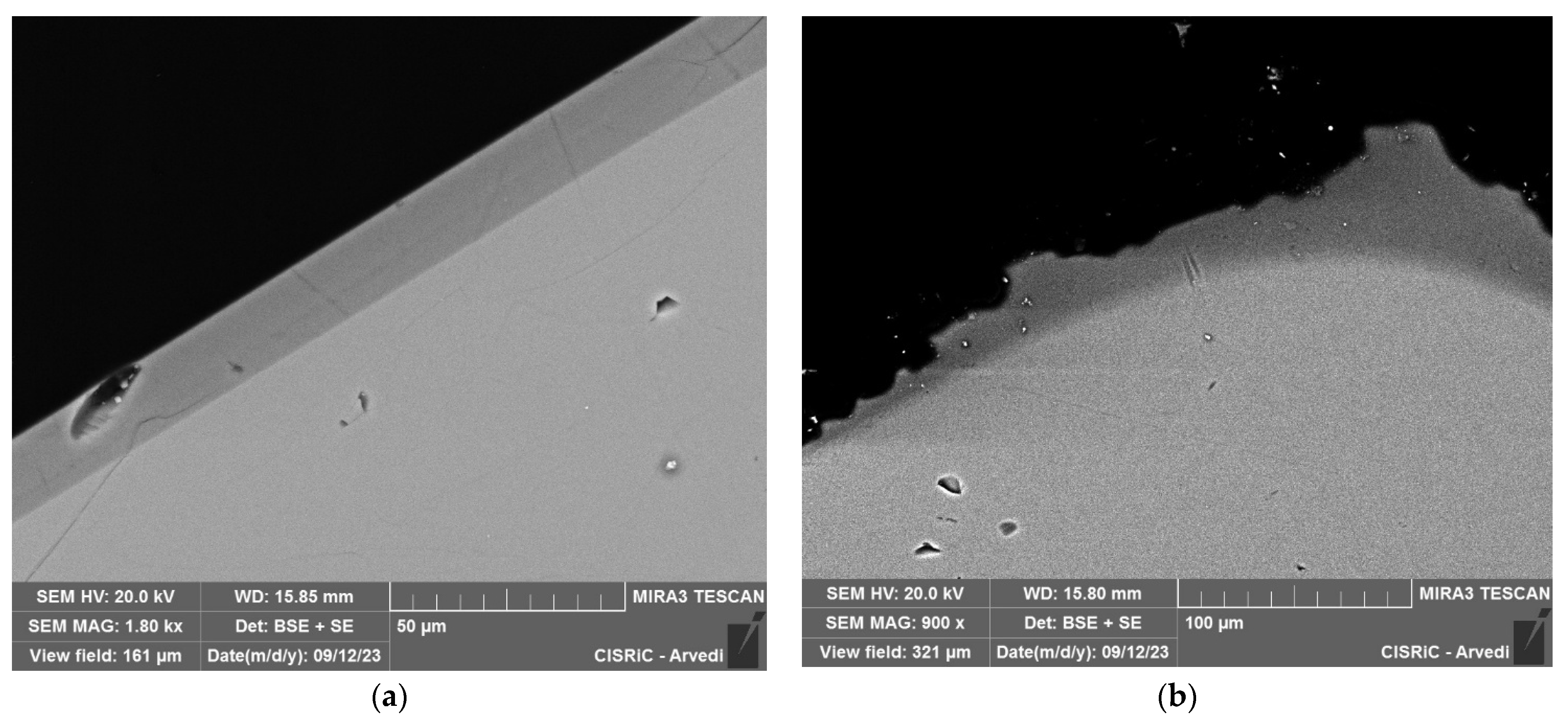
Samples AA1, AA2, and AC1 are made of homogeneous glass, while AC2 shows a layered appearance (Figure 5a) that runs uniformly and regularly along the entire perimeter of the sample, and which has a very different composition from that of the internal part of the sample. Also, sample CR1 shows a surface layer with a different composition (Figure 5b), but with an irregular and nonuniform profile and a very different composition compared to the pristine glass. HE1 and HE2 are composed of homogeneous glass—apart from the presence of two quartz-like particles adhered to the edges of sample HE2.
The BSE analyses of sample AC2 show a morphology characterized by a dark-grey stripe that runs in an extremely regular and defined way along the entire perimeter of the object (Figure 5a) and which has a different composition from that of the internal area of the sample (richer in heavy elements). The composition of the inner zone of AC2 is characterized by higher flux and lower stabilizer. The outer band of the AC2 has a composition strongly depleted in flux, i.e., sodium with Na2O lowered from almost 15% to about 1.5% and K2O from around 7 to 4%. These compositional differences could be interpreted as the phenomenon of glass leaching. This type of degradation develops more easily in conditions of high humidity and involves the replacement of alkaline elements with H3O+ ions [32]. This process involves the creation of a hydrated silica layer which therefore has a chemical composition in which the content of silicon oxide is higher, while the amounts of sodium and potassium oxides are lower. Although these compositional differences would correspond exactly with those observed in the AC2 sample, the extreme regularity of the outer layer and its clear separation from the inner area do not exactly reflect the leaching phenomenon, whose morphology appears quite irregular. The regularity of this layer and the clear separation from the interior of the sample could correspond, instead, to the hypothesis formulated to explain the horizontal streaks visible at high magnification of the tentacles of the Anthea cereus model (Figure S7). This type of two-layered glass is generally referred to as flashed glass, in which a thin layer of coloured glass is superimposed on to a colourless glass. Differently, in this case the layers are both colourless and the layered structure could possibly correspond to an effect of the lampworking technique for which continuous rotation and repeated heating was necessary. For a comparison between the two morphologies, refer to the image of the CR1 sample and its discussion. The image of the CR1 sample shows two zones, one external and one internal (Figure 5b), characterized by two different chemical compositions: the differences between the compositions of the inner and outer layers of the CR1 sample concern silica, sodium, and calcium. These compositional differences are indubitably due to the phenomenon of leaching or de-alkalization of the glass, undoubtedly due to the atmospheric environment’s interaction with glass. The slightly higher content of potassium in the leached layer—although not consistent with the effects of the lixiviation—could be due to inhomogeneities of the glass matrix. The presence of the leaching phenomenon is also confirmed by the irregular morphology of the outer layer and by the fact that the boundary with the inner layer is not well defined.
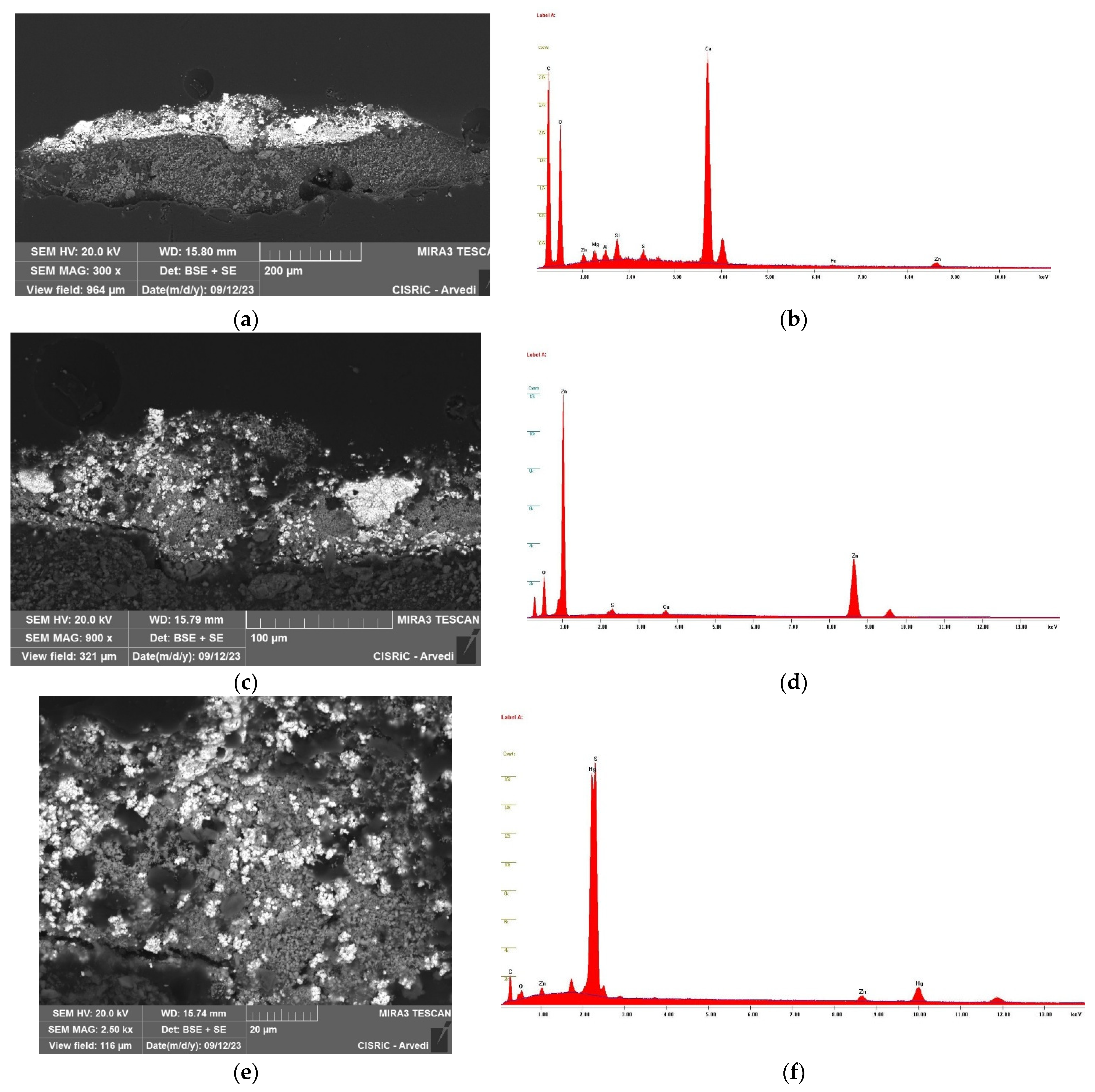
As stated above, the CT shows a higher radiopacity of the paint layer of the Corallium rubrum. In the case of the CR1 sample, the fact that a portion of the entire stratigraphy of paint layer was adhered to the glass allowed analysis of the latter to be carried out. The paint layer does not appear homogeneous, and stereomicroscope observations show that it is composed of a pink matrix with white particles of less than or equal to 0.1mm in size. The stratigraphy of the paint layer adhered to the CR1 sample is composed of two layers (Figure 6a). The compositional analysis of the external layer (bottom layer in Figure 6a) shows a very high content of calcium (Figure 6b). Given the high levels of carbon and oxygen also, the orientation of the stratigraphy, and the methods of application of the polyps, it can be assumed that this layer corresponds to the adhesive of animal origin used to apply the small glass polyps to the paint layer of the main structure of the Corallium rubrum, with the possible addition of a filler containing organic components (Ca, Si, Al, Mg, Zn—as detected) to improve mechanical properties. The lower stratigraphy (upper layer in Figure 6a) is given by two pigments (Figure 6c): one based on Zn (possibly zinc white—Figure 6d) and one on Hg and S (possibly cinnabar or vermilion—Figure 6e–f).
The samples of Hydractinia echinata, HE1 and HE2, have two very different compositions: the first is a mixed alkali glass with Na as the main flux; the second has a high content of Pb. In the latter, two inclusions of a quartz-like material are embedded in the outer perimeter. It can be hypothesized that the spheres were cooled on the surface of a clay-type material, which is also made of quartz minerals. During the cooling phase, the hot sphere could have melted, incorporating the minerals on its surface. Quartz could also be the result of an un-melted glass-making ingredient. A summary scheme of the samples analysed, and the results obtained, is reported in Table 4.

4.2. Analysis of the Adhesive (UVF and FTIR)

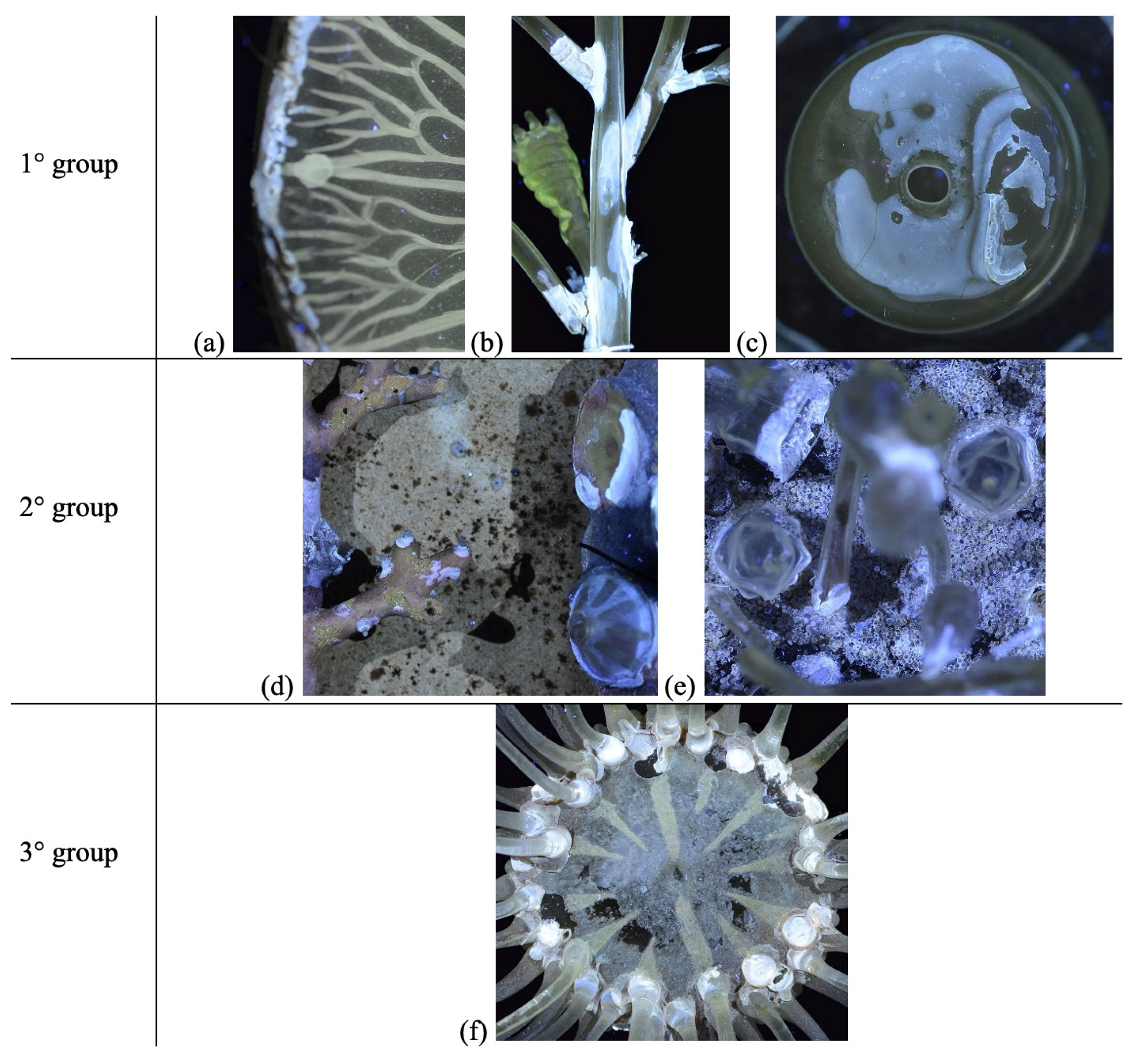
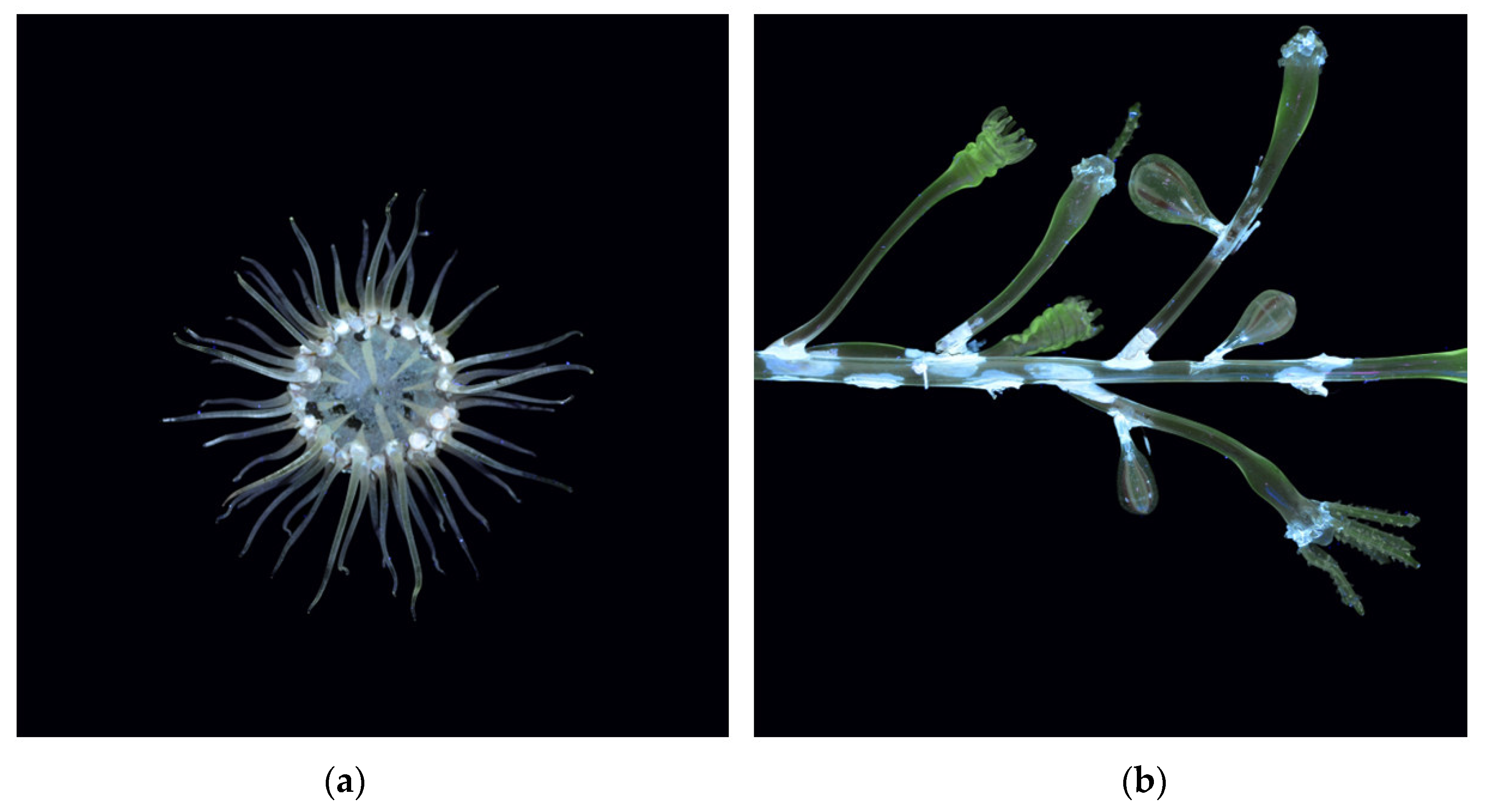
UVF analysis of the Blaschka glass models revealed variations in the fluorescence of adhesives, allowing for a visual distinction between heat-fused joints and those bonded with adhesive pastes. Based on the fluorescence observation, the adhesives could be divided into three different groups, according to their response intensity (Figure 7). The first, characterized by a pale sky-blue fluorescence with icy white highlights, was observed in Aurelia aurita, Bougainvillia fruticosa, and in element A of the Anthea cereus model (Figure 8a). The second, with a slightly cooler blue tone, was identified in Corallium rubrum and Hydractinia echinata. The third, presenting intermediate sky-blue nuances, was seen in element B of Anthea cereus. These intensity differences, while visually discernible, are likely attributable to variations in adhesive thickness or slightly different mixtures, as also supported by spectroscopic analysis.
For the glass models of Bougainvillia fruticosa and Hydractinia echinata, the presence of fluorescence at the lower part of the tentacles indicates the use of adhesives; by contrast, the absence of any kind of fluorescence at other junction points suggests an assembly method through heat fusion. For the Bougainvillia fruticosa and Hydractinia echinata, UVF analyses show the use of both production modes. The intense blue fluorescence observed at the base of Anthea cereus (Figure 8a) is attributable to a high concentration of adhesive. Another observed phenomenon is the green-yellow fluorescence of the glass surface, particularly evident in fractures of the Aurelia aurita and Bougainvillia fruticosa models (Figure 8b).
According to van Giffen [4], this effect—more intense in the thicker areas of glass—may be due to the use of manganese, which was identified in all samples through EDS analyses and is often employed as a decolourizing agent in sodium-based glasses. This hypothesis has indeed been confirmed in the case of Aurelia aurita.
Lastly, no fluorescence indicative of biodeterioration agents was detected in the adhesives or on the analysed surfaces.
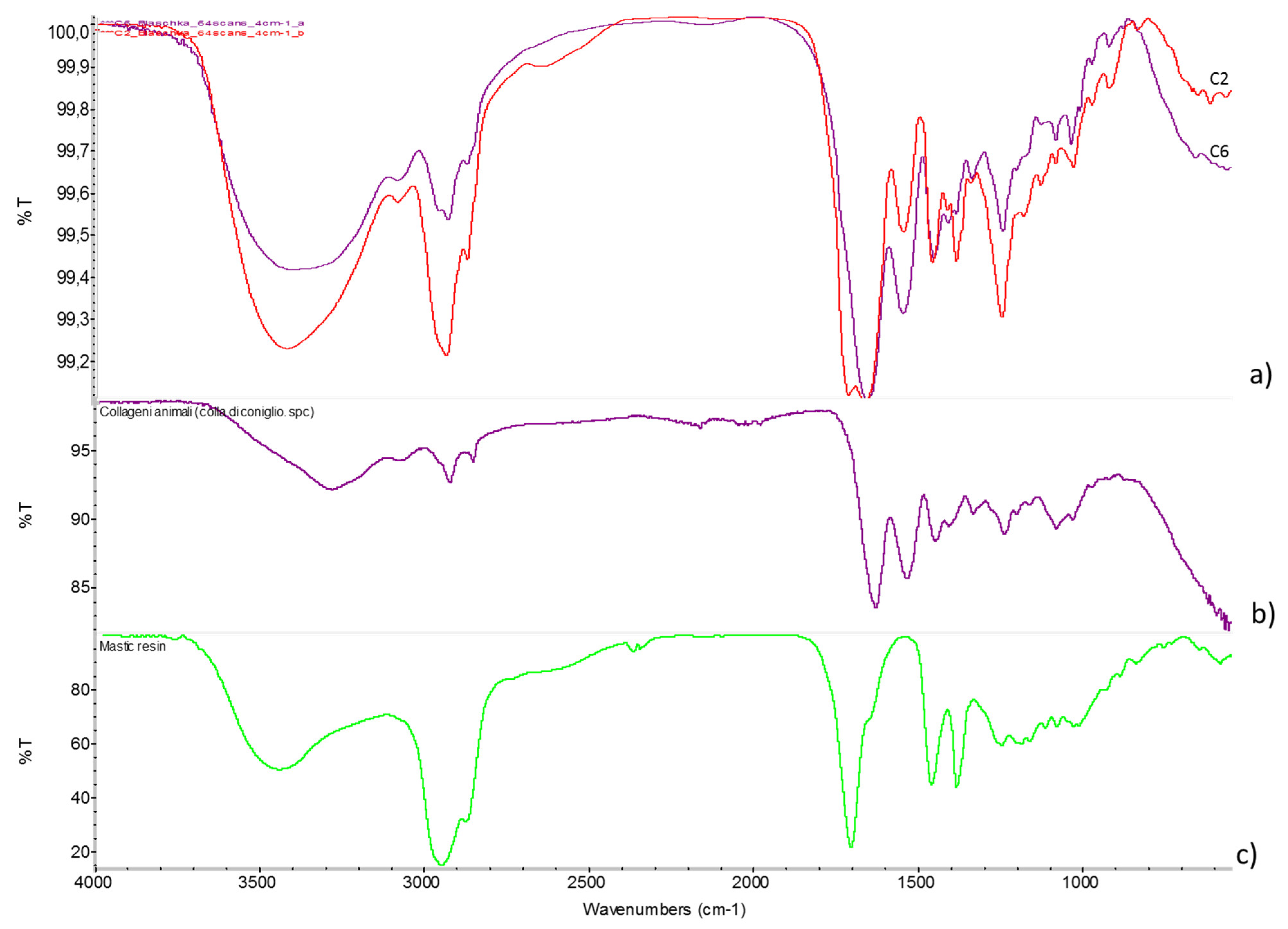
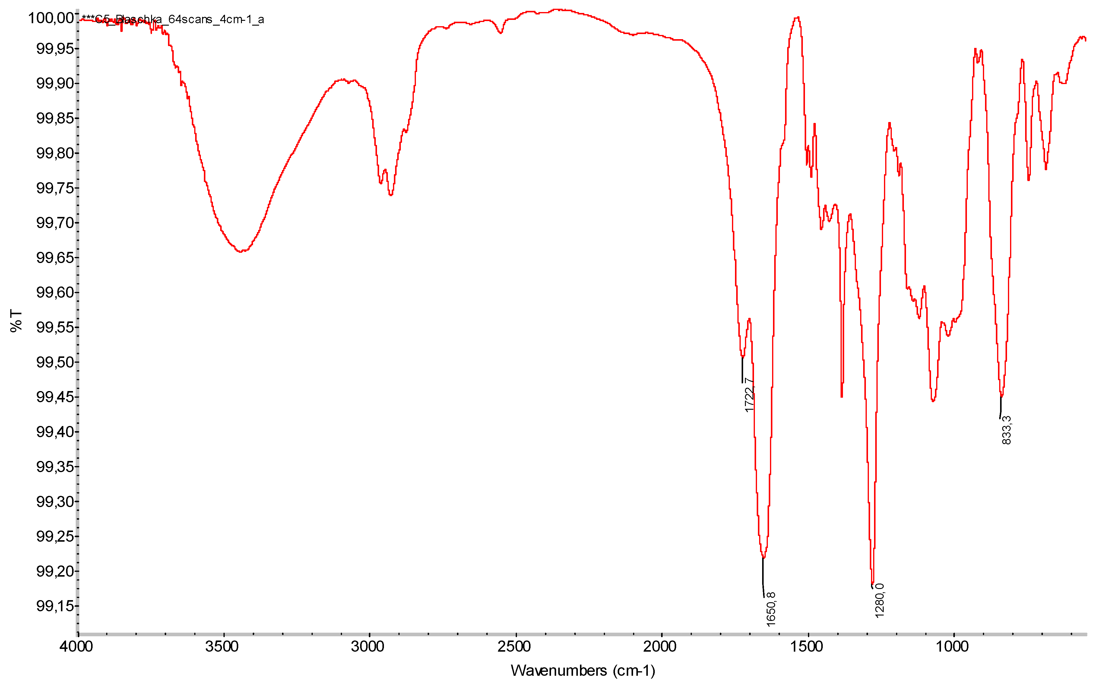
FTIR analyses confirmed that the adhesives at the jointing areas of the artefacts vary in composition, as also highlighted by UVF. A list of the analysed samples and related results is provided in Table 5. Animal glue is the most common: in fact, only one of the six samples examined had cellulose nitrate, whereas the other four contained a protein compound with a natural resin added, probably in various amounts. Spectra acquired on the samples taken from Bougainvillia fruticosa (C1), Aurelia aurita (C2, C3), and Hydractinia echinata (C6) show the strong and broad amide I and amide II bands, respectively, at 1655 (C=O stretching carbonyl group) and 1545 (C–N stretching and N–H bending) cm−1; the presence of proteins is further confirmed by the N–H stretching band around 3370 cm−1 and the amide III C–N stretching vibration (1245 cm−1) and CH2/CH3 in-plane bending vibrations (1455 cm−1) [33,34,35,36]. Although the vibrational bands of protein adhesives predominate in most of the spectra, additional compounds were also detected. This is particularly visible in sample C2, taken from the manubrium under the bell-shaped body of Aurelia aurita, where the spectrum (Figure 9) only partially matches the standard of proteinaceous glue. The detected additional bands can be attributed to the chemical structure of tree resin: the symmetric and asymmetric stretching of CH3 and CH2 groups of its aliphatic three-ring structure (2937 and 2875 cm−1); the O–H vibration of the dimerized carboxyl group (about 2645 cm−1); the strong carbonyl stretch (1712 cm−1); the bending of CH2 and asymmetric bending of CH3 groups (1457 cm−1); the symmetric bending of CH3 groups (1383 cm−1) [37]. Differently from the other analysed samples, on sample C5—taken from the element B of Corallium rubrum—the IR spectrum shows intense vibration bands at 1650, 1280, and 833 cm−1 (Figure 10). These bands can be, respectively, attributed to O–NO2 asymmetrical stretching, NO2 symmetrical stretching, and N–O stretching of cellulose nitrate [38]. A vibrational band at 1722 cm−1, ascribable to the C=O stretching, was detected: it could indicate either the addition of a carbonyl-containing plasticizer, such as a camphor or a phthalate ester, or the formation of some degradation products due to a photooxidative process [39,40]. UV analysis did not reveal the characteristic yellow-green fluorescence typically associated with cellulose nitrate under long-wave UV radiation. This absence can plausibly be attributed to the localization of the material in areas that were inaccessible or partially obscured during the examination, likely corresponding to later restoration interventions.

4.3. Conservation Measures

The intervention provided an opportunity to observe and address conservation and degradation issues related not only to glass but also to other materials, given the multi-material nature of the Blaschka models. The conservative operations were carried out with the dual aim of improving the preservation condition of the individual specimens and developing a methodological approach to such objects, with particular focus on consolidation, adhesion, and integration treatments.
An initial phase, common to all five glass models, involved general dry cleaning using soft-bristled brushes combined with light vacuuming. Subsequently, wet cleaning was planned where deemed necessary. As some objects presented different types of surfaces and consequently specific conditions and needs, targeted cleaning tests were conducted to remove the pulverulent deposit. Since adhesives of animal origin are soluble in polar solvents, this category of product was eliminated from the cleaning tests. As the objects were small and had many areas where this type of substance was present, the risk of solvent contact during the cleaning phase was significant. Consequently, solvents that could not interact with the adhesive were chosen, i.e., apolar solvents. These included ligroin, petrol, white spirit, and turpentine.
For the Anthea cereus model, the intervention included re-adhering the detached fragments of the oral disc and the cardboard ring at the base. Once a correspondence had been verified between the imprint on the adhesive on the lower part of element A and that of element B, the two parts were assembled. The unstable tentacles were stabilized, and the detached ones were reattached.
The most complex procedure involved the model of Aurelia aurita, which was completely reconstructed. After an initial dry-fit assembly, the need for structural integration and the most suitable operational approach were assessed. The object was assembled by joining fragments using narrow adhesive tape strips applied transversely to the fractures. Two losses were confirmed, one significantly larger than the other, separated by a small group of fragments with minimal contact surfaces. Due to the lack of stable connection, it was decided not to include this fragment group in the reconstruction but to preserve it separately. This decision did not compromise the legibility of the object’s overall form and eliminated a major structural weakness, contributing to improved stability. The reconstruction of the missing section aimed to enhance both the readability of the form and the structural stability of the Aurelia aurita model. Without such integration, the already fragile and thin fracture edges would remain vulnerable to external stress. An indirect reconstruction was therefore carried out by manually shaping thin support plates. Two materials were tested for comparison: Paraloid B72® (an acrylic resin) and Hxtal NYL-1® (an epoxy resin). Of the two resins, the best result was given by Hxtal NYL-1, which produced a transparent, colourless, bubble-free sheet of the desired thickness. The fragment reattachment was carried out using Hxtal NYL-1® resin, applied by capillary action. Once assembly was complete, excess adhesive was removed immediately with absorbent paper to minimize subsequent cleaning. Nevertheless, after curing, any remaining residues were mechanically removed with a scalpel. Following the restoration, the Aurelia aurita specimen showed greatly improved legibility, thanks to both cleaning and the reassembly of fragments, and exhibited enhanced structural stability.
The Bougainvillia fruticosa model required stabilization of the original adhesives, some of which were partially detached. Adhesion was performed using Paraloid B72® at 30% in a solvent mixture of 35% acetone and 65% petroleum ether (ligroin) 100–140. Given the minimal contact area and the weight the adhesive had to support, additional small, visually unobtrusive supports were created from Hxtal NYL-1® resin plates. The weeping degradation phenomenon inside the jellyfish buds was treated by washing with demineralized water. The removal of alkalis was monitored by measuring the pH of the rinsing water. As the alkalis leach out during washing, the pH of the solution becomes more basic. After the conservation treatment, the glass model was both structurally and chemically more stable, and its surface was more legible.
For the Corallium rubrum, the intervention involved pre-consolidation of the paint layer and re-adhesion of both the lifted areas and the polyps on the surface of element A. The re-adhesion of the lifted paint layer was carried out with Aquazol 200® at 7% in demineralized water, applied with a brush. No mechanical pressure was required, as the paint layer flattened spontaneously due to the swelling of the binder caused by the water. This was also due to the small size of the detached areas. After this procedure, wet cleaning was performed using solvents established through preliminary tests. The solvent mixtures tested were as follows: ligroin, ligroin–ethanol (90–10%), ligroin–acetone (80–20%), ligroin–ethanol (70–30%), and ligroin–acetone (50–50%) where the percentages indicate the ratio (w/w) of the solvents in the solution. The best results were given by ligroin–acetone (50–50%) applied with a soft-bristled brush. Then, the entire surfaces of elements A and B were consolidated using Aquazol 200® at 5% in demineralized water, again applied with a brush, due to the overall fragility of the paint layer. The interior paint layers of both elements were not treated due to inaccessibility. After conservation, the Corallium rubrum model showed improved surface legibility and greater stability of the paint layer.
For the final model, Hydractinia echinata, the main intervention was stabilization of partially detached elements and reattachment of those already separated. Adhesion was performed using Paraloid B72® at 50% in acetone, applied from a tube. Given the very small bonding surfaces of the stalks, the adhesive alone was deemed insufficient to guarantee stability. Despite the low weight, the height of the elements could have affected the adhesive’s performance. Therefore, additional unobtrusive supports were applied, created from Hxtal NYL-1® resin plates. The previously detached gonophore was also reattached using Paraloid B72® at 30% in a 35% acetone and 65% ligroin mixture; in this case, no support was necessary. Finally, detached spheres were reattached using Paraloid B72® at 10% in the same solvent mixture. After the intervention, the Hydractinia echinata displayed improved morphological legibility and significantly enhanced structural stability. In order to ensure the best possible preservation of the restored Blaschka models, it was recommended that the temperature be kept between 20 and 24 degrees Celsius, and the relative humidity between 40% and 45% [41].

5. Conclusions

The conservation of the marine invertebrate models from Trento and Pavia offered a valuable opportunity to investigate the composition, technical characteristics, and production techniques of Blaschka glass. The data obtained in this study demonstrate that the integration of a multi-analytical approach with knowledge of historical glassmaking practices provides a robust foundation for both conservation strategies and further scholarly research into these unique artifacts.
The interpretation of tomographic data suggests specific techniques for assembling the models, such as the use of metal wire in the base of the Hydractinia echinata models. This technical detail highlights the methodical approach in the creation of these models, focusing not only on aesthetic qualities but also on structural stability. These insights offer a broader perspective on the functionality of the models, in addition to their material characteristics. The descriptions of varying radiopacity in specific areas of the models, such as the tentacles or gonads, help to understand the diverse applications of glass with different physical properties.
The SEM-EDS analyses of the Blaschka glass models provide valuable insights into their chemical composition and manufacturing techniques. Although a one-to-one correspondence between models cannot be confirmed, correlation with a late set of glass models from the Natural History Museum in London allows us to set the possible time range of production of the Italian models in this study in the late stage of the Blaschkas’ activity. The study reveals that most of the glass samples are mixed alkali glass, with some exceptions of high-lead glass, particularly in samples like the Anthea cereus hemispheres (AC1). The use of different glass types is likely related to differences in working properties and melting temperatures, or aimed to achieve specific visual or structural effects in various parts of the models. Compositional analyses show that the average flux (Na2O + K2O) is around 18% (Table 3). This could be in line with the requirements of lampworking, for which the Blaschkas used oil or paraffin as fuel. This fuel does not allow high temperatures to be reached, so they needed to use glass that melted more easily. Notably, the high potassium content in AC1 suggests that this glass was designed for different purposes, such as the creation of a milky colour typical of lead glass. Additives and impurities such as manganese (used as a decolourizer), phosphorus, and chlorine were also identified, each playing a role in the glass’s properties. For instance, the chlorine in AC1 could be a result of industrial refining rather than an unintended impurity. The presence of phosphorus and potassium in certain samples suggests potential interactions during the glass manufacturing process. Although the nature of the raw materials cannot be ascertained, the compositional features proves that the Blaschkas could access the desired raw materials, possibly customized for their purposes. The SEM-EDS results also show layered structures in the Anthea cereus tentacle (AC2) and Corallium rubrum polyp (CR1) samples. In sample CR1, the alteration is connected with the leaching phenomenon due to the irregular morphology of the surface layer and the compositional differences. However, in sample AC2, while the compositional differences may also be related to the leaching phenomenon, the regularity of the surface layer suggests the deliberate addition of a second layer of glass—also visible in the high magnification optical microscopy images of the object—possibly of flashed glass, although the layers are both coloured. Additionally, the study highlights detailed analysis of the paint layer on the CR1 sample. Two distinct pigment layers were identified, one containing calcium and the other zinc and mercury, which may indicate the use of animal-based adhesives and specific pigments like zinc white and cinnabar (vermilion). These findings underscore the sophisticated artistic techniques employed in creating the Blaschka models, as well as the careful selection of materials to achieve specific aesthetic effects.
UVF analyses allowed the identification of distinct fluorescent properties in the adhesives. The mention of green-yellow fluorescence in fractures, particularly in the Aurelia aurita and Bougainvillea fruticosa models, suggests possible material degradation or the influence of environmental factors on the glass surface. The absence of fluorescence indicative of biodeterioration agents is also a notable observation, as it suggests that the adhesives and glass surfaces have remained relatively unaffected by biological attack. Additionally, FTIR spectroscopy provided further insights into the adhesive compositions, confirming the presence of protein-based compounds and natural resins in the adhesives.
The conservation treatment of the Blaschka glass models involved comprehensive interventions to address both material degradation and the complex multi-material nature of the objects. The primary goals were to improve the preservative condition and develop a methodological approach to their treatment, focusing on consolidation, adhesion, and gap-filling. By establishing a conservation methodology valid for the Blaschka model macro-category, we were able to adhere to guidelines as well as react flexibly to the specific needs of each object. In conclusion, the conservation intervention for these models not only involved the careful stabilization and reassembly of the glass components but also addressed the unique challenges posed by the combination of glass, adhesives, and paint layers. The use of different resins and consolidation methods tailored to each model’s specific needs led to significant improvements in both the structural integrity and visual clarity of the objects.

Supplementary Materials

The following supporting information can be downloaded at: https://www.mdpi.com/article/10.3390/heritage8090376/s1; Table S1: Blaschka models before and after conservation; Table S2: Blashcka models: state of conservation.

Author Contributions

Conceptualization, M.V.; Methodology, S.F.; Validation, S.F.; Investigation, C.M., S.A.A., G.T., M.P.M., M.B. and M.P.R.; Data curation, G.G.; Writing—original draft, G.G.; Supervision, M.V.; Funding acquisition, M.V. All authors have read and agreed to the published version of the manuscript.

Funding

The project is funded by the European Union—NextGenerationEU under the National Recovery and Resilience Plan (PNRR)—Mission 4 Education and research—Component 2 From research to business—Investment 1.3, Notice D.D. 341 of 15/03/2022, entitled: Cultural Heritage Active Innovation for Sustainable Society proposal code PE0000020-CUPJ33C22002850006, duration until 28 February 2026.

Data Availability Statement

Data are contained within the article or Supplementary Materials.

Acknowledgments

This study was made possible thanks to the availability of the glass models from the Northern Italian didactic high school labs in the Liceo Giovanni Prati in Trento (Trentino-Alto Adige) and the Liceo Ugo Foscolo in Pavia (Lombardy). The work was conducted within the framework of a 5-year Master’s thesis in Conservation and Restoration of Cultural Heritage at the University of Bologna (by Gemma Giani, academic year 2022–23). The authors would like to thank Sandra Linguerri for her suggestions concerning the technical art history aspects.

Conflicts of Interest

The authors declare no conflicts of interest.

References

  1. Carter, J.; Meechan, C. Nature in glass. The models of Leopold and Rudolf Blaschka. In Things Fall Apart. Museum Conservation in Practice; Buttler, C., Davis, M., Eds.; National Museum of Wales Books: Cardiff, UK, 2006; pp. 164–171. [Google Scholar]
  2. Reiling, H. The Blaschkas’ glass animal models: Origins of design. J. Glass Stud. 1998, 40, 105–126. [Google Scholar]
  3. Ruggiero, A.; Larson, K.A. The Blaschka legacy in worldwide collections: A new resource. J. Glass Stud. 2017, 59, 419–428. [Google Scholar]
  4. van Giffen, N.A.R. The Case of the Hydrating Hydra: Examination and Treatment of a Blaschka Glass Invertebrate Model. In Objects Specialty Group Postprints; Hamilton, E., Dodson, K., Eds.; American Institute for Conservation of Historic and Artistic Works: Washington, DC, USA, 2017; pp. 351–371. [Google Scholar]
  5. Rossi-Wilcox, S.M.; Reiling, P.; Bisaga, P. The Blaschkas’ Lampworking Tables. J. Glass Stud. 2003, 45, 167–176. [Google Scholar]
  6. van Giffen, N.A.R.; Eremin, K.; Drier, T.O. Imitating Nature: The Materials and Preservation Needs of the Blaschka Glass Models. J. Glass Stud. 2015, 27, 213–224. [Google Scholar]
  7. Koob, S.P.; Fulton, S.E.; Rossi-Wilcox, S.M. Botanical wonders: The conservation and exhibition of the Harvard glass flowers. Stud. Conserv. 2008, 53 (Suppl. S1), 44–49. [Google Scholar] [CrossRef]
  8. Smith McNally, R.; Buschini, N. The Harvard Glass Flowers: Materials and Techniques. J. Am. Inst. Conserv. 1993, 32, 231–240. [Google Scholar] [CrossRef]
  9. Rossi-Wilcox, S.M.; Whitehouse, D. Drawing upon Nature: Studies for the Blaschkas’ Glass Models; The Corning Museum of Glass: Corning, NY, USA, 2007. [Google Scholar]
  10. Rossi-Wilcox, S.M. A Brief History of Harvard’s Glass Flowers Collection and Its Development. J. Glass Stud. 2015, 57, 197–211. [Google Scholar]
  11. Bertini, M.; Verveniotou, E.; Lowe, M.; Miller, C.G. Laser ablation inductively coupled plasma mass spectrometry investigation of late 19th Century Blaschka marine invertebrate glass models. J. Archaeol. Sci. Rep. 2016, 6, 506–517. [Google Scholar] [CrossRef]
  12. van Giffen, N.A.R.; Eremin, K.; Kirk, S.; Tate, J. Deterioration and Preservation of Blaschka Glass. In Proceedings of the Interim Meeting of the ICOM-CC Working Group, Corning NY, USA, 3–6 October 2010; Roemich, H., Ed.; The Corning Museum of Glass: New York, NY, USA, 2010; pp. 53–62. [Google Scholar]
  13. van Giffen, N.A.R.; Eremin, K.; Newman, R. The Harvard Glass Flowers and more: A technical study. In Proceedings of the Annales du 18 Congrès de l’Association Internationale Pour l’Histoire du Verre, Thessaloniki, Greece, 21–25 September 2009; pp. 475–480. [Google Scholar]
  14. Harvell, D. A Sea of Glass; University of California Press: Oakland, CA, USA, 2016. [Google Scholar]
  15. Fulton, S.E.; Rossi-Wilcox, S.M. Harvard’s glass flowers: A case study in travelling a fragile collection. J. Am. Inst. Conserv. 2008, 47, 15–26. [Google Scholar] [CrossRef]
  16. Brill, E.R.; Huber, F.; Brown, D.O. Sea Creatures in Glass: The Blaschka Marine Animals at Harvard; Scala Arts Publishers: New York, NY, USA, 2016. [Google Scholar]
  17. Doyle, H.; Callaghan, E.; Reynaud, E.G. Blaschka invertebrate models in Irish institutions. J. Hist. Collect. 2017, 29, 439–450. [Google Scholar] [CrossRef]
  18. Sigwart, J.D. Crystal creatures: Context for the Dublin Blaschka Congress. Hist. Biol. 2008, 20, 1–10. [Google Scholar] [CrossRef]
  19. Kessler, C.G.; Russell, H.D. Leopold and Rudolf Blaschka’s nudibranch glass models. Nautilus 1978, 92, 167–172. [Google Scholar]
  20. Swinney, G.N. Enchanted invertebrates: Blaschka models and other simulacra in National Museums Scotland. Hist. Biol. 2008, 20, 39–50. [Google Scholar] [CrossRef]
  21. Brierley, L. Art forms in nature, examination and conservation of a Blaschka glass, model of the protozoan Aulosphaera elegantissima. Stud. Conserv. 2009, 54, 255–267. [Google Scholar] [CrossRef]
  22. Reiling, H. The Blaschkas’ glass animal models: Illustrations of 19 century zoology. Sci. Hist. 2000, 26, 131–144. [Google Scholar]
  23. Callaghan, E.; Egger, B.; Doyle, H.; Reynaud, E.G. Taxonomic revision of Leopold and Rudolf Blaschkas’ Glass Models of Invertebrates 1888 Catalogue, with correction of authorities. J. Nat. Sci. Collect. 2020, 7, 34–43. [Google Scholar]
  24. Shaw, M.D.; Szczepanski, J.Z.; Murray, S.F.; Hodge, S.; Vink, C.J. Ideas made glass: Blaschka glass models at Canterbury Museum. Rec. Canterb. Mus. 2017, 31, 5–84. [Google Scholar]
  25. Whitman, J.D.; Viscardi, P.; Reynaud, E.G. Recording of Blaschka glass invertebrate models: A method and workflow for imaging using standardized methods. J. Nat. Sci. Collect. 2022, 10, 115–145. [Google Scholar]
  26. Dublin Blaschka Congress. Proceedings of the Dublin Blaschka Congress; Taylor & Francis: Abingdon, Oxon, UK, 2008. [Google Scholar]
  27. Fried, P.; Woodward, J.; Brown, D.; Harvell, D.; Hanken, J. 3D scanning of antique glass by combining photography and computed tomography. Digit. Appl. Archaeol. Cult. Herit. 2020, 18, e00147. [Google Scholar] [CrossRef]
  28. Jungck, J.R.; Wagner, R.; Van Loo, D.; Grossman, B.; Khiripet, N.; Khiripet, J.; Khantuwan, W.; Hagan, M. Art Forms in Nature: Radiolaria from Haeckel and Blaschka to 3D Nanotomography, Quantitative Image Analysis, Evolution, and Contemporary Art. Theory Biosci. 2019, 138, 159–187. [Google Scholar] [CrossRef]
  29. van Giffen, N.A.R.; Koob, S.P. Cleaning and Repairing Blaschka Glass Models: Challenges in the Preservation of Exquisite Reproductions of Nature. In Recent Advances in Glass and Ceramics Conservation 2016, Proceedings of the ICOM-CC Working Group Glass and Ceramics Interim Meeting, The Eugeniusz Geppert Academy of Art and Design, Wrocław, Poland, 25–29 May 2016; International Council of Museums—Committee for Conservation (ICOM-CC): Paris, France, 2016; pp. 205–214. [Google Scholar]
  30. Weyl, W.A. Coloured Glasses; Society of Glass: Sheffield, UK, 1951. [Google Scholar]
  31. Janssens, K. Electron Microscopy. In Modern Methods for Analysisng Archaeological and Historical Glass; Janssens, K., Ed.; John Wiley & Sons Ltd.: Chichester, UK, 2013; pp. 129–154. [Google Scholar]
  32. Davidson, S. Conservation and Restoration of Glass, 2nd ed.; Butterworth-Heinemann: London, UK, 2003. [Google Scholar]
  33. Pellegrini, D.; Duce, C.; Bonaduce, I.; Biagi, S.; Ghezzi, L.; Colombini, M.P.; Tinè, M.R.; Bramanti, E. Fourier transform infrared spectroscopic study of rabbit glue/inorganic pigments mixtures in fresh and aged reference paint reconstructions. Microchem. J. 2016, 124, 31–35. [Google Scholar] [CrossRef]
  34. Vahur, S.; Teearu, A.; Peets, P.; Joosu, L.; Leito, I. ATR-FT-IR spectral collection of conservation materials in the extended region of 4000–80 cm−1. Anal. Bioanal. Chem. 2016, 408, 3373–3379. [Google Scholar] [CrossRef] [PubMed]
  35. Zou, W.; Yeo, S.Y. Characterization of Animal Protein-Based Binders in Ancient Chinese Wall Paintings Using Atomic Force Microscopy and Fourier Transform Infrared Spectroscopy. Appl. Spectrosc. 2022, 76, 1191–1205. [Google Scholar] [CrossRef]
  36. Derrick, M.R.; Stulik, D.; Landry, J.M. Infrared Spectroscopy in Conservation Science; The Getty Conservation Institute: Los Angeles, CA, USA, 1999. [Google Scholar]
  37. Bruni, S.; Guglielmi, V. Identification of archaeological triterpenic resins by the non-separative techniques FTIR and 13C NMR: The case of Pistacia resin (mastic) in comparison with frankincense. Spectrochim. Acta—Part A Mol. Biomol. Spectrosc. 2014, 121, 613–622. [Google Scholar] [CrossRef]
  38. Noake, E.; Lau, D.; Nel, P. Identification of cellulose nitrate based adhesive repairs in archaeological pottery of the University of Melbourne’s Middle Eastern archaeological pottery collection using portable FTIR-ATR spectroscopy and PCA. Herit. Sci. 2017, 5, 3. [Google Scholar] [CrossRef]
  39. Berthumeyrie, S.; Collin, S.; Bussiere, P.O.; Therias, S. Photooxidation of cellulose nitrate: New insights into degradation mechanisms. J. Hazard. Mater. 2014, 272, 137–147. [Google Scholar] [CrossRef]
  40. Mitchell, G.; France, F.; Nordon, A.; Tang, P.L.; Gibson, L.T. Assessment of historical polymers using attenuated total reflectance-Fourier transform infra-red spectroscopy with principal component analysis. Herit. Sci. 2013, 1, 1. [Google Scholar] [CrossRef]
  41. NORMA UNI 10829:1999; Works of Art of Historical Importance—Ambient Conditions or the Conservation—Measurement and Analysis. UNI: Milan/Rome, Italy, 1999.
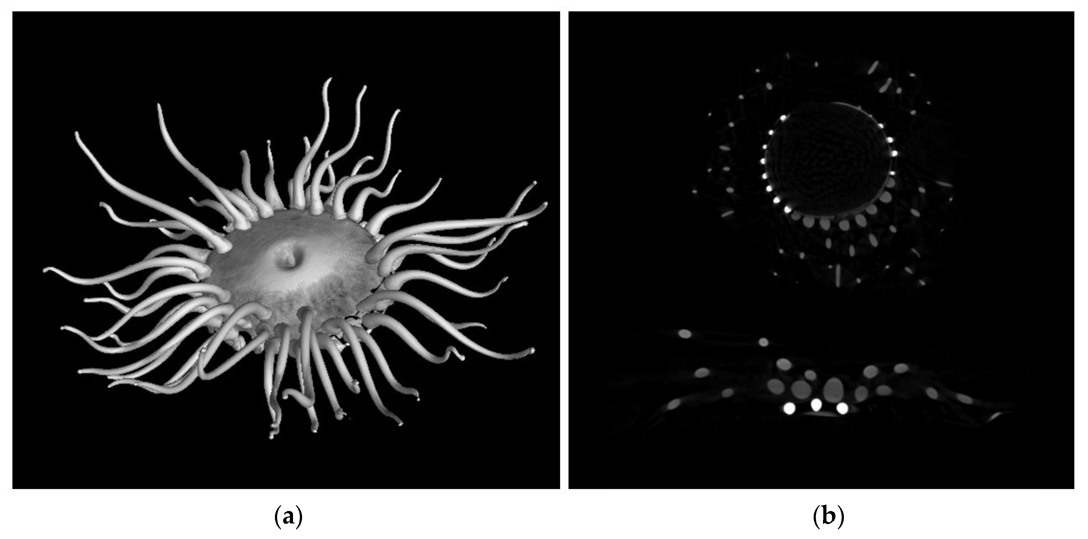
Figure 1. CT analysis of Anthea cereus: (a) 3D rendering; (b) axial tomographic slice. Radiopaque elements appear in brighter tones.
Heritage 08 00376 g001
Figure 2. (a) False-colour 3D rendering of tomographic data relating to Aurelia aurita; the most radiopaque areas are in light grey; (b) false-colour 3D rendering of tomographic data of Bougainvillia fruticosa, with the most radiopaque areas in yellow.
Heritage 08 00376 g002
Figure 3. Three-dimensional rendering of the tomographic data of Hydractinia echinata. The most radiopaque areas are in yellow.
Heritage 08 00376 g003
Figure 4. (a) Three-dimensional rendering of the tomographic data of Corallium rubrum, with the most radiopaque regions highlighted in yellow; (b) frontal CT slice of element B and (c) sagittal CT slice of element A, where the bright vertical line corresponds to the nail. Images (b,c) show the most radiopaque areas in white.
Heritage 08 00376 g004
Figure 5. (a) BSE image of the layered superficial layer of sample AC2. (b) BSE image of the layered superficial layer of sample CR1. The image only shows the top part of the specimen, i.e., the paint layer. As noted, the samples from Anthea cereus, AC1 and AC2, have two very different compositions, the first being a high-lead glass, the second being a mixed alkali glass. In sample AC1, there is also a fair amount of chlorine corresponding to ClO2 around 1%, which could be justified by the fact that chlorine, in industrial times, was used as a refiner of the glass mass [31]. The presence of 0.8% phosphorus (P2O5) in sample AC1 (Pb-glass)—as measured by preliminary LA-ICP-MS analyses that are not fully reported in this work—and not detected in the other samples of the set, could result from impurities present in the raw materials or in the environment in which the glass was melted. Since a slightly lower content of phosphorus (P2O5 around 0.4%) is present in the other P glass sample HE2, its presence could be related either to Pb or K.
Heritage 08 00376 g005
Figure 6. (a) BSE SEM-EDS stratigraphy of the paint layer of sample CR1 (the bottom layer is the external one). (b) EDS spectrum of the external layer. Semi-quantitative estimation of detected elements: C +++, O ++, Mg +, Al +, Si +, S +, Ca +++, Zn +. (c) BSE SEM-EDS stratigraphy of the internal paint layer of sample CR1. (d) EDS spectrum of the internal layer—grey area. Semi-quantitative estimation of detected elements: C +, O +, S +, Ca +, Zn +++. (e) BSE SEM-EDS detail of the internal paint layer of sample CR1. (f) EDS spectrum of the internal layer—red pigment (white BSE area). Semi-quantitative estimation of detected elements: C +, O +, S +++, Zn +, Hg +++.
Heritage 08 00376 g006
Figure 7. UVF imaging details showing variations in fluorescence intensity of adhesives: group one (pale sky-blue with icy white highlights) in Aurelia aurita (a), Bougainvillia fruticosa (b), and element A of Anthea cereus (c); group two (cooler blue tone) in Corallium rubrum (d) and Hydractinia echinata (e); group three (intermediate sky-blue) in element B of Anthea cereus (f).
Heritage 08 00376 g007
Figure 8. UVF images of (a) Anthea cereus and (b) Bougainvillia fruticosa. In both cases, note the differences in fluorescence intensity—light sky-blue fluorescence tending toward ice white—observed in the junction areas where parts have been joined. Additionally, a clear green-yellow fluorescence of the glass surface is visible in the Bougainvillia fruticosa model (b).
Heritage 08 00376 g008
Figure 9. FT-IR spectra of C2 and C6 samples (a), animal glue (b), and mastic resin (c).
Heritage 08 00376 g009
Figure 10. FT-IR spectrum of C5: O–NO2 asymmetrical stretching (1650 cm−1), NO2 symmetrical stretching (1280 cm−1), and N–O stretching of cellulose nitrate (833 cm−1) of cellulose nitrate; C=O stretching (1722 cm−1) indicates the presence of a carbonyl-containing compound (i.e., camphor or a phthalate ester added as plasticizer, or degradation products).
Heritage 08 00376 g010
Table 1. The analysed Blaschka models.
ModelNameCollection
Heritage 08 00376 i001Anthea cereus
sea anemone
(A: oral disc and tentacles; B: body and base)
Liceo Ugo Foscolo, Pavia
Heritage 08 00376 i002Aurelia aurita
jellyfish
Liceo Ugo Foscolo, Pavia
Heritage 08 00376 i003Bougainvillia fruticosaLiceo Giovanni Prati, Trento
Heritage 08 00376 i004Corallium rubrum
Red coral
(A: Corallium rubrum; B: enlargement of Corallium rubrum terminal branch)
Liceo Ugo Foscolo, Pavia
Heritage 08 00376 i005Hydractinia echiantaLiceo Giovanni Prati, Trento
Table 2. Position of sampling for SEM-EDS analyses.
ModelSamplesSample MaterialPosition of SamplingCT Indication for Sampling
Aurelia auritaAA1 (bell)
Heritage 08 00376 i006
AA2 (tentacle)
Heritage 08 00376 i007
GlassHeritage 08 00376 i013Bell and tentacles have a different radiopacity. No sampling was allowed of the pink glass
Anthea cereusAC1 (hemispheres—highly radiopaque area)
Heritage 08 00376 i008
AC2 (tentacle)
Heritage 08 00376 i009
GlassHeritage 08 00376 i014The tentacles and the corolla are less radiopaque than the small hemispheres attached to the edge of the central part
Corallium rubrumCR1 (polyp with paint layer)
Heritage 08 00376 i010
Glass + paint layerHeritage 08 00376 i015Higher radiopacity of the paint layer
Hydractinia echinataHE1 (tentacle—low-radiopacity area)
Heritage 08 00376 i011
HE2 (spheres)
Heritage 08 00376 i012
GlassHeritage 08 00376 i016Highly radiopaque small spheres are glued to the base and the ends of some tentacles are also quite radiopaque. No sampling was allowed of the yellow glass
Table 3. Semi-quantitative compositions of the samples analysed by SEM-EDS (* sup = superficial layer).
wt%AA1AA2AC1AC2AC2_sup *CR1CR1_sup *HE1HE2
Na2O13.914.364.1214.851.4914.743.1414.337.58
MgO0.210.170.53n.d.n.d.0.140.180.190.89
Al2O32.131.90.141.241.893.043.421.880.72
SiO272.171.5447.4969.4783.8469.9979.4971.3561.08
SO30.330.742.2n.d.n.d.0.790.680.78n.d.
PbO20.32n.d.31.46n.d.n.d.n.d.n.d.n.d.15.29
Cl2O0.230.210.981.221.410.300.180.220.28
K2O3.913.949.877.434.313.584.043.9410.79
CaO6.326.551.755.286.316.798.176.632.65
MnO0.310.350.180.250.370.310.480.400.33
Fe2O30.260.240.470.270.380.300.230.270.38
Table 4. Summary of SEM-EDS results.
ModelSampleZone of ProvenanceType of Glass
Aurelia auritaAA1BellMixed alkali
AA2TentacleMixed alkali
Anthea cereusAC1Lower edge
of the A element
Lead glass
AC2TentacleMixed alkali
Bougainvillia fruticosa---
Corallium rubrumCR1GlassPolyp of the A elementMixed alkali
ColourExterior layerPolyp of the A elementAdhesive
Interior layerPolyp of the A elementZinc oxide and cinnabar
Hydractinia echinataHE1TentacleMixed alkali
HE2Sphere on the baseLead glass
Table 5. Summary table of the samples and FT-IR analysis results. (*) the amount of the sample was insufficient for analysis.
ModelsSampleSampling AreaDescriptionFT-IRComposition
Bougainvillia fruticosaC1Central stemYellow; hard and brittle~3370, 2935, 2974, 1714, 1655, 1543, 1453, 1384, 1245Protein adhesive (i.e., animal glue) + natural resin (i.e., mastic)
Aurelia auritaC2ManubriumYellow/orange; plastic consistency~3440, 2937, 2875, ~2645, 1712, 1659, 1544, 1457, 1383, 1246Protein adhesive (i.e., animal glue) + natural resin (i.e., mastic)
C3Tentacle attachmentYellow/orange~3370, 2935, 2872, ~2650, 1712, 1653, 1542, 1460, 1384, 1247Protein adhesive (i.e., animal glue) + natural resin (i.e., mastic)
Anthea cereusC4Element BYellow; hard and brittleNo valuable results (*)-
Corallium rubrumC5Cylindrical elementYellow/orange; hard and brittle2962, 2923, 2872, 1722, 1650, 1280, 1066, 833, 742, 687Cellulose nitrate
Hydractinia echinataC6BaseYellow; hard and brittle~3370, 2959, 2935, 2974, 1720, 1655, 1543, 1453, 1250Protein adhesive (i.e., animal glue) + natural resin (i.e., mastic)
Disclaimer/Publisher’s Note: The statements, opinions and data contained in all publications are solely those of the individual author(s) and contributor(s) and not of MDPI and/or the editor(s). MDPI and/or the editor(s) disclaim responsibility for any injury to people or property resulting from any ideas, methods, instructions or products referred to in the content.