Exploring How Micro-Computed Tomography Imaging Technology Impacts the Preservation of Paleontological Heritage
Abstract
1. Introduction
1.1. Micro-CT Imaging for Ichnology
1.2. Micro-CT for Restoring Samples
1.3. Micro-CT for Digitalizing Samples
2. Materials and Methods
2.1. Micro-CT Devices
2.2. Investigated Samples
2.2.1. Sealed Bee Cells
2.2.2. O.P.D. Institute of Conservation and Restoration
2.2.3. G.A.M.P.S. Collection
3. Results
3.1. The Case Study of Sealed Bee Cells
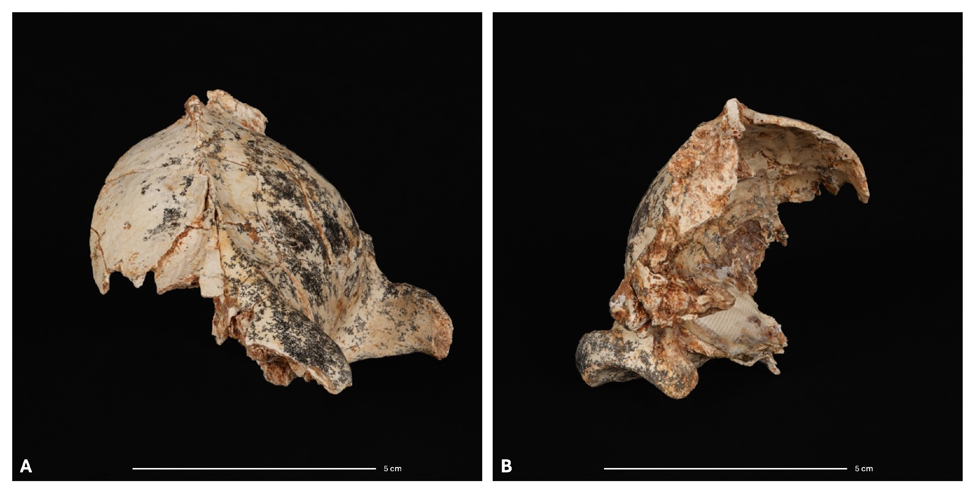
3.2. The Case Study of Menthol in Restoration Artifacts
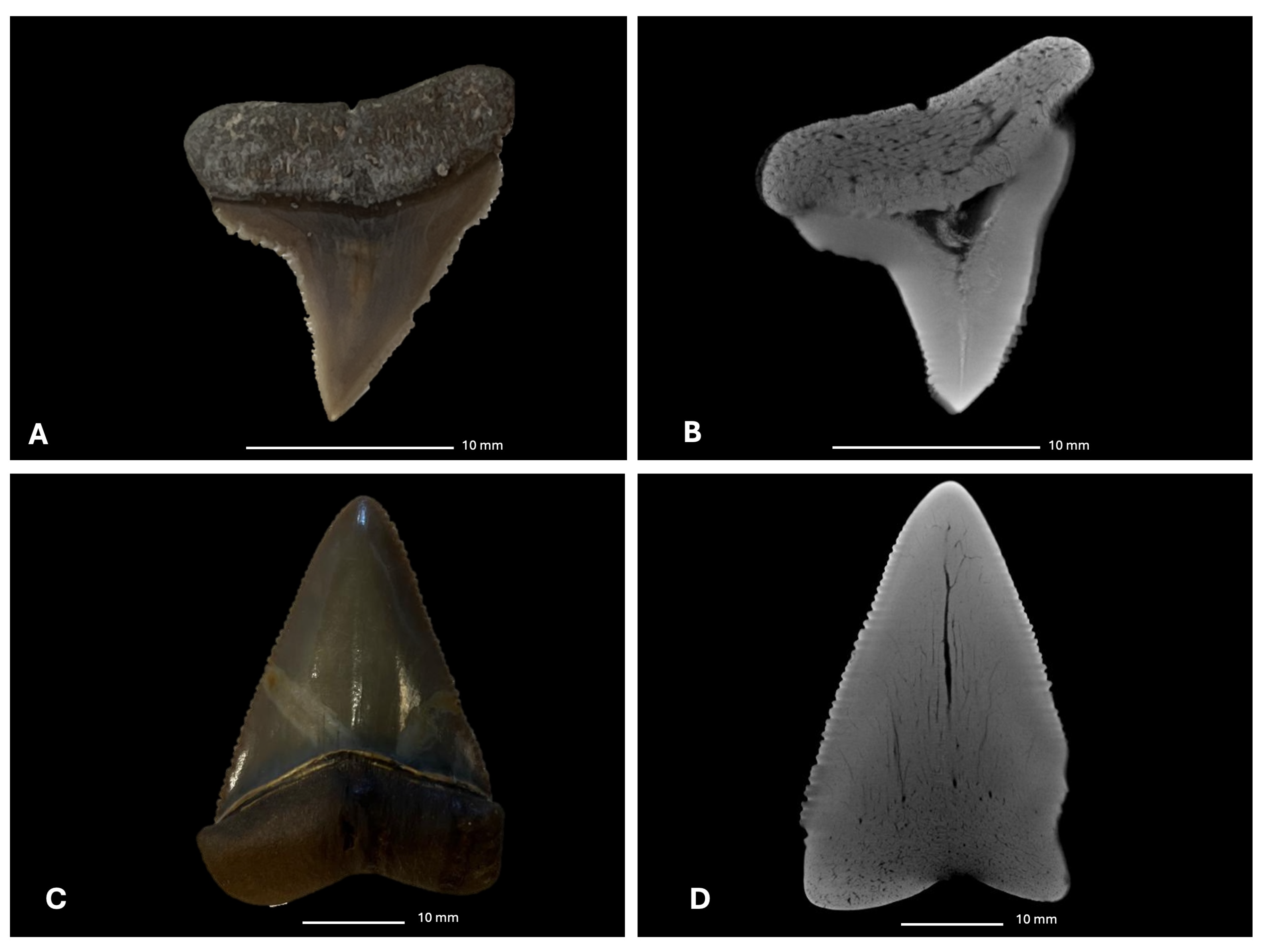
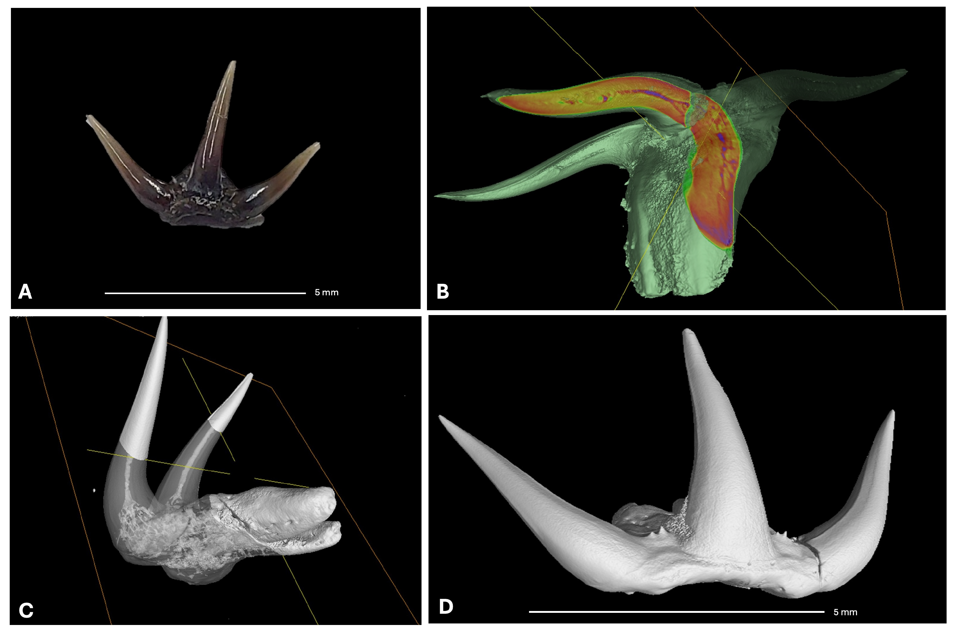
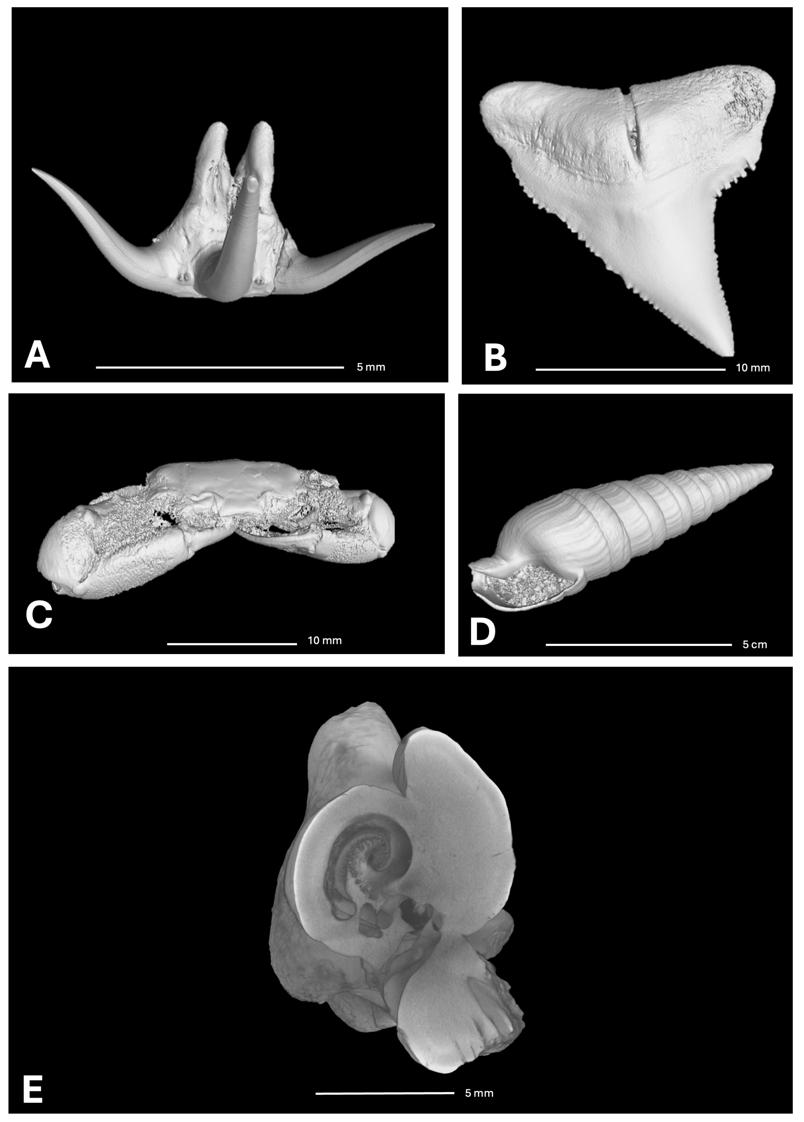
3.3. The G.A.M.P.S. Case Study
4. Discussion
4.1. Trace Fossils: Challenges and Advantages of Micro-CT in Paleontology
4.2. Menthol as a Restoration Material: Insights and Limitations
4.3. Three-Dimensional Modeling and Digital Conservation: Implications for Museums
5. Conclusions
Author Contributions
Funding
Institutional Review Board Statement
Informed Consent Statement
Data Availability Statement
Acknowledgments
Conflicts of Interest
References
- Hipsley, C.A.; Aguilar, R.; Black, J.R.; Hocknull, S.A. High-throughput microCT scanning of small specimens: Preparation, packing, parameters and post-processing. Sci. Rep. 2020, 10, 13863. [Google Scholar]
- Ritman, E.L. Micro-computed tomography—Current status and developments. Annu. Rev. Biomed. Eng. 2004, 6, 185–208. [Google Scholar] [CrossRef] [PubMed]
- Tate, J.R.; Cann, C.E. High-resolution computed tomography for the comparative study of fossil and extant bone. Am. J. Phys. Anthropol. 1982, 58, 67–73. [Google Scholar] [CrossRef]
- Elliott, J.; Davis, G.; Dover, S. X-ray microtomography: Past and present. In Proceedings of the Developments in X-Ray Tomography VI, SPIE, San Diego, CA, USA, 12–14 August 2008; Volume 7078, pp. 33–43. [Google Scholar]
- Ritman, E.L. Current status of developments and applications of micro-CT. Annu. Rev. Biomed. Eng. 2011, 13, 531–552. [Google Scholar] [CrossRef]
- Clark, D.; Badea, C. Advances in micro-CT imaging of small animals. Phys. Medica 2021, 88, 175–192. [Google Scholar] [CrossRef]
- Keklikoglou, K.; Arvanitidis, C.; Chatzigeorgiou, G.; Chatzinikolaou, E.; Karagiannidis, E.; Koletsa, T.; Magoulas, A.; Makris, K.; Mavrothalassitis, G.; Papanagnou, E.D.; et al. Micro-CT for biological and biomedical studies: A comparison of imaging techniques. J. Imaging 2021, 7, 172. [Google Scholar] [CrossRef]
- Zabler, S.; Maisl, M.; Hornberger, P.; Hiller, J.; Fella, C.; Hanke, R. X-ray imaging and computed tomography for engineering applications. Tm-Tech. Mess. 2021, 88, 211–226. [Google Scholar] [CrossRef]
- Reedy, C.L.; Reedy, C.L. High-resolution micro-CT with 3D image analysis for porosity characterization of historic bricks. Herit. Sci. 2022, 10, 83. [Google Scholar] [CrossRef]
- Piroddi, L.; Abu Zeid, N.; Calcina, S.V.; Capizzi, P.; Capozzoli, L.; Catapano, I.; Cozzolino, M.; D’Amico, S.; Lasaponara, R.; Tapete, D. Imaging cultural heritage at different scales: Part I, the micro-scale (manufacts). Remote Sens. 2023, 15, 2586. [Google Scholar] [CrossRef]
- Calo, C.M.; Marconetto, B. Sobre el uso imágenes microtomográficas para estudios de carbón de madera arqueológico. Rev. Mus. Antropol. 2024, 17, 13–28. [Google Scholar] [CrossRef]
- Jaques, V.A.; Zemek, M.; Šalplachta, J.; Zikmund, T.; Ožvoldík, D.; Kaiser, J. X-ray high resolution computed tomography for cultural heritage material micro-inspection. In Proceedings of the Optics for Arts, Architecture, and Archaeology VIII, SPIE, Online, 21–25 June 2021; Volume 11784, pp. 111–118. [Google Scholar]
- Coletti, G.; Stainbank, S.; Fabbrini, A.; Spezzaferri, S.; Foubert, A.; Kroon, D.; Betzler, C. Biostratigraphy of large benthic foraminifera from Hole U1468A (Maldives): A CT-scan taxonomic approach. Swiss J. Geosci. 2018, 111, 523–536. [Google Scholar] [CrossRef]
- Hutchinson, J.C.; Shelmerdine, S.C.; Simcock, I.C.; Sebire, N.J.; Arthurs, O.J. Early clinical applications for imaging at microscopic detail: Microfocus computed tomography (micro-CT). Br. J. Radiol. 2017, 90, 20170113. [Google Scholar] [CrossRef] [PubMed]
- Schmidt, J.; Scholz, S.; Wiesner, J.; Will, K. MicroCT data provide evidence correcting the previous misidentification of an Eocene amber beetle (Coleoptera, Cicindelidae) as an extant species. Sci. Rep. 2023, 13, 14743. [Google Scholar] [CrossRef] [PubMed]
- Edie, S.M.; Collins, K.S.; Jablonski, D. High-throughput micro-CT scanning and deep learning segmentation workflow for analyses of shelly invertebrates and their fossils: Examples from marine Bivalvia. Front. Ecol. Evol. 2023, 11, 1127756. [Google Scholar] [CrossRef]
- Wisshak, M.; Titschack, J.; Kahl, W.A.; Girod, P. Classical and new bioerosion trace fossils in Cretaceous belemnite guards characterised via micro-CT. Foss. Rec. 2017, 20, 173–199. [Google Scholar] [CrossRef]
- Racicot, R. Fossil secrets revealed: X-ray CT scanning and applications in paleontology. Paleontol. Soc. Pap. 2016, 22, 21–38. [Google Scholar] [CrossRef]
- Wang, Y.-F.; Wei, C.-F.; Que, J.-M.; Zhang, W.-D.; Sun, C.-L.; Shu, Y.-F.; Hou, Y.-M.; Zhang, J.-C.; Shi, R.-J.; Wei, L. Development and applications of paleontological computed tomography. Vertebr. PalAsiatica 2019, 57, 84. [Google Scholar]
- Sutton, M.D. Tomographic techniques for the study of exceptionally preserved fossils. Proc. R. Soc. B Biol. Sci. 2008, 275, 1587–1593. [Google Scholar] [CrossRef]
- Ball, A.; Abel, R.; Ambers, J.; Brierley, L.; Howard, L. Micro-computed tomography applied to museum collections. Microsc. Microanal. 2011, 17, 1794–1795. [Google Scholar] [CrossRef][Green Version]
- Caloi, I.; Bernardini, F. Revealing primary forming techniques in wheel-made ceramics with X-ray microCT. J. Archaeol. Sci. 2024, 169, 106025. [Google Scholar] [CrossRef]
- Abate, F.; De Bernardin, M.; Stratigaki, M.; Franceschin, G.; Albertin, F.; Bettuzzi, M.; Brancaccio, R.; Bressan, A.; Morigi, M.P.; Daniele, S.; et al. X-ray computed microtomography: A non-invasive and time-efficient method for identifying and screening Roman copper-based coins. J. Cult. Herit. 2024, 66, 436–443. [Google Scholar] [CrossRef]
- Dierick, M.; Cnudde, V.; Masschaele, B.; Vlassenbroeck, J.; Van Hoorebeke, L.; Jacobs, P. Micro-CT of fossils preserved in amber. Nucl. Instruments Methods Phys. Res. Sect. A Accel. Spectrometers Detect. Assoc. Equip. 2007, 580, 641–643. [Google Scholar] [CrossRef]
- Dierick, M.; Van Hoorebeke, L.; Jacobs, P.; Masschaele, B.; Vlassenbroeck, J.; Cnudde, V.; De Witte, Y. The use of 2D pixel detectors in micro-and nano-CT applications. Nucl. Instruments Methods Phys. Res. Sect. A Accel. Spectrometers Detect. Assoc. Equip. 2008, 591, 255–259. [Google Scholar] [CrossRef]
- Silbiger, N.J.; Guadayol, Ò.; Thomas, F.I.; Donahue, M.J. A novel μ CT analysis reveals different responses of bioerosion and secondary accretion to environmental variability. PLoS ONE 2016, 11, e0153058. [Google Scholar] [CrossRef] [PubMed]
- Merella, M.; Farina, S.; Scaglia, P.; Caneve, G.; Bernardini, G.; Pieri, A.; Collareta, A.; Bianucci, G. Structured-Light 3D Scanning as a Tool for Creating a Digital Collection of Modern and Fossil Cetacean Skeletons (Natural History Museum, University of Pisa). Heritage 2023, 6, 6762–6776. [Google Scholar] [CrossRef]
- Das, A.J.; Murmann, D.C.; Cohrn, K.; Raskar, R. A method for rapid 3D scanning and replication of large paleontological specimens. PLoS ONE 2017, 12, e0179264. [Google Scholar] [CrossRef]
- Skublewska-Paszkowska, M.; Milosz, M.; Powroznik, P.; Lukasik, E. 3D technologies for intangible cultural heritage preservation—literature review for selected databases. Herit. Sci. 2022, 10, 3. [Google Scholar] [CrossRef]
- Erolin, C.; Jarron, M.; Csetenyi, L.J. Zoology 3D: Creating a digital collection of specimens from the D’Arcy Thompson Zoology Museum. Digit. Appl. Archaeol. Cult. Herit. 2017, 7, 51–55. [Google Scholar] [CrossRef]
- Frey, R.W.; Pemberton, S.G. Biogenic structures in outcrops and cores. I. Approaches to ichnology. Bull. Can. Pet. Geol. 1985, 33, 72–115. [Google Scholar]
- Meyer, M.; Polys, N.; Yaqoob, H.; Hinnov, L.; Xiao, S. Beyond the stony veil: Reconstructing the Earth’s earliest large animal traces via computed tomography x-ray imaging. Precambrian Res. 2017, 298, 341–350. [Google Scholar] [CrossRef]
- Baucon, A.; Piazza, M.; Cabella, R.; Bonci, M.C.; Capponi, L.; de Carvalho, C.N.; Briguglio, A. Buildings that ‘speak’: Ichnological geoheritage in 1930s buildings in Piazza della Vittoria (Genova, Italy). Geoheritage 2020, 12, 70. [Google Scholar] [CrossRef]
- Fu, S.; Werner, F.; Brossmann, J. Computed tomography: Application in studying biogenic structures in sediment cores. Palaios 1994, 116–119. [Google Scholar] [CrossRef]
- Dorador Rodríguez, J.; Rodríguez Tovar, F.J.; Titschack, J. Exploring computed tomography in ichnological analysis of cores from modern marine sediments. Sci. Rep. 2020, 10, 201. [Google Scholar] [CrossRef]
- Marenco, K.N.; Bottjer, D.J. Quantifying bioturbation in Ediacaran and Cambrian rocks. In Quantifying the Evolution of Early Life: Numerical Approaches to the Evaluation of Fossils and Ancient Ecosystems; Springer: Dordrecht, The Netherlands, 2011; pp. 135–160. [Google Scholar]
- Bromley, R.G. Trace Fossils. Biology, Taphonomy and Applications; Chapman & Hall: London, UK, 1996; p. 361. [Google Scholar]
- Heřmanová, Z.; Bruthansová, J.; Holcová, K.; Mikuláš, R.; Kočová Veselská, M.; Kočí, T.; Dudák, J.; Vohník, M. Benefits and limits of x-ray micro-computed tomography for visualization of colonization and bioerosion of shelled organisms. Palaeontol. Electron 2020, 23, a23. [Google Scholar] [CrossRef] [PubMed]
- Noffke, N.; Gerdes, G.; Klenke, T.; Krumbein, W.E. Microbially induced sedimentary structures: A new category within the classification of primary sedimentary structures. J. Sediment. Res. 2001, 71, 649–656. [Google Scholar] [CrossRef]
- De Carvalho, C.N.; Couto, H.; Figueiredo, M.V.; Baucon, A. Microbial-related biogenic structures from the Middle Ordovician slates of Canelas (northern Portugal). Comun. Geológicas 2016, 103, 23–38. [Google Scholar]
- Neto de Carvalho, C.; Baucon, A.; Badano, D.; Proença Cunha, P.; Ferreira, C.; Figueiredo, S.; Muñiz, F.; Belo, J.; Bernardini, F.; Cachão, M. Eucera Bees (Hymenoptera, Apidae, Eucerini) Preserved Their Brood Cells Late Holocene (middle Neoglacial) Palaeosols Southwest Port. Pap. Palaeontol. 2023, 9, e1518. [Google Scholar] [CrossRef]
- Zhang, W.; Wang, X.; Han, X.; Meng, C.; Huang, X.; Luo, H. Laboratory research of solvent-assisted menthol sols as temporary consolidants in archaeological excavation applications. Herit. Sci. 2022, 10, 74. [Google Scholar] [CrossRef]
- Vincenzo, A. Il Restauro di un Crocifisso Ligneo Attribuito alla Bottega di Giovanni Teutonico, Distrutto Durante il Terremoto del 2016. Consolidamento Temporaneo del Colore con Adesivi Volatili, Risanamento Strutturale e Ricomposizione. Ph.D. Thesis, Opificio delle Pietre Dure e Laboratori di Restauro di Firenze—OPD, Florence, Italy, 2021. [Google Scholar]
- Langdon, K.; Skinner, L.; Shugar, A. Archaeological Block-Lifting with Volatile Binding Media: Exploring Alternatives to Cyclododecane; University of Cambridge Museums: Cambridge, UK, 2019. [Google Scholar] [CrossRef]
- Bartolini-Lucenti, S.; Rook, L. Nurturing Italian Geo-palaeontological Heritage with Virtual Palaeontology: Preliminary Report of Its Application in Two Natural History Museums. Geoheritage 2023, 15, 40. [Google Scholar] [CrossRef]
- YXLON. International YXLON GmbH, Essener Bogen 15, 22419 Hamburg, Germany. Available online: https://yxlon.comet.tech/ (accessed on 29 November 2024).
- VGMAX. Software by Volume Graphics Part of Hexagon. Available online: https://www.volumegraphics.com/en/products/vgsm.htm (accessed on 29 November 2024).
- Baucon, A.; de Carvalho, C.N. Can AI Get a Degree in Geoscience? Performance Analysis of a GPT-Based Artificial Intelligence System Trained for Earth Science (GeologyOracle). Geoheritage 2024, 16, 121. [Google Scholar] [CrossRef]
- Lorenzo, R. The 1980s field researches at Pirro Nord were developed thanks to the inspired and energetic activity of Claudio De Giuli (1938–1988). Palaeontogr. Abt. A Paläozoologie Stratigr. 2013, 298, 1–3. [Google Scholar]
- Zunino, M.; Pavia, M.; Arzarello, M.; Bertok, C.; Di Carlo, M.; DI DONATO, V.; Graziano, R.; Matteucci, R.; Nicosia, U.; Petronio, C.; et al. Il Gargano, un archivio della diversità geologica dal Mesozoico al Pleistocene. Geol. Field Trips 2012, 4, 1–137. [Google Scholar]
- Collareta, A.; Casati, S.; Di Cencio, A.; Bianucci, G. The Deep Past of the White Shark, Carcharodon Carcharias, Mediterr. Sea: A Synth. Its Palaeobiology Palaeoecol. Life 2023, 13, 2085. [Google Scholar] [CrossRef]
- Cigala Fulgosi, F.; Casati, S.; Orlandini, A.; Persico, D. A small fossil fish fauna, rich in Chlamydoselachus Teeth, Late Pliocene Tuscany (Siena, Cent. Italy). Cainozoic Res. 2009, 6, 3–23. [Google Scholar]
- Bianucci, G.; Vaiani, S.C.; Casati, S. A new delphinid record (Odontoceti, Cetacea) from the Early Pliocene of Tuscany (central Italy): Systematics and biostratigraphic considerations. Neues Jahrb. Geol. Paläontologie Abh. 2009, 11, 275. [Google Scholar] [CrossRef]
- Barucci, A.; Ciacci, G.; Liò, P.; Azevedo, T.; Di Cencio, A.; Merella, M.; Bianucci, G.; Bosio, G.; Casati, S.; Collareta, A. An explainable Convolutional Neural Network approach to fossil shark tooth identification. Boll. Soc. Paleontol. Ital. 2024, 63, 216. [Google Scholar]
- Bosio, G.; Bianucci, G.; Collareta, A.; Landini, W.; Urbina, M.; Di Celma, C. Ultrastructure, composition, and 87Sr/86Sr dating of shark teeth from lower Miocene sediments of southwestern Peru. J. South Am. Earth Sci. 2022, 118, 103909. [Google Scholar] [CrossRef]
- Cappetta, H. Chondrichthyes: Mesozoic and Cenozoic Elasmobranchii: Teeth. In Handbook of Paleoichthyology; Gustav Fischer Verlag: Stuttgart, Germany, 2012. [Google Scholar]
- Jambura, P.L.; Türtscher, J.; Kindlimann, R.; Metscher, B.; Pfaff, C.; Stumpf, S.; Weber, G.W.; Kriwet, J. Evolutionary trajectories of tooth histology patterns in modern sharks (Chondrichthyes, Elasmobranchii). J. Anat. 2020, 236, 753–771. [Google Scholar] [CrossRef] [PubMed]
- Glickman, L. Class Chondrichthyes, Subclass Elasmobranchii. Osn. Paleontol. 1964, 11, 195–236. [Google Scholar]
- Moyer, J.K.; Riccio, M.L.; Bemis, W.E. Development and microstructure of tooth histotypes in the blue shark, P rionace glauca (C archarhiniformes: C archarhinidae) and the great white shark, C archarodon carcharias (L amniformes: L amnidae). J. Morphol. 2015, 276, 797–817. [Google Scholar] [CrossRef] [PubMed]
- Ørvig, T. Histologic Studies of Placoderms and Fossil Elasmobranchs; Almqvist & Wiksell: Stockholm, Sweden, 1951. [Google Scholar]
- Sutton, M.; Rahman, I.; Garwood, R. Virtual paleontology—An overview. Paleontol. Soc. Pap. 2016, 22, 1–20. [Google Scholar] [CrossRef]
- Travel + Leisure. 15 Museums Around the World You Can Visit Virtually. 2025. Available online: https://www.travelandleisure.com/attractions/museums-galleries (accessed on 14 April 2025).
Disclaimer/Publisher’s Note: The statements, opinions and data contained in all publications are solely those of the individual author(s) and contributor(s) and not of MDPI and/or the editor(s). MDPI and/or the editor(s) disclaim responsibility for any injury to people or property resulting from any ideas, methods, instructions or products referred to in the content. |
© 2025 by the authors. Licensee MDPI, Basel, Switzerland. This article is an open access article distributed under the terms and conditions of the Creative Commons Attribution (CC BY) license (https://creativecommons.org/licenses/by/4.0/).
Share and Cite
Amendola, M.; Barucci, A.; Baucon, A.; Zini, C.; Borrelli, C.; Casati, S.; Cencio, A.d.; Fiore, S.; Siano, S.; Agresti, J.; et al. Exploring How Micro-Computed Tomography Imaging Technology Impacts the Preservation of Paleontological Heritage. Heritage 2025, 8, 310. https://doi.org/10.3390/heritage8080310
Amendola M, Barucci A, Baucon A, Zini C, Borrelli C, Casati S, Cencio Ad, Fiore S, Siano S, Agresti J, et al. Exploring How Micro-Computed Tomography Imaging Technology Impacts the Preservation of Paleontological Heritage. Heritage. 2025; 8(8):310. https://doi.org/10.3390/heritage8080310
Chicago/Turabian StyleAmendola, Michela, Andrea Barucci, Andrea Baucon, Chiara Zini, Claudia Borrelli, Simone Casati, Andrea di Cencio, Sandra Fiore, Salvatore Siano, Juri Agresti, and et al. 2025. "Exploring How Micro-Computed Tomography Imaging Technology Impacts the Preservation of Paleontological Heritage" Heritage 8, no. 8: 310. https://doi.org/10.3390/heritage8080310
APA StyleAmendola, M., Barucci, A., Baucon, A., Zini, C., Borrelli, C., Casati, S., Cencio, A. d., Fiore, S., Siano, S., Agresti, J., Neto de Carvalho, C., Bernardini, F., Lo Russo, G., Collareta, A., & Bosio, G. (2025). Exploring How Micro-Computed Tomography Imaging Technology Impacts the Preservation of Paleontological Heritage. Heritage, 8(8), 310. https://doi.org/10.3390/heritage8080310


