
In the Likeness of a God: The Non-Invasive Investigation of Animal Votives


Abstract
1. Introduction
2. Materials and Methods
2.1. Radiographic Method
2.2. Photogrammetric Method
3. Results
3.1. Radiographic Results
3.2. Photogrammetric Results
4. Discussion
5. Conclusions
Funding
Data Availability Statement
Acknowledgments
Conflicts of Interest
References
- Scalf, F. The role of birds within the religious landscape of ancient Egypt. In Between Heaven & Earth: Birds in Ancient Egypt; Bailleul-LeSuer, R., Ed.; Oriental Institute of the University of Chicago: Chicago, IL, USA, 2012; pp. 33–40. [Google Scholar]
- Frankfort, H. The Egyptian Gods. In Ancient Egyptian Religion: An Interpretation; Columbia University Press: New York, NY, USA, 1948; pp. 1–29. [Google Scholar] [CrossRef]
- Assmann, J. The Search for God in Ancient Egypt; Cornell University Press: Ithaca, NY, USA, 2001. [Google Scholar]
- Teeter, E. Religion and Ritual in Ancient Egypt; Cambridge University Press: Cambridge, UK, 2011. [Google Scholar]
- Barbash, Y. How the Ancient Egyptians Viewed the Animal World. In Soulful Creatures: Animal Mummies in Ancient Egypt; Bleiberg, E., Barbash, Y., Bruno, L., Eds.; Brooklyn Museum Press: New York, NY, USA, 2013; pp. 19–62. [Google Scholar]
- Meeks, D.; Farvard-Meeks, C. Daily Life of the Egyptian Gods; Cornell University Press: Ithaca, NY, USA, 1996. [Google Scholar]
- Kessler, D. Tierkult. In Lexikon der Ägyptologie; Helck, W., Otto, E., Westendorf, W., Eds.; O. Harrassowitz: Wiesbaden, Germany, 1986; Volume 6, pp. 571–587. [Google Scholar]
- Baines, J.; Lesko, L.H.; Silverman, D.P. Religion in Ancient Egypt: Gods, Myths, and Personal Practice; Cornell University Press: Ithaca, NY, USA, 1991. [Google Scholar]
- Baines, J. Egyptian Deities in Context. Multiplicity, Unity, and the Problem of Change. In One God or Many? Concepts of Divinity in the Ancient World; Porter, B.N., Ed.; Casco Bay Assyriological Institute: Chebeague, ME, USA, 2000; pp. 9–78. [Google Scholar]
- Oberhelman, S.M. Anatomical Votive Reliefs as Evidence for Specialization at healing Sanctuaries in the Ancient Mediterranean World. Athens J. Health 2014, 1, 47–62. [Google Scholar] [CrossRef]
- Weinryb, I. Votive materials: Bodies and beyond. In Agents of Faith: Votive Objects in Time and Place; Bard Graduate Center: New York, NY, USA, 2018; pp. 33–59. [Google Scholar]
- Roberts, D.; Rainsford, C. Animal offerings in ritual, economic and social contexts in Britannia. Theor. Rom. Archaeol. J. 2024, 7, 1–28. [Google Scholar] [CrossRef]
- Pinch, G.; Waraksa, E.A. Votive Practices. UCLA Encycl. Egyptol. 2009, 1, 1–9. Available online: https://escholarship.org/uc/item/7kp4n7rk (accessed on 1 November 2024).
- Price, C. Votive Practice in Ancient Egypt. In Gifts for the Gods: Ancient Egyptian Animal Mummies and the British; McKnight, L.M., Atherton-Woolham, S., Eds.; Liverpool University Press: Liverpool, UK, 2015; pp. 59–60. [Google Scholar]
- Ray, J.D. The Archive of Hor; Egypt Exploration Society: London, UK, 1976. [Google Scholar]
- Nicholson, P.T. Sacred Animals at Saqqara. Heritage 2022, 5, 1240–1252. [Google Scholar] [CrossRef]
- Von den Driesch, A.; Kessler, D.; Steinmann, F.; Berteaux, V.; Peters, J. Mummified, deified and buried at Hermopolis Magna—The sacred birds from Tuna el-Gebel, Middle Egypt. Ägypten Levante XV Int. J. Egypt. Archaeol. Relat. Discip. 2005, 15, 203–244. [Google Scholar] [CrossRef]
- Baber, T. Ancient Corpses as Curiosities: Mummymania in the age of early travel. J. Anc. Egypt. Interconnect. 2016, 8, 60–93. [Google Scholar] [CrossRef]
- Edwards, A.B. A Thousand Miles Up the Nile; Routledge and Sons: London, UK, 1888. [Google Scholar]
- Edwards, A.B. Pharaohs, Fellahs and Explorers; Harper and Brothers: New York, NY, USA, 1891. [Google Scholar]
- Fagan, B.M. The Rape of the Nile; Scribner’s: New York, NY, USA, 1975. [Google Scholar]
- Larson, F. An Infinity of Things: How Sir Henry Wellcome Collected the World; Oxford University Press: Oxford, UK, 2009. [Google Scholar]
- Murray, J. A Handbook for Travellers in Lower and Upper Egypt; John Murray: London, UK, 1888. [Google Scholar]
- Yallop, J. Magpies, Squirrels and Thieves: How the Victorians Collected the World; Atlantic: London, UK, 2011. [Google Scholar]
- Brocklehurst, M. Miss Brocklehurst on the Nile: Diary of a Victorian Traveller in Egypt; Millrace: Stockport, UK, 2004. [Google Scholar]
- Brettell, R.; Martin, W.; Atherton-Woolham, S.; Stern, B.; McKnight, L. Organic residue analysis of Egyptian votive mummies and their research potential. Stud. Conserv. 2017, 62, 68–82. [Google Scholar] [CrossRef]
- Buckley, S.A.; Clark, K.A.; Evershed, R.P. Complex organic chemical balms of Pharaonic animal mummies. Nature 2004, 431, 294–299. [Google Scholar] [CrossRef]
- Łucejko, J.J.; Lluveras-Tenorio, A.; Modugno, F.; Ribechini, E.; Colombini, M.P. An analytical approach based on X-ray diffraction, Fourier transform infrared spectroscopy and gas chromatography/mass spectrometry to characterize Egyptian embalming materials. Microchem. J. 2012, 103, 110–118. [Google Scholar] [CrossRef]
- Ikram, S. Manufacturing Divinity: The Technology of Mummification. In Divine Creatures: Animal Mummies in Ancient Egypt; Ikram, S., Ed.; The American University in Cairo Press: Cairo, Egypt, 2015; pp. 17–44. [Google Scholar]
- McKnight, L.M. Imaging Applied to Animal Mummification in Ancient Egypt; BAR International Series 2175; Archaeopress: Oxford, UK, 2010. [Google Scholar]
- McKnight, L.; Bibb, R.; Chamberlain, A.; Mazza, R. Appearance and Reality in Ancient Egyptian Votive Animal Mummies. J. Anc. Egypt. Interconnect. 2018, 20, 52–57. [Google Scholar]
- Riggs, C. Unwrapping Ancient Egypt; Bloomsbury Academic: London, UK, 2014. [Google Scholar]
- Davies, S. Bronzes from the sacred animal necropolis at North Saqqara. In Gifts for the Gods: Images from Egyptian Temples; Hill, M., Schorsch, D., Eds.; The Metropolitan Museum of Art: New York, NY, USA, 2007; pp. 174–187. [Google Scholar]
- Kessler, D.; Nur el-Din, A. Tuna al-Gebel: Millions of Ibises and Other Animals. In Divine Creatures: Animal Mummies in Ancient Egypt; Ikram, S., Ed.; The American University in Cairo Press: Cairo, Egypt, 2015; pp. 120–163. [Google Scholar]
- Moschenska, G. Unrolling Egyptian mummies in nineteenth-century Britain. Br. J. Hist. Sci. 2014, 47, 451–477. [Google Scholar] [CrossRef]
- Stienne, A. Mummified; Manchester University Press: Manchester, UK, 2022. [Google Scholar]
- Raven, M.; Taconis, J.; Wybren, K. Egyptian Mummies: Radiological Atlas of the Collections in the National Museum of Antiquities in Leiden; Brepols: Turnhout, Belgium, 2005. [Google Scholar]
- Bruno, L. The Scientific Examination of Animal Mummies. In Soulful Creatures: Animal Mummies in Ancient Egypt; Bleiberg, E., Barbash, Y., Bruno, L., Eds.; Brooklyn Museum Press: New York, NY, USA, 2013; pp. 108–137. [Google Scholar]
- Adams, J. Imaging animal mummies: History and techniques. In Gifts for the Gods: Ancient Egyptian Animal Mummies and the British; McKnight, L.M., Atherton-Woolham, S., Eds.; Liverpool University Press: Liverpool, UK, 2015; pp. 68–71. [Google Scholar]
- McKnight, L.M.; Atherton-Woolham, S.D. The Evolution of Imaging Ancient Egyptian Animal Mummies at the University of Manchester, 1972–2014. In Mummies, Magic and Medicine in Ancient Egypt: Multidisciplinary Essays for Rosalie David; Price, C., Forshaw, R., Chamberlain, A., Nicholson, P.T., Morkot, R., Tyldesley, J., Eds.; Manchester University Press: Manchester, UK, 2016; pp. 345–354. [Google Scholar]
- Adams, J.E.; Alsop, C.W. Imaging in Egyptian Mummies. In Egyptian Mummies and Modern Science; David, A.R., Ed.; Cambridge University Press: Cambridge, UK, 2008; pp. 21–42. [Google Scholar]
- McKnight, L.M.; Atherton-Woolham, S.D.; Adams, J.E. Clinical Imaging of Ancient Egyptian Animal Mummies. RadioGraphics 2015, 35, 2108–2120. [Google Scholar] [CrossRef] [PubMed]
- Bibb, R.; McKnight, L.M. Identification of bird taxa species in ancient Egyptian mummies: Part 2, A qualitative evaluation of the utility of CT scanning and 3D Printing. J. Archaeol. Sci. Rep. 2022, 46, 103668. [Google Scholar] [CrossRef]
- McKnight, L.M.; White, J.; Rosier, A.; Cooper, J.; Bibb, R. Identification of avian remains contained within wrapped ancient Egyptian mummies: Part 1, A critical assessment of identification techniques. J. Archaeol. Sci. Rep. 2022, 45, 103585. [Google Scholar] [CrossRef]
- McKnight, L.M.; Bibb, R.; Cooper, F. Seeing is believing—The application of Three-Dimensional modelling technologies to reconstruct the final hours in the life of an ancient Egyptian Crocodile. Digit. Appl. Archaeol. Cult. Herit. 2024, 34, e00356. [Google Scholar] [CrossRef]
- Cox, S.L. A Critical Look at Mummy CT Scanning. Anat. Rec. 2015, 298, 1099–1110. [Google Scholar] [CrossRef]
- Lewin, P.K.; Harwood-Nash, D.C. Computerized axial tomography in medical archaeology. Paleopathol. Newsl. 1977, 17, 8–9. [Google Scholar]
- Croci, S.; Es Sebar, L.; Lombardo, L.; Di Iorio, F.; Buscaglia, P.; Taverni, F.; Aicardi, S.; Grassini, S. Dimensional accuracy assessment of 3D models based on photogrammetry and 3D scanner: A case study from the Museo Egizio of Turin. In Proceedings of the 2024 IEEE International Instrumentation and Measurement Technology Conference, Glasgow, UK, 20–23 May 2024. [Google Scholar] [CrossRef]
- Hanke, K. The Photogrammetric Contribution to Archaeological Documentation of Prehistory. Int. Arch. Photogramm. Remote Sens. 2000, 33, 355–357. [Google Scholar]
- Adamopoulos, E.; Rinaudo, F.; Ardissono, L. A Critical Comparison of 3D Digitization Techniques for Heritage Objects. ISPRS Int. J. Geo-Inf. 2021, 10, 10. [Google Scholar] [CrossRef]
- Konstantakis, M.; Trichopoulos, G.; Aliprantis, J.; Michalakis, K.; Caridakis, G.; Thanou, A.; Zafeiropoulos, A.; Sklavounou, S.; Psarras, C.; Papavassiliou, S.; et al. An Enhanced Methodology for Creating Digital Twins within a Paleontological Museum Using Photogrammetry and Laser Scanning Techniques. Heritage 2023, 6, 5967–5980. [Google Scholar] [CrossRef]
- Alfahal, T.; Alaabar, M. The Creative Implications of Photogrammetry in Museum Experience. In Proceedings of the International Conference on Sustaining Heritage: Innovative and Digital Approaches (ICSH), Sakhir, Bahrain, 18–19 June 2023; pp. 153–159. [Google Scholar] [CrossRef]
- Magnani, M.; Douglass, M.; Schroder, W.; Reeves, J.; Braun, D.R. The Digital Revolution to Come: Photogrammetry in Ar-chaeological Practice. Am. Antiq. 2020, 85, 737–760. [Google Scholar] [CrossRef]
- 3D Flow. Available online: https://www.3dflow.net/zephyr-doc/en/3DFMasquerade.html (accessed on 22 October 2024).
- Sportun, S. Conservation and care of animal mummies. In Gifts for the Gods: Ancient Egyptian Animal Mummies and the British; McKnight, L.M., Atherton-Woolham, S., Eds.; Liverpool University Press: Liverpool, UK, 2015. [Google Scholar]
- Nicolae, C.; Nocerino, E.; Menna, F.; Remondino, F. Photogrammetry applied to problematic artefacts. Int. Arch. Photogramm. Remote Sens. Spat. Inf. Sci. 2014, 40, 451–456. [Google Scholar] [CrossRef]
- Schaich, M.; Fritsch, D. Combined 3D scanning and photogrammetry surveys with 3D database support for archaeology & cultural heritage. A practice report on ArcTron’s information system aSPECT3D. In Photogrammetric Week’13; Wichmann/VDE Verlag: Belin, Germany, 2013; pp. 233–246. [Google Scholar]
- SketchFab. Available online: https://sketchfab.com/egyptbiobank (accessed on 22 October 2024).
- Kerr, J.P.; Knapp, D.; Frake, B.; Sellberg, M. “True” color surface anatomy: Mapping the Visible Human to patient-specific CT data. Comput. Med. Imaging Graph. 2000, 24, 153–164. [Google Scholar] [CrossRef]
- Visible Human Project. Available online: https://www.nlm.nih.gov/research/visible/visible_human.html (accessed on 13 June 2025).
- Zhan, K.; Song, Y.; Fritsch, D.; Mammadov, G.; Wagner, J. Computed Tomography Data Colouring Based on Photogrammetric Images. In Proceedings of the International Archives of the Photogrammetry, Remote Sensing and Spatial Information Sciences, XXIV ISPRS Congress, Nice, France, 31 August–2 September 2020; Volume XLIII-B2-2020. [Google Scholar]
- Larentis, O.; Gorini, I.; Campus, M.; Lorenzetti, M.; Mansueto, G.; Bortolotto, S.; Zappa, E.; Gregorini, A.; Rampazzi, L.; Vanin, S.; et al. Integrated multidisciplinary analysis of mobile digital radiographic acquisitions of the mummies of the hermits from the Sanctuary of Madonna della Corona (Trentino-Alto Adige, Italy—17th to 19th century CE). Front. Med. 2025, 11, 1492328. [Google Scholar] [CrossRef]
- Mainieri, S. Photogrammetry and Face Carvings: Exploring the ‘Face’ of the Egyptian Anthropoid Coffins by 3D Modeling. In Ancient Egypt, New Technology: The Present and Future of Computer Visualization, Virtual Reality and Other Digital Humanities in Egyptology; Lucarelli, R., Roberson, J.A., Vinson, S., Eds.; Brill: Leiden, The Netherlands, 2023; pp. 261–280. [Google Scholar]


| Accession No. | Image (Not to Scale) | External Description |
|---|---|---|
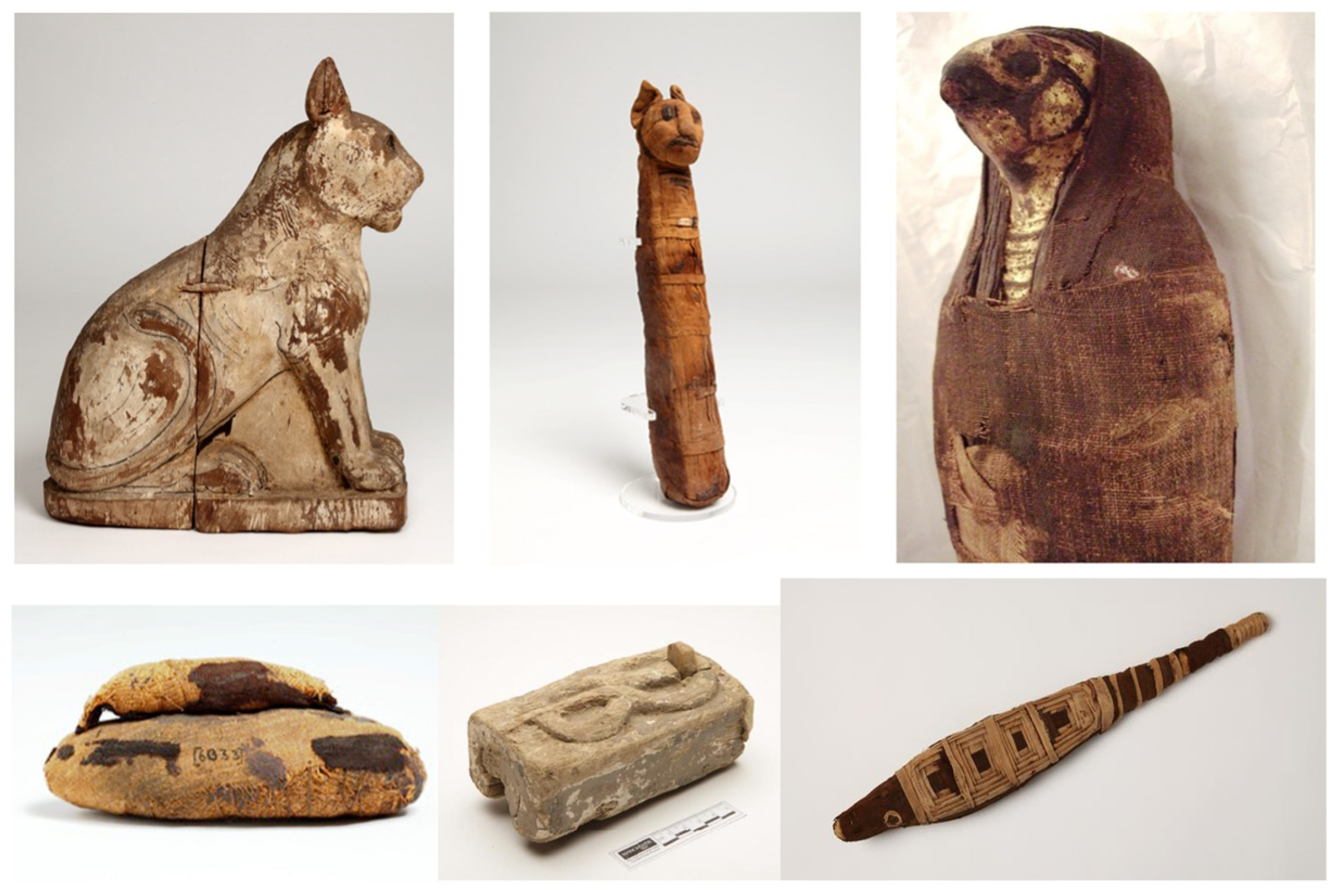
| 13861 | 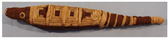 | Fully wrapped bundle with no discernable areas of damage. The upper aspect is decorated with four geometric square lozenges completed with a striped section over the tail area. The head is covered in a darker brown linen and has two false eyes. Excellent condition with no loose linen wrappings evident. Dimensions 410 × 70 × 40 (mm) |
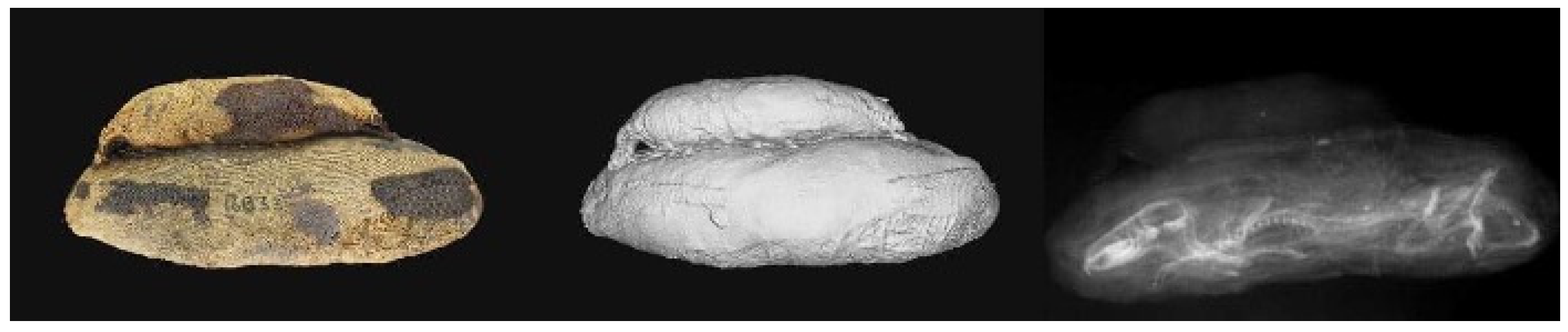
| 6033 | 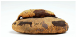 | Amorphous-shaped bundle wrapped in medium brown linen with a smaller ovoid-shaped bundle attached to the upper aspect. The bundle is wrapped in pale brown linen, with patches of a darker linen fixed to the surface. The museum number is written on the side in parentheses. Excellent condition. Dimensions 120 × 45 × 35 (mm). |
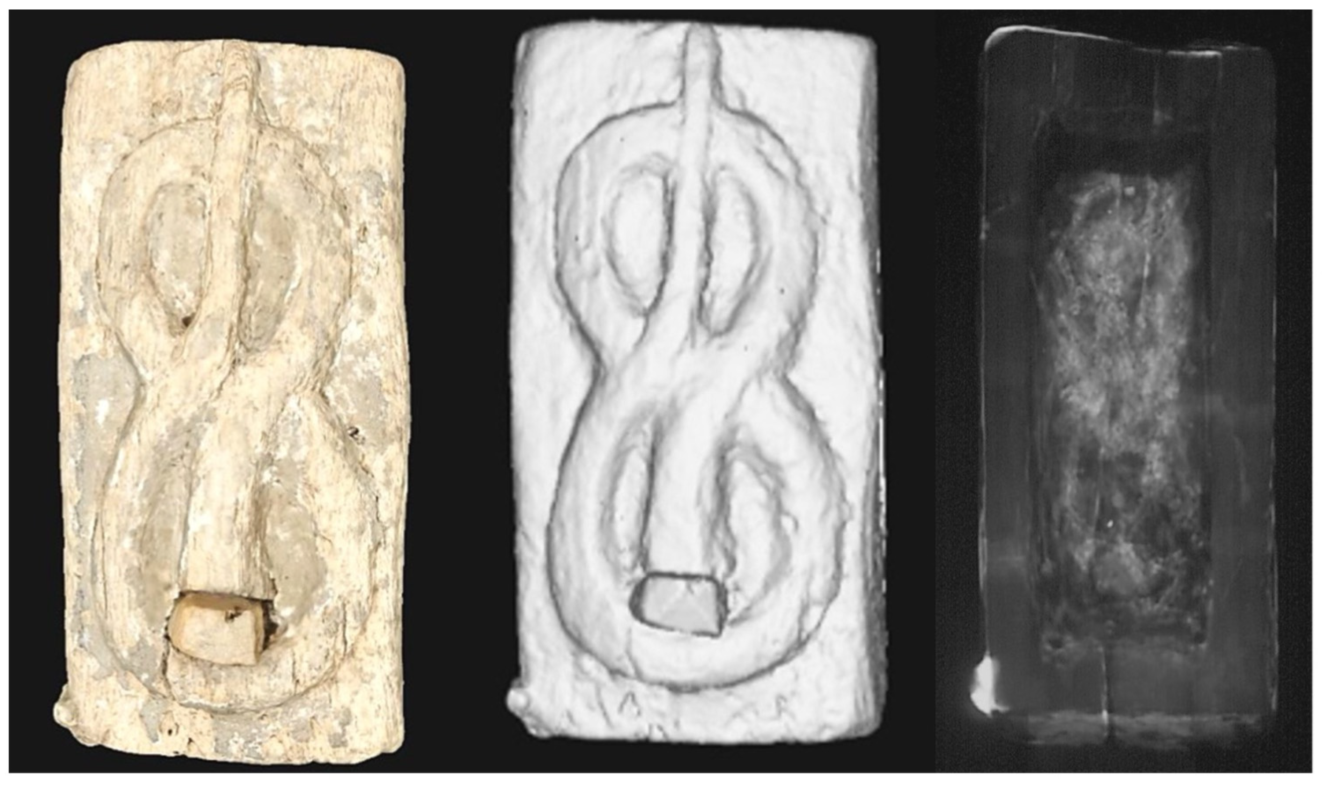
| 1195 | 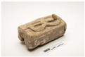 | Crudely carved wooden box with a removeable base. The upper aspect depicts a snake emblem in a ‘figure of eight’ shape. The wood is rough to touch and the base is loose with some debris having become separated from the main artefact. This artefact should not be excessively handled to avoid the further loss of contents. Dimensions 145 × 75 × 55 (mm). |
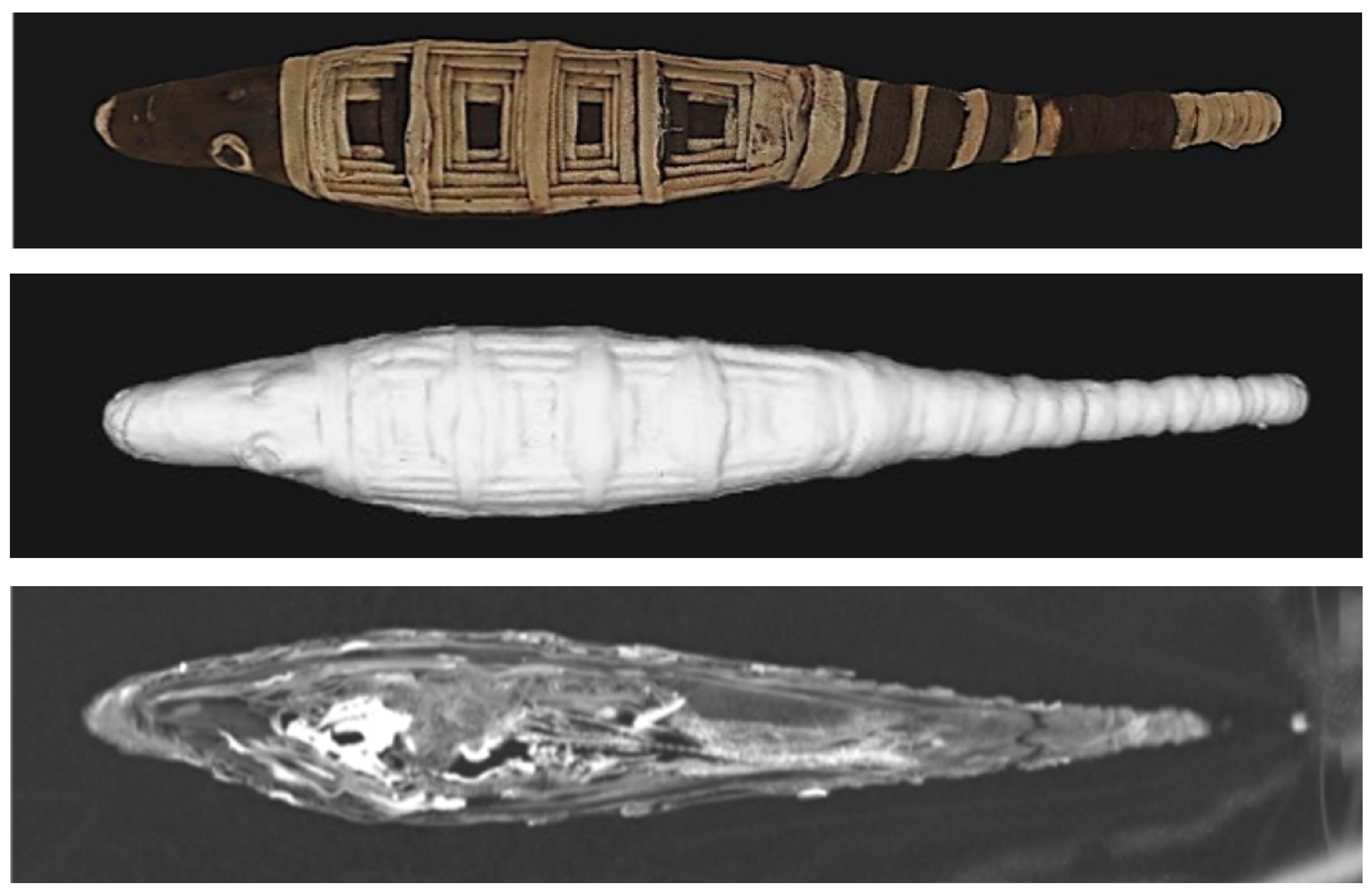
| 6293 | 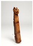 | Completely wrapped cylindrical bundle which narrows towards the modelled head. The front is adorned with a geometrically patterned panel. The modelled head displays two eyes and mouth created from a darker linen than the base colour from which the ears and muzzle are moulded. A small paper label is fixed to the front of the bundle, giving the museum no. 6293. Excellent condition. Dimensions 345 × 75 × 55 (mm). |
| 11293 | 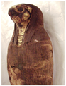 | Fully wrapped bundle in a tightly compressed anthropoid form with a false head and feet. The face and breast area are gilded and depict the facial features of a hawk. A linen shroud covers the back of the head and wraps around the front to leave a small gilded area visible. Excellent condition. Dimensions 360 × 100 × 60 (mm). |
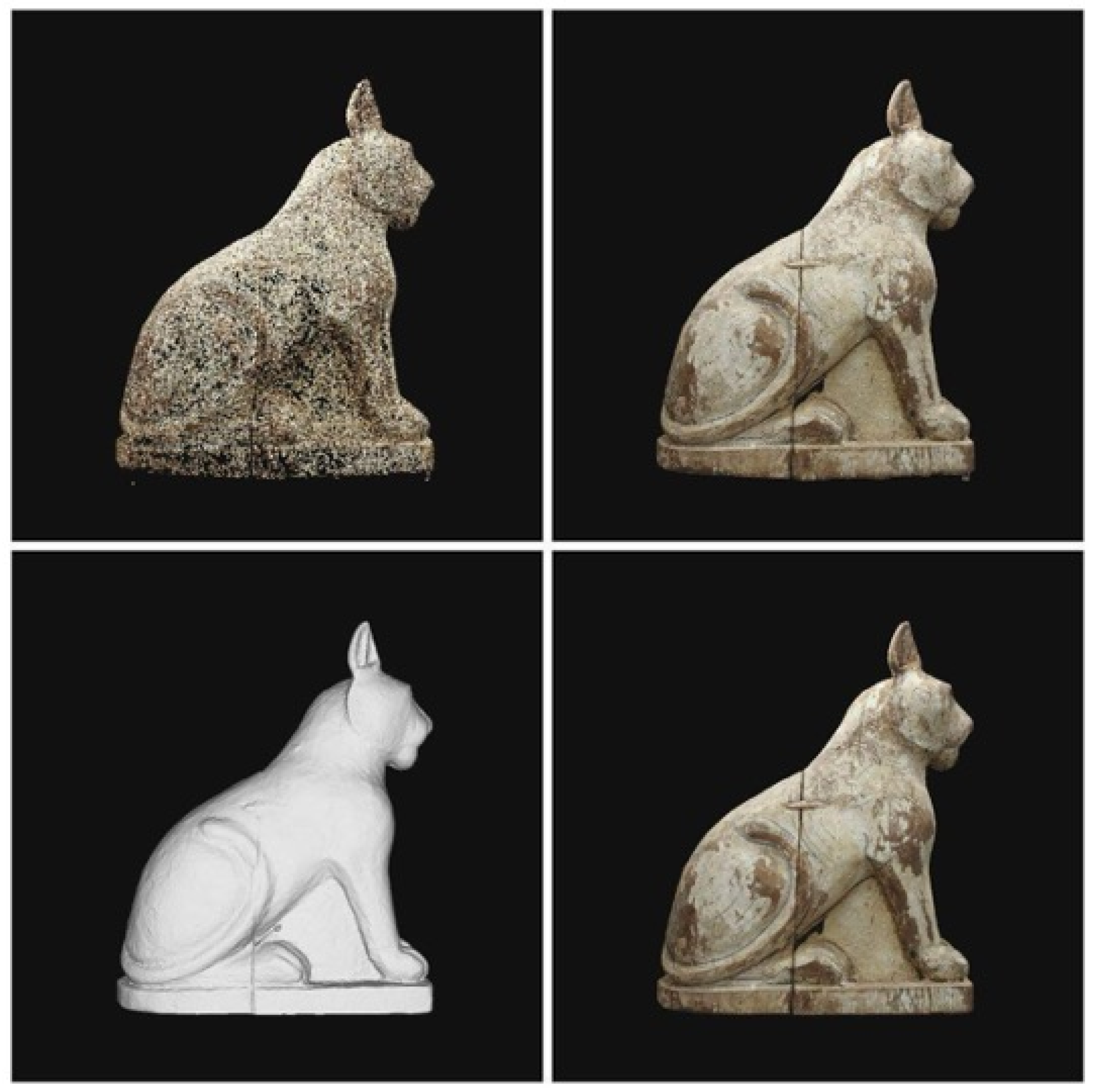
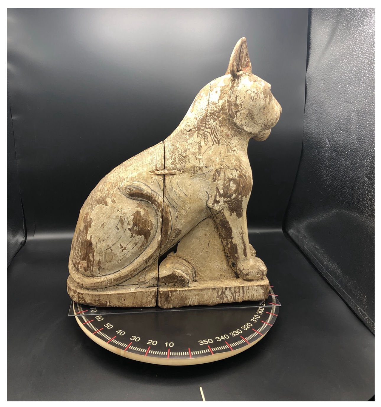
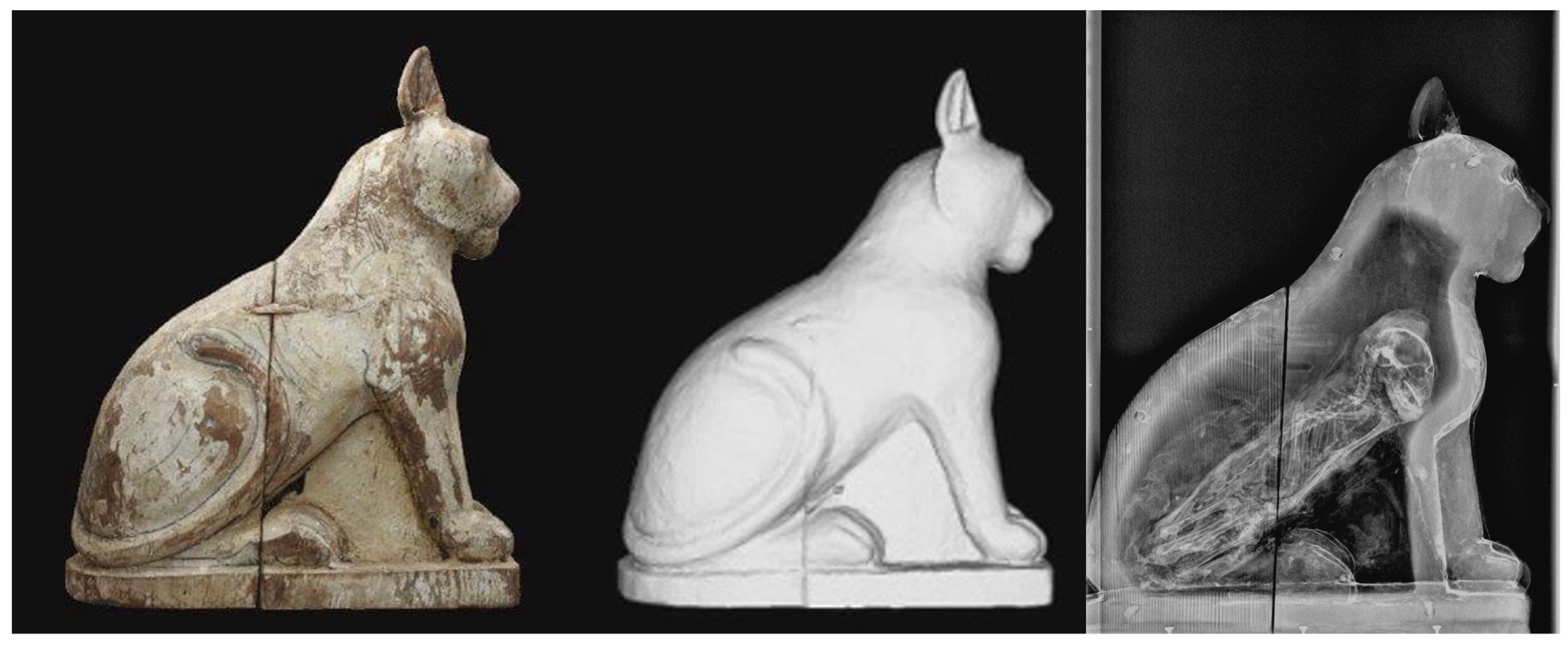
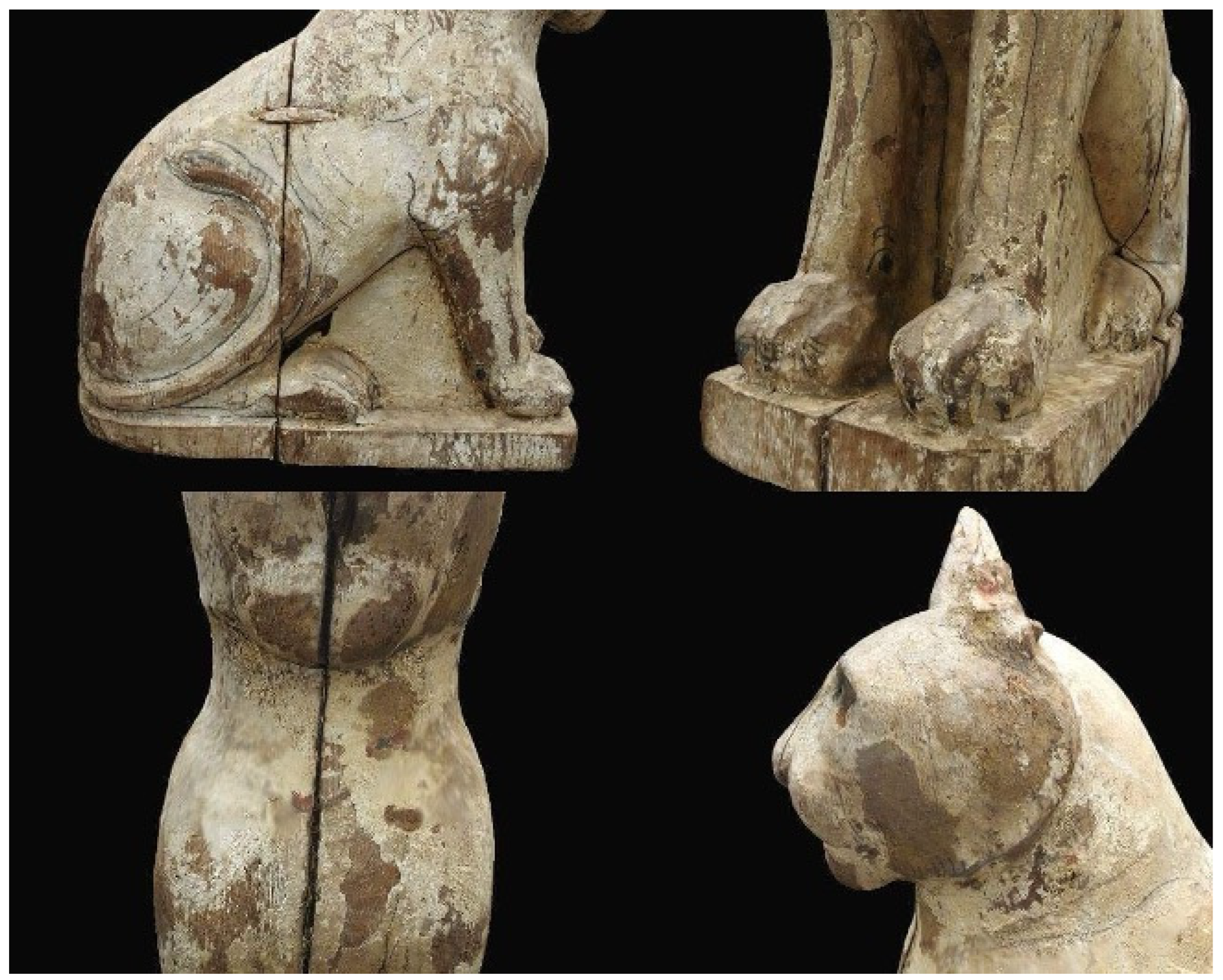
| 9303a-b | 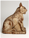 | A substantial wooden artefact in the form of a seated cat constructed in four sections, joined together with wooden dowels. Some attempts to depict musculature and anatomic features (such as the tail, legs and face) are evident in the form of carved attributes and black lines. It appears that the surface may once have been painted or covered with a layer of a gesso-type material. Excellent condition. Dimensions 45 × 360 × 145 (mm), weight 4.45 kg. |
| Accession No./Corresponding Figure | Provenance/Accession Information | Represented Species (Suggested by External Form) | Species (Radiographic Identification) |
|---|---|---|---|
| 13861/5 | Naukratis. Robinow Collection, no.72 | Crocodile | Crocodylus niloticus (Nile crocodile) Partial skeleton |
| 6033/6 | Unknown. Donated by Mr. J. and A. Williams | Indeterminate | Shrew—complete and articulated |
| 1195/7 | Distributed from Egypt Exploration Society excavations at Hibeh | Sealed rectangular wooden coffinette with carved snake decoration | Snake (MNI > 1) |
| 6293/8 | Beni Hasan, 22nd Dynasty. Collected by Prof. Garstang, Liverpool Institute of Archaeology in Egypt (1910-11 or 1920 season) | Feline | Bundle containing an isolated vertebra of feline origin |
| 11293/9 | Acquired from the Robinow Collection, January 1959 | Falcon | Pseudo mummy constructed from organic matter |
| 9303a-b/10 | Excavated by Cecil Firth at the Sacred Animal Necropolis, North Saqqara | Sealed wooden coffinette resembling a seated feline | Feline–complete and articulated |
Disclaimer/Publisher’s Note: The statements, opinions and data contained in all publications are solely those of the individual author(s) and contributor(s) and not of MDPI and/or the editor(s). MDPI and/or the editor(s) disclaim responsibility for any injury to people or property resulting from any ideas, methods, instructions or products referred to in the content. |
© 2025 by the author. Licensee MDPI, Basel, Switzerland. This article is an open access article distributed under the terms and conditions of the Creative Commons Attribution (CC BY) license (https://creativecommons.org/licenses/by/4.0/).
Share and Cite
McKnight, L. In the Likeness of a God: The Non-Invasive Investigation of Animal Votives. Heritage 2025, 8, 286. https://doi.org/10.3390/heritage8070286
McKnight L. In the Likeness of a God: The Non-Invasive Investigation of Animal Votives. Heritage. 2025; 8(7):286. https://doi.org/10.3390/heritage8070286
Chicago/Turabian StyleMcKnight, Lidija. 2025. "In the Likeness of a God: The Non-Invasive Investigation of Animal Votives" Heritage 8, no. 7: 286. https://doi.org/10.3390/heritage8070286
APA StyleMcKnight, L. (2025). In the Likeness of a God: The Non-Invasive Investigation of Animal Votives. Heritage, 8(7), 286. https://doi.org/10.3390/heritage8070286