A Case of Developmental Dysplasia of the Hip with Dislocation from Ancient Rome
Abstract
1. Introduction
2. Materials and Methods
3. Results
| Paleopathological Finding | Anatomical Evaluation | Imaging Evaluation |
|---|---|---|
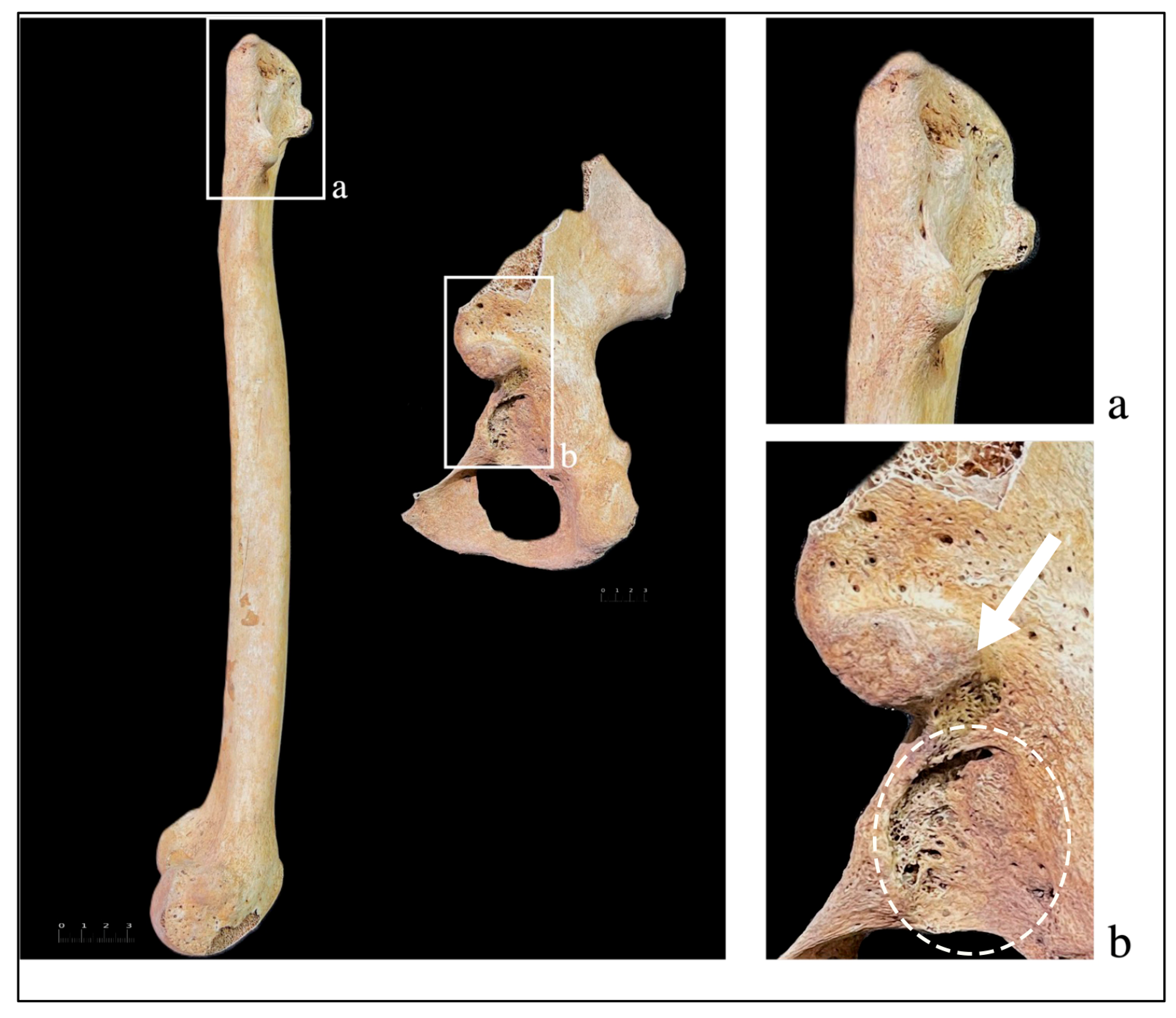
| Shallow, flattened real acetabulum (Figure 2b) | X | X |
| False acetabulum AIIS (Figure 2b) | X | X |
| Antero-lateral shifting of the ischial spine | X | X |
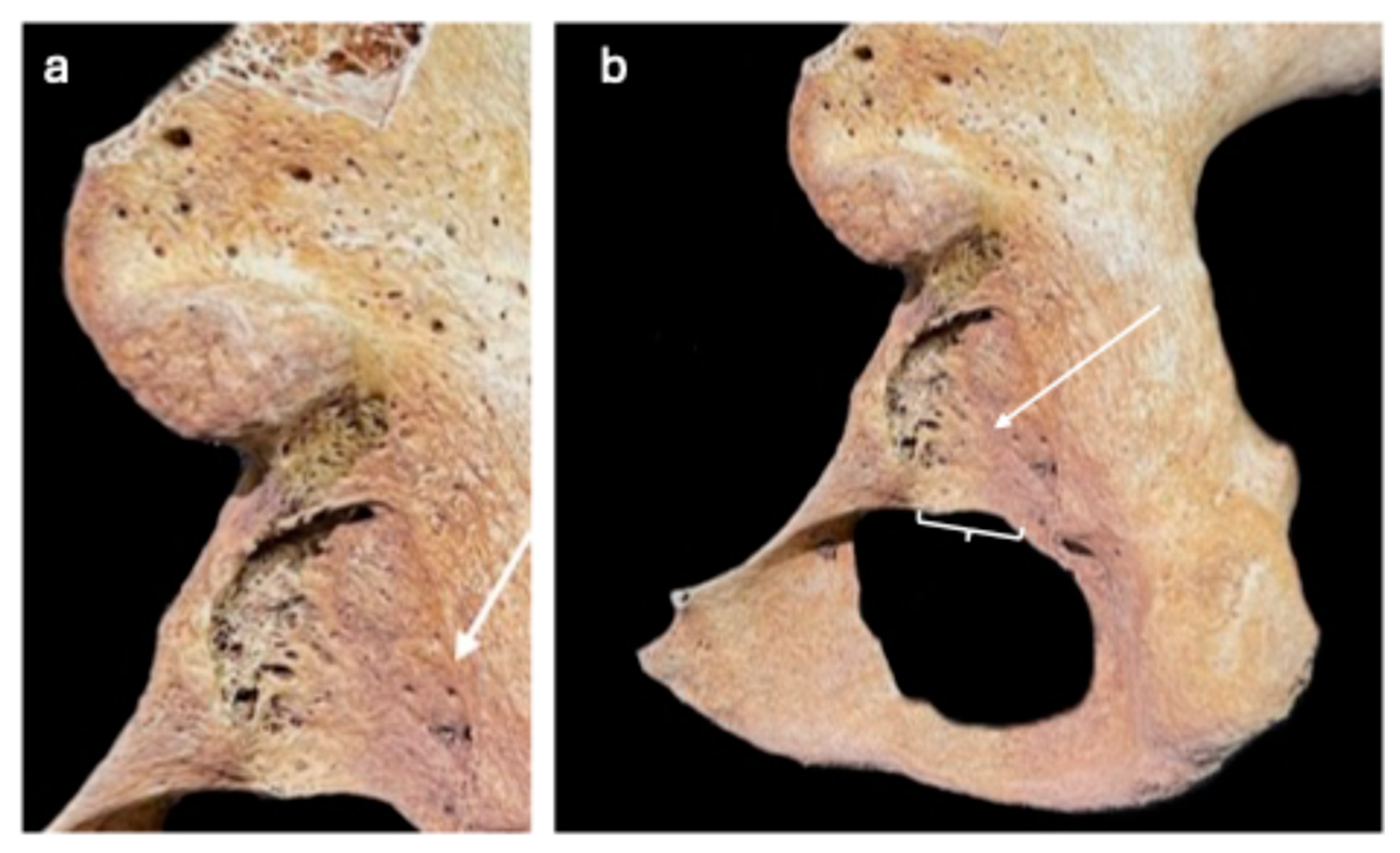
| Flattened groove for the Obturator Externus (Figure 3a) | X | X |
| Widening of the acetabular notch (Figure 3a) | ||
| Lack of bony deposit around the joint (Figure 3b) | X | |
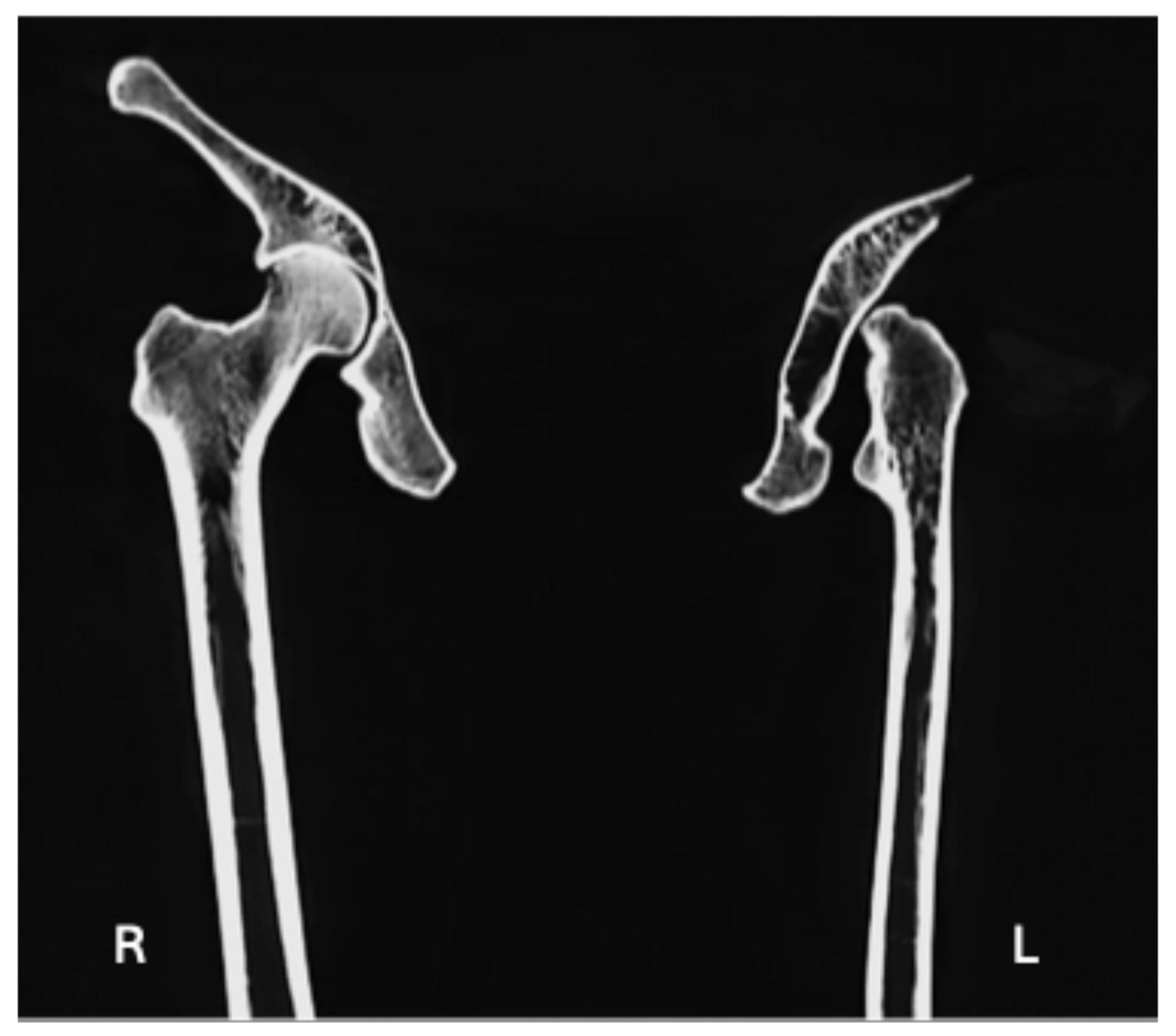
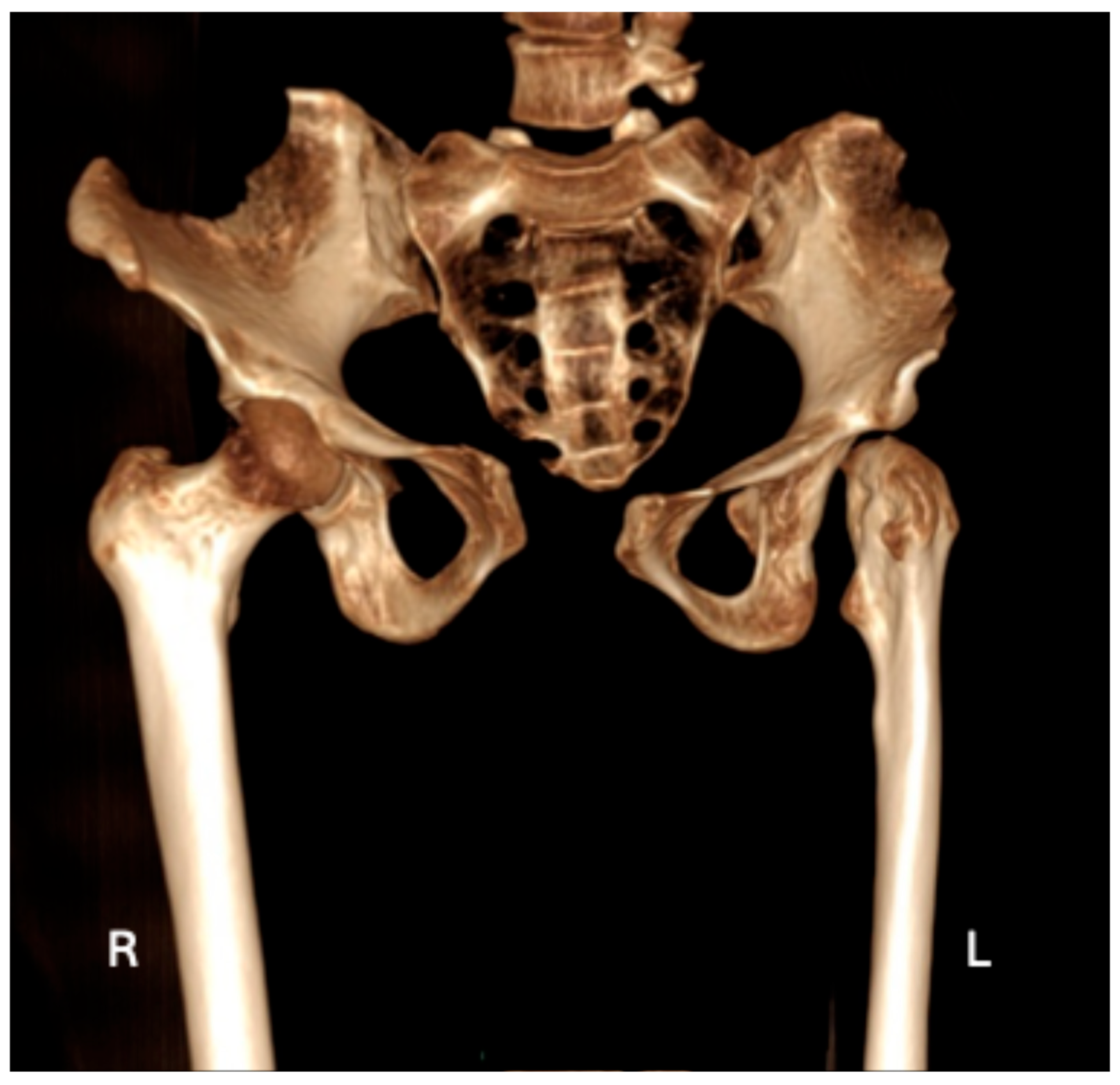
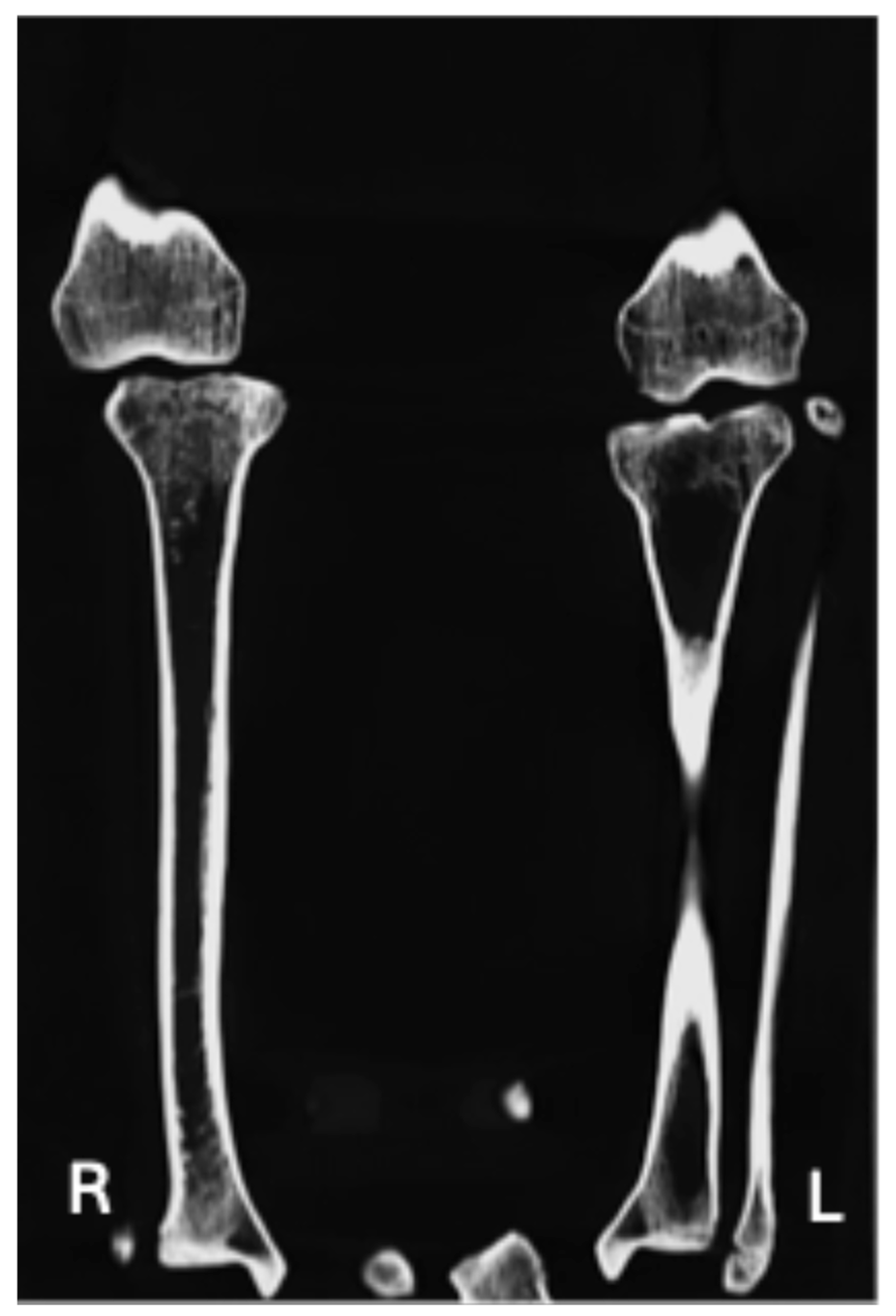
| Contralateral hip joint is definitively unaffected (Figure 4 and Figure 5) | X | X |
| Periarticular osteoporosis (Figure 4) | X | |
| Hypoplastic/aplastic femoral head (Figure 2b) | X | X |
| Subchondral sclerosis (Figure 4) | X | |
| Lack of development of the intertrochanteric crest (Figure 2a and Figure 4) | X | X |
| Lack of development of the trochanteric fossa (Figure 2a and Figure 4) | X | X |
| Occult fractures (Figure 4) | X | |
| Uppermost origin area for Vastus Lateralis as a flat spike (Figure 2a) | X |
4. Discussion
Author Contributions
Funding
Data Availability Statement
Acknowledgments
Conflicts of Interest
Abbreviations
| AIIS | Anterior Inferior Iliac Spine |
| CT | Computed tomography |
| DDH | Developmental dysplasia of the hip |
| PFGD | Proximal femoral growth disturbance |
References
- Afshar, Z. Bioarchaeology: Scientific Studies of Archaeological Human Skeletal Remains. J. Res. Archaeom. 2018, 4, 81–92. [Google Scholar] [CrossRef][Green Version]
- Stodder, A.L.W.; Byrnes, J.F. (Re)Discovering Paleopathology: Integrating Individuals and Populations in Bioarchaeology. In Evaluating Evidence in Biological Anthropology: The Strange and the Familiar; Willermet, C., Lee, S.-H., Eds.; Cambridge Studies in Biological and Evolutionary Anthropology; Cambridge University Press: Cambridge, UK, 2019; pp. 103–125. ISBN 978-1-108-47684-3. [Google Scholar]
- Buikstra, J.E. Bioarchaeologists Speak Out: Deep Time Perspectives on Contemporary Issues; Bioarchaeology and Social Theory; Springer International Publishing: Cham, Switzerland, 2019; ISBN 978-3-319-93011-4. [Google Scholar]
- Buikstra, J.; Ubelaker, D. Standards for Data Collection from Human Skeletal Remains: Proceedings of a Seminar at the Field Museum of Natural History, Organized by Jonathan Haas; Arkansas Archeological Survey: Fayetteville, AK, USA, 1994. [Google Scholar]
- Buikstra, J.E. A Bioarchaeological Perspective: What’s in a Name? Annu. Rev. Anthropol. 2024, 53, 1–19. [Google Scholar] [CrossRef]
- Armelagos, G.J. Bioarchaeology as Anthropology. Archeol. Pap. Am. Anthropol. Assoc. 2003, 13, 27–40. [Google Scholar]
- Armelagos, G.J.; Gerven, D.P.V. A Century of Skeletal Biology and Paleopathology: Contrasts, Contradictions, and Conflicts. Am. Anthropol. 2003, 105, 53–64. [Google Scholar] [CrossRef]
- Michalik, R.; Rissel, V.; Migliorini, F.; Siebers, H.L.; Betsch, M. Biomechanical Evaluation and Comparison of Clinically Relevant versus Non-Relevant Leg Length Inequalities. BMC Musculoskelet. Disord. 2022, 23, 174. [Google Scholar] [CrossRef]
- Filograna, L.; Pugliese, L.; Muto, M.; Tatulli, D.; Guglielmi, G.; Thali, M.J.; Floris, R. A Practical Guide to Virtual Autopsy: Why, When and How. In Seminars in Ultrasound, CT and MRI; WB Saunders: Philadelphia, PA, USA, 2019; Volume 40, pp. 56–66. [Google Scholar] [CrossRef]
- Giovannetti, G.; Guerrini, A.; Minozzi, S.; Panetta, D.; Salvadori, P.A. Computer Tomography and Magnetic Resonance for Multimodal Imaging of Fossils and Mummies. Magn. Reson. Imaging 2022, 94, 7–17. [Google Scholar] [CrossRef]
- Shelmerdine, S.C.; Gerrard, C.Y.; Rao, P.; Lynch, M.; Kroll, J.; Martin, D.; Miller, E.; Filograna, L.; Martinez, R.M.; Ukpo, O.; et al. Joint European Society of Paediatric Radiology (ESPR) and International Society for Forensic Radiology and Imaging (ISFRI) Guidelines: Paediatric Postmortem Computed Tomography Imaging Protocol. Pediatr. Radiol. 2019, 49, 694–701. [Google Scholar] [CrossRef]
- Profico, A.; Buzi, C.; Di Vincenzo, F.; Boggioni, M.; Borsato, A.; Boschian, G.; Marchi, D.; Micheli, M.; Cecchi, J.M.; Samadelli, M.; et al. Virtual Excavation and Analysis of the Early Neanderthal Cranium from Altamura (Italy). Commun. Biol. 2023, 6, 316. [Google Scholar] [CrossRef]
- Montiel, G.; Lorenzo, C. A New Virtual Reconstruction of the Ndutu Cranium. Heritage 2023, 6, 2822–2850. [Google Scholar] [CrossRef]
- Buzi, C.; Boggioni, M.; Borsato, A.; Boschian, G.; Marchi, D.; Moggi-Cecchi, J.; Profico, A.; Riga, A.; Samadelli, M.; Manzi, G. Virtual Paleoanthropology in Karstic Environments. The Challenging Case of the Neanderthal Skeleton from Altamura (Southern Italy). Quat. Sci. Rev. 2024, 338, 108833. [Google Scholar] [CrossRef]
- Filograna, L.; Manenti, G.; Mecchia, D.; Tatulli, D.; Pasqualetto, M.; Perlangeli, V.; Rossi, P.F.; De Angelis, F.; Floris, R. Investigation of Human Remains from the Archaeological Areas of “Parco Archeologico Di Ostia Antica”: The Role of CT Imaging. Forensic Imaging 2022, 31, 200521. [Google Scholar] [CrossRef]
- Singh, S.; Bray, T.J.P.; Hall-Craggs, M.A. Quantifying Bone Structure, Micro-Architecture, and Pathophysiology with MRI. Clin. Radiol. 2018, 73, 221–230. [Google Scholar] [CrossRef] [PubMed]
- Ministero Italiano Cultura. I Resti Scheletrici Umani: Dallo Scavo al Laboratorio al Museo; Ministero Della Cultura ICCD—Istituto Centrale per Il Catalogo e La Documentazione ICA—Istituto Centrale per l’Archeologia: Rome, Italy, 2022. [Google Scholar]
- Mitchell, P.; Brickley, M. Professional Practice Papers|Chartered Institute for Archaeologists. Available online: https://www.archaeologists.net/publications/papers (accessed on 25 January 2023).
- Beckett, R.G. Paleoimaging: A Review of Applications and Challenges. Forensic Sci. Med. Pathol. 2014, 10, 423–436. [Google Scholar] [CrossRef]
- Villa, C.; Frohlich, B.; Lynnerup, N. Chapter 7—The Role of Imaging in Paleopathology. In Ortner’s Identification of Pathological Conditions in Human Skeletal Remains, 3rd ed.; Buikstra, J.E., Ed.; Academic Press: San Diego, CA, USA, 2019; pp. 169–182. ISBN 978-0-12-809738-0. [Google Scholar]
- Bansal, G.J. Digital Radiography. A Comparison with Modern Conventional Imaging. Postgrad. Med. J. 2006, 82, 425–428. [Google Scholar] [CrossRef]
- Filograna, L.; Tartaglione, T.; Vetrugno, G.; Guerra, C.; Fileni, A.; Bonomo, L. Freshwater Drowning in a Child: A Case Study Demonstrating the Role of Post-Mortem Computed Tomography. Med. Sci. Law 2015, 55, 304–311. [Google Scholar] [CrossRef]
- Filograna, L.; Guglielmi, G.; Floris, R.; Marchetti, D. The Development of Forensic Imaging in Italy. A Systematic Review of the Literature. J. Forensic Radiol. Imaging 2018, 15, 14–20. [Google Scholar] [CrossRef]
- Villa, C.; Larsen, S.; Zink, A.; Lynnerup, N. Ötzi the Iceman: Forensic 3D Reconstructions of a 5300-Year-Ago Murder Case. Int. J. Legal Med. 2025, 139, 2263–2271. [Google Scholar] [CrossRef]
- Thompson, M.W. N.P. Stanley Price (Ed.): Conservation on Archaeological Excavations, with Particular Reference to the Mediterranean Area. Rome: ICCROM, 1984, 158 pp., Illustrated. Antiquity 1985, 59, 59–60. [Google Scholar] [CrossRef]
- Nicolosi, T.; Mariotti, V.; Talamo, S.; Miari, M.; Minarini, L.; Nenzioni, G.; Lenzi, F.; Pietrobelli, A.; Sorrentino, R.; Benazzi, S.; et al. On the Traces of Lost Identities: Chronological, Anthropological and Taphonomic Analyses of the Late Neolithic/Early Eneolithic Fragmented and Commingled Human Remains from the Farneto Rock Shelter (Bologna, Northern Italy). Archaeol. Anthropol. Sci. 2023, 15, 36. [Google Scholar] [CrossRef]
- Brooks, S.; Suchey, J.M. Skeletal Age Determination Based on the Os Pubis: A Comparison of the Acsádi-Nemeskéri and Suchey-Brooks Methods. Hum. Evol. 1990, 5, 227–238. [Google Scholar] [CrossRef]
- Işcan, M.Y.; Loth, S.R.; Wright, R.K. Age Estimation from the Rib by Phase Analysis: White Females. J. Forensic Sci. 1985, 30, 853–863. [Google Scholar] [CrossRef] [PubMed]
- Meindl, R.S.; Lovejoy, C.O. Ectocranial Suture Closure: A Revised Method for the Determination of Skeletal Age at Death Based on the Lateral-Anterior Sutures. Am. J. Phys. Anthropol. 1985, 68, 57–66. [Google Scholar] [CrossRef] [PubMed]
- Djurić, M.; Djonić, D.; Nikolić, S.; Popović, D.; Marinković, J. Evaluation of the Suchey-Brooks Method for Aging Skeletons in the Balkans. J. Forensic Sci. 2007, 52, 21–23. [Google Scholar] [CrossRef]
- Trotter, M.; Gleser, G.C. A Re-Evaluation of Estimation of Stature Based on Measurements of Stature Taken during Life and of Long Bones after Death. Am. J. Phys. Anthropol. 1958, 16, 79–123. [Google Scholar] [CrossRef]
- Redfern, R. Diagnostic Criteria for Developmental Dislocation of the Hip in Human Skeletal Remains. Int. J. Osteoarchaeol. 2008, 18, 61–71. [Google Scholar] [CrossRef]
- Dalrymple, N.C.; Prasad, S.R.; Freckleton, M.W.; Chintapalli, K.N. Informatics in Radiology (infoRAD): Introduction to the Language of Three-Dimensional Imaging with Multidetector CT. Radiogr. Rev. Publ. Radiol. Soc. N. Am. Inc. 2005, 25, 1409–1428. [Google Scholar] [CrossRef]
- Giannecchini, M.; Moggi-Cecchi, J. Stature in Archeological Samples from Central Italy: Methodological Issues and Diachronic Changes. Am. J. Phys. Anthropol. 2008, 135, 284–292. [Google Scholar] [CrossRef]
- Danubio, E.; Martella, M. Sanna Changes in Stature from the Upper Paleolithic to the Medieval Period in Western Europe. J. Anthropol. Sci. Riv. Antropol. JASS 2017, 95, 269–280. [Google Scholar] [CrossRef]
- Edinborough, M.; Rando, C. Stressed Out: Reconsidering Stress in the Study of Archaeological Human Remains. J. Archaeol. Sci. 2020, 121, 105197. [Google Scholar] [CrossRef]
- Beltran, L.S.; Rosenberg, Z.S.; Mayo, J.D.; De Tuesta, M.D.; Martin, O.; Neto, L.P.; Bencardino, J.T. Imaging Evaluation of Developmental Hip Dysplasia in the Young Adult. Am. J. Roentgenol. 2013, 200, 1077–1088. [Google Scholar] [CrossRef]
- Binder, H.; Schurz, M.; Aldrian, S.; Fialka, C.; Vécsei, V. Physeal Injuries of the Proximal Humerus: Long-Term Results in Seventy Two Patients. Int. Orthop. 2011, 35, 1497–1502. [Google Scholar] [CrossRef]
- Murphy, S.B.; Ganz, R.; Müller, M.E. The Prognosis in Untreated Dysplasia of the Hip. A Study of Radiographic Factors That Predict the Outcome. J. Bone Joint Surg. Am. 1995, 77, 985–989. [Google Scholar] [CrossRef]
- Wedge, J.H.; Wasylenko, M.J. The Natural History of Congenital Dislocation of the Hip: A Critical Review. Clin. Orthop. 1978, 137, 154–162. [Google Scholar] [CrossRef]
- Momodu, I.I.; Savaliya, V. Septic Arthritis. In StatPearls; StatPearls Publishing: Treasure Island, FL, USA, 2024. [Google Scholar]
- Saraf, S.K.; Tuli, S.M. Tuberculosis of Hip: A Current Concept Review. Indian J. Orthop. 2015, 49, 1–9. [Google Scholar] [CrossRef] [PubMed]
- Mills, S.; Burroughs, K.E. Legg-Calve-Perthes Disease. In StatPearls; StatPearls Publishing: Treasure Island, FL, USA, 2024. [Google Scholar]
- Pountney, T.; Green, E.M. Hip Dislocation in Cerebral Palsy. BMJ 2006, 332, 772–775. [Google Scholar] [CrossRef] [PubMed]
- Dawson-Amoah, K.; Raszewski, J.; Duplantier, N.; Waddell, B.S. Dislocation of the Hip: A Review of Types, Causes, and Treatment. Ochsner J. 2018, 18, 242–252. [Google Scholar] [CrossRef]
- Biko, D.M.; Davidson, R.; Pena, A.; Jaramillo, D. Proximal Focal Femoral Deficiency: Evaluation by MR Imaging. Pediatr. Radiol. 2012, 42, 50–56. [Google Scholar] [CrossRef]
- Huser, A.J.; Kwak, Y.H.; Rand, T.J.; Paley, D.; Feldman, D.S. Anatomic Relationship of the Femoral Neurovascular Bundle in Patients with Congenital Femoral Deficiency. J. Pediatr. Orthop. 2021, 41, e111–e115. [Google Scholar] [CrossRef]
- Jarraya, M.; Hayashi, D.; Roemer, F.W.; Crema, M.D.; Diaz, L.; Conlin, J.; Marra, M.D.; Jomaah, N.; Guermazi, A. Radiographically Occult and Subtle Fractures: A Pictorial Review. Radiol. Res. Pract. 2013, 2013, e370169. [Google Scholar] [CrossRef]
- Park, J.-M.; Im, G.-I. The Correlations of the Radiological Parameters of Hip Dysplasia and Proximal Femoral Deformity in Clinically Normal Hips of a Korean Population. Clin. Orthop. Surg. 2011, 3, 121–127. [Google Scholar] [CrossRef] [PubMed]
- Pinto, A.; Berritto, D.; Russo, A.; Riccitiello, F.; Caruso, M.; Paola Belfiore, M.; Roberto Papapietro, V.; Carotti, M.; Pinto, F.; Giovagnoni, A.; et al. Traumatic Fractures in Adults: Missed Diagnosis on Plain Radiographs in the Emergency Department. Acta Bio Medica Atenei Parm. 2018, 89, 111–123. [Google Scholar] [CrossRef]
- Subbarao, K. Proximal Femoral Focal Deficiency (PFFD). J. Med. Sci. Res. 2015, 3, 90–93. [Google Scholar] [CrossRef]
- Uduma, F.U.; Dim, E.M.; Njeze, N.R. Proximal Femoral Focal Deficiency—A Rare Congenital Entity: Two Case Reports and a Review of the Literature. J. Med. Case Rep. 2020, 14, 27. [Google Scholar] [CrossRef]
- Baumgart, M.; Wiśniewski, M.; Grzonkowska, M.; Badura, M.; Małkowski, B.; Szpinda, M. Quantitative Anatomy of the Primary Ossification Center of the Femoral Shaft in Human Fetuses. Surg. Radiol. Anat. 2017, 39, 1235–1242. [Google Scholar] [CrossRef]
- Weinstein, S.L.; Dolan, L.A. Proximal Femoral Growth Disturbance in Developmental Dysplasia of the Hip: What Do We Know? J. Child. Orthop. 2018, 12, 331–341. [Google Scholar] [CrossRef]
- Cheng, R.; Huang, M.; Kernkamp, W.A.; Li, H.; Zhu, Z.; Wang, L.; Tsai, T.-Y. The Severity of Developmental Dysplasia of the Hip Does Not Correlate with the Abnormality in Pelvic Incidence. BMC Musculoskelet. Disord. 2020, 21, 623. [Google Scholar] [CrossRef]
- Dygut, J.; Piwowar, M. Distinction Between Dysplasia, Malformation, and Deformity—Towards the Proper Diagnosis and Treatment of Hip Development Disorders. Diagnostics 2025, 15, 1547. [Google Scholar] [CrossRef]
- Ren, N.; Zhang, Z.; Li, Y.; Zheng, P.; Cheng, H.; Luo, D.; Zhang, J.; Zhang, H. Effect of Hip Dysplasia on the Development of the Femoral Head Growth Plate. Front. Pediatr. 2023, 11, 1247455. [Google Scholar] [CrossRef]
- England, P.; Schaeffer, E.; Price, C.; Mulpuri, K.; Sankar, W.N. Proximal Femoral Growth Alterations Can Be Seen Prior to Treatment of Developmental Dysplasia of the Hip: A Multicenter Cohort Study. J. Pediatr. Orthop. Soc. N. Am. 2024, 5, 669. [Google Scholar] [CrossRef]
- Strayer, L.M. The Embryology of the Human Hip Joint. Yale J. Biol. Med. 1943, 16, 13–26.6. [Google Scholar] [CrossRef]
- Ahmed, M.F.; Ferdous, S.; Azad, A.K. Progressive Pseudo-Rheumatoid Dysplasia: Two Cases in One Family. Mymensingh Med. J. 2020, 29, 734–737. [Google Scholar]
- Lievense, A.M.; Bierma-Zeinstra, S.M.A.; Verhagen, A.P.; Verhaar, J.A.N.; Koes, B.W. Influence of Hip Dysplasia on the Development of Osteoarthritis of the Hip. Ann. Rheum. Dis. 2004, 63, 621–626. [Google Scholar] [CrossRef] [PubMed]
- Nandhagopal, T.; De Cicco, F.L. Developmental Dysplasia of The Hip. In StatPearls; StatPearls Publishing: Treasure Island, FL, USA, 2022. [Google Scholar]
- Pone, M.V.d.S.; Gomes da Silva, T.O.; Ribeiro, C.T.M.; de Aguiar, E.B.; Mendes, P.H.B.; Gomes Junior, S.C.D.S.; Hamanaka, T.; Zin, A.A.; Pereira Junior, J.P.; Moreira, M.E.L.; et al. Acquired Hip Dysplasia in Children with Congenital Zika Virus Infection in the First Four Years of Life. Viruses 2022, 14, 2643. [Google Scholar] [CrossRef] [PubMed]
- Trousdale, R.T.; Ganz, R. Posttraumatic Acetabular Dysplasia. Clin. Orthop. 1994, 305, 124–132. [Google Scholar] [CrossRef]
- Katz, J.N.; Arant, K.R.; Loeser, R.F. Diagnosis and Treatment of Hip and Knee Osteoarthritis: A Review. JAMA 2021, 325, 568–578. [Google Scholar] [CrossRef]
- Pineda, C.; Espinosa, R.; Pena, A. Radiographic Imaging in Osteomyelitis: The Role of Plain Radiography, Computed Tomography, Ultrasonography, Magnetic Resonance Imaging, and Scintigraphy. Semin. Plast. Surg. 2009, 23, 80–89. [Google Scholar] [CrossRef]
- Cooperman, D.R.; Wallensten, R.; Stulberg, S.D. Acetabular Dysplasia in the Adult. Clin. Orthop. Relat. Res. 1983, 175, 79. [Google Scholar] [CrossRef]
- Bakarman, K.; Alsiddiky, A.M.; Zamzam, M.; Alzain, K.O.; Alhuzaimi, F.S.; Rafiq, Z. Developmental Dysplasia of the Hip (DDH): Etiology, Diagnosis, and Management. Cureus 2023, 15, e43207. [Google Scholar] [CrossRef]
- Killen, M.-C.; DeKiewiet, G. Genu Varum in Children. Paediatr. Child Health 2022, 32, 141–150. [Google Scholar] [CrossRef]
- Ferguson, J.; Wainwright, A. Tibial Bowing in Children. Orthop. Trauma 2013, 27, 30–41. [Google Scholar] [CrossRef]
- Bowen, J.R. Developmental Dysplasia of the Hip Book. Available online: https://www.thriftbooks.com/w/developmental-dysplasia-of-the-hip_j-richard-bowen/38321599/ (accessed on 22 November 2023).
- Mitchell, P.D.; Redfern, R.C. The Prevalence of Dislocation in Developmental Dysplasia of the Hip in Britain over the Past Thousand Years. J. Pediatr. Orthop. 2007, 27, 890–892. [Google Scholar] [CrossRef] [PubMed]
- Mafart, B.; Kéfi, R.; Béraud-Colomb, E. Palaeopathological and Palaeogenetic Study of 13 Cases of Developmental Dysplasia of the Hip with Dislocation in a Historical Population from Southern France. Int. J. Osteoarchaeol. 2007, 17, 26–38. [Google Scholar] [CrossRef]
- Saccheri, P.; Travan, L. The Limping Nuns. Two Cases of Hip Dislocation in a Medieval Female Monastery. Ital. J. Anat. Embryol. 2022, 126, 81–91. [Google Scholar] [CrossRef]
- Traversari, M.; Feletti, F.; Vazzana, A.; Gruppioni, G.; Frelat, M.A. Trois cas de dysplasie développementale de la hanche chez des individus partiellement momifiés (Roccapelago, Modène, 18e siècle): Étude des indicateurs paléopathologiques par analyses directe et virtuelle. BMSAP 2016, 28, 202–212. [Google Scholar] [CrossRef]
- Cesana, D.; Benedictow, O.J.; Bianucci, R. The Origin and Early Spread of the Black Death in Italy: First Evidence of Plague Victims from 14th-Century Liguria (Northern Italy). Anthropol. Sci. 2017, 125, 15–24. [Google Scholar] [CrossRef]
- Piccioli, A.; Gazzaniga, V.; Catalano, P. Bones: Orthopaedic Pathologies in Roman Imperial Age; Softcover Reprint of the Original 1st ed. 2015 Edition; Springer: Berlin/Heidelberg, Germany, 2016; ISBN 978-3-319-36802-3. [Google Scholar]
- Belfiglio, V.J. Orthopedic Surgery in Ancient Roman Military Hospitals: 27 BCE to 476 CE. Int. J. Interdiscip. Cult. Stud. 2023, 18, 81–90. [Google Scholar] [CrossRef]
- Mitchell, P.D.; Redfern, R.C. Brief Communication: Developmental Dysplasia of the Hip in Medieval London. Am. J. Phys. Anthropol. 2011, 144, 479–484. [Google Scholar] [CrossRef]
- Van Sleuwen, B.E.; Engelberts, A.C.; Boere-Boonekamp, M.M.; Kuis, W.; Schulpen, T.W.J.; L’Hoir, M.P. Swaddling: A Systematic Review. Pediatrics 2007, 120, e1097–e1106. [Google Scholar] [CrossRef]
- Laes, C. Infants Between Biological and Social Birth in Antiquity: A Phenomenon of the “Longue Durée”. Hist. Z. Alte Gesch. 2014, 63, 364–383. [Google Scholar] [CrossRef]
- Lawson, A.; Beckett, A.E. The Social and Human Rights Models of Disability: Towards a Complementarity Thesis. Int. J. Hum. Rights 2021, 25, 348–379. [Google Scholar] [CrossRef]
- Bohling, S.; Croucher, K.; Buckberry, J. The Bioarchaeology of Disability: A Population-Scale Approach to Investigating Disability, Physical Impairment, and Care in Archaeological Communities. Int. J. Paleopathol. 2022, 38, 76–94. [Google Scholar] [CrossRef] [PubMed]
- Upson-Saia, K.; Goodey, C.F.; Rose, M.L. Review of Disabilities in Roman Antiquity: Disparate Bodies. A Capite Ad Calcem. Mnemosyne, Supplements, History and Archaeology of Classical Antiquity, 356. Bull. Hist. Med. 2014, 88, 570–572. [Google Scholar]
- Laes, C. Mobility Impairments: History of Pain and Toil. In Disabilities and the Disabled in the Roman World: A Social and Cultural History; Cambridge University Press: Cambridge, UK, 2018; pp. 149–167. ISBN 978-1-107-16290-7. [Google Scholar]
- Munyi, C.W. Past and Present Perceptions Towards Disability: A Historical Perspective. Disabil. Stud. Q. 2012, 32. [Google Scholar] [CrossRef]
- Gismondi, A.; D’Agostino, A.; Di Marco, G.; Scuderi, F.; De Angelis, F.; Rickards, O.; Catalano, P.; Canini, A. Archaeobotanical Record from Dental Calculus of a Roman Individual Affected by Bilateral Temporo-Mandibular Joint Ankylosis. Quat. Int. 2023, 653–654, 82–88. [Google Scholar] [CrossRef]
- López-Chamizo, S. Caring for the Dead, Understanding the Living: Bioarchaeology of Care in a 2nd–3rd Century CE Burial from Roman Malaca. J. Archaeol. Sci. Rep. 2025, 66, 105254. [Google Scholar] [CrossRef]
- Laes, C. Disabilities and the Disabled in the Roman World: A Social and Cultural History; Cambridge University Press: Cambridge, UK, 2018; ISBN 978-1-107-16290-7. [Google Scholar]
- Tilley, L. Theory and Practice in the Bioarchaeology of Care; Bioarchaeology and Social Theory; Springer International Publishing: Cham, Switzerland, 2015; ISBN 978-3-319-18859-1. [Google Scholar]
- Dettwyler, K.A. Can Paleopathology Provide Evidence for “Compassion”? Am. J. Phys. Anthropol. 1991, 84, 375–384. [Google Scholar] [CrossRef]
- Brent, L. Disturbed, Damaged and Disarticulated: Grave Reuse in Roman Italy. Theor. Roman Archaeol. J. 2017, 2016, 37–50. [Google Scholar] [CrossRef]
- Draycott, J. Reconstructing the Lived Experience of Disability in Antiquity: A Case Study from Roman Egypt. Greece Rome 2015, 62, 189–205. [Google Scholar] [CrossRef][Green Version]
- Shrader, M.J. Moving Beyond the Medical Model: Assessing the Bioarchaeology of Disability in the Roman Empire; Texas Tech University Libraries: Lubbock, TX, USA, 2023. [Google Scholar]
- Sáez, R.; Edén Fernández Suárez, M.; David Candela, G.; Andrea Barrio Fioresta, P.; León-Cristóbal, A.; Romero, J.; Velayos Castelo, C. Care in Late Antiquity: Applying the Bioarchaeology of Care Method in the Case of an Unprecedented Pathology in an Individual from Herrera de Pisuerga, Northern Spain. J. Archaeol. Sci. Rep. 2024, 60, 104867. [Google Scholar] [CrossRef]
- Cilione, M.; Gazzaniga, V. Conceptualizing Disabilities from Antiquity to the Middle Ages: A Historical-Medical Contribution. Int. J. Paleopathol. 2023, 40, 41–47. [Google Scholar] [CrossRef]
- Verbruggen, S.W.; Kainz, B.; Shelmerdine, S.C.; Arthurs, O.J.; Hajnal, J.V.; Rutherford, M.A.; Phillips, A.T.M.; Nowlan, N.C. Altered Biomechanical Stimulation of the Developing Hip Joint in Presence of Hip Dysplasia Risk Factors. J. Biomech. 2018, 78, 1–9. [Google Scholar] [CrossRef]
- Haber, C.K.; Sacco, M. Scoliosis: Lower Limb Asymmetries during the Gait Cycle. Arch. Physiother. 2015, 5, 4. [Google Scholar] [CrossRef]
- Rossetti, N.; Fusco, R.; Messina, C.; Vanni, A.; Licata, M. Health and Heritage: The Bioarchaeological Discovery of a Probable Case of Developmental Dysplasia in an Adult Subject. Heritage 2024, 7, 5295–5306. [Google Scholar] [CrossRef]
| Etiology | Epidemiology | Bone Signs | Imaging | |
|---|---|---|---|---|
| Septic Arthritis [41] | Secondary to an infectious etiology spreading through bloodstream to hip joint | Neonates to elderly. Among children, the hip is the joint most affected; in adults, the hip is second to the knee. Monoarticular; but polyarticular may occur | Subchondral bone changes. Associated osteomyelitis may be present | Early stage: periarticular osteopenia. Later stages may reveal chronic bony changes and calcium deposits |
| Joint Tuberculosis [42] | Mycobacterim tuberculosis spreads from the lungs to the hips | First three decades but no age is immune | Gross destruction of the femoral head or the superior acetabular margin (wandering acetabulum). Irregular and hazy joint margins. Osteonecrosis has been reported. Hypertrophy of the cephalic and subtrochanteric ossification centers is not infrequent | Periarticular osteoporosis, peripheral erosion, and progressive diminution of the joint space (Phemister’s triad) |
| Legg–Calvé–Perthes disease [43] | Blood supply to the head of the femur is interrupted idiopathically | More common in boys than girls. Age range 4–8 | The necroses weakens the bone and can lead to multiple fractures. Height retardation | Loss of structural integrity of the femoral head and acetabular subchondral sclerosis |
| Hip dislocation in Cerebral Palsy [44] | Spasticity causes the hip to displace in posterior superior direction | Most frequent 3–5 | Lateralization and proximal migration of the femoral head. Progressive erosion of lateral acetabular lip. Deformity of femoral head due to pressure from the capsule. Acetabular angle, iliac angle, iliac index and femoral neck shaft angle were all significantly increased. Asymmetrical growth of the legs | Percent of the diameter of the femoral head no longer covered by the lateral margin of the acetabulum (Migration Percentage) > 30%. Angle between the Hilgenreiner’s line and the line connecting the inferomedial and superolateral aspects of the acetabulum (Acetabular Index) > 34° |
| Traumatic dislocation [45] | Trauma | More frequent in young males | Presence of concomitant injuries. Femoral heads of both limbs should be equal in size and congruent within the acetabulum. | Detection of occult fractures, especially of the femoral head or neck |
| True Acetabulum | False Acetabulum | Femur | Tibia |
|---|---|---|---|
| Triangular: the base facing the obturator foramen and the apex was directed postero-superiorly | Type 1 [32]: smooth shallow depression as articular surface developed just around the AIIS area, which was shifted laterally | Head: hypoplastic/aplastic | Subchondral sclerosis occurring at the knee joint margins |
| Extremely shallow and flatted | Head: Articular facet on the top for an altered joint architecture for the hip bone | Both medial and lateral joint erosion on the tibial plate bilaterally, particular in the left joint. | |
| Change in the posterodistal area, leading to the shifting of the spine of ischium antero-laterally | Trochanteric fossa not developed | ||
| Flat groove for the Obturator Externus | Intertrochanteric crest not fully developed | ||
| Wide acetabular notch | Uppermost attachment area for Vastus Lateralis as a flat spike | ||
| Inverted limbus | Shortening of the bone | ||
| No smooth articulating surface |
Disclaimer/Publisher’s Note: The statements, opinions and data contained in all publications are solely those of the individual author(s) and contributor(s) and not of MDPI and/or the editor(s). MDPI and/or the editor(s) disclaim responsibility for any injury to people or property resulting from any ideas, methods, instructions or products referred to in the content. |
© 2025 by the authors. Licensee MDPI, Basel, Switzerland. This article is an open access article distributed under the terms and conditions of the Creative Commons Attribution (CC BY) license (https://creativecommons.org/licenses/by/4.0/).
Share and Cite
De Angelis, F.; Filograna, L.; Battistini, A.; Chirico, F.; Iorio, S.; Carini, A.; Papa, M.; Gazzaniga, V.; D’Agostini, C.; Manenti, G.; et al. A Case of Developmental Dysplasia of the Hip with Dislocation from Ancient Rome. Heritage 2025, 8, 489. https://doi.org/10.3390/heritage8110489
De Angelis F, Filograna L, Battistini A, Chirico F, Iorio S, Carini A, Papa M, Gazzaniga V, D’Agostini C, Manenti G, et al. A Case of Developmental Dysplasia of the Hip with Dislocation from Ancient Rome. Heritage. 2025; 8(11):489. https://doi.org/10.3390/heritage8110489
Chicago/Turabian StyleDe Angelis, Flavio, Laura Filograna, Andrea Battistini, Flavia Chirico, Silvia Iorio, Alessandro Carini, Michele Papa, Valentina Gazzaniga, Cristina D’Agostini, Guglielmo Manenti, and et al. 2025. "A Case of Developmental Dysplasia of the Hip with Dislocation from Ancient Rome" Heritage 8, no. 11: 489. https://doi.org/10.3390/heritage8110489
APA StyleDe Angelis, F., Filograna, L., Battistini, A., Chirico, F., Iorio, S., Carini, A., Papa, M., Gazzaniga, V., D’Agostini, C., Manenti, G., & Garaci, F. (2025). A Case of Developmental Dysplasia of the Hip with Dislocation from Ancient Rome. Heritage, 8(11), 489. https://doi.org/10.3390/heritage8110489


