Article

Integrating Material Analysis, Radiocarbon Dating, and Technical Examination in the Dating and Provenance Study of a Copy of Raphael’s “The Great Holy Family of Francis I”

by
Ester S. B. Ferreira
1,*,
Charlotte Hoffmann
1,
Laura Hendriks
2,3,†,
Irka Hajdas
2,
Stefan Kradolfer
3,
Detlef Günther
3,
Katharina Hünerfauth
1,
Juliane Reinhardt
1,
Hans Portsteffen
1 and
Susanne Müller-Bechtel
4
1
Cologne Institute for Conservation Sciences (CICS), Cologne University of Applied Sciences TH Köln, Ubierring, 40, 50678 Cologne, Germany
2
Laboratory of Ion Beam Physics, ETH-Zürich, Otto-Stern-Weg 5, 8093 Zürich, Switzerland
3
Laboratory of Inorganic Chemistry, ETH Zürich, Vladimir-Prelog-Weg 1-5/10, 8093 Zürich, Switzerland
4
Institut für Kunstgeschichte, Julius-Maximilians-Universität, Sanderring 2, 97070 Würzburg, Germany
*
Author to whom correspondence should be addressed.
Current address: School of Engineering and Architecture (HEIA-FR), Institute of Chemical Technology, HES-SO University of Applied Sciences and Arts Western Switzerland, Pérolles 80, 1700 Fribourg, Switzerland.
Heritage 2025, 8(10), 424; https://doi.org/10.3390/heritage8100424 (registering DOI)
Submission received: 29 August 2025 / Revised: 28 September 2025 / Accepted: 30 September 2025 / Published: 6 October 2025

Abstract

In 2016, five fragments from a copy of “The Great Holy Family of Francis I” were brought to the Cologne Institute of Conservation Sciences (CICS) for research and conserva-tion/restoration. A comprehensive technical and material analysis was carried out to as-sist provenance studies. From the analysis of pigments, binder, additives, and canvas fi-bres alongside radiocarbon dating of the lead white pigment, oil binder, and canvas sup-port, as well as the lead stable isotope study, it could be determined that, with high proba-bility, the copy was created in Northern Europe between the late 16th century and the mid-17th century. During this period the original painting was initially displayed in Fon-tainebleau in the “Chapelle Haute” before being transferred in the early 17th century to the newly built “Cabinet des Peintures”, also in Fontainebleau, where it would probably have been more accessible for copying. Interestingly, the written sources describe a copy made during this period to replace the original in the “Chapelle Haute”, the location of which is currently not known. However, the different overall dimensions of the present copy speak against it, having been created to replace the original.

1. Introduction

The topic of this research is a painting that underwent conservation at the Cologne Institute of Conservation Sciences (CICS). It is evidently a copy of “The Great Holy Family of Francis I” by Raphael (Raffaello Santi 1483–1520) (Figure 1, left), now at the Louvre. The copy (Figure 1, right) was delivered to the Cologne Institute of Conservation Science (CICS) for research and conservation in 2016. It had been cut into several fragments, five of which were handed in to the institute in 2016 and a further sixth fragment in 2021. In the context of the conservation of the painting fragments, and in view of the high pictorial quality art technological and scientific analysis were carried out with focus on dating and provenance. This research would be a first step towards an answer to questions such as when, where, how, for, and by whom the copy was created.

1.1. The Original Raphael “The Great Holy Family of Francis I”

The painting “The Great Holy Family of Francis I” (“La Grande Sainte Famille de François Ier “, Figure 1, left) was commissioned by Lorenzo de’ Medici on behalf of Pope Leo X, intended as a diplomatic gift to Francis I [1] (p.170).
Meyer zur Capellen describes the painting as follows:
“In the near foreground Mary, wearing a red dress and a blue mantle, sits on an undefined support on a marble ground. (…) she is slightly leaning forward and with both hands holds the naked Christ Child (…). On the left St. Elizabeth (…) clasps the young St. John who holds the reed cross between his arms and turns to the Child in adoration. Above this group an angel is leaning forward with outstretched arms to scatter flowers over the Virgin. (…) Behind Mary to the right stands Joseph, pensively resting his head on a praying desk draped with part of his mantle.”
[1] (p. 170)
The panel painting arrived in France in August 1518 and was transferred to the Château de Fontainebleau between 1530 and 1537 [2] (p. 205). Published in the middle of the 16th century (1568), Vasari’s biography of the painter Giulio Romano (1499–1546), one of Raphael’s assistants, provides the first reference to the painting’s location in the King’s chapel [3] (p. 1178). According to Cox-Rearick, this refers to the “Saint Saturnin” upper chapel (“Chapelle Haute”), which was only accessible to the king [2] (p. 206). This report is consistent with the information provided by the Dutch humanist Arnold van Buchell, who visited Fontainebleau in February 1586 and also described the painting in the upper chapel (“Chapelle Haute”) [3]. Before its relocation to Fontainebleau, the painting was possibly displayed in the Francis I palace of Amboise [2] (p. 205).
Between 1594 and 1600, King Henry IV built the “Cabinet des Peintures” in the west wing of the Château [2], relocating many of the original paintings to this new gallery and commissioning copies to replace them in the original location. This included Raphael’s original painting, which was replaced in the upper chapel by a copy [4]. In 1695, “The Great Holy Family of Francis I” can be traced to Versailles. In 1777, the painting underwent radical treatment and was transferred from a wooden to a textile support [5]. Since 1793 it has belonged to the then newly created Musée du Louvre in Paris [1] (p. 170).
Several copies of “The Great Holy Family of Francis I”, including early ones, are in existence [1,2,3].

1.2. The Copy at CICS

The object (Figure 1, right, after conservation and lining), a copy of remarkable artistic quality of the Raphael painting “The Great Holy Family of Francis I” (1518) (Figure 1, left) by an unknown artist, is presented in six fragments (overall size approximately 135 × 132 cm). All figurative depictions have been preserved; however, the background and the lower edge of the painting can no longer be found today. Infrared reflectography (Supplementary Information Figure S1) does not reveal a clear underdrawing; therefore, the copying technique remains unclear.
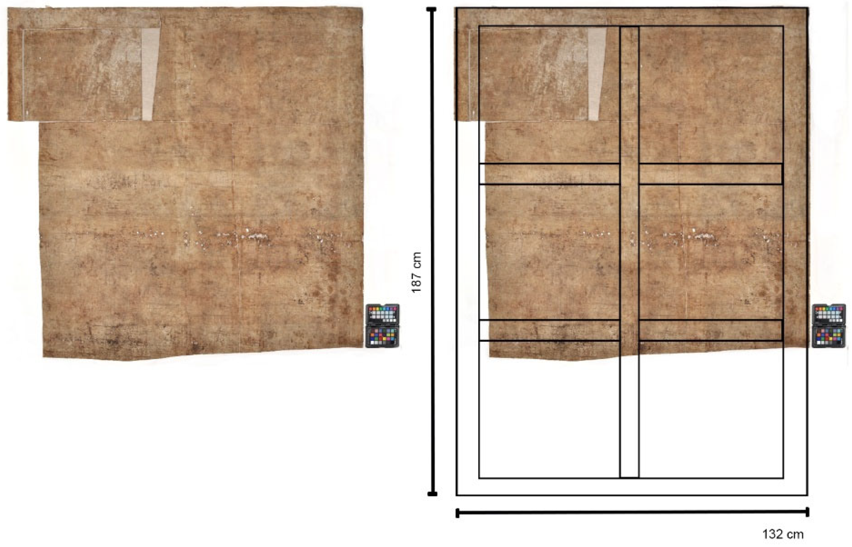
Since the lower part of the painting is missing it is difficult to ascertain its original size. Nevertheless, due to the markings of the stretcher frame on the back of the painting (Figure 2), an original size of approx. 187 cm × 132 cm can be reconstructed.
The results of the estimation suggest that the copy was, in its original state, slightly smaller than the original (207 cm × 140 cm). The digital overlay of both versions shows that there are only minor differences in scale and detail of the figures and the reason for the difference in dimension is that the upper part is absent in the copy. The painting had been restored in the past including relining and retouching. During treatment at CICS, the relining was reversed and the lining canvas removed.
In terms of provenance, the only information available was provided by the current owner who acquired the painting fragments. Although it is impossible to verify this information, the painting was allegedly acquired by a previous owner in a street market in Paris in the 1950s. It was subsequently cut into several fragments for ease of transport.

1.3. Strategy for Provenance and Dating Studies

To establish the possible date and geographical origin of the painting, a combination of lead isotope analysis and radiocarbon dating of different paint components as well as characterisation of painting materials and techniques were carried out.
The identification of the main pigments and additives combined with the historical context of introduction and use of these materials can help limit the possible creation date of a painting.

2. Materials and Methods

2.1. Canvas Fibre Identification

To analyse the fibre material, individual fibres were taken from both thread systems (vertical and horizontal) and examined using the “modified Herzog-Test” or red-plate test as described in [6], using a polarisation microscope Leica DM750P (Leica, Wetzlar, Germany). This test uses polarized light to determine the fibrillar orientation of bast fibres. Bast fibres are birefringent. Under polarised light and in the presence of a red-plate compensator, interference occurs. Depending on the fibre fibrillar orientation (S- or Z-twist), additive or subtractive compensation occurs, enabling its characterisation.

2.2. Stratigraphy Analysis in Cross Section

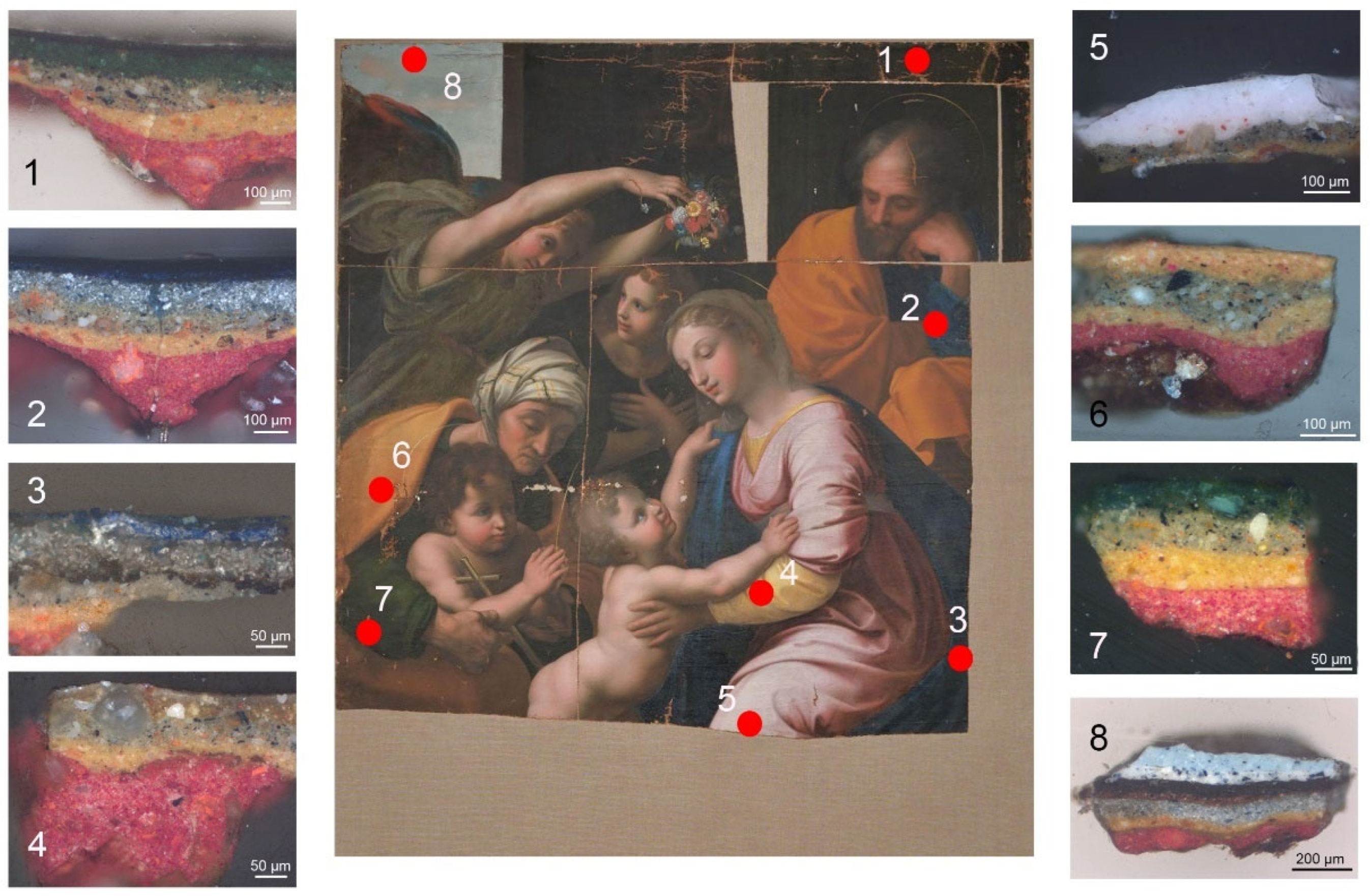
Samples comprising the complete paint package (including ground layers) were collected on the edge of fragments or damaged areas. Eight samples were collected from four fragments (Figure 3). The samples were embedded in Technovit LC 2000 (Kulzer GmbH, Wehrheim, Germany). The embedded sample was placed in a MOPAS hand polishing device (JAAP Enterprise, Amsterdam, The Netherlands) and the cross-section was prepared by dry polishing with Micromesh (grade up to 12,000). Documentation was carried out through light microscopy on a Zeiss Axioscope fluorescence microscope (Zeiss, Oberkochen, Germany).

2.3. Raman Spectroscopy

Raman spectroscopy was carried out directly on embedded and polished cross sections. The instrument is a Bruker Senterra I Raman microscope (Bruker, Billerica, MA, USA)used in conjunction with the OPUS version 7.5 software (Bruker, Billerica, MA, USA). All measurements were carried out with the 785 nm laser at 1 mW laser power and the 50× objective on cross sections.

2.4. SEM-EDX

Scanning electron microscopy energy dispersive x-ray spectroscopy was carried out on Sigma VP FESEM (Zeiss, Oberkochen, Germany) equipped with a Quantax XFlash EDX detector 430M (Bruker Nano GmbH, Berlin, Germany). EDX analysis parameters include an acceleration voltage of 20 kV and performed at a working distance of 9 mm.

2.5. Py-GC/MS

Samples from the paint (5_01) and preparatory layers (5_02), collected in the identical location as sample 5, were analysed by Py-GC/MS for characterisation of the binding medium. Each sample was suspended in 2 µL of tetramethylammonium hydroxide (TMAH, Sigma-Aldrich, Schnelldorf, Germany) 2.5% in methanol. An amount of 1 µL was transferred to an ECO cup (Frontier Lab, Koriyama, Japan) and the methanol allowed to evaporate. Py-GC/MS was carried out in a TraceTM GC Ultra (Thermo Scientific, Waltham, MA, USA) coupled to an ISQ 7000 (Thermo Scientific, Waltham, MA, USA) mass spectrometer and a Multishot Pyrolyser, EGA/Py-3030D (Frontier Lab, Koriyama, Japan). The pyrolysis temperature was set to 590 °C and maintained for 1 min. The GC was equipped with an Ultra alloy® capillary column (length 30 m, i.d. 0.25 mm, film thickness 0.25 µm, Frontier Lab, Koriyama, Japan). Helium was used as carrier gas (1.2 mL/min) and the system was operated in split flow mode (1:80 split ratio). The GC oven programme started at 40 °C for 1 min, then increased to 200 °C by heating at a rate of 10 °C/min, slowing down to reach 300 °C at 6 °C/min, for a final isothermal period of 10 min. The GC inlet was held at 260 °C, while the mass transfer line at 290 °C. A standard ionisation energy of 70 eV was used, and the temperature of the ion source was set at 270 °C. The range of mass scanning was set at 45–750 m/z.

2.6. Radiocarbon Dating

Both the support material, i.e., canvas and relining material, as well as the pictorial layer were 14C dated. The textile samples (≈2.5 mg) were first cleaned with solvents in the Soxhlet before being subjected to the standard acid–base–acid (ABA) protocol [7]. The cleaned textiles were converted to graphite in an automated graphitisation system (AGE) [8]. Flakes of paint (total weight approx. 2 mg detailed information in Table S12), removed from the Madonna’s dress, were split into multiple sub-preparations (average weight 500 µg), where the 14C age of both the organic binder and lead white was targeted [9]. The 14C signature of the carbonate was accessed by thermal decomposition of the lead carbonate at 350 °C. Following the isolation of the carbonate’s 14C signature, the dating of the organic binder in the carbonate-free paint fraction was pursued coupled to Pb-isotopic analysis [9]. The 14C analysis of both graphitised and gaseous samples was carried out on the MICADAS 14C dedicated system [10,11]. Data reduction was performed using in house BATS software [12]. Finally, the obtained radiocarbon ages were calibrated using the software Oxcal v.4.4.4 (https://c14.arch.ox.ac.uk/oxcal/OxCal.html (accessed on 28 September 2025)) [13] with the IntCal20 atmospheric calibration curve [14].

2.7. Lead Isotope Analysis

The remaining material after 14C-analysis was prepared for the lead isotope determination as described by Hendriks et al. [9]. The samples were digested; the lead concentration was determined and accordingly diluted. A Thallium spike served as an internal standard for the analysis by an HR MC-ICPMS. The operating settings, operating conditions, data correction (for the background and 204Hg), and data treatment followed the procedure as described in Hendriks et al. [9]. The given ratios are calculated based on the treated data and given as mean plus relative standard deviation. Experimental parameters are given in Supplementary Information Table S13.

3. Results and Discussion

3.1. Material Characterisation

3.1.1. Canvas Fibres and Sizing

The textile support is woven in a linen weave and consists of two fabric panels originally sewn together. The weft and warp direction cannot be clearly determined due to the absence of the selvedges, thread count: 14 (H) × 12 (V)/cm2 (relatively coarsely woven fabric), Z-twist of the threads with relatively tight torsion of the threads. The fibres show the characteristic features of bast fibres such as a smooth surface with cross-markings and dislocations (nodes). Both thread systems show a S-twist fibrillar orientation supporting the identification as hemp fibres [6]. No sizing layer could be detected. The first red preparation layer fills the canvas weave gaps and can be seen in the reverse side (Figure 4). In the literature, a widespread hemp tradition from the 17th century is mentioned [15,16].

3.1.2. Ground Layers

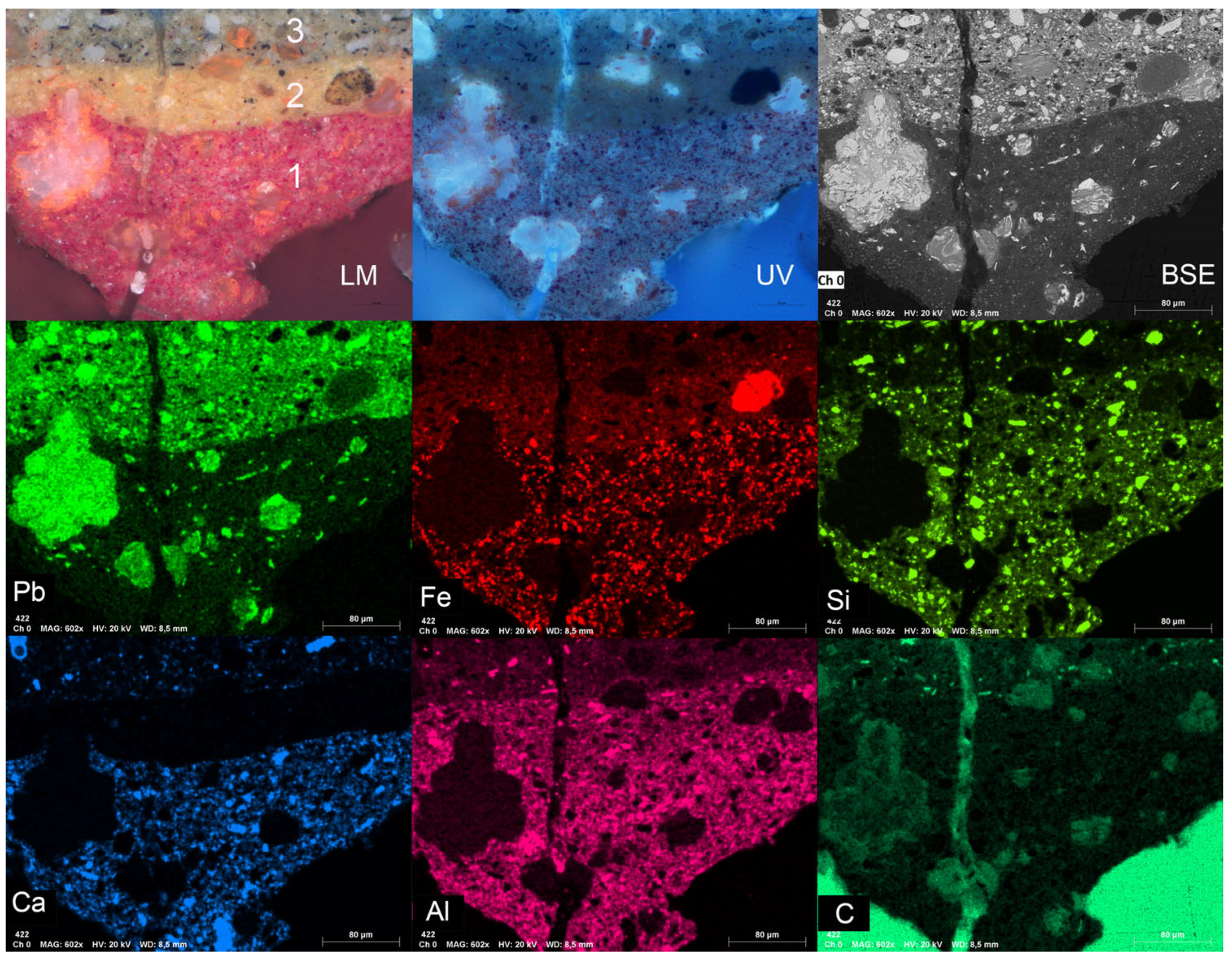
The canvas preparation is composed of three layers (Figure 5). Starting from the canvas surface a red layer (layer 1) is detected in all areas of the painting. The thickness of the layer follows the canvas profile and fills the gaps between weft and warp as can be seen in Figure 4. The second preparation layer is a yellow/orange (layer 2) and finally a grey/beige layer (layer 3).
The material characterisation of the observed three preparative layers of the preparation (Figure 5 and Table 1) can be described as follows:
Applied directly to the canvas, a red layer is detected in all areas of the painting. This layer is composed of iron oxide (haematite), red lead/minium (Pb3O4), calcium carbonate, aluminium silicates (clay minerals), and quartz in an organic lead matrix. The presence of minium is closely, but not exclusively, associated with the partially re-mineralised lead soap inclusions. Minium particles are also found dispersed in the matrix. Higgit et al. 2004 interprets this as evidence that the metal soaps form from a reaction between minium and the fatty acids in oil binding medium [17]. As an alternative interpretation, Boon et al. (2002, 2006) proposed that minium might also be a product of the remineralisation process; however, this has never been successfully reproduced in model systems [18,19]. The addition of minium in the original formulation cannot be excluded. This suggests a first preparation layer composed of a red earth and calcium carbonate pigmented leaded oil, possibly with the addition of minium.
The second preparation layer is a yellow/orange layer consisting of yellow ochre, lead carbonate, minium, quartz, and aluminium silicates in a lead rich organic matrix.
The third preparation layer is a grey layer composed of lead white and carbon black. In this layer, calcium carbonate (visible foraminifera; therefore, chalk), quartz, and minium particles are equally detected.
It was not possible to separate the ground layers for binding medium analysis identified as a drying oil (Table S11).

3.1.3. Paint Layers

The identification of the main pigments and additives is fundamental in technological, provenance, and dating studies. The analytical results are summarised in Table 2 for the paint layers.
Natural minerals can provide geographical and technological information. Calcium carbonate was identified as chalk due to the presence of foraminifera, which has been reported in painting ground layers from early medieval times, in particular in northern European paintings [20]. The use of red iron oxide (haematite) and yellow ochre since antiquity has been reported [21] and so the pigments provide little dating information in this case. Green copper-based minerals were detected in the green paint layers. In sample 1, the correlation between light microscopy, SEM-EDX, and Raman shows that copper carbonate and a copper sulphate are present. The spectrum at a measurement point with high copper and sulphur content in EDX mapping shows a very strong SO42− vibration band at 972 cm−1, characteristic for copper sulphates [22,23]. The location of the bands at 445 and 972 cm−1 indicates the presence of Posnjakite (hydrated copper sulphate) [23]. Posnjakite is not an abundant pigment, but it has been reported in green paint layers [24,25,26]. Copper sulphates often occur in mixtures with malachite, presumably because the minerals often occur together and are mined together [27,28]. It has been suggested that Posnjakite found alongside malachite may be a degradation product of the malachite [22]. Further research is required to enable the extraction of dating or provenance information from the identified copper green pigments.
Mercury sulphide was identified as the red pigment based on combined LM, SEM-EDX, and Raman spectroscopy. Exclusively Hg and S are detected and other elements if present are below the level of detection (LoD) of EDX. Franquelo et al. have compared cinnabar mineral with wet and dry processed vermilion and identified trace elements that could be associated to either wet or dry process of the pigment [29]. No trace elements could be detected, which could indicate that mineral ground cinnabar is present rather than synthetic wet or dry processed vermilion. According to Gettens et al. [30] the mines in Almadén (Spain) were the main source of cinnabar and mercury in the world and have been known since antiquity.
Lead tin yellow type I and type II (an earlier pigment) both have a distinct composition (type I Pb2SnO4 and type II Pb(Sn,Si)O3/PbSn1-xSixO3) and Raman spectrum [31]. No silicon could be detected with SEM-EDX, and the Raman spectrum matches that of type I pigment [31]. Lead tin yellow type I (Pb2SnO4) was frequently used in European paintings from the 15th century to the 1750s [32].
Basic lead carbonate or lead white is a synthetic pigment which has been manufactured and used since antiquity. Until the invention and introduction of zinc white, basic lead white was the only white pigment used in European paintings [33].
Several blue pigments are used in the original layers of the painting, including smalt, natural ultramarine, and azurite. In the first blue layer from sample 2, smalt was identified, containing the elements K, Co, As, Fe, Ni, and Bi. Smalt, a synthetic pigment, is a potassium glass made with the addition of cobalt oxide and was widely used in European paintings from the 15th to 18th centuries [34,35]. The history of cobalt mining in Europe in relation to ceramic, glass, and paint pigments has been recently reviewed [36], and its long-standing use in glass and ceramics is well documented [37]. Although smalt is chemically similar to cobalt glass, it was seldom found in paintings before 1450 [36]. The situation changes in the early 1500s, with the re-invention of smalt as pigment. This is associated with the mining development in the Erzgebirge (Ore Mountains in the Czech–German border). Evidence of a change of cobalt source can be inferred from a change in trace elements. The presence of nickel, arsenic, and bismuth is consistent with the use of cobalt ore from Schneeberg in the Erzgebirge region in Germany [36,37,38,39]. The smalt pigment was not prepared from the cobalt ore directly but from an intermediate product called zaffre made by roasting the cobalt ores with sand and a potassium containing flux. The commercialisation of zaffre increased after 1520. The change in composition of cobalt blue glazes occurring between 1517 and 1540 coincided with the technological change in the preparation of zaffre [36,40] and the increased use of smalt as a pigment in paintings. By 1550, it was found in a large number of European paintings [41,42]. The dating of smalt based on trace elements still requires further investigation. However, the research conducted by Robinet et al. provides evidence supporting the thesis that smalt pigments containing trace amounts of iron, arsenic, bismuth, and nickel first appeared in the mid-16th century [43]. These elements have been identified in the smalt pigments of several paintings from the late 16th and 17th centuries across various European regions. Notable examples include works by Paolo Veronese (dated 1562), Frans Floris I (dated 1562), and Murillo (dated 1672–1682).
Finally, in the top layer of sample 2, Prussian blue, an 18th century pigment, was detected [44]. However, it has filled the paint craquelure and therefore was clearly applied long after the original smalt blue paint layer had dried, most likely during the restoration of the painting.
The binding medium of the paint layer and ground layers were analysed by Py-GC/MS. The results mainly show markers for long and short chain fatty acids and diacids (Table S11). The results indicate the presence of a drying oil.

3.2. Radiocarbon Dating

Due to the invasiveness of the method, radiocarbon dating has historically only been considered for the analysis of the canvas of an easel painting. The recent reduction of the sample size required for the analysis enables the dating of other materials, such as the pictorial layer, by targeting the natural organic binder, or lead white pigment if it is present. While offsets between the 14C ages and the act of painting cannot be excluded, the possibility of dating different materials from an object can allow us to gain a better picture of how and when it was created. The radiocarbon results of the selected samples are summarized in Table S12 in SI, including their calibration to the corresponding calendar age.
As illustrated in Figure 6, all the materials (canvas, organic binder, and lead carbonate) originate from the same period. The dating of the canvas, i.e., when the hemp plant was harvested, yields a time range between the beginning of the 16th century and the mid-17th century. The dating of the organic binder has been recognised as more representative of the time of creation of an object than the support, as the binder can less easily be recycled [45]. However, this approach requires a detailed characterisation of all paint components prior to 14C analysis to exclude contamination from other materials like organic pigments, a varnish, or carbonates [46]. Here, the samples collected from the Madonna’s dress (sample 5—material characterisation) were identified as a mixture of quartz and lead white in an oil binder and were therefore suitable for 14C dating of the natural organic binder. The sample also contains a small portion of the lead white/vermilion sublayer, which is equally original and contemporary to the lead white quartz layer and compatible with radiocarbon dating. In the same sample cerussite and hydrocerussite, commonly known as lead white, were identified. This was therefore also dated with 14C dating [9]. The collected paint sample enabled the measurement of four replicas and yielded a mean value of 339 ± 34 yrs BP for the drying oil, which calibrates to 1470–1640. In contrast, despite three replicas having been measured, the dating of the lead white pigment corresponds to a much wider time range covering the mid-15th to mid-20th century. The 14C signature of the lead carbonate indicates an atmospheric carbon dioxide source, which is characteristic of the traditional Dutch process of fermenting organic material [47]. From the 19th century, the means of production evolved and the CO2 source varied considerably, typically bearing a geological isotopic ratio, so the time range for the possible production of the pigment can be narrowed to the mid-15th to the beginning of the 19th century. On its own, this dating bears little chronological information.
The three individual time frames provided by the radiocarbon dating of the canvas, the binder and the lead carbonate pigment overlap significantly. Using the R-combine function in Oxcal, the canvas and mean value of the paint and lead white successfully pass the null hypothesis χ2-test, hence indicating that all materials originate from the same time period between 1500–1642 (R_Combine (319, 17), χ2-Test: df = 2 T = 1.4 (5%)).

3.3. Lead Isotope Analysis

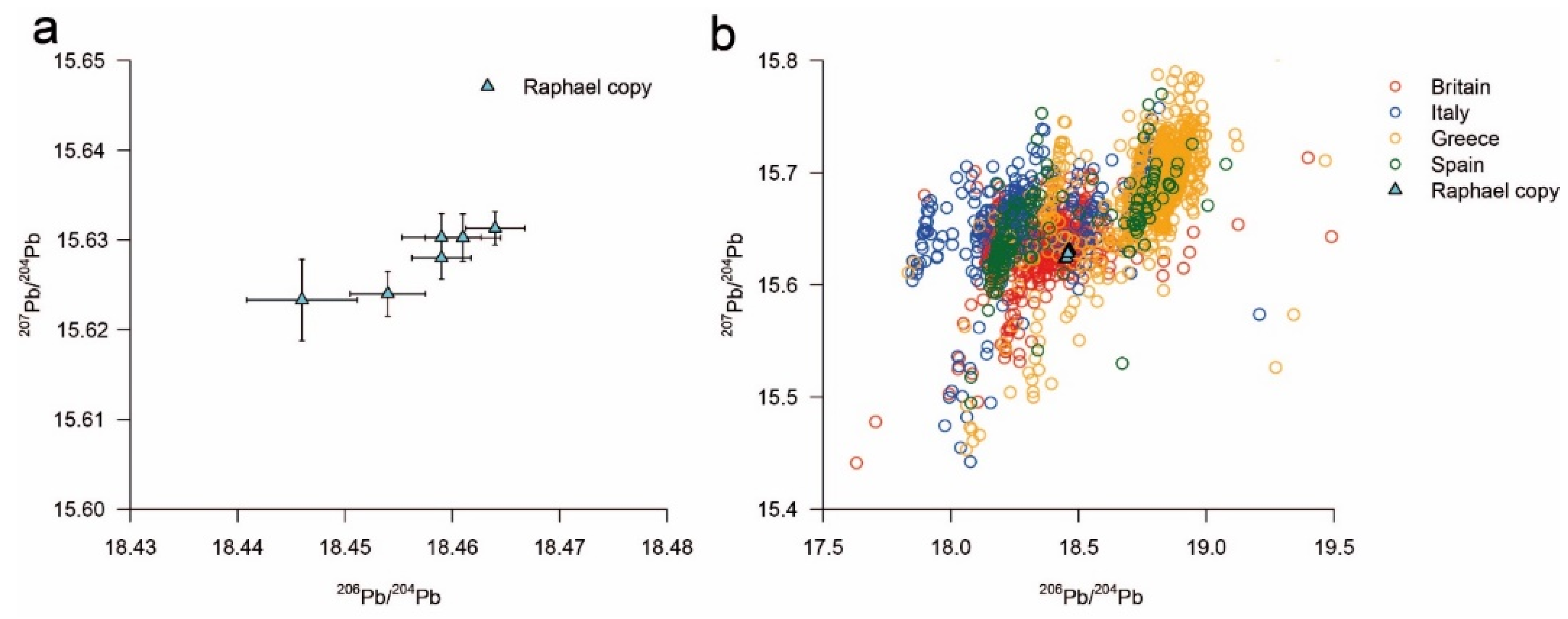
As shown in Table 3 and Figure 7a,b, all the samples show a close grouping in their lead isotope ratios. Additionally, if the ratios relative to the 204Pb are plotted against each other, the sample collection shows a characteristic linearity (Figure 7a). This feature is often reported in Pb-isotope determination as being given by the uncertainty on the 204Pb-signal. The heterogeneity of the date is similar to that reported in the work of D’Imporzano et al. [48]. The data therefore support the assumption of a common lead or lead white source. In combination with the determined 14C-age, a geographical classification with other lead isotope ratio data may enable the possible origin of the lead white and its corresponding lead ore source(s) to be more precisely identified [9,49]. A direct comparison of the presented data here with the collection of the data in D’Imporzano et al. indicates that the painting, or the lead white pigment used, dates to before 1647, which is in line with the radiocarbon dating, but must be considered with caution as the work of D’Imporzano et al. (2021) only discusses paintings of Dutch origin [48].
The direct comparison with data from European lead ore sources is shown in Figure 7b (OXALID database: Spain, Great Britain, Italy, Greece, and Bulgaria), in a similar way to Van Loon et al. in their study about the geographic source of lead found in Vermeer’s “Girl with a Pearl Earring” painting [50]. The data from the different ore sites in Europe form clusters but show extensive overlap with each other. Although the lead isotope ratio values for the lead white pigment in the copy of Raphael’s “The Great Holy Family” fits within the data of the European ore sites, absolute statements regarding the exact geographical origin are not possible. Specifically, the data overlap with British ore sites and are close to data from Bulgaria and Italy. Other studies have also followed this approach with similar findings [51,52,53]. A comparison with their data similarly shows a grouping of European ore sites and a direct overlap of the data presented here with English/British and German ore sources.
Non-European ore sources, like those from America and Australia, can most probably be excluded due to their generally higher radiogenic lead ores in combination with the dated age of the lead white, as already described by Keisch et al. [49]. The works of Stevenson et al. and Hendriks et al. present the same line of argument [9,53].

4. Conclusions and Future Work

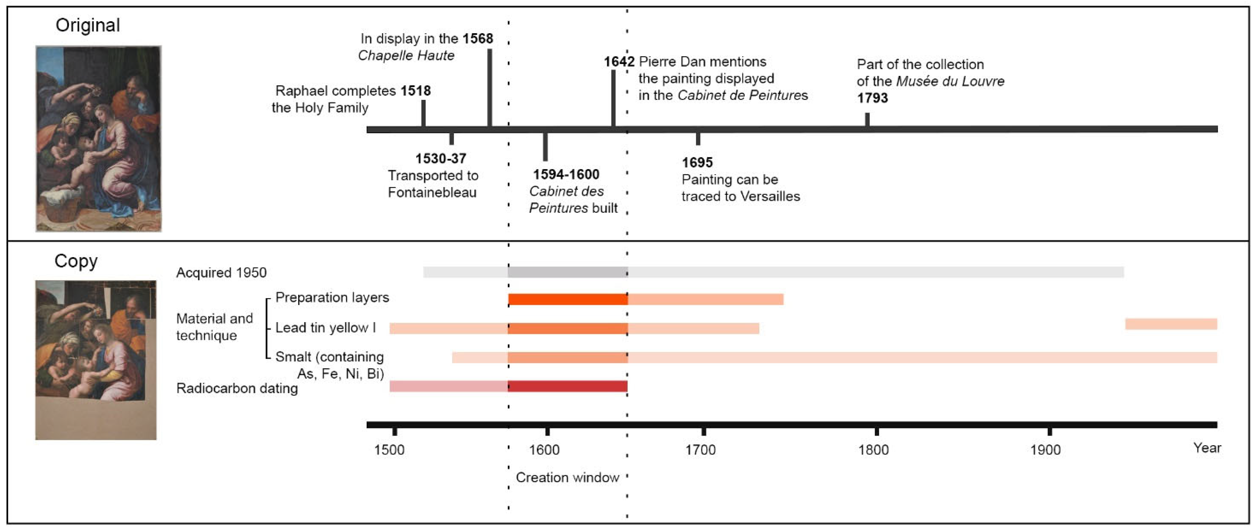
The copy of Raphael’s “The Great Holy Family” under study here is neither dated nor attributed to an artist. In addition, there is very limited provenance information. Material characterisation, radiocarbon dating of multiple materials, and lead isotope study enable the narrowing of the chronological window of the materials used. The combined information is shown in Figure 8, enabling the visualisation of the possible window of creation of the copy.
Radiocarbon dating of the binder, lead carbonate, and canvas fibres indicate that the painting can be dated to between 1500 and 1642. As the support of the work of art, the age of the canvas sets the earliest possible date of creation, namely 1496. However, the original painting by Raphael Santi was commissioned in 1518, hereby setting the terminus post-quem for the creation of the copy. The combination of 14C dating with art historical knowledge therefore points to a time range of 1518–1642 for the materials used to make the copy under investigation.
Significant systematic research has been devoted to the changing use of ground layers in the south and north of Europe [54,55,56,57]. In her review, Stols-Witlox indicates that the use of coloured grounds gradually spread through Europe during the 16th century and, in particularly in northern Europe, the introduction of coloured grounds seems to be dated to the late 16th century [55]. This is supported by the survey of contemporary recipes for the preparation of paintings [56]. Until then, calcium sulphate grounds were most commonly used in Italy and chalk grounds in northern Europe. Starting in Italy, the painters of the generation of Titian (16th century Italian painter) began to omit the gypsum layer and applied their coloured layers directly to the sized canvas. In Venice these grounds had grey reddish-brown hues. Multi-layered coloured grounds, in particular double grounds, appeared in the 16th century and became popular in the 17th century. These typically consisted of a lower layer of mainly earth pigments covered by a layer composed of lead white toned with pigments such as carbon black, ochre, umber, minium, or vermilion. Triple grounds as described in the current study are rare. In the late 17th to early 18th century, coloured grounds lost their popularity. The research by Duval (1992) and Salvant et al. (2021) [54,58] of both written sources and the ground layers of paintings of the “Écóle Francaise” in the 17th and 18th centuries, clearly show how established the use of coloured grounds prepared with iron oxides was, with the addition of lead containing pigment and/or siccative. The use of calcium carbonate as an additive in the coloured grounds was particularly evident in the paintings of the first half of the 17th century. Within the period researched, the presence of minium was restricted to the paintings of the first half of the 17th century [54]. However, minium has been identified in preparation layers in earlier paintings. For example, minium or red lead has been detected together with lead white and chalk in the painting “Penance of St. Jerome” by Lucas Cranach the Elder dated to 1502 [59]. According to Heydenreich [59] it is unclear if the addition of minium or read lead is intentional or the result of reactivity.
The materials and techniques of the preparatory layers, based on current knowledge, give a higher probability date of copy of “The Great Holy Family” to the late 16th century to mid-18th century. All the pigments found were available and popular during this period.
Lead isotope analysis and comparison with data of lead ore sources (Oxford Archaeological Lead Isotope Database from the Isotrace Laboratory (OXALID)) and the work D’imporzano et al. (2021) [48] indicate a northern Europe origin, with a match to German or, most probably, British ore.
Combining radiocarbon dating, lead isotope analysis, material, and technological characterisation of preparation and paint layers, it can be concluded that the copy of Raphael’s “The Great Holy Family” was most likely created in northern Europe in the period between the late 16th and mid-17th century.
In the late 16th century, the original painting was displayed in the “Chapelle Haute” and in the early 17th century it was transferred to the newly built “Cabinet des Peintures et Tableaux du Roi” [4]. The same source mentions that a copy had replaced the original in the “Chapelle Haute”. The location of this copy is not known, according to Cox-Rearick [2]. Another, later copy of the painting was made by the painter Jean Michelin (1623–95) and is still located in Fontainebleau [1]. The establishment of the Art Academy in Paris in 1648 and the introduction of the copying of old masters in the curriculum facilitated the production of copies of paintings such as “The Great Holy Family” [60]. Meyer zur Capellen lists thirty-nine paintings and drawings which are copies of “The Great Holy Family” currently part of collections worldwide [1] (p. 175). The painting under study in this work is not referred to and can now be added to the list. Additionally, although the combination of sources research, technical analysis, radiocarbon dating, and lead isotope analysis reveal when, where, and to some extent how the copy was prepared, the questions of by whom and for whom the painting was made remain unanswered. There are other examples from the first half of the 17th century of the movement of precious altarpieces from the altars to collections and replaced in situ by copies [61]. However, there were many different motivations for producing or commissioning faithful copies during the period in question [62]. Further art historical research should clarify which actors at the French court in Fontainebleau (or outside) could have had an interest in owning a copy after Raphael’s “The Great Holy Family” and thus what the motivation for the production of such a copy could have been.

Supplementary Materials

The following supporting information can be downloaded at https://www.mdpi.com/article/10.3390/heritage8100424/s1, Figure S1: Infrared reflectography (IRR); Table S1: Light microscopy (LM) and scanning electron microscopy energy dispersive spectroscopy/SEM/EDX spot analysis of the ground layers. Table S2: Light microscopy (LM) and scanning electron microscopy energy dispersive spectroscopy/SEM/EDX element mapping of the ground layers. Table S3: Raman spectroscopy of selected spot in the ground layers. Table S4: Light microscopy (LM) and scanning electron microscopy energy dispersive spectroscopy/SEM/EDX spot analysis of smalt containing paint layers. Table S5: Raman spectroscopy of Prussian blue containing overpaint. Table S6: Light microscopy (LM) and scanning electron microscopy energy dispersive spectroscopy/SEM/EDX spot analysis and element mapping of copper green pigment containing paint layers. Table S7: Raman spectroscopy of the copper green pigment. Table S8: Light microscopy (LM) and scanning electron microscopy energy dispersive spectroscopy/SEM/EDX spot analysis and element mapping of lead tin yellow and cinnabar pigment containing paint layers. Table S9: Raman spectroscopy of lead tin yellow and cinnabar. Table S10: Light microscopy (LM) and scanning electron microscopy energy dispersive spectroscopy (SEM/EDX) spot analysis and element mapping white and light red/pink paint layers. Table S11: Pyrolysis gas chromatography mass spectrometric analysis of binding medium of paint and ground layers. Table S12: Radiocarbon results gained on the paint samples. The summary of the results is organized by ETH laboratory code and targeted material (lead carbonate or binder), 14C ages with 1σ uncertainty, and the respective calibrated calendar ages using the software Oxcal 4.4 with the IntCal20 calibration curve. Mean values for both the drying oil and the lead white were generated by the combination of the individual results, which is automatically checked for internal consistency by a chi-square test. Table S13: Lead isotope ratio analysis parameters.

Author Contributions

Conceptualisation: E.S.B.F., L.H. and H.P.; Investigation: E.S.B.F., L.H., S.K., K.H., J.R., C.H. and S.M.-B.; Writing—original draft: E.S.B.F., L.H., S.K., K.H. and C.H.; Writing—review and editing: E.S.B.F., L.H., S.K., K.H., H.P., D.G., I.H. and S.M.-B.; Supervision: E.S.B.F., H.P., I.H. and D.G. All authors have read and agreed to the published version of the manuscript.

Funding

The radiocarbon dating and combination with lead isotope analysis was carried out during L.H.’s Ph.D.’s project, supported by grant ETH-21 15-1, and S.K.’s Ph.D.’s project. L.H. acknowledges current support from The Branco Weiss Fellowship–Society in Science, administered by the ETH Zurich, which made it possible to complete the manuscript.

Data Availability Statement

Data is contained within the article or Supplementary Materials.

Acknowledgments

The authors would like to thank Dietmar Weinem, for providing access to the painting fragments and agreeing with the publication of the research results. Diana Blumenroth is acknowledged for the IRR of the fragments and Sarah Critchley for reviewing the text. The authors would also like to thank Barbara Berrie for insightful discussions on the history of cobalt and copper pigments.

Conflicts of Interest

The authors declare no conflicts of interest. The funders had no role in the design of the study; in the collection, analyses, or interpretation of data; in the writing of the manuscript; or in the decision to publish the results.

References

  1. Meyer zur Capellen, J. Raphael. A Critical Catalogue of His Paintings. Volume II, The Roman Religious Paintings ca. 1508–1520; ARCOS: Landhut, Germany, 2005; pp. 170–177. [Google Scholar]
  2. Cox-Rearick, J. The Collection of Francis I. Royal Treasures; Fonds Mercator Paribas: Antwerp, Belgium, 1995. [Google Scholar]
  3. Fagnart, L. L’appartement des bains et le cabinet des peintures du château de Fontainebleau sous le règne d’Henri IV. In Henri IV. Art et Pouvoir; Nativel, C., Ed.; Presses universitaires François-Rabelais: Tours, France, 2016; pp. 53–65. [Google Scholar] [CrossRef]
  4. Dan, P. Le Trésor des Merveilles de la Maison Royale de Fontainebleau, Paris, Sébastien Cramoisy, 1642, Gallica Digital archive of the Bibliothèque Nationale de France. Available online: https://gallica.bnf.fr/ark:/12148/bpt6k96507143 (accessed on 27 August 2025).
  5. Mottin, B.; Ravaud, E.; Bastian, G.; Eveno, M. Raphael and his Entourage in Rome: Laboratory Study of the Works in the Musée du Louvre. In Late Raphael. Exhibition Catalogue; Henry, T., Joannides, P., Eds.; Musée du Louvre: Paris, France; Museo Nacional del Prado: Madrid, Spain, 2012; pp. 351–364. [Google Scholar]
  6. Haugan, E.; Holst, B. Determining the fibrillar orientation of bast fibers with polarizes light microscopy: The modified Herzog test (red plate test) explained. J. Microsc. 2013, 252, 159–168. [Google Scholar] [CrossRef]
  7. Hajdas, I.; Cristi, C.; Bonani, G.; Maurer, M. Textiles and Radiocarbon Dating. Radiocarbon 2014, 56, 637–643. [Google Scholar] [CrossRef]
  8. Wacker, L.; Nemec, M.; Bourquin, J. A revolutionary graphitisation system: Fully automated, compact and simple. Nucl. Instrum. Methods B 2010, 268, 931–934. [Google Scholar] [CrossRef]
  9. Hendriks, L.; Kradolfer, S.; Lombardo, T.; Hubert, V.; Küffner, M.; Khandekar, N.; Hajdas, I.; Synal, H.-A.; Hattendorf, B.; Günther, D. Dual isotope system analysis of lead white in artworks. Analyst 2020, 145, 1310–1318. [Google Scholar] [CrossRef]
  10. Synal, H.A.; Stocker, M.; Suter, M. MICADAS: A new compact radiocarbon AMS system. Nucl. Instrum. Methods B 2007, 259, 7–13. [Google Scholar] [CrossRef]
  11. Wacker, L.; Fahrni, S.M.; Hajdas, I.; Molnar, M.; Synal, H.A.; Szidat, S.; Zhang, Y.L. A versatile gas interface for routine radiocarbon analysis with a gas ion source. Nucl. Instrum. Methods B 2013, 294, 315–319. [Google Scholar] [CrossRef]
  12. Wacker, L.; Christl, M.; Synal, H.A. Bats: A New Tool for AMS Data Reduction. Nucl. Instrum. Methods B 2010, 268, 976–979. [Google Scholar] [CrossRef]
  13. Ramsey, C.B. Radiocarbon Calibration and Analysis of Stratigraphy: The OxCal Program. Radiocarbon 1995, 37, 425–430. [Google Scholar] [CrossRef]
  14. Reimer, P.J.; Austin, W.E.; Bard, E.; Bayliss, A.; Blackwell, P.G.; Ramsey, C.B.; Butzin, M.; Cheng, H.; Edwards, R.L.; Friedrich, M.; et al. The IntCal20 Northern Hemisphere Radiocarbon Age Calibration Curve (0–55 Cal kBP). Radiocarbon 2020, 62, 725–757. [Google Scholar] [CrossRef]
  15. Koller, M.; Kühn, H.; Roosen-Runge, H.; Straub, R.E. Das Staffeleibild der Neuzeit. In Reclams Handbuch der Künstlerischen Techniken 1, Farbmittel, Buchmalerei, Tafel- und Leinwandmalerei; Reclam, P., Jr., Ed.; GmbH Publishers: Stuttgart, Germany, 1984. [Google Scholar]
  16. Vanderbilt Carbonnel, K. A study of French painting canvas. J. Am. Inst. Conserv. 1980, 20, 3–20. [Google Scholar] [CrossRef]
  17. Higgit, C.; Spring, M.; Saunders, D. Pigment-medium Interactions in oil paint films containing red lead or lead-tin yellow. Natl. Gallery Tech. Bull. 2003, 24, 75–95. [Google Scholar]
  18. Boon, J.J.; van der Weerd, J.P.; Keune, K.; Noble, P.; Wadum, J. Mechanical and chemical changes in Old Master paintings: Dissolution, metal soap formation and remineralization processes in lead pigmented ground/intermediate paint layers of 17th century paintings. In Proceedings of the ICOM-CC 13th Triennial Conference Preprints, Rio de Janeiro, Brazil, 20–27 September 2002; James & James (Science Publishers) Ltd.: London, UK, 2002. [Google Scholar]
  19. Boon, J.J. Processes inside paintings that affect the picture: Chemical changes at, near and underneath the paint surface. In Reporting Highlights of the De Mayerne Programme. Research Programme on Molecular Studies in Conservation and Technical Studies in Art History MOLART Reports 13; Boon, J.J., Ferreira, E.S.B., Eds.; Nederlandse Organisatie voor Wetenschappelijk Onderzoek (NWO): The Hague, The Netherlands, 2006. [Google Scholar]
  20. Gettens, R.J.; Fitzhugh, E.W.; Feller, R.L. Calcium Carbonate Whites. In Artists’ Pigments: A Handbook of Their History and Characteristics; Roy, A., Ed.; Archetype Publications Ltd.: London, UK, 1993; Volume 2, pp. 203–226. [Google Scholar]
  21. Helwig, K. Iron Oxide Pigments. In Artists’ Pigments: A Handbook of Their History and Characteristics; Berrie, B.H., Ed.; Archetype Publications Ltd.: London, UK, 2007; Volume 4, pp. 39–109. [Google Scholar]
  22. Martens, W.; Frost, R.L.; Kloprogge, T.; Williams, P.A. Raman spectroscopic study of the basic copper sulphates—Implications for the copper corrosion and bronze disease. J. Raman Spect. 2003, 34, 145–151. [Google Scholar] [CrossRef]
  23. Gilbert, B.; Denoel, S.; Weber, G.; Allart, D. Analysis of green copper pigments in illuminated manuscripts by micro-Raman spectroscopy. Analyst 2003, 128, 1213–1217. [Google Scholar] [CrossRef] [PubMed]
  24. Eastaugh, N.; Walsh, V.; Chaplin, T.; Siddal, R. Pigment Compendium; Elsevier Butterworth-Heinemann: Oxford, UK, 2008. [Google Scholar]
  25. Scott, D.A. Basic sulfates. In Copper and Bronze in Art; Scott, D.A., Ed.; Getty Publications: Hong Kong, China, 2002. [Google Scholar]
  26. Svarcova, S.; Hradil, D.; Hradilova, J.; Cermakova, Z. Pigments copper-based greens and blues. Archaeol. Anthropol. Sci. 2021, 13, 190. [Google Scholar] [CrossRef]
  27. Gettens, R.J.; Fitzburg, E.W. Malachite and Green Verditer. In Artists Pigments A Handbook of Their History and Characteristics; Roy, A., Ed.; Archetype Publications Ltd.: London, UK, 1993; Volume 2, pp. 183–201. [Google Scholar]
  28. Bersani, D.; Antonioli, G.; Lottici, P.; Casoli, A. Raman microspectrometric investigation of wall paintings in S.Giocanni Envagelista Abbey in Parma: A comparison between two artists of the 16th century. Spectrochim. Acta Part A Mol. Biomol. Spectrosc. 2003, 59, 2409–2417. [Google Scholar] [CrossRef] [PubMed]
  29. Franquelo, M.L.; Perez-Rodriguez, J.L. A new approach to the determination of the synthetic or natural origin of red pigments through spectroscopic analysis. Spectrochim. Acta Part A Mol. Biomol. Spectrosc. 2016, 166, 103–111. [Google Scholar] [CrossRef]
  30. Gettens, R.J.; Feller, R.L.; Chase, W.T. Vermilion and Cinnabar. In Artists Pigments A Handbook of Their History and Characteristics; Roy, A., Ed.; Archetype Publications Ltd.: London, UK, 1993; Volume 2, pp. 159–182. [Google Scholar]
  31. Sefcu, R.; Chlumska, S.; Hostasova, A. An investigation of the lead tin yellow type I and II and their use in Bohemian panel paintings from the Gothic period. Herit. Sci. 2015, 3, 16. [Google Scholar] [CrossRef]
  32. Kühn, H. 1993 Lead tin yellow. In Artists Pigments A Handbook of Their History and Characteristics; Roy, A., Ed.; Archetype Publications Ltd.: London, UK, 1993; Volume 2, pp. 83–112. [Google Scholar]
  33. Gettens, R.J.; Kühn, H.; Chase, W.T. Lead White. In Artists’ Pigments A Handbook of Their History and Characteristics; Roy, A., Ed.; Archetype Publications Ltd.: London, UK, 1993; Volume 2, pp. 67–82. [Google Scholar]
  34. Seldes, A.M. A note on the pigments and media in some Spanish Colonial paintings from Argentina. Stud. Conserv. 1994, 39, 272–276. [Google Scholar] [CrossRef]
  35. Gil, M.; Carvalho, M.L.; Longelin, S.; Ribeiro, I.; Valadas, S.; Mirão, J.; Candeias, A.E. Blue pigment colors from wall painting churches in danger (Portugal 15th to 18th century): Identification, diagnosis, and color evaluation. Appl. Spectrosc. 2011, 65, 782–789. [Google Scholar] [CrossRef]
  36. Berrie, B.H. Mining for Color: New Blues, Yellows, and Translucent Paint. Early Sci. Med. 2015, 20, 308–334. [Google Scholar] [CrossRef]
  37. Seccaroni, C.; Haldi, J.P. Impuresse ed elementi associate al cobalto. In Cobalto, Zaffera, Smalto dall Antichita al XVIII Secolo; Seccaroni, C., Haldi, J.P., Eds.; ENEA: Rome, Italy, 2016; pp. 65–102. [Google Scholar]
  38. Giannini, R.; Freestone, I.C.; Shortland, A.J. European Cobalt Sources Identified in the Production of Chinese Famille Rose Porcelain. J. Archaeol. Sci. 2017, 80, 27–36. [Google Scholar] [CrossRef]
  39. Gratuze, B. Provenance Analysis of Glass Artefacts. In Modern Methods for Analysing Archaeological and Historical Glass; Janssens, K., Ed.; John Wiley & Sons: Hoboken, NJ, USA, 2013; Volume I, pp. 311–343. [Google Scholar]
  40. Gratuze, B.; Soulier, I.; Barrandon, J.N.; Foy, D. The origin of cobalt blue pigments in French glass from the thirteenth to the eighteenth centuries. In Trade and Discovery: The Scientific Study of Artefacts from Post-Medieval Europe and Beyond; British Museum: London, UK, 1995; Volume 109, pp. 123–132. [Google Scholar]
  41. Mühlenthaler, B.; Theissen, J. Smalt. In Artists Pigments A Handbook of Their History and Characteristics; Roy, A., Ed.; Archetype Publications Ltd.: London, UK, 1993; Volume 2, pp. 113–130. [Google Scholar]
  42. Stege, H. Out of the Blue? Considerations on the early use of smalt as blue pigment in Europe easel painting. ZKK 2004, 18, 121–142. [Google Scholar]
  43. Robinet, L.; Spring, M.; Pages-Camagna, S. Vibrational Spectroscopy correlated with elemental analysis for the investigation of smalt pigments and its alteration in paintings. Anal. Methods 2013, 5, 4628. [Google Scholar] [CrossRef]
  44. Berrie, B.H. Prussian Blue. In Artists Pigments. A Handbook of Their History and Characteristics; Fitzhugh, E.W., Ed.; Archetype Publications Ltd.: London, UK, 1997; Volume 3, pp. 191–218. [Google Scholar]
  45. Hendriks, L.; Hajdas, I.; Ferreira, E.S.B.; Scherrer, N.C.; Zumbühl, S.; Smith, G.D.; Welte, C.; Wacker, L.; Synal, H.; Günther, D. Uncovering modern paint forgeries by radiocarbon dating. Proc. Natl. Acad. Sci. USA 2019, 116, 13210–13214. [Google Scholar] [CrossRef]
  46. Hendriks, L.; Hajdas, I.; Ferreira, E.S.B.; Scherrer, N.C.; Zumbühl, S.; Küffner, M.; Wacker, L.; Synal, H.-A.; Günther, D. Combined 14C Analysis of Canvas and Organic Binder for Dating a Painting. Radiocarbon 2018, 60, 207–218. [Google Scholar] [CrossRef]
  47. Messager, C.; Beck, L.; de Viguerie, L.; Jaber, M. Thermal Analysis of Carbonate Pigments and Linseed Oil to Optimize CO2 Extraction for Radiocarbon Dating of Lead White Paintings. Microchem. J. 2020, 154, 104637. [Google Scholar] [CrossRef]
  48. D’imporzano, P.; Batur, K.; Keune, K.; Koornneef, J.M.; Hermens, E.; Noble, P.; van Zuilen, K.; Davies, G.R. Lead isotope heterogeneity in lead white: From lead white raw pigment to canvas. Microchem. J. 2021, 163, 105897. [Google Scholar] [CrossRef]
  49. Keisch, B.; Callahan, R.C. Lead isotope ratios in artists’ lead white: A progress report. Archaeometry 1976, 18, 181–193. [Google Scholar] [CrossRef]
  50. van Loon, A.; Vandivere, A.; Delaney, J.K.; Dooley, K.A.; De Meyer, S.; Vanmeert, F.; Gonzalez, V.; Janssens, K.; Leonhardt, E.; Haswell, R.; et al. Beauty is skin deep: The skin tones of Vermeer’s Girl with a Pearl Earring. Herit. Sci. 2019, 7, 1–20. [Google Scholar] [CrossRef]
  51. Fortunato, G.; Ritter, A.; Fabian, D. Old Masters’ lead white pigments: Investigations of paintings from the 16th to the 17th century using high precision lead isotope abundance ratio. Analyst 2005, 130, 898–906. [Google Scholar] [CrossRef] [PubMed]
  52. Fabian, D.; Fortunato, G. Tracing white: A study of lead white pigments found in seventeenth-century paintings using high precision lead isotope abundance ratios. In Trade in Artists’ Materials. Markets and Commerce in Europe to 1700; Kirby, J., Nash, S., Cannon, J., Eds.; Archetype: London, UK, 2010; pp. 426–443. [Google Scholar]
  53. Stevenson, R.K.; Moffatt, E.A.; Corbeil, M.C.; Poirier, A. Pb and Sr Isotopes and the Provenance of the Painting Materials of Cornelius Krieghoff in 19th-Century Canada. Archaeometry 2016, 58, 673–687. [Google Scholar] [CrossRef]
  54. Duval, A. Les préparations colorées des tableaux de l’Ecole Française des dixseptième et dixhuitième siècles. Stud. Conserv. 1992, 37, 239–258. [Google Scholar]
  55. Stols-Witlox, M. Grounds 1400–1900. In Conservation of Easel Paintings, 2nd ed.; Stoner, J.H., Rushfiled, R., Eds.; Routledge: London, UK, 2012; Chapter 7; pp. 161–188. [Google Scholar]
  56. Stols-Witlox, M. “By no means a trivial matter”. The influence of the colour of ground layers on artists’ working methods and on the appearance of oil paintings, according to historical recipes from North West Europe, c. 1550–1900. Oud Holl.-J. Art Low Ctries. 2015, 128, 171–186. [Google Scholar] [CrossRef]
  57. Martin, E. Grounds on canvas 1600-1640 in various European artistic centres. In Preparation for Painting: The Artist’s Choice and Its Consequences g, Proceedings of the 2007 ICOM-CC Interim Meeting, London, UK, 31 May –1 June 2007; Townsend, J.H., Doherty, T., Heydenreich, G., Eds.; Archetype Publications Ltd.: London, UK, 2008. [Google Scholar]
  58. Salvant, J.; Eveno, M.; Betelu, C.; Negrello, C.; Bastian, G.; Faroult, G.; Ravaud, E. Investigating the transition period from colored to white preparatory layers in 18th-century French canvas paintings: A retrospective study. In Transcending Boundaries: Integrated Approaches to Conservation. Proceedings of the ICOM-CC 19th Triennial Conference Preprints, Beijing, China, 17–21 May 2021; Bridgland, J., Ed.; International Council of Museums: Paris, France, 2021. [Google Scholar]
  59. Heydenreich, G. Lucas Cranach the Elder. Painting Materials, Techniques and Workshop Practice; Amsterdam University Press: Amsterdam, The Netherlands, 2007. [Google Scholar]
  60. Voermann, I. Die Kopie als Element Fürstlicher Gemäldesammlungen des 19 Jahrhundert; Lukas Publishers: Berlin, Germany, 2012. [Google Scholar]
  61. Putzger, A. Kult und Kunst-Kopie und Original. Altarbilder von Rogier van der Weyden, Jan van Eyck und Albrecht Dürer in Ihrer Frühneuzeitlichen Rezeption; Bild+Bild; Reimer: Berlin, Germany, 2021; Volume 5. [Google Scholar]
  62. Gaggetta, C. The faithful copy as a medium of appropriation and propaganda. French commissions after Leonardo’s “Last Supper”. In Nichts Neues Schaffen. Perspektiven auf die Treue Kopie 1300–1900; Putzger, A., Heisterberg, M., Müller-Bechtel, S., Eds.; De Gruyter Brill: Berlin, Germany, 2018; pp. 133–152. [Google Scholar] [CrossRef]
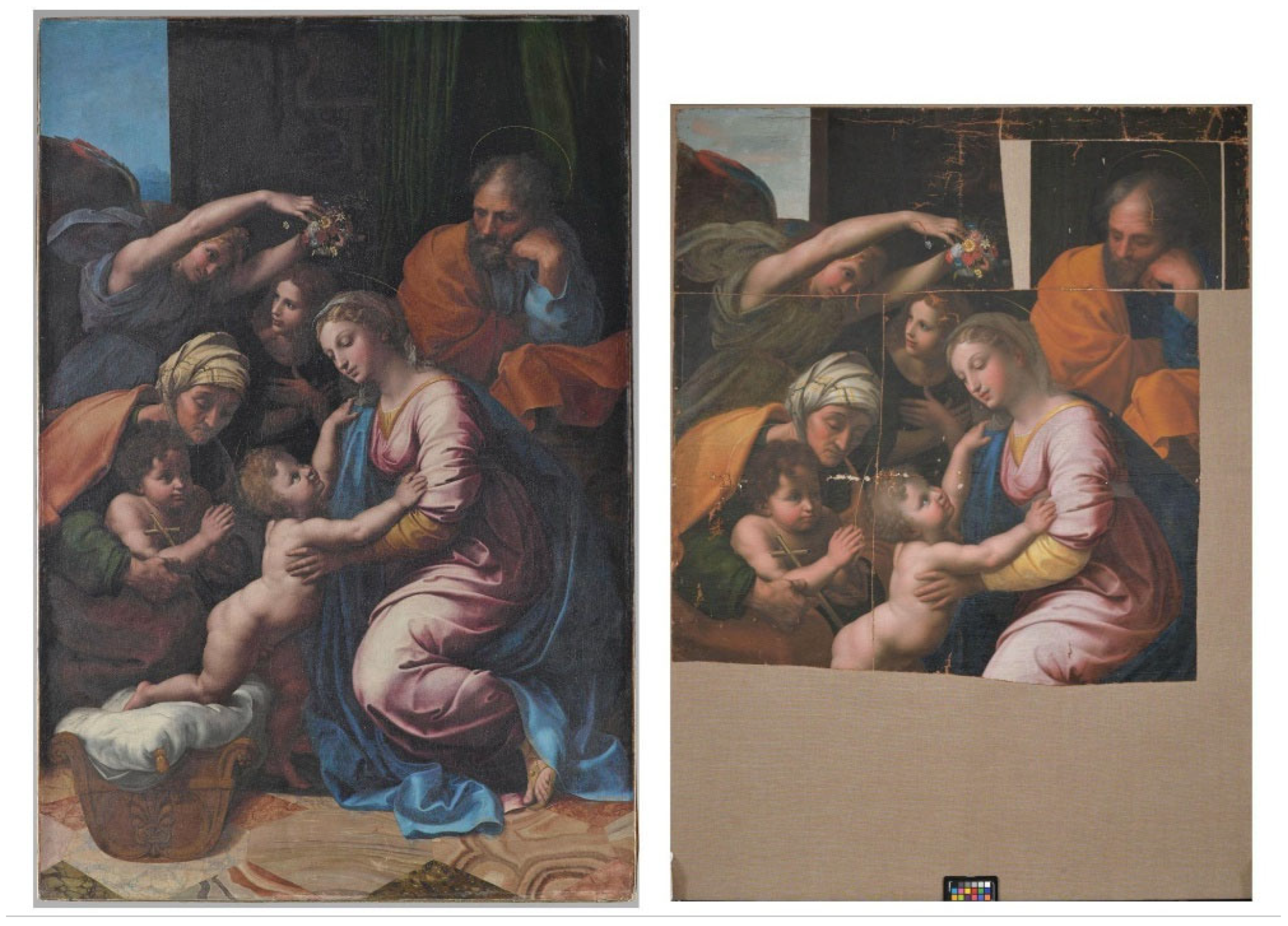
Figure 1. “The Great Holy Family of Francis I” (“La Grande Sainte Famille de François Ier”) by Raphael (Raffaelo di Santi), 1518, INV 604 MR 432, 207 cm × 140 cm (left) (Musée du Louvre, reproduced with permission). Copy after Raphael, artist and date unknown, state after conservation in 2022 (right).
Heritage 08 00424 g001
Figure 2. Reverse side of the recombined fragments (left). The markings from a stretcher can be seen faintly (left) and enable the reconstruction of the stretcher size (right).
Heritage 08 00424 g002
Figure 3. Sample location of the 8 investigated cross-sections (red dots mark the sampling locations and numbers identifys the samples).
Heritage 08 00424 g003
Figure 4. Polarised light microscopy: (a) Red/orange interference colour in 0° position, (b) blue interference colour in 90° position indicating an S-twist fibrillar orientation. (c) Detail of the textile support (reverse).
Heritage 08 00424 g004
Figure 5. Cross-section of sample 2, highlighting the three preparation layers. Top row: light microscopy under visible light (top left) (the numbers 1–3 identify the preparation layers), UV illumination (top centre), BSE image (top right). Second and third row: elemental distribution maps obtained by EDX, with Pb (middle left), Fe (middle centre), Si (middle right), Ca (bottom left), Al (bottom centre), C (bottom right).
Heritage 08 00424 g005
Figure 6. Respective calibration plots for the different dated materials, canvas (a), mean binder age (n = 4) (b), and mean lead carbonate age (n = 3) (c). The measured 14C ages are plotted against the calibration curve IntCal20 (blue). The intersection between the measured data (red), expressed in years before present (yrs BP), can be converted to calendar years on the x-axis and where the grey histograms depict the probability distribution.
Heritage 08 00424 g006
Figure 7. (a) Lead isotope ratios (206Pb/204Pb vs. 207Pb/204Pb) for the sample collected from the copy of “The Great Saint Family” after Raffael Santi (see Table 3). The error bars indicate the standard deviation of the acquired data. (b) Comparison of the determined isotope ratios (206Pb/204Pb vs. 207Pb/204Pb) from the case study “Copy of Raphael’s Great Holy Family” to the European lead ore sources. Data: Oxford Archaeological Lead Isotope Database from the Isotrace Laboratory OXALID, (3 February 2020), only for ores mainly containing lead.
Heritage 08 00424 g007
Figure 8. Timeline of the overall results: provenance (grey), materials and techniques, including the use of multi-layered coloured grounds (dark orange), availability of the pigment lead tin yellow (middle orange) and the use of smalt containing the trace elements As, Fe, Ni, and Bi (light orange), and the combined 14C dating results (dark red). The boxed area indicates the most likely time of production.
Heritage 08 00424 g008
Table 1. Chemical characterisation of ground layers measured in sample 2 (raw data in Supplementary Information Tables S1–S3).
LayerLight MicroscopySEM-EDXRaman
Peaks (cm−1)
Material Identification
3. Grey/beige groundwhite particlesPb, C, O1050Lead carbonate
black particlesC1599, 1302Carbon black
orange particlesPb, O549, 391, 314, 223, 152, 122Minium (Pb3O4)
white particles (foraminifera)Ca, C, O--Calcium carbonate
white translucent particlesSi, O--Quartz
2. Yellow/orange groundwhite particlesSi, Al, O--Aluminium silicates
white translucent particlesSi, O--Quartz
yellow particlesFe, O--Yellow Ochre
orange particlesPb, O549, 391, 310, 225, 151, 122Minium (Pb3O4)
1. Red groundred particles (1–3 µm), non-fluorescentFe, O610, 501, 410, 293, 225Iron oxide (Haematite) Fe2O3
orange particles in the matrix and associated with the metal soap aggregatesPb, O550, 391, 310, 225, 151, 122Minium (Pb3O4)
white particlesCa, C, O--Calcium carbonate
white particlesAl, Si, O--Aluminium silicates
white particlesSi, O--Quartz
translucent fluorescent phasePb, C, O--Lead carboxylates
Table 2. Chemical characterisation of pigments in paint layers (raw data in Supplementary Information Tables S4–S10).
LayerLight MicroscopySEM-EDXRaman Peaks cm−1Material Identification
Yellow paint layer (sample 4)yellow particlePb, Sn, O196, 129Lead tin yellow type I (Pb2SnO4)
red particleHg, S344, 290, 254Cinnabar
Blue paint layer 4 (sample 2)blue particleO, Si, K, Co, Fe, As, Bi, Ni (Al, Cl) --Smalt
Blue paint layer 5 (sample 3)blue particle Cu, C, O --Azurite (Cu3(CO3)2(OH)2)
blue particleO, Si, C, Co, Fe, As, K, Bi, Ni (Al)--Smalt
Blue paint layer 6 (sample 3)blue particleO, C, Al, Si, Na, S, Ca, K--Ultramarine
Green paint (sample 1)green particleCu, C, O1085Copper carbonate Malachite (Cu2CO3(OH)2)
green bluish particleCu, C, O, S445, 972Hydrated copper sulphate Posnjakite Cu4(SO4)(OH)6 H2O
Green paint layer 4 (sample 7)green particleCu, O, C, S--Copper carbonate copper sulphate,
green particleCu, O, C--Copper carbonate Malachite (Cu2CO3(OH)2)
White layer
(sample 5)
white particlePb, C, O--Lead carbonate
white translucent particleSi, O, (Ca, Al)--Quartz
Light red/pink layer (sample 5)white particle
Pb, C, O--Lead carbonate
red particleHg, S--Cinnabar
Overpaint
(sample 2)
blue particleFe, K, O, (Al, Ca)2154, 2092, 950, 592, 537 Prussian blue
Table 3. Overview of the lead isotope ratios for the investigated samples, namely 208Pb/204Pb, 207Pb/204Pb, and 206Pb/204Pb. The data are given sample-wise with the corresponding ETH laboratory codes (14C- and MC-ICPMS-label), their lead isotope ratios with the corresponding relative standard deviation (RSD in%), and the delta value ∆ (in%) to the corresponding not Hg-corrected ratios.
Sample CodeMC-ICPMS labelMean 208Pb/204PbRSD%Mean 206Pb/204PbRSD%Mean 207Pb/204PbRSD%Hg ∆
92174.2.1SK15838.3920.02818.4460.02915.623310.0230.05%
92174.2.2SK15938.4220.01518.4640.01215.631300.0130.5%
92174.3.1SK16038.4140.02018.4590.01715.630290.0171.2%
92174.1.1SK18838.4010.01918.4540.01615.6240.0230.5%
92174.4.1SK20638.4160.01918.4610.01715.630250.0160.6%
92174.1.1 *SK22638.4120.01518.4590.01515.6280.0160.5%
* Duplicate measurement.
Disclaimer/Publisher’s Note: The statements, opinions and data contained in all publications are solely those of the individual author(s) and contributor(s) and not of MDPI and/or the editor(s). MDPI and/or the editor(s) disclaim responsibility for any injury to people or property resulting from any ideas, methods, instructions or products referred to in the content.