Article

Entomological Evidence Reveals Burial Practices of Three Mummified Bodies Preserved in Northeast Italy

1
DISTAV—Dipartimento di Scienze della Terra dell’Ambiente e della Vita, Università di Genova, 16132 Genova, Italy
2
LaBAAF—Laboratorio Bagolini Archeologia, Archeometria, Fotografia, Dipartimento di Lettere e Filosofia, Università di Trento, 38122 Trento, Italy
3
CROP—Centro di Ricerca in Osteoarcheologia e Paleopatologia, Dipartimento di Biotecnologie e Scienze della Vita, Università dell’Insubria, 21100 Varese, Italy
4
CNR-IAS—National Research Council, Institute for the Study of Anthropic Impact and Sustainability in the Marine Environment, 16149 Genova, Italy
*
Author to whom correspondence should be addressed.
Heritage 2025, 8(10), 406; https://doi.org/10.3390/heritage8100406
Submission received: 29 July 2025 / Revised: 8 September 2025 / Accepted: 20 September 2025 / Published: 28 September 2025
(This article belongs to the Special Issue Advanced Analysis of Bioarchaeology, Skeletal Biology and Evolution)

Abstract

Funerary archaeoentomology is the discipline that studies insects and other arthropods in archaeological contexts, with a particular focus on the funerary domain. The presence of specific species, such as necrophagous beetles or saprophagous flies, can provide crucial evidence regarding post-mortem conditions—whether bodies were left exposed to the air or buried suddenly after death—and whether they underwent particular preservation practices, such as desiccation or embalming. This study concentrates on entomological specimens collected from three mummified bodies at the Sanctuary of Madonna della Corona in the province of Verona (northeast Italy), aiming to reconstruct aspects of funerary practices, especially the season of death and the authenticity of the garments worn by the mummified individuals. Insects were manually collected from bodies belonging to three hermits living between the 17th and 19th centuries. A complex entomofauna consisting of Diptera, Coleoptera, Lepidoptera, and minor taxa was collected and analyzed. Diptera puparia, primarily from the families Calliphoridae, Muscidae, and Fanniidae, were the most abundant entomological elements recovered. Their presence suggests potential exposure of the bodies before burial and indicates that death likely occurred during a mild period of the year (end of spring/beginning of autumn). The co-occurrence of holes caused by maggots on the hermits’ skin and their garments allows us to speculate about the authenticity of the clothing used during the funerary rituals. By combining entomological evidence with textile analysis, this research offers a more precise understanding of historical funerary practices within this devotional context. It sheds light on methods of managing human remains, burial traditions, and preservation techniques, particularly regarding the clothing of the deceased.

1. Introduction

Insects, with over 1,300,000 species identified, constitute approximately 75% of all known animals and are found in every terrestrial and freshwater habitat on Earth. The only part of the planet devoid of insects is the deep ocean; however, crabs and shrimps—also members of the phylum Arthropoda—inhabit these dark, mysterious depths. Due to their large numbers, wide distribution, rapid reproduction rates, and high adaptability, insects are very common in environments inhabited by humans. They often occupy microhabitats similar to their natural habitats and benefit from their facultative or obligatory associations with humans, which explains their frequent presence in both modern and historical anthropic contexts. Indeed, because of the resilience of their chitinous exoskeletons, insect fragments can be preserved in archaeological records for thousands of years [1].
Archaeoentomology is the discipline concerned with the study of insect remains recovered from archaeological contexts. It provides crucial information not only about environmental changes from the past to the present [2,3] but also about aspects of past human life, including human activities and funerary practices [4,5].
From an environmental perspective, insect remains serve as highly sensitive biological indicators, offering valuable insights into past climatic and habitat conditions. Many species are restricted to well-defined ecological niches, making them especially useful for reconstructing local environments [6].
Importantly, since most insect taxa have remained morphologically stable for over two million years, species recovered from archaeological contexts can often be directly compared with modern equivalents. This evolutionary continuity significantly enhances the interpretive value of archaeoentomological data, allowing researchers to bridge ancient and contemporary ecosystems [7,8].
However, the reliability of these reconstructions largely depends on proper sampling and the preservation quality of the material. Interpretation can be challenging when diagnostic features are missing, degraded, or fragmented, which may make species-level identification difficult or even impossible. Additionally, incorrect or incomplete collection can significantly reduce the number of specimens and taxa from a specific context, thereby affecting the database used for subsequent interpretations. Sampling issues are especially problematic when conclusions are based on entomological evidence, a concern shared with forensic entomology. In this discipline, the accuracy of results critically depends on the precision of the sampling process, particularly when insects collected from a body or crime scene are involved.
Funerary archaeoentomology is the discipline that focuses on the analysis of insects and other arthropods from archaeo-funerary contexts. It was first defined by the French entomologist Jean-Bernard Huchet [4,9], who theorized that the knowledge acquired and utilized in forensic entomology could also be applied to archaeological contexts. Although both fields deal with insect colonization of human and animal remains and share some methods for sample collection and analysis, funerary archaeoentomology and forensic entomology are two distinct and clearly separate disciplines. In fact, it is not appropriate to refer to forensic entomology (from the Latin “forum,” meaning related to court) in an archaeological context, as the term “forensic” specifically pertains to legal or court-related matters [10,11].
Insect remains collected from archaeological contexts can provide valuable information about funerary rituals, including food offerings or sacrifices, as well as hygiene and sanitary conditions of past populations and taphonomic processes [12,13,14,15]. Furthermore, insects from archaeological contexts can yield significant insights into post-mortem phenomena such as the circumstances of death, the season of death, the possible exposure of the body, delayed burial, tomb reopening, and other critical factors useful for reconstructing past events [16,17]. All these reconstructions depend on the correct identification of the specimens and the availability of ecological and biological data for the recovered species. It is worth noting that, in archaeological contexts, morphological identification remains the most commonly used method due to the limited quantity and poor preservation of DNA that can be extracted from insect remains. Additionally, the high presence of PCR inhibitors further hampers the possibility of DNA amplification [18].
Although funerary archaeoentomology holds great potential, there are still relatively few studies that effectively demonstrate past funerary practices through the analysis of insects. A notable study was conducted on human remains from the Mochica site (100–750 AD) of Huaca de la Luna in Peru by Huchet and Greenberg. The authors demonstrated, through insect analysis, that the bodies had been exposed for at least 3 to 4 weeks prior to complete burial [14]. In addition to studying insects from the archaeo-funerary site, analyses of bone alterations caused by insect activity also provide important information to elucidate funerary practices [19,20,21]. In these cases, reported from different continents and periods, researchers focused on traces generated by beetles (Dermestidae), which, with their strong mandibles, can perforate the inorganic matrix of bones [22].
In his review of funerary archaeontomological studies conducted in Italy, Tuccia and colleagues [12] listed the following fields of analysis within the discipline: funerary practices reconstruction, season of death, contamination, bone alterations, paleopathology, and biodiversity studies.
A very specific context where funerary archaeoentomology has been applied is within analyses of Christian burials, including Saints, blessed individuals, and clergy members, such as Saint Davino of Armenia, Nun Angela Veronica Bava, and Saint Luc [23,24,25]. These studies confirmed the authenticity of the remains and provided valuable information about their preservation states. Burials of Christian (particularly Catholic) saints and clergy are especially numerous in Italy due to both historical and climatic (e.g., very hot and dry or very cold areas) and geographic reasons.
A particular case where climatic and geographic factors have contributed to the good preservation of some bodies is the focus of this paper. The site’s climatic and environmental conditions also facilitated the preservation of insects associated with the bodies, which are thus easily identifiable. The uniqueness of the site and the novelty of this study lie in the fact that this is the first location—and the first time—where entomology has been applied to investigate funerary behavior in a hermitage.
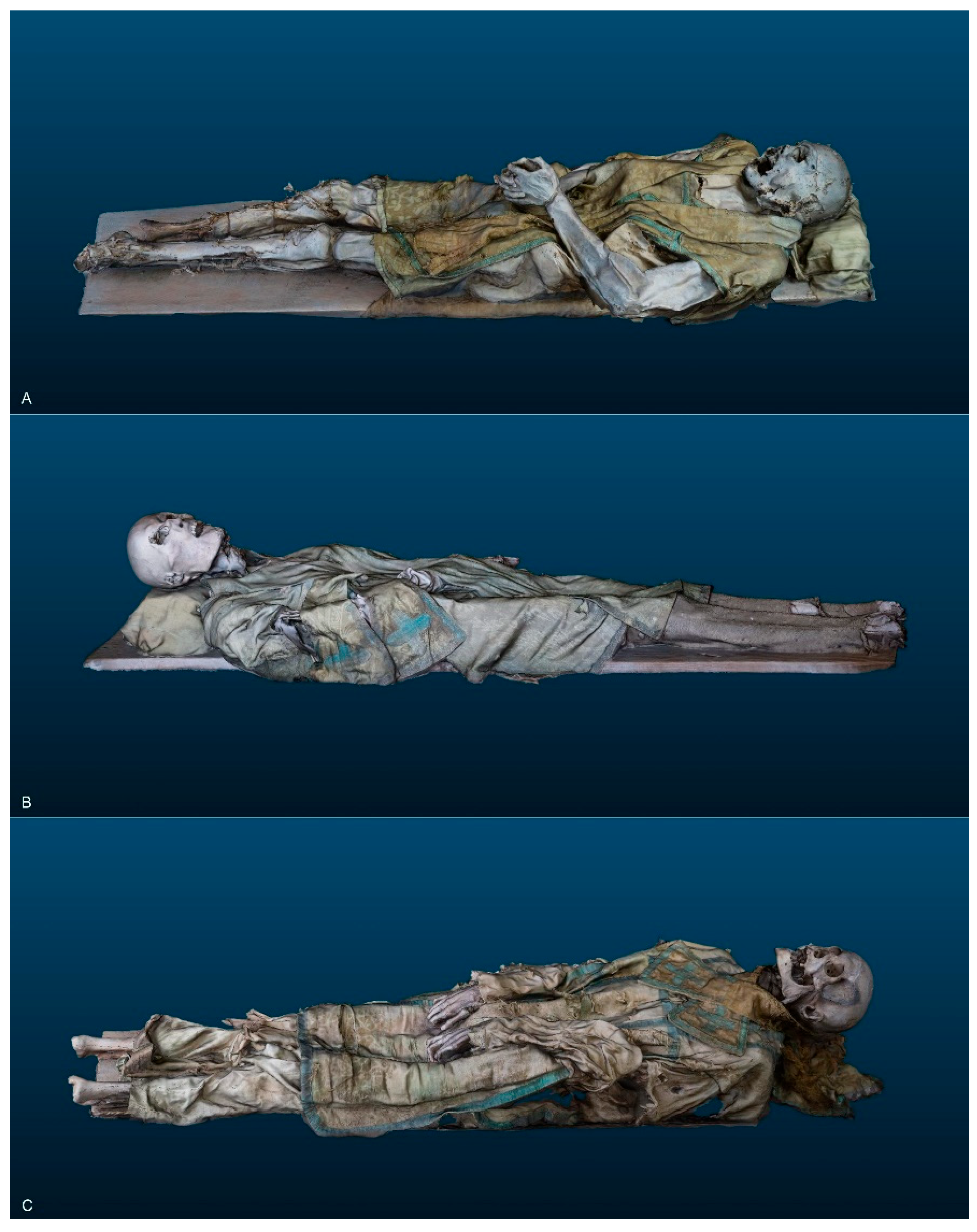
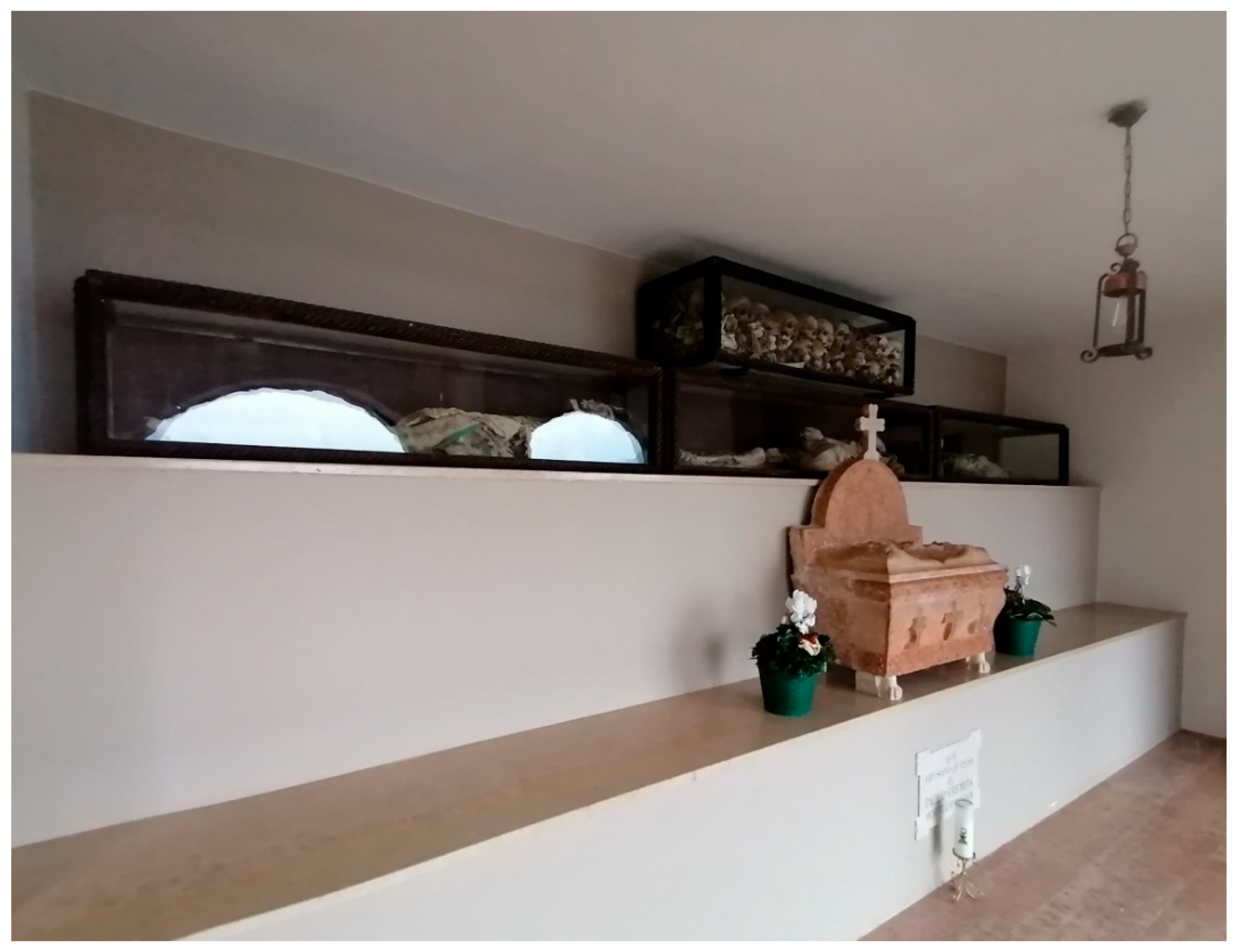
In fact, the research presented here focuses on entomological samples from three well-preserved, male, naturally mummified bodies housed in the Sanctuary of the Madonna della Corona (northeastern Italy) (Figure 1). These three hermits, dressed in mid-17th-, 18th-, and early 19th-century liturgical vestments, are displayed in glass cases with wooden frames (Figure 2 and Figure 3).
An interdisciplinary study has been conducted on these mummified bodies, involving anthropology and paleopathology through investigations and X-ray analysis [26].
Since some questions regarding the funerary practices and the season of death remained unanswered, an entomological inspection of the bodies was carried out. The main questions addressed, based on the information derived from the insects recovered from the bodies and their clothing, are as follows:
In which season(s) did the individuals die?
Were the bodies exposed or buried in a sheltered place shortly after death?
Were the garments the same as those worn at the time of deposition/burial, or were non-original liturgical vestments applied to the bodies after death?
Geographical and Historical context
The Sanctuary of Madonna della Corona is situated at an altitude of 775 m above sea level, mounted on the Monte Baldo rock, which also constitutes the left side of the building (Figure 1b).
The Madonna della Corona Sanctuary has ancient origins, likely dating back to the phenomenon of hermitic and monastic settlements in the early Middle Ages, specifically during the 8th century, a period when numerous hermitages were built in the Adige Valley and its connected side valleys. However, the first written evidence of hermits in the area dates to 1193. Although it has been suggested that there was an earlier use of the caves as a place of prayer by hermits associated with the Abbey of San Zeno in Verona, the presence of a monastery and a chapel dedicated to Santa Maria di Monte Baldo is documented in texts from the 13th century. The chapel was accessible via a narrow and dangerous path along the rocky terrain.
During the late Middle Ages, a new church was constructed above the cave, while an underground chamber became a sepulcher for the hermits. The presence of these hermits is documented until 1598, as evidenced by a funerary slab commemorating the burial of the last hermit, preserved within the Sanctuary. Over the centuries, the sepulcher was expanded to accommodate the burial of common people and members of the clergy. Between 1975 and 1978, the Sanctuary was demolished and reconstructed. This process removed the tomb oratory and the hermits’ sepulcher, creating space for new areas, including the current exhibition site for the three bodies.
The bodies are displayed in wooden coffins fitted with glass panels (Figure 3), which have been in use since the 1930s; however, their craftsmanship likely dates to the late 19th century [26]. Based on archival research and clothing analyses, Larentis et al. [26] proposed the following chronological dating for the three individuals: individual 1, early 19th century; individual 2, mid-17th century; and individual 3, 18th century.

2. Materials and Methods

All the procedures used in this study, from the collection to the data interpretation, followed the best practices reported in Vanin et al. [27].
Entomological specimens were collected manually from the three bodies, from their garments, and from the base of the glass cases in which they are preserved. Tweezers and soft brushes were used for the collection to ensure the preservation of the specimens and to prevent any damage to the bodies and to their garments.
Samples were subsequently weighed using a 0.1 mg precision scale (Mettler Toledo New Classic), and their volumes were also measured using a graduated cylinder. Due to the quality of the collected material (absence of debris), sieving of the samples was not necessary.
Entomological samples were observed and photographed using an S9i Leica stereomicroscope (Leica, Wetzlar, Germany).
The identification of the species was performed using specific identification keys, taxa descriptions [28,29,30,31,32,33,34,35,36,37], and by comparison with the SV reference collection.

3. Results and Discussion

3.1. The Entomofauna Associated with Corona Hermits

The presence of feeding activity of insects on the Corona mummified bodies was evident and documented by direct and indirect evidence. In fact, several fragments of insects (direct evidence) and traces of feeding (indirect evidence) were observed, carefully sampled, and studied in the laboratory.
Fragments of insects belonging to Diptera, Coleoptera, and Lepidoptera were collected from all the bodies, with Diptera puparia representing the predominant fraction in all the bodies (Table 1).

3.1.1. Diptera

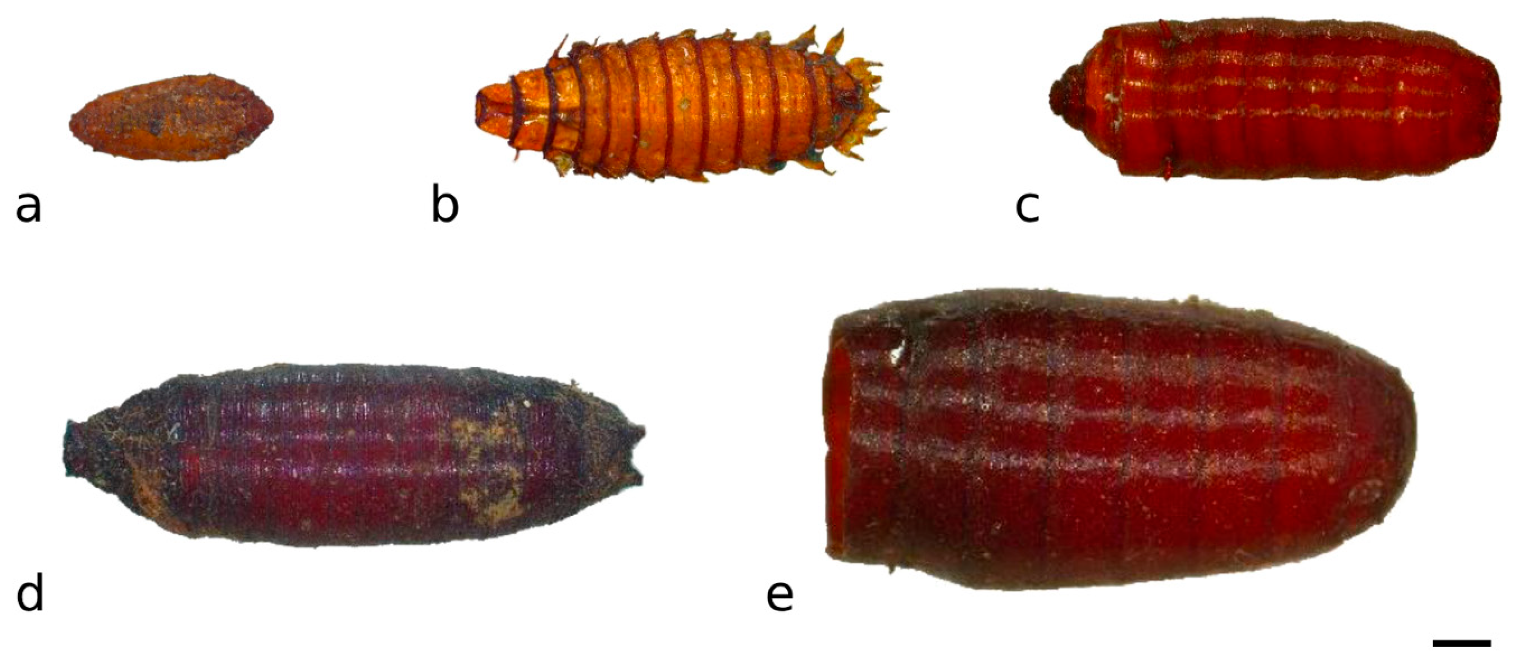
Despite issues related to the scarcity of diagnostic characters and the lack of any specific literature for some taxa, Diptera puparia were identified as Hydrotaea capensis (Wiedemann, 1818) (Muscidae)—the most common finding—Calliphora vicina Robineau-Desvoidy, 1830 (Calliphoridae), Fannia scalaris (Fabricius, 1794) (Fanniidae), and unidentified Phoridae and Heleomyzidae (Table 1, Figure 4). The presence of puparia indicates that the complete developmental cycle of these species had occurred on the body while the tissues were still fresh; Diptera are, in fact, the first colonizers of a body and are not capable of feeding on dried tissue.
Calliphora vicina is a synanthropic species belonging to the first wave of colonization of an exposed body. This species has been reported in several forensic cases in Italy and is known to be active, along with its congeners C. vomitoria and C. loewi, mainly in spring and fall [38], in contrast to species in the genus Lucilia, which are typical of the summer period [39]. These observations primarily refer to low-altitude areas in northern Italy, particularly the Po Plain, and other southern Italian regions, so they cannot be directly applied to the context of the site in question due to differences in altitude, geographical position, and the presence of several caves where temperatures are lower compared to the external environment. It is worth mentioning that the presence of Calliphora sp. was reported by Henriquez and colleagues [40] during an archaeoentomological study of a cave at 600 m above sea level in the Canary Islands. The two locations differ in latitude by approximately 18°, and they exhibit very different climate conditions.
The most common elements collected from the bodies were Hydrotaea capensis puparia. This species is considered a late colonizer of exposed cadavers and is widely distributed, being more common in warm regions and scarce in excessively dry environments [41]. However, it is also the most frequently found species in mummified bodies and, more generally, in bodies buried in crypts and/or other hypogean contexts [17,42,43]. It is considered one of the first colonizers of buried bodies, as noted by Smith [33].
Phoridae species were also present among the analyzed material; this taxon is commonly associated with buried bodies, indoor settings, or cadavers located in enclosed environments [17,42,43,44].
Additionally, the Fanniidae family, specifically the species Fannia scalaris, known as the “latrine fly,” was also identified. The common name of this species derives from its frequent presence in particularly humid environments and its tendency to colonize bodies during advanced decomposition phases when decomposition fluids are abundant. This species, along with the congeneric Fannia canicularis Linnaeus, 1761, has been reported from bodies in hypogean contexts [43].
Furthermore, puparia of Heleomyzidae were also isolated among the samples. These flies are typically attracted to rotten materials, feces, and occasionally cadavers. They prefer humid habitats, cool environments, or underground settings. A recent study documented their presence inside an intact human femur, highlighting their ability to colonize internal bone cavities [45].

3.1.2. Coleoptera

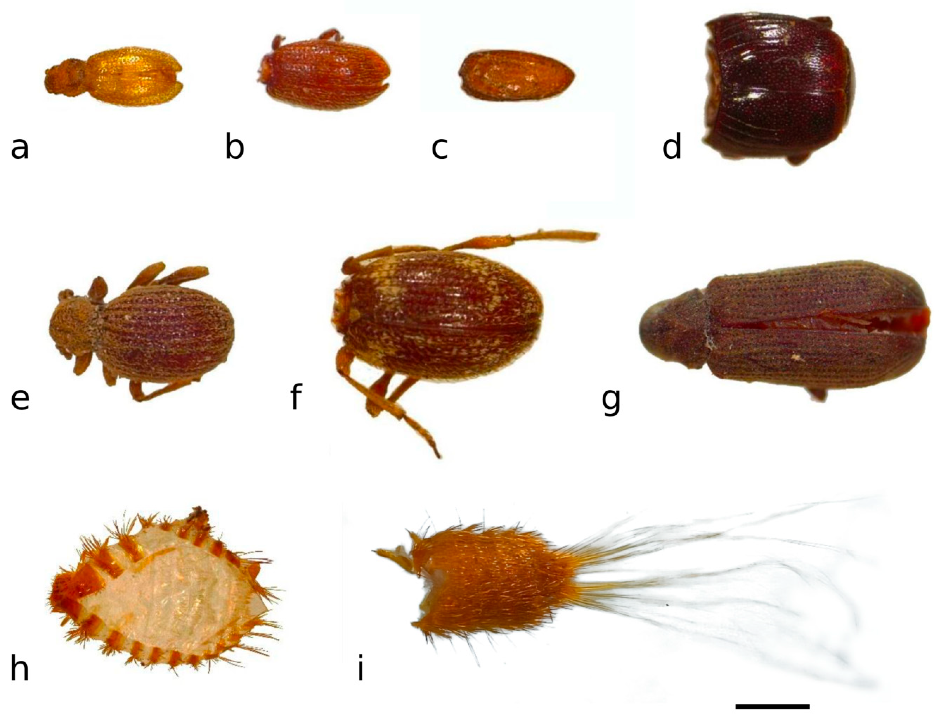
Among Coleoptera (Figure 5), three species from the Ptinidae family were identified: Ptinus fur (Linnaeus, 1758), Tipnus unicolor (Piller & Mitterpacher, 1783), and Anobium punctatum (De Geer, 1774).
Ptinus fur and T. unicolor are commonly found in domestic environments and storage areas, where they infest a variety of organic materials. Both species have been reported from archaeological contexts; T. unicolor is considered characteristic of Roman and late or post-medieval occupation deposits in England, where it is regarded as an indicator of long-lived buildings [46].
Anobium punctatum, a species that typically infests wood and is known as the “furniture beetle,” is a xylophagous species. Dermestidae exuviae, belonging to two different genera—Attagenus Latreille, 1802, and Anthrenus Geoffroy, 1762—were also collected. These taxa feed on a wide range of organic materials rich in keratin, collagen, and other proteins. This feeding habit makes them particularly relevant in forensic and funerary contexts, as they colonize human remains during the later stages of decomposition.
Unfortunately, they also play a significant role as museum pests (museophagous insects) due to their ability to damage historical artifacts such as textiles, stuffed animals, and preserved biological specimens [47,48]. For this reason, particular attention must be paid to distinguish whether their presence is associated with body decomposition or if it is secondary contamination occurring during collection or museum storage.
An additional species of beetle in the family Endomychidae, Mycetaea subterranea (Fabricius, 1801), was found. This species is typically found in underground environments, as its name suggests, and it is associated with decaying fungi, wood, and other organic materials in cool and humid settings.
Other mycetophagous (fungus-feeding) species belonging to the families Latridiidae and Cryptophagidae were also collected. The Latridiidae family, comprising around 500 species, includes very small insects (1–3 mm) that are often found in mouldy food and decomposing plant matter [49]. The specimens collected from the bodies belong to a species in the genus Corticaria Marsham, 1802.
The specimens in the family Cryptophagidae, known as silken fungus beetles, belong to the genus Cryptophagus Herbst, 1863. This family has a cosmopolitan distribution and includes more than 600 species, which are commonly found on mold and fungi, under bark, and in decaying vegetation [49].
A specimen belonging to Gnathoncus communis (Marseul, 1862) in the family Histeridae was also collected. The family Histeridae is primarily composed of predators of other insects, especially larvae of Diptera. Species in the genus Gnathoncus Jacquelin-Duval, 1858 have already been reported from archaeo-funerary contexts in Italy [16], where the genus includes six species, one of which is potentially extinct. In Italy, G. communis has been reported from bird nests, bat guano in caves, and chicken coops, and it is also possible to find it on food commodities [35]. In New Zealand, this species is mainly found in bird nests and chicken coops, with less frequent occurrences on bird carrion [50].

3.1.3. Lepidoptera

The cocoons and exuviae collected from the bodies and garments of the three individuals belong to clothes moths in the family Tineidae (Lepidoptera) (Figure 6). Clothes moths are typically found in domestic environments, infesting wardrobes and other areas where animal-derived materials are stored. These insects feed on a variety of materials, including wool, silk, fur, feathers, dried plant matter, and fungi. They can also feed on the dried tissue of a body, either at the end of decomposition (skeletonization) or, in this context, when the skin and other tissues are mummified.

3.2. Comparison of the Colonization of the Bodies

Hydrotaea capensis, Phoridae, and Fanniidae puparia were collected from all three bodies, whereas C. vicina puparia were only found on individuals 1 and 3. Heleomyzidae were found on individual 3, with more than 100 specimens. In general, the number of specimens collected from individual 2 appears to be lower compared to the other two bodies. This difference may be related to variations in colonization or to cleaning processes that occurred years after death.
The fact that bodies of individuals 1 and 3 exhibit evidence of insects typically associated with the colonization of exposed corpses suggests that the bodies were deposited in an environment either open or accessible to entomofauna characteristic of exposed remains. The presence of the synanthropic species Calliphora vicina indicates that the deposition site was near human dwellings or shelters where hermits lived, suggesting a connection between the burial site and nearby human activity. This supports the hypothesis that the bodies were deposited in an accessible area.
Calliphora vicina is active and relatively abundant during the cooler months of the year. Although this point is somewhat controversial due to the location of the sanctuary (high altitude, presence of caves, etc.), the deposition of at least individual 3 during a cooler season is supported by the presence of Heleomyzidae puparia, which are typical colonizers of cadavers during autumn and winter.
Specimens of Anobium punctatum were recorded, in varying numbers, on all three bodies. However, these are not associated with the time of death but are linked to the wooden frame of the display case where the mummified bodies are currently housed, which shows clear signs of their trophic activity. Nonetheless, the possibility of their burial in a wooden coffin or their deposition on a wooden board cannot be excluded. This is further supported by the presence of some specimens of Tipnus unicolor, a species associated with aged wood [51].

3.3. Clothes and Skin Analyses and Evaluations

The liturgical vestments of individual 1 consisted of a chasuble, a stole, an alb, and a pillow cover, whereas individual 2 was dressed in a chasuble, a stole, a pillow cover, and what might be a maniple. Individual 3 wore a chasuble, a stole, a maniple, an alb, and footwear. Several peculiar damages have been found on the clothing worn by the bodies. Insects cause different types of alterations to fabrics depending on the taxa involved.
Saprophagous Diptera, such as blowflies, do not feed on the textiles themselves but can pass through the fibers, moving away from the body to pupariate or for other reasons such as disturbance or the presence of predators. In performing this, they push through the fabrics, widening the fibers without causing significant structural damage. Conversely, Lepidoptera (e.g., clothes moths) and Coleoptera (e.g., carpet beetles) actively feed on natural fibers like wool, silk, and cotton. Their larvae chew through the material, weakening and breaking the fibers, which leads to more severe damage.
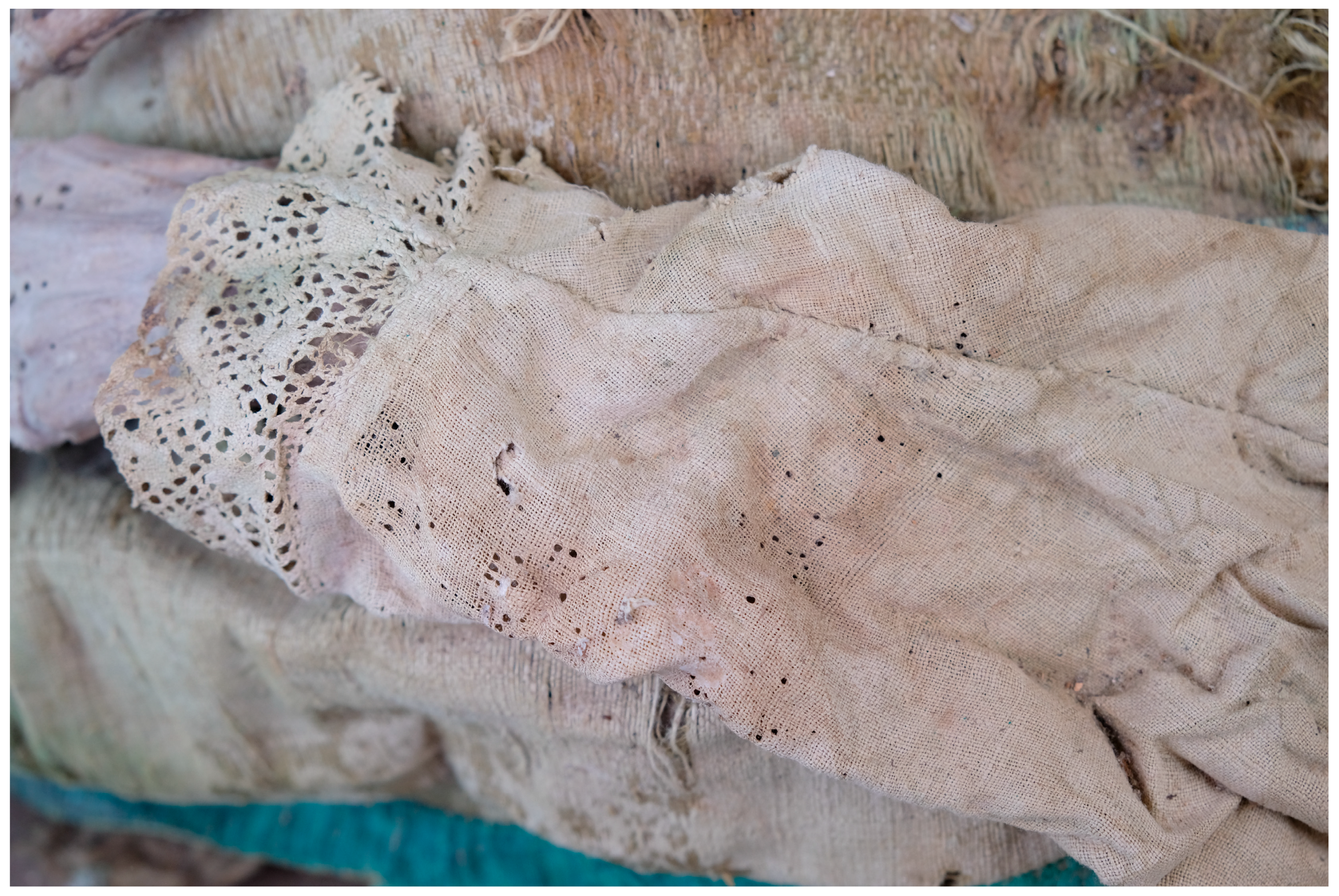
These differences in insect behavior result in distinct types of textile deterioration, with Diptera typically leaving the fabric largely intact, while other groups cause more destructive alterations (Figure 7): both types of insect-created holes were present on the clothing.
However, while the holes made by clothes moths or beetles could be attributed to secondary colonization, the marks left by dipteran larvae can only be linked to the time of body deposition, as the dipterans responsible for this type of damage (e.g., Calliphoridae, Sarcophagidae, Muscidae) are associated with the early stages of decomposition when the body is still fresh. Additionally, dipterans cause similar circular holes on the skin of the corpse, feeding on the body during their developmental process [52] (Figure 8).
In various parts of the bodies, a distinct continuity of emergence holes was observed between the clothing and the skin. This continuity suggests that the garments currently worn by the three bodies are the same as those in which they were originally buried at the time of death, because the insects feeding directly on the body were responsible for the damage to the garments passing through. The alignment of these holes provides valuable insights into insect behavior and decomposition dynamics, indicating that both the body and the clothing were colonized by the same entomofauna during the decomposition process.
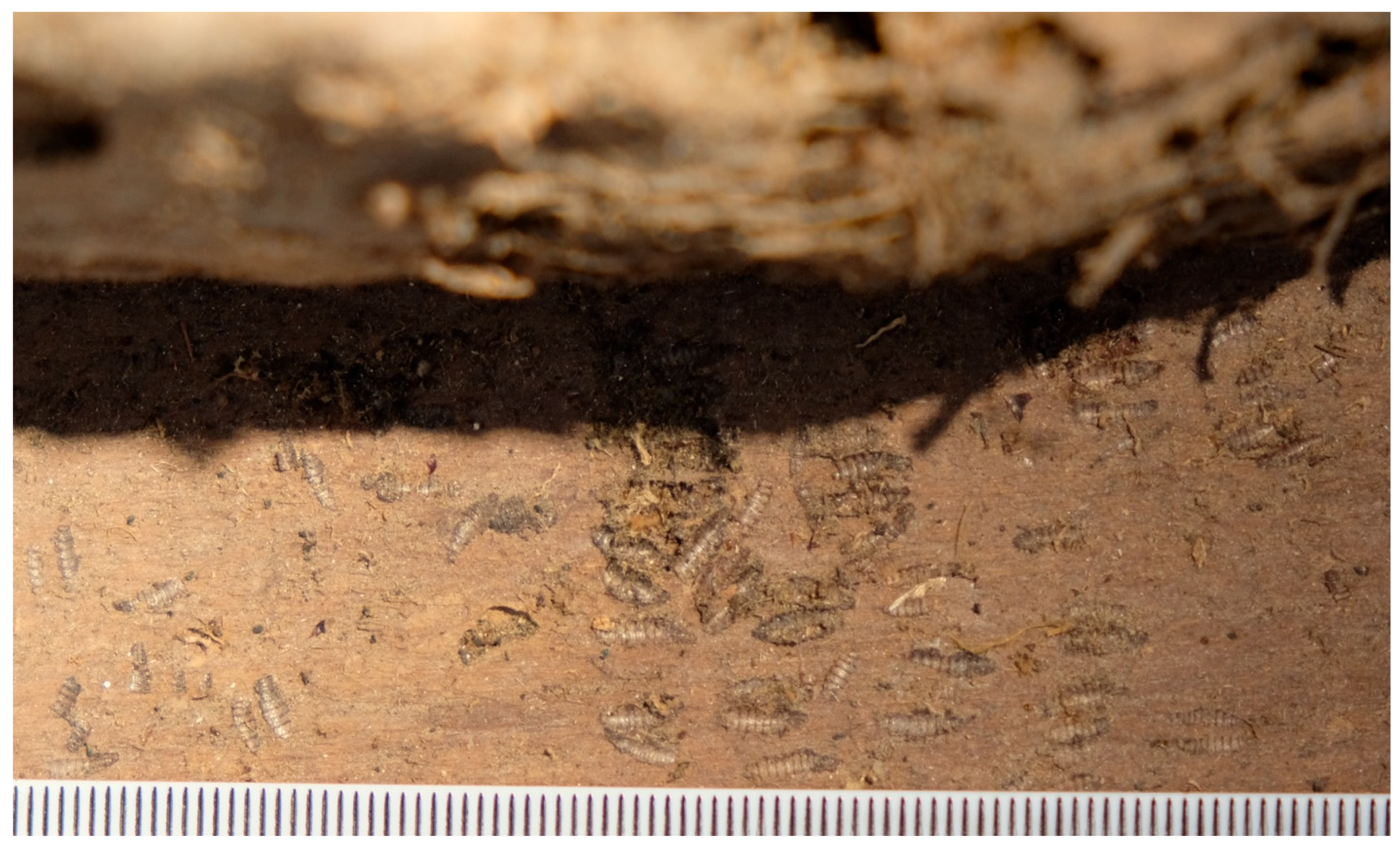
Additionally, textile analysis conducted by a specialist confirmed that the fabrics found on the bodies are consistent with garments produced during the period of the individuals’ death [26]. This finding supports the conclusion that the clothing worn is indeed original, reinforcing the evidence from entomological traces on the garments. Furthermore, it is worth mentioning that Fanniidae puparia were observed and sampled from the boards on which the bodies lay and among the garments (Figure 9). This may suggest that the board belonged to the initial deposition; however, the reuse of a board from another deposition cannot be excluded.
An interesting observation concerns individual 2, which appears to have belonged to an overweight individual, as described by Larentis et al. [26]. Unlike the other two mummified bodies, individual 2 was found with only partial clothing, and a significantly smaller number of entomological specimens were collected from the body. It is plausible that, due to the larger body size, the decomposition process produced a greater amount of decomposition fluids, which may have necessitated some form of intermediate cleaning. The traces of insects found on the garments suggest that they are indeed original; however, the absence of certain clothing layers and the reduced insect presence may indicate that, at some point during decomposition, the body was cleaned. It is possible that the heavily soiled undergarments, which could not be preserved, were removed, while the external liturgical garments—though original—were reapplying after cleaning.
This scenario raises questions about the role of cleaning or partial re-dressing in funerary practices. The missing clothing layers and less pronounced insect colonization could reflect cultural practices involving the handling of the body during the later stages of decomposition. For this reason, the relatively few insects found on individual 2 may also be the result of cleaning procedures that removed some of the insects already present on the body.

4. Conclusions

This study aims to provide insights into funerary practices and burial conditions through entomological analysis, thereby contributing to the historical and cultural knowledge of the populations to which the bodies belonged. The corpses were primarily colonized by insects from the orders Coleoptera, Diptera, and Lepidoptera.
The entomological findings suggest that all three bodies likely underwent similar funerary procedures. The deaths and depositions of individuals 1 and 3 probably occurred during the colder months of the year (potentially late spring or early fall), based on the garments they currently wear. This is clearly indicated by the continuity of holes caused by dipteran maggots on both the skin and the garments.
In contrast, individual 2 appears to have undergone a cleaning process, which resulted in a limited number of specimens collected from the body and its clothing. The presence of Calliphora suggests that the bodies were exposed for several days prior to burial, a common practice in Italy and other countries with Christian Catholic traditions—especially for members of the clergy, venerable figures, and saints. After this exposition, the bodies were enclosed in wooden coffins.
Furthermore, the wooden boards on which the bodies currently rest are very likely the same as those on which they were placed after death, during the decomposition and mummification process. This is supported by the presence of Fanniidae puparia traces on the boards themselves and among the garments of the examined bodies.
Overall, insects play a crucial role in this study, providing valuable information for reconstructing funerary practices and understanding the post-mortem history of the three mummified bodies.

Author Contributions

Conceptualization, O.L. and S.V.; investigation, G.C., S.V., E.T. and O.L.; writing—original draft preparation, S.V. and G.C.; writing—review and editing, S.V., O.L. and I.G.; supervision, S.V. All authors have read and agreed to the published version of the manuscript.

Funding

This research received no external funding.

Data Availability Statement

All the data are reported in the main text and in the tables. No other data have been generated.

Acknowledgments

We would like to thank the Diocese of Verona and the Superintendence for Archaeology, Fine Arts, and Landscape for the provinces of Verona, Rovigo, and Vicenza (Soprintendenza Archeologia Belle Arti e Paesaggio per le province di Verona, Rovigo e Vicenza), particularly Irene Dori, for authorizing this study. We also extend our gratitude to Don Martino Signoretto, Vicar for Culture of the Diocese of Verona and Rector of the Sanctuary, Don Manuel Magalini, and Francesco Teris for their logistical support and for ensuring we could work under the best possible conditions, meeting all our needs. Finally, we sincerely thank the Sisters of the Lovers of the Cross congregation for their warm hospitality.

Conflicts of Interest

The authors declare no conflicts of interest.

References

  1. Erickson, J.M. Fossil oribatid mites as tools for Quaternary paleoecologists: Preservation quality, quantities, and taphonomy. Bull. Buffalo Soc. Nat. Sci. 1988, 33, 207–226. [Google Scholar]
  2. Buckland, P.C. The use of insect remains in the interpretation of archaeological environments. In Geoarchaeology: Earth Science and the Past; Davidson, D.A., Shackley, M.L., Eds.; West View Press: Boulder, CO, USA, 1976; pp. 369–396. [Google Scholar]
  3. Buckland, P.; Wagner, P. Is there an insect signal for the” Little Ice Age”? Clim. Change 2001, 48, 137–149. [Google Scholar] [CrossRef]
  4. Huchet, J.B. L’Archéoentomologie funéraire: Une approche originale dans l’interprétation des sépultures. Bull. Mem. Soc. Anthropol. Paris 1996, 3–4, 299–311. [Google Scholar] [CrossRef]
  5. Kenward, H.K. The analysis of archaeological insect assemblages: A new approach. In Archaeology of York; Council for British Archaeology for York Archaeological Trust: York, UK, 1978; Volume 19/1. [Google Scholar]
  6. Bain, A. Archaeoentomological and Archaeoparasitological Reconstructions at Îlot Hunt (CeEt-110): New Perspectives in Historical Archaeology (1850–1900). Ph.D. Thesis, Laval University, Quebec, QC, Canada, 1999. [Google Scholar]
  7. Buckland, P.I.; Buckland, P.C.; Olsson, F. Paleoentomology: Insects and other arthropods in environmental archaeology. In Encyclopedia of Global Archaeology; Smith, C., Ed.; Springer: New York, NY, USA, 2014. [Google Scholar]
  8. Forbes, V.; Dussault, F.; Bain, A. Contributions of ectoparasite studies in archaeology with two examples from the North Atlantic region. Int. J. Paleopathol. 2013, 3, 158–164. [Google Scholar] [CrossRef] [PubMed]
  9. Huchet, J.B. L’archéoentomologie funéraire (chap. 15). In Insectes, Cadavres et Scènes de Crime, Principes et Applications de L’entomologie Médico-Légale; Charabidze, D., Gosselin, M., Eds.; Editions De Boeck: Bruxelles, Belgium, 2014; pp. 201–224. [Google Scholar]
  10. Giordani, G.; Tuccia, F.; Floris, I.; Vanin, S. First record of Phormia regina (Meigen, 1826) (Diptera: Calliphoridae) from mummies at the Sant’Antonio abate Cathedral of Castelsardo, Sardinia, Italy. PeerJ 2018, 6, e4176. [Google Scholar] [CrossRef]
  11. Magni, P.A.; Harvey, A.D.; Guareschi, E.E. Insects Associated with Ancient Human Remains: How Archaeoentomology Can Provide Additional Information in Archaeological Studies. Heritage 2023, 6, 435–465. [Google Scholar] [CrossRef]
  12. Tuccia, F.; Giordani, G.; Vanin, S. State of the art of the funerary archaeoentomological investigations in Italy. Archaeol. Anthropol. Sci. 2022, 14, 70. [Google Scholar] [CrossRef]
  13. Giordani, G.; Erauw, C.; Eeckhout, P.A.; Owens, L.S.; Vanin, S. Patterns of camelid sacrifice at the site of Pachacamac, Peruvian Central Coast, during the Late Intermediate Period (AD1000–1470): Perspectives from funerary archaeoentomology. J. Archaeol. Sci. 2020, 114, 105065. [Google Scholar] [CrossRef]
  14. Huchet, J.B.; Greenberg, B. Flies, Mochicas and burial practices: A case study from Huaca de la Luna, Peru. J. Archaeol. Sci. 2010, 37, 2846–2856. [Google Scholar] [CrossRef]
  15. Vanin, S. When Entomological studies meet Archaeology: Archaeoentomology an old, new discipline for investigation of the Past. JBR 2023, 1, e2023007. [Google Scholar]
  16. Vanin, S.; Turchetto, M.; Galassi, A.; Cattaneo, C. Forensic Entomology and Archaeology of War. J. Confl. Archaeol. 2010, 50, 127–139. [Google Scholar] [CrossRef]
  17. Pradelli, J.; Rossetti, C.; Tuccia, F.; Giordani, G.; Licata, M.; Birkhoff, J.M.; Verzeletti, A.; Vanin, S. Environmental necrophagous fauna selection in a funerary hypogeal context: The putridarium of the Franciscan monastery of Azzio (Northern Italy). J. Archaeol. Sci. Rep. 2019, 24, 683–692. [Google Scholar] [CrossRef]
  18. Vanin, S.; Tuccia, F.; Pradelli, J.; Carta, G.; Giordani, G. Identification of Diptera Puparia in Forensic and Archeo-Funerary Contexts. Insects 2024, 15, 599. [Google Scholar] [CrossRef]
  19. Huchet, J.-B.; Le Mort, F.; Rabinovich, R.; Blau, S.; Coqueugniot, H.; Arensburg, B. Identification of dermestid pupal chambers on Southern Levant human bones: Inference for reconstruction of Middle Bronze Age mortuary practices. J. Archaeol. Sci. 2013, 40, 3793–3803. [Google Scholar] [CrossRef]
  20. Wrobel, G.D.; Biggs, J. Osteophageous insect damage on human bone from Je’reftheel, a Maya mortuary cave site in west-central Belize. Int. J. Osteoarchaeol. 2018, 28, 745–756. [Google Scholar] [CrossRef]
  21. Thompson, J.E.; Martín-Vega, D.; Buck, L.T.; Power, R.K.; Stoddart, S.; Malone, C. Identification of dermestid beetle modification on Neolithic Maltese human bone: Implications for funerary practices at the Xemxija tombs. J. Archaeol. Sci. Rep. 2018, 22, 123–131. [Google Scholar] [CrossRef]
  22. Britt, B.B.; Scheetz, R.D.; Dangerfield, A. A Suite of Dermestid Beetle Traces on Dinosaur Bone from the Upper Jurassic Morrison Formation, Wyoming, USA. Ichnos 2008, 15, 59–71. [Google Scholar] [CrossRef]
  23. Loni, A.; Vanin, S.; Fornaciari, A.; Tomei, P.E.; Giuffra, V.; Benelli, G. Back to the Middle Ages: Entomological and Botanical Elements Reveal New Aspects of the Burial of Saint Davino of Armenia. Insects 2022, 13, 1113. [Google Scholar] [CrossRef] [PubMed]
  24. Vanin, S.; Boano, R.; Giordani, G.; Carta, G.; Fulcheri, E. Description of the entomofauna associated with the remains of the Cistercian nun Angela Veronica Bava (1591–1637). Med. Histor. 2022, 6, e2022006. [Google Scholar]
  25. Zangheri, S.; Fontana, P. Indagini sugli insetti rinvenuti nella bara attribuita a San Luca Evangelista. In San Luca Evangelista Testimone della Fede che Unisce. Atti del Congresso Internazionale, Padova, 16–21 Ottobre 2000, I Risultati Scientifici sulla Ricognizione delle Reliquie Attribuite a san Luca. Fonti e Ricerche di Storia Ecclesiatica Padovana; Terribile Wiel Marin, V., Trolese, F.G.B., Eds.; Istituto di Storia Ecclesiastica Padovana: Padova, Italia, 2003; Volume 2. [Google Scholar]
  26. Larentis, O.; Gorini, I.; Campus, M.; Lorenzetti, M.; Mansueto, G.; Bortolotto, S.; Zappa, E.; Gregorini, A.; Rampazzi, L.; Vanin, S.; et al. Integrated multidisciplinary analysis of mobile digital radiographic acquisitions of the mummies of the Hermits from the Sanctuary of Madonna della Corona (Trentino-Alto Adige, Italy-17th to 19th Century CE). Front. Med. 2025, 11, 1492328. [Google Scholar] [CrossRef]
  27. Vanin, S.; Giordani, G.; Carta, G. From the field to the microscope: Funerary archaeoentomology workflow. JBR 2023, 1, 2023017. [Google Scholar]
  28. Giordani, G.; Grzywacz, A.; Vanin, S. Characterization and identification of puparia of Hydrotaea Robineau-Desvoidy, 1830 (Diptera: Muscidae) from forensic and archaeological contexts. J. Med. Entomol. 2019, 56, 45–54. [Google Scholar] [CrossRef]
  29. Giordani, G.; Vanin, S. Morphological characterization of puparia and molecular analysis of Heleomyza serrata (Linnaeus, 1758) (Diptera: Heleomyzidae): A species of potential forensic interest. Eur. Zool. J. 2023, 90, 604–613. [Google Scholar] [CrossRef]
  30. Rozkosny, R.; Gregor, F.; Pont, A.C. The European Fanniidae (Diptera); Institute of Landscape Ecology: Brno, Czech Republic, 1997. [Google Scholar]
  31. Lyneborg, L. Taxonomy of European Fannia larvae (Diptera, Fanniidae); Staatliches Museum für Naturkunde: Karlsruhe, Germany, 1970. [Google Scholar]
  32. Skidmore, P. The Biology of the Muscidae of the World; Springer Netherlands: Dordrecht, The Netherlands, 1985. [Google Scholar]
  33. Smith, K.G.V. A Manual of Forensic Entomology; London Trustees of the British Museum: London, UK, 1986. [Google Scholar]
  34. Szpila, K. Key for the identification of third instars of European blowflies (Diptera: Calliphoridae) of forensic importance. In Current Concepts in Forensic Entomology; Amendt, J., Goff, M.L., Campobasso, P.C., Grassberger, M., Eds.; Springer Verlag: Dordrecht, The Netherlands, 2010; pp. 43–56. [Google Scholar]
  35. Vienna, P. Histeridae. Fauna d’Italia Vol. XVI; Edizioni Calderini: Bologna, Italy, 1980; 386p. [Google Scholar]
  36. Zahradaník, P. Beetles of the Family Ptinidae of Central Europe/Brouci Čeledi Červotočovití (Ptinidae) Střední Evropy; Zoological Keys/Zoologické Klíče, Vol. 2; Academia Press: Gand, Belgium, 2013. [Google Scholar]
  37. Háva, J. Beetles of the Family Dermestidae of the Czech and Slovak Republics; Academia: Praha, Czech Republic, 2021. [Google Scholar]
  38. Vanin, S.; Gherardi, M.; Bugelli, V.; Di Paolo, M. Insects found on a human cadaver in central Italy including the blowfly Calliphora loewi (Diptera, Calliphoridae), a new species of forensic interest. Forensic Sci. Int. 2011, 207, e30–e33. [Google Scholar] [CrossRef] [PubMed]
  39. Vanin, S.; Tasinato, P.; Ducolin, G.; Terranova, C.; Zancaner, S.; Montisci, M.; Ferrara, P.; Turchetto, M. Use of Lucilia species (Diptera: Calliphoridae) for forensic investigations in Southern Europe. Forensic Sci. Int. 2008, 177, 37–41. [Google Scholar] [CrossRef]
  40. Henríquez-Valido, P.; Santana, J.; Morquecho-Izquier, A.; Rodríguez-Rodríguez, A.; Huchet, J.-B. Insects in the far West: Burial practices on El Hierro Island (Canary Islands, Spain; ca. 6th-11th centuries) reconstructed via funerary archaeoentomology. J. Arch. Sci. 2025, 173, 106120. [Google Scholar]
  41. Pérez-Marcos, M.; García, M.D.; López-Gallego, E.; Ramírez-Soria, M.J.; Arnaldos, M.I. Life Cycle and Biometric Study of Hydrotaea capensis (Wiedemann, 1818) (Diptera, Muscidae), a Species of Forensic Interest. Insects 2022, 13, 531. [Google Scholar] [CrossRef]
  42. Loni, A.; Fornaciari, A.; Canale, A.; Giuffra, V.; Vanin, S.; Benelli, G. Insights on funeral practices and insects associated with the tombs of King Ferrante II d’Aragona and other Renaissance nobles. J. Med. Entomol. 2019, 56, 1582–1589. [Google Scholar] [CrossRef]
  43. Lunardini, A.; Carta, C.; Costantini, L.; Minozzi, S.; Giuffra, V.; Giordani, G.; Vanin, S. Entomological analysis for archaeological reconstruction and conservation strategies design: The mummies of Cerreto di Spoleto (Central Italy). Archaeol. Anthrop Sci. 2023, 15, 149. [Google Scholar] [CrossRef]
  44. Reibe, S.; Madea, B. Use of Megaselia scalaris (Diptera: Phoridae) for post-mortem interval estimation indoors. Parasitol. Res. 2010, 106, 637–640. [Google Scholar] [CrossRef] [PubMed]
  45. Sessa, F.; Varotto, E.; Salerno, M.; Vanin, S.; Bertozzi, G.; Galassi, F.M.; Maglietta, F.; Salerno, M.; Tuccia, F.; Pomara, C.; et al. First report of Heleomyzidae (Diptera) recovered from the inner cavity of an intact human femur. J. Forensic Leg. Med. 2019, 66, 4–7, Erratum in: J. Forensic Leg. Med. 2023, 97, 102545. [Google Scholar] [CrossRef] [PubMed]
  46. Kenward, H.; Hall, A.R.; Allison, E.; Carrott, J. Environment, Activity and Living Conditions at Deer Park Farms: Evidence from Plant and Invertebrate Remains. In Deer Park Farms: The Excavation of a Raised Rathin the Glenarm Valley, County Antrim (Northern Ireland); Lynn, C.J., McDowell, J.A., Eds.; Stationery Office Books: London, UK, 2011. [Google Scholar]
  47. Vanin, S.; Azzoni, M.; Giordani, G.; Belcastro, M.G. Bias and potential misinterpretations in the analysis of insects collected from human remains of archaeological interest. Archaeol. Anthropol. Sci. 2021, 13, 201. [Google Scholar] [CrossRef]
  48. Hagstrum, D.; Klejdysz, T.; Subramanyam, B.; Nawrot, J. Atlas of Stored-Product Insects and Mites; AACC International Press: St. Paul, MN, USA, 2017; ISBN 978-0-12-810431-6. [Google Scholar]
  49. Otero, J.C.; Benyahia, Y.; Brustel, H. Faunistic notes on Cryptophagidae and Latridiidae of Talassemtane National Park, Western Rif, Morocco, with the description of a new species (Coleoptera, Cucujoidea). ZooKeys 2017, 668, 69–82. [Google Scholar] [CrossRef]
  50. Lackner, T.; Leschen, R.A.B. A monograph of the Australopacific Saprininae (Coleoptera, Histeridae). ZooKeys 2017, 689, 1–263. [Google Scholar] [CrossRef] [PubMed]
  51. Forbes, V.; Bain, A.; Gísladóttir, G.A.; Milek, K.B. Reconstructing aspects of the daily life in late 19th and early 20th-century Iceland: Archaeoentomological analysis of the Vatnsfjörður Farm, NW Iceland. Archaeol. Isl. 2010, 8, 77–110. [Google Scholar]
  52. Viero, A.; Montisci, M.; Pelletti, G.; Vanin, S. Crime Scene and Body Alterations caused by Arthropods: Implications in Death Investigation. Int. J. Leg. Med. 2019, 133, 307–316. [Google Scholar] [CrossRef]
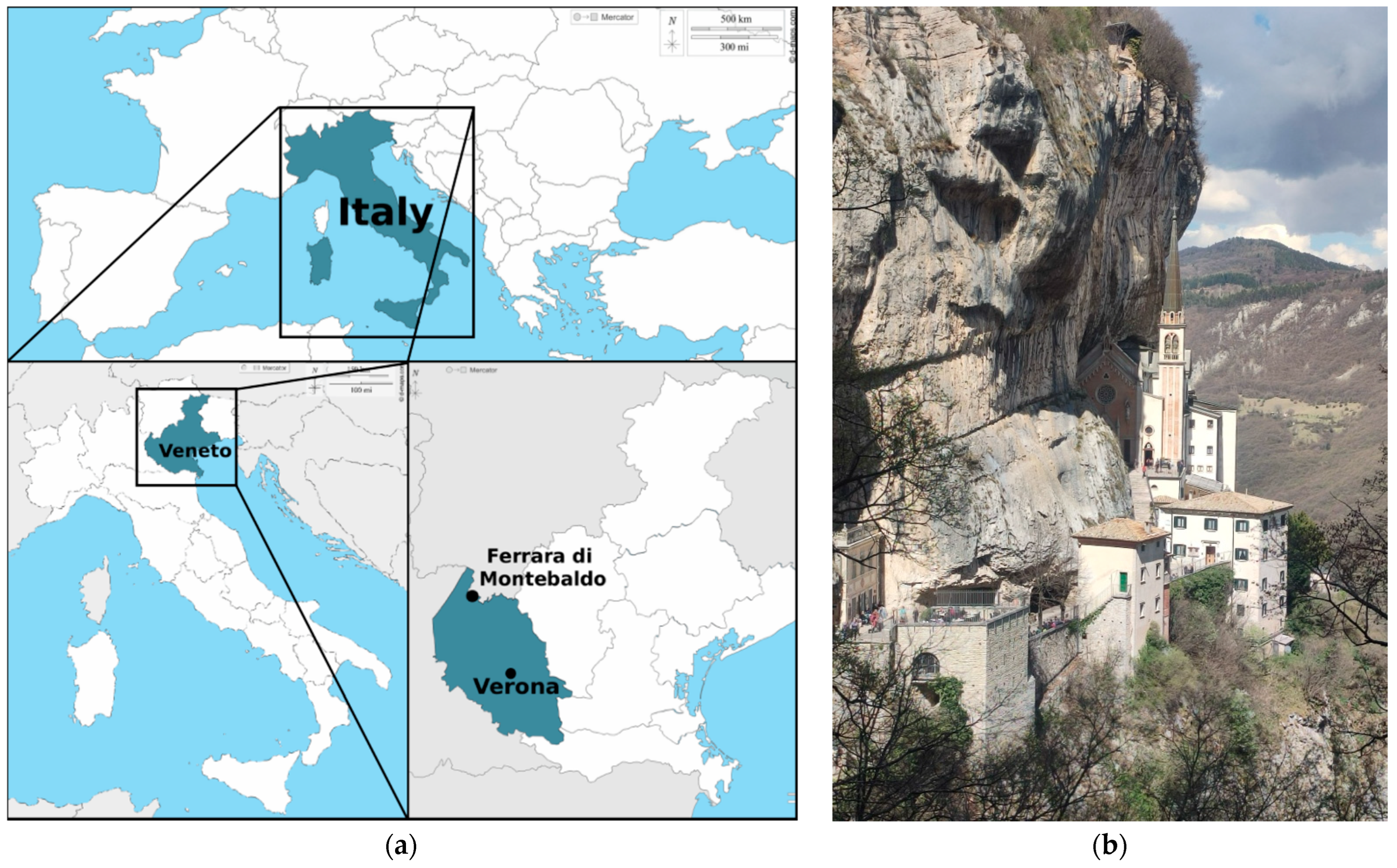
Figure 1. (a) Map of the location (45°38′58″ N 10°51′22″ E) (© 2007–2018 d-maps.com) and (b) view of the Sanctuary.
Heritage 08 00406 g001
Figure 2. The bodies preserved in the Sanctuary are arranged in chronological order: (A) individual 2—mid-17th century; (B) individual 3—18th century; (C) individual 1—early 19th century. The 3D models of the bodies were created by Dr. Andrea Gregorini and Professor Emanuele Zappa from the Politecnico di Milano, Milan, Italy. A Fuji FDR Xair mobile digital radiographic system (Fujifilm, Tokyo, Japan) was placed directly in the storage area with the three bodies, whereas pictures were captured using a digital camera Fujifilm X-T30, with a sensor resolution of 6246 × 4170 pixels and the following settings: ISO-2000, f/4, and a focal length of 18 mm. The Agisoft Metashape Professional software v 2.2.0 was used for the photogrammetry. The obtained 3D models were made of 157,175 points for individual 1, 403,681 points for individual 2, and 234,827 points for individual 3 [26].
Heritage 08 00406 g002
Figure 3. The current placement of the three wooden coffins with glass panels containing the mummified bodies, with a fourth coffin containing skeletal remains placed above the central case. In front is positioned a marble urn containing the remains of “Paolo l’Eremita”, who died in 1598.
Heritage 08 00406 g003
Figure 4. Diptera puparia: (a) Phoridae; (b) Fannia scalaris; (c) Hydrotaea capensis; (d) Heleomyzidae; (e) Calliphora vicina [scale bar: 1 mm].
Heritage 08 00406 g004
Figure 5. Coleoptera: (a) Corticaria sp.; (b) Cryptophagus sp.; (c) Mycetaea subterranea (elitra); (d) Gnathoncus communis; (e) Tipnus unicolor; (f) Ptinus fur; (g) Anobium punctatum; (h) Athrenus sp. (exuvia); (i) Attagenus sp. (exuvia) [scale bar: 1 mm].
Heritage 08 00406 g005
Figure 6. Clothes moths from the family Tineidae: (a) exuvia; (b) cocoon [scale bar: 1 mm].
Heritage 08 00406 g006
Figure 7. Alteration to the fabric caused by Diptera larvae.
Heritage 08 00406 g007
Figure 8. Emergence holes caused by Diptera in the skin. The pattern of the holes (clustered) and the absence of signs of mandibles allow for distinguishing this kind of alteration from the holes caused by larvae of beetles and lepidoptera.
Heritage 08 00406 g008
Figure 9. Traces of Fanniidae puparia on the wooden board [scale: mm].
Heritage 08 00406 g009
Table 1. Summary of the taxa collected from the mummified bodies of The Sanctuary of Madonna della Corona (northern Italy) (developmental stage: A adult; C cocoon; E exuvia; Pu puparium; number of specimens: x = 1 specimen; xx = 2–10 specimens; xxx = 11–100 specimens; xxxx = >100 specimens).
D. StageIndividual 1
(18th Cent)
Individual
2
(Mid-17th Cent)
Individual 3
(18th Cent)
ArachnidaAranea x
Acaridida x
MalacostracaIsopoda x
InsectaColeopteraHisteridaeGnathoncus communisA x
DermestidaeAttagenus sp.Exx
Anthrenus sp.Exxxxxxxx
PtinidaeAnobium punctatumAxxxxxxx
Ptinus furA x
Tipnus unicolorA xx
CryptophagidaeCryptophagus sp.Axxxxx
MycetaeidaeMycetaea subterraneaA x
LatridiidaeCorticaria sp.A x
DipteraPhoridaeGen. sp.Pxxxxxx
HeleomyzidaeGen. sp.P xxxx
FanniidaeFannia scalarisPxxxxxxxx
MuscidaeHydrotaea capensisPxxxxxxxxxx
CalliphoridaeCalliphora vicinaPxxxx xxxx
LepidopteraTineidae Cxxxxxxxxx
Hymenoptera Ax
Disclaimer/Publisher’s Note: The statements, opinions and data contained in all publications are solely those of the individual author(s) and contributor(s) and not of MDPI and/or the editor(s). MDPI and/or the editor(s) disclaim responsibility for any injury to people or property resulting from any ideas, methods, instructions or products referred to in the content.