Article

Evaluation of the Irradiation Treatment Effects on Ancient Parchment Samples

1
Particle Accelerator for Medical Application Laboratory, Italian National Agency for New Technologies, Energy and Sustainable Economic Development (ENEA), Science and Research Direction, Italian Space Agency (ASI), Via del Politecnico, 00133 Rome, Italy
2
Department of Literary, Philosophical and Art History Studies, University of Rome “Tor Vergata”, Via Columbia 1, 00133 Rome, Italy
3
Department of Chemical Science and Technologies, University of Rome “Tor Vergata”, Via della Ricerca Scientifica, 00133 Rome, Italy
4
Department of Biology and Biotechnology “Charles Darwin,” Sapienza University of Rome, 00185 Rome, Italy
5
Department of Industrial Engineering, University of Rome “Tor Vergata”, Via del Politecnico 1, 00133 Rome, Italy
6
Beamline IUVS, Sincrotrone Trieste in Area Science Park, S.S. 14 km 163.5, 34012 Basovizza, Italy
7
Istituto Officina dei Materiali, CNR, S.S. 14 km 163.5 in Area Science Park, 34149 Basovizza, Italy
8
Nanoinnovation Laboratory, Sincrotrone Trieste, S.S. 14 km 163.5 in Area Science Park, 34149 Basovizza, Italy
*
Author to whom correspondence should be addressed.
Heritage 2023, 6(2), 1308-1324; https://doi.org/10.3390/heritage6020072
Submission received: 8 December 2022 / Revised: 20 January 2023 / Accepted: 27 January 2023 / Published: 30 January 2023

Abstract

:
In this work, the effect of X-ray irradiation as a disinfection treatment in original ancient parchment samples, belonging to a discarded book cover of a 16th-century archival register, has been evaluated. Specifically, the bacterial and fungal species isolated from the book cover have been characterized and then irradiated with increasing doses of X-rays with the aim to evaluate the effectiveness of the antimicrobial protocol on the isolated microorganisms. The deterioration effects induced by the X-ray treatment as well as the natural aging on the collagen matrix of the parchment sample have been tested by employing several techniques, namely, Light Transmission Analysis, Fiber Optic Reflectance Spectroscopy, Attenuated Total Reflectance-Fourier Transformed Infrared spectroscopy, UV Resonant Raman spectroscopy and Atomic Force Microscopy. The results reveal that the irradiation treatment applied to our ancient parchment samples deteriorated by biological attack and other naturally occurring phenomena, possibly associated with inappropriate conservation conditions, does not seem to induce further damage factors even when large doses of irradiation are employed. The X-rays-based disinfection treatment effects are limited on the collagen support and this confirms the potential of this method in mass disinfection of library and archival materials.

1. Introduction

Since Late Antiquity and until the diffusion of paper, parchment has been the most frequently used writing support for codices and documents and also used for the manufacture of at least part of bookbindings [1]. A complex manufacturing process turns the animal skin into a resistant material able to be stored for centuries if preserved in adequate environmental conditions [2,3]. Nevertheless, as all collagen-based materials, parchment constitutes a culture medium exposed to microorganism growth, such as bacteria or fungi, that can colonize the substrate when preserved in inadequate conditions of temperature and humidity [4,5]. In order to preserve parchment based-artifacts, such as manuscripts or ancient archival documents [6,7,8,9], from bio-deterioration, when in progress, the infection must be halted [10,11].
At present, several disinfection methods have been proposed in the literature, all of them showing some kind of drawbacks which has made them non adequate for extensive application in collagen-based artifacts. Though the use of ethylene oxide is still permitted in Europe, it is strongly discouraged because of its toxic nature for the environment, the operator and even for some of the library and archival substrates [12]. Some attempts have been made to use UV or gamma irradiation [13,14,15,16,17], with encouraging results. Recently, the use of essential oils [18,19] or plant extracts [20] has also been proposed, in the frame of a green strategy able to reduce the environmental impact of the restoration/conservation treatments [21], the disinfection ones in the specific case.
In the last few years, with the aim to find innovative approaches to disinfect parchment artifacts, a new method based on X-ray irradiation has been tested and its capacity to inhibit the microorganism’s growth on parchment, without inducing alterations in the collagen molecules, was assessed [22,23]. In particular, the irradiation process induces the transfer of the energy of the radiation to the target material. When the objective is an artistic artifact, the main effect of this transfer is the modification of the chemical components that occur in living organisms: irradiation produces biological effects [24,25,26,27] of the DNA macromolecules and the structural change that prevents the replication leads to cell death [28]. The developed disinfection protocol has been applied on modern parchment samples, demonstrating to be effective for its purpose at irradiation doses insufficient to induce alterations in the fibrillary structure of the collagen (gelatinization, hydrolysis and depolymerization). Indeed, different analytical and microscopic techniques confirmed the safety of the proposed method, since no induced alteration on the samples following X-ray exposure doses sufficient to cure the biological attack (~350 Gy) [29]. Moreover, in modern parchment, the cumulative effects of artificial aging on top of previously induced irradiation damage in X-ray irradiated samples was studied. It was found that, when the irradiation was performed in samples of originally undamaged modern parchment, the ageing induced damage added up on top of that, caused by the irradiation pretty much independent of the irradiation dose [30].
Based on the results of the above-mentioned studies, the irradiation-based method of disinfection seems to be a valid candidate as a mass disinfection method for library and archival material. Nevertheless, a certain number of aspects still need to be thoroughly tested and, in particular, the impact of irradiation on naturally aged and bio-deteriorated parchment. In this work, a preliminary analysis of the irradiation effects produced on historical bio-deteriorated parchment was carried out. The samples derive from a discarded book cover belonging to an archival registry originating from around the 16th century, from a private collection (Figure 1). The samples used in this work were retrieved from an area highly damaged by biodeteriogens’ attack. The involved bacterial and fungal species were isolated and then characterized, with the aim of evaluating the effectiveness of the antimicrobial protocol on the isolated microorganisms. Indeed, parchment biodeterioration causes undesirable and irreversible changes in the aesthetical, physicochemical and mechanical properties of historical documents [31]. In particular, parchment is made up of collagen which is rich in nitrogen and therefore easily degradable by microorganisms. The major biodeteriogens of parchment produce collagenases, weak acids and pigments, causing localized collagen damage. Specifically, Actinobacteria are responsible for parchment discoloration and degradation and subsequent purple spots formation [32].
In order to evaluate the potential harmful effects of the X-rays on the fibrillar structure of the collagen in the parchment, several experiments were carried out. Different samples were irradiated with increasing X-rays doses (350, 500, 1000, 2000 and 4000 Gy) by means of the REX irradiation facility available in the laboratories of the ENEA research center of Frascati.
Several techniques were employed to evaluate and characterize the detrioration effects associated both with the natural ageing and to the X-ray irradiation. Based on the results of previous investigations carried out in samples of modern parchment which had been iraddiated and artificially aged [33], it turned out that only Light Transmission Analysis (LTA) [34] proved sensitive enough to detect not only the changes induced in the parchment collagen stability by the artificial ageing treatments but also those associated with the different degree of irradiation damage, while the other employed techniques (Attenuated Total Reflectance-Fourier Transformed Infrared (ATR-FTIR) spectroscopy and Reflectance Spectroscopy) were only able to account for the ageing effects. So, in this work, the effects of the various irradiation doses on the naturally aged parchment were analyzed primarily by LTA, which also enabled to detect the ageing effects. The other techniques, listed in the following, were used to investigate mainly the deterioration effects associated with ageing. Fiber Optic Reflectance Spectroscopy (FORS) was used to evaluate macroscopic alteration in the reflectance properties of the sample surface induced by the deterioration processes [35,36]. ATR-FTIR spectroscopy was used to characterize the changes induced on the collagen protein [37,38]. The UV Resonant Raman (UVRR) spectroscopy at an excitation wavelength of 213 nm was used in order to evaluate the effect of the natural aging on the collagen protein [39]. Finally, the Atomic Force Microscopy (AFM) mechanical analysis was applied to evaluate the changes induced in the elastic properties of the parchment, in its stiffness [29].

2. Materials and Methods

2.1. Bacterial and Fungal Strains Identification

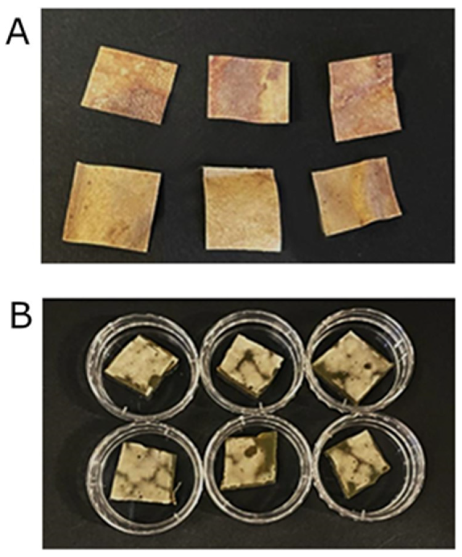
The parchment samples were macroscopically examined and 1 × 1 cm2 pieces were retrieved directly from the original document using sterile scalpels, following a selection of heavily biodegraded areas (Figure 2A,B).
To isolate bacterial and fungal biodeteriogens from the parchment, samples were placed in sterile tubes containing 2 mL of Nutrient Broth (NB) and then vortexed for 2 min. Aliquots with serial dilutions were then seeded on NB agar plates and incubated at 30 °C for 10 days. Morphologically different colonies were then isolated and stocked at -80 °C in 30% glycerol solution. Bacterial DNA was extracted by scraping up a colony and pipette up and down in 20 μL of 20 mM sodium hydroxide to disperse the colony in the liquid. After 10 min at 95 °C, an aliquot of DNA extract was taken for PCR amplification. As described in Ref. [40], a region of about 1400 bp from the 16S rRNA gene was amplified using the primers F8 (5′-AGAGTTTGATCCTGGCTCAG-3′) and R1492 (5′-GGTTACCTTGTTACGACTT-3′).
Fungal DNA was extracted according to Ref. [41] with some modifications. Fungal mycelia were taken in a 1.5 mL micro-centrifuge tube to which glass beads and 500 μL of lysis buffer (1% SDS, 40 mM Tris HCl, 20 mM sodium acetate, 1 mM EDTA) were added. The tube was vortexed for 5 cycles (2 min vortex and 5 min ice). Then, 165 μL NaCl 5M were added and the mixture was vortexed and then centrifuged at 13,000 rpm for 20 min at 4 °C. Next, 500 μL of Phenol-Chlorophorm-Isoamyl alcohol (PCI) in the ratio of 25:24:1 were added to the supernatant, centrifuged at 10,000 rpm for 15 min at 4 °C. Then, 400 μL of isopropanol were added to the supernatant and incubated at room temperature for 30 min, then centrifuged at 13,000 rpm for 15 min. The sediment containing DNA was washed twice with ethanol (4 °C/200 μL) at 6000 rpm for 2 min and dried at 68 °C for 5 min. The DNA was treated with RNAse, re-suspended in 30 μL of Tris–EDTA buffer and stored at −20 °C for further processing.
An aliquot of DNA was used for PCR amplification: a region of about 600 bp from the 18S rRNA gene was amplified using the primers ITS4 (5′-TCCTCCGCTTATTGATATGC-3′) and ITS5 (5′-GGAAGTAAAAGTCGAACAAGG-3′). The PCR reaction was performed utilizing the Taq DNA polymerase from Accuzyme DNA Polymerase (Bioline). The amplified region was sequenced BMR Genomics (Padova, Italy) and the obtained sequences were analyzed with BLAST database.

2.2. The Disinfection by Irradiation

For in vitro irradiation treatments, 1 × 1 cm2 portions of NB agar plates used for biodeteriogens’ isolation and thus covered with bacterial and fungal colonies were cut and placed in 3.5 cm Petri plates (Figure 2B).
The disinfection method by X-ray irradiation employs a source based on an electron accelerator. The treatment proposed and tested on artistic and cultural heritage collagen-based items is a specific application of a machine that accelerates electrons to produce radiation devoted to clinical applications. This type of electron accelerator is equipped with radiation conversion heads adequate for the generation of X-rays commonly used in hospitals for the radiotherapy of cancer. At the ENEA Frascati research center, the REX facility is a machine built as part of a program related to applications of nuclear methods to medical practice, then used also in activities inherent to the conservation of cultural heritage and, specifically, to the removal of bioagents inducing degradation in archival assets in a non-invasive way. The REX facility thus constitutes an irradiation system able to deliver both electrons and/or X-rays depending on the experimental needs. The machine generates high-performance, highly customizable, high-quality pulsed beams involved in research studies for radiation processing and for the evaluation and the characterization of radiation-induced effects in materials. Its operational mode allows to provide access and safety procedures at a significantly lower cost and less complex than other ionizing radiation facilities, which could possibly be used in the context of the sanification of bio-deteriorated cultural heritage items (i.e., machines that deliver directly with accelerated particles or the radioactive sources such as gamma rays) and also allows to actively modulate the processing speeds according to requirements. The technical characteristics of the REX machine and of its operations are detailed in the references [22,42]. The radiation monitoring applied during the biodegradation removal campaigns with the REX machine is carried out with an on-line system consisting of an ionization chamber with high spatial resolution and radiochromic films used for the dose distribution evaluation.
In the reported experimental campaigns, the treatments were performed in air, at ambient temperature and pressure conditions. The selected samples were exposed, stationary, a few centimeters in front of the X-ray source, to ensure the homogenous coverage of the entire samples area. In general, this procedure can be simplified by changing the beam diaphragm, thus investing a larger irradiated area. If need arises, the object itself can also be translated and rotated in front of the radiation beam. Given the density of the parchment, the photon beam completely traverses the samples, delivering the dose in the entire volume. In the processing facility, the linear movement system of the object holder allows treatments to be performed on series of assets in a completely safe, automated and remote controlled way [42].
After irradiation, agar samples were put in a sterile tube containing 2 mL of liquid NB and vortexed for 2 min. Then, aliquots of serial dilutions were then plated on NB agar plates and incubated at 30 °C for 10 days and colonies obtained were counted by the colony forming units (CFU) method, measuring the capacity of a single viable cell to form a colony. The experiment was performed in triplicate and repeated three times. Data are presented as mean ± SD. The statistical significance was determined by one-way ANOVA analysis coupled with a Bonferroni post-test (GraphPad Prism 5.0 software, GraphPad Software Inc., La Jolla, CA, USA), and defined as * p < 0.05, ** p < 0.01 and *** p < 0.001.

2.3. LTA

LTA is a technique able to characterize the hydrothermal stability of collagen proteins in animal skin derived substrates, such as parchment, leather and alum tawed skin, through the determination of their respective denaturation temperature Td. During the analysis, a suspension of fibers in water, obtained by pulping by a blade in a 1 mm2 area of the parchment sample in the condition of saturated hydration, was then placed in a 0.1 mm thick quartz cell and finally inserted in an oven where it was heated with a constant temperature rate of about 1 °C/min. During the temperature scan, from room temperature up to 90 °C, the light produced by an He-Ne laser beam passed through the sample so as to probe the occurring hydrothermal denaturation of the fibers, following which, the parchment fibers reduced their capability to scatter light. This lead to the increase of the transmitted unscattered fraction of the light beam which was collected by a lens and focused onto a photodiode. During the sample heating, the signal was recorded to determine Td, which is defined as the temperature at which the maximum denaturation activity and therefore change of the transmitted light occurs. This corresponds to the temperature where the derivative of the transmission data with respect to temperature displays a maximum in a peaked feature. The Td value is related to the hydrothermal stability of the sample under investigation: the higher the denaturation temperature, the better the chemical integrity of the collagen fibers and, therefore, of the sample preservation condition [34]. In this work, the reported Td values were obtained as an average of three different measurements.

2.4. FORS

Fiber optics reflectance spectroscopy (FORS) is a non-destructive technique that makes it possible to perform in situ inspection of artworks. It provides scientists and conservators with useful data for the identification of pigments and for the spectral analysis of color and its variation.
In this work, the reflectance spectra were obtained by averaging the data from 5 measurements recorded with StellarNet GREEN-Wave spectrometers equipped with a D65 illuminant in the range of 400–1100 nm. The instrument is optically coupled by means of fiber to a cube with an internal integrating sphere able to probe an area with a diameter of approximately 1 cm2 (StellarNet FORS Systems, 2021). The calibration of the system was performed using a reference target with a >97% certificated reflectance to the light from 300–1700 nm. In this work, the FORS investigations were conducted on the parchment samples to characterize its chromatism and brightness with the aim of possibly detecting any eventual optical effect induced by the disinfection procedure [36,40]. As explained further on, given the substantially observed invariability of the spectra obtained on the samples after the irradiation, it was decided not to present and discuss the results in terms of colorimetric coordinates due to the obtained color differences being extremely small.

2.5. ATR-FTIR spectroscopy

FTIR absorption spectra were acquired with the Thermo-Scientific instrument (model Is50) (Thermo Scientific Inc., Madison, WI, USA) in ATR mode using a single reflection diamond cell. Spectra were recorded from 4000 to 750 cm−1, averaging over 32 scans with a resolution of 2 cm−1. All experiments were performed in triplicate, yielding consistent and reproducible results. Deconvolution was performed in the 1360−1800 cm−1 region on normalized absorbance ATR-FTIR spectra by means of Voigt bands of full width at half maximum (FWHM) of 25 cm−1 maximum, using OMNIC software. In detail, ATR-FTIR investigations were carried out to detect possible deterioration effects, borne by collagen molecules in parchments, induced by natural aging and X-rays exposure. Indeed, ATR-FTIR spectroscopy allows monitoring changes in the proteins’ secondary structures, in a non-destructive way, through the analysis of Amide I and II bands, which are most commonly used in conformational protein studies (i.e., monitoring protein denaturation and/or aggregation processes) [43,44]. For clarity, the Amide I band is found between 1700 and 1600 cm−1, it is mainly associated with the C = O stretching vibration belonging to the polypeptide chain and is sensitive to local order and different secondary structures producing Amide I bands centered at different frequencies and in turn, to water bound to the macromolecule. The Amide II band occurs between 1570 and 1500 cm−1 and originates mainly from NH in-plane bending and CN stretching. By exploiting the study of the intensity ratio of these vibrational bands (IAmideI/IAmideII), it is possible to evaluate the degree of hydrolysis of the sample under examination. An increase in the value of this ratio indicates an increase in the degree of hydrolysis that took place in the sample, in fact in this case, there will be a contribution to the intensity of the Amide I band attributable to the higher percentage of water molecules contained [45]. This parameter is essential to trace the state of conservation of the parchment.

2.6. UV Resonant Raman Spectroscopy

UVRR measurements were performed at the IUVS beamline, in the Elettra Synchrotron radiation facility (Trieste, Italy). A full description of the Raman experimental apparatus is reported in Ref. [46]. Briefly, Raman spectra were collected in backscattering configuration employing a 213 nm laser as excitation source, focalized on the sample surface with a spot size of approximately 100 μm and a beampower of 200 μW. During the measurements, samples were kept oscillating for 5 mm at a frequency of 1 Hz to avoid photodegradation. The diffused Raman signal was collected and analyzed by means of a single stage Czerny–Turner spectrometer (Princeton Instruments), with a 750 mm of focal length, equipped with an 1800 lines/mm holographic reflection grating, 250 nm ruled and a Peltier-cooled back thinned CCD (Princeton Instruments) as a detector. To remove the elastic contribution, an HV (horizontal-vertical) geometrical configuration was adopted, i.e., it was selectively collected only the diffused radiation with polarization perpendicular (horizontal with respect to the optical plane) to the incident one (vertical with respect to the optical plane) [47]. The spectral bandwidth was approximately 20 cm−1. The final spectra were obtained averaging 4 spectra of 15 min, taken at different sample positions, for a cumulative data acquisition time of 60 min per sample.

2.7. Atomic Force Microscopy Nano-Indentation Measurements

Measurements were carried out under ambient conditions using an MFP-3D (Asylum Research/Oxford Instruments) in contact mode with standard silicon cantilevers (NSC36A, nominal stiffness 0.6 N/m, nominal radius of curvature <10 nm). We acquired force–distance curves in at least three 2 × 2 μm2 areas per sample in a 4 × 4 grid (16 curves per area). Cantilever stiffness and optical lever sensitivity were measured through the Sader method and linear fitting of the lever deflection tested on a glass substrate, respectively. After calibration, the force–distance curves (at least 40 curves per sample) were analyzed using MFP-3D software on an Igor platform and values of Young’s modulus extracted by Hertz model (fixed parameters: Poisson ratio = 0.5, radius of the tip = 100 nm). The high roughness and inhomogeneity of the parchments caused an almost immediate dulling of the tip shape, from 10 nm to roughly 100 nm.

3. Results

3.1. Identification of Bacterial and Fungal Colonies and Cell Viability Test

Morphologically different bacterial and fungal colonies were isolated from parchment and identified at the molecular level by the amplification of bacterial 16S rDNA or fungal ITS sequences. Comparison between FASTA sequences obtained from sequencing and those held in the BLAST database allowed to identify Arthrobacter echini, Micrococcus yunnanensis, Brevibacterium frigotolerans, Psychrobacillus psychrodurans and the Gram-negative Achromobacter xylosoxidans. Isolated fungal colonies belonged to Penicillium chrysogenum, Cladosporium cladosporioides and Aspergillus versicolor species.
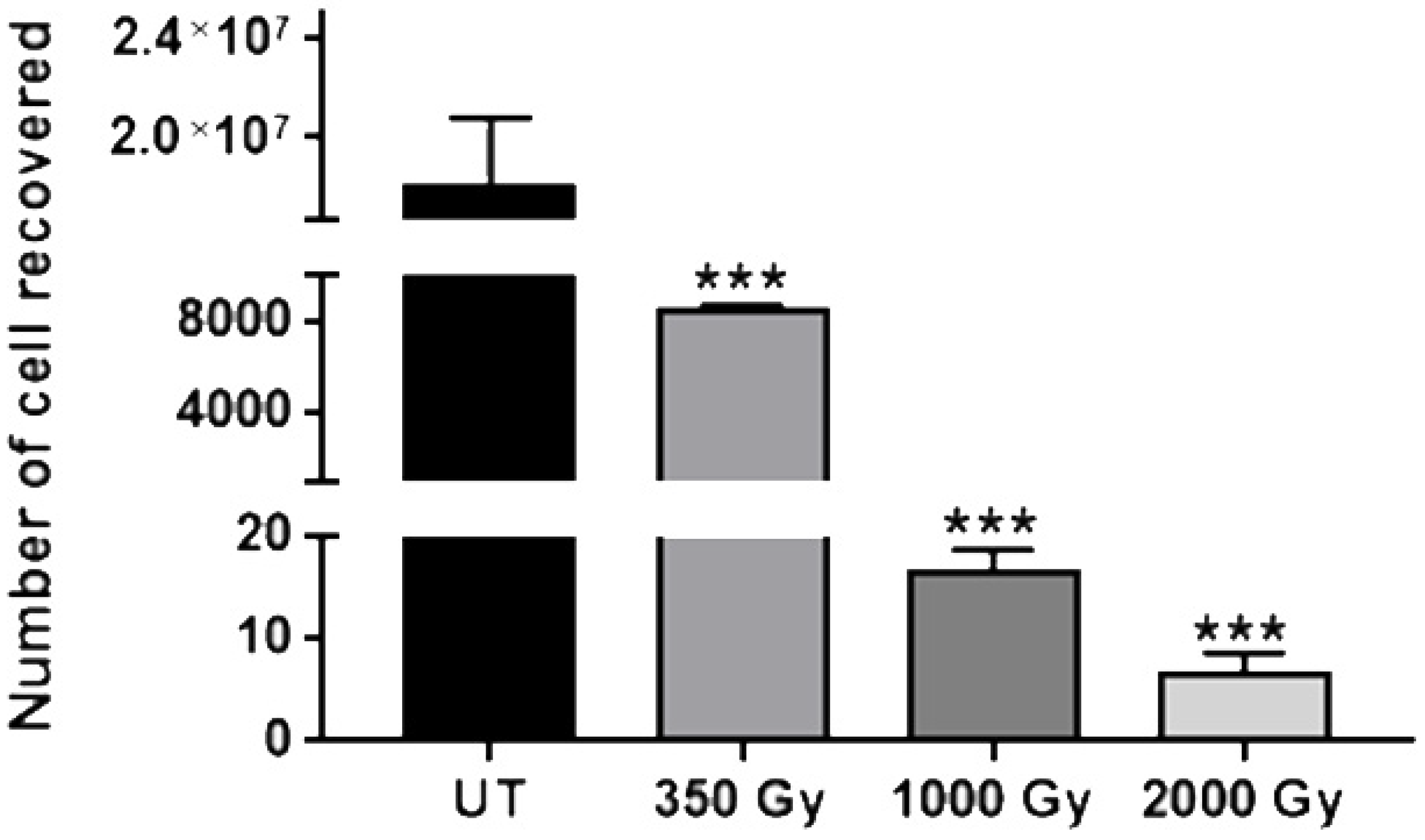
Cell viability tests after irradiation of agar colonized specimens showed a high rate of mortality: Already with a dose of 350 Gy, the substantial total inhibition of cell survival was observed (Figure 3). Notably, when irradiation doses increased to values of 1000 Gy and 2000 Gy, the mortality rate reached the complete inhibition of cell survival.
Among the different species isolated, some of them have already been reported to be involved in parchment biodeterioration, as described in in the literature [31,32,48].
In agreement with our data, the most frequent fungi genera isolated from parchment were Cladosporium, Penicillium and Aspergillus [49].

3.2. Characterization of the Parchment Deterioration

3.2.1. LTA

LTA was performed on each sample to evaluate possible deterioration induced changes in Td and, therefore, in their thermal stability. The obtained results are reported in the graph of Figure 4. It shows that all the Td values of the samples before irradiation are smaller than 52.2 °C, revealing a rather poor thermal stability characterizing all the investigated samples [33]; bearing in mind that, for example, in new modern parchment, Td can be as large as 59–60 °C. It can be also observed that the Td values of the unirradiated samples are spread over a range of about 2 °C. This indicates that the deterioration of the different samples is significantly inhomogeneous, in spite of the samples having been retrieved from areas close to each other. For this reason, in this work, the LTA characterization of the irradiation induced effects cannot be based on the comparison of the absolute values of Td obtained in the differently irradiated samples, but, on the contrary, by analyzing the relative change induced in each irradiated sample before and after the considered irradiation dose. In this respect, it is worthwhile pointing out that, even though LTA is a destructive technique, it involves such a small sample area (1 mm2) that, by performing the measurements before and after the irradiation on neighbouring areas of the sample, the results can be considered obtained on the same area before and after irradiation.
The graph in Figure 4 also shows the Td values (red empty square) obtained after irradiating five of the studied samples with different X-rays doses (350, 500, 1000, 2000 and 4000 Gy). Comparing each Td value with that obtained on the same sample before the irradiation treatment, no significant change is observed, even at the largest dose. It seems therefore that no additional damage by the X-ray treatments can be detected in these historical parchments. In this regard, it is worth noting that in a previous study carried out on undamaged modern parchment, it was shown that irradiation doses above 500 Gy produce significant damage in the structure of the parchment [29,30]. The lack of irradiation effects observed in this work on historical samples can be explained by considering that in the investigated ancient parchment, the deterioration of the most fragile elements of the collagen microstructure, which irradiation might also cause, has already been produced by environmental interaction processes, including hygrothermal aging and biodeterioration, which occurred during the history of the parchment.

3.2.2. FORS

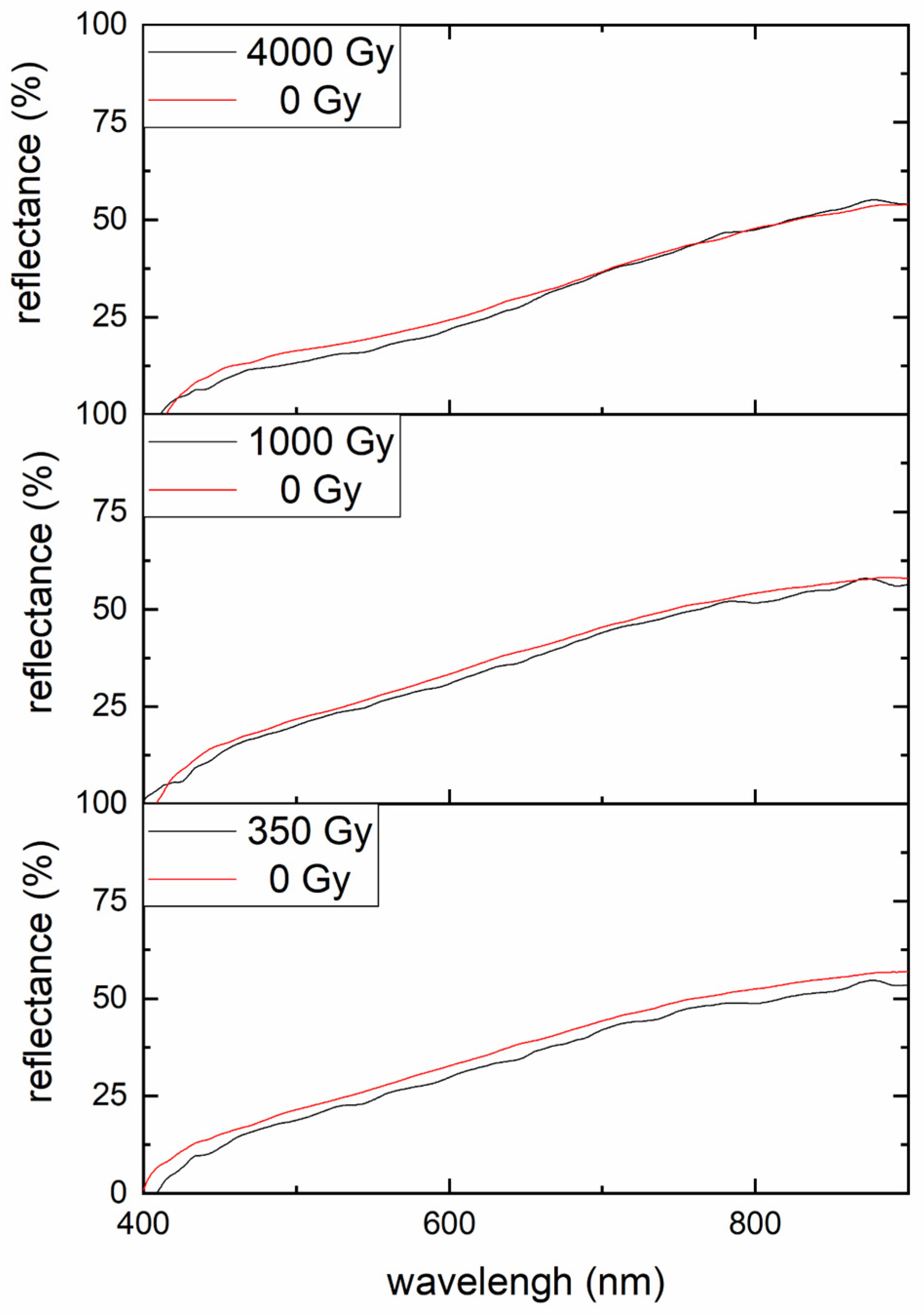
The samples were also studied by FORS in order to evaluate alteration in the macroscopic reflectance properties of the parchment induced by the irradiation. The mesurements were performed on each sample before and after the corresponding irradiation dose. Figure 5 shows the reflectance spectra obtained on samples irradiated with 350 Gy, 1000 Gy and 4000 Gy dose (black curves) and the corresponding spectra before the irradiation treatment (red curves). Similarly to the results obtained by LTA, no significant changes associated with the X-ray treatments can be detected even in the reflectance spectra, witnessing that no substantial changes in the opacification and yellowing of the samples has occurred.

3.2.3. ATR-FTIR Spectroscopy Results

A previously reported analysis indicated that no deterioration effects (i.e., conformational changes due to gelatinization), due to X-rays exposure, occurred. Therefore, it is reasonable to assume that every change in FTIR spectra is attributable to heterogeneity of parchments, rather than X-ray treatments. Data reported in Table 1 indicate that the variation of values of the intensity ratio of the Amide I and Amide II bands does not follow the irradiation dose trend, thus confirming our hypothesis.
The values of such a ratio, which generally characterize the degree of hydrolysis of the samples, indicate that the degree of deterioration in the analyzed parchments caused by the ageing is very heterogeneous, as also observed by the previously shown LTA results and also somehow predictable for an ancient material subjected not only to the natural effect of time but, in this specific case, also to a microbiological attack [45,50].
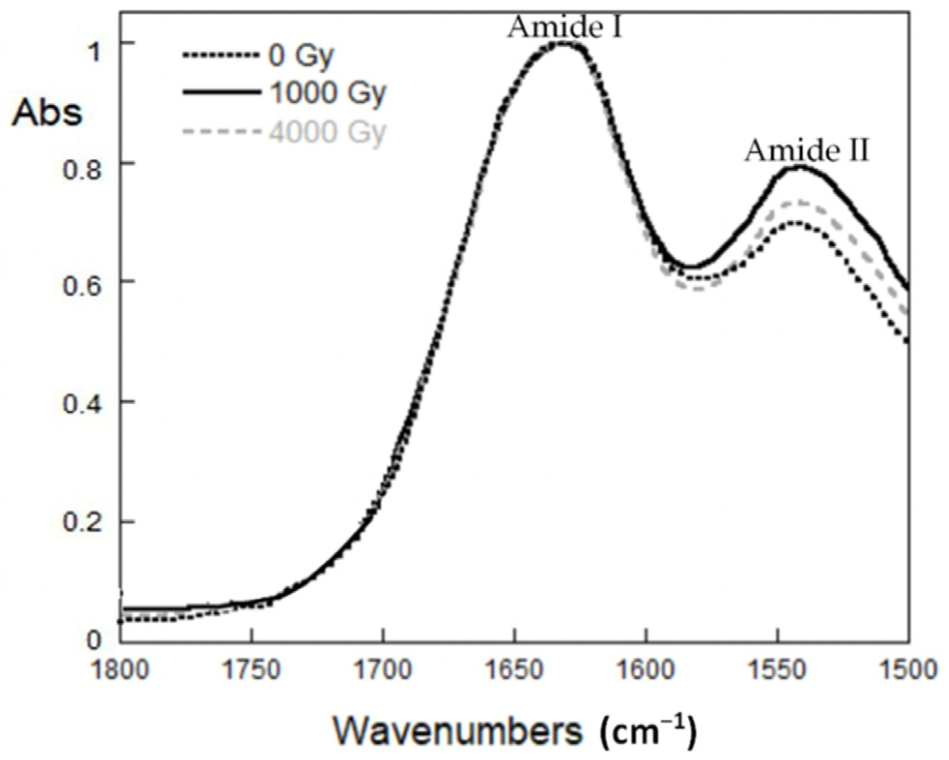
Figure 6 presents the spectra related to the samples untreated (0 Gy) and treated with X-rays doses of 1000 Gy and 4000 Gy. From the spectra, it is gathered that the value of the relative distance between Amide I and Amide II bands remains unchanged (~93 cm−1) in spite of the irradiation doses being considerably larger than that sufficient to induce the curing of the infection. This indicates that the degree of gelatinization in the samples [51] is not affected by the irradiation process and it is substantially related to the deterioration associated with the ageing. The extent of degradation of the parchment, due to aging alone, is better highlighted when compared to that of a modern parchment in good condition. Indeed, as can be seen from Table 1, a naturally aged parchment, not irradiated (0 Gy), shows a value of the ratio between the intensities of the Amide I and Amide II bands of 3.0 ± 0.1, while in the case of modern parchment, this value is 1.2 ± 0.1. Furthermore, the relative distance between the bands of Amides I and II is about 93 cm−1 and 89 cm−1, for ancient and modern parchment, respectively. These results clearly indicate that natural aging has a marked effect on the material, producing its hydrolysis and denaturation and/or gelatinization.

3.2.4. UV Resonant Raman Spectroscopy

To verify the presence of the aforementioned natural aging, we also employed UV Resonant Raman spectroscopy with an excitation wavelength of 213 nm. At this excitation wavelength, it is possible to match the resonance with the protein’s amide component, and at the same time, to avoid the presence of a fluorescence background affecting the spectra [52,53]. By this way, it is possible to selectively follow the spectral behavior of the collagen inside the parchment. Attempts made to collect spectra employing excitation wavelengths of 266 nm, 532 nm and 633 nm resulted in spectral profiles dominated by a very large fluorescence background.
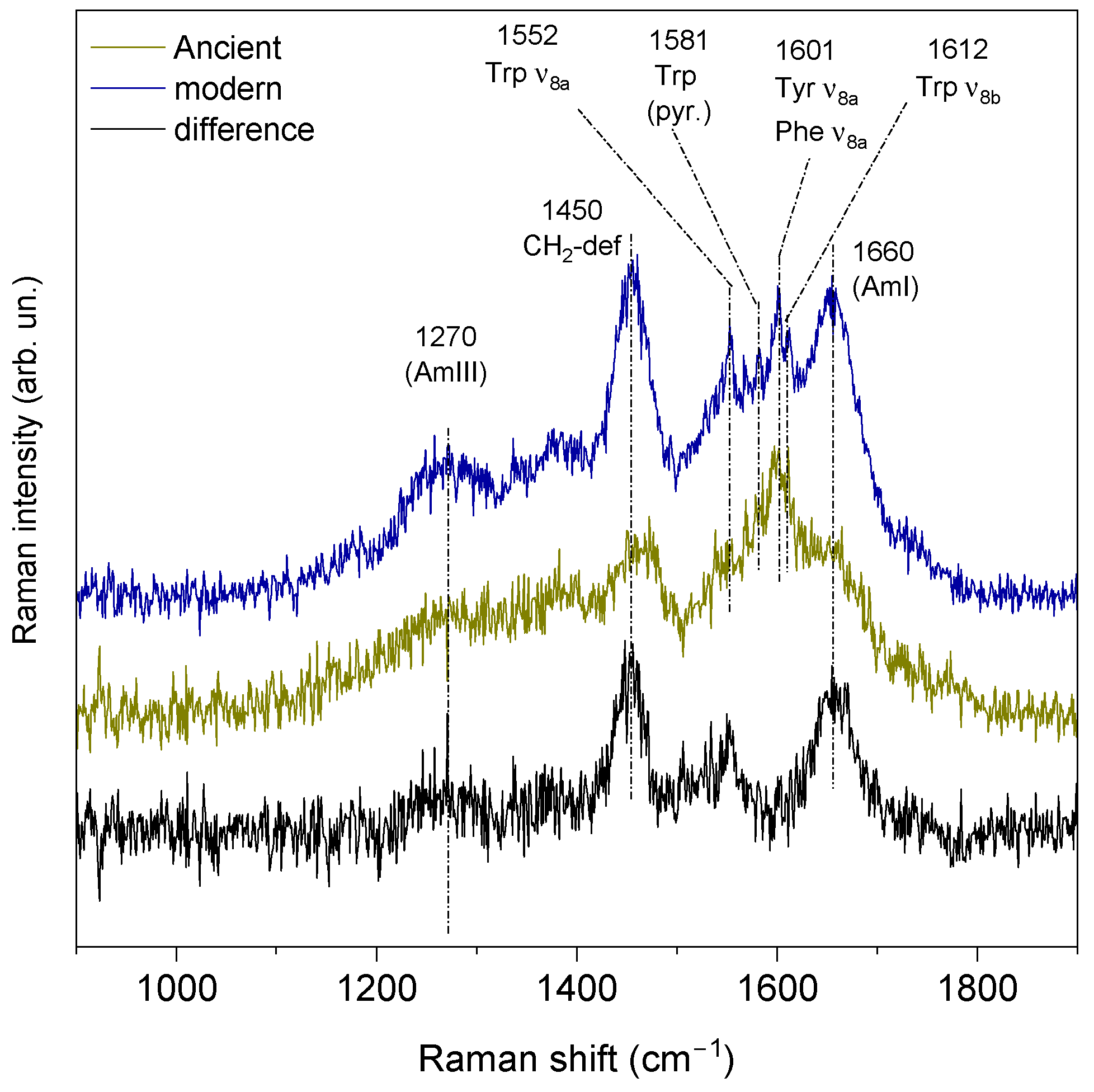
The mesurements were carried out only in the samples after the irradiation treatment to highlight possible phenomena of hydrolysis and/or denaturation associated with the aging, since irradiation was previously shown not to cause significant additional deterioration. Figure 7 reports the UVRR spectra, in the wavenumber range of 900–1900 cm−1, relating to ancient and modern parchment samples. The latter sample was prepared following the traditional artisan recipe and it constitutes a good reference of a well-preserved parchment sample to be compared with the ancient ones. The comparison of the results shown in Figure 7 is useful to evaluate the very significant variation occurring in the protein structure to which the aged parchment samples have undergone.
The peaks' positions agree with the presence of collagen on the sample surface [54]. Specifically, we can identify the Amide I and Amide III spectral features located respectively at 1660 cm−1 and 1270 cm−1 [55,56]. Further important spectral features at the peak at 1450 cm−1 and 1380 cm−1 can be assigned to CH2-def and Cα-H vibrations derived from the amide contribution. Moreover, in the spectral range between 1550 and 1650 cm−1, the spectrum contains some peaks which can be specifically assigned to aromatic amino acids vibrations [35], namely the rings stretching vibrational bands related to aromatic of tryptophan at 1612, 1581 and 1552, and in addition, the tyrosine and phenylalanine ones overlapped at 1601 cm−1 [57,58] (see details in Figure 7). Moreover, in Figure 7, it is possible to appreciate a decrease of the Amide I, Amide III and CH2-def spectral bands in the ancient sample with respect to the modern one that clearly indicates a strong degradation of collagen.

3.2.5. Atomic Force Microscopy Mechanical Analysis

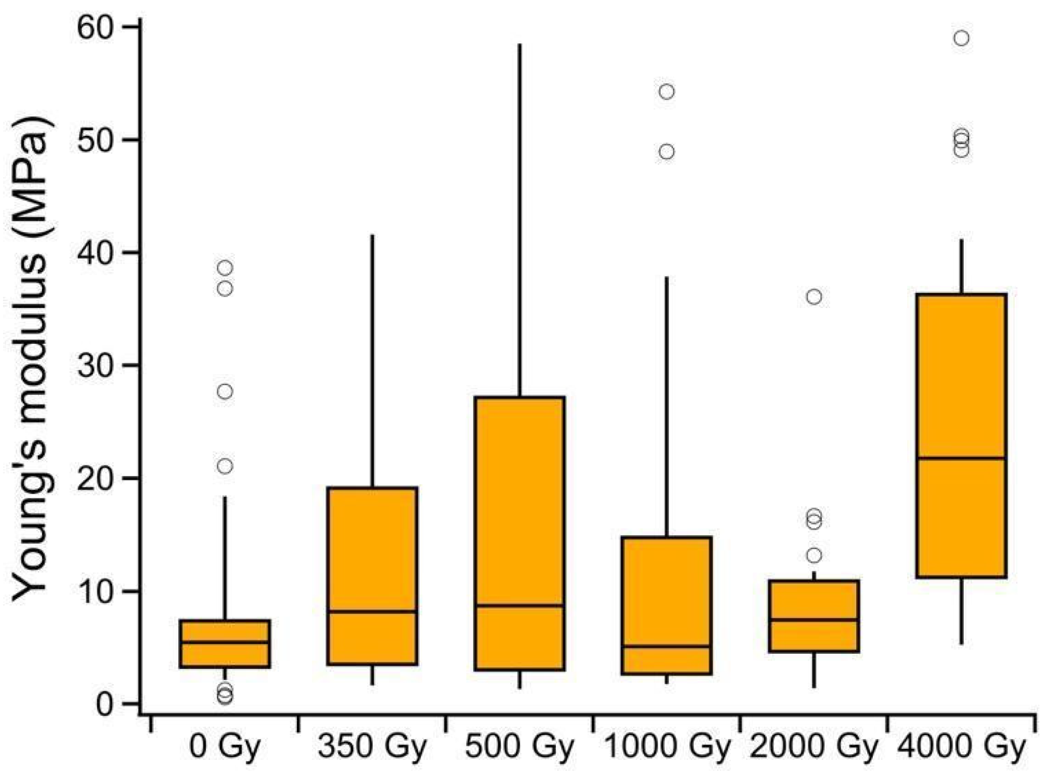
We also tested the mechanical response of the irradiated and non-irradiated samples, as explained in our recent paper [29]. The mesurements were carried out only in the samples after the irradiation treatment to highlight possible phenomena of hydrolysis and/or denaturation associated with the aging, since irradiation was previously shown not to cause significant additional deterioration. In Figure 8, we report the Young’s modulus distribution extrapolated from the analysis of force–distance curves, as detailed in the Materials and Methods section, for the non-irradiated (0 Gy) and irradiated samples (350, 500, 1000, 3000 Gy). All values of Young’s modulus fall in the range of tens of MPa, in agreement with previous reports. We cannot observe a clear correlation between dose and Young’s modulus and thus, a significant effect which can be directly associated with the irradiation treatment on the collagen matrix and to consequent effects on its mechanical properties. Even the results of the present analysis reveal a non systematic change with the increasing irradiation dose. In fact, a mild stiffening effect with respect to the non-irradiated sample is observed for the 350 Gy and 500 Gy irradiated samples, non-significant changes for those irradiated with 1000 Gy and a stiffness increase for those irradiated with 2000 Gy and 4000 Gy.

4. Discussion

Parchment is a handcrafted complex and non-homogeneous material with quality strictly related to the time and the method it was produced during the centuries as well as the preservation conditions. Its intrinsic inhomogeneity is mainly due to its nature (different parts of the animal skin have different physical and mechanical properties) and also to the peculiar manufacturing process that seldom leads to obtaining similar quality samples even when originating from the same animal skin. These intrinsic inhomogeneities are amplified by the effect of the bacterial and fungi infections that can make the samples extremely uneven, like in the case of the presented study. Nevertheless, in order to evaluate the applicability of the proposed disinfection treatment by means of X-ray irradiation, some considerations can be deduced by comparing the results obtained by the different analytical techniques employed in this study.
At first, based on our results, we can certainly conclude that the irradiation method is efficient to inhibit in vitro the microbial growth of the species of the fungi and bacteria isolated from the selected samples. As a matter of fact, the microbiological analysis of the cell viability test performed after the irradiation treatment has shown that even with a dose as low as 350 Gy, it is possible to obtain a complete mortality rate. The X-rays dose of the REX machine, 350 Gy, that achieves the inhibition of microorganisms’ survival is lower than the threshold identified in other studies in which ionizing radiation is applied as a bio-sanitation treatment on mixed microorganisms (bacteria and fungi) [59,60].
Regarding the effect of the irradiation protocol on the collagen molecules of the historical parchment samples analyzed in this work, LTA revealed no significant alteration in the parchment microstructural integrity induced by the irradiation treatments, regardless of their significant uneven initial state of deterioration, and suggested that, in all the analyzed cases, the natural deterioration associated with hygrothermal and bioagents agents effects overshadowed the damage related to the X-ray irradiation process. In fact, the different samples showed intrinsic deterioration inhomogeneity despite having been retrieved from neighboring areas, no correlation being found between the absolute Td values and the applied X-rays dose, and no change could be found in the Td values obtained on each sample before and after the respective irradiation treatment, even at the largest employed dose. So, no additional damage produced by the X-ray treatment could be detected in such historical naturally aged parchment.
The FTIR analysis confirmed the inhomogeneity of the initial naturally induced deterioration and showed that it was associated with hydrolysis and gelatinization of the collagen molecules. In fact, the values of the intensity ratio of the Amide I/Amide II bands are different for the different irradiation doses but no correlation with the increasing irradiation dose appears, revealing the differences to be related only to the natural ageing effects. The distance between the two previously mentioned bands appears on the contrary, to remain pretty much constant in the different samples, highlighting a homogeneous gelatinization degree of the samples.
The strong natural deterioration of the investigated parchment was confirmed by the UVRR results that show a decrease in the spectral bands of the Amide I, Amide III and Ch2 def in the ancient sample with respect to the ones recordable on a modern one.
Furthermore, even the AFM mechanical analysis does not find a direct relation between the value of the Young’s modulus of the investigated sample and the irradiation treatment of the samples. The recorded increase in the stiffness of some samples (350, 500 and 4000 Gy) does not follow the dose trend, suggesting the possibility that, also in this case, the recorded deterioration cannot be directly related to the treatment. Finally, similarly to the results obtained with the other analysis techniques, no significant changes also in the reflectance spectra are produced by the X-ray treatments. In fact, for all the samples, the reflectance profiles recorded before the irradiation were maintained after the treatment.
It can thus be stated that the results obtained by all the techniques suggest that the deterioration effects highlighted by the employed analyses can only be associated with the natural aging effects on the samples, worsened by the biodeterioration processes. The results show the possibility that, for significantly deteriorated samples like the present naturally aged and biodeteriorated ones, even high doses of irradiation do not lead to detectable alteration of the stability of the already partially deteriorated collagen molecule of the parchment. When the parchment is not originally heavily deteriorated, the minimum dose sufficient to sterilize the sample without introducing additional damage (i.e., ≈350 Gy) would be advisable. Based on these results, the irradiation-based method of disinfection seems to be a valid candidate as a mass disinfection method for library and archival material, at least for heavy contaminated and deteriorated assets.
One of the strong points of the use of X-rays for the sanitization of the archival-library assets is the possibility to directly act on the artifacts to be preserved without first identifying the infesting biodeteriogen. In particular, this constitutes an advantage with respect to other treatments for which it is necessary to know the infesting agent both for the specific treatment actions (in order to find the more effective one) and to avoid any contamination of the environment in which the process takes place (such us the radiation systems based on radionuclides). The proposed sterilization treatment is non-polluting and seems to be effectively working with the doses used in the experiments presented in this paper. In fact, it must be reminded that not all common disinfectant processes are adequate to clean and preserve cultural heritage items. The conventionally used sanitization procedures cannot be considered unambiguously applicable in the same way to any asset or environment. The current common procedures and rules of intervention and behavior, aimed at guaranteeing the safety of the operators, the public and the protection of archival and library assets, are generally different and often not applicable if the deterioration degree is high. In that case, treatments with disinfectant and bactericidal products become more complex, involving a large number of operators, extended times and also high costs. Furthermore, in some cases, the conservation of library and archival heritage can be exposed to danger by unpredictable events, such as flooding. Even in this eventuality, when urgent and more complex interventions are required, the application of radiation is a strong advantage for the remediation of assets thanks to the faster and more practical use compared to other physical, biological and chemical interventions.
Moreover, unlike the radionuclide sources, which emit nearly monoenergetic photons, the proposed process creates a broad energy spectrum of X-rays and, the maximum energy limit of 5 MeV set by the REX machine prevents, with a very high level of confidence, any radioactivity induction in the irradiated products through photonuclear reactions.
Finally, an additional advantage of the applications of this X-ray source is that the objects to be treated can be irradiated as they are without any specific preparation (i.e., preliminary dry cleaning) or environmental conditions control since the treatments are carried out in the air, under normal temperature and pressure conditions. This system can also offer costs and operating advantages compared to gamma sources thanks to their easy radiation control, convenience of easy-on and easy-off electrical equipment that can also operate actively according to the treatments requirements. Some plants delivering high power and high energy already offer the opportunity to produce and use X-rays for different industrial applications [61,62,63,64,65,66,67,68].
It must be finally remarked that additional research is required, finalized at promoting X-ray irradiation as a possible standard disinfectant remedy against bio-infections for library and documentary assets. In fact, it remains to be established what are the possible damaging effects of the radiation on the materials, other than parchment, of which books are constituted and also the possible damaging effects on parchment, which is initially not as heavily degraded as the one considered in this work. To date, as stated earlier on, only investigations on undamaged modern parchment have been carried out, and experiments on parchment with a progressively increasing initial degree of degradation are underway.

5. Conclusions and Perspectives

In this work, a method based on X-ray irradiation was proposed as a disinfection treatment of microorganisms involved in the biodeterioration of parchment. In particular, some ancient parchment samples, belonging to a 16th century book cover, were treated and then analyzed by different diagnostics techniques, namely LTA, FORS, ATR-FTIR spectroscopy, UVRR spectroscopy and AFM analysis.
Our results demonstrated the high curing efficiency of the proposed irradiation method in vitro tests, highlighting the potential of this method in mass disinfection of library and archival materials without no introduction of addittional damage in the treated parchment samples. It is worth noting that biodeteriogens should be studied directly on the original samples and in their natural environment. This is not always possible due to the scarcity of material from original ancient documents. Therefore, preliminary tests are needed to fine-tune protocols without wasting material belonging to important documents. On this basis, further research is underway to evaluate the differences between in vitro and in vivo results, given the sensitivity of the microorganisms to the treatments under different growth conditions.
Finally, it should be remarked that the ability to treat library materials without first having to identifying the biodeteriogen, together with the effectiveness of the treatment with the safe doses used in this work, make this disinfection method a valuable alternative to those commonly applied. Indeed, the use of X-ray radiation can be a valuable tool for the disinfection of library materials, even in emergency situations, because of its rapidity of application, which makes it cost-effective compared to other physical, biological and chemical interventions. All these aspects encourage the application of the X-ray irradiation method for disinfection of these kinds of materials. Since the proposed procedure does not require the use of high-risk substances, this bioremediation treatment can be considered as a "green" method compared to conventional disinfection techniques and to other techniques using different sources of nuclear radiation.

Author Contributions

Conceptualization, C.C. and M.V.; Irradiation treatment, M.V.; Microbiological formal analysis and data curation, E.S. and D.U.; ATR-FTIR formal analysis and data curation, C.M. and L.S.; UVRR formal analysis and data curation, F.D.; LTA formal analysis and data curation, C.C. and F.M.; FORS formal analysis and data curation, N.O. and U.Z.; AFM formal analysis and data curation, P.P.; writing—original draft preparation, C.C.; writing—review and editing, all the authors. All authors have read and agreed to the published version of the manuscript.

Funding

Part of this research has been realized with the co-funding of the European Union FSE REACT-EU-PON Ricerca e Innovazione 2014–2020, DM 1062/2021. The authors acknowledge the CERIC-ERIC Consortium for the access to the IUVS-OFF experimental facility and financial support with the proposal number 20217067.

Institutional Review Board Statement

Not applicable.

Informed Consent Statement

Not applicable.

Data Availability Statement

Not applicable.

Acknowledgments

The authors acknowledge the Regione Lazio Centro di Eccellenza for the opportunity to collaborate in their network.

Conflicts of Interest

The authors declare no conflict of interest.

References

  1. Agati, M. The Manuscript Book, a Compendium of Codicology, 1st ed.; L’Erma di Bretschneider: Roma, Italy, 2017. [Google Scholar]
  2. Kennedy, C.J.; Wess, T.J. The Structure of Collagen within Parchment—A Review. Restaurator 2003, 24, 61–80. [Google Scholar] [CrossRef]
  3. Kite, M.; Thomson, R. Conservation of Leather and Related Materials, 1st ed.; Routledge: London, UK, 2005. [Google Scholar]
  4. Hansen, E.F.; Lee, S.N.; Sobel, H. The Effects of Relative Humidity on Some Physical Properties of Modern Vellum: Implications for the Optimum Relative Humidity for the Display and Storage of Parchment. JAIC 1992, 31, 325–342. [Google Scholar] [CrossRef]
  5. Karbowska-Berent, J.; Górny, R.L.; Strzelczyk, A.B.; Wlazło, A. Airborne and dust borne microorganisms in selected Polish libraries and archives. Build. Environ. 2011, 46, 1872–1879. [Google Scholar] [CrossRef]
  6. Piñar, G.; Sterflinger, K.; Ettenauer, J.; Quandt, A.; Pinzari, F. A Combined Approach to Assess the Microbial Contamination of the Archimedes Palimpsest. Microb. Ecol. 2015, 69, 118–134. [Google Scholar] [CrossRef] [PubMed] [Green Version]
  7. Cicero, C.; Pinzari, F.; Mercuri, F. 18th Century knowledge on microbial attacks on parchment: Analytical and historical evidence. Int. Biodeterior. Biodegrad. 2018, 134, 76–82. [Google Scholar] [CrossRef]
  8. Migliore, L.; Thaller, M.C.; Vendittozzi, G.; Mejia, A.Y.; Mercuri, F.; Orlanducci, S.; Rubechini, A. Purple spot damage dynamics investigated by an integrated approach on a 1244 A.D. parchment roll from the Secret Vatican Archive. Sci. Rep. 2017, 7, 9521. [Google Scholar] [CrossRef]
  9. Migliore, L.; Perini, N.; Mercuri, F.; Orlanducci, S.; Rubechini, A.; Thaller, M.C. Three ancient documents solve the jigsaw of the parchment purple spot deterioration and validate the microbial succession model. Sci. Rep. 2019, 9, 1623. [Google Scholar] [CrossRef] [Green Version]
  10. Craig, R. Alternative approaches to the treatment of mould biodeterioration—An international problem. Pap. Conserv. 1986, 10, 27–30. [Google Scholar] [CrossRef]
  11. Sequeira, S.; Cabrita, E.J.; Macedo, M.F. Antifungals on paper conservation: An overview. Int. Biodeterior. Biodegrad. 2012, 74, 67–86. [Google Scholar] [CrossRef]
  12. Michaelsen, A.; Pinzari, F.; Barbabietola, N.; Piñar, G. Monitoring the effects of different conservation treatments on paper-infecting fungi. Int. Biodeterior. Biodegrad. 2013, 84, 333–341. [Google Scholar] [CrossRef]
  13. Pfendler, S.; Borderie, F.; Bousta, F.; Alaoui-Sosse, L.; Alaoui-Sosse, B.; Aleya, L. Comparison of biocides, allelopathic substances and UV-C as treatments for biofilm proliferation on heritage monuments. J. Cult. Herit. 2018, 33, 117–124. [Google Scholar] [CrossRef]
  14. Adamo, A.M.; Giovannotti, M.; Magaudda, G.; PLoSsi Zappalà, M.; Rocchetti, F.; Rossi, G. Effect of Gamma Rays on Pure Cellulose Paper as a Model for the Study of a Treatment of “Biological Recovery” of Biodeteriorated Books. Restaurator 1998, 19, 41–59. [Google Scholar] [CrossRef]
  15. Adamo, M.; Brizzi, M.; Magaudda, G.; Martinelli, G.; PLoSsi-Zappalà, M.; Rocchetti, F.; Savagnone, F. Gamma Radiation Treatment of Paper in Different Environmental Conditions: Chemical, Physical and Microbiological Analysis. Restaurator 2001, 22, 107–131. [Google Scholar] [CrossRef]
  16. Magaudda, G. The recovery of biodeteriorated books and archive documents through gamma radiation: Some considerations on the results achieved. J. Cult. Herit. 2004, 5, 113–118. [Google Scholar] [CrossRef]
  17. Otero D’Almeida, M.L.; de Souza Medeiros Barbosa, P.; Guerra Boaratti, M.F.; Borrely, S.Y. Radiation effects on the integrity of paper. Radiat. Phys. Chem. 2009, 78, 489–492. [Google Scholar] [CrossRef]
  18. Pietrzak, K.; Otlewska, A.; Danielewicz, D.; Dybka, K.; Pangallo, D.; Kraková, L.; Puškárová, A.; Bučková, M.; Scholtz, V.; Ďurovič, M.; et al. Disinfection of archival documents using thyme essential oil, silver nanoparticles misting and low temperature plasma. J. Cult. Herit. 2017, 24, 69–77. [Google Scholar] [CrossRef]
  19. Macedo-Arantes, S.; Piçarra, A.; Caldeira, A.T.; Candeias, A.E.; Martins, M.R. Essential oils of Portuguese flavouring plants: Potential as green biocides in cultural heritage. Eur. Phys. J. Plus 2021, 136, 1106. [Google Scholar] [CrossRef]
  20. Ismael, S.; Omar, A.; Maher, M. Comparative Inhibition Study by Nanomaterial, Plant Extract and Chemical Microcide on the Screaming Mummy in Egyptian Museum Store. Heritage 2021, 4, 2481–2493. [Google Scholar] [CrossRef]
  21. Gueidão, M.; Vieira, E.; Bordalo, R.; Moreira, P. Available green conservation methodologies for the cleaning of cultural heritage: An overview. Estud. Conserv. E Restauro 2021, 12, 22–44. [Google Scholar] [CrossRef]
  22. Vadrucci, M.; Borgognoni, F.; Cicero, C.; Perini, N.; Migliore, L.; Mercuri, F.; Orazi, N.; Rubechini, A. Parchment processing and analysis: Ionizing radiation treatment by the REX source and multidisciplinary approach characterization. Appl. Radiat. Isot. 2019, 149, 159–164. [Google Scholar] [CrossRef]
  23. Vadrucci, M.; De Bellis, G.; Mazzuca, C.; Mercuri, F.; Borgognoni, F.; Schifano, E.; Uccelletti, D.; Cicero, C. Effects of the Ionizing Radiation Disinfection Treatment on Historical Leather. Front. Mater. 2020, 11, 7. [Google Scholar] [CrossRef] [Green Version]
  24. Imlay, J.A.; Linn, S. DNA damage and oxygen radical toxicity. Science 1988, 240, 1302–1309. [Google Scholar] [CrossRef] [PubMed] [Green Version]
  25. Cadet, J.; Delatour, T.; Douki, T.; Gasparutto, D.; Pouget, J.P.; Ravanat, J.L.; Sauvaigo, S. Hydroxyl radicals and DNA base damage. Mutat Res. 1999, 424, 9–21. [Google Scholar] [CrossRef] [PubMed]
  26. Tilquin, B. Actions Biologique et Chimique des Rayonnements Ionisants; Nauwelaerts: Paris, France, 2001; p. 115. [Google Scholar]
  27. Borrely, S.I.; Cruz, A.C.; Del Mastro, N.L.; Sampa, M.H.O.; Somessari, E.S. Radiation processing of sewage and sludge: A review. Prog. Nucl. Energy 1998, 33, 3–21. [Google Scholar] [CrossRef]
  28. Condón-Abanto, S.; Pedrós-Garrido, S.; Cebrián, G.; Raso, J.; Condón, S.; Lyng, J.G.; Álvarez, I. Crab-meat-isolated psychrophilic spore forming bacteria inactivation by electron beam ionizing radiation. Food Microbiol. 2018, 76, 374–381. [Google Scholar] [CrossRef] [Green Version]
  29. Vadrucci, M.; Cicero, C.; Parisse, P.; Casalis, L.; De Bellis, G. Surface evaluation of the effect of X-rays irradiation on parchment artefacts through AFM and SEM. Appl. Surf. Sci. 2020, 513, 145881. [Google Scholar] [CrossRef]
  30. Vadrucci, M.; Cicero, C.; Mazzuca, C.; Mercuri, F.; Missori, M.; Orazi, N.; Severini, L.; Zammit, U. Effect of X-ray and artificial aging on parchment. Eur. Phys. J. Plus 2021, 136, 873. [Google Scholar] [CrossRef]
  31. Sterflinger, K.; Pinzari, F. The revenge of time: Fungal deterioration of cultural heritage with particular reference to books, paper and parchment. Environ. Microbiol. 2012, 14, 559–566. [Google Scholar] [CrossRef]
  32. Piñar, G.; Tafer, H.; Schreiner, M.; Miklas, H.; Sterflinger, K. Decoding the biological information contained in two ancient Slavonic parchment codices: An added historical value. Environ. Microbiol. 2020, 22, 3218–3233. [Google Scholar] [CrossRef]
  33. Cicero, C.; Mercuri, F.; Paoloni, S.; Orazi, N.; Zammit, U.; Glorieux, C.; Thoen, J. Integrated adiabatic scanning calorimetry, light transmission and imaging analysis of collagen deterioration in parchment. Thermochim. Acta 2019, 676, 263–270. [Google Scholar] [CrossRef]
  34. Palmieri, E.; Cicero, C.; Orazi, N.; Mercuri, F.; Zammit, U.; Mazzuca, C.; Orlanducci, S. Nanodiamond composites: A new material for the preservation of parchment. J. Appl. Polym. Sci. 2022, 139, e52742. [Google Scholar] [CrossRef]
  35. Ganz, E.; Pauli, H.K.A. Whiteness and tint formulas of the Commission Internationale de l'Eclairage: Approximations in the L*a*b* color space. Appl. Opt. 1995, 34, 2998–2999. [Google Scholar] [CrossRef]
  36. Oleari, C. Misurare il Colore, 1st ed.; Hoepli: Milano, Italy, 2008. [Google Scholar]
  37. De Campos Vidal, B.; Mello, M.L.S. Collagen type I amide I band infrared spectroscopy. Micron 2011, 42, 283–289. [Google Scholar] [CrossRef]
  38. Schwing, K.; Gerhards, M. Investigations on isolated peptides by combined IR/UV spectroscopy in a molecular beam—Structure, aggregation, solvation and molecular recognition. Int. Rev. Phys. Chem. 2016, 35, 569–677. [Google Scholar] [CrossRef]
  39. Asher, S.A.; Ludwig, M.; Johnson, C.R. UV resonance Raman excitation profiles of the aromatic amino acids. J. Am. Chem. Soc. 1986, 108, 3186–3197. [Google Scholar] [CrossRef]
  40. Schifano, E.; Cavallini, D.; De Bellis, G.; Bracciale, M.P.; Felici, A.C.; Santarelli, M.L.; Sarto, M.S.; Uccelletti, D. Antibacterial Effect of Zinc Oxide-Based Nanomaterials on Environmental Biodeteriogens Affecting Historical Buildings. Nanomaterials 2020, 10, 335. [Google Scholar] [CrossRef] [Green Version]
  41. Kumar, M.; Mugunthan, M. Evaluation of three DNA extraction methods from fungal cultures. Med. J. Armed. Forces India 2018, 4, 333–336. [Google Scholar] [CrossRef]
  42. Vadrucci, M.; Ferrari, P.; Borgognoni, F.; Campani, L. The REX irradiation facility and its applications. Nucl. Instrum. 2019, 930, 126–131. [Google Scholar] [CrossRef]
  43. Nebojsa, P.; Nils, K.A.; Ragni, O.; Achim, K. Monitoring Protein Structural Changes and Hydration in Bovine Meat Tissue Due to Salt Substitutes by Fourier Transform Infrared (FTIR) Microspectroscopy. J. Agric. Food Chem. 2011, 59, 10052–10061. [Google Scholar] [CrossRef] [Green Version]
  44. Shivu, B.; Seshadri, S.; Li, J.; Oberg, K.A.; Uversky, V.N.; Fink, A.L. Distinct β-Sheet Structure in Protein Aggregates Determined by ATR–FTIR Spectroscopy. Biochemistry 2013, 52, 5176–5183. [Google Scholar] [CrossRef]
  45. Plavan, V.; Giurginca, M.; Budrugeac, P.; Vilsan, M.; Miu, L. Evaluation of the physico-chemical characteristics of leather samples of some historical objects from kiev. Rev. Chim. 2010, 61, 627–631. [Google Scholar]
  46. D'Amico, F.; Saito, M.; Bencivenga, F.; Marsi, M.; Gessini, A.; Camisasca, G.; Principi, E.; Cucini, R.; Di Fonzo, S.; Battistoni, A.; et al. UV resonant Raman scattering facility at Elettra. Nucl. Instrum. 2013, 703, 33–37. [Google Scholar] [CrossRef]
  47. Ferraro, J.R.; Nakamoto, K.; Brown, C.W. Introductory Raman Spectroscopy, 2nd ed.; Academic Press: San Diego, CA, USA, 2003. [Google Scholar]
  48. Lech, T. Evaluation of a Parchment Document, the 13th Century Incorporation Charter for the City of Krakow, Poland, for Microbial Hazards. Appl. Environ. Microbiol. 2016, 82, 2620–2631. [Google Scholar] [CrossRef] [Green Version]
  49. Mesquita, N.; Portugal, A.; Videira, S.; Rodríguez-Echeverría, S.; Bandeira, A.M.L.; Santos, M.J.A.; Freitas, H. Fungal diversity in ancient documents. A case study on the Archive of the University of Coimbra. Int. Biodeterior. Biodegrad. 2009, 63, 626–629. [Google Scholar] [CrossRef]
  50. Vyskočilová, G.; Ebersbach, M.; Kopecká, R.; Prokeš, L.; Príhoda, J. Model study of the leather degradation by oxidation and hydrolysis. Herit. Sci. 2019, 7, 26. [Google Scholar] [CrossRef] [Green Version]
  51. Vichi, A.; Eliazyan, G.; Kazarian, S.G. Study of the Degradation and Conservation of Historical Leather Book Covers with Macro Attenuated Total Reflection–Fourier Transform Infrared Spectroscopic Imaging. ACS Omega 2018, 3, 7150–7157. [Google Scholar] [CrossRef]
  52. Pachetti, M.; D’Amico, F.; Pascolo, L.; Pucciarelli, S.; Gessini, A.; Parisse, P.; Vaccari, L.; Masciovecchio, C. UV Resonance Raman explores protein structural modification upon fibrillation and ligand interaction. Biophys. J. 2021, 120, 4575–4589. [Google Scholar] [CrossRef]
  53. Asher, S.A.; Johnson, C.R. Raman Spectroscopy of a Coal Liquid Shows That Fluorescence Interference Is Minimized with Ultraviolet Excitation. Science 1984, 225, 311–313. [Google Scholar] [CrossRef] [Green Version]
  54. Gullekson, C.; Lucas, L.; Hewitt, K.; Kreplak, L. Surface-sensitive Raman spectroscopy of collagen I fibrils. Biophys. J. 2011, 100, 1837–1845. [Google Scholar] [CrossRef] [Green Version]
  55. Asher, S.A.; Ianoul, A.; Mix, G.R.; Boyden, M.N.; Karnoup, A.S.; Diem, M.; Schweitzer-Stenner, R. Dihedral psi angle dependence of the amide III vibration: A uniquely sensitive UV resonance Raman secondary structural probe. J. Am. Chem. Soc. 2001, 123, 11775–11781. [Google Scholar] [CrossRef]
  56. Asher, S.A.; Mikhonin, A.V.; Bykov, S. UV Raman Demonstrates that α-Helical Polyalanine Peptides Melt to Polyproline II Conformations. J. Am. Chem. Soc. 2004, 126, 8433–8440. [Google Scholar] [CrossRef]
  57. Pieridou, G.K.; Hayes, S.C. UV resonance Raman spectroscopy of TTR(105–115): Determination of the pKa of tyrosine. Phys. Chem. Chem. Phys. 2009, 11, 5302–5309. [Google Scholar] [CrossRef]
  58. Hernández, B.; Pflüger, F.; Kruglik, S.G.; Ghomi, M. Characteristic Raman lines of phenylalanine analyzed by a multiconformational approach. J. Raman Spectrosc. 2013, 44, 827–833. [Google Scholar] [CrossRef]
  59. Singh, R.; Singh, D.; Singh, A. Radiation sterilization of tissue allografts: A review. World J. Radiol. 2016, 8, 355–369. [Google Scholar] [CrossRef]
  60. Linh, N.T. Application of Gamma Radiations and X-rays for Disinfection of Fungi in Historical Archives. Ph.D. Thesis, Osaka Prefecture University, Sakai, Japan, 2020. Available online: https://www.semanticscholar.org/paper/Application-of-gamma-radiations-and-X-rays-for-of-Linh/a37c2acaa4f1eec02b1f623bbea5596b667c553a (accessed on 1 December 2022).
  61. Cleland, M.R. X-ray processing: A review of the status and prospects. Radiat. Phys. Chem. 1993, 42, 499–503. [Google Scholar] [CrossRef]
  62. Takehisa, M.; Saito, T.; Takahashi, T.; Sato, Y.; Sato, T. Characteristics of a contract electron beam and bremsstrahlung (X-ray) irradiation facility of Radia Industry. Radiat. Phys. Chem. 1993, 42, 495–498. [Google Scholar] [CrossRef]
  63. Uehara, S.; Taniguchi, S.; Kashiwagi, M.; Mizusawa, K.; Sakamoto, I.; Hoshi, Y.; Tomita, K. A 5 MV 30 mA EB/X-ray processing system. Radiat. Phys. Chem. 1993, 42, 515–518. [Google Scholar] [CrossRef]
  64. Mizusawa, K.; Kashiwagi, M.; Hoshi, Y. 5 MeV electron beam facilities in Japan. Radiat. Phys. Chem. 1998, 52, 475–477. [Google Scholar] [CrossRef]
  65. Stichelbaut, F.; Bol, J.-L.; Cleland, M.R.; Grégoire, O.; Herer, A.S.; Jongen, Y.; Mullier, B. The Palletron™: A high-dose uniformity pallet irradiator with X-rays. Radiat. Phys. Chem. 2004, 71, 291–295. [Google Scholar] [CrossRef]
  66. Gregoire, O.; Cleland, M.R.; Mittendorfer, J.; Vander Donckt, M.; Meissner, J. Radiological safety of medical devices sterilized with X-rays at 7.5 MeV. Radiat. Phys. Chem. 2003, 67, 149–167. [Google Scholar] [CrossRef]
  67. Meissner, J.; Abs, M.; Cleland, M.R.; Herer, A.S.; Jongen, Y.; Kuntz, F.; Strasser, A. X-ray treatment at 5 MeV and above. Radiat. Phys. Chem. 2000, 57, 647–651. [Google Scholar] [CrossRef]
  68. Jongen, Y.; Abs, M.; Bol, J.-L.; Mullier, B.; Poncelet, E.; Rose, G.; Stichelbaut, F. Advances in sterilization with X rays, using a very high power Rhodotron and a very low DUR pallet irradiator. In Proceedings of the Technical Meeting on Emerging Applications of Radiation Processing, Vienna, Austria, 28–30 April 2003. [Google Scholar]
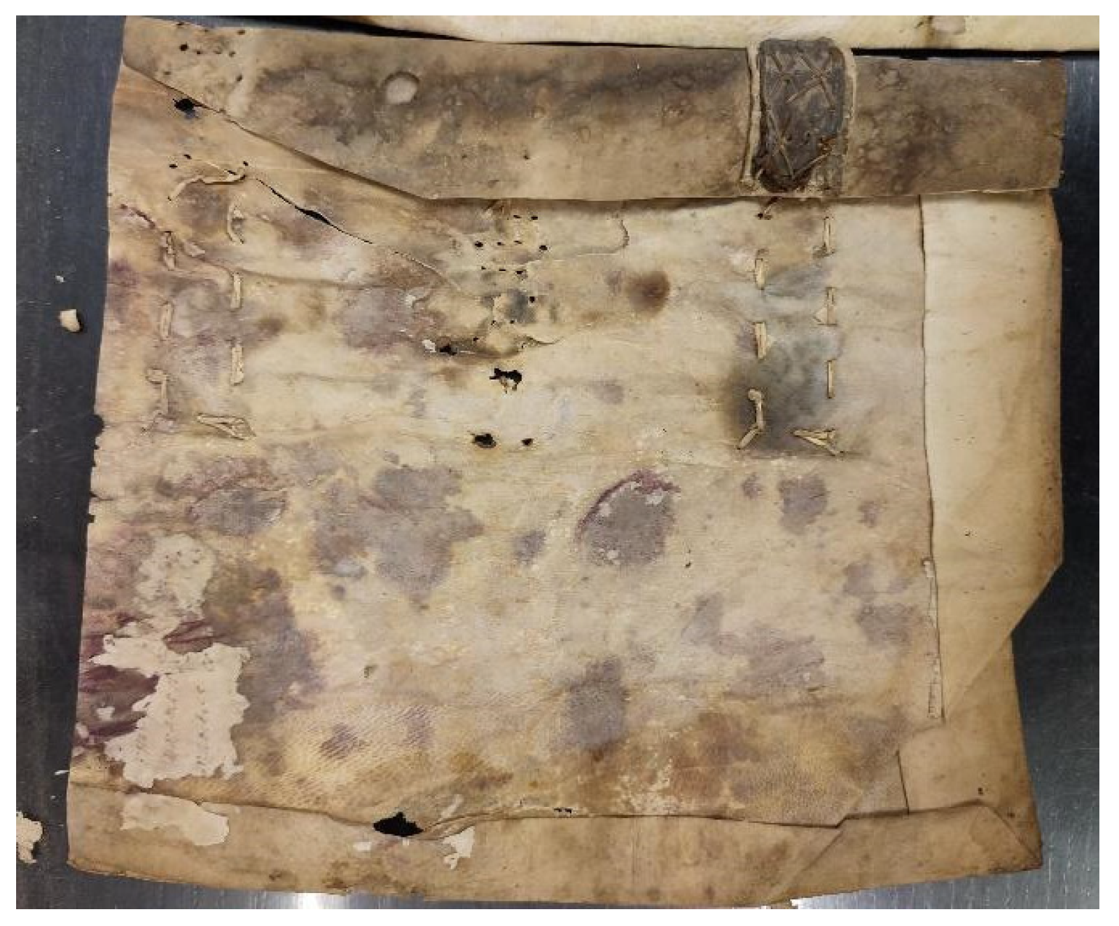
Figure 1. Portion of the discarded book cover of an archival registry from around the 16th century from which the samples were cut.
Heritage 06 00072 g001
Figure 2. (A) About 1 × 1 cm2 specimens used for microorganisms’ isolation and irradiation. (A) Parchment specimens before immersion in NB broth and vortexed, to isolate biodeteriogens. (B) Agar 1 × 1 cm2 parchment specimens derived from NB plates after 10 days of growth at 30 °C and composed by bacterial and fungal colonies. Agar portions were then cut and used for irradiation.
Heritage 06 00072 g002
Figure 3. Effect of irradiation on agar specimens. Bacterial and fungal colonies grown on 1 × 1 cm2 agar pieces were treated or not (UT: untreated) with different irradiation doses. Microorganisms survival was evaluated by CFU counting analysis. Statistical significance was performed through a one-way ANOVA analysis with the Bonferroni post-test (*** p < 0.001 with respect to UT).
Heritage 06 00072 g003
Figure 4. Td recorded by LTA for each sample before (black symbol) and after (red symbol) the irradiation treatment.
Heritage 06 00072 g004
Figure 5. FORS spectra for the parchment samples irradiated with a dose of 350 Gy, 1000 Gy and 4000 Gy with respect to the spectra of the non-irradiated samples (0 Gy).
Heritage 06 00072 g005
Figure 6. ATR-FTIR spectra in the Amide I and Amide II region of non-irradiated sample 0Gy (dotted line), of parchment irradiated with a dose of 1000 Gy (full line) and with a dose of 4000 Gy (dashed line).
Heritage 06 00072 g006
Figure 7. UVRR spectra of ancient (yellow) and modern (blue) parchment samples in the wavenumber range of 900–1900 cm−1. The difference spectrum (black is also reported for comparison).
Heritage 06 00072 g007
Figure 8. Young’s modulus of parchment samples at different irradiation doses.
Heritage 06 00072 g008
Table 1. FTIR data of the ratio between the intensities of the Amide I and Amide II bands as a function of X-rays dose.
X-rays Dose (Gy)IAmideI/IAmideII
Non-irradiated (0 Gy)3.0 ± 0.1
3502.8 ± 0.4
5002.1 ± 0.3
10001.0 ± 0.2
20001.9 ± 0.4
40001.8 ± 0.3
Disclaimer/Publisher’s Note: The statements, opinions and data contained in all publications are solely those of the individual author(s) and contributor(s) and not of MDPI and/or the editor(s). MDPI and/or the editor(s) disclaim responsibility for any injury to people or property resulting from any ideas, methods, instructions or products referred to in the content.