Identification of Lithol Red Synthetic Organic Pigment Reveals the Cause of Paint Layer Degradation on the Lazar Vozarević Painting “Untitled” with Copper Plates


Abstract
:1. Introduction
Painter and the Paintings
2. Materials and Methods
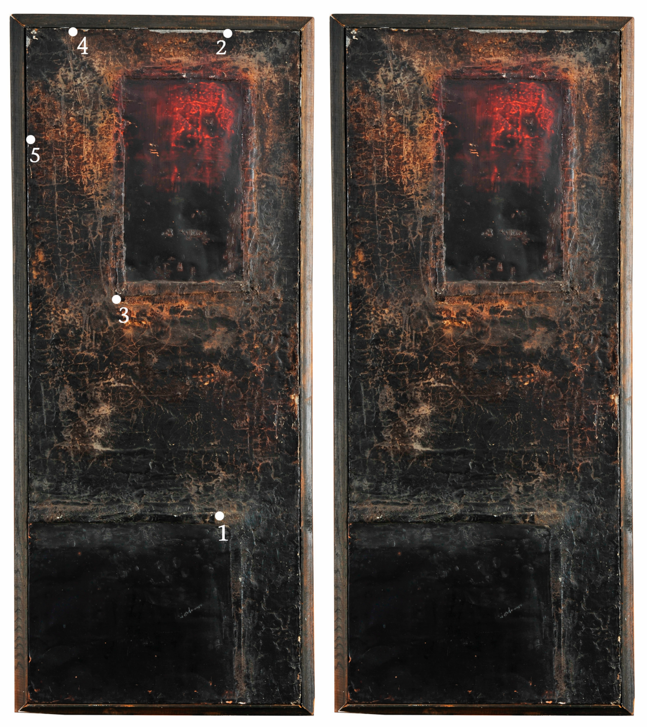
2.1. Sampling
2.2. Methods
3. Results and Discussion
Conservation and Restoration Treatment
4. Conclusions
Supplementary Materials
Author Contributions
Funding
Acknowledgments
Conflicts of Interest
References
- Vandenabeele, P.; Edwards, H.G.M.; Moens, L. A Decade of Raman Spectroscopy in Art and Archaeology. Chem. Rev. 2007, 107, 675–686. [Google Scholar] [CrossRef] [PubMed]
- Lee, A.S.; Otieno-Alego, V.; Creagh, D.C. Identification of iron-gall inks with near-infrared Raman microspectroscopy. J. Raman Spectrosc. 2008, 39, 1079–1084. [Google Scholar] [CrossRef]
- Baraldi, P.; Baraldi, C.; Curina, R.; Tassi, L.; Zannini, P. A micro-Raman archaeometric approach to Roman wall paintings. Vib. Spectrosc. 2007, 43, 420–426. [Google Scholar] [CrossRef]
- Stanzani, E.; Bersani, D.; Lottici, P.P.; Colomban, P. Analysis of artist’s palette on a 16th century wood panel painting by portable and laboratory Raman instruments. Vib. Spectrosc. 2016, 85, 62–70. [Google Scholar] [CrossRef]
- Trentelman, K.; Turner, N. Investigation of the painting materials and techniques of the late-15th century manuscript illuminator Jean Bourdichon. J. Raman Spectrosc. 2009, 40, 577–584. [Google Scholar] [CrossRef]
- Edwards, H.G.M.; Farwell, D.W. The conservational heritage of wall paintings and buildings: An FT-Raman spectroscopic study of prehistoric, Roman, mediaeval and Renaissance lime substrates and mortars. J. Raman Spectrosc. 2008, 39, 985–992. [Google Scholar] [CrossRef]
- Vandenabeele, P.; De Paepe, P.; Moens, L. Study of the 19th century porcelain cards with direct Raman analysis. J. Raman Spectrosc. 2008, 39, 1099–1103. [Google Scholar] [CrossRef]
- Goodall, R.A.; Hall, J.; Viel, R.; Agurcia, F.R.; Edwards, H.G.M.; Fredericks, P.M. Raman microscopic investigation of paint samples from the Rosalila building, Copan, Honduras. J. Raman Spectrosc. 2006, 37, 1072–1077. [Google Scholar] [CrossRef]
- Kokkori, M.; Hubert, M.-O.; Balcar, N.; Barabant, G.; Sutherland, K.; Casadio, F. Gloss paints in late paintings by Francis Picabia: A multi-analytical study. Appl. Phys. 2016, 122, 11. [Google Scholar] [CrossRef]
- Casadio, M.; Bezúr, A.; Fiedler, I.; Muir, K.; Trad, T.; Maccagnola, S. Pablo Picasso to Jasper Johns: A Raman study of cobalt-based synthetic inorganic pigments. J. Raman Spectrosc. 2012, 43, 1761–1771. [Google Scholar] [CrossRef]
- Ropret, P.; Centeno, S.A.; Bukovec, P. Raman identification of yellow synthetic organic pigments in modern and contemporary paintings: Reference spectra and case studies. Spectrochim. Acta Part A 2008, 69, 486–497. [Google Scholar] [CrossRef]
- Scherrer, N.C.; Zumbuehl, S.; Delavy, F.; Fritsch, A.; Kuehnen, R. Synthetic organic pigments of the 20th and 21st century relevant to artist’s paints: Raman spectra reference collection. Spectrochim. Acta Part A 2009, 73, 505–524. [Google Scholar] [CrossRef]
- Castro, K.; Perez-Alonso, M.; Rodrıguez-Laso, M.D.; Fernandez, L.A.; Madariaga, J.M. On-line FT-Raman and dispersive Raman spectra database of artists’ materials (e-VISART database). Anal Bioanal. Chem. 2005, 382, 248–258. [Google Scholar] [CrossRef] [PubMed]
- Colomban, P. The Destructive/Non-Destructive Identification of Enamelled Pottery, Glass Artefacts and Associated Pigments—A Brief Overview. Arts 2013, 2, 77–110. [Google Scholar] [CrossRef]
- Fremout, W.; Saverwyns, S. Identification of synthetic organic pigments: The role of a comprehensive digital Raman spectral library. J. Raman Spectrosc. 2012, 43, 1536–1544. [Google Scholar] [CrossRef]
- Sendova, M.; Kaiser, B.; Scalera, M.; Zhelyaskov, V. Della Robbia blue glaze: Micro-Raman temperature study and X-ray fluorescence spectroscopy characterization. J. Raman Spectrosc. 2010, 41, 469–472. [Google Scholar] [CrossRef]
- Burgio, L.; Melessanaki, K.; Doulgeridis, M.; Clark, R.J.H.; Anglos, D. Pigment identification in paintings employing laser induced breakdown spectroscopy and Raman microscopy. Spectrochim. Acta Part B 2001, 56, 905–913. [Google Scholar] [CrossRef]
- Merenik, L. Umetnost i Vlast. Srpsko Slikarstvo 1945–1968 [Art and Power. Serbian Painting 1945–1968]; Fond Vujicic: Belgrade, Serbian, 2010; p. 113. [Google Scholar]
- Trifunović, L. Od Impesionizma Do Enformela [From Impressionism to Art Informel]; Nolit: Belgrade, Serbian, 1982; p. 85. [Google Scholar]
- Stanojević, Đ. Slikanje Lazara Vozarevića Ili Traganje Za Identitetom [Lazara Vozarević’s Painting or Identity Search] Živopis 7; The Academy of the Serbian Orthodox Church for Arts and Conservation: Belgrade, Serbia, 2013; p. 179. [Google Scholar]
- Vandenabeele, P.; Moens, L.; Edwards HG, M.; Dams, R. Raman spectroscopic database of azo pigments and application to modern art studies. J. Raman Spectrosc. 2000, 31, 509–517. [Google Scholar] [CrossRef]
- Maquoi, M.-C.; Fremout, W. KIK IRPA SOP Spectral Library. 2010. Available online: https://soprano.kikirpa.be/index.php?lib=sop&id=PR49_1_A_785_kikirpa (accessed on 7 December 2019).
- Standeven, H.A.L. The History and Manufacture of Lithol Red, a Pigment Used by Mark Rothko in his Seagram and Harvard Murals of the 1950s and 1960s. Tate Pap. 2008, 10, 1–8. [Google Scholar]
- Živković, V.; Džikić, V. Return to basics—Environmental management for museum collections and historic houses. Energy Build. 2015, 95, 116–123. [Google Scholar] [CrossRef]
- Džikić, V.; Ćosic, N. Препоруке за обезбеђивање адекватних услова чувања фонда у депоу Галерије Лазар Возаревић у Сремској Митровици [Recommendations for providing adequate conditions for keeping the fund in the depot of the Lazar Vozarević Gallery in Sremska Mitrovica]. Unpublished report. 2013. [Google Scholar]
- Jovanović, V. The Conservation оf Petar Lubarda’s Painting Prisoner—Challenges and Results, Public paintings by Edvard Munch and his contemporaries. In Change and Conservation Challenges, Proceedings International Conference in Oslo; Frysaker, T., Ed.; Archetype Publications: London, UK, 2012; pp. 325–333. [Google Scholar]
- Roberts, C. 2009. Available online: http://carolineroberts.blogspot.com/2009/01/toxicity-of-pigments.html (accessed on 7 December 2019).
- Stenger, J.; Khandekar, N.; Raskar, R.; Cuellar, S.; Mohan, A.; Gschwind, R. Conservation of a room: A treatment proposal for Mark Rothko’s Harvard Murals. Stud. Conserv. 2016, 61, 348–361. [Google Scholar] [CrossRef]
- Stenger, J.; Kwan, E.E.; Eremin, K.; Speakman, S.; Kirby, D.; Stewart, H.; Huang, S.G.; Kennedy, A.R.; Newman, R.; Khandekar, N. Lithol red salts: Characterization and deterioration. E-Preserv. Sci. 2010, 7, 147–157. [Google Scholar]
- Janke, M.; Riedl, N. Missing piece of 16th-century mural recreated virtually. The Art Newspaper, 4 November 2010. [Google Scholar]
- Aleksic, M.; Jovanovic, V. Painting Restoration in Improved Reality. Communications in Computer and Information Science 904. In Proceedings of the 1st International Conference on VR Technologies in Cultural Heritage, Brasov, Romania, 29–30 May 2018; pp. 206–214. [Google Scholar]
- Culka, A.; Jehlička, J. Raman microspectrometric investigation of urea in calcite and gypsum powder matrices. J. Raman Spectrosc. 2010, 41, 1743–1747. [Google Scholar] [CrossRef]
- Baraldi, P.; Fagnano, C.; Bensi, P. Raman study of a «Tabula Colorum Physiologica» in a 1686 printed journal. J. Raman Spectrosc. 2006, 37, 1104–1110. [Google Scholar] [CrossRef]
- Bell, I.M.; Clark, R.J.H.; Gibbs, P.J. Raman spectroscopic library of natural and synthetic pigments (pre-~1850 AD). Spectrochim. Acta Part A 1997, 53, 2159–2179. [Google Scholar] [CrossRef]
- Benquerenca, M.-J.; Mendes, N.F.C.; Castelluci, E.; Gaspar, V.M.F.; Gil, F.P.S.C. Micro-Raman spectroscopy analysis of 16th century Portuguese Ferreirim Masters oil paintings. J. Raman Spectrosc. 2009, 40, 2135–2143. [Google Scholar] [CrossRef]
- Edwards, H.G.M.; Chalmers, J.M. Raman Spectroscopy in Archaeology and Art History. RSC Anal. Spectrosc. Monogr. 2005, 740, 192–206. [Google Scholar]
- Kaminska, A.M.; Sawczak, M.; Ouija, M.; Domingo, C.; Castillejo, M.; Sliwinski, G. Pigment identification of a XIV/XV c. wooden crucifix by means of the Raman spectroscopic technique. J. Raman Spectrosc. 2006, 37, 1125–1130. [Google Scholar] [CrossRef]
- Correia, A.M.; Clark, R.J.H.; Ribeiro, M.I.M.; Duarte, M.L.T.S. Pigment study by Raman microscopy of 23 paintings by the Portuguese artist Henrique Pousão (1859–1884). J. Raman Spectrosc. 2007, 38, 1390–1405. [Google Scholar] [CrossRef]
- Baroni, S. Restaurationet Conservation des Tableaux Manuel Pratique; Celiv: Paris, France, 1993. [Google Scholar]
- Burgio, L.; Clark, R.J.H. Library of FT-Raman spectra of pigments, minerals, pigment media and varnishes, and supplement to existing library of Raman spectra of pigments with visible excitation. Spectrochim. Acta Part A 2001, 57, 1491–1521. [Google Scholar] [CrossRef]
- Griffith, W.P. Raman Spectroscopy of Terrestrial Minerals, in Infrared and Raman Spectroscopy of Lunar and Terrestrial Minerals; Academic Press Inc.: Cambridge, MA, USA, 1975; pp. 299–323. [Google Scholar]
- Andrikopoulos, K.S.; Danillia, S.; Roussel, B.; Janssens, K. In vitro validation of a mobile Raman-XRF micro-analytical instrument’s capabilities on the diagnosis of Byzantine icons. J. Raman Spectrosc. 2006, 37, 1026–1034. [Google Scholar] [CrossRef]
- Schulte, F.; Brzezinka, K.-W.; Lutzenberger, K.; Stege, H.; Panne, U. Raman spectroscopy of synthetic organic pigments used in 20th century works of art. J. Raman Spectrosc. 2008, 39, 1455–1463. [Google Scholar] [CrossRef]
- Lafuente, B.; Downs, R.T.; Yang, H.; Stone, N. The power of databases: The RRUFF project. In Highlights in Mineralogical Crystallography; Armbruster, T., Danisi, R.M., Eds.; W. De Gruyter: Berlin, Germany, 2015; pp. 1–30. [Google Scholar]
- Froment, F.; Tournie, A.; Colomban, P. Raman identification of natural identification red to yellow pigments: Ochre and iron-containing ores. J. Raman Spectrosc. 2008, 39, 560–568. [Google Scholar] [CrossRef]
| 1 | It should be noted that painting on copper is an old technique that was mastered in Italy and the Low Countries during the 16th century. |
| 2 | MAIMERI® Restauro colours. |
| 3 | J.G. VIBERT retouching varnish (LEFRANC & BOURGEOIS) with acrylic and ketone resin quick drying petroleum. Contain the most resin with 25% dry extract after drying. |


| The Sample # | Photomicrograph | The Color | Image for Raman Analysis | RamanAnalysis | SEM/EDS Analysis | the Suggested Composition of the Sample |
|---|---|---|---|---|---|---|
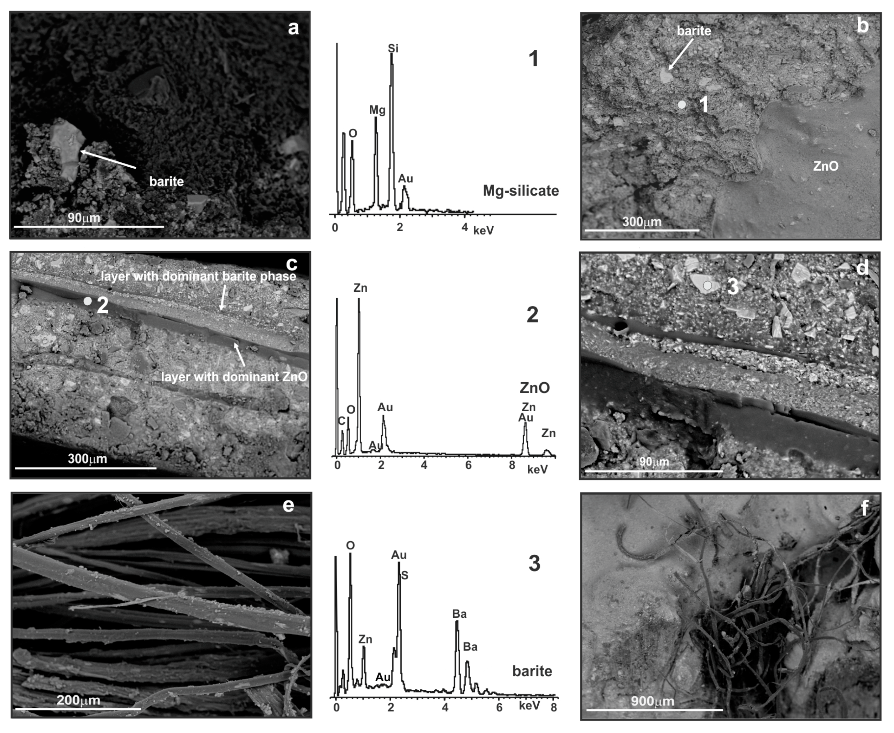
| 1 |  | white and black | 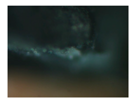 | ZnO, carbon black | BaSO4, ZnO, Mg-silicate | BaSO4, ZnO, carbon black, Mg-silicate |
| 2 | 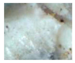 | white |  | CaCO3, BaSO4 | BaSO4, ZnO | CaCO3, BaSO4, ZnO, |
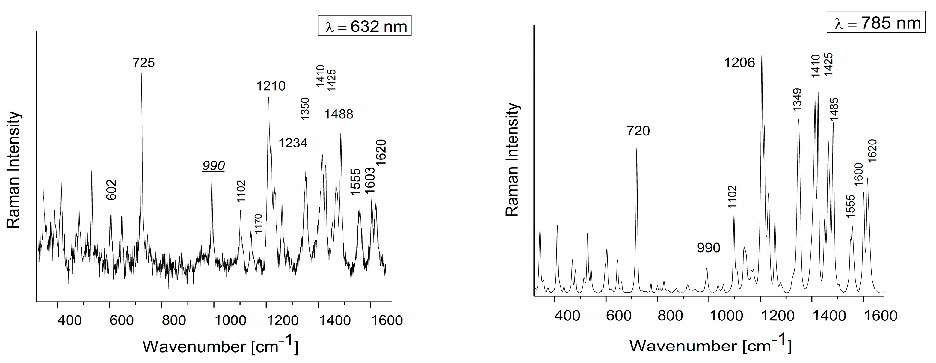
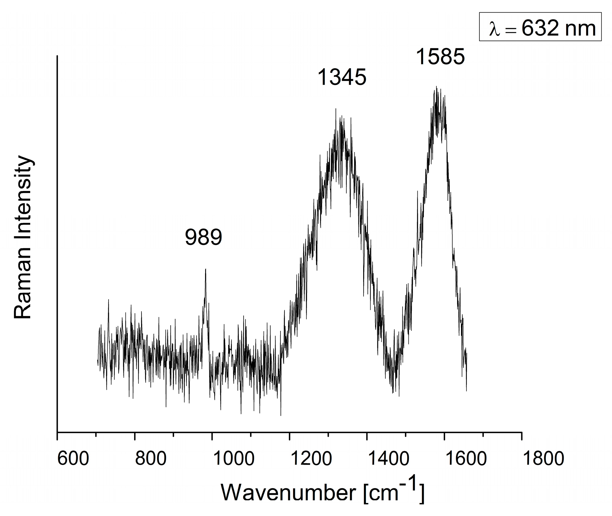
| 3 | 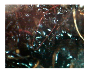 | red and black | 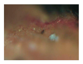 | Lithol red, BaSO4 | organic material, BaSO4 | Lithol red, BaSO4, |
| 4 | 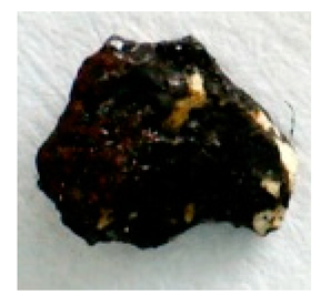 | red, black and white | 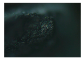 | BaSO4, carbon black | organic material, BaSO4 | BaSO4, carbon black |
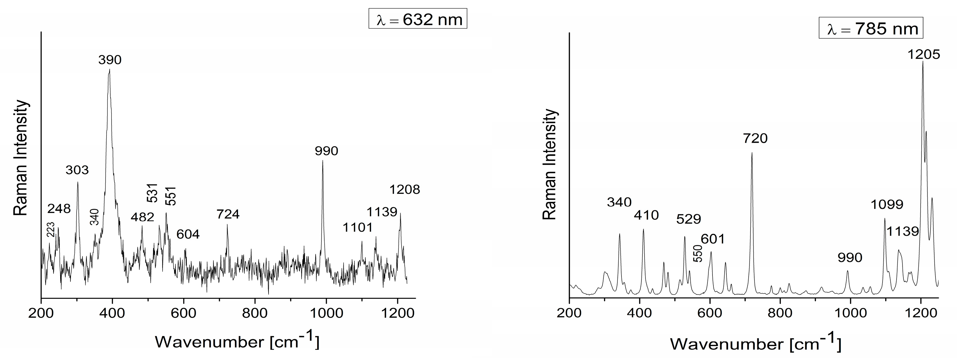
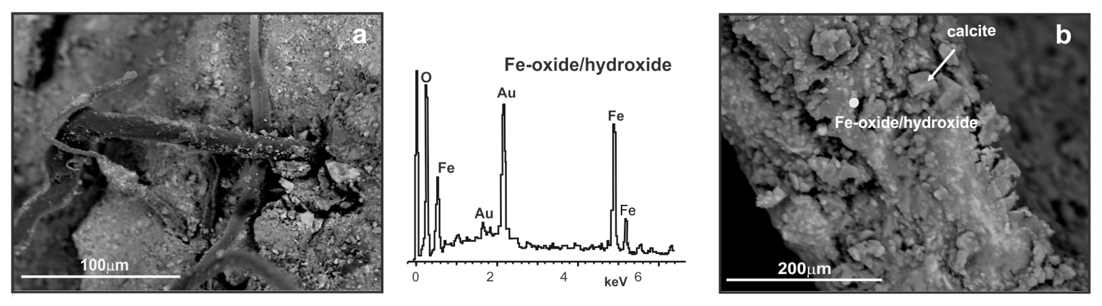
| 5 |  | light brownish red and black | 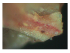 | Fe2O3, Goethite, BaSO4, Lithol red | Fe-oxide/ hydroxide, CaCO3, BaSO4 | Fe2O3, Goethite, CaCO3, BaSO4, Lithol red |
© 2019 by the author. Licensee MDPI, Basel, Switzerland. This article is an open access article distributed under the terms and conditions of the Creative Commons Attribution (CC BY) license (http://creativecommons.org/licenses/by/4.0/).
Share and Cite
Jovanović, V.; Erić, S.; Colomban, P.; Kremenović, A. Identification of Lithol Red Synthetic Organic Pigment Reveals the Cause of Paint Layer Degradation on the Lazar Vozarević Painting “Untitled” with Copper Plates. Heritage 2019, 2, 2612-2624. https://doi.org/10.3390/heritage2030160
Jovanović V, Erić S, Colomban P, Kremenović A. Identification of Lithol Red Synthetic Organic Pigment Reveals the Cause of Paint Layer Degradation on the Lazar Vozarević Painting “Untitled” with Copper Plates. Heritage. 2019; 2(3):2612-2624. https://doi.org/10.3390/heritage2030160
Chicago/Turabian StyleJovanović, Vanja, Suzana Erić, Philippe Colomban, and Aleksandar Kremenović. 2019. "Identification of Lithol Red Synthetic Organic Pigment Reveals the Cause of Paint Layer Degradation on the Lazar Vozarević Painting “Untitled” with Copper Plates" Heritage 2, no. 3: 2612-2624. https://doi.org/10.3390/heritage2030160
APA StyleJovanović, V., Erić, S., Colomban, P., & Kremenović, A. (2019). Identification of Lithol Red Synthetic Organic Pigment Reveals the Cause of Paint Layer Degradation on the Lazar Vozarević Painting “Untitled” with Copper Plates. Heritage, 2(3), 2612-2624. https://doi.org/10.3390/heritage2030160