Magic Lantern Glass Slides Materials and Techniques: The First Multi-Analytical Study

 , , and
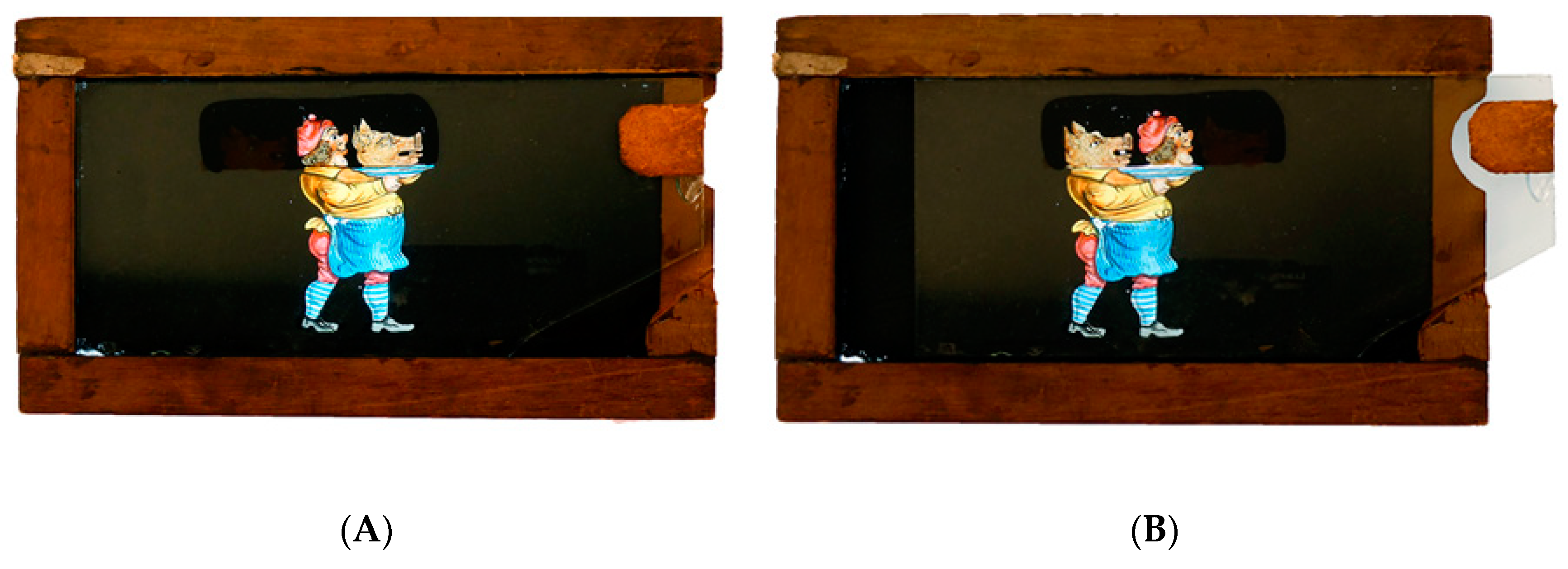
Abstract
1. Introduction
1.1. Historical Sources on Painting Techniques
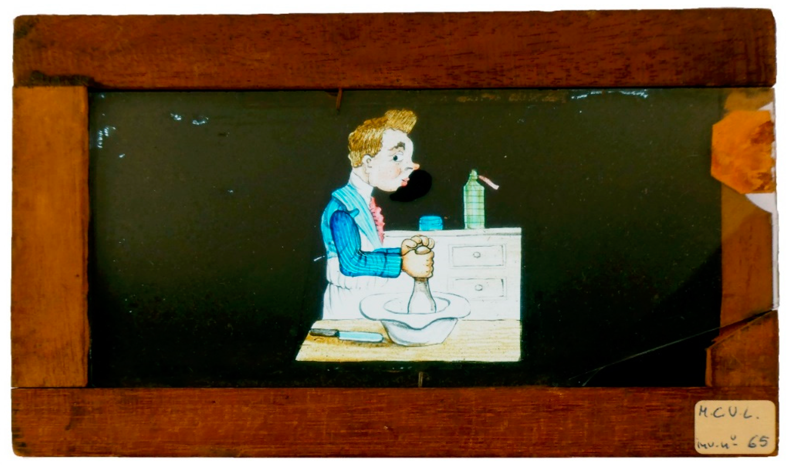
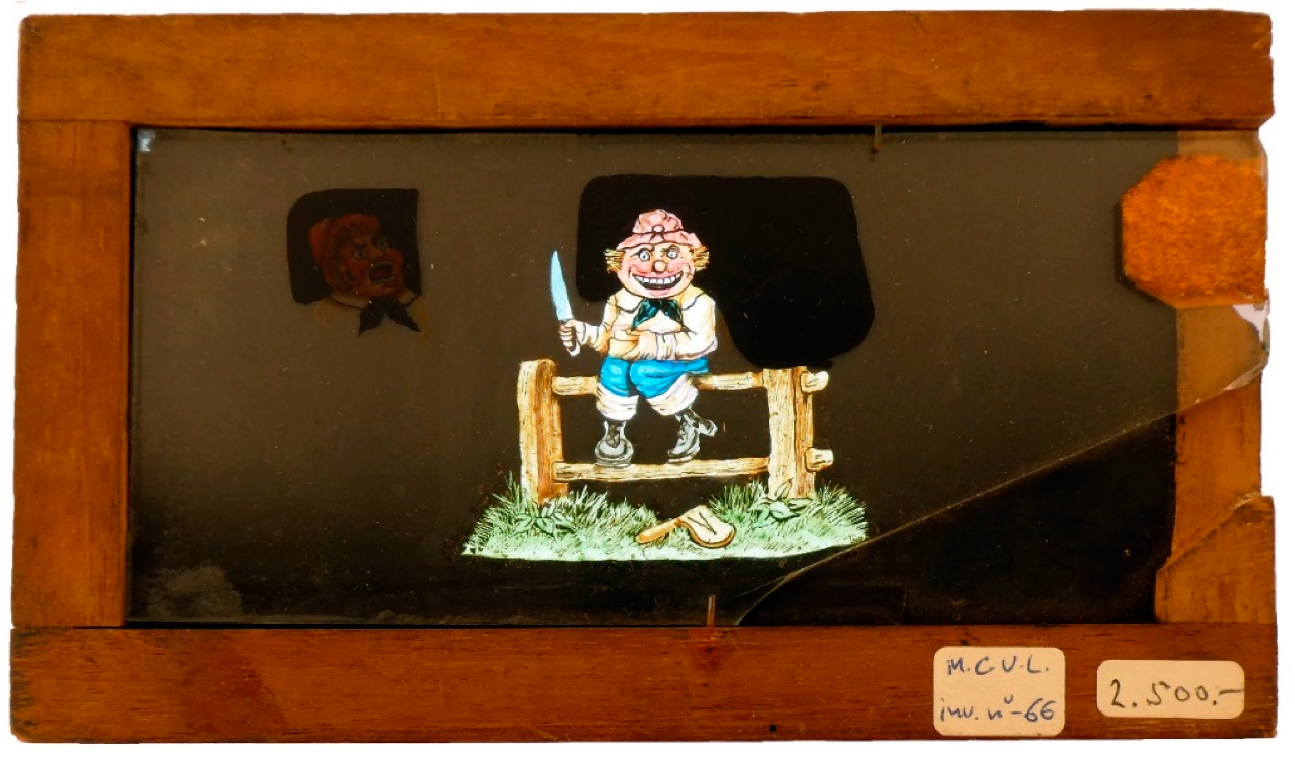
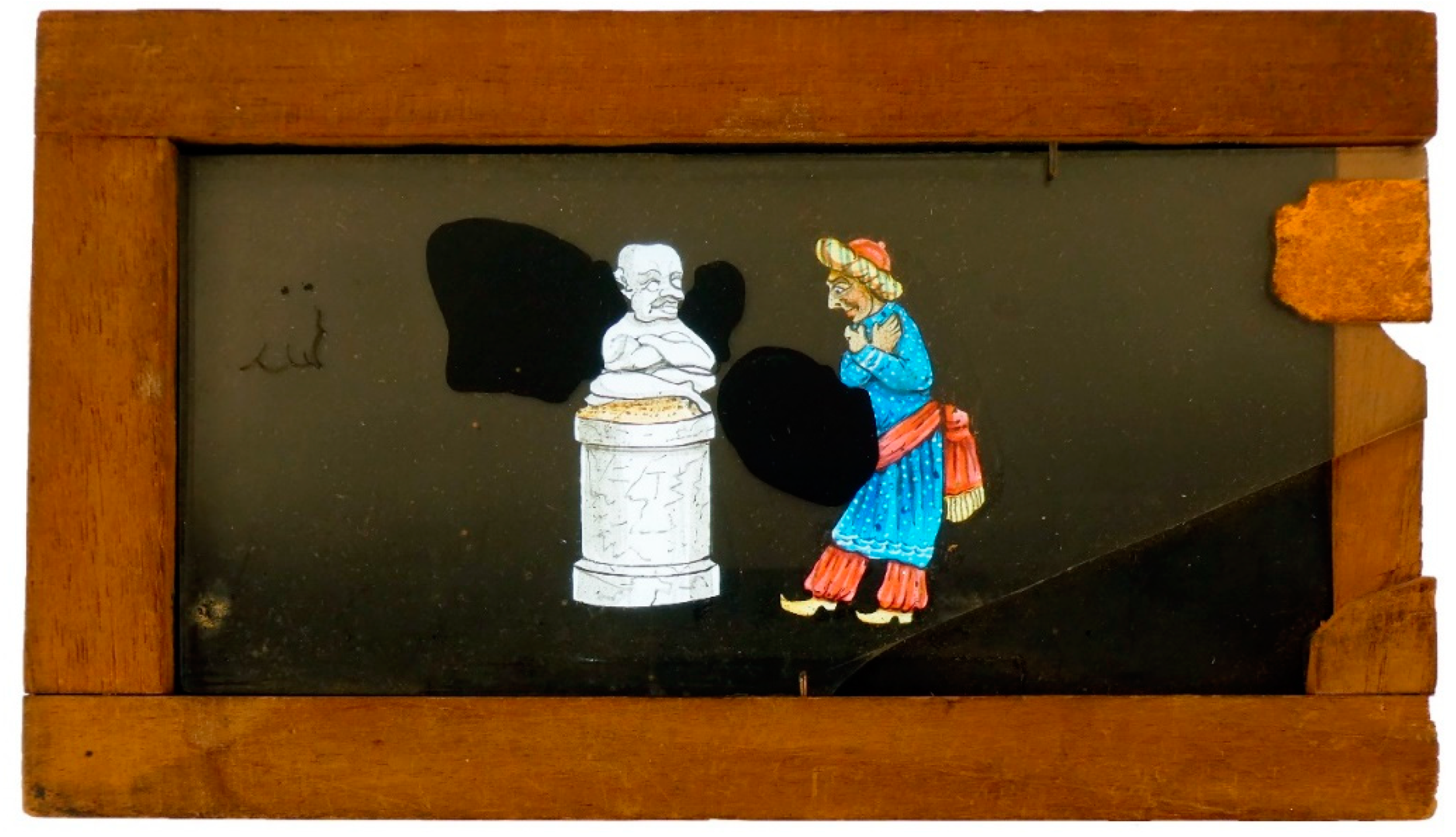
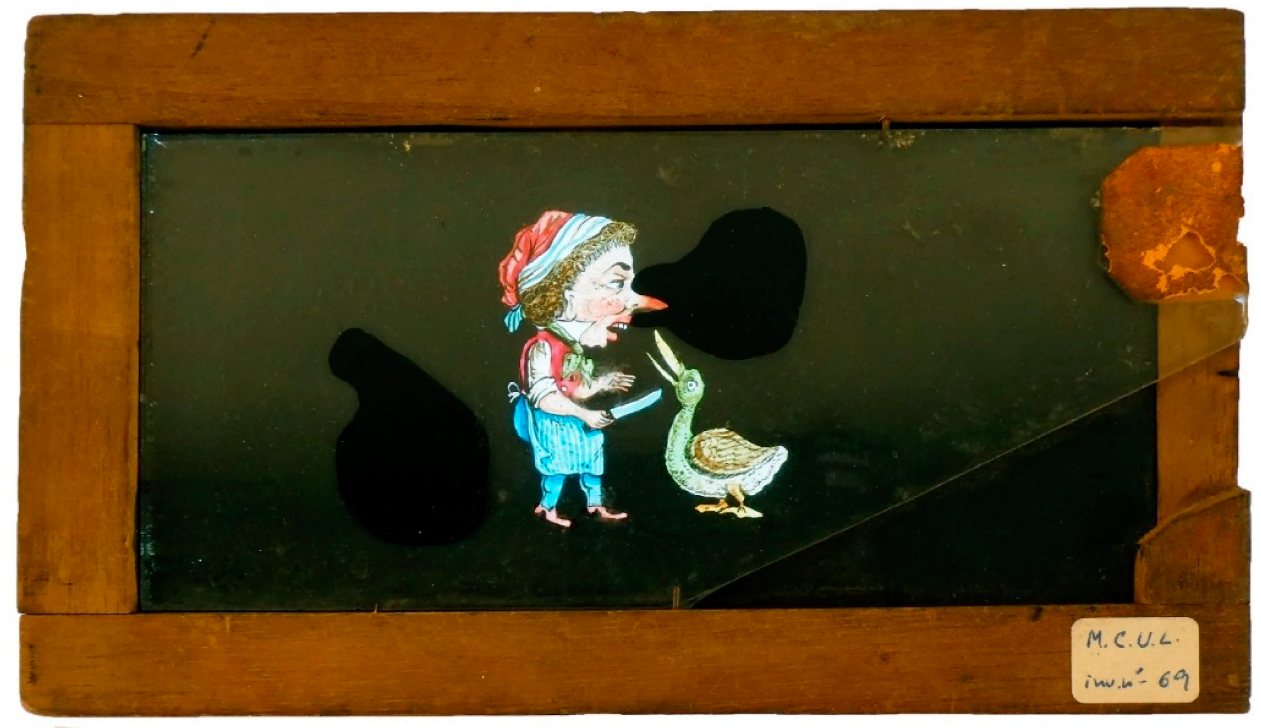
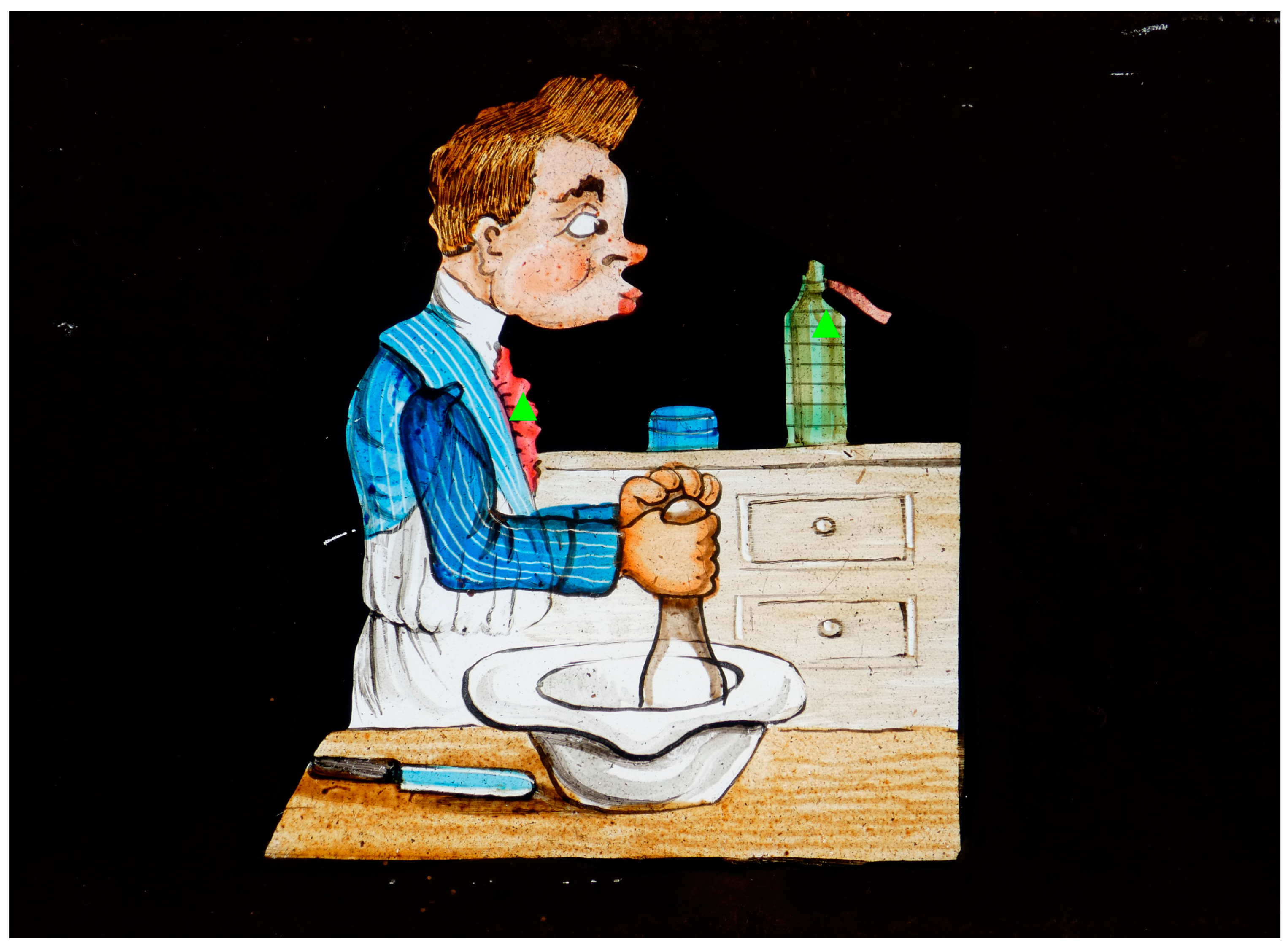
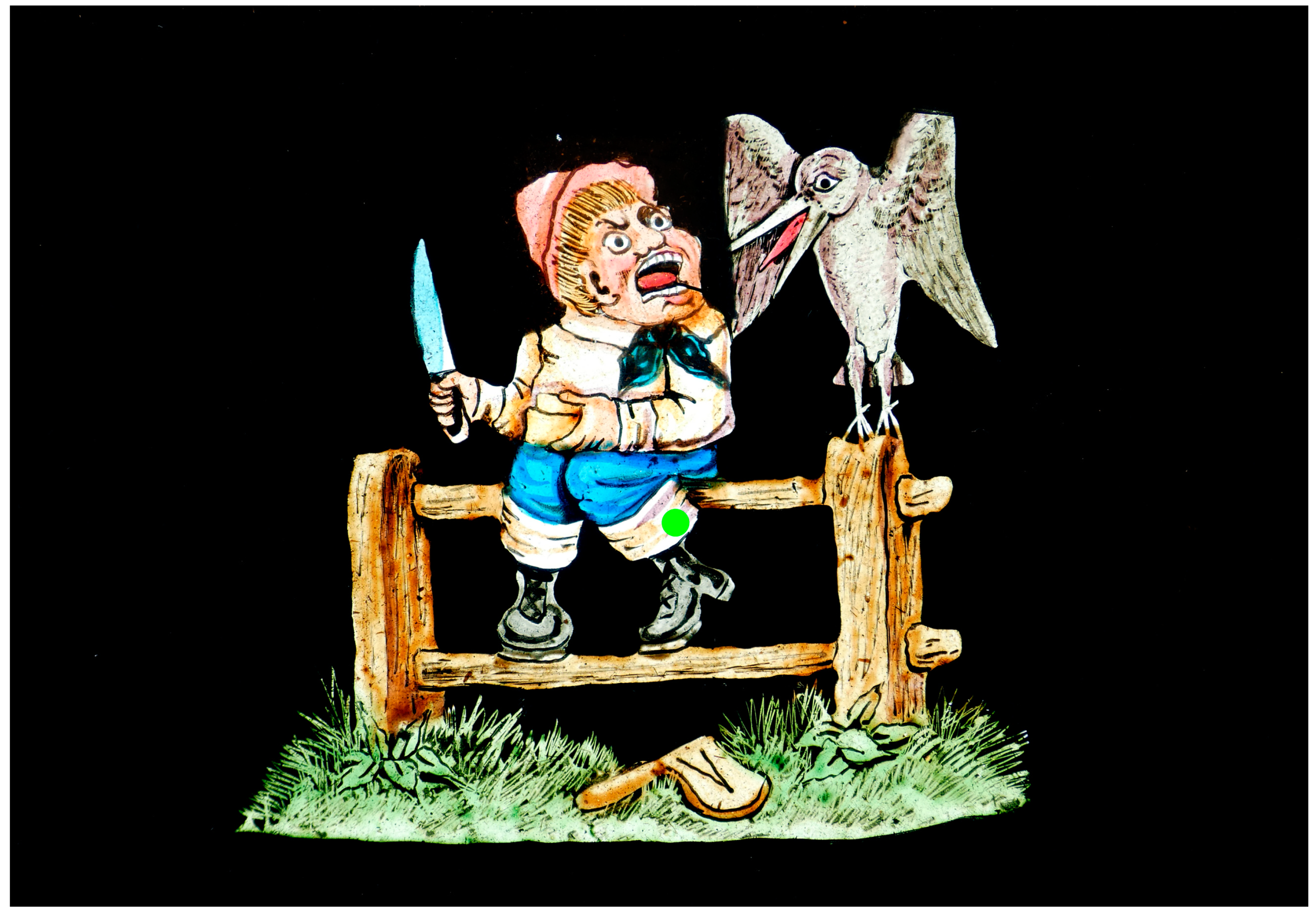
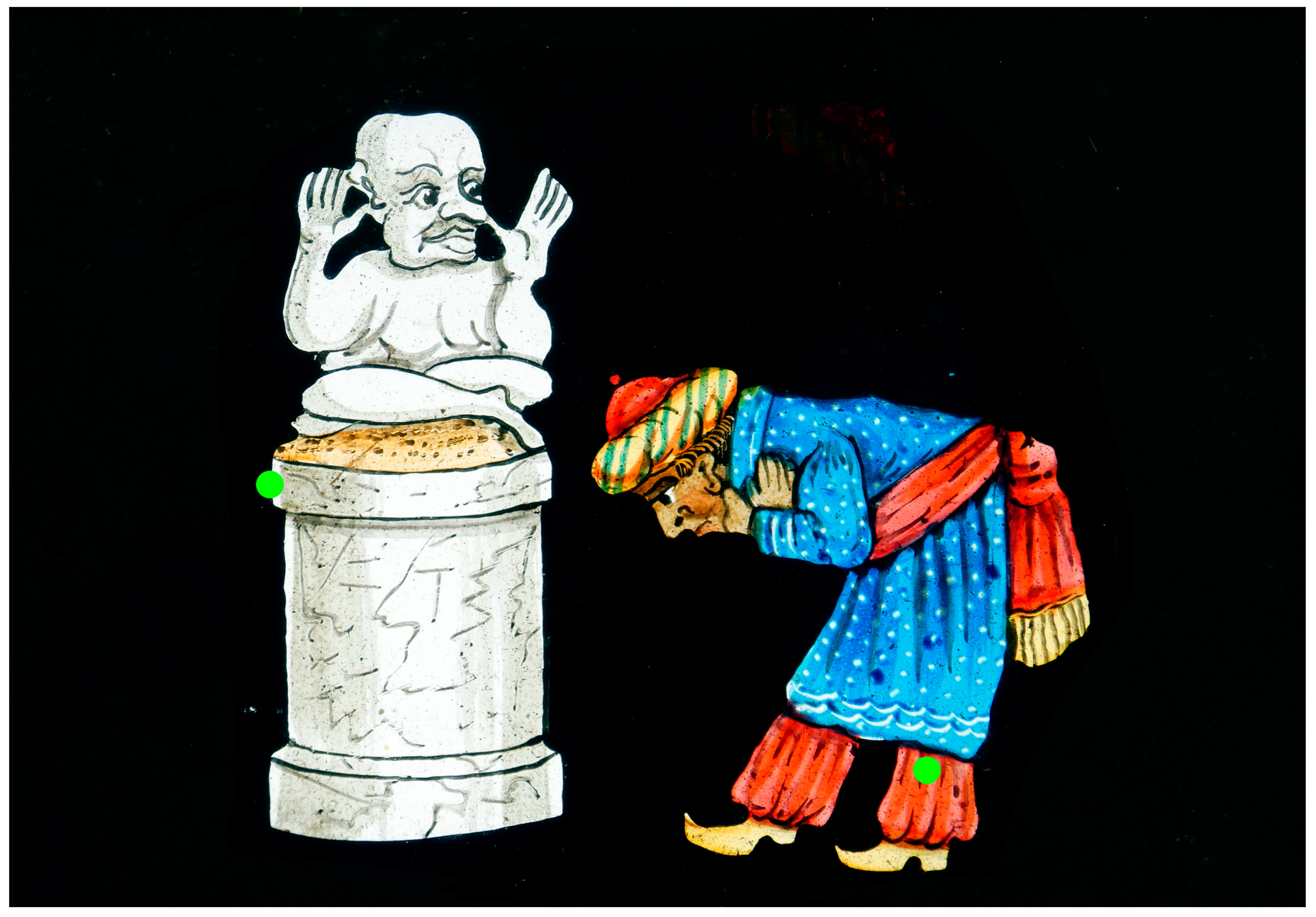
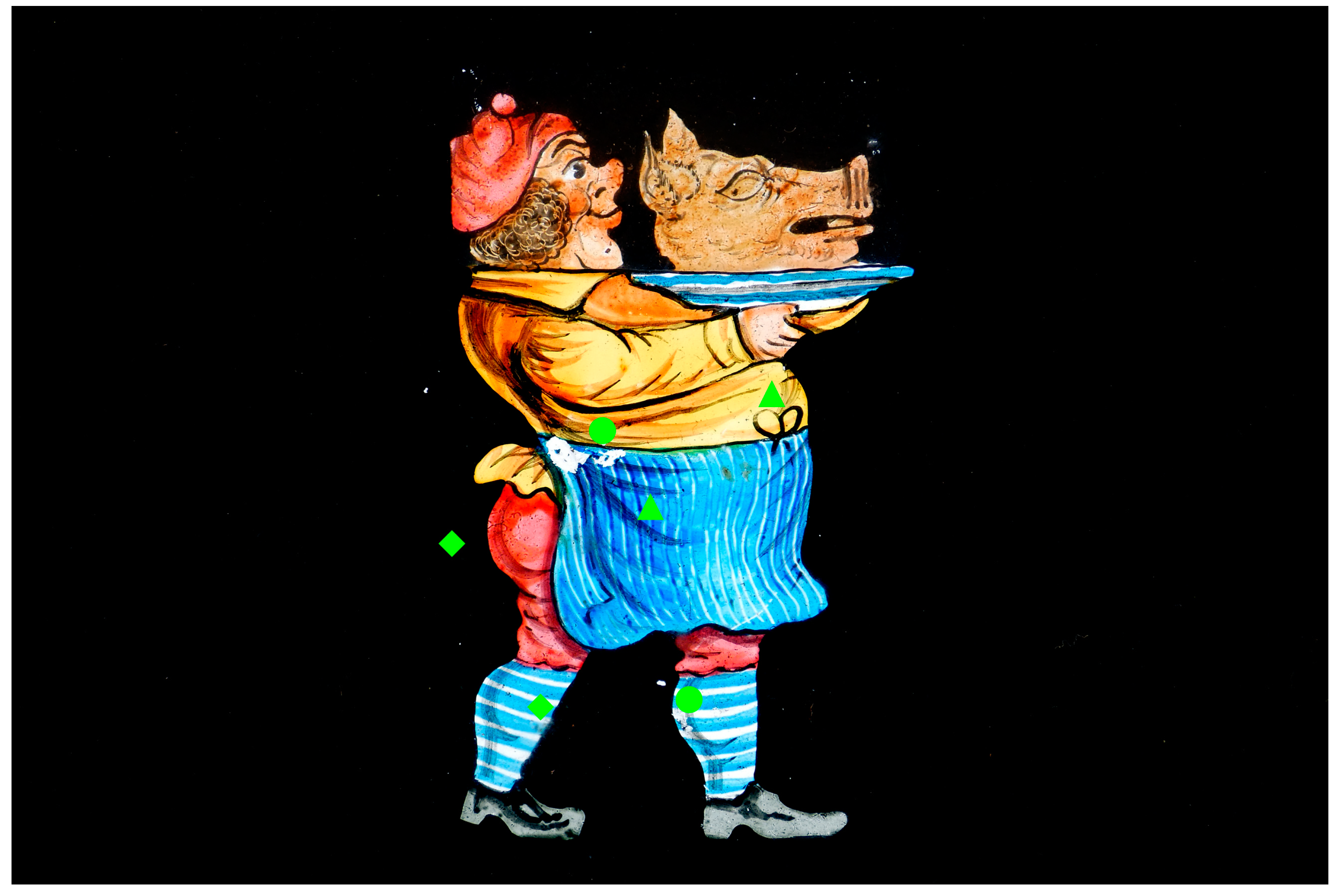
1.2. Case Study
2. Materials and Methods
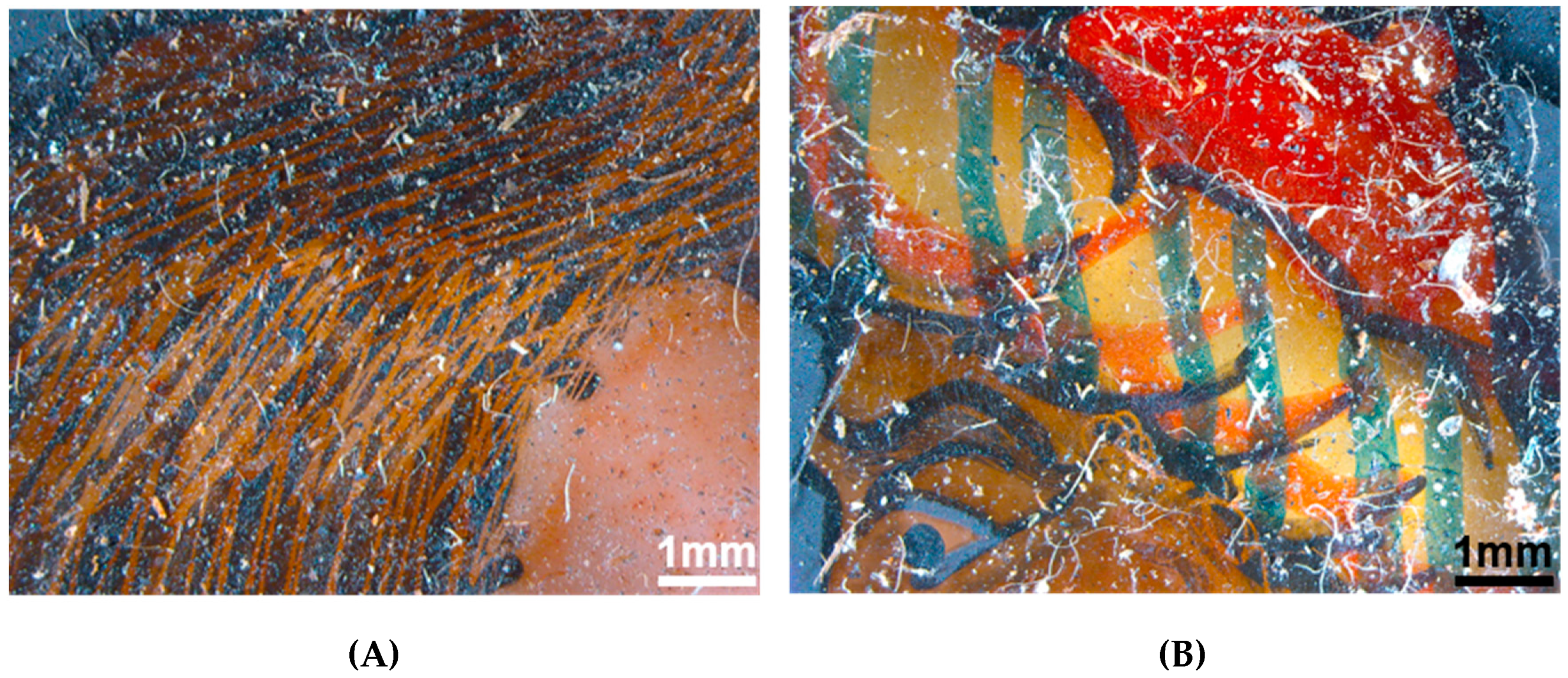
2.1. Stereomicroscope
2.2. Micro-Energy Dispersive X-Ray Fluorescence Spectrometry (EDXRF)
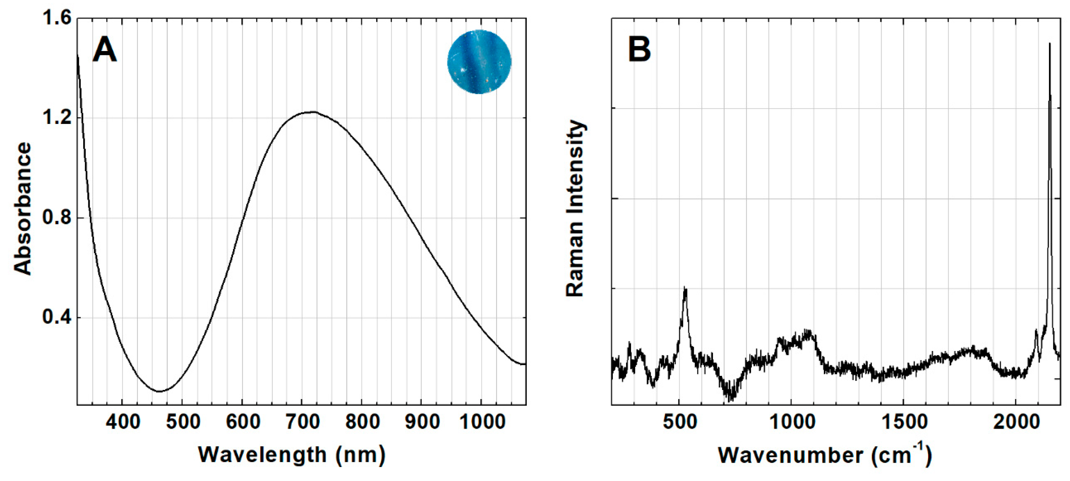
2.3. UV-Vis Spectroscopy (UV-Vis)
2.4. Micro-Raman Spectroscopy (µ-Raman)
2.5. Micro-Fourier Transform Infrared Spectroscopy (µ-FTIR)
3. Results and Discussion
3.1. Characterization of the Glass
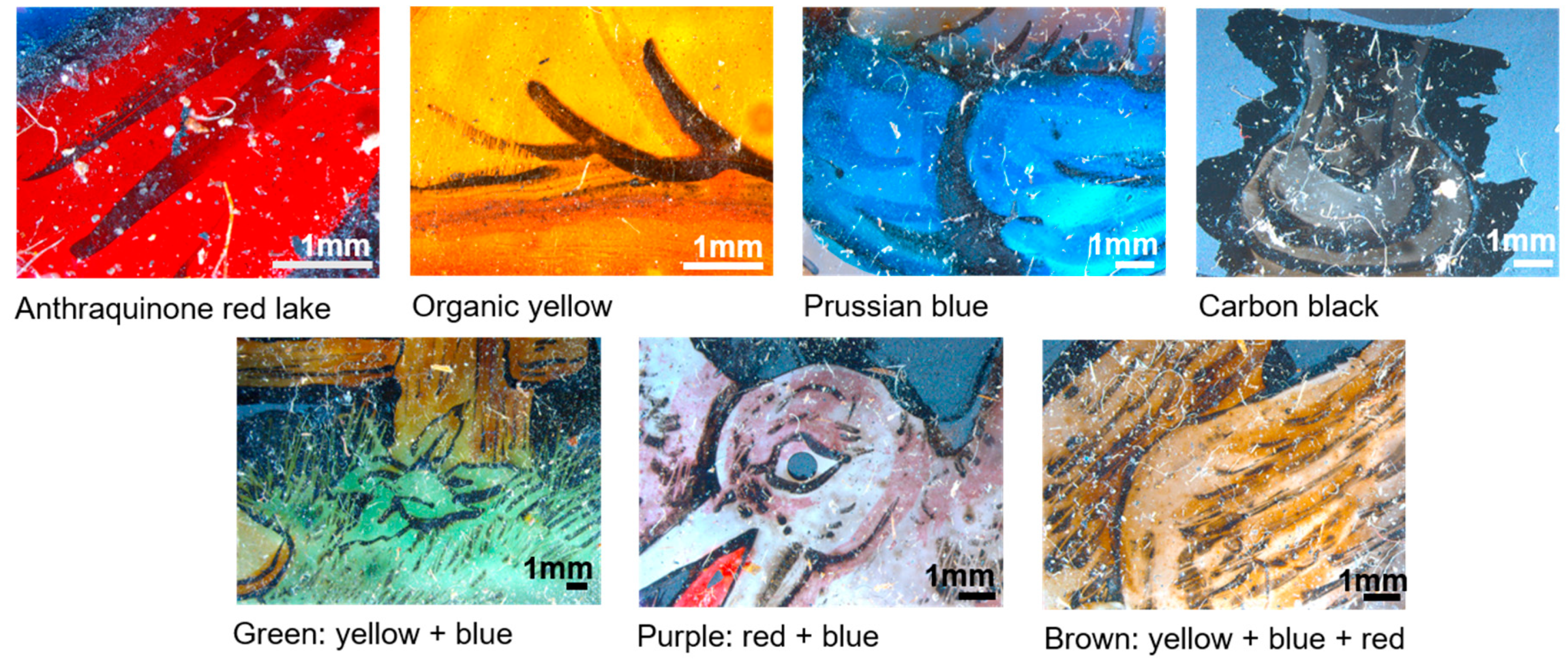
3.2. Identification of the Color Palette
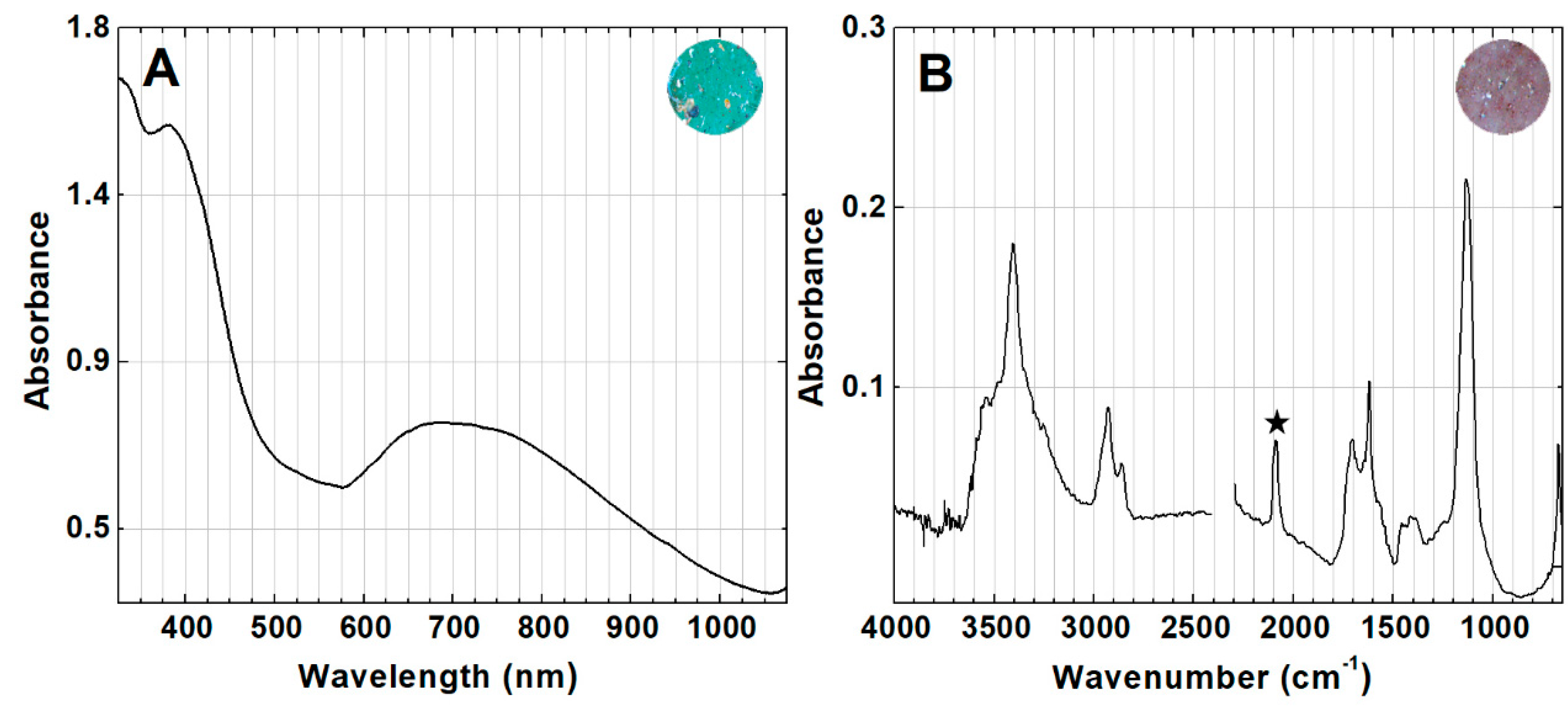
3.2.1. Blue Color
3.2.2. Red Color
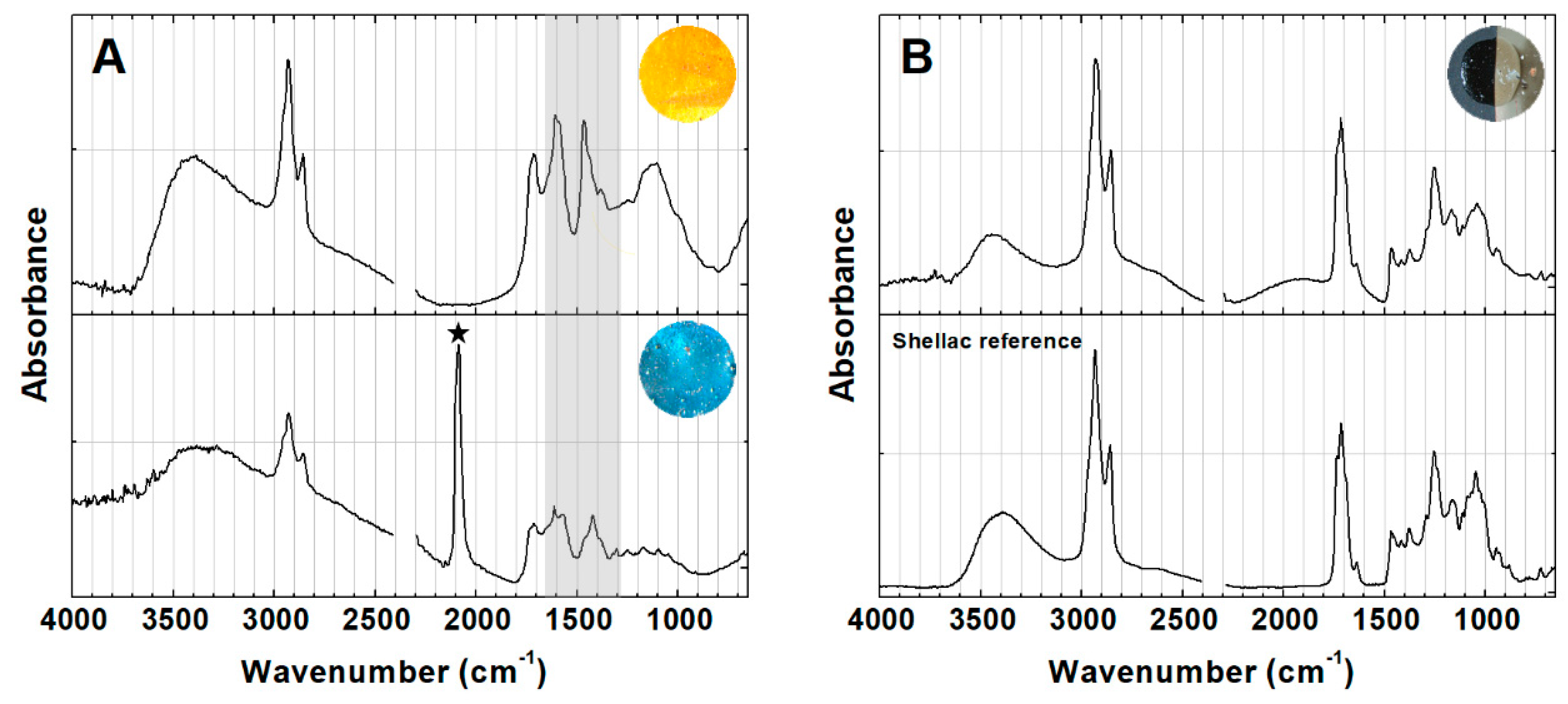
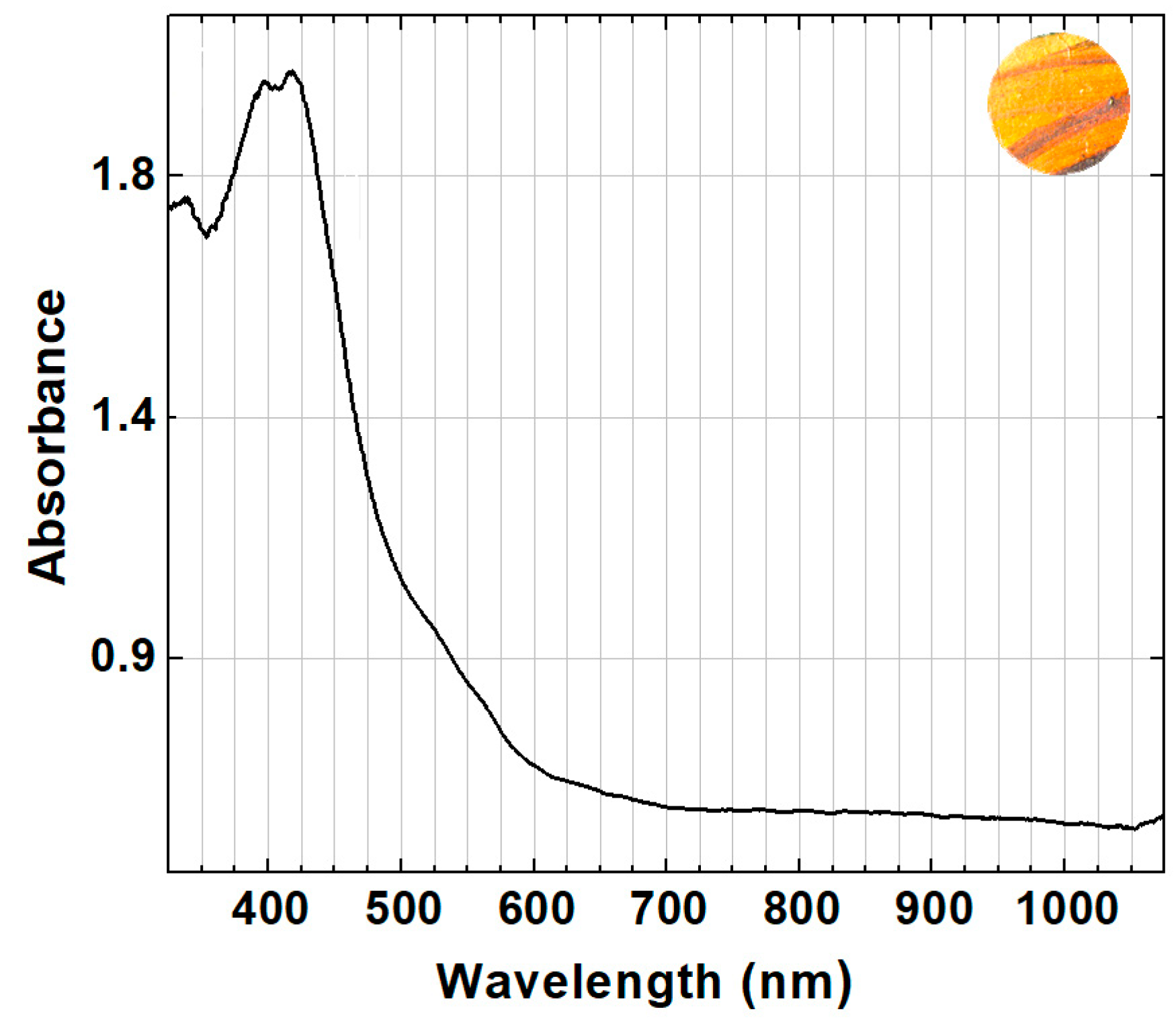
3.2.3. Yellow Color
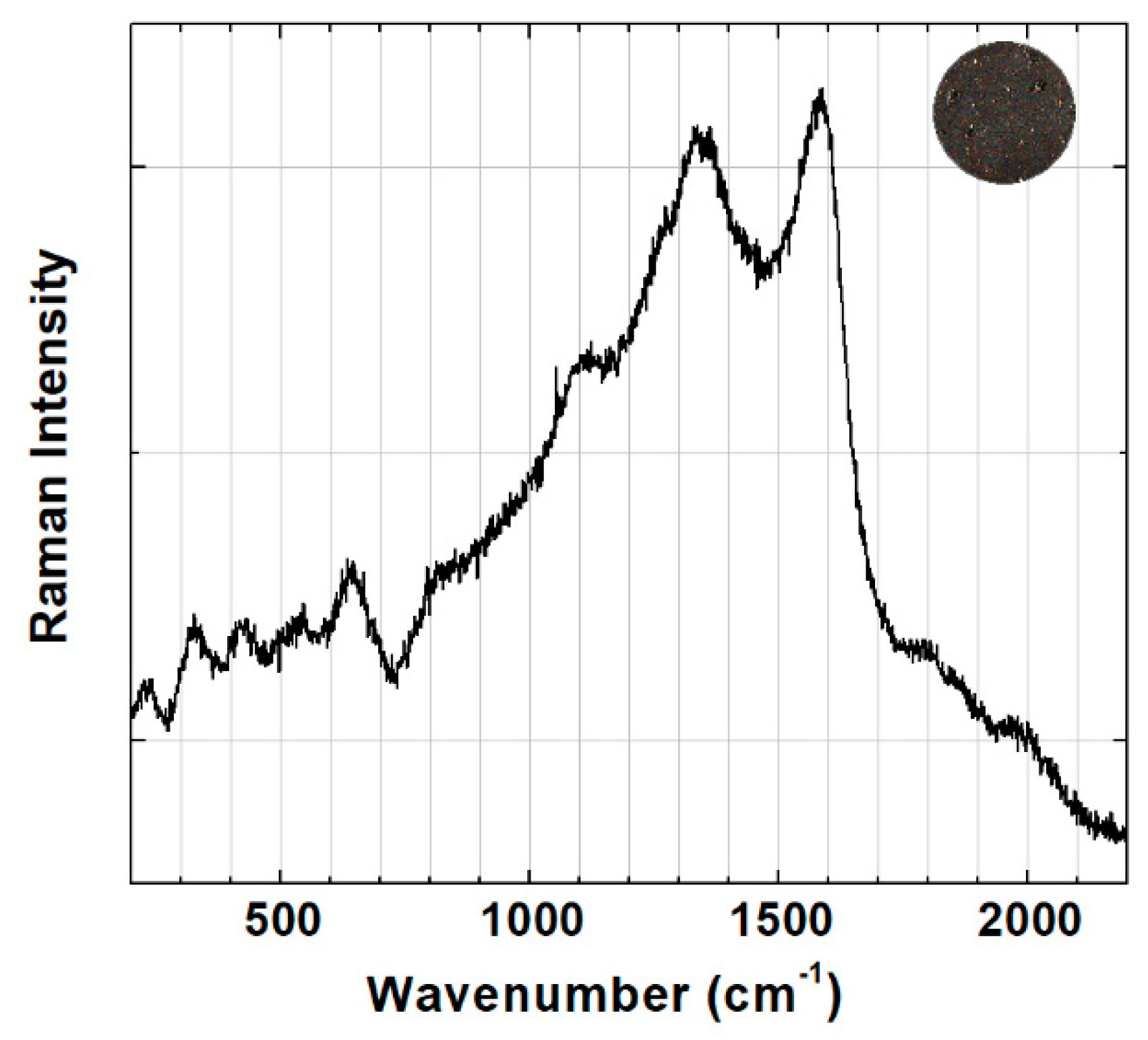
3.2.4. Black Color
3.2.5. Mixed Colors: Green, Purple and Brown
3.3. Identification of the Organic Media and Its State of Conservation
4. Conclusions
Author Contributions
Funding
Acknowledgments
Conflicts of Interest
Appendix A


References
- Frutos, F.J. Un público encantado. Las proyecciones audiovisuales mediante linterna mágica al servicio de la divulgación científica. Cult. Educ. 2009, 21, 305–318. [Google Scholar] [CrossRef]
- Balzer, R. Optical Amusements: Magic Lanterns and Other Transforming Images. A Catalog of Popular Entertainments; Museum of Our National Heritage: Watertown, MA, USA, 1987. [Google Scholar]
- Frutos, F.J. From Luminous Pictures to Transparent Photographs: The Evolution of Techniques for Making Magic Lantern Slides. Magic Lantern Gaz. 2013, 25, 3–11. [Google Scholar]
- Magic Lantern Slide Show; Tasmanian School of Art Gallery: Hobart, Australia, 1979.
- Campagnoni, D.P.; Bartetto, P. História da Lanterna Megalográphica vulgarmente dita Lanterna Mágica. In A Magia da Imagem—A Arqueologia do Cinema do Museu Nacional do Cinema de Turim; Cinemateca Portuguesa, Centro Cultural de Belém: Lisboa, Portugal, 1996. [Google Scholar]
- Heard, M. The lantern is not dead: Lanterns and slides in the 20th century and beyond. In Realms of Light: Uses and Perceptions of the Magic Lantern from the 17th to the 21st Century, Crangle, R., Heard, M., van Dooren, I., Eds.; Magic Lantern Society: London, UK, 2005; pp. 193–194. [Google Scholar]
- Mannoni, L.; Campagnoni, D.P. Lanterne Magique et Film Peint: 400 ans de Cinéma; Cinémathèque Française: Paris, France, 2009. [Google Scholar]
- Gray, F. Engaging with the Magic Lantern’s History. In Screen Culture and the Social Question, 1880–1914; Crangle, R., Vogl-Bienek, L., Eds.; Indiana University Press, John Libbey Publishing: Bloomington, IN, USA, 2014; pp. 173–181. [Google Scholar]
- Vogl-Bienek, L. Lichtspiele im Schatten der Armut: Historische Projektionskunst und Soziale Frage; Stroemfeld Verlag: Frankfurt, Germany, 2016. [Google Scholar]
- Scalarone, D.; Agostino, A.; Chiantore, O.; Basano, R. Vetri da proiezione dipinti per lanterne magiche: Analisi non invasive di leganti e pigmenti. In Proceedings of the IV Congresso Nazionale IGIIC—Lo Stato dell’Arte, Sienna, Italy, 28–30 September 2006. [Google Scholar]
- Ploeger, R.; Scalarone, D.; Chiantore, O. Non-invasive characterisation of binding media on painted glass magic lantern plates using mid-infrared fibre-optic reflectance spectroscopy. J. Cult. Herit. 2010, 11, 35–41. [Google Scholar] [CrossRef]
- Groom, E. The Art of Transparent Painting on Glass; Winsor & Newton: London, UK, 1855. [Google Scholar]
- Directions for the Graduation and Mixture of Colors, to Which Are Added Directions for Transparent Painting on Glass; M. J. Whipple & Co.: Boston, MA, USA, 1856.
- Hepworth, T.C. The Book of the Lantern. Being a Practical Guide to the Working of the Optical (or Magic) Lantern; Wyman & Sons: London, UK, 1888. [Google Scholar]
- Middleton, C. Magic Lantern Dissolving View Painting; Brodie & Middleton: London, UK, 1876. [Google Scholar]
- Hughes, W.C. The Art of Projection and Complete Magic Lantern Manual; E. A. Beckett: London, UK, 1893. [Google Scholar]
- Rintoul, A.N. Transparent Painting on Glass for the Magic Lantern, in Water, Oil, & Varnish colors; Brodie & Middleton: London, UK, 1867. [Google Scholar]
- Chadwick, W.I. The Magic Lantern Manual; Scovill Manufacturing Company: New York, NY, USA, 1886. [Google Scholar]
- Crompton, D.; Henry, D.; Herbert, S. Magic Images: The art of Hand-Painted and Photographic Lantern Slides; The Magic Lantern Society of Great Britain: London, UK, 1990. [Google Scholar]
- Brill, R.H. Chemical Analyses of Early Glasses. Volume 2: Tables of Analyses; The Corning Museum of Glass: New York, NY, USA, 1999; p. 544. [Google Scholar]
- Dungworth, D. Historic Window Glass. J. Archit. Conserv. 2012, 18, 7–25. [Google Scholar] [CrossRef]
- Boselli, L. Non-Invasive Spectroscopic Study of 19th Century Artists’ Materials. Ph.D. Thesis, Università degli Studi di Ferrara, Ferrara, Italy, 2010. [Google Scholar]
- Bacci, M.; Magrini, D.; Picollo, M.; Vervat, M. A study of the blue colors used by Telemaco Signorini (1835–1901). J. Cult. Herit. 2009, 10, 275–280. [Google Scholar] [CrossRef]
- Moretti, G.; Gervais, C. Raman spectroscopy of the photosensitive pigment Prussian blue. J. Raman Spectrosc. 2018, 49, 1198–1204. [Google Scholar] [CrossRef]
- Aceto, M.; Agostino, A.; Fenoglio, G.; Idone, A.; Gulmini, M.; Picollo, M.; Ricciardi, P.; Delaney, J.K. Characterisation of colourants on illuminated manuscripts by portable fibre optic UV-visible-NIR reflectance spectrophotometry. Anal. Methods 2014, 6, 1488–1500. [Google Scholar] [CrossRef]
- Vitorino, T.; Casini, A.; Cucci, C.; Melo, M.J.; Picollo, M.; Stefani, L. Non-invasive identification of traditional red lake pigments in fourteenth to sixteenth centuries paintings through the use of hyperspectral imaging technique. Appl. Phys. A Mater. Sci. Process. 2015, 121, 891–901. [Google Scholar] [CrossRef]
- Vitorino, T.; Otero, V.; Carlyle, L.; Melo, M.J.; Parola, A.J.; Picollo, M. Nineteenth-century cochineal lake pigments from Winsor & Newton: Insight into their methodology through reconstructions. In Proceedings of the ICOM-CC 18th Triennial Conference, Copenhagen, Denmark, 4–8 September 2017. [Google Scholar]
- Vitorino, T. Red Lake Pigments: From the Study of Their Chromophores and Historically Accurate References to a Comprehensive Hyperspectral Imaging Database. Ph.D. Thesis, Universidade Nova de Lisboa, Lisbon, Portugal. forthcoming.
- Bell, I.M.; Clark, R.H.; Gibbs, P.J. Raman spectroscopic library of natural and synthetic pigments (pre-∼ 1850 AD). Spectrochim. Acta Part A Mol. Biomol. Spectrosc. 1997, 53, 2159–2179. [Google Scholar] [CrossRef]
- Tomasini, E.P.; Halac, E.B.; Reinoso, M.; Di Liscia, E.J.; Maier, M.S. Micro-Raman spectroscopy of carbon-based black pigments. J. Raman Spectrosc. 2012, 43, 1671–1675. [Google Scholar] [CrossRef]
- Vetter, W.; Schreiner, M. Characterization of pigment-binding media systems comparison of non-invasive in-situ reflection FTIR with Transmission FTIR Microscopy. e-Preserv. Sci. 2011, 8, 10–22. [Google Scholar]
- Montagner, C.; Sanches, D.; Pedroso, J.; Melo, M.J.; Vilarigues, M. Ochres and earths: Matrix and chromophores characterization of 19th and 20th century artist materials. Spectrochim. Acta Part A Mol. Biomol. Spectrosc. 2013, 103, 409–416. [Google Scholar] [CrossRef] [PubMed]
- Sarkar, P.C.; Shrivastava, A.K. FTIR spectroscopy of lac resin and its derivatives FTIR spectroscopy of lac resin and its derivatives. Pigment Resin Technol. 1997, 26, 370–377. [Google Scholar] [CrossRef]
- Ortega-Avilés, M.; Vandenabeele, P.; Tenorio, D.; Murillo, G.; Jiménez-Reyes, M.; Gutiérrez, N. Spectroscopic investigation of a ‘Virgin of Sorrows’ canvas painting: A multi-method approach. Anal. Chim. Acta 2005, 550, 164–172. [Google Scholar] [CrossRef][Green Version]
- Tamburini, D.; Dyer, J.; Bonaduce, I. The characterisation of shellac resin by flow injection and liquid chromatography coupled with electrospray ionisation and mass spectrometry. Sci. Rep. 2017, 7, 14784. [Google Scholar] [CrossRef] [PubMed]
- Sutherland, K. Bleached shellac picture varnishes: Characterization and case studies. J. Inst. Conserv. 2010, 33, 129–145. [Google Scholar] [CrossRef]
- Carlyle, L. The Artist’s Assistant: Oil Painting Instruction Manuals and Handbooks. In Britain 1800–1900 with Reference to Selected Eighteenth-Century Sources; Archetype Publications: London, UK, 2001. [Google Scholar]
- Otero, V.; Sanches, D.; Montagner, C.; Vilarigues, M.; Carlyle, L.; Lopes, J.A.; Melo, M.J. Characterisation of metal carboxylates by Raman and infrared spectroscopy in works of art. J. Raman Spectrosc. 2014, 45, 1197–1206. [Google Scholar] [CrossRef]
- Monico, L.; Rosi, F.; Miliani, C.; Daveri, A.; Brunetti, B.G. Non-invasive identification of metal-oxalate complexes on polychrome artwork surfaces by reflection mid-infrared spectroscopy. Spectrochim. Acta Part A Mol. Biomol. Spectrosc. 2013, 116, 270–280. [Google Scholar] [CrossRef]
- Poli, T.; Piccirillo, A.; Zoccali, A.; Conti, C.; Nervo, M.; Chiantore, O. The role of zinc white pigment on the degradation of shellac resin in artworks. Polym. Degrad. Stab. 2014, 102, 138–144. [Google Scholar] [CrossRef]
- Doménech-Carbó, M.T.; Kuckova, S.; de la Cruz-Cañizares, J.; Osete-Cortina, L. Study of the influencing effect of pigments on the photoageing of terpenoid resins used as pictorial media. J. Chromatogr. A 2006, 1121, 248–258. [Google Scholar] [CrossRef]
- Poli, T.; Piccirillo, A.; Nervo, M.; Chiantore, O. Interactions of natural resins and pigments in works of art. J. Colloid Interface Sci. 2017, 503, 1–9. [Google Scholar] [CrossRef] [PubMed]


© 2019 by the authors. Licensee MDPI, Basel, Switzerland. This article is an open access article distributed under the terms and conditions of the Creative Commons Attribution (CC BY) license (http://creativecommons.org/licenses/by/4.0/).
Share and Cite
Rodrigues, B.; Santos, Â.; Melo, M.J.; Otero, V.; Vilarigues, M. Magic Lantern Glass Slides Materials and Techniques: The First Multi-Analytical Study. Heritage 2019, 2, 2513-2530. https://doi.org/10.3390/heritage2030154
Rodrigues B, Santos Â, Melo MJ, Otero V, Vilarigues M. Magic Lantern Glass Slides Materials and Techniques: The First Multi-Analytical Study. Heritage. 2019; 2(3):2513-2530. https://doi.org/10.3390/heritage2030154
Chicago/Turabian StyleRodrigues, Beatriz, Ângela Santos, Maria J. Melo, Vanessa Otero, and Márcia Vilarigues. 2019. "Magic Lantern Glass Slides Materials and Techniques: The First Multi-Analytical Study" Heritage 2, no. 3: 2513-2530. https://doi.org/10.3390/heritage2030154
APA StyleRodrigues, B., Santos, Â., Melo, M. J., Otero, V., & Vilarigues, M. (2019). Magic Lantern Glass Slides Materials and Techniques: The First Multi-Analytical Study. Heritage, 2(3), 2513-2530. https://doi.org/10.3390/heritage2030154