Review

Experimental Fish Models in the Post-Genomic Era: Tools for Multidisciplinary Science

by
Camila Carlino-Costa
1 and
Marco Antonio de Andrade Belo
2,*
1
Department of One Health, Sao Paulo State University (UNESP), Jaboticabal 14884-900, SP, Brazil
2
Laboratory of Animal Pharmacology and Toxicology, Brazil University, Descalvado 13690-000, SP, Brazil
*
Author to whom correspondence should be addressed.
Submission received: 16 August 2025 / Revised: 16 September 2025 / Accepted: 24 September 2025 / Published: 2 October 2025
(This article belongs to the Special Issue Feature Papers of J—Multidisciplinary Scientific Journal in 2025)

Abstract

Fish have become increasingly prominent as experimental models due to their unique capacity to bridge basic biological research with translational applications across diverse scientific disciplines. Their biological traits, such as external fertilization, high fecundity, rapid embryonic development, and optical transparency, facilitate in vivo experimentation and real-time observation, making them ideal for integrative research. Species like zebrafish (Danio rerio) and medaka (Oryzias latipes) have been extensively validated in genetics, toxicology, neuroscience, immunology, and pharmacology, offering robust platforms for modeling human diseases, screening therapeutic compounds, and evaluating environmental risks. This review explores the multidisciplinary utility of fish models, emphasizing their role in connecting molecular mechanisms to clinical and environmental outcomes. We address the main species used, highlight their methodological advantages, and discuss the regulatory and ethical frameworks guiding their use. Additionally, we examine current limitations and future directions, particularly the incorporation of high-throughput omics approaches and real-time imaging technologies. The growing scientific relevance of fish models reinforces their strategic value in advancing cross-disciplinary knowledge and fostering innovation in translational science.

1. Introduction

Experimental models are essential tools for investigating biological processes, elucidating disease mechanisms, and developing therapeutic interventions under controlled conditions [1]. They allow researchers to observe, manipulate, and reproduce complex phenomena that would be unfeasible or ethically impractical in humans, thus accelerating discoveries and contributing to both human and veterinary medicine [1,2,3].
Among the diverse organisms used in research, fish have gained prominence due to a unique combination of biological and practical attributes [4]. Their remarkable biodiversity, shaped by adaptation to a wide variety of ecological niches, offers unparalleled genetic and physiological variability [5,6]. This diversity enables studies ranging from embryonic development and organ physiology to immune responses, behavior, and evolutionary adaptations across multiple contexts [7,8].
Well-established models such as zebrafish (Danio rerio) and medaka (Oryzias latipes) share a high degree of genetic, anatomical, and physiological similarity with mammals, supporting translational research on human diseases and drug testing [6]. Features such as prolific reproduction, rapid external development, and optical transparency of embryos facilitate real-time, non-invasive observation of processes like organogenesis, disease progression, and pharmacological effects [9,10,11,12].
The post-genomic era has transformed the potential of these models. Genome sequencing and the development of multi-omics approaches, combined with gene-editing tools such as CRISPR/Cas9 (Clustered Regularly Interspaced Short Palindromic Repeats/CRISPR-associated protein 9), have expanded the capacity to link molecular changes to phenotypic outcomes, identify conserved biological pathways, and generate precise models of human pathologies [6,13,14,15]. Integrating these molecular tools with real-time imaging technologies further enhances the resolution and depth of biological insights [16,17,18,19,20].
Despite these advantages, direct extrapolation to mammals requires caution due to physiological, immunological, and anatomical differences [21]. Rather than diminishing their relevance, these distinctions highlight the complementary role of fish in reducing mammalian use, enabling high-throughput experimentation, and generating hypotheses for validation in more complex systems [9,22].
This review critically examines the use of fish as experimental models in the post-genomic era, discussing the main species employed, their advantages and limitations, multidisciplinary applications, from biomedicine to ecotoxicology, and the ethical frameworks guiding their use (Figure 1). By identifying research gaps and future perspectives, it aims to support the strategic, integrated, and sustainable application of fish models in contemporary science.

2. Main Species Used

The choice of fish species as experimental models is a critical decision guided by the specific research objectives, the biological attributes of the species, and practical considerations related to laboratory maintenance and experimental feasibility [23,24,25]. Due to their immense biodiversity and evolutionary adaptations, fish encompass a wide range of physiological, genetic, and ecological traits, offering versatile platforms for investigating fundamental biological processes and applied biomedical or environmental questions [26].
Some species have become standard-bearers in experimental research owing to their unique features, well-characterized genomes, and the availability of advanced genetic tools. These models serve as powerful systems for dissecting developmental mechanisms, disease pathways, toxicological responses, and environmental adaptations [6,27]. Additionally, the use of native or regional species has gained traction in recent years, complementing traditional models by providing ecologically relevant insights into local environmental challenges and biodiversity conservation [28,29].
Several fish species have gained prominence as experimental models due to their distinctive biological traits and suitability for diverse research applications. These species offer complementary advantages that enable investigations ranging from basic developmental biology to applied biomedical and environmental sciences [3,7]. Among the most widely utilized are zebrafish and medaka, whose well-characterized genomes and genetic tools have made them indispensable in translational research [4,9]. Additionally, freshwater species such as Nile tilapia (Oreochromis niloticus) and common carp (Cyprinus carpio) are valued for studies in physiology, nutrition, and ecotoxicology, owing to their ecological relevance and adaptability [30,31,32]. The incorporation of native or region-specific fish species into experimental frameworks further enriches research by providing models that reflect local environmental conditions and biodiversity [33,34]. Together, these diverse fish models form a robust toolkit for multidisciplinary science, each contributing unique insights into complex biological and ecological phenomena.

2.1. Zebrafish (Danio rerio)

Zebrafish have become one of the most prominent and versatile non-mammalian models in biomedical research, widely employed in fields such as developmental biology, toxicology, genetics, immunology, oncology, neuroscience, and nutritional science [35]. This tropical freshwater teleost, originally native to the rivers of Northern India, exhibits several biological and technical characteristics that have solidified its position as a powerful vertebrate model system [36].
One of the primary reasons for its increasing popularity is the remarkable genetic similarity between zebrafish and humans [37]. According to Howe et al. [6], the zebrafish genome comprises over 26,000 protein-coding genes, making it the largest gene set of any vertebrate sequenced to date. For these authors, comparative genomic analyses indicate that approximately 71.4% of human protein-coding genes have at least one zebrafish orthologue, and reciprocally, around 69% of zebrafish genes share orthology with humans. Among these, 47% of human genes have a one-to-one orthology relationship, while others exhibit one-to-many relationships due to the teleost-specific whole genome duplication, providing zebrafish with expanded paralogue repertoires useful for functional studies. Most importantly, 82% of the 3176 human morbid genes catalogued in the OMIM (Online Mendelian Inheritance in Man) database, as well as 76% of human genes identified in genome-wide association studies (GWAS), have at least one zebrafish orthologue. This genomic homology allows zebrafish to be effectively used in modeling a broad spectrum of human pathologies [9]. Since the sequencing of its genome and the expansion of molecular tools in the late 20th century, zebrafish have gained traction as a robust organism for translational research across the globe [38].
A distinct and scientifically advantageous trait of zebrafish is the optical transparency of their embryos during early development [39]. This feature allows for continuous in vivo imaging of organogenesis, cell migration, vascular development, and tumor progression in real time [39,40,41]. Moreover, zebrafish embryos develop externally and rapidly, with major organs forming within the first 72 h post-fertilization, enabling detailed observation of developmental stages without the need for invasive procedures [11].
The species supports a broad spectrum of genetic and molecular tools, such as morpholino antisense oligonucleotides, Tol2-based transgenesis, and, more recently, CRISPR/Cas9 gene editing. These tools have greatly expanded the capacity to manipulate the zebrafish genome with high precision, allowing for the generation of knock-in and knock-out lines to study gene function, model human genetic diseases, and evaluate gene-environment interactions [14,42].
Another major strength of zebrafish is its applicability in high-throughput screening. Due to their small size, external fertilization, and the capacity of a single mating pair to produce hundreds of embryos weekly, zebrafish are ideally suited for large-scale chemical and genetic screens [43]. These characteristics reduce experimental cost and time while maintaining the complexity of a whole-organism model, thereby enabling efficient screening of drugs, environmental toxicants, and bioactive compounds [44].
In the field of nutrition and metabolism, zebrafish have emerged as a relevant model due to their omnivorous diet and flexible feeding behaviors. Despite these advantages, one of the challenges in zebrafish-based nutritional research is the lack of standardized diets [45]. While commercial diets are frequently used in laboratory settings, they often have undefined compositions, which can lead to experimental variability [46]. To mitigate such issues, standardized diets with well-defined nutritional profiles are recommended, particularly when evaluating dietary interventions or modeling metabolic diseases. Differences in the dietary requirements of larvae and adults must also be considered, as nutritional needs vary with developmental stage [47].
Moreover, zebrafish have been extensively used to model a wide range of human diseases, including obesity, diabetes, inflammatory bowel disease, liver disease, and cancer [48,49]. Their susceptibility to diet-induced metabolic disturbances and their conserved endocrine system enables mechanistic studies of lipid metabolism, glucose homeostasis, and inflammatory pathways. In addition, the zebrafish gut and liver share key structural and functional features with their human counterparts, making them appropriate for studying gastrointestinal and hepatic disorders [50,51].
Zebrafish combine the advantages of genetic tractability, optical accessibility, cost-effectiveness, and physiological relevance to human biology. These qualities make them an indispensable tool in modern biomedical research. In the post-genomic era, zebrafish continue to play a pivotal role in dissecting disease mechanisms, validating therapeutic targets, and bridging the translational gap between in vitro systems and mammalian models.

2.2. Medaka (Oryzias latipes)

The Japanese medaka, is a small freshwater teleost fish native to East Asia, with a long-standing history as a vertebrate genetic model. Diverging from the zebrafish approximately 150 million years ago, medaka presents a complementary system in biomedical and genetic research, distinguished by several unique biological and genetic features [27,52]. Its relatively compact genome of approximately 800 Mb has been fully sequenced and annotated, facilitating high-resolution genetic and comparative studies [27]. Unlike many other fish models, medaka exhibits a remarkable tolerance to inbreeding, enabling the establishment of numerous highly inbred lines that are invaluable for genetic mapping, association studies, and elucidation of gene functions [53,54].
According to o Kasahara et al. [27], analysis of the medaka draft genome identified 20,141 predicted genes, of which 11,617 (57.7%) have human orthologues, including 4342 that constitute strict one-to-one orthologous pairs. Importantly, 925 of the 1395 human disease genes listed in the OMIM database (~66%) show strong orthology with medaka genes, encompassing key loci such as TP53 (tumor suppressor), PSEN1 (implicated in Alzheimer’s disease), and DLEC1 (lung cancer–associated gene). These statistics underscore the significant conservation of human disease pathways in medaka and highlight its translational potential alongside other teleost models.
Medaka’s transparent embryos and larvae allow detailed in vivo analysis of developmental processes, organogenesis, and physiological function, similar to zebrafish, but with some distinctive aspects related to its temperate habitat and physiological adaptations [55,56]. Adult medaka reach lengths of up to 4 cm and display sexual dimorphism readily distinguishable by dorsal fin morphology and pigmentation patterns, which are also genetically tractable traits [57,58]. The species has a relatively short generation time of 8 to 12 weeks under laboratory conditions, with embryos hatching after 7 to 8 days at 28 °C, supporting rapid experimental cycles [53].
Medaka is native to a wide range of freshwater habitats across Japan, Korea, China, and Taiwan, and can tolerate a broad temperature range (4–40 °C), which can be leveraged experimentally to modulate developmental speed without compromising viability [59]. Additionally, medaka populations from northern and southern Japan exhibit distinct genetic differences, including chromosomal polymorphisms and potential speciation, offering a rich resource for evolutionary biology and population genetics [60,61]. These geographic variants, together with over 60 wild-derived strains and more than a dozen highly inbred laboratory lines, create a powerful platform for genome-wide association studies and phenotype-genotype correlations [62,63].
Molecular and genetic tools available for medaka research continue to expand. The fully sequenced genome provides a reference for CRISPR/Cas9-mediated genome editing, transgenesis, and mutagenesis studies. This enables precise functional characterization of genes implicated in development, physiology, and disease [27,64]. Medaka has been instrumental in studies of sex determination and differentiation, exhibiting a well-characterized XX–XY system where genes such as DMY/DMRT1Y on the Y chromosome act as master sex-determining factors [65,66]. Intriguingly, closely related Oryzias species display diverse sex determination mechanisms, making medaka a valuable model for studying rapid evolution of sex chromosomes and associated gene regulation [67].
Moreover, medaka is increasingly used in toxicology and environmental sciences due to its robust tolerance to laboratory conditions and responsiveness to pollutants, rendering it suitable for ecotoxicological screenings and developmental toxicity assessments [68]. Its well-annotated genome and ease of genetic manipulation allow detailed mechanistic studies on toxicant effects at molecular and physiological levels.
Overall, medaka complements the zebrafish model by offering distinct advantages such as a fully sequenced, compact genome, high tolerance to inbreeding, and unique genetic and physiological traits related to its temperate distribution. Its extensive genetic resources, combined with established molecular tools and diverse natural populations, make it a powerful vertebrate model for genetic, developmental, evolutionary, and toxicological research.

2.3. Tilapia and Carp

Among freshwater teleosts, Nile tilapia and common carp have emerged as valuable models in experimental biology, particularly in the fields of physiology, nutrition, aquaculture, and ecotoxicology. These species are extensively farmed and rank among the most important fish in global aquaculture due to their rapid growth, environmental adaptability, and tolerance to a wide range of water conditions [69]. Their broad distribution and availability make them ideal candidates for applied research and translational studies. For instance, recent advances in nutrition demonstrate how dietary supplementation, such as inclusion of microalgae like Chlorella pyrenoidosa, can significantly enhance the shelf-life and quality of O. niloticus fillets under refrigerated storage conditions [70].
Common carp has historically served as a model for classical immunological and endocrine research, owing to its well-characterized physiology and responsiveness to hormonal manipulations [71,72]. Seasonal variations in immune activity have been documented, highlighting the species’ complex immunophysiology and its utility in studying environmental influences on immune function [73]. Additionally, exposure to environmental contaminants such as sewage-derived chemical mixtures triggers measurable immune responses in juvenile carp, underscoring its value as a sentinel species in ecotoxicology [74].
Tilapia, on the other hand, has gained prominence in recent years for its utility in studies of neuroendocrine regulation, stress physiology, osmoregulation, and behavior under various environmental stressors [30,75,76,77]. Their relatively large body size compared to smaller model organisms facilitates sampling of tissues and biofluids for biochemical and molecular analyses, enabling multi-omics approaches in experimental trial [78]. In ecotoxicology, both tilapia and carp are used as sentinel species for environmental monitoring, due to their sensitivity to pollutants and capacity to bioaccumulate toxicants such as heavy metals, pesticides, and endocrine disruptors [79,80]. Moreover, genetic tools and transcriptomic resources are increasingly available for these species, which has enhanced their value in systems biology and functional genomics studies [81,82]. As such, tilapia and carp bridge the gap between basic fish biology and applied aquatic science, serving both as experimental vertebrate models and key species for sustainable aquaculture development.

2.4. Native or Regional Species

Although globally recognized model organisms such as Danio rerio and Oryzias latipes dominate biomedical and environmental research due to their extensive genetic characterization and laboratory standardization, there has been a growing interest in incorporating native or region-specific fish species into experimental frameworks. These locally adapted species offer unique ecological, physiological, and behavioral traits that are often underrepresented in mainstream model systems, but are crucial for understanding species-specific and habitat-specific responses to environmental challenges.
One of the most prominent areas where native species contribute significantly is ecotoxicology. Utilizing endemic species allows for more ecologically valid assessments of how local pollutants, such as heavy metals, pesticides, and endocrine disruptors affect aquatic biota within their natural habitats [83,84]. For instance, South American species like Astyanax spp. and Hoplias malabaricus have been widely used in Brazil for environmental monitoring due to their abundance, ecological relevance, and well-documented responses to environmental stressors [85,86].
In conservation biology, regional fish models help evaluate the effects of habitat degradation, water scarcity, and invasive species on local biodiversity. These models are particularly important for endangered or endemic species, where controlled experimental studies can inform management and recovery programs. By studying their reproductive cycles, physiological tolerance ranges, and genetic variability under changing conditions, researchers can provide insights into species vulnerability and resilience [34].
Furthermore, native fish serve as valuable models for climate adaptation research, as they often inhabit extreme or fluctuating environments that impose selective pressures on traits like thermal tolerance, hypoxia resistance, and salinity adaptability. Investigating these traits in local species enhances our understanding of evolutionary and physiological mechanisms underpinning adaptation to global climate change and anthropogenic stress [87,88].
Despite their advantages, native species often suffer from limited genetic and molecular tools, posing challenges for translational or mechanistic studies. However, recent advances in genomic sequencing, transcriptomics, and biomarker discovery have begun to address these limitations, expanding the experimental utility of regional species. Moreover, their inclusion in experimental research reinforces biocultural conservation, valuing local biodiversity and knowledge systems while promoting context-specific scientific inquiry [33].

3. Advantages of Using Fish as Experimental Models

Fish have become indispensable model organisms in a wide range of experimental research fields due to a unique combination of biological and practical advantages. Their suitability stems from evolutionary proximity to other vertebrates as well as life history traits and husbandry features that facilitate efficient and reproducible experimentation [89,90]. Notably, species such as zebrafish and medaka produce large numbers of offspring with rapid development, enabling the generation of synchronized cohorts for high-throughput studies [11,91]. Unlike seasonal fish, zebrafish breed continuously under laboratory conditions, allowing embryos to be obtained year-round, which greatly facilitates their use in experimental research [90].
In addition, fish models are easy to maintain in laboratory environments, requiring less space and simpler aquatic systems than mammalian models, resulting in lower infrastructure and operational costs [9,44]. This practical advantage has contributed to the growing popularity of fish in genetics, developmental biology, toxicology, pharmacology, and environmental research.
Moreover, the ethical considerations regarding fish embryos and larvae favor their use in early-stage research. Regulatory frameworks such as the European Directive 2010/63/EU exclude early developmental stages (typically up to 5 days post-fertilization in zebrafish) from animal protection regulations, allowing extensive experimentation with reduced ethical and administrative burdens [92,93]. This aspect aligns fish models with the 3Rs principles, replacement, reduction, and refinement, making them attractive alternatives for in vivo studies [41,94].
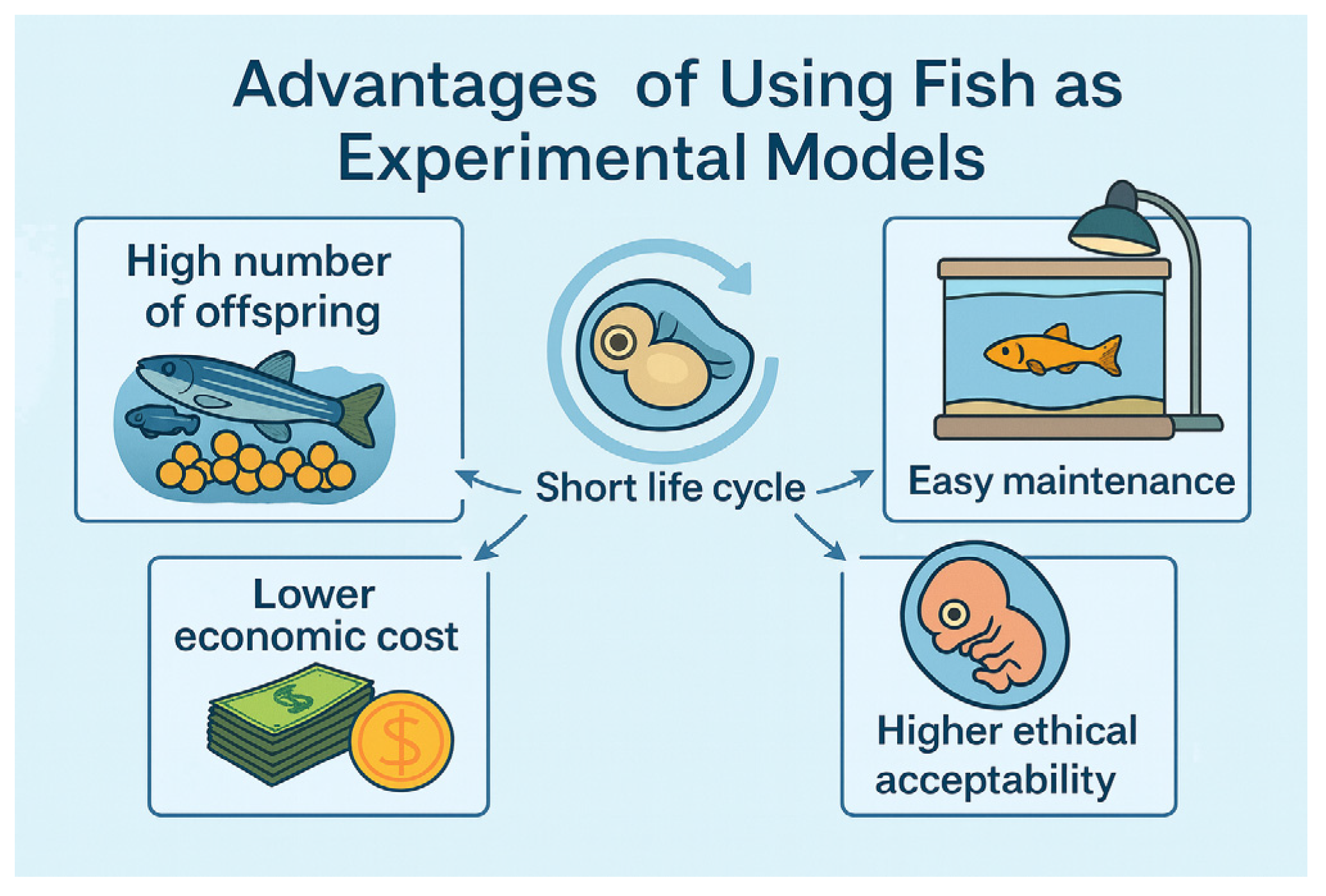
Collectively, these biological, practical, economic, and ethical advantages underpin the widespread adoption of fish as versatile and powerful experimental models across multiple scientific disciplines (Figure 2).

3.1. High Number of Offspring

Fish species commonly used in research, such as zebrafish and medaka, exhibit high reproductive capacity [89,95]. A single mating pair can produce hundreds of embryos weekly under controlled laboratory conditions. This prolific reproduction enables the collection of statistically significant sample sizes, which is particularly advantageous for high-throughput screening in genetic, toxicological, pharmacological, and developmental biology studies. For example, zebrafish embryo assays routinely involve 96- to 384-well plate formats, enabling the simultaneous testing of hundreds of compounds with replicates sufficient for robust dose–response curve [96]. It allows researchers to conduct large-scale experiments with reduced interindividual variability and increased statistical power.
A single pair of zebrafish, for instance, can generate 200 to 300 embryos per spawning event, often several times per week, while medaka are known for their daily spawning and consistent egg production, typically yielding 20–30 eggs per female per day [89,97]. This prolific reproductive output enables researchers to maintain stable breeding colonies and obtain embryos at defined developmental stages with minimal logistical constraints.
Such high embryo availability facilitates statistically robust experimental designs by enabling the generation of large, synchronized cohorts. This is particularly advantageous for fields requiring high-throughput analysis, such as developmental biology, toxicology, drug screening, and genetic manipulation [44,98]. The production of hundreds or thousands of embryos in a short time frame allows researchers to screen multiple experimental conditions or genetic variants in parallel while maintaining appropriate replication and control groups.
Moreover, because fish embryos develop ex utero, they are exceptionally amenable to imaging, microinjection, and other forms of experimental manipulation. This ease of access, combined with prolific reproduction, allows for longitudinal studies of gene function, organogenesis, behavior, and environmental response across a broad population base, minimizing the influence of individual variation and enhancing statistical power [6,22].
In biomedical research, these characteristics support the use of fish embryos in phenotypic screening, where subtle morphological or behavioral changes in response to drugs or toxicants can be detected at scale. For example, zebrafish embryos are frequently used in automated platforms to evaluate cardiovascular effects, neurotoxicity, teratogenicity, and metabolic changes in response to novel compounds [40,41]. The abundance of embryos not only accelerates data acquisition but also contributes to more reliable dose–response modeling and reproducibility, which are critical for translational research.
Importantly, the ethical and regulatory frameworks governing the use of early-stage fish embryos are often less restrictive than those applied to mammalian vertebrates, particularly during the first 5 days post-fertilization [92,93]. This aspect makes zebrafish and medaka attractive alternatives for early-stage in vivo testing, including genetic screens and environmental impact assessments. Therefore, this efficiency underlies the scalability and statistical strength of fish-based experimental systems, reinforcing their utility across a wide range of disciplines in both basic and applied biosciences.

3.2. Short Life Cycle and Rapid Development

Commonly used experimental fish models, such as zebrafish and medaka, are characterized by a short life cycle coupled with rapid and externally visible development. Embryogenesis proceeds swiftly in these species, with critical organ systems, including the heart, eyes, brain, and notochord, forming within the first 24 to 72 h post-fertilization [11,97]. The transparent nature of embryos enables real-time in vivo observation of morphogenesis without invasive procedures.
For example, zebrafish embryos begin spontaneous movement by 24 h and exhibit heartbeat activity around 26 h post-fertilization, with circulation becoming visible shortly thereafter. Major anatomical structures develop within three days, and larvae commence free-swimming and feeding by 5 days post-fertilization. Full sexual maturity is typically reached within 2.5 to 3 months under optimal laboratory conditions [11,89]. Medaka follow a similarly rapid developmental timeline and exhibit daily egg production upon adulthood, with transparent chorions facilitating developmental imaging [97].
This compressed life cycle allows researchers to observe multiple generations within a short timeframe, a feature particularly valuable for studies on genetic inheritance, mutagenesis, developmental programming, gene–environment interactions, and transgenerational effects [41,99]. The rapid generational turnover accelerates forward and reverse genetic screens, enabling swift functional validation of candidate genes associated with human diseases.
Moreover, the fast progression from fertilization to organogenesis supports timely phenotypic assessment in disease models such as neurodegeneration, cancer, congenital defects, and metabolic disorders. Early-stage exposure to chemicals or environmental stressors permits detailed tracking of phenotypic outcomes throughout development and adulthood, refining the understanding of cause-effect relationships and developmental toxicity [9,44].
In ecological and evolutionary contexts, the short generation times facilitate experimental evolution studies and population analyses, allowing investigation of adaptive responses and reproductive fitness over several life cycles within a single year [100]. Collectively, the rapid development, external embryogenesis, and brief generation intervals enhance the experimental versatility and throughput of fish models, making them particularly suitable for longitudinal, multigenerational, and high-throughput research applications across diverse scientific fields.

3.3. Easy Maintenance in Laboratory Environments

Compared to mammalian models, fish are considerably easier to house and maintain under laboratory conditions. They require relatively simple aquatic systems with controlled temperature, lighting, and water quality, alongside standardized feeding protocols. Recirculating aquaculture systems (RAS) or flow-through tanks effectively maintain water parameters, reduce waste, and optimize operational costs. The small size and robust physiology of these teleost species facilitate high-density housing within compact spaces, making large-scale breeding and experiments feasible even in limited facilities [89,91].
Routine husbandry, including feeding, tank cleaning, and health monitoring, is straightforward and requires less specialized personnel compared to mammalian models. Most research fish accept commercial diets, reducing the complexity of nutritional management. Their adaptability to controlled laboratory environments supports continuous breeding cycles under artificial photothermal regimes, ensuring a steady supply of embryos and adults for experimentation [90]. These factors combine to make fish models highly practical for long-term and large-scale research, enhancing reproducibility and accessibility in diverse institutional settings.

3.4. Lower Economic Cost Compared to Mammals

Fish models offer significant cost advantages over traditional mammalian systems in biomedical and environmental research. These species require fewer resources for housing, feeding, and veterinary care, resulting in lower operational expenses. The infrastructure for aquatic husbandry, particularly recirculating aquaculture systems, is generally less expensive to establish and maintain than rodent facilities. Additionally, temperature and lighting control for fish systems are simpler and more energy-efficient [89,101].
Due to their small size, fish can be housed at high densities, allowing thousands of individuals within minimal spatial footprints. This scalability enables large-scale experimental designs with extensive replication without proportional increases in cost. Fish also consume less food per capita and have lower veterinary needs due to reduced disease incidence, further reducing maintenance overhead [90].
The affordability of these models makes them particularly suitable for high-throughput genetic, pharmacological, and toxicological screenings, facilitating early-phase research where multiple conditions or variables must be tested. They allow researchers to generate comprehensive datasets efficiently before validation in more complex and costly mammalian systems [41,44].

3.5. Higher Ethical Acceptability, Especially at Early Developmental Stages (Embryos and Larvae)

A notable advantage of fish models lies in their favorable ethical profile compared to mammalian systems. Regulatory frameworks often do not classify early-stage fish embryos and larvae as protected animals until they reach critical developmental milestones associated with neural maturity and nociceptive capacity [92]. For zebrafish, this stage is typically 5 days post-fertilization, during which embryos develop ex utero and remain transparent, allowing non-invasive, real-time in vivo studies [93].
This regulatory distinction enables extensive early developmental research, genetic manipulations, and high-throughput chemical screening without the need for full ethical review or licensing. Fish embryos thus provide an ethically accessible model aligning with the 3Rs principles (Replacement, Reduction, Refinement), minimizing animal suffering while supporting rigorous scientific inquiry [94,102].
The external fertilization and transparent development of fish embryos reduce invasiveness and facilitate precise microinjection, live imaging, and phenotypic analyses at high resolution [11,91,103]. These attributes enhance experimental rigor and animal welfare, accelerating project timelines and promoting innovative experimental designs across developmental biology, toxicology, neurobiology, and pharmacology [104].
Beyond regulatory compliance, public acceptance of research involving fish embryos tends to be higher than those involving mammals, potentially improving funding prospects and collaborative opportunities [105]. Together, these ethical benefits reinforce the strategic value of fish models in contemporary biomedical research.

4. Fields of Application for Fish as Experimental Models

The utility of biological models for elucidating disease mechanisms, developing diagnostics, and testing therapies is widely recognized. Numerous biomedical advances, including insights into human pathologies and the development of effective therapeutics, have arisen from studies using diverse model organisms. While basic biomedical research often relies on a variety of species, clinically focused studies tend to favor models with closer physiological similarity to humans, historically establishing mammals, particularly rodents, as the dominant experimental system due to their ease of genetic manipulation [106,107].
More recently, fish have gained recognition as valuable models for human diseases. Despite diverging from humans over 400 million years ago, fish share sufficient molecular and biological similarities to support translational research. For instance, the human HRAS gene, frequently mutated in cancer, exhibits over 95% identity with its medaka ortholog, with minor differences outside the protein’s functional core [108].
Fish have become indispensable in genetics and developmental biology due to transgenic lines, genome editing tools, and advanced imaging techniques that allow real-time visualization of gene expression and organogenesis [109,110]. They also serve as ethical and cost-effective models in toxicology, pharmacology, immunology, and behavioral neuroscience, providing translational relevance to human health [111,112]. Moreover, fish models are widely used to study human diseases, including cancer, cardiovascular, renal, hematopoietic, and skeletal disorders, with transgenic and mutant lines facilitating investigation of genetic interactions, disease progression, and therapeutic responses [106,108,113].
Fish represent versatile and integrative experimental platforms that combine genetic tractability, high-resolution imaging, behavioral assays, pharmacological screening, immunological studies, and disease modeling (Figure 3). The combination of classical laboratory species with evolutionary mutants, together with cutting-edge genomic and multi-omics technologies, positions fish at the forefront of vertebrate research, offering mechanistic insights, translational relevance, and ethical alternatives to traditional mammalian models [114,115].

4.1. Genetics and Developmental Biology

Fish models have become indispensable in modern genetics and developmental biology due to their experimental tractability, rapid development, and the availability of powerful genetic and imaging tools. A key breakthrough in the field was the generation of transgenic fish lines capable of faithfully expressing reporter genes under tissue-specific promoters [116]. Higashijima et al. [117] demonstrated the reliable expression of GFP in zebrafish muscle tissues using an endogenous α-actin promoter, while β-actin-driven constructs enabled ubiquitous fluorescence throughout the body. These findings not only validated zebrafish as a genetically tractable organism but also established foundational tools for dissecting spatiotemporal patterns of gene expression in vivo.
The advent of programmable nucleases, ZFN, TALEN, and CRISPR/Cas systems, has further propelled fish into the post-genomic era by enabling targeted gene disruptions, knock-ins, and regulatory element modifications with high precision. These genome editing technologies have been broadly applied across species, from developmental models like medaka to aquaculture-relevant species such as salmon and tilapia [118]. In the African turquoise killifish (Nothobranchius furzeri), a species appreciated for its exceptionally short lifespan and aging-associated traits, precise CRISPR/Cas9-mediated knock-in techniques have recently been established, enabling the generation of humanized disease models and fluorescent reporter lines. These tools allow for the investigation of genetic determinants of senescence and regeneration, aligning fish models with key priorities in biomedical research [119].
Recent advances in single-cell and spatial transcriptomic technologies are revolutionizing developmental biology by providing unprecedented resolution into the cellular and molecular architecture of embryogenesis. Techniques such as single-cell RNA sequencing (scRNA-seq), single-cell ATAC sequencing (scATAC-seq), and spatial transcriptomics have been successfully applied in zebrafish to map lineage trajectories, transcriptional heterogeneity, and epigenetic remodeling during organogenesis [120,121]. Furthermore, hybrid multi-omics approaches enable the simultaneous profiling of mRNA expression, chromatin accessibility, and protein abundance at the single-cell level, offering a systems-level perspective on developmental programs [122]. These methodologies, when combined with advanced imaging and genome editing techniques, provide powerful tools for dissecting the complex regulatory networks governing vertebrate development.
In particular, spatial transcriptomics has emerged as a transformative tool for developmental studies in fish. By preserving the spatial context of gene expression within intact tissues, this technique enables researchers to visualize molecular zonation across developing organs. Recent studies have utilized the 10× Genomics Visium platform (10× Genomics Space Ranger and Loupe Browser) to map gene expression in the zebrafish retina and optic tectum following optic nerve injury, revealing localized induction of regenerative markers such as pdgfra and kitb [120]. Inhibition of these genes impaired axonal regrowth, underscoring the value of spatially resolved data in identifying functional regenerative modules. Complementary in situ techniques, including smFISH, hybridization chain reaction (HCR), and CRISPR-based CASFISH, have further refined our ability to detect gene expression at single-molecule resolution within embryonic tissues [123,124,125,126].
The integration of spatial data with multi-omics pipelines, such as MERFISH and SEFI (Spatial Embedded Feature Identification), now enables transcriptome-wide, subcellular-resolution profiling without the need for cell segmentation [127]. These approaches have already been adapted to zebrafish and other teleost fish, facilitating the reconstruction of developmental trajectories in both healthy and injured tissues. Such strategies are particularly useful in mapping Müller glia responses in retinal regeneration, a process extensively characterized in zebrafish due to its robust neurogenic potential [111,128].
Fish models are especially suited for probing conserved developmental signaling pathways. Canonical pathways such as Wnt, Notch, Hedgehog, and BMP are functionally conserved between teleost fish and mammals, and disruptions in these cascades often recapitulate phenotypes observed in human congenital disorders. Zebrafish have been central to studies of somitogenesis, heart morphogenesis, neural tube formation, and craniofacial development, with multiple human disease genes first characterized in fish embryos [129,130]. Their optical transparency facilitates dynamic imaging of these processes in real-time, while transgenic biosensor lines allow the visualization of pathway activation in situ.
Importantly, model fish species differ in genome size, chromosomal arrangements, and life history traits, which can be leveraged to address distinct biological questions. For instance, medaka offers a compact genome and robust transgenesis protocols, while killifish serves as a comparative model for aging and epigenetic reprogramming due to its natural senescence. Sticklebacks, on the other hand, are invaluable in evo-devo studies because of their ecological plasticity and genomic divergence among populations.
Finally, the scalability of fish models makes them ideal for high-throughput genetic screens and phenotype-driven investigations. Pooled CRISPR screens, barcode-based lineage tracing, and optogenetic manipulation have been adapted for zebrafish embryos, enabling systematic dissection of gene networks and developmental hierarchies in a vertebrate context [131]. Integration with computational models and machine learning tools further enhances the predictive power of these datasets, facilitating genotype-to-phenotype mapping at a systems biology level.
Altogether, the synergy between molecular genetics, cutting-edge imaging, and emerging multi-omics platforms in diverse fish models positions them at the forefront of developmental biology. As post-genomic technologies continue to evolve, experimental fish models will remain pivotal for elucidating fundamental principles of vertebrate development, gene regulation, and the molecular basis of disease.

4.2. Toxicology and Ecotoxicology

Fish models are indispensable tools in environmental toxicology and ecotoxicology, providing relevant biological systems for assessing the effects of contaminants on aquatic organisms and ecosystems. The Fish Embryo Acute Toxicity (FET) test, notably standardized with zebrafish embryos, is recognized by the OECD as an ethical and cost-effective alternative to mammalian testing, allowing rapid evaluation of acute toxicity of diverse chemicals [132].
Water quality is fundamental to sustaining aquatic life, and contamination by environmental pollutants such as endocrine disrupting chemicals (EDCs), pharmaceuticals, personal care products (PPCPs), and other xenobiotics poses significant risks to aquatic ecosystems and human health [112]. These pollutants are often persistent, bioaccumulate in aquatic organisms, especially fish, and may not be fully removed by conventional water treatment processes [112].
Beyond acute effects, fish models allow detailed investigation of chronic toxicity, endocrine disruption, genotoxicity, and developmental defects induced by environ-mental pollutants. For example, a study by Do Nascimento et al. [111] demonstrated that chronic dietary exposure to polypropylene microplastics in Nile tilapia causes systemic inflammation, altered hepatic enzyme activity, immune cell profile changes, and intestinal microbiota dysbiosis, highlighting sub-lethal deleterious effects relevant for ecological risk assessment.
Fish species are invaluable models in toxicology and ecotoxicology because they integrate complex physiological, biochemical, and molecular responses to contaminants present in their environment. In particular, fish are sensitive to EDCs, which interfere with hormonal regulation affecting reproduction, development, and homeostasis [133]. Studies using zebrafish have demonstrated that exposure to synthetic estrogens such as 17α-ethinylestradiol (EE2) induces feminization and vitellogenin production, while androgens like 17β-trenbolone cause masculinization and reproductive impairments, evidencing conserved endocrine disruption pathways relevant to other vertebrates [134,135].
Fish are recognized as fundamental environmental sentinels for assessing aquatic ecosystem quality due to their sensitivity to a wide range of chemical and biological contaminants [136]. Their capacity to integrate physiological and molecular responses to pollutants makes them excellent early indicators of environmental stress, aiding in contamination detection before irreversible damage occurs in natural populations and habitats [137]. Thus, the use of fish as bioindicators directly contributes to environmental monitoring strategies and public policies for pollutant control and remediation.
Multi-omics approaches have revolutionized ecotoxicological studies by enabling comprehensive profiling of molecular alterations in exposed fish. Morrison et al. [121] applied single-cell RNA sequencing to characterize conserved cellular responses during liver fibrosis in zebrafish, demonstrating molecular parallels with human pathophysiology and validating zebrafish as a translational model for environmental liver toxicity. Recent multi-omics studies have illustrated this capacity; for example, Wang et al. [138] demonstrated that combined exposure to cadmium and triazophos in hook snout carp significantly altered hepatic metabolism, disrupted steroid hormone biosynthesis, and impaired mitochondrial function. Metabolomic profiling identified nearly 200 differentially expressed metabolites per treatment group, while 16S rRNA sequencing revealed pronounced shifts in gut microbial diversity and composition. These integrated findings highlight the statistical and mechanistic depth achievable in modern fish-based toxicological assays, showing how co-exposure to heavy metals and pesticides can synergistically exacerbate oxidative stress, metabolic dysfunction, and gut–liver axis disruption.
Epigenetic modifications represent a key layer of toxicant effects, as environmental contaminants can induce heritable DNA methylation changes in fish, affecting gene expression and potentially population resilience. Early-life exposures to endocrine-disrupting chemicals, heavy metals, or pharmaceuticals can lead to adverse adult phenotypes by altering genes important for germ cell development, such as vasa [139]. These effects have been observed in both model species, like zebrafish, and non-model species, such as Kryptolebias marmoratus and Menidia beryllina, emphasizing the ecological relevance of epigenetic studies in fish.
The integration of traditional toxicological methods with post-genomic and multi-omics technologies in diverse fish species is advancing environmental risk assessment. This multidisciplinary approach provides detailed mechanistic understanding of pollutant effects at cellular, molecular, and organismal levels, supporting regulatory decision-making and environmental protection.

4.3. Neuroscience and Behavior

Fish models have emerged as valuable tools in behavioral neuroscience due to their combination of vertebrate complexity and experimental practicality. Species such as zebrafish, medaka, killifish (Nothobranchius furzeri), and goldfish (Carassius auratus) offer advantages such as low cost, ease of large-scale maintenance, great breeding potential, and rapid developmental cycles, allowing behavioral, pharmacological, and genetic studies to be conducted efficiently [140,141]. The behavioral repertoire of these species includes learning, memory, stress and anxiety responses, social interactions, sleep patterns, and reward-related behaviors, which can be quantified using automated tracking systems [41,142].
Neuroanatomical conservation with higher vertebrates, together with neurotransmitter systems and receptors similar to those of mammals, provides translational relevance to these studies. For example, dopaminergic, serotonergic, and GABAergic receptors present in zebrafish and medaka respond to drugs developed for humans, allowing reliable investigation of neurochemical mechanisms [143,144]. Modern tools, such as transgenic lines, optogenetics, and calcium imaging, enable real-time manipulation and monitoring of neuronal activity during complex behaviors, while imaging techniques like light-sheet and two-photon microscopy allow dynamic mapping of entire neural circuits in larvae and juvenile brains [145,146,147].
Studies demonstrate the application of these models in investigating neurological and neuropsychiatric disorders. Zebrafish have been used to analyze drug-associated reward using conditioned place preference (CPP) paradigms, showing effects of ethanol, caffeine, and nicotine on behavior and expression of dopaminergic and nicotinic receptors [148,149]. Medaka has been employed in carcinogenesis and metabolic disorder studies, while killifish, due to its short lifespan, is used to study neural aging and cognitive decline [150]. Goldfish have proven useful in associative learning and spatial memory research, providing complementary data to studies using zebrafish and medaka [151].
The post-genomic era has further expanded the potential of these models. Complete genome sequencing of zebrafish and medaka, together with genome-editing tools such as CRISPR/Cas9 and TALENs, enables precise manipulation of genes orthologous to humans, facilitating therapeutic compound screening and mechanistic studies of neural circuits [106,152]. Applications include the assessment of drug-induced cardiotoxicity, modeling of neurodegenerative diseases, and the development of “avatar” cancer models for personalized drug testing [113].
Thus, fish models provide a powerful, ethical, and cost-effective platform for behavioral neuroscience. The combination of a complex behavioral repertoire, advanced genetic and optical tools, and large-scale screening capability allows researchers to investigate learning, memory, reward, attention, stress, and neuropsychiatric disorders, offering translational relevance for preclinical studies and a promising outlook for future research.

4.4. Pharmacology and Drug Screening

The use of fish models, particularly zebrafish and medaka, has grown substantially in preclinical pharmacology due to their combination of biological relevance, experimental versatility, and compatibility with high-throughput screening (HTS) platforms. This feature eliminates the need for invasive procedures, enabling longitudinal assessment of drug efficacy, toxicity, teratogenicity, and organ-specific effects under controlled conditions [6,13,44].
From a pharmacological perspective, zebrafish offer significant translational value due to the conservation of key drug metabolism pathways with mammals, including cytochrome P450 (CYP) enzymes. The CYP superfamily includes 32 direct human orthologs, supporting the evaluation of drug biotransformation, pharmacokinetics, and potential drug–drug interactions [153]. Such conservation underpins their utility in mechanism-based drug discovery, as metabolic activation and detoxification processes can be reliably modeled.
The post-genomic era has markedly enhanced the applicability of fish in drug screening. The complete sequencing of the zebrafish genome [6] and the advent of genome-editing tools such as CRISPR/Cas9 and TALENs have enabled precise genetic manipulations to generate disease-specific models. These advances facilitate in vivo functional validation of therapeutic targets and allow the screening of candidate molecules in a disease-relevant genetic background [14]. Examples include CRISPR-engineered zebrafish lines modeling congenital heart defects for cardioprotective drug identification, or tumor-prone lines for targeted anticancer compound evaluation.
High-throughput zebrafish embryo assays have been successfully applied across diverse pharmacological domains. In oncology, zebrafish xenograft or “avatar” models, where patient-derived tumor cells are implanted into embryos, allow rapid, personalized drug response profiling, contributing to precision medicine strategies [154]. In cardiovascular research, zebrafish embryos exposed to doxorubicin have been used to screen cardioprotective agents such as visnagin and diphenyl urea, identified through real-time imaging of cardiac function [155]. In angiogenesis studies, compounds such as SU5416 and flavopiridol have been evaluated using transgenic zebrafish lines expressing fluorescent endothelial markers, enabling quantitative analysis of vascular development inhibition [156].
Toxicological screening has also benefited from zebrafish-based HTS. Embryos can be arrayed in multiwell plates (e.g., 96- or 384-well formats), where compounds are directly dissolved in the water, ensuring homogeneous exposure [157]. This approach supports the simultaneous evaluation of acute toxicity, teratogenicity, neurotoxicity, hepatotoxicity, and cardiotoxicity with reduced cost and time compared to mammalian models [11]. Moreover, automated imaging and behavioral tracking systems have expanded the scope of endpoints measurable in HTS, including locomotor activity changes, seizure-like behavior, and sensory impairments.
Beyond zebrafish, other fish species offer valuable models for specialized pharmacological and toxicological studies. Medaka have been used in long-term carcinogenesis bioassays due to their well-characterized tumorigenesis patterns [158], while rainbow trout hepatocyte spheroids serve as robust in vitro systems for investigating xenobiotic metabolism and enzyme induction, including both oxidative and conjugative detoxification pathways. This diversity allows researchers to choose the most appropriate model based on metabolic capacity, lifespan, or organ-specific relevance [159].
Despite these advances, certain limitations must be acknowledged. Physiological differences, such as the absence of lungs, the presence of gills, and variations in circulatory architecture, can restrict the direct extrapolation of findings to humans, particularly in pulmonary pharmacology or complex cardiovascular conditions [160]. Environmental factors, including water temperature, significantly influence drug absorption, metabolism, and clearance rates. Furthermore, species-specific genome duplication events (as in teleosts) can result in multiple paralogs for human genes, complicating the interpretation of knockout or knockdown studies [161].
Overall, the integration of genomic tools, disease modeling, and HTS in fish systems has established them as powerful platforms for pharmacological and toxicological research. Their role is not to replace mammalian models entirely but to bridge the translational gap between in vitro assays and costly mammalian in vivo studies, accelerating drug discovery pipelines, improving mechanistic understanding, and contributing to the development of safer and more effective therapeutic agents.

4.5. Immunology and Pathogen Response

Fish possess both innate and adaptive immune systems that share numerous structural and functional characteristics with mammals, making them ideal models for studying host–pathogen interactions and immune responses [162]. Among the most commonly used species are zebrafish and tilapia, extensively employed in research on bacterial, viral, and parasitic infections relevant to both aquaculture and biomedicine. Zebrafish, in particular transgenic lines with fluorescently labeled innate immune cells, allow real-time tracking of macrophage behavior, neutrophil recruitment, and inflammation resolution [163,164]. This model has also been employed to study dysregulated macrophage activation and hyperinflammatory responses, such as those observed in macrophage activation syndrome (MAS) during severe infections, highlighting its utility for disease modeling and drug screening in contexts like COVID-19 [165].
At the molecular level, zebrafish possess genes encoding immunoglobulins IgM, IgD, and the exclusive IgZ/T isotype, restricted to bony fish, with differential expression (IgM > IgZ > IgD) [166]. In situ hybridization studies have identified key hematopoietic genes such as GATA1, GATA2, c-myb, and LMO2, enabling the mapping of hematopoietic sites during embryogenesis [164]. Zebrafish provide a cost-effective system for gene mapping and positional cloning, with techniques for analyzing sequence polymorphisms and genetic linkage maps using simple sequence repeats (CA)n and RAPD markers [167,168].
The exceptional imaging potential of zebrafish has allowed the study of fundamental biological processes, including phagocytosis, neutrophil apoptosis during inflammation resolution, and the role of antigen-presenting cells (APCs), including dendritic cells (DCs) [162,169]. Characterization of eosinophils and mast cells, often difficult to study in mammals, has also been achieved using specific transgenic lines [170,171,172,173]. All major mammalian T cell subtypes (CD8 cytotoxic, CD4 helper, and CD4/CD25 regulatory) have been identified in zebrafish, along with functional aspects of B cells [162,169].
With the advent of the post-genomic era, omics-based approaches have significantly enhanced understanding of immune responses in fish. Recent single-cell RNA sequencing (scRNA-seq) of zebrafish spleen leukocytes provided a comprehensive atlas of immune diversity, revealing 11 major immune cell categories and more than 50 transcriptionally distinct subsets. These populations displayed marked quantitative shifts in gene expression when challenged with spring viremia of carp virus (SVCV), including the induction of interferons and other antiviral mediators. Notably, trained immunity was demonstrated in neutrophil and M1-macrophage subsets following vaccination, underscoring the statistical depth and high-resolution capacity of fish models for dissecting complex immune landscapes [174]. Transcriptomic studies in tilapia infected with Streptococcus agalactiae revealed rapid induction of genes involved in pathogen recognition, cytoskeletal reorganization, and activation of TLR, PI3K-Akt, Jak-STAT, and MAPK pathways, in addition to piscidins as potential antimicrobial agents [175]. In Tilapia Lake Virus (TiLV) infections, RNA-seq identified thousands of differentially expressed genes associated with antigen presentation, interferon signaling, NF-κB, apoptosis, and acute-phase responses [176]. In zebrafish, transcriptomic and single-cell RNA-seq analyses highlighted critical genes such as acod1 and gpr84 and identified 11 immune cell categories, each responding distinctly to viral infections and demonstrating trained immunity in specific cell subsets [177]. Multi-omics studies in goldfish (Carassius auratus) identified microRNAs regulating local immune profiles in gills during Aeromonas hydrophila infection [178], while combined metabolomic and transcriptomic analyses in zebrafish demonstrated interactions between metabolism and immunity [179].
Furthermore, teleost fish have been successfully employed as experimental models in vaccine development, since their translationally relevant immune responses for human applications and facilitate large-scale assessment of vaccine efficacy and safety, as well as testing of immunomodulators [180]. Consequently, fish models are valuable for evaluating vaccines, immunostimulants, and antimicrobial agents, complementing chemical screening studies [162,164].
These findings demonstrate that fish, especially zebrafish and tilapia, constitute powerful platforms for investigating defense mechanisms, trained immunity, and susceptibility to infections, while also providing valuable insights for the development of preventive and therapeutic strategies against emerging diseases. Comparative fish immunology thus not only contributes to understanding the evolution of the immune system but also offers robust models for advancing vaccine research and immunotherapeutic approaches.

4.6. Modeling Human Diseases

Fish models have become indispensable tools in biomedical research due to their versatility, genetic tractability, and physiological similarities with humans. Species such as zebrafish, medaka, and live-bearing fish like Xiphophorus and Poecilia are widely used to study the molecular and cellular mechanisms underlying various human pathologies [108]. These models allow real-time visualization of biological processes and facilitate the identification of therapeutic targets, the screening of pharmacological compounds, and the investigation of disease progression in vivo [48].
In oncology, zebrafish and medaka have proven powerful for studying tumor initiation, progression, and metastasis. Early models, such as the tp53 M214K mutant, enabled research on multiple cancer types, including peripheral nerve sheath tumor, breast cancer, brain tumors, and leukemia [181]. Transgenic lines expressing oncogenes such as BRAF V600E or KRAS G12D allow detailed functional analyses of melanoma, pancreatic adenocarcinoma, and rhabdomyosarcoma [108,182,183]. Zebrafish carrying tp53 and brca2 mutations provide additional insight into genetic interactions, highlighting the influence of genotype and sex on tumor development, while evolutionary mutant species such as Xiphophorus and Poecilia offer a complementary perspective on naturally occurring genetic variation and modifier genes affecting disease susceptibility [108,184,185].
A notable application involves brca1-associated breast cancer. Transgenic zebrafish with targeted disruption of brca1 exhibit impaired DNA repair mechanisms, genomic instability, and enhanced tumorigenesis, closely recapitulating human brca1-deficient phenotypes. Moreover, zebrafish xenografts of brca1-mutated human breast cancer cells enable real-time visualization of tumor growth, invasion, and angiogenesis, while providing a rapid platform for assessing chemosensitivity to PARP inhibitors and other targeted therapies [186]. This case exemplifies how zebrafish models translate genomic alterations into clinically relevant cancer biology and therapeutic discovery.
Fish models have also significantly contributed to understanding cardiovascular, hematopoietic, and renal diseases [187,188,189]. The embryonic zebrafish heart is easily accessible and oxygenated by passive diffusion, facilitating studies of development and disease progression. Mutant and transgenic lines, including sih, placoglobin, kif20a, pbx4, hspb7, kcnh2, prrx1a/b, kcip1, lmcd1, and tensin1, model a variety of cardiovascular conditions such as congenital cardiomyopathies, arrhythmogenic cardiomyopathy, atrial fibrillation, long QT syndrome, and mitral valve prolapse [48,190,191,192,193]. Similarly, lines such as Tg (podocin:GFP), sos2-acp1, plce1 knockdown, Tg (wt1b:EGFP), elmo1, ctns, and Na+/K+ ATPase variants have been used to study renal disorders, including glomerular diseases, nephrotic syndromes, Noonan syndrome, diabetic nephropathy, Fanconi syndrome, and autosomal dominant polycystic kidney disease [194,195,196,197,198].
Beyond these applications, fish models have proven valuable in studying skeletal diseases such as osteoporosis. Small teleost fish like zebrafish and medaka exhibit skeletal structures and developmental pathways highly conserved with mammals, including endochondral and perichondral ossification, dermal bone formation, and the regulatory genes controlling skeletogenesis [199]. While osteocytes are absent in certain teleost skeletal elements, and osteoclast morphology differs slightly from mammals, resorption and remodeling processes are maintained, making these models suitable for studying bone homeostasis, mechanotransduction, and pharmacological interventions in osteoporosis [199].
The post-genomic era has further expanded the potential of fish models. Advanced technologies such as single-cell RNA sequencing, CRISPR-based genome editing, and high-throughput drug screening now enable detailed molecular characterization of disease mechanisms and rapid identification of therapeutic candidates [48,108]. The combination of classical laboratory species with evolutionary mutant models and natural genetic variation provides a rich platform to explore gene-environment interactions, disease modifiers, and translational approaches across a wide spectrum of human diseases.
Fish models represent a versatile and powerful platform for biomedical research, offering insights into cancer, cardiovascular, hematopoietic, neurological, and skeletal disorders. Continued technological advancements, coupled with the exploration of natural mutants, promise to further enhance the relevance of fish as experimental models in understanding human disease and developing therapeutic strategies [48,108,199].

5. Ethical Aspects and Regulations

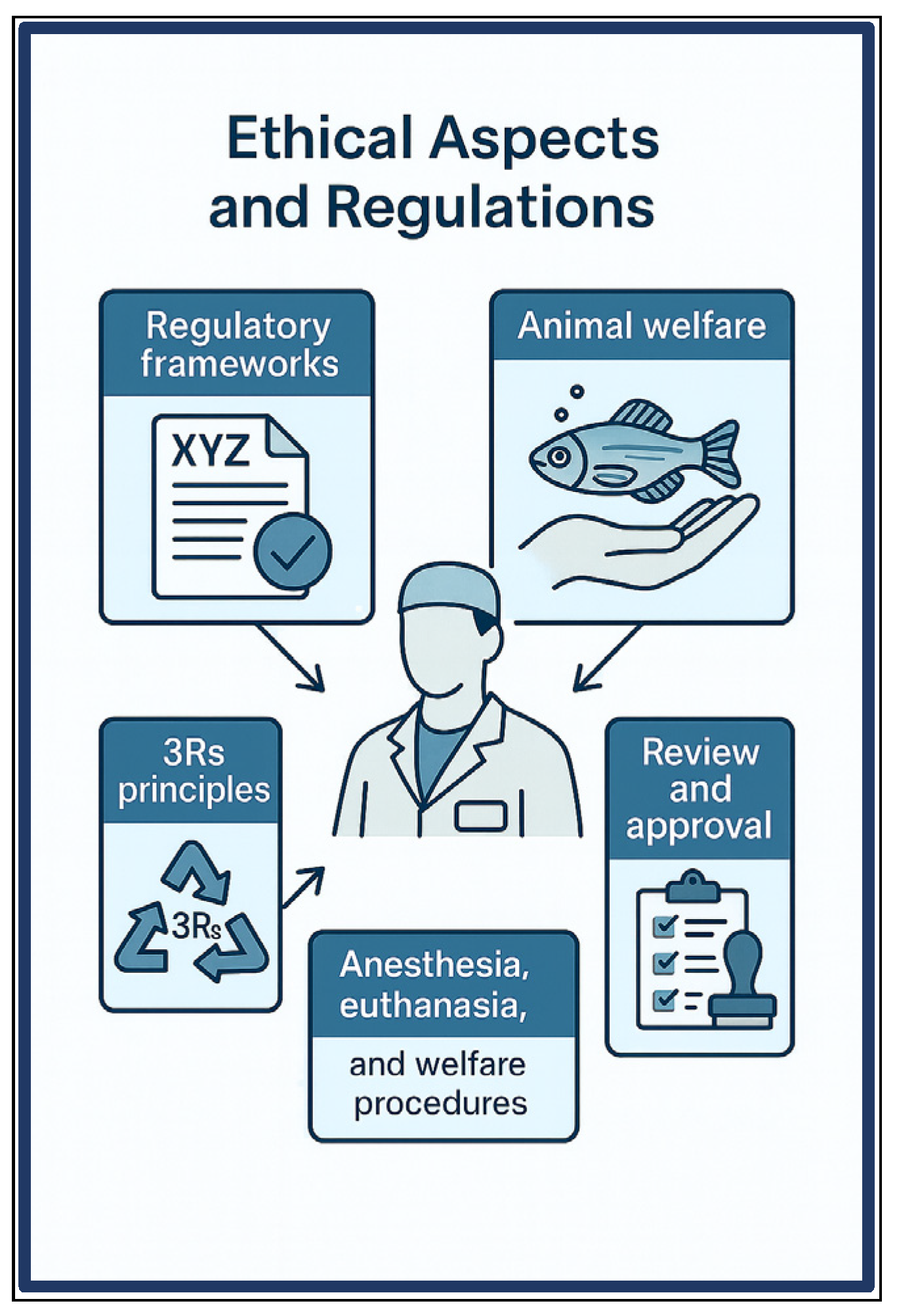
The use of fish as experimental models in scientific research is increasingly regulated by international frameworks designed to balance scientific advancement with the protection of animal welfare [93,200]. Guidelines such as the European Union Directive 2010/63/EU on the protection of animals used for scientific purposes, and the OECD Guidelines for toxicity and safety testing, establish clear requirements for scientific justification, ethical approval, and performance of procedures by trained personnel [93,201]. These instruments recognize that, as vertebrates, fish possess the capacity to experience pain and stress, and thus require specific consideration of their physiological, behavioral, and environmental needs [201,202].
Globally, these regulations are complemented by recommendations from scientific bodies such as the Scientific Committee on Health, Environmental and Emerging Risks (SCHEER), which regularly updates parameters for housing, environmental enrichment protocols, and appropriate euthanasia methods for species such as zebrafish [93,203]. The application of the 3Rs principles, Replacement, Reduction, and Refinement, remains central, promoting not only the reduction in the number of animals used but also the refinement of procedures to minimize suffering and improve data quality [102,204].
The international harmonization of such guidelines strengthens ethical responsibility in fish research, enhances the reliability and reproducibility of results, and fosters greater public acceptance of these models [205,206]. Therefore, strict adherence to these standards is not only a legal obligation but also a fundamental pillar of responsible and sustainable science (Figure 4).

5.1. Ethical Considerations According to International Guidelines (OECD, EU Directive 2010/63)

The ethical use of fish in scientific research is governed by an expanding body of international legislation and guidelines that aim to ensure animal welfare while supporting scientific advancement. Regulatory frameworks such as the European Union’s Directive 2010/63/EU on the protection of animals used for scientific purposes and the American Veterinary Medical Association (AVMA) Guidelines on Euthanasia establish detailed requirements for minimizing pain, distress, and suffering in fish during experimentation [93,200].
Fish, as vertebrates, are increasingly recognized as sentient beings capable of experiencing pain and stress, which necessitates careful ethical consideration in experimental design and conduct [24]. These guidelines emphasize the need for species-specific welfare measures addressing physiological, behavioral, and environmental needs. This includes the mandatory use of appropriate anesthesia and analgesia protocols, the definition of humane endpoints, and the provision of environmental enrichment aimed at reducing stress and improving well-being throughout the research lifecycle [201].
In addition to legal mandates requiring the establishment of institutional Animal Care and Use Committees (IACUCs) or equivalent bodies to review and approve experimental protocols involving fish, expert bodies and scientific committees regularly update best practices. For example, the European Scientific Committee on Health, Environmental and Emerging Risks (SCHEER) has issued recommendations revising housing parameters and acceptable euthanasia methods, such as the conditional use of hypothermic shock, specifically for zebrafish, underscoring the importance of species-tailored guidelines [93,202]. These committees also promote the implementation of the 3Rs principles, encouraging the replacement of sentient animals when feasible, reduction in the number of animals used, and refinement of procedures to enhance welfare [102,203].
Ethical oversight extends beyond experimental procedures to include husbandry practices, transportation, housing, and post-procedural care, recognizing that welfare is a continuum throughout the animal’s life in the research environment. Continuous welfare assessment using behavioral and physiological indicators is critical to detect and mitigate suffering promptly, as emphasized by Sloman et al. [24].
Finally, the international harmonization of ethical frameworks fosters responsible research practices and strengthens public confidence in the use of fish models. Compliance with these guidelines not only protects animal welfare but also improves the quality, reliability, and reproducibility of scientific data by reducing stress-induced variability [204,205].

5.2. From 3Rs to 10Rs: Ethical Principles in Fish Model Research

The ethical conduct of research involving fish models has evolved significantly, reflecting broader concerns about animal welfare, scientific rigor, and responsible practice. Central to this evolution are the well-established principles of the 3Rs, Replacement, Reduction, and Refinement, which serve as a foundational framework to minimize animal use and suffering in scientific studies [94]. Replacement encourages the use of alternative methods that avoid or substitute the use of live animals, Reduction emphasizes minimizing the number of animals required to obtain valid results, and Refinement focuses on improving experimental procedures to enhance animal welfare [102].
In fish research, these principles are exemplified by the increasing use of embryonic and larval stages, which are often considered to have less developed nervous systems and lower capacity for pain perception. Utilizing these early life stages not only aligns with the Replacement principle by reducing reliance on adult fish but also can improve experimental efficiency and reproducibility [206,207].
More recently, the ethical landscape has expanded to include additional principles, leading to the concept of the 10Rs [203]. These extend beyond animal welfare to encompass scientific integrity and ethical responsibility in research practices. The additional Rs include Registration, Reporting, Robustness, Reproducibility, Relevance, Responsibility, and Respect. These principles promote transparent study design and data reporting, ensure methodological soundness, encourage reproducibility of findings, focus on the biological relevance of the research, and uphold the responsibility and respect researchers owe to their animal models [203].
Adopting the 10Rs framework in fish model research fosters a holistic approach that integrates welfare considerations with scientific quality. For instance, rigorous reporting and registration help avoid unnecessary duplication of experiments, thus supporting Reduction. Refinements in husbandry and anesthesia protocols align with both animal welfare and the Responsibility and Respect principles. Furthermore, focusing on study robustness and relevance ensures that fish models are used effectively, maximizing scientific gain while minimizing animal use [203].
Incorporating these ethical principles is particularly critical as fish models like zebrafish continue to expand their role in multidisciplinary research fields. It not only guarantees high standards of animal welfare but also strengthens the scientific validity and reproducibility of research outcomes. Institutions, ethics committees, and researchers are encouraged to implement the 10Rs framework, supported by guidelines and training, to advance responsible and ethical fish research.

5.3. Anesthesia, Euthanasia, and Welfare Procedures Adapted for Fish

Ensuring humane treatment of fish in research demands that anesthesia, euthanasia, and welfare procedures be carefully adapted to their unique physiology. Proper management of these procedures is essential not only for ethical reasons but also for obtaining reliable and reproducible results. As the use of fish species in laboratory and aquaculture contexts increases, so does the importance of minimizing stress and pain through tailored protocols.
Fish anesthesia encompasses sedation, immobilization, unconsciousness (narcosis), amnesia, and analgesia. Commonly used agents include benzocaine, tricaine methanesulfonate (MS-222), metomidate, isoeugenol, 2-phenoxyethanol, and quinaldine [208,209]. These are administered primarily via gill absorption, and dosages must consider species-specific variables such as size, age, and water temperature, which influence anesthetic depth and recovery [210].
While many fish anesthesia protocols rely on single agents, experiences from veterinary and human medicine highlight that combining anesthetics with analgesics can enhance anesthesia quality, reduce doses, and promote smoother induction and recovery [208,211]. MS-222 is widely used due to its effectiveness and approval for aquaculture, particularly in zebrafish research [212]. However, prolonged sedation with MS-222 alone in adult zebrafish can reduce heart rate and increase mortality, limiting its applicability in long-term experiments. Studies have shown that combining MS-222 with isoflurane extends anesthesia duration while minimizing adverse physiological effects such as bradycardia, and allows faster recovery. This combination also enables long-term electrocardiogram recording and microscopic observation with minimal impact on overall fish physiology, highlighting the importance of optimizing anesthetic protocols to balance efficacy and welfare in adult fish models [213].
For euthanasia, overdose of anesthetics like MS-222 or benzocaine is common, applied by immersion or directly to the gills, especially in larger species or terminal procedures [208,214]. Physical methods such as cranial concussion or spinal transection may follow deep anesthesia to ensure rapid, humane death. Although CO2 is frequently used as an anesthetic in other animals, its application in fish is generally discouraged due to limited analgesia and adverse physiological effects [205,215].
In addition to anesthetic overdose with tricaine (MS-222), immersion in ice-chilled water (rapid cooling/hypothermic shock) has been proposed as a humane method of euthanasia for zebrafish. This approach induces rapid loss of consciousness at 2–4 °C, provided fish are not placed in direct contact with ice prior to unconsciousness, and has been reported to cause fewer stress responses compared to anesthetic-based methods [216]. The American Veterinary Medical Association Guidelines for the Euthanasia of Animals [200] recognize rapid cooling as an acceptable method in some contexts, especially when preservation of tissues for histological or biochemical analyses is required. Furthermore, Valentim et al. [217] highlighted the need for its formal inclusion in European regulations, given its practicality and alignment with welfare considerations. While debate remains regarding its universal adoption, cold exposure represents a viable alternative that, in certain jurisdictions, is even preferred over MS-222 for small-bodied fish such as zebrafish.
Welfare concerns extend beyond anesthesia and euthanasia protocols. Handling, confinement, and environmental changes provoke stress responses in fish, and anesthetic agents themselves can cause physiological alterations like elevated cortisol, acid-base imbalance, or respiratory depression [208,214]. Thus, welfare monitoring and pre-anesthetic sedation are increasingly advocated to mitigate acute stress and enhance outcomes, especially in sensitive models like zebrafish and medaka used in genetic and toxicological studies.
As fish models gain prominence in multidisciplinary research, from genetics to environmental toxicology, standardized, species-specific anesthesia and euthanasia protocols are indispensable. These ensure ethical treatment and improve data reliability and reproducibility [204,205]. Refinement of these procedures, alongside welfare monitoring, supports sustainable research and aligns with evolving regulatory and ethical standards for vertebrate animal use in science.

6. Challenges and Limitations

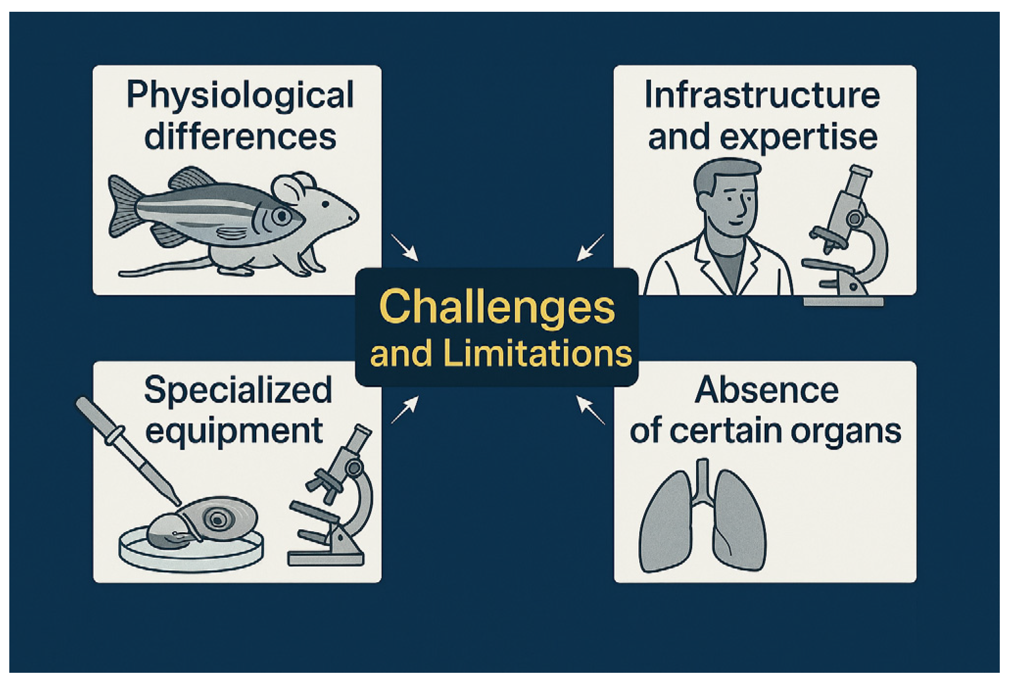
Fish models, especially zebrafish, have become invaluable tools in biomedical and environmental research due to their genetic tractability, rapid development, and suitability for high-throughput studies [44]. However, several inherent challenges limit their broad applicability (Figure 5). Significant physiological and anatomical differences between fish and mammals can hinder direct extrapolation of experimental results [218,219]. Moreover, the specialized infrastructure and expertise required to maintain aquatic facilities and perform precise experimental manipulations create practical barriers for many laboratories [89,205]. In addition, the absence of certain mammalian organs, such as lungs, restricts the use of fish models in specific disease contexts [44]. Overcoming these challenges demands careful study design, investment in dedicated resources, and often, complementary use of mammalian models to ensure translational relevance.

6.1. Physiological Differences That May Limit Direct Extrapolation to Mammals

Fish models, particularly zebrafish, provide significant advantages for biomedical and environmental research due to their genetic tractability, rapid development, and suitability for high-throughput experimentation [44]. Nevertheless, important physiological divergences from mammals must be carefully considered to ensure responsible and accurate extrapolation of findings.
One major difference lies in the poikilothermic nature of fish, whose metabolic rates depend on ambient temperature, in contrast to the homeothermic regulation found in mammals. This temperature dependence influences drug pharmacokinetics and pharmacodynamics as well as disease progression, making fish metabolism, especially in zebrafish, highly variable and sensitive to environmental conditions. Consequently, absorption, distribution, metabolism, and clearance of compounds in fish may differ substantially from human models [218].
The immune system of fish also exhibits distinct features. Although zebrafish possess both innate and adaptive immunity, their adaptive immune responses are less mature and functionally different from those of mammals. For example, memory lymphocyte development in zebrafish is not as robust, which can limit the direct modeling of vaccine efficacy, infectious diseases, and immunotoxicological responses [220,221].
Respiration is another key physiological difference. Fish rely on gill-based gas exchange, affecting oxygen uptake, systemic circulation, and acid–base balance differently than the mammalian lung system. These factors may influence both physiological homeostasis and responses to pharmacological agents (general physiology).
Regarding disease modeling, although zebrafish are phylogenetically more distant from humans than rodents, they possess orthologs for approximately 82% of human disease-associated genes and often exhibit physiological and pharmacological conservation comparable to, or sometimes exceeding, that of rodent models [44]. However, zebrafish still lack many complex tissue-specific and cellular mechanisms characteristic of mammals. This limitation is particularly relevant in fields such as oncology and neurodegenerative diseases, where mammal-specific pathways and microenvironments play crucial roles in disease progression and therapeutic responses [21].
Further distinctions appear in tissue regeneration capacities. For instance, adult zebrafish cardiomyocytes maintain proliferative ability, enabling cardiac regeneration after injury, an ability that adult mammals largely lack, favoring hypertrophy over true regeneration [222].
A key limitation of using zebrafish to model human neurodegenerative diseases lies in their extensive adult neurogenesis and regenerative potential, which are far greater than in mammals. Although zebrafish can present pathologies such as amyloid-β deposition, tau aggregation, and dopaminergic neuron loss [223], their intrinsic ability to replace neurons and restore synaptic function complicates direct extrapolation to human conditions characterized by progressive and irreversible degeneration. Thus, while zebrafish are invaluable for investigating mechanisms of neural plasticity and regeneration, their biology presents a translational challenge in faithfully modeling the chronic course of human neurodegenerative disorders.
Environmental and ecological variability inherent to fish models, such as fluctuating water conditions, environmental stressors, and genetic diversity among strains, adds additional layers of complexity, potentially affecting reproducibility and translational relevance [218,221].
While fish models remain invaluable for a wide array of scientific inquiries, careful interpretation and cautious extrapolation to mammalian biology are imperative. Integrating complementary mammalian studies and employing comparative physiological analyses can improve the predictive power and translational applicability of fish-based research.

6.2. Need for Infrastructure and Specialized Expertise for Proper Handling

Research involving fish models demands highly specialized infrastructure and trained personnel to ensure animal welfare, experimental reproducibility, and data reliability. Aquatic facilities must provide rigorously controlled environmental parameters including temperature, pH, dissolved oxygen, ammonia levels, and light cycles, as fluctuations in these factors can significantly impact fish physiology and behavior [89,205].
Such facilities also require robust biosecurity protocols to prevent pathogen introduction and spread, alongside life support systems such as advanced filtration and aeration to maintain water quality. The complexity of these systems mandates continuous monitoring and maintenance by staff with expertise in aquatic husbandry and veterinary care tailored to fish species.
Beyond husbandry, many experimental procedures necessitate additional technical skills and specialized equipment. Techniques like microinjection for genetic manipulation, live imaging of embryonic development, and waterborne toxicology assays require not only precision instruments but also personnel trained in handling delicate embryos and adults without inducing undue stress or injury [11,91].
Moreover, successful breeding and genetic line maintenance demand thorough knowledge of reproductive biology and environmental cues to optimize spawning and viability [89]. These multifaceted requirements for infrastructure and expertise can pose significant barriers to laboratories without dedicated aquatic research programs, potentially limiting the adoption and scalability of fish models in certain scientific fields.
Thus, investment in tailored aquatic systems and ongoing training for researchers and animal care staff are critical components for advancing fish model research while adhering to welfare and ethical standards.

6.3. Limitations in Studying Certain Organs (e.g., Lungs)

A notable limitation of fish models in biomedical research is the absence of specific mammalian organs, particularly lungs, which constrains their utility in studying diseases related to pulmonary function, such as asthma, chronic obstructive pulmonary disease, and respiratory infections [44]. This anatomical difference limits direct modeling of respiratory pathologies and associated systemic inflammatory responses observed in humans.
Beyond the respiratory system, structural divergences in cardiovascular anatomy, such as the single-circuit heart in fish compared to the double-circuit system in mammals, affect the extrapolation of cardiovascular disease models [218]. Neuroanatomical variations, including differences in brain region complexity and connectivity, further restrict the use of fish for certain neurological disorders where mammalian brain structures play critical roles [221].
Skin physiology also differs markedly; fish skin serves as a primary barrier to the aquatic environment with specialized mucosal functions, unlike the stratified epidermis of mammals, impacting studies of dermatological conditions and transdermal drug delivery [21].
Although advances in genetic and molecular tools have enhanced the translational scope of fish models, it remains essential to consider these anatomical and functional limitations in study design and when interpreting data, particularly for organ-specific diseases. Integrative approaches combining fish models with mammalian systems can help overcome these constraints by leveraging complementary strengths.

7. Future Perspectives

The continuous advancement of omics platforms, coupled with innovations in biotechnology and genetic engineering, is exponentially expanding the potential of experimental fish models for multidisciplinary research in the post-genomic era. Established approaches such as RNA sequencing (RNA-seq) now allow high-resolution profiling of transcriptomes, detecting alternative isoforms and non-coding RNAs that remain undetectable by traditional microarrays [224,225,226]. This capacity has enhanced our understanding of gene regulation in model species including zebrafish and Nothobranchius furzeri [225,227]. For instance, Liu et al. [228], integrated transcriptomics and proteomics to uncover molecular pathways driving heart regeneration in zebrafish, identifying novel regulatory genes with therapeutic potential.
Emerging tools such as single-cell RNA-seq and spatial transcriptomics are further transforming the field, enabling precise mapping of cellular heterogeneity and spatial gene expression patterns within intact tissues [229,230]. These techniques open previously inaccessible windows into developmental dynamics, tissue microenvironments, and disease progression. However, the vast complexity and volume of multi-omics data present ongoing challenges for analysis and integration, underscoring the need for advanced bioinformatics pipelines, standardized analytical frameworks, and cross-disciplinary collaborations [231].
In biotechnology, CRISPR/Cas9 genome editing has become a transformative tool for generating precise disease models and conducting functional gene analyses in fish [14,232]. Beyond CRISPR, next-generation editing platforms such as base editors and prime editing promise greater specificity and reduced off-target effects, while expanding the spectrum of achievable genetic modifications [233]. When combined with transgenic lines carrying fluorescent biosensors and advanced in vivo imaging, these tools enable real-time visualization of processes such as tumor progression and immune cell dynamics with unprecedented temporal and spatial resolution [9].
Complementary to engineered models, naturally occurring evolutionary mutants in fish provide unique perspectives for studying human diseases, often offering phenotypes that reveal the influence of natural allelic variation [234]. For example, transgenic melanoma models in medaka expressing the xmrk oncogene from Xiphophorus exhibit distinct tumor phenotypes depending on genetic background, highlighting the role of inherent variation in modulating disease outcomes [63,234]. While these models offer powerful opportunities to identify disease-modifier genes, limitations remain in genomic resource availability, breeding standardization, and phenotypic characterization. Advances in sequencing technologies are now enabling the rapid generation of full genomic resources for these species at relatively low cost, expanding their accessibility beyond a few well-established laboratory models [235,236].
Experimental fish systems are increasingly pivotal in understanding and predicting ecological responses to environmental change. Climate stressors such as temperature fluctuations, pH shifts, and oxygen variability have been shown to shape phenotypic traits. Long-term studies by Crozier and Hutchings [237] demonstrate that changes in reproductive timing, growth, and survival are closely linked to temperature trends. Predictive modeling by Lefevre et al. [238] reinforces the critical role of accurate physiological parameters, such as metabolic scaling and gill surface area, in forecasting population trajectories under global warming scenarios. Despite these advances, uncertainties remain regarding multigenerational adaptation, species-specific thresholds, and the synergistic effects of multiple stressors.
In conclusion, the convergence of advanced omics technologies, precision genetic tools, and evolutionary mutant models positions experimental fish as indispensable platforms for translational, multidisciplinary, and ecological research. Expanding these integrated approaches will deepen mechanistic understanding, enhance translational relevance, guide conservation strategies, and support evidence-based policy in the face of global environmental challenges. Looking ahead, artificial intelligence (AI) and machine learning approaches hold great promise for optimizing experimental design, predicting disease phenotypes, and integrating multi-omics datasets, thereby accelerating the development of more accurate and efficient fish models for translational research. Achieving these goals requires coordinated investment in genomic resources, bioinformatics infrastructure, and international collaboration, ensuring that the full potential of fish models is realized in the post-genomic era.

8. Final Considerations

Fish have established themselves as versatile, accessible, and ethically advantageous experimental models across numerous scientific disciplines. Their unique biological characteristics, including external fertilization, high fecundity, rapid developmental cycles, and transparent embryonic stages, facilitate non-invasive real-time observation and enable high-throughput experimentation, particularly in developmental biology. Furthermore, their relatively low maintenance costs compared to mammalian models make them highly attractive for academic research, pharmaceutical testing, and regulatory applications.
Beyond these logistical benefits, fish models have proven invaluable for advancing fundamental understanding of key biological processes such as organogenesis, neurodevelopment, immune system function, and metabolic regulation. Many species, notably zebrafish and medaka, share considerable genetic and physiological homology with humans, allowing researchers to derive translational insights into disease mechanisms, toxicological responses, and therapeutic interventions. The growing use of fish to model complex human diseases, including cancer, cardiovascular disorders, and neurodegenerative conditions, further highlights their expanding biomedical significance.
Importantly, the application of fish models is strongly aligned with the ethical principles of the 3Rs (Replacement, Reduction, and Refinement), especially when utilizing embryonic or larval stages that are often exempt from stringent regulatory oversight. As technological innovations such as genomics, CRISPR gene editing, and advanced in vivo imaging continue to evolve and integrate into experimental pipelines, the scope and impact of fish models are poised to grow. These developments promise more ethical, efficient, and informative strategies for exploring both basic biological questions and translational challenges in human health.
In summary, fish models occupy a unique and growing niche in biomedical and environmental research, bridging fundamental biology with applied science in a cost-effective and ethically responsible manner. Their continued development and adoption will undoubtedly play a pivotal role in shaping the future of multidisciplinary science.

Author Contributions

Conceptualization, C.C.-C. and M.A.d.A.B.; methodology, C.C.-C. and M.A.d.A.B.; investigation, C.C.-C. and M.A.d.A.B.; writing—original draft preparation, C.C.-C.; writing—review and editing, M.A.d.A.B.; visualization, C.C.-C.; supervision, M.A.d.A.B.; project administration, M.A.d.A.B. All authors have read and agreed to the published version of the manuscript.

Funding

This research received no external funding.

Institutional Review Board Statement

Not applicable.

Informed Consent Statement

Not applicable.

Data Availability Statement

No new data were created or analyzed in this study. Data sharing is not applicable to this article.

Conflicts of Interest

The authors declare no conflicts of interest.

Abbreviations

The following abbreviations are used in this manuscript:
APCsAntigen-presenting cells
AVMAAmerican veterinary medical association
CRISPR/Cas9Clustered Regularly Interspaced Short Palindromic Repeats/CRISPR-associated protein 9
CYPCytochrome P450
DCsDendritic cells
EDCsEndocrine disrupting chemicals
EE217α-ethinylestradiol
FETFish embryo acute toxicity
GFPGreen fluorescent protein
HTSHigh-throughput screening
IACUCInstitutional Animal Care and Use Committee
Jak-STATJanus Kinase—Signal Transducer and Activator of Transcription pathway
MAPKMitogen-Activated Protein Kinase pathway
MERFISH Multiplexed error-robust fluorescence in situ hybridization
MS-222Tricaine methanesulfonate
OECDOrganisation for Economic Co-operation and Development
OECDOrganization for Economic Co-operation and Development
PI3K-AktPhosphatidylinositol 3-Kinase—Protein Kinase B pathway
PPCPsPharmaceuticals and Personal Care Products
RASRecirculating aquaculture systems
RNARibonucleic acid
scATAC-seqSingle-cell Assay for Transposase-Accessible Chromatin sequencing
SCHEERScientific Committee on Health, Environmental and Emerging Risks
scRNA-seqSingle-cell RNA sequencing
SEFISpatial embedded feature identification
TALENsTranscription activator-like effector nucleases
TiLVTilapia lake virus
TLRToll-like receptor
ZFNZinc finger nuclease

References

  1. Kirk, R.G. Recovering the Principles of Humane Experimental Technique: The 3Rs and the Human Essence of Animal Research. Sci. Technol. Hum. Values 2018, 43, 622–648. [Google Scholar] [CrossRef]
  2. Balls, M. It’s Time to Reconsider the Principles of Humane Experimental Technique. Altern. Lab. Anim. 2020, 48, 40–46. [Google Scholar] [CrossRef]
  3. Aitman, T.J.; Boone, C.; Churchill, G.A.; Hengartner, M.O.; Mackay, T.F.C.; Stemple, D.L. The Future of Model Organisms in Human Disease Research. Nat. Rev. Genet. 2011, 12, 575–582. [Google Scholar] [CrossRef] [PubMed]
  4. Lin, C.-Y.; Chiang, C.-Y.; Tsai, H.-J. Zebrafish and Medaka: New Model Organisms for Modern Biomedical Research. J. Biomed. Sci. 2016, 23, 19. [Google Scholar] [CrossRef]
  5. Henning, F.; Meyer, A. The Evolutionary Genomics of Cichlid Fishes: Explosive Speciation and Adaptation in the Postgenomic Era. Annu. Rev. Genom. Hum. Genet. 2014, 15, 417–441. [Google Scholar] [CrossRef]
  6. Howe, K.; Clark, M.D.; Torroja, C.F.; Torrance, J.; Berthelot, C.; Muffato, M.; Collins, J.E.; Humphray, S.; McLaren, K.; Matthews, L. The Zebrafish Reference Genome Sequence and Its Relationship to the Human Genome. Nature 2013, 496, 498–503. [Google Scholar] [CrossRef]
  7. Volff, J.N. Genome Evolution and Biodiversity in Teleost Fish. Heredity 2005, 94, 280–294. [Google Scholar] [CrossRef] [PubMed]
  8. Ravi, V.; Venkatesh, B. Rapidly Evolving Fish Genomes and Teleost Diversity. Curr. Opin. Genet. Dev. 2008, 18, 544–550. [Google Scholar] [CrossRef] [PubMed]
  9. Lieschke, G.J.; Currie, P.D. Animal Models of Human Disease: Zebrafish Swim into View. Nat. Rev. Genet. 2007, 8, 353–367. [Google Scholar] [CrossRef]
  10. Santoriello, C.; Zon, L.I. Hooked! Modeling Human Disease in Zebrafish. J. Clin. Investig. 2012, 122, 2337–2343. [Google Scholar] [CrossRef]
  11. Kimmel, C.B.; Ballard, W.W.; Kimmel, S.R.; Ullmann, B.; Schilling, T.F. Stages of Embryonic Development of the Zebrafish. Dev. Dyn. 1995, 203, 253–310. [Google Scholar] [CrossRef] [PubMed]
  12. White, R.M.; Sessa, A.; Burke, C.; Bowman, T.; LeBlanc, J.; Ceol, C.; Bourque, C.; Dovey, M.; Goessling, W.; Burns, C.E. Transparent Adult Zebrafish as a Tool for in Vivo Transplantation Analysis. Cell Stem Cell 2008, 2, 183–189. [Google Scholar] [CrossRef]
  13. White, R.; Rose, K.; Zon, L. Zebrafish Cancer: The State of the Art and the Path Forward. Nat. Rev. Cancer 2013, 13, 624–636. [Google Scholar] [CrossRef]
  14. Hwang, W.Y.; Fu, Y.; Reyon, D.; Maeder, M.L.; Tsai, S.Q.; Sander, J.D.; Peterson, R.T.; Yeh, J.J.; Joung, J.K. Efficient Genome Editing in Zebrafish Using a CRISPR-Cas System. Nat. Biotechnol. 2013, 31, 227–229. [Google Scholar] [CrossRef]
  15. Liu, J.; Zhou, Y.; Qi, X.; Chen, J.; Chen, W.; Qiu, G.; Wu, Z.; Wu, N. CRISPR/Cas9 in Zebrafish: An Efficient Combination for Human Genetic Diseases Modeling. Hum. Genet. 2017, 136, 1–12. [Google Scholar] [CrossRef]
  16. Kaufmann, A.; Mickoleit, M.; Weber, M.; Huisken, J. Multilayer Mounting Enables Long-Term Imaging of Zebrafish Development in a Light Sheet Microscope. Development 2012, 139, 3242–3247. [Google Scholar] [CrossRef]
  17. Keller, P.J.; Schmidt, A.D.; Wittbrodt, J.; Stelzer, E.H. Reconstruction of Zebrafish Early Embryonic Development by Scanned Light Sheet Microscopy. Science 2008, 322, 1065–1069. [Google Scholar] [CrossRef]
  18. Weber, M.; Huisken, J. In Vivo Imaging of Cardiac Development and Function in Zebrafish Using Light Sheet Microscopy. Swiss Med. Wkly. 2015, 145, w14227. [Google Scholar] [CrossRef] [PubMed]
  19. Lawson, N.D.; Weinstein, B.M. In Vivo Imaging of Embryonic Vascular Development Using Transgenic Zebrafish. Dev. Biol. 2002, 248, 307–318. [Google Scholar] [CrossRef] [PubMed]
  20. Okuda, K.S.; Hogan, B.M. Endothelial Cell Dynamics in Vascular Development: Insights from Live-Imaging in Zebrafish. Front. Physiol. 2020, 11, 842. [Google Scholar] [CrossRef]
  21. Hason, M.; Bartůněk, P. Zebrafish Models of Cancer—New Insights on Modeling Human Cancer in a Non-Mammalian Vertebrate. Genes 2019, 10, 935. [Google Scholar] [CrossRef]
  22. Dooley, K.; Zon, L.I. Zebrafish: A Model System for the Study of Human Disease. Curr. Opin. Genet. Dev. 2000, 10, 252–256. [Google Scholar] [CrossRef]
  23. Jenkins, J.A.; Bart, H.L., Jr.; Bowker, J.D.; Bowser, P.R.; MacMillan, J.R.; Nickum, J.G.; Rose, J.D.; Sorensen, P.W.; Whitledge, G.W.; Rachlin, J.W. Guidelines for the Use of Fishes in Research; American Fisheries Society: Bethesda, MD, USA, 2014. [Google Scholar]
  24. Sloman, K.A.; Bouyoucos, I.A.; Brooks, E.J.; Sneddon, L.U. Ethical Considerations in Fish Research. J. Fish Biol. 2019, 94, 556–577. [Google Scholar] [CrossRef]
  25. D’Angelo, L.; Lossi, L.; Merighi, A.; de Girolamo, P. Anatomical Features for the Adequate Choice of Experimental Animal Models in Biomedicine: I. Fishes. Ann. Anat.-Anat. Anz. 2016, 205, 75–84. [Google Scholar] [CrossRef] [PubMed]
  26. Villéger, S.; Brosse, S.; Mouchet, M.; Mouillot, D.; Vanni, M.J. Functional Ecology of Fish: Current Approaches and Future Challenges. Aquat. Sci. 2017, 79, 783–801. [Google Scholar] [CrossRef]
  27. Kasahara, M.; Naruse, K.; Sasaki, S.; Nakatani, Y.; Qu, W.; Ahsan, B.; Yamada, T.; Nagayasu, Y.; Doi, K.; Kasai, Y. The Medaka Draft Genome and Insights into Vertebrate Genome Evolution. Nature 2007, 447, 714–719. [Google Scholar] [CrossRef] [PubMed]
  28. De la Torre, F.R.; Ferrari, L.; Salibián, A. Biomarkers of a Native Fish Species (Cnesterodon decemmaculatus) Application to the Water Toxicity Assessment of a Peri-Urban Polluted River of Argentina. Chemosphere 2005, 59, 577–583. [Google Scholar] [CrossRef]
  29. PARK, Y.-S.; Chang, J.; Lek, S.; Cao, W.; Brosse, S. Conservation Strategies for Endemic Fish Species Threatened by the Three Gorges Dam. Conserv. Biol. 2003, 17, 1748–1758. [Google Scholar] [CrossRef]
  30. Monier, M.N.; Abd El-Naby, A.S.; Fawzy, R.M.; Samir, F.; Shady, S.H.H.; Grana, Y.S.; Albaqami, N.M.; Abdel-Tawwab, M. Growth Performance, Antioxidant, and Immune Responses of Nile Tilapia (Oreochromis niloticus) Fed on Low-Fishmeal Diets Enriched with Sodium Chloride and Its Adaptability to Different Salinity Levels. Fish Physiol. Biochem. 2025, 51, 6. [Google Scholar] [CrossRef] [PubMed]
  31. Zhao, L.; Luo, J.; Du, J.; Liu, Q.; Xu, G.; Yang, S. Study on the Adaptability of Common Carp (Cyprinus carpio) to Diets from the Perspective of Digestion and Absorption. Aquac. Res. 2020, 51, 2495–2504. [Google Scholar] [CrossRef]
  32. Vilizzi, L.; Tarkan, A.S. Bioaccumulation of Metals in Common Carp (Cyprinus carpio L.) from Water Bodies of Anatolia (Turkey): A Review with Implications for Fisheries and Human Food Consumption. Environ. Monit. Assess. 2016, 188, 243. [Google Scholar] [CrossRef] [PubMed]
  33. Mastrochirico Filho, V.A.; Freitas, M.V.; Ariede, R.B.; Lira, L.V.; Mendes, N.J.; Hashimoto, D.T. Genetic Applications in the Conservation of Neotropical Freshwater Fish. In Biological Resources of Water; IntechOpen: London, UK, 2018; ISBN 1-78923-081-0. [Google Scholar]
  34. Abell, R.; Thieme, M.L.; Revenga, C.; Bryer, M.; Kottelat, M.; Bogutskaya, N.; Coad, B.; Mandrak, N.; Balderas, S.C.; Bussing, W. Freshwater Ecoregions of the World: A New Map of Biogeographic Units for Freshwater Biodiversity Conservation. BioScience 2008, 58, 403–414. [Google Scholar] [CrossRef]
  35. Dubey, A.; Ghosh, N.S.; Singh, R. Zebrafish as an Emerging Model: An Important Testing Platform for Biomedical Science. J. Pharm. Negat. Results 2022, 13, 2. [Google Scholar] [CrossRef]
  36. Sarasamma, S.; Varikkodan, M.M.; Liang, S.-T.; Lin, Y.-C.; Wang, W.-P.; Hsiao, C.-D. Zebrafish: A Premier Vertebrate Model for Biomedical Research in Indian Scenario. Zebrafish 2017, 14, 589–605. [Google Scholar] [CrossRef]
  37. Teame, T.; Zhang, Z.; Ran, C.; Zhang, H.; Yang, Y.; Ding, Q.; Xie, M.; Gao, C.; Ye, Y.; Duan, M. The Use of Zebrafish (Danio rerio) as Biomedical Models. Anim. Front. 2019, 9, 68–77. [Google Scholar] [CrossRef]
  38. Phillips, J.B.; Westerfield, M. Zebrafish Models in Translational Research: Tipping the Scales toward Advancements in Human Health. Dis. Models Mech. 2014, 7, 739–743. [Google Scholar] [CrossRef]
  39. Bauer, B.; Mally, A.; Liedtke, D. Zebrafish Embryos and Larvae as Alternative Animal Models for Toxicity Testing. Int. J. Mol. Sci. 2021, 22, 13417. [Google Scholar] [CrossRef]
  40. Kari, G.; Rodeck, U.; Dicker, A.P. Zebrafish: An Emerging Model System for Human Disease and Drug Discovery. Clin. Pharmacol. Ther. 2007, 82, 70–80. [Google Scholar] [CrossRef]
  41. Kalueff, A.V.; Stewart, A.M.; Gerlai, R. Zebrafish as an Emerging Model for Studying Complex Brain Disorders. Trends Pharmacol. Sci. 2014, 35, 63–75. [Google Scholar] [CrossRef]
  42. Varshney, G.K.; Pei, W.; LaFave, M.C.; Idol, J.; Xu, L.; Gallardo, V.; Carrington, B.; Bishop, K.; Jones, M.; Li, M. High-Throughput Gene Targeting and Phenotyping in Zebrafish Using CRISPR/Cas9. Genome Res. 2015, 25, 1030–1042. [Google Scholar] [CrossRef]
  43. Driever, W.; Solnica-Krezel, L.; Schier, A.F.; Neuhauss, S.C.F.; Malicki, J.; Stemple, D.L.; Stainier, D.Y.; Zwartkruis, F.; Abdelilah, S.; Rangini, Z. A Genetic Screen for Mutations Affecting Embryogenesis in Zebrafish. Development 1996, 123, 37–46. [Google Scholar] [CrossRef] [PubMed]
  44. MacRae, C.A.; Peterson, R.T. Zebrafish as Tools for Drug Discovery. Nat. Rev. Drug Discov. 2015, 14, 721–731. [Google Scholar] [CrossRef]
  45. Watts, S.A.; D’Abramo, L.R. Standardized Reference Diets for Zebrafish: Addressing Nutritional Control in Experimental Methodology. Annu. Rev. Nutr. 2021, 41, 511–527. [Google Scholar] [CrossRef]
  46. Gonzales Jr, J.M.; Law, S.H.W. Feed and Feeding Regime Affect Growth Rate and Gonadosomatic Index of Adult Zebrafish (Danio rerio). Zebrafish 2013, 10, 532–540. [Google Scholar] [CrossRef]
  47. Ulloa, P.E.; Iturra, P.; Neira, R.; Araneda, C. Zebrafish as a Model Organism for Nutrition and Growth: Towards Comparative Studies of Nutritional Genomics Applied to Aquacultured Fishes. Rev. Fish Biol. Fish. 2011, 21, 649–666. [Google Scholar] [CrossRef]
  48. Adhish, M.; Manjubala, I. Effectiveness of Zebrafish Models in Understanding Human Diseases—A Review of Models. Heliyon 2023, 9, e14557. [Google Scholar] [CrossRef]
  49. Spitsbergen, J.M.; Kent, M.L. The State of the Art of the Zebrafish Model for Toxicology and Toxicologic Pathology Research—Advantages and Current Limitations. Toxicol. Pathol. 2003, 31, 62–87. [Google Scholar] [CrossRef]
  50. Goessling, W.; Sadler, K.C. Zebrafish: An Important Tool for Liver Disease Research. Gastroenterology 2015, 149, 1361–1377. [Google Scholar] [CrossRef]
  51. Wilkins, B.J.; Pack, M. Zebrafish Models of Human Liver Development and Disease. Compr. Physiol. 2011, 3, 1213–1230. [Google Scholar] [CrossRef]
  52. Furutani-Seiki, M.; Wittbrodt, J. Medaka and Zebrafish, an Evolutionary Twin Study. Mech. Dev. 2004, 121, 629–637. [Google Scholar] [CrossRef]
  53. Kirchmaier, S.; Naruse, K.; Wittbrodt, J.; Loosli, F. The Genomic and Genetic Toolbox of the Teleost Medaka (Oryzias latipes). Genetics 2015, 199, 905–918. [Google Scholar] [CrossRef]
  54. Naruse, K.; Fukamachi, S.; Mitani, H.; Kondo, M.; Matsuoka, T.; Kondo, S.; Hanamura, N.; Morita, Y.; Hasegawa, K.; Nishigaki, R. A Detailed Linkage Map of Medaka, Oryzias latipes: Comparative Genomics and Genome Evolution. Genetics 2000, 154, 1773–1784. [Google Scholar] [CrossRef]
  55. Wakamatsu, Y.; Pristyazhnyuk, S.; Kinoshita, M.; Tanaka, M.; Ozato, K. The See-through Medaka: A Fish Model That Is Transparent throughout Life. Proc. Natl. Acad. Sci. USA 2001, 98, 10046–10050. [Google Scholar] [CrossRef]
  56. Chowdhury, K.; Lin, S.; Lai, S.-L. Comparative Study in Zebrafish and Medaka Unravels the Mechanisms of Tissue Regeneration. Front. Ecol. Evol. 2022, 10, 783818. [Google Scholar] [CrossRef]
  57. Takeda, H.; Shimada, A. The Art of Medaka Genetics and Genomics: What Makes Them so Unique? Annu. Rev. Genet. 2010, 44, 217–241. [Google Scholar] [CrossRef]
  58. Kimura, T.; Shimada, A.; Sakai, N.; Mitani, H.; Naruse, K.; Takeda, H.; Inoko, H.; Tamiya, G.; Shinya, M. Genetic Analysis of Craniofacial Traits in the Medaka. Genetics 2007, 177, 2379–2388. [Google Scholar] [CrossRef]
  59. Sampetrean, O.; Iida, S.; Makino, S.; Matsuzaki, Y.; Ohno, K.; Saya, H. Reversible Whole-Organism Cell Cycle Arrest in a Living Vertebrate. Cell Cycle 2009, 8, 620–627. [Google Scholar] [CrossRef] [PubMed]
  60. Takehana, Y.; Nagai, N.; Matsuda, M.; Tsuchiya, K.; Sakaizumi, M. Geographic Variation and Diversity of the Cytochrome b Gene in Japanese Wild Populations of Medaka, Oryzias latipes. Zoolog. Sci. 2003, 20, 1279–1291. [Google Scholar] [CrossRef] [PubMed]
  61. Asai, T.; Senou, H.; Hosoya, K. Oryzias sakaizumii, a New Ricefish from Northern Japan (Teleostei: Adrianichthyidae). Ichthyol. Explor. Freshw. 2011, 22, 289. [Google Scholar]
  62. Naruse, K.; Hori, H.; Shimizu, N.; Kohara, Y.; Takeda, H. Medaka Genomics: A Bridge between Mutant Phenotype and Gene Function. Mech. Dev. 2004, 121, 619–628. [Google Scholar] [CrossRef]
  63. Spivakov, M.; Auer, T.O.; Peravali, R.; Dunham, I.; Dolle, D.; Fujiyama, A.; Toyoda, A.; Aizu, T.; Minakuchi, Y.; Loosli, F. Genomic and Phenotypic Characterization of a Wild Medaka Population: Towards the Establishment of an Isogenic Population Genetic Resource in Fish. G3 Genes Genomes Genet. 2014, 4, 433–445. [Google Scholar] [CrossRef]
  64. Myosho, T.; Takehana, Y.; Sato, T.; Hamaguchi, S.; Sakaizumi, M. The Origin of the Large Metacentric Chromosome Pair in Chinese Medaka (Oryzias sinensis). Ichthyol. Res. 2012, 59, 384–388. [Google Scholar] [CrossRef]
  65. Matsuda, M.; Nagahama, Y.; Shinomiya, A.; Sato, T.; Matsuda, C.; Kobayashi, T.; Morrey, C.E.; Shibata, N.; Asakawa, S.; Shimizu, N. DMY Is a Y-Specific DM-Domain Gene Required for Male Development in the Medaka Fish. Nature 2002, 417, 559–563. [Google Scholar] [CrossRef]
  66. Myosho, T.; Otake, H.; Masuyama, H.; Matsuda, M.; Kuroki, Y.; Fujiyama, A.; Naruse, K.; Hamaguchi, S.; Sakaizumi, M. Tracing the Emergence of a Novel Sex-Determining Gene in Medaka, Oryzias luzonensis. Genetics 2012, 191, 163–170. [Google Scholar] [CrossRef] [PubMed]
  67. Takehana, Y.; Matsuda, M.; Myosho, T.; Suster, M.L.; Kawakami, K.; Shin-i, T.; Kohara, Y.; Kuroki, Y.; Toyoda, A.; Fujiyama, A. Co-Option of Sox3 as the Male-Determining Factor on the Y Chromosome in the Fish Oryzias dancena. Nat. Commun. 2014, 5, 4157. [Google Scholar] [CrossRef]
  68. Inoue, K.; Takei, Y. Diverse Adaptability in Oryzias Species to High Environmental Salinity. Zoolog. Sci. 2002, 19, 727–734. [Google Scholar] [CrossRef]
  69. Naylor, R.L.; Hardy, R.W.; Buschmann, A.H.; Bush, S.R.; Cao, L.; Klinger, D.H.; Little, D.C.; Lubchenco, J.; Shumway, S.E.; Troell, M. A 20-Year Retrospective Review of Global Aquaculture. Nature 2021, 591, 551–563. [Google Scholar] [CrossRef] [PubMed]
  70. Rodrigues, L.F.; Aracati, M.F.; Luporini de Oliveira, S.; Carlino-Costa, C.; Alves Rodrigues, R.; Pereira, M.R.; Borba, H.; Menegasso Mansano, C.F.; Marques Rossi, G.A.; Galindo-Villegas, J.; et al. Dietary Supplementation with Green Alga (Chlorella pyrenoidosa) Enhances the Shelf Life of Refrigerated Nile Tilapia (Oreochromis niloticus) Fillets. Foods 2025, 14, 1642. [Google Scholar] [CrossRef]
  71. Šimková, A.; Vojtek, L.; Halačka, K.; Hyršl, P.; Vetešník, L. The Effect of Hybridization on Fish Physiology, Immunity and Blood Biochemistry: A Case Study in Hybridizing Cyprinus carpio and Carassius Gibelio (Cyprinidae). Aquaculture 2015, 435, 381–389. [Google Scholar] [CrossRef]
  72. Rohlenová, K.; Morand, S.; Hyršl, P.; Tolarová, S.; Flajšhans, M.; Šimková, A. Are Fish Immune Systems Really Affected by Parasites? An Immunoecological Study of Common Carp (Cyprinus carpio). Parasit. Vectors 2011, 4, 120. [Google Scholar] [CrossRef]
  73. Saha, N.R.; Usami, T.; Suzuki, Y. Seasonal Changes in the Immune Activities of Common Carp (Cyprinus carpio). Fish Physiol. Biochem. 2002, 26, 379–387. [Google Scholar] [CrossRef]
  74. Tarnawska, M.; Augustyniak, M.; Łaszczyca, P.; Migula, P.; Irnazarow, I.; Krzyżowski, M.; Babczyńska, A. Immune Response of Juvenile Common Carp (Cyprinus carpio L.) Exposed to a Mixture of Sewage Chemicals. Fish Shellfish Immunol. 2019, 88, 17–27. [Google Scholar] [CrossRef]
  75. Liu, Y.; Li, E.; Xu, C.; Su, Y.; Qin, J.G.; Chen, L.; Wang, X. Brain Transcriptome Profiling Analysis of Nile Tilapia (Oreochromis niloticus) under Long-Term Hypersaline Stress. Front. Physiol. 2018, 9, 219. [Google Scholar] [CrossRef]
  76. Xing, S.; Li, P.; He, S.; Cao, Z.; Wang, X.; Cao, X.; Liu, B.; Chen, C.; You, H.; Li, Z.-H. Physiological Responses in Nile Tilapia (Oreochromis niloticus) Induced by Combined Stress of Environmental Salinity and Triphenyltin. Mar. Environ. Res. 2022, 180, 105736. [Google Scholar] [CrossRef]
  77. Copatti, C.E.; Baldisserotto, B. Osmoregulation in Tilapia: Environmental Factors and Internal Mechanisms. In Biology and Aquaculture of Tilapia; CRC Press: Boca Raton, FL, USA, 2021; pp. 104–118. ISBN 1-003-00413-X. [Google Scholar]
  78. Li, Y.; Yang, H.; Fu, B.; Kaneko, G.; Li, H.; Tian, J.; Wang, G.; Wei, M.; Xie, J.; Yu, E. Integration of Multi-Omics, Histological, and Biochemical Analysis Reveals the Toxic Responses of Nile Tilapia Liver to Chronic Microcystin-LR Exposure. Toxins 2024, 16, 149. [Google Scholar] [CrossRef]
  79. Mahboob, S.; Al-Ghanim, K.A.; Al-Balawi, H.F.; Al-Misned, F.; Ahmed, Z. Toxicological Effects of Heavy Metals on Histological Alterations in Various Organs in Nile Tilapia (Oreochromis niloticus) from Freshwater Reservoir. J. King Saud Univ.-Sci. 2020, 32, 970–973. [Google Scholar] [CrossRef]
  80. Scott, G.R.; Sloman, K.A. The Effects of Environmental Pollutants on Complex Fish Behaviour: Integrating Behavioural and Physiological Indicators of Toxicity. Aquat. Toxicol. 2004, 68, 369–392. [Google Scholar] [CrossRef] [PubMed]
  81. Chu, P.-Y.; Li, J.-X.; Hsu, T.-H.; Gong, H.-Y.; Lin, C.-Y.; Wang, J.-H.; Huang, C.-W. Identification of Genes Related to Cold Tolerance and Novel Genetic Markers for Molecular Breeding in Taiwan Tilapia (Oreochromis Spp.) via Transcriptome Analysis. Animals 2021, 11, 3538. [Google Scholar] [CrossRef]
  82. Rasal, K.D.; Kumar, P.V.; Risha, S.; Asgolkar, P.; Harshavarthini, M.; Acharya, A.; Shinde, S.; Dhere, S.; Rasal, A.; Sonwane, A. Genetic Improvement and Genomic Resources of Important Cyprinid Species: Status and Future Perspectives for Sustainable Production. Front. Genet. 2024, 15, 1398084. [Google Scholar] [CrossRef] [PubMed]
  83. Martins, S.E.; Bianchini, A. Toxicity Tests Aiming to Protect Brazilian Aquatic Systems: Current Status and Implications for Management. J. Environ. Monit. 2011, 13, 1866–1875. [Google Scholar] [CrossRef] [PubMed]
  84. Van der Oost, R.; Beyer, J.; Vermeulen, N.P. Fish Bioaccumulation and Biomarkers in Environmental Risk Assessment: A Review. Environ. Toxicol. Pharmacol. 2003, 13, 57–149. [Google Scholar] [CrossRef]
  85. Loro, V.L.; Murussi, C.; Menezes, C.; Leitemperger, J.; Severo, E.; Guerra, L.; Costa, M.; Perazzo, G.X.; Zanella, R. Spatial and Temporal Biomarkers Responses of Astyanax jacuhiensis (Cope, 1894)(Characiformes: Characidae) from the Middle Rio Uruguai, Brazil. Neotrop. Ichthyol. 2015, 13, 569–578. [Google Scholar] [CrossRef]
  86. Barletta, M.; Jaureguizar, A.J.; Baigun, C.; Fontoura, N.F.; Agostinho, A.A.; Almeida-Val, V.M.F.; Val, A.L.; Torres, R.A.; Jimenes-Segura, L.F.; Giarrizzo, T.; et al. Fish and Aquatic Habitat Conservation in South America: A Continental Overview with Emphasis on Neotropical Systems. J. Fish Biol. 2010, 76, 2118–2176. [Google Scholar] [CrossRef]
  87. Fangue, N.A.; Hofmeister, M.; Schulte, P.M. Intraspecific Variation in Thermal Tolerance and Heat Shock Protein Gene Expression in Common Killifish, Fundulus heteroclitus. J. Exp. Biol. 2006, 209, 2859–2872. [Google Scholar] [CrossRef]
  88. Madeira, D.; Narciso, L.; Cabral, H.N.; Vinagre, C. Thermal Tolerance and Potential Impacts of Climate Change on Coastal and Estuarine Organisms. J. Sea Res. 2012, 70, 32–41. [Google Scholar] [CrossRef]
  89. Lawrence, C. The Husbandry of Zebrafish (Danio rerio): A Review. Aquaculture 2007, 269, 1–20. [Google Scholar] [CrossRef]
  90. Aleström, P.; D’Angelo, L.; Midtlyng, P.J.; Schorderet, D.F.; Schulte-Merker, S.; Sohm, F.; Warner, S. Zebrafish: Housing and Husbandry Recommendations. Lab. Anim. 2020, 54, 213–224. [Google Scholar] [CrossRef] [PubMed]
  91. Westerfield, M. The Zebrafish Book. In A Guide for the Laboratory Use of Zebrafish (Danio rerio); University of Oregon Press: Eugene, OR, USA, 2007. [Google Scholar]
  92. Habjan, E.; Schouten, G.K.; Speer, A.; Van Ulsen, P.; Bitter, W. Diving into Drug-Screening: Zebrafish Embryos as an in Vivo Platform for Antimicrobial Drug Discovery and Assessment. FEMS Microbiol. Rev. 2024, 48, fuae011. [Google Scholar] [CrossRef]
  93. European Parliament and Council. Directive 2010/63/EU on the Protection of Animals Used for Scientific Purposes. Off. J. Eur. Union 2010, 276, 33–79. Available online: https://eur-lex.europa.eu/LexUriServ/LexUriServ.do?uri=OJ:L:2010:276:0033:0079:en:PDF (accessed on 21 May 2025).
  94. Clark, J.M. The 3Rs in Research: A Contemporary Approach to Replacement, Reduction and Refinement. Br. J. Nutr. 2018, 120, S1–S7. [Google Scholar] [CrossRef] [PubMed]
  95. Leaf, R.T.; Jiao, Y.; Murphy, B.R.; Kramer, J.I.; Sorensen, K.M.; Wooten, V.G. Life-History Characteristics of Japanese Medaka Oryzias latipes. Ichthyol. Herpetol. 2011, 2011, 559–565. [Google Scholar] [CrossRef]
  96. Lessman, C.A. The Developing Zebrafish (Danio rerio): A Vertebrate Model for High-throughput Screening of Chemical Libraries. Birth Defects Res. Part C Embryo Today Rev. 2011, 93, 268–280. [Google Scholar] [CrossRef]
  97. Shima, A.; Mitani, H. Medaka as a Research Organism: Past, Present and Future. Mech. Dev. 2004, 121, 599–604. [Google Scholar] [CrossRef]
  98. Strähle, U.; Scholz, S.; Geisler, R.; Greiner, P.; Hollert, H.; Rastegar, S.; Schumacher, A.; Selderslaghs, I.; Weiss, C.; Witters, H. Zebrafish Embryos as an Alternative to Animal Experiments—A Commentary on the Definition of the Onset of Protected Life Stages in Animal Welfare Regulations. Reprod. Toxicol. 2012, 33, 128–132. [Google Scholar] [CrossRef]
  99. Ton, C.; Lin, Y.; Willett, C. Zebrafish as a Model for Developmental Neurotoxicity Testing. Birt. Defects Res. A. Clin. Mol. Teratol. 2006, 76, 553–567. [Google Scholar] [CrossRef]
  100. Terekhanova, N.V.; Logacheva, M.D.; Penin, A.A.; Neretina, T.V.; Barmintseva, A.E.; Bazykin, G.A.; Kondrashov, A.S.; Mugue, N.S. Fast Evolution from Precast Bricks: Genomics of Young Freshwater Populations of Threespine Stickleback Gasterosteus Aculeatus. PLoS Genet. 2014, 10, e1004696. [Google Scholar] [CrossRef]
  101. Avdesh, A.; Chen, M.; Martin-Iverson, M.T.; Mondal, A.; Ong, D.; Rainey-Smith, S.; Taddei, K.; Lardelli, M.; Groth, D.M.; Verdile, G. Regular Care and Maintenance of a Zebrafish (Danio rerio) Laboratory: An Introduction. J. Vis. Exp. JoVE 2012, 69, e4196. [Google Scholar] [CrossRef]
  102. Ameen-Ali, K.E.; Allen, C. The 3Rs in Zebrafish Research. In Zebrafish: A Practical Guide to Husbandry, Welfare and Research Methodology; CABI GB: Wallingford, UK, 2024; pp. 225–250. [Google Scholar]
  103. Maqsood, H.M.; Jaias, I.; Mattoo, A.I.; Akram, T.; Akram, W.; Ganai, N.A. Breeding of Zebrafish and Its Life Cycle. In Zebrafish as a Model for Parkinson’s Disease; CRC Press: Boca Raton, FL, USA, 2024; pp. 29–42. [Google Scholar]
  104. Hill, A.J.; Teraoka, H.; Heideman, W.; Peterson, R.E. Zebrafish as a Model Vertebrate for Investigating Chemical Toxicity. Toxicol. Sci. 2005, 86, 6–19. [Google Scholar] [CrossRef] [PubMed]
  105. Steenbergen, P.J.; Richardson, M.K.; Champagne, D.L. The Use of the Zebrafish Model in Stress Research. Prog. Neuropsychopharmacol. Biol. Psychiatry 2011, 35, 1432–1451. [Google Scholar] [CrossRef] [PubMed]
  106. Patton, E.E.; Zon, L.I. The Art and Design of Genetic Screens: Zebrafish. Nat. Rev. Genet. 2001, 2, 956–966. [Google Scholar] [CrossRef]
  107. Rennekamp, A.J.; Peterson, R.T. 15 Years of Zebrafish Chemical Screening. Curr. Opin. Chem. Biol. 2015, 24, 58–70. [Google Scholar] [CrossRef] [PubMed]
  108. Schartl, M. Beyond the Zebrafish: Diverse Fish Species for Modeling Human Disease. Dis. Models Mech. 2014, 7, 181–192. [Google Scholar] [CrossRef] [PubMed]
  109. Lee, O.; Green, J.M.; Tyler, C.R. Transgenic Fish Systems and Their Application in Ecotoxicology. Crit. Rev. Toxicol. 2015, 45, 124–141. [Google Scholar] [CrossRef]
  110. Mahanayak, B. Zebrafish as a Model Organism in Bio-Medical Research. Int. J. Res. Publ. Rev. 2024, 5, 3637–3646. [Google Scholar]
  111. Do Nascimento, L.S.; De Oliveira, S.L.; Da Costa, C.C.; Aracati, M.F.; Rodrigues, L.F.; Charlie-Silva, I.; Conde, G.; Mansano, C.F.M.; Andreani, D.I.K.; de Andrade Belo, M.A. Deleterious Effects of Polypropylene Microplastic Ingestion in Nile Tilapia (Oreochromis niloticus). Bull. Environ. Contam. Toxicol. 2023, 111, 13. [Google Scholar] [CrossRef]
  112. Caballero-Gallardo, K.; Olivero-Verbel, J.; Freeman, J.L. Toxicogenomics to Evaluate Endocrine Disrupting Effects of Environmental Chemicals Using the Zebrafish Model. Curr. Genom. 2016, 17, 515–527. [Google Scholar] [CrossRef]
  113. Yoganantharjah, P.; Gibert, Y. The Use of the Zebrafish Model to Aid in Drug Discovery and Target Validation. Curr. Top. Med. Chem. 2017, 17, 2041–2055. [Google Scholar] [CrossRef]
  114. Zhang, Q.; Jiang, S.; Schroeder, A.; Hu, J.; Li, K.; Zhang, B.; Dai, D.; Lee, E.B.; Xiao, R.; Li, M. Leveraging Spatial Transcriptomics Data to Recover Cell Locations in Single-Cell RNA-Seq with CeLEry. Nat. Commun. 2023, 14, 4050. [Google Scholar] [CrossRef] [PubMed]
  115. Choe, S.-K.; Kim, C.-H. Zebrafish: A Powerful Model for Genetics and Genomics. Int. J. Mol. Sci. 2023, 24, 8169. [Google Scholar] [CrossRef]
  116. Maclean, N.; Laight, R.J. Transgenic Fish: An Evaluation of Benefits and Risks. Fish Fish. 2000, 1, 146–172. [Google Scholar] [CrossRef]
  117. Higashijima, S.; Okamoto, H.; Ueno, N.; Hotta, Y.; Eguchi, G. High-Frequency Generation of Transgenic Zebrafish Which Reliably Express GFP in Whole Muscles or the Whole Body by Using Promoters of Zebrafish Origin. Dev. Biol. 1997, 192, 289–299. [Google Scholar] [CrossRef]
  118. Lu, J.; Fang, W.; Huang, J.; Li, S. The Application of Genome Editing Technology in Fish. Mar. Life Sci. Technol. 2021, 3, 326–346. [Google Scholar] [CrossRef]
  119. Bedbrook, C.N.; Nath, R.D.; Nagvekar, R.; Deisseroth, K.; Brunet, A. Rapid and Precise Genome Engineering in a Naturally Short-Lived Vertebrate. eLife 2023, 12, e80639. [Google Scholar] [CrossRef] [PubMed]
  120. Newland, R.I.; Hu, M.; Speer, W.; Veldman, M.B. Spatial Transcriptomic Identification of Regional Responses and Ligand-Receptor Interactions during Optic Nerve Regeneration in Zebrafish. Investig. Ophthalmol. Vis. Sci. 2024, 65, 3436. [Google Scholar]
  121. Morrison, J.K.; DeRossi, C.; Alter, I.L.; Nayar, S.; Giri, M.; Zhang, C.; Cho, J.H.; Chu, J. Single-cell transcriptomics reveals conserved cell identities and fibrogenic phenotypes in zebrafish and human liver. Hepatol. Commun. 2022, 6, 1711. [Google Scholar] [CrossRef] [PubMed]
  122. Cui, C.; Shu, W.; Li, P. Fluorescence In Situ Hybridization: Cell-Based Genetic Diagnostic and Research Applications. Front. Cell Dev. Biol. 2016, 4, 89. [Google Scholar] [CrossRef]
  123. Saraswathy, V.M.; Zhou, L.; Mokalled, M.H. Single-Cell Analysis of Innate Spinal Cord Regeneration Identifies Intersecting Modes of Neuronal Repair. Nat. Commun. 2024, 15, 6808. [Google Scholar] [CrossRef]
  124. Baek, S.; Tran, N.T.; Diaz, D.C.; Tsai, Y.-Y.; Acedo, J.; Lush, M.E.; Piotrowski, T. Single-Cell Transcriptome Analysis Reveals Three Sequential Phases of Gene Expression during Zebrafish Sensory Hair Cell Regeneration. Dev. Cell 2022, 57, 799–819.e6. [Google Scholar] [CrossRef]
  125. Shah, S.; Lubeck, E.; Schwarzkopf, M.; He, T.-F.; Greenbaum, A.; Sohn, C.H.; Lignell, A.; Choi, H.M.; Gradinaru, V.; Pierce, N.A. Single-Molecule RNA Detection at Depth by Hybridization Chain Reaction and Tissue Hydrogel Embedding and Clearing. Development 2016, 143, 2862–2867. [Google Scholar] [CrossRef]
  126. Xia, C.; Colognori, D.; Jiang, X.S.; Xu, K.; Doudna, J.A. Single-Molecule Live-Cell RNA Imaging with CRISPR–Csm. Nat. Biotechnol. 2025, 1–8. [Google Scholar] [CrossRef]
  127. Ren, J.; Luo, S.; Shi, H.; Wang, X. Spatial Omics Advances for in Situ RNA Biology. Mol. Cell 2024, 84, 3737–3757. [Google Scholar] [CrossRef]
  128. Bludau, O.; Weber, A.; Bosak, V.; Kuscha, V.; Dietrich, K.; Hans, S.; Brand, M. Successful Regeneration of the Adult Zebrafish Retina Is Dependent on Inflammatory Signaling. bioRxiv 2023. bioRxiv:2023.08.11.552956. [Google Scholar] [CrossRef]
  129. Lange, M.; Granados, A.; VijayKumar, S.; Bragantini, J.; Ancheta, S.; Kim, Y.-J.; Santhosh, S.; Borja, M.; Kobayashi, H.; McGeever, E. A Multimodal Zebrafish Developmental Atlas Reveals the State-Transition Dynamics of Late-Vertebrate Pluripotent Axial Progenitors. Cell 2024, 187, 6742–6759.e17. [Google Scholar] [CrossRef]
  130. Gutierrez-Triana, J.A.; Mateo, J.L.; Ibberson, D.; Ryu, S.; Wittbrodt, J. iDamIDseq and iDEAR: An Improved Method and Computational Pipeline to Profile Chromatin-Binding Proteins. Development 2016, 143, 4272–4278. [Google Scholar] [CrossRef]
  131. He, J.; Mo, D.; Chen, J.; Luo, L. Combined Whole-Mount Fluorescence in Situ Hybridization and Antibody Staining in Zebrafish Embryos and Larvae. Nat. Protoc. 2020, 15, 3361–3379. [Google Scholar] [CrossRef]
  132. OECD. Test Guideline number 236: Fish Embryo Acute Toxicity (FET) test. In OECD Guidelines for the Testing of Chemicals; OECD Publishing: Paris, France, 2013; Volume 2. [Google Scholar]
  133. Socha, M.; Chyb, J.; Suder, A.; Bojarski, B. How Endocrine Disruptors Affect Fish Reproduction on Multiple Levels: A Review. Fish. Aquat. Life 2024, 32, 128–136. [Google Scholar] [CrossRef]
  134. Örn, S.; Holbech, H.; Norrgren, L. Sexual Disruption in Zebrafish (Danio rerio) Exposed to Mixtures of 17α-Ethinylestradiol and 17β-Trenbolone. Environ. Toxicol. Pharmacol. 2016, 41, 225–231. [Google Scholar] [CrossRef] [PubMed]
  135. Morthorst, J.E.; Holbech, H.; Bjerregaard, P. Trenbolone Causes Irreversible Masculinization of Zebrafish at Environmentally Relevant Concentrations. Aquat. Toxicol. 2010, 98, 336–343. [Google Scholar] [CrossRef] [PubMed]
  136. Frame, L.; Dickerson, R.L. Fish and Wildlife as Sentinels of Environmental Contamination. In Endocrine Disruption: Biological Bases for Health Effects in Wildlife and Humans; Oxford University Press: Oxford, UK, 2006; p. 202. [Google Scholar]
  137. Sedeño-Díaz, J.E.; López-López, E. Freshwater Fish as Sentinel Organisms: From the Molecular to the Population Level. In New Advances and Contributions to Fish Biology; IntechOpen Limited: London, UK, 2012; p. 151. [Google Scholar]
  138. Wang, Y.; Gu, W.; Xu, Z.; Lv, L.; Wang, D.; Jin, Y.; Wang, X. Comprehensive Multi-Omics Investigation of Sub-Chronic Toxicity Induced by Cadmium and Triazophos Co-Exposure in Hook Snout Carps (Opsariichthys bidens). J. Hazard. Mater. 2024, 476, 135104. [Google Scholar] [CrossRef]
  139. Brander, S.M.; Biales, A.D.; Connon, R.E. The Role of Epigenomics in Aquatic Toxicology. Environ. Toxicol. Chem. 2017, 36, 2565–2573. [Google Scholar] [CrossRef]
  140. Gerlai, R. Using Zebrafish to Unravel the Genetics of Complex Brain Disorders. Behav. Neurogenet. 2012, 12, 3–24. [Google Scholar]
  141. Sison, M.; Gerlai, R. Associative Learning in Zebrafish (Danio rerio) in the plus Maze. Behav. Brain Res. 2010, 207, 99–104. [Google Scholar] [CrossRef]
  142. Geng, Y.; Peterson, R.T. The Zebrafish Subcortical Social Brain as a Model for Studying Social Behavior Disorders. Dis. Models Mech. 2019, 12, dmm039446. [Google Scholar] [CrossRef]
  143. Shams, S.; Chatterjee, D.; Gerlai, R. Chronic Social Isolation Affects Thigmotaxis and Whole-Brain Serotonin Levels in Adult Zebrafish. Behav. Brain Res. 2015, 292, 283–287. [Google Scholar] [CrossRef]
  144. Müller, T.E.; Ziani, P.R.; Fontana, B.D.; Duarte, T.; Stefanello, F.V.; Canzian, J.; Santos, A.R.; Rosemberg, D.B. Role of the Serotonergic System in Ethanol-Induced Aggression and Anxiety: A Pharmacological Approach Using the Zebrafish Model. Eur. Neuropsychopharmacol. 2020, 32, 66–76. [Google Scholar] [CrossRef]
  145. Ahrens, M.B.; Li, J.M.; Orger, M.B.; Robson, D.N.; Schier, A.F.; Engert, F.; Portugues, R. Brain-Wide Neuronal Dynamics during Motor Adaptation in Zebrafish. Nature 2012, 485, 471–477. [Google Scholar] [CrossRef]
  146. de Vito, G.; Turrini, L.; Müllenbroich, C.; Ricci, P.; Sancataldo, G.; Mazzamuto, G.; Tiso, N.; Sacconi, L.; Fanelli, D.; Silvestri, L. Fast Whole-Brain Imaging of Seizures in Zebrafish Larvae by Two-Photon Light-Sheet Microscopy. Biomed. Opt. Express 2022, 13, 1516–1536. [Google Scholar] [CrossRef]
  147. Turrini, L.; Ricci, P.; Sorelli, M.; de Vito, G.; Marchetti, M.; Vanzi, F.; Pavone, F.S. Two-Photon All-Optical Neurophysiology for the Dissection of Larval Zebrafish Brain Functional and Effective Connectivity. Commun. Biol. 2024, 7, 1261. [Google Scholar] [CrossRef]
  148. Kedikian, X.; Faillace, M.P.; Bernabeu, R. Behavioral and Molecular Analysis of Nicotine-Conditioned Place Preference in Zebrafish. PLoS ONE 2013, 8, e69453. [Google Scholar] [CrossRef] [PubMed]
  149. Collier, A.D.; Khan, K.M.; Caramillo, E.M.; Mohn, R.S.; Echevarria, D.J. Zebrafish and Conditioned Place Preference: A Translational Model of Drug Reward. Prog. Neuropsychopharmacol. Biol. Psychiatry 2014, 55, 16–25. [Google Scholar] [CrossRef] [PubMed]
  150. Kim, Y.; Nam, H.G.; Valenzano, D.R. The Short-Lived African Turquoise Killifish: An Emerging Experimental Model for Ageing. Dis. Models Mech. 2016, 9, 115–129. [Google Scholar] [CrossRef] [PubMed]
  151. Gerlai, R. Associative Learning in Zebrafish (Danio rerio). Methods Cell Biol. 2011, 101, 249–270. [Google Scholar] [PubMed]
  152. Penberthy, W.T.; Shafizadeh, E.; Lin, S. The Zebrafish as a Model for Human Disease. Front. Biosci. 2002, 7, D1439–D1453. [Google Scholar] [CrossRef]
  153. Goldstone, J.V.; McArthur, A.G.; Kubota, A.; Zanette, J.; Parente, T.; Jönsson, M.E.; Nelson, D.R.; Stegeman, J.J. Identification and Developmental Expression of the Full Complement of Cytochrome P450 Genes in Zebrafish. BMC Genom. 2010, 11, 643. [Google Scholar] [CrossRef]
  154. Yan, C.; Brunson, D.C.; Tang, Q.; Do, D.; Iftimia, N.A.; Moore, J.C.; Hayes, M.N.; Welker, A.M.; Garcia, E.G.; Dubash, T.D. Visualizing Engrafted Human Cancer and Therapy Responses in Immunodeficient Zebrafish. Cell 2019, 177, 1903–1914.e14. [Google Scholar] [CrossRef]
  155. Liu, Y.; Asnani, A.; Zou, L.; Bentley, V.L.; Yu, M.; Wang, Y.; Dellaire, G.; Sarkar, K.S.; Dai, M.; Chen, H.H. Visnagin Protects against Doxorubicin-Induced Cardiomyopathy through Modulation of Mitochondrial Malate Dehydrogenase. Sci. Transl. Med. 2014, 6, 266ra170. [Google Scholar] [CrossRef]
  156. Choi, T.-Y.; Kim, J.-H.; Ko, D.H.; Kim, C.-H.; Hwang, J.-S.; Ahn, S.; Kim, S.Y.; Kim, C.-D.; Lee, J.-H.; Yoon, T.-J. Zebrafish as a New Model for Phenotype-based Screening of Melanogenic Regulatory Compounds. Pigment Cell Res. 2007, 20, 120–127. [Google Scholar] [CrossRef]
  157. Delvecchio, C.; Tiefenbach, J.; Krause, H.M. The Zebrafish: A Powerful Platform for in Vivo, HTS Drug Discovery. Assay Drug Dev. Technol. 2011, 9, 354–361. [Google Scholar] [CrossRef] [PubMed]
  158. Takashima, F.; Hibiya, T. An Atlas of Fish Histology: Normal and Pathological Features; Gustav Fischer Verlag: Stuttgart, Germany, 1995. [Google Scholar]
  159. Kleinow, K.M.; Melancon, M.J.; Lech, J.J. Biotransformation and Induction: Implications for Toxicity, Bioaccumulation and Monitoring of Environmental Xenobiotics in Fish. Environ. Health Perspect. 1987, 71, 105–119. [Google Scholar] [CrossRef]
  160. Hughes, G.M.; Morgan, M. The Structure of Fish Gills in Relation to Their Respiratory Function. Biol. Rev. 1973, 48, 419–475. [Google Scholar] [CrossRef]
  161. Glasauer, S.M.; Neuhauss, S.C. Whole-Genome Duplication in Teleost Fishes and Its Evolutionary Consequences. Mol. Genet. Genom. 2014, 289, 1045–1060. [Google Scholar] [CrossRef]
  162. Renshaw, S.A.; Trede, N.S. A Model 450 Million Years in the Making: Zebrafish and Vertebrate Immunity. Dis. Models Mech. 2012, 5, 38–47. [Google Scholar] [CrossRef] [PubMed]
  163. Martin, J.S.; Renshaw, S.A. Using in Vivo Zebrafish Models to Understand the Biochemical Basis of Neutrophilic Respiratory Disease. Biochem. Soc. Trans. 2009, 37, 830–837. [Google Scholar] [CrossRef] [PubMed]
  164. Rauta, P.R.; Nayak, B.; Das, S. Immune System and Immune Responses in Fish and Their Role in Comparative Immunity Study: A Model for Higher Organisms. Immunol. Lett. 2012, 148, 23–33. [Google Scholar] [CrossRef]
  165. Galindo-Villegas, J. The Zebrafish Disease and Drug Screening Model: A Strong Ally against Covid-19. Front. Pharmacol. 2020, 11, 680. [Google Scholar] [CrossRef]
  166. Zimmerman, A.M.; Moustafa, F.M.; Romanowski, K.E.; Steiner, L.A. Zebrafish Immunoglobulin IgD: Unusual Exon Usage and Quantitative Expression Profiles with IgM and IgZ/T Heavy Chain Isotypes. Mol. Immunol. 2011, 48, 2220–2223. [Google Scholar] [CrossRef] [PubMed]
  167. Postlethwait, J.H.; Johnson, S.L.; Midson, C.N.; Talbot, W.S.; Gates, M.; Ballinger, E.W.; Africa, D.; Andrews, R.; Carl, T.; Eisen, J.S. A Genetic Linkage Map for the Zebrafish. Science 1994, 264, 699–703. [Google Scholar] [CrossRef]
  168. Ransom, D.G.; Haffter, P.; Odenthal, J.; Brownlie, A.; Vogelsang, E.; Kelsh, R.N.; Brand, M.; van Eeden, F.J.; Furutani-Seiki, M.; Granato, M. Characterization of Zebrafish Mutants with Defects in Embryonic Hematopoiesis. Development 1996, 123, 311–319. [Google Scholar] [CrossRef]
  169. Mathias, J.R.; Perrin, B.J.; Liu, T.-X.; Kanki, J.; Look, A.T.; Huttenlocher, A. Resolution of Inflammation by Retrograde Chemotaxis of Neutrophils in Transgenic Zebrafish. J. Leukoc. Biol. 2006, 80, 1281–1288. [Google Scholar] [CrossRef]
  170. Dobson, J.T.; Seibert, J.; Teh, E.M.; Da’as, S.; Fraser, R.B.; Paw, B.H.; Lin, T.-J.; Berman, J.N. Carboxypeptidase A5 Identifies a Novel Mast Cell Lineage in the Zebrafish Providing New Insight into Mast Cell Fate Determination. Blood J. Am. Soc. Hematol. 2008, 112, 2969–2972. [Google Scholar] [CrossRef]
  171. Liang, Z.; Kong, Y.; Luo, C.; Shen, Y.; Zhang, S. Molecular Cloning, Functional Characterization and Phylogenetic Analysis of B-Cell Activating Factor in Zebrafish (Danio rerio). Fish Shellfish Immunol. 2010, 29, 233–240. [Google Scholar] [CrossRef]
  172. Ryo, S.; Wijdeven, R.H.; Tyagi, A.; Hermsen, T.; Kono, T.; Karunasagar, I.; Rombout, J.H.; Sakai, M.; Verburg-van Kemenade, B.L.; Savan, R. Common Carp Have Two Subclasses of Bonyfish Specific Antibody IgZ Showing Differential Expression in Response to Infection. Dev. Comp. Immunol. 2010, 34, 1183–1190. [Google Scholar] [CrossRef]
  173. Balla, K.M.; Lugo-Villarino, G.; Spitsbergen, J.M.; Stachura, D.L.; Hu, Y.; Banuelos, K.; Romo-Fewell, O.; Aroian, R.V.; Traver, D. Eosinophils in the Zebrafish: Prospective Isolation, Characterization, and Eosinophilia Induction by Helminth Determinants. Blood J. Am. Soc. Hematol. 2010, 116, 3944–3954. [Google Scholar] [CrossRef]
  174. Hu, C.-B.; Wang, J.; Hong, Y.; Li, H.; Fan, D.-D.; Lin, A.-F.; Xiang, L.-X.; Shao, J.-Z. Single-cell Transcriptome Profiling Reveals Diverse Immune Cell Populations and Their Responses to Viral Infection in the Spleen of Zebrafish. FASEB J. 2023, 37, e22951. [Google Scholar] [CrossRef] [PubMed]
  175. Ken, C.-F.; Chen, C.-N.; Ting, C.-H.; Pan, C.-Y.; Chen, J.-Y. Transcriptome Analysis of Hybrid Tilapia (Oreochromis Spp.) with Streptococcus Agalactiae Infection Identifies Toll-like Receptor Pathway-Mediated Induction of NADPH Oxidase Complex and Piscidins as Primary Immune-Related Responses. Fish Shellfish Immunol. 2017, 70, 106–120. [Google Scholar] [CrossRef]
  176. Sood, N.; Verma, D.K.; Paria, A.; Yadav, S.C.; Yadav, M.K.; Bedekar, M.K.; Kumar, S.; Swaminathan, T.R.; Mohan, C.V.; Rajendran, K.V. Transcriptome Analysis of Liver Elucidates Key Immune-Related Pathways in Nile Tilapia Oreochromis niloticus Following Infection with Tilapia Lake Virus. Fish Shellfish Immunol. 2021, 111, 208–219. [Google Scholar] [CrossRef]
  177. Hu, C.; Zhang, N.; Hong, Y.; Tie, R.; Fan, D.; Lin, A.; Chen, Y.; Xiang, L.; Shao, J. Single-Cell RNA Sequencing Unveils the Hidden Powers of Zebrafish Kidney for Generating Both Hematopoiesis and Adaptive Antiviral Immunity. eLife 2024, 13, RP92424. [Google Scholar] [CrossRef] [PubMed]
  178. Huo, J.; Hu, X.; Bai, J.; Lv, A. Multiomics Analysis Revealed miRNAs as Potential Regulators of the Immune Response in Carassius auratus Gills to Aeromonas hydrophila Infection. Front. Immunol. 2023, 14, 1098455. [Google Scholar] [CrossRef] [PubMed]
  179. Vivekaa, A.; Nellore, J.; Sunkar, S. Zebrafish Metabolomics: A Comprehensive Approach to Understanding Health and Disease. Funct. Integr. Genom. 2025, 25, 110. [Google Scholar] [CrossRef]
  180. de Andrade Belo, M.A.; Charlie-Silva, I. Teleost Fish as an Experimental Model for Vaccine Development. In Vaccine Design: Methods and Protocols, Volume 2. Vaccines for Veterinary Diseases; Springer: Berlin/Heidelberg, Germany, 2021; pp. 175–194. [Google Scholar]
  181. Berghmans, S.; Murphey, R.D.; Wienholds, E.; Neuberg, D.; Kutok, J.L.; Fletcher, C.D.; Morris, J.P.; Liu, T.X.; Schulte-Merker, S.; Kanki, J.P. Tp53 Mutant Zebrafish Develop Malignant Peripheral Nerve Sheath Tumors. Proc. Natl. Acad. Sci. USA 2005, 102, 407–412. [Google Scholar] [CrossRef]
  182. Patton, E.E.; Zon, L.I. Taking Human Cancer Genes to the Fish: A Transgenic Model of Melanoma in Zebrafish. Zebrafish 2005, 1, 363–368. [Google Scholar] [CrossRef]
  183. Ignatius, M.S.; Hayes, M.N.; Moore, F.E.; Tang, Q.; Garcia, S.P.; Blackburn, P.R.; Baxi, K.; Wang, L.; Jin, A.; Ramakrishnan, A. Tp53 Deficiency Causes a Wide Tumor Spectrum and Increases Embryonal Rhabdomyosarcoma Metastasis in Zebrafish. eLife 2018, 7, e37202. [Google Scholar] [CrossRef]
  184. Park, S.W.; Davison, J.M.; Rhee, J.; Hruban, R.H.; Maitra, A.; Leach, S.D. Oncogenic KRAS Induces Progenitor Cell Expansion and Malignant Transformation in Zebrafish Exocrine Pancreas. Gastroenterology 2008, 134, 2080–2090. [Google Scholar] [CrossRef]
  185. Santoriello, C.; Deflorian, G.; Pezzimenti, F.; Kawakami, K.; Lanfrancone, L.; d’Adda di Fagagna, F.; Mione, M. Expression of H-RASV12 in a Zebrafish Model of Costello Syndrome Causes Cellular Senescence in Adult Proliferating Cells. Dis. Models Mech. 2009, 2, 56–67. [Google Scholar] [CrossRef] [PubMed]
  186. Singhal, S.S.; Garg, R.; Mohanty, A.; Garg, P.; Ramisetty, S.K.; Mirzapoiazova, T.; Soldi, R.; Sharma, S.; Kulkarni, P.; Salgia, R. Recent Advancement in Breast Cancer Research: Insights from Model Organisms—Mouse Models to Zebrafish. Cancers 2023, 15, 2961. [Google Scholar] [CrossRef]
  187. Bournele, D.; Beis, D. Zebrafish Models of Cardiovascular Disease. Heart Fail. Rev. 2016, 21, 803–813. [Google Scholar] [CrossRef]
  188. Gore, A.V.; Pillay, L.M.; Venero Galanternik, M.; Weinstein, B.M. The Zebrafish: A Fintastic Model for Hematopoietic Development and Disease. Wiley Interdiscip. Rev. Dev. Biol. 2018, 7, e312. [Google Scholar] [CrossRef] [PubMed]
  189. Kulkeaw, K.; Sugiyama, D. Zebrafish Erythropoiesis and the Utility of Fish as Models of Anemia. Stem Cell Res. Ther. 2012, 3, 55. [Google Scholar] [CrossRef] [PubMed]
  190. Tsai, C.-T.; Hsieh, C.-S.; Chang, S.-N.; Chuang, E.Y.; Ueng, K.-C.; Tsai, C.-F.; Lin, T.-H.; Wu, C.-K.; Lee, J.-K.; Lin, L.-Y. Genome-Wide Screening Identifies a KCNIP1 Copy Number Variant as a Genetic Predictor for Atrial Fibrillation. Nat. Commun. 2016, 7, 10190. [Google Scholar] [CrossRef]
  191. Dina, C.; Bouatia-Naji, N.; Tucker, N.; Delling, F.N.; Toomer, K.; Durst, R.; Perrocheau, M.; Fernandez-Friera, L.; Solis, J.; PROMESA investigators. Genetic Association Analyses Highlight Biological Pathways Underlying Mitral Valve Prolapse. Nat. Genet. 2015, 47, 1206–1211. [Google Scholar] [CrossRef]
  192. Asimaki, A.; Kapoor, S.; Plovie, E.; Karin Arndt, A.; Adams, E.; Liu, Z.; James, C.A.; Judge, D.P.; Calkins, H.; Churko, J. Identification of a New Modulator of the Intercalated Disc in a Zebrafish Model of Arrhythmogenic Cardiomyopathy. Sci. Transl. Med. 2014, 6, 240ra74. [Google Scholar] [CrossRef]
  193. Louw, J.J.; Nunes Bastos, R.; Chen, X.; Verdood, C.; Corveleyn, A.; Jia, Y.; Breckpot, J.; Gewillig, M.; Peeters, H.; Santoro, M.M. Compound Heterozygous Loss-of-Function Mutations in KIF20A Are Associated with a Novel Lethal Congenital Cardiomyopathy in Two Siblings. PLoS Genet. 2018, 14, e1007138. [Google Scholar] [CrossRef] [PubMed]
  194. Elmonem, M.A.; Khalil, R.; Khodaparast, L.; Khodaparast, L.; Arcolino, F.O.; Morgan, J.; Pastore, A.; Tylzanowski, P.; Ny, A.; Lowe, M. Cystinosis (Ctns) Zebrafish Mutant Shows Pronephric Glomerular and Tubular Dysfunction. Sci. Rep. 2017, 7, 42583. [Google Scholar] [CrossRef] [PubMed]
  195. He, B.; Ebarasi, L.; Hultenby, K.; Tryggvason, K.; Betsholtz, C. Podocin-Green Fluorescence Protein Allows Visualization and Functional Analysis of Podocytes. J. Am. Soc. Nephrol. 2011, 22, 1019–1023. [Google Scholar] [CrossRef]
  196. Li, M.; Li, Y.; Weeks, O.; Mijatovic, V.; Teumer, A.; Huffman, J.E.; Tromp, G.; Fuchsberger, C.; Gorski, M.; Lyytikäinen, L.-P. SOS2 and ACP1 Loci Identified through Large-Scale Exome Chip Analysis Regulate Kidney Development and Function. J. Am. Soc. Nephrol. 2017, 28, 981–994. [Google Scholar] [CrossRef]
  197. Hinkes, B.; Wiggins, R.C.; Gbadegesin, R.; Vlangos, C.N.; Seelow, D.; Nürnberg, G.; Garg, P.; Verma, R.; Chaib, H.; Hoskins, B.E. Positional Cloning Uncovers Mutations in PLCE1 Responsible for a Nephrotic Syndrome Variant That May Be Reversible. Nat. Genet. 2006, 38, 1397–1405. [Google Scholar] [CrossRef]
  198. Gee, H.Y.; Sadowski, C.E.; Aggarwal, P.K.; Porath, J.D.; Yakulov, T.A.; Schueler, M.; Lovric, S.; Ashraf, S.; Braun, D.A.; Halbritter, J. FAT1 Mutations Cause a Glomerulotubular Nephropathy. Nat. Commun. 2016, 7, 10822. [Google Scholar] [CrossRef]
  199. Rosa, J.T.; Laizé, V.; Gavaia, P.J.; Cancela, M.L. Fish Models of Induced Osteoporosis. Front. Cell Dev. Biol. 2021, 9, 672424. [Google Scholar] [CrossRef] [PubMed]
  200. Underwood, W.; Raymond, A. AVMA Guidelines for the Euthanasia of Animals: 2020 Edition; Retrieved March; American Veterinary Medical Association: Schaumburg, IL, USA, 2020; Volume 2013, pp. 2020–2021. [Google Scholar]
  201. Huntingford, F.A.; Adams, C.; Braithwaite, V.A.; Kadri, S.; Pottinger, T.G.; Sandøe, P.; Turnbull, J.F. Current Issues in Fish Welfare. J. Fish Biol. 2006, 68, 332–372. [Google Scholar] [CrossRef]
  202. Borges, T.; De Jong, W.; Testai, E.; Vighi, M.; Bakkers, J.; Fiedler, W.; Hawkins, P.; Ohnesorge, N.; Parker, M.; Schroeder, J. SCHEER (Scientific Committee on Health, Environmental and Emerging Risks), Opinion on the Revision of Annexes III and IV of Directive 2010/63/EU on the Protection of Animals Used for Scientific Purposes Regarding Accommodation Parameters and Methods of Killing for Zebrafish, and Accommodation Parameters for Passerine Birds, Adopted on 25 September 2023; European Commission: Brussels, Belgium, 2023; pp. 1–90. [Google Scholar] [CrossRef]
  203. Canedo, A.; Saiki, P.; Santos, A.L.; da Carneiro, K.S.; de Souza, A.M.; Qualhato, G.; da Brito, R.S.; Mello-Andrade, F.; Rocha, T.L. Zebrafish (Danio rerio) Meets Bioethics: The 10Rs Ethical Principles in Research. Ciênc. Anim. Bras. 2022, 23, e-70884. [Google Scholar] [CrossRef]
  204. Ackerman, P.A.; Morgan, J.D.; Iwama, G.K. Les Anesthésiques. In Information Additionnelle au Sujet des Lignes Directrices du CCPA Sur le Soin et L’Utilisation des Poissons en Recherche, en Enseignement et dans les Tests; Conseil Canadien de Protection des Animaux: Ottawa, ON, Canada, 2005; Volume 2005, pp. 1–25. [Google Scholar]
  205. Neiffer, D.L.; Stamper, M.A. Fish Sedation, Anesthesia, Analgesia, and Euthanasia: Considerations, Methods, and Types of Drugs. ILAR J. 2009, 50, 343–360. [Google Scholar] [CrossRef]
  206. Dang, Z.; van der Ven, L.T.; Kienhuis, A.S. Fish Embryo Toxicity Test, Threshold Approach, and Moribund as Approaches to Implement 3R Principles to the Acute Fish Toxicity Test. Chemosphere 2017, 186, 677–685. [Google Scholar] [CrossRef] [PubMed]
  207. Lammer, E.; Carr, G.J.; Wendler, K.; Rawlings, J.M.; Belanger, S.E.; Braunbeck, T. Is the Fish Embryo Toxicity Test (FET) with the Zebrafish (Danio rerio) a Potential Alternative for the Fish Acute Toxicity Test? Comp. Biochem. Physiol. Part C Toxicol. Pharmacol. 2009, 149, 196–209. [Google Scholar] [CrossRef] [PubMed]
  208. Zahl, I.H.; Samuelsen, O.; Kiessling, A. Anaesthesia of Farmed Fish: Implications for Welfare. Fish Physiol. Biochem. 2012, 38, 201–218. [Google Scholar] [CrossRef]
  209. Posner, L.P.; Harms, C.A.; Smith, S.A. Sedation, Anesthesia, Analgesia and Euthanasia. In Fish Diseases and Medicine; CRC Press: Boca Raton, FL, USA, 2019; pp. 283–304. [Google Scholar]
  210. Readman, G.D.; Owen, S.F.; Knowles, T.G.; Murrell, J.C. Species Specific Anaesthetics for Fish Anaesthesia and Euthanasia. Sci. Rep. 2017, 7, 7102. [Google Scholar] [CrossRef]
  211. Brønstad, A. Good Anesthesia Practice for Fish and Other Aquatics. Biology 2022, 11, 1355. [Google Scholar] [CrossRef]
  212. Nordgreen, J.; Tahamtani, F.M.; Janczak, A.M.; Horsberg, T.E. Behavioural Effects of the Commonly Used Fish Anaesthetic Tricaine Methanesulfonate (MS-222) on Zebrafish (Danio rerio) and Its Relevance for the Acetic Acid Pain Test. PLoS ONE 2014, 9, e92116. [Google Scholar] [CrossRef]
  213. Huang, W.-C.; Hsieh, Y.-S.; Chen, I.-H.; Wang, C.-H.; Chang, H.-W.; Yang, C.-C.; Ku, T.-H.; Yeh, S.-R.; Chuang, Y.-J. Combined Use of MS-222 (Tricaine) and Isoflurane Extends Anesthesia Time and Minimizes Cardiac Rhythm Side Effects in Adult Zebrafish. Zebrafish 2010, 7, 297–304. [Google Scholar] [CrossRef]
  214. Harms, C.A.; Lewbart, G.A.; Swanson, C.R.; Kishimori, J.M.; Boylan, S.M. Behavioral and Clinical Pathology Changes in Koi Carp (Cyprinus carpio) Subjected to Anesthesia and Surgery with and without Intra-Operative Analgesics. Comp. Med. 2005, 55, 221–226. [Google Scholar] [PubMed]
  215. Matthews, M.; Varga, Z.M. Anesthesia and Euthanasia in Zebrafish. ILAR J. 2012, 53, 192–204. [Google Scholar] [CrossRef]
  216. Wallace, C.K.; Bright, L.A.; Marx, J.O.; Andersen, R.P.; Mullins, M.C.; Carty, A.J. Effectiveness of Rapid Cooling as a Method of Euthanasia for Young Zebrafish (Danio rerio). J. Am. Assoc. Lab. Anim. Sci. 2018, 57, 58–63. [Google Scholar]
  217. Valentim, A.M.; van Eeden, F.J.; Strähle, U.; Olsson, I.A.S. Euthanizing Zebrafish Legally in Europe: Are the Approved Methods of Euthanizing Zebrafish Appropriate to Research Reality and Animal Welfare? EMBO Rep. 2016, 17, 1688–1689. [Google Scholar] [CrossRef] [PubMed]
  218. Choi, T.-Y.; Choi, T.-I.; Lee, Y.-R.; Choe, S.-K.; Kim, C.-H. Zebrafish as an Animal Model for Biomedical Research. Exp. Mol. Med. 2021, 53, 310–317. [Google Scholar] [CrossRef]
  219. Tavares, B.; Lopes, S.S. The Importance of Zebrafish in Biomedical Research. Acta Med. Port. 2013, 26, 583–592. [Google Scholar] [CrossRef] [PubMed]
  220. Bailone, R.L.; Fukushima, H.C.S.; Ventura Fernandes, B.H.; De Aguiar, L.K.; Corrêa, T.; Janke, H.; Grejo Setti, P.; Roça, R.D.O.; Borra, R.C. Zebrafish as an Alternative Animal Model in Human and Animal Vaccination Research. Lab. Anim. Res. 2020, 36, 13. [Google Scholar] [CrossRef]
  221. Supriya, R.H.; Reshma, K.J.; Sreekanth, G.B. Fish Models in Experimental Pharmacology: On the Mark or off the Mark. Curr. Sci. 2022, 123, 1199–1206. [Google Scholar] [CrossRef]
  222. Matrone, G.; Tucker, C.S.; Denvir, M.A. Cardiomyocyte Proliferation in Zebrafish and Mammals: Lessons for Human Disease. Cell. Mol. Life Sci. 2017, 74, 1367–1378. [Google Scholar] [CrossRef] [PubMed]
  223. Chia, K.; Klingseisen, A.; Sieger, D.; Priller, J. Zebrafish as a Model Organism for Neurodegenerative Disease. Front. Mol. Neurosci. 2022, 15, 940484. [Google Scholar] [CrossRef]
  224. Sanches, P.H.G.; de Melo, N.C.; Porcari, A.M.; de Carvalho, L.M. Integrating Molecular Perspectives: Strategies for Comprehensive Multi-Omics Integrative Data Analysis and Machine Learning Applications in Transcriptomics, Proteomics, and Metabolomics. Biology 2024, 13, 848. [Google Scholar] [CrossRef]
  225. Qian, X.; Ba, Y.; Zhuang, Q.; Zhong, G. RNA-Seq Technology and Its Application in Fish Transcriptomics. Omics J. Integr. Biol. 2014, 18, 98–110. [Google Scholar] [CrossRef]
  226. Collins, F.S.; Morgan, M.; Patrinos, A. The Human Genome Project: Lessons from Large-Scale Biology. Science 2003, 300, 286–290. [Google Scholar] [CrossRef]
  227. Valenzano, D.R.; Benayoun, B.A.; Singh, P.P.; Zhang, E.; Etter, P.D.; Hu, C.-K.; Clément-Ziza, M.; Willemsen, D.; Cui, R.; Harel, I. The African Turquoise Killifish Genome Provides Insights into Evolution and Genetic Architecture of Lifespan. Cell 2015, 163, 1539–1554. [Google Scholar] [CrossRef]
  228. Liu, L.; Yang, T.; Jiang, Q.; Sun, J.; Gu, L.; Wang, S.; Li, Y.; Chen, B.; Zhao, D.; Sun, R. Integrated Transcriptomic and Proteomic Analysis Reveals Potential Targets for Heart Regeneration. Biomol. Biomed. 2023, 23, 101. [Google Scholar] [CrossRef]
  229. Liu, C.; Li, R.; Li, Y.; Lin, X.; Zhao, K.; Liu, Q.; Wang, S.; Yang, X.; Shi, X.; Ma, Y. Spatiotemporal Mapping of Gene Expression Landscapes and Developmental Trajectories during Zebrafish Embryogenesis. Dev. Cell 2022, 57, 1284–1298.e5. [Google Scholar] [CrossRef]
  230. Sun, K.; Liu, X.; Xu, R.; Liu, C.; Meng, A.; Lan, X. Mapping the Chromatin Accessibility Landscape of Zebrafish Embryogenesis at Single-Cell Resolution by SPATAC-Seq. Nat. Cell Biol. 2024, 26, 1187–1199. [Google Scholar] [CrossRef] [PubMed]
  231. Korsunsky, I.; Millard, N.; Fan, J.; Slowikowski, K.; Zhang, F.; Wei, K.; Baglaenko, Y.; Brenner, M.; Loh, P.; Raychaudhuri, S. Fast, Sensitive and Accurate Integration of Single-Cell Data with Harmony. Nat. Methods 2019, 16, 1289–1296. [Google Scholar] [CrossRef]
  232. Moreno-Mateos, M.A.; Fernandez, J.P.; Rouet, R.; Vejnar, C.E.; Lane, M.A.; Mis, E.; Khokha, M.K.; Doudna, J.A.; Giraldez, A.J. CRISPR-Cpf1 Mediates Efficient Homology-Directed Repair and Temperature-Controlled Genome Editing. Nat. Commun. 2017, 8, 2024. [Google Scholar] [CrossRef] [PubMed]
  233. Anzalone, A.V.; Randolph, P.B.; Davis, J.R.; Sousa, A.A.; Koblan, L.W.; Levy, J.M.; Chen, P.J.; Wilson, C.; Newby, G.A.; Raguram, A. Search-and-Replace Genome Editing without Double-Strand Breaks or Donor DNA. Nature 2019, 576, 149–157. [Google Scholar] [CrossRef] [PubMed]
  234. Schartl, M.; Wilde, B.; Laisney, J.A.; Taniguchi, Y.; Takeda, S.; Meierjohann, S. A Mutated EGFR Is Sufficient to Induce Malignant Melanoma with Genetic Background-Dependent Histopathologies. J. Investig. Dermatol. 2010, 130, 249–258. [Google Scholar] [CrossRef]
  235. Hartmann, N.; Englert, C. A Microinjection Protocol for the Generation of Transgenic Killifish (Species: Nothobranchius furzeri). Dev. Dyn. 2012, 241, 1133–1141. [Google Scholar] [CrossRef]
  236. Yergeau, D.A.; Cornell, C.N.; Parker, S.K.; Zhou, Y.; Detrich III, H.W. Bloodthirsty, an RBCC/TRIM Gene Required for Erythropoiesis in Zebrafish. Dev. Biol. 2005, 283, 97–112. [Google Scholar] [CrossRef] [PubMed]
  237. Crozier, L.G.; Hutchings, J.A. Plastic and Evolutionary Responses to Climate Change in Fish. Evol. Appl. 2014, 7, 68–87. [Google Scholar] [CrossRef] [PubMed]
  238. Lefevre, S.; McKenzie, D.J.; Nilsson, G.E. Models Projecting the Fate of Fish Populations under Climate Change Need to Be Based on Valid Physiological Mechanisms. Glob. Change Biol. 2017, 23, 3449–3459. [Google Scholar] [CrossRef] [PubMed]
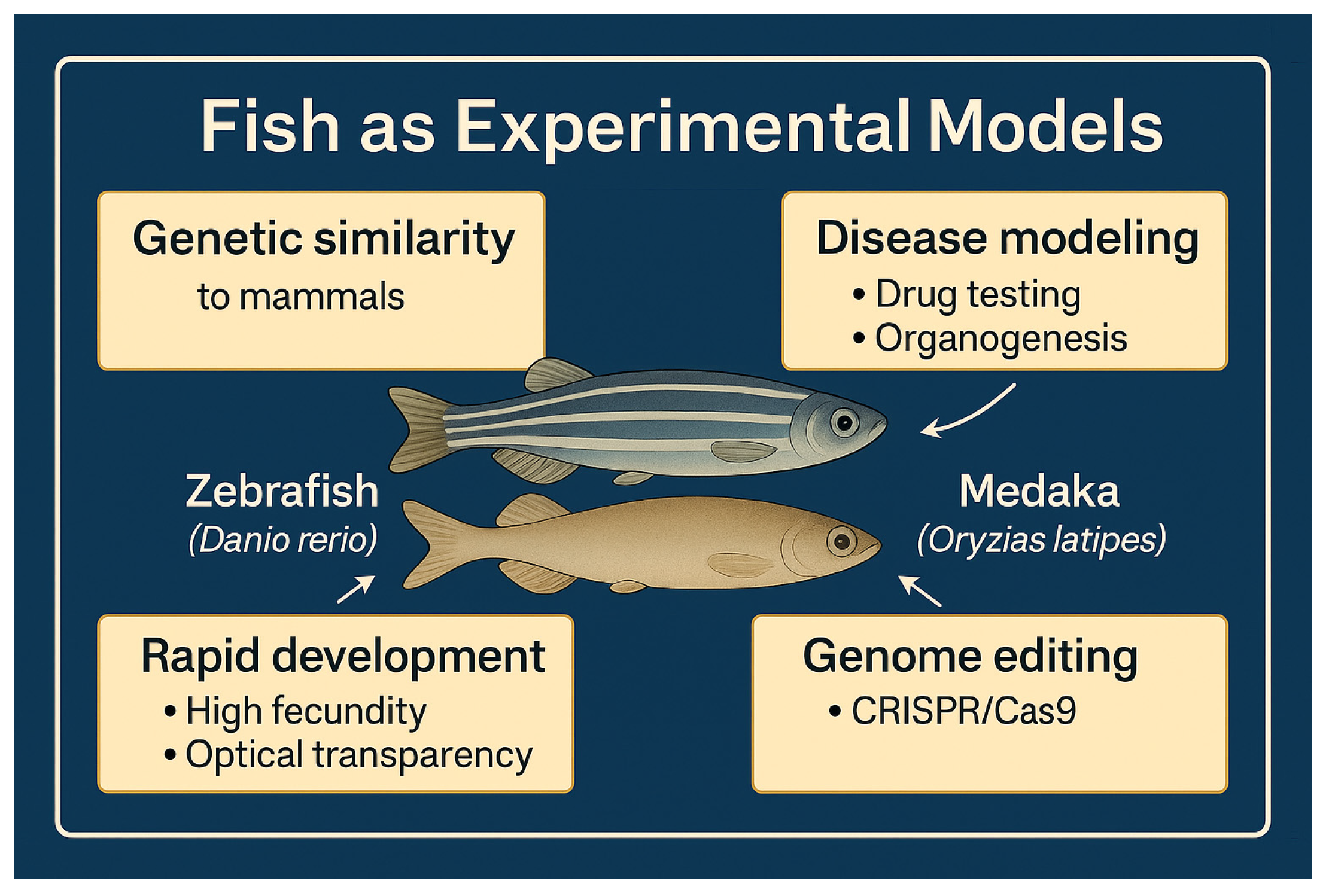
Figure 1. Fish as Experimental Models: Zebrafish (Danio rerio) and medaka (Oryzias latipes) are widely used in biomedical and toxicological research due to their genetic similarity to mammals, suitability for disease modeling (e.g., drug testing and organogenesis studies), rapid development with high fecundity and optical transparency, and accessibility to genome editing techniques such as CRISPR/Cas9.
J 08 00039 g001
Figure 2. Advantages of Using Fish as Experimental Models: Fish models offer several benefits for research, including a high number of offspring, short life cycle, easy maintenance, lower economic cost, and higher ethical acceptability, making them efficient and practical for experimental studies.
J 08 00039 g002
Figure 3. Fields of Application for Fish as Experimental Models: Fish models are used in diverse research areas, including disease modeling (e.g., cancer, cardiovascular, renal, hematopoietic, and skeletal disorders), genetics and genomics (transgenic lines, genome editing), developmental biology (transgenic lines, advanced imaging), immunology (immunological studies), pharmacology and toxicology (drug discovery and testing), and neuroscience (behavioral assays).
J 08 00039 g003
Figure 4. Ethical Aspects and Regulations in the Use of Fish as Experimental Models: Key ethical considerations include adherence to regulatory frameworks, ensuring animal welfare, following the 3Rs principles (Replacement, Reduction, Refinement), and implementing appropriate anesthesia, euthanasia, and welfare procedures. All research activities require review and approval by competent ethical committees.
J 08 00039 g004
Figure 5. Challenges and Limitations of Using Fish as Experimental Models: Major constraints include physiological differences compared to mammals, the need for specialized equipment, and infrastructure with trained expertise. Additionally, the absence of certain organs in fish limits their applicability in specific research areas.
J 08 00039 g005
Disclaimer/Publisher’s Note: The statements, opinions and data contained in all publications are solely those of the individual author(s) and contributor(s) and not of MDPI and/or the editor(s). MDPI and/or the editor(s) disclaim responsibility for any injury to people or property resulting from any ideas, methods, instructions or products referred to in the content.