Article

Fungal and Prokaryotic Communities in Soil Samples of the Aral Sea Dry Bottom in Uzbekistan

by
Alexandra Šimonovičová
1,
Eva Pauditšová
2,
Sanja Nosalj
1,*,
Medetbay Oteuliev
3,
Nikola Klištincová
4,
Francesca Maisto
4,
Lucia Kraková
4,
Jelena Pavlović
4,
Katarína Šoltys
5,6 and
Domenico Pangallo
4,7
1
Department of Soil Science, Faculty of Natural Sciences, Comenius University in Bratislava, Ilkovičova 6, 842 15 Bratislava, Slovakia
2
Department of Spatial Planning, Institute of Management, Slovak University of Technology, Vazovova 5, 812 43 Bratislava, Slovakia
3
Department of Economic and Social Geography, Karakalpak State University Named after Berdakh, Abdirov st. 1, Nukus 742012, Uzbekistan
4
Institute of Molecular Biology, Slovak Academy of Sciences, Dúbravská Cesta 21, 841 04 Bratislava, Slovakia
5
Department of Microbiology and Virology, Faculty of Natural Sciences, Comenius University in Bratislava, 842 15 Bratislava, Slovakia
6
Comenius University Science Park, Comenius University in Bratislava, 841 04 Bratislava, Slovakia
7
Caravella, s.r.o., Tupolevova 2, 851 01 Bratislava, Slovakia
*
Author to whom correspondence should be addressed.
Soil Syst. 2024, 8(2), 58; https://doi.org/10.3390/soilsystems8020058
Submission received: 3 April 2024 / Revised: 11 May 2024 / Accepted: 16 May 2024 / Published: 21 May 2024

Abstract

:
Due to the falling water level in the Aral Sea and Muynak Lake, the content of salts dissolved in the water has gradually increased, and toxic elements have been deposited at the lake’s bottom and subsequently washed into the Aral region by the river. Bacteria, archaea and fungi are crucial for the cycling of several important inorganic nutrients in soils. From 15 genera and 31 species of recovered microscopic filamentous fungi, a big group was melanized, of which most of them were also phytopathogenic. The second group consisted of keratinophilic species. Isolated bacteria mainly included members of the genera Arthrobacter, Bacillus, Massilia, Rhodococcus and Nocardiopsis. High-throughput sequencing analysis permitted a better view of the mycobiome and prokaryotic communities (comprising archaea). The cultivation and sequencing approaches were shown to be complementary. The aim of the work was to identify soil microorganisms, including the order Halobacteriales, and to discover the differences in species diversity depending on soil salinity and the presence of PTEs in soil.

1. Introduction

The Aral Sea (a fresh water lake) is situated in the Central Asian deserts, on the border between Kazakhstan and Uzbekistan (in the autonomous Republic of Karakalpakstan). Up until the third quarter of the 20th century, the Aral Sea was the fourth largest lake in the world, with an area of 68,000 km2, and the largest inland body of salty reservoirs in the world. The Aral Sea has been steadily shrinking since the 1960s after the rivers that fed it were diverted by Soviet irrigation projects. In the past, the Aral Sea was supplied with water from the rivers Amu Darya (from the south) and Syr Darya (from the northeast). Those rivers were diverted in the 1960s for the irrigation of the desert region surrounding the Sea in order to favor agriculture over supplying the Aral Sea basin. Much of the Amu Darya and Syr Darya rivers feeding the lake were diverted to irrigate cotton fields and rice paddies. Irrigation works during that period turned many desolate areas into flourishing oases and provided water and tillable soil. However, in the last fifty years, more than 85 percent of the Aral Sea has disappeared.
From 1973 to 2020, there has been a profound reduction in the overall area of the Aral Sea and an increase in land area as the basin. Due to the shortage of water in the Aral Sea region, the watering of the natural lakes in the Amu Darya delta has sharply decreased. Currently, there are only about 10 lakes formed as a result of waste and collector-drainage waters, and the share of natural lakes is only about 5 thousand hectares [1].
The remnants of the Aral Sea water and exposed lake bottom provide extreme environmental conditions for organisms. A secondary problem of this area is the strong winds that pick up and deposit bed soil. The crops from agricultural land have been appreciably affected due to heavily salt-laden particles falling on arable land in and around the former Aral Sea. The intense scattering of saline soil by wind also contributes to a significant reduction in breathable air quality and causes human health problems [2].
Soil salinization is the source of many of environmental problems, such as land degradation, reduced crop yields, contaminated freshwater, biodiversity reduction, etc. Anthropogenic salinization of soils is a global threat. This process has been recorded, e.g., in the Euphrates Basin in Syria [3], the Yellow River Basin in China [4] and also in other regions of the world. Together with salinity, the presence of heavy metals or other types of potential toxic elements (PTEs) is to be considered as another limiting factor to the reduced biodiversity. In fact, recent studies have shown that over the years, various quantities of toxic material coming from different types of industrial manufactures [5] have been deposited in the lake sediment. The results of these works highlighted the high presence of Al, As, Cd, Pb, U and PTEs, but unfortunately, they are related only to regions belonging to Kazakhstan. According to information found in the literature, data on the conditions of the Aral Sea in Uzbekistan are scarce and relate to the Amu Darya River Basin or to Dautkul Lake, situated on the right bank of the Amu Darya River, 47 km north of the city of Nukus [6]. On the other hand, it is known that the contaminated sediment of the former seabed (in the Karakalpakstan region, in the vicinity of the Aral Sea) has been disseminated over the surrounding area by strong winds, and this deterioration of the ecosystem has created a hazardous situation for the health of the people of this region [7].
The desiccated Aral Sea Basin, rich in salt and toxins, offers a unique natural setting for the examination of microbial community structures and their ecological functions. Many microorganisms are able to adapt to extreme environmental conditions of natural origin [8]. However, the extreme conditions of anthropogenic origin which man has caused by his careless and inappropriate interference with nature are much worse. In addition to soil salinity, these are, for example, contaminated environment by radiation from 3 to 5 orders higher than the background radioactivity, as at the Chernobyl Nuclear power Plant [9], contamination by heavy metals and toxic elements after mining activities [10], and many others. The soil salinity reduces microbial activity and changes the microbial community structure [11]. Salinity reduces microbial biomass mainly because the osmotic stress results in the drying and lysis of cells [12]. On the other hand, bacteria, archaea and fungi are crucial for the cycling of several important inorganic nutrients in soils [13].
The combination of salinity and heavy metals contributes to the presence of halophilic and halotolerant bacterial and archaeal species capable of resisting and metabolizing these dangerous elements [14]. In addition to the ecological value brought by the identification of these microbial species, it is also very interesting to learn about them for their possible use in various biotechnological fields. For example, some groups of archaea can be used in agriculture so that plants can tolerate soil salinity [15]. Bacteria can be exploited as plant promoters and also to remediate potential agricultural soil from heavy metals [16].
Microscopic fungi represent an extensive group of organisms that occur under extreme conditions, such as a halophilic environment [17,18], a hypersaline environment [19] or an alkaline environment [20]. The adaptation mechanisms of microfungi are different. In a hypersaline environment, some important mechanisms are the high osmolarity glycerol signaling pathway for sensing and responding to increased salt concentration [8], the accumulation of inorganic ions intracellularly to balance the salt concentrations in their environment [21], and adaptation to low water activity and high concentrations of toxic ions [22]. In other extreme environments, they are able to translocate an array of naturally occurring waste, such as man-made radionuclides in the mycelium, and have also been shown to adapt to stress conditions caused by melanin pigments, which are among the most stable and resistant of biochemical materials [9]. Despite the difficulties in both sample collection and cultivation of microscopic fungi from extreme environments, this environment can also be a promising source of novel species [23]. They are considered to be potential sources for the discovery of bioactive compounds and compatible solutes, including novel and/or extraordinary enzymes. In addition, some extremophilic microorganisms are capable of producing novel bioactive secondary metabolites [24], as well as massive amounts of compatible solutes that are useful as stabilizers for biomolecules and in various fields in biotechnology [25], as remediation, for biosorption of heavy metals [26] and for the application of indigenous species as biorefineries [17].
Because macroscopic species have all but disappeared with the reduction in the Aral Sea water area and the extreme climatic conditions of the desert region have limited the biodiversity, we focused our study on the soil microbiota. We compared the species diversity of bacteria, archaea and fungi in localities situated at different distances from the Muynak (Moynaq; Uzbekistan) water body, which is among the remnants of the Aral Sea in the southern part of the original water reservoir. The aim of the work was to identify soil microorganisms and to discover the differences in species diversity depending on soil salinity and the presence of PTEs in soil.

2. Materials and Methods

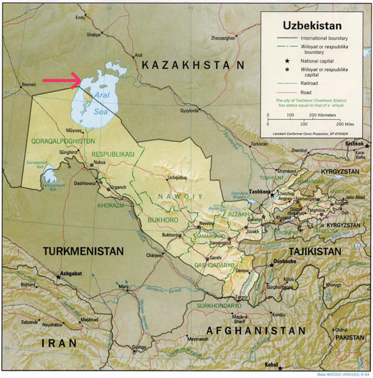
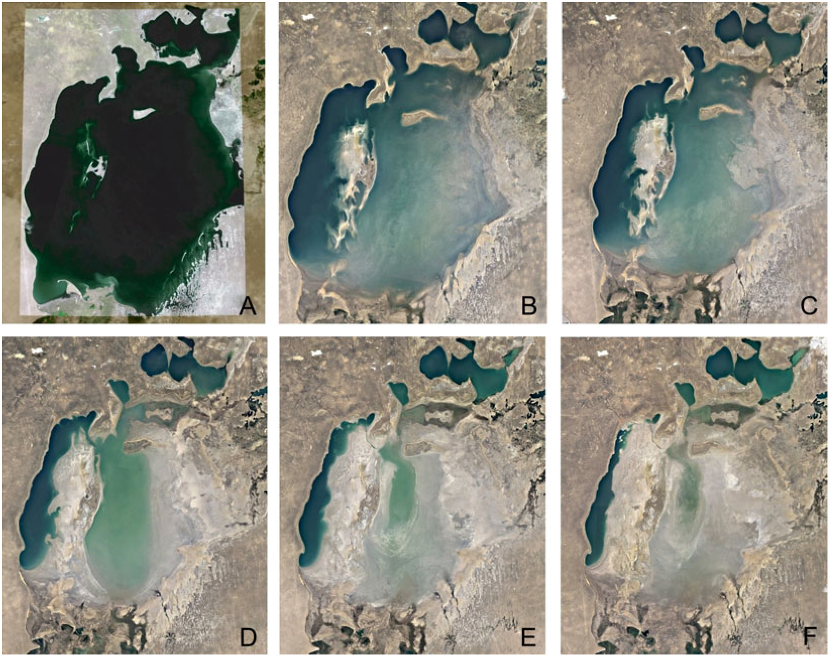
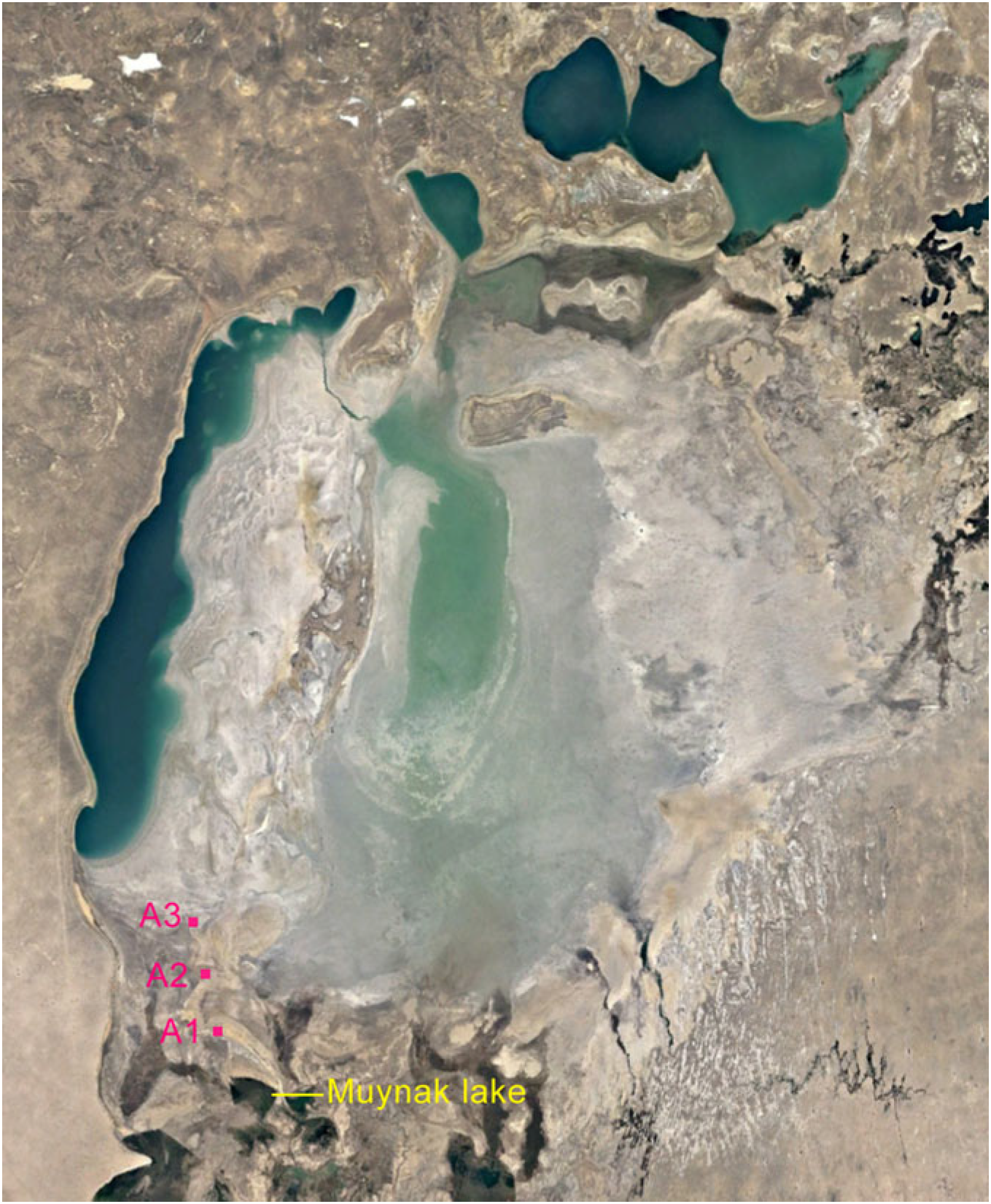
2.1. Study Area and Soil Sampling

Soil samples were taken from the dry bottom of the southern part of the Aral Sea in Uzbekistan (Figure 1), from three sites near Lake Muynak between which the distance is approximately 8–9 km (Figure 2). This is a freshwater lake fed by water from the Amu Darya River. The water supply in the lake from the river is currently limited. River water is used to irrigate cotton fields in Uzbekistan. Much of the water evaporates before it reaches the fields and the original river delta, and only a small volume of water reaches Muynak Lake. As a result of increased evaporation, water is declining in all surface water bodies that have been supported by river water in the past. The Amu Darya delta supported about 2600 lakes in the 1960s; in 1985 it supported only 400 [27], and the current number is even lower. The samples were taken from this territory because the concentrations of salts and minerals began to rise in the shrinking body of water (Figure 3). This process was the reason for changes in the lake’s ecology, causing an extreme reduction in biodiversity. The level of salinity rose from approximately 10 g/L to often more than 100 g/L in the remaining Southern Aral [28]. The selection criterion of the sites was the different distances from the water body of different parts of the Aral Sea. The mutual distance between the research sites (A1, A2, A3) was 25 km. Samples from site A3 are situated closest to the former water body of the Aral Sea, with brackish water, and farthest from the Muynak Lake. Site A2 is between A1 and A3. Soil samples were taken at two soil depths, up to 5 cm (samples marked a) and from a depth of 50 cm (samples marked b). Soil samples from each localityA1a, A1b, A2a, A2b, A3a and A3b—were taken from five sampling points. In the laboratory, soil samples from each sampling point were homogenized by quartering and sieved through a 2 mm sieve to obtain a fine soil. The fine soils were stored at 4 °C in darkness for approximately 10 days, until the soil samples were processed. The resulting fine soils were used for all chemical and microbiological analyses.

2.2. Chemical Analyses

Basic chemical parameters, such as pHH2O, the amount of TC, TN, humus and CaCO3, and the amount of available nutrients and salts (as Ca2+, Na2+, Mg2+, K+, Al3+, Mn2+, Fe2+ and H+), were analyzed in the certified laboratory of the National Forest Centre, Zvolen, Slovakia. All soil samples were also analyzed for their metal content (Ag, Al, Be, Bi, Cd, Co, Cr, Cu, Fe, Hf, Ga, In, La, Mg, Mo, Nb, Ni, Pb, Re, Sc, Sn, Ta, Ti, Tl, V, W, Y, Zn, Zr), lanthanide content (Ce, Dy, Eu, Gd, Nd, Pr, Sm, Tb), alkali metal content (Cs, Na, K, Li, Rb), alkali earth metals content (Ba, Ca, Sr), semi-metal content (As, Sb and Te), non-metals (P, S, Se) and actinides content (U and Th) in the Certified Laboratories of Bureau Veritas Commodities Canada, Ltd., Vancouver, BC, Canada (Tables as Supplementary Material). Analyses of all elements are included in the Supplementary Material.

2.3. Mycological Analyses

The soil mycobiota was isolated by the plating method up to a dilution of 104 CFU (Colony Forming Units) per 1 g of dry sample plates of SAB (Sabouraud Dextrose Agar), CDY (Czapek Dox Yeast Extract), MEA (Malt Extract Agar Base w/Mycological Peptone) and DMB (Dichloran Medium Base w/Rose Bengal; all media from HiMedia, Mumbai, India). An adequate concentration of NaCl in % (NaCl p. a., Merck, Darmstadt, Germany) was always added to the agar media used, depending on the sampling point A1, A2 and A3. Cultivation was carried out in the dark at 25 °C for 7–10 days. Each soil sample was always processed in three replications. All morphologically distinctive colonies were selected from the resulting mixed culture and purified. The isolates were then maintained on the original isolation agar media. Pure cultures were identified according to the phenotype using mycological diagnostic keys [29] and according to their Internal Transcribed Spacer (ITS) sequences of the amplicons produced by the primers ITS1–ITS4 [30]. The obtained sequences were deposited to the GenBank database under the accession numbers: PP085461–PP085492.
Valid names of microscopic filamentous fungi were modified according to Hubka et al. [31] and Visagie et al. [32]. All figures of microscopic filamentous fungi were observed under an Axio Scope A 1 Carl Zeiss Jena light (Carl Zeiss Microscopy GmbH, Jena, Germany) microscope in a drop of lactic acid enriched with cotton blue stain (0.01%).

2.4. Bacterial Analyses

Ten grams (wet weight) of each soil sample (A1a, A1b, A2a, A2b, A3a and A3b) was combined in sterile 250 mL Erlenmeyer flasks with 90 mL of a 0.9% (w/v) NaCl solution. The mixture was then incubated at 28 °C in a shaker incubator at 90 rpm for 2 h. After incubation, the resulting suspensions were filtered through Whatman 1 filter paper (Merck, Darmstadt, Germany). Subsequently, these filtered soil suspensions were decimally diluted, and the dilutions were inoculated onto two distinct agar media for the isolation of bacteria: tenfold-diluted Luria–Bertani (LB10 agar; peptone 1 g L−1, yeast extract 0.5 g L−1, NaCl 1 g L−1, agar 15 g L−1; [33] and Reasoner’s 2A (R2A; Himedia, Mumbai, India)) supplemented with cycloheximide (80 mg L−1; Sigma-Aldrich, Seelze, Germany). The incubation lasted a minimum of two weeks at room temperature (24–26 °C). Following this incubation period, isolated bacterial colonies were selected on the basis of their macro-morphological characteristics and maintained on R2A plates.
Bacterial representatives were identified by the sequencing of their 16S rRNA genes using the primers 27F and 685R [30]. The PCR amplicons of bacterial isolates were subjected to purification using ExoSAP-IT (Affymetrix, Cleveland, OH, USA) and subsequently sent for sequencing at a commercial facility, GATC-Biotech, in Konstanz, Germany. The resulting sequences were directly compared with those available in GenBank using the BLAST program (http://blast.ncbi.nlm.nih.gov/Blast.cgi, accessed on 3 January 2024). The sequences were then deposited in GenBank, with accession numbers PP078790–PP078818.
The similarity of soil micromycocoenoses and bacteriocenoses based on identified fungi and bacteria was determined according to Sőrensen SS = 2a/(2a + b + c) and Jaccard SJ = a/(a + b + c), where a is the number of species in sample a; b is the number of species in sample b; and c is the number of species in common in both (a and b) samples [34].

2.5. Next-Generation Sequencing Analysis

2.5.1. DNA Amplification of Bacterial, Archaeal and Fungal Communities

DNA extraction from the six soil samples was carried out using the DNeasy PowerSoil Pro Kit (Qiagen, Hilden, Germany), following the manufacturer’s instructions. Subsequently, the elution process was performed multiple times, concluding with a final volume of 2 × 30 µL of TE buffer.
The total DNA of bacterial, archaeal and fungal communities was amplified using the sets of primers oriented to bacterial (27f/685r) and archaeal (Arc344f-mod/Arch958r-mod) 16S rRNA [35] and to fungal ITS (ITS1/ITS4; [36]).

2.5.2. High-Throughput Sequencing and Data Analysis

PCR amplicons (amplified by primer sets for 16S rRNA gene for bacteria and archaea, ITS for fungi) were transposon-tagmented using the Nextera XT DNA Library Preparation Kit (Illumina Inc., San Diego, CA, USA) and consequently indexed and amplified with low-cycle pcr. The DNA profile of the sequencing libraries was verified using an Agilent 2100 Bioanalyzer (Agilent Technologies, Santa Clara, CA, USA) and a High-Sensitivity DNA Kit (Agilent Technologies, Santa Clara, CA, USA) and quantified using a Qubit 2.0 Fluorometer (Thermo Fisher Scientific, Waltham, MA, USA). The DNA libraries were analyzed using the Illumina MiSeq platform via 200 bp paired-end reads.
The quality of sequencing reads was verified using the FastQC tool (Version 0.12.0). The paired reads were trimmed for sequence quality and length and taxonomically classified with kraken2 using [37] and the Standard RefSeq database, including archaea and bacteria (v:2022-06-07). The produced sequences were deposited in GenBank (https://www.ncbi.nlm.nih.gov/, accessed on 17 January 2024) Bioproject PRJNA1065900.
Alpha-diversity of bacterial, fungal and archaeal communities was performed using Simpson’s diversity index. The statistical test used was Mann–Whitney U.

3. Results and Discussion

Due to the loss of water in the Aral Sea, the conditions for living organisms have changed in the studied area from the 1960s to the present. A fundamental change occurred in the hydrological regime and subsequently in the chemical and physical parameters of the soils. Increasing soil salinity, circulation change and oxygenation have had a direct negative impact on biodiversity [13]. Plants and animals that got stuck in the gradually drying lake and subsequently died became a source of organic matter for microorganisms. Soil microscopic filamentous fungi have gradually adapted to the new extreme conditions.

3.1. Soil Characteristic

According to the pH range [38], the soil reaction of all the study soil samples from the depth of 5 cm (assigned as “a”) is medium alkali, and of all the study soil samples from the depth of 50 cm (assigned as “b”) are highly alkali. The amount of organic matter is also very low. The calcium content in the soils is variable and significantly influenced by the parent rock and especially by precipitation (Table 1). Therefore, it accumulates in areas with limited precipitation, which is evident in samples from a depth of 50 cm (A3b, A2b and A1b). The salinity content of the analyzed soil samples is shown in Table 2. In the studied soil samples, a total of 59 elements were detected, including metals, lanthanides, alkali metals, alkali earth metals, semi-metals, non-metals and actinides. From this analysis, there is an evident increase in the values of metals in both soil samples from locality A2 (Cu, Pb, Zn, Ag, Ni, Co, Fe, V, La, Cr, Mg, Al, W Zr, Sn, Sc, Y, Nb and Ga). The same situation also occurred in the analysis of lanthanides (Ce, Pr, Nd, Dy, Er). From localities A2 and A3, increased values of Sr were detected. From the soil samples of locality A3, the highest amount of U was detected compared to the other soil samples (Table in Supplementary Material).

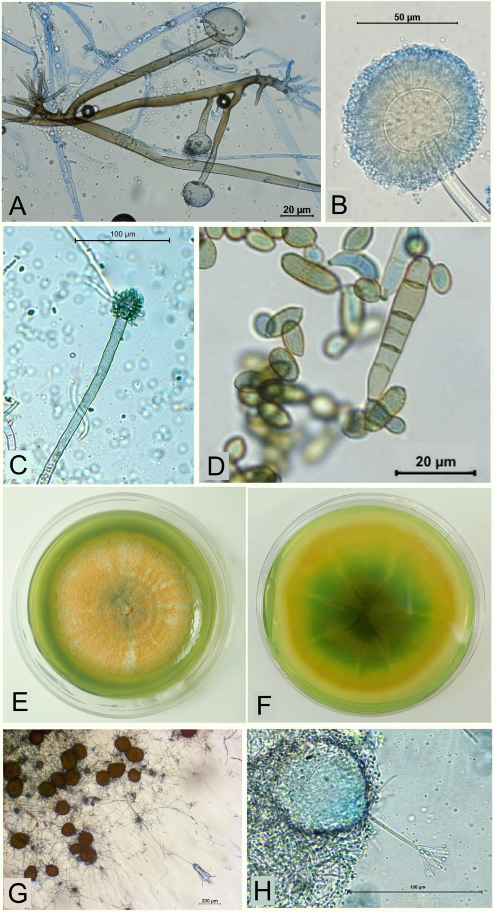
3.2. Cultivable Soil Microscopic Filamentous Fungi

From the analyzed soil samples, 15 genera and 31 species of soil microscopic filamentous fungi were recovered (Table 3). The species Rhizopus microsporus (Figure 4A) and Saksena vasiformis, from the strain Zygomycota, occurred only in samples A1a, A2a and A2b. All other species belonging to the strain Ascomycota were much more abundant. The most species (seven) were recorded in the genera Aspergillus, as A. flavus, A. fumigatus, A. pseudoglaucus, A. jensenii, A. niger (Figure 4B), A. oryzae and A. proliferans (Figure 4C). In the genus Cladosporium, five species were identified, as C. cladosporioides, C. floccosum, C. herbarum, C. iridis (Figure 4D) and C. sphaerospermum. Also, in the genus Penicillium, five species were identified, as P. chrysogenum, P. corraligenum (Figure 4E–H), P. echinulatum, P. expansum and P. rugulosum. Four species were identified in the genus Alternaria, as A. alternata, A. atra (Figure 5A), A. japonica and A. tenuissima.
All of the identified species of the genus Alternaria were isolated from soil sample A1a (the depth of 5 cm). Alternaria is one of the most ubiquitous fungal genera, inhabiting nearly every environmental substrate, but most species have been recorded on an extremely wide range of plants. Among the less common species is Alternaria japonica, occurring on seeds, which causes black spot on turnips, and head rot and leaf spots on Brassicaceae [39]. Also, Cladosporium sphaerospermum belongs to the group of melanized fungi isolated from the same sample. Aspergillus fumigatus and Rhizopus microsporus also occurred in soil sample A1a. The identification of Rhizopus microsporus is very interesting, because of its pathogenicity on plants such as maize, sunflower or rice. It has also been isolated from pristine soils from sites in Antarctica. According to Durán et al. [40], the extreme conditions that coexist in Antarctica produce a strong selective pressure that could lead to the evolution of novel mechanisms for stress tolerance by indigenous microorganisms. The lowest number of species (Paraengyodontium album, Penicillium expansum, P. chrysogenum and melanized Stachybotrys chartarum) was isolated from soil sample A1b (the depth of 50 cm). All of these species are known as cosmopolitan, occurring in variable soils [41,42]. According to Jaouani et al. [43], the keratinophilic species Paraengyodontium album is halotolerant and able to grow in solid and liquid media with a salt concentration from 10% to 15% NaCl. This strain is even able to tolerate 20% NaCl and alkaline tress at pH 10. It was identified from locality A1, together with 11 species of soil microscopic filamentous fungi (Table 3).
From a mycological point of view, locality A2 is very interesting. From the depth of 5 cm (sample A2a), only six species were isolated (Chaetomium globosum, Cladosporium floccosum, C. herbarum, Neomicrosphaeropsis italica (Figure 5B), Rhizopus microsporus and Simplicillium sympodiophorum). All these species occur in soil but all of them are also plant pathogens [44,45,46]. Of all the studied soil samples, the most microfungal species (11) were identified from sample A2b (from the depth of 50 cm). A total of six cosmopolitan aspergili were identified. According to Hubka et al. [31], Aspergillus proliferans is an economically important strain, and it seems to be relatively common. It has been isolated from variable soils in Tibet or China, from moldy wood, cave sediment, inside books from a library, from air in a living room and from unknown sources [47], as well as from onychomycoses [48]. The species Auxarthron umbrinum (Figure 5C,D), in contrast, was until now isolated only from onychomycoses [49]. The phytopathogenic species Cladosporium iridis has been isolated from leaf spot and blotch of Iris sp. from many countries, such as Africa and Cyprus and also from Uzbekistan, Turkmenistan, Kazakhstan, Kyrgyzstan and others [50]. The species Myriodontium keratinophilum (Figure 5E) is widespread in the environment and able to colonize keratinous surfaces of human body [51]. It has been isolated from soil samples in India [52] and also from the funeral clothes of Cardinal Peter Pázmany [53]. Penicillium species are among the most widespread fungal organisms on the Earth, but marine environments and marine subaqueous soils have been poorly studied. Penicillium coralligerum (Figure 4E–H) is a marine species sometimes referred to as a deep-sea fungus and in some languages named the equivalent of a “deep-sea mold”, isolated from subaqueous soil in the Sakhalin shelf [54]. According to the study of Takahashi et al. [55], among 91 deep-sea fungal strains, Penicillium coralligerum could produce notable anti-Saprolegnia parasitica activity. Saksena vasiformis is a species able to cause severe human infections. It has also been isolated from soils, driftwood or grains [56].
Locality A3 is closest to the southern part of the Aral Sea, where there is still water. From the soil depth of 5 cm (sample A3a), three aspergili were isolated, from which Aspergillus jensenii was described as a new species in 2012. This species belongs to the section Versicolores, and according to Jurjevic et al. [57], variable propagules of A. versicolor have been recovered from the highly saline Dead Sea, showing an ability to survive conditions of salinity or drying. High tolerance to salinity may extend to other species in the section Versicolores. Aspergillus jensenii was also isolated from an old manuscript from Indonesia [58] and from the soil of potted plants [59]. All other microscopic fungi isolated from this soil sample belong to ubiquitous species, including in the case of the sample from a depth of 50 cm (A3b). Among the species isolated are the phytopathogenic Cladosporium cladosporioides, C. iridis and Epicoccum nigrum and the entomopathogenic Isaria farinosa [60].
Based on the calculation according to Sörensen and Jaccard [34]. (Table 4), the similarity of mycocoenoses is the highest between samples A1a and A1b (0.64). The similarity values of 0.57 (between samples A2b and A3b) and 0.54 (between samples A1a and A3a) are on the border of similarity and difference of mycocoenoses.
The species composition of microscopic filamentous fungi of the arid and slightly halophile environment of the Aral Sea dry bottom is characterized by a big group of melanized species of the genera Alternaria, Cladosporium, Epicoccum and Stachybotrys, from which most of them are also phytopathogenic. The second group consists of keratinophilic species, such as Auxarthron umbrinum, Isaria farinosa, Myriodontium keratinophilum, Paraengyodontium album and Saksena vasiformis, which are widespread in nature but found most abundantly in keratin-rich environments, such as insects, feathers, animal fur, nails and hair. Fungal species belonging to the genera Alternaria, Aspergillus, Cladosporium, Chaetomium and Penicillium have been isolated from saline or hypersaline environments by many other authors [17,19,22], and for this reason they have been assigned as halophiles. Halophiles are defined as organisms requiring > 3% NaCl for growth [24]. Fungal species which can tolerate salt concentrations of 2 to 5% w/v are known as slight halophiles [17]. However, the long-term adaptation of microscopic fungi to salinity requires cellular and metabolic responses that differ from short-term osmotic stress signaling [18].

3.3. Bacterial Isolates

According to the isolated bacteria, the soil that had the richest bacterial diversity was from sample A1a. From this sample, 11 different species were isolated, mainly members of the genera Arthrobacter, Bacillus, Massilia and Rhodococcus. Soils from A3a and A3b displayed the poorest bacterial diversity, with four and three species isolated, respectively. Half of these species belong to the Bacillus genus (Table 5). Bacillus species were the only isolates spread in all the samples. Members of this genus have already been isolated from Aral soils, and some of them were able to mineralize [61] and to protect plants against pathogenic fungi [62]. Bacteria isolated exclusively from soils at A1a were Massilia strains; these bacteria have also been isolated in other arid soils from Uzbekistan [63] and Morocco and show interesting hydrolytic properties [64].
Other soil-specific isolates were members of the genera Rhodococcus and Nocardiopsis, which occurred in the soils at A1 and A2, respectively. The Rhodococcus species was previously isolated in Aral Sea areas in association with halophytes [62] and detected by metagenomics analysis in water samples [65]. However, Rhodococcus strains are able to adapt to different soil conditions, including high concentrations of salts and pollutants. These characteristics permit them to be versatile bacteria for bioremediation applications [66]. Several Nocardiopsis species have also been isolated from halophytes of the west Aral Sea basin, and they display strong proteolytic and cellulolytic activities [62]. They can be considered as free-living entities occurring in a variety of soils and other saline or hypersaline habitats, and they are producers of diverse bioactive compounds and extracellular enzymes [67].
Typical strains of the soils from A3 were as follows: Sphingomonas sp., Rathayibacter sp. and Paenisporosarcina sp. Wicaksono et al. [68] found that Sphingomonas were part of the bacterial community in the rhizosphere of the plant Suaeda acuminata, rather than being present in Aral soil samples, and Osman et al. [63] frequently found Sphingomonas in soil samples from the Kyzyl-Kum desert in Uzbekistan. Rathayibacter sp. and Paenisporosarcina sp. were not previously identified in Aral-related samples. However, Rathayibacter species are known mainly as opportunistic phytopathogenic bacteria that frequently occur in arid areas [69].
The highest similarity of bacteria is between samples A1a and A3b (0.76), A1a and A3b (0.75), A2b and A3b (0.69), A1a and A3b (0.67), A1a and A2a (0.65) and A2a and A3b (0.64). Much less samples show dissimilarity of bacteriocenosis (Table 6).

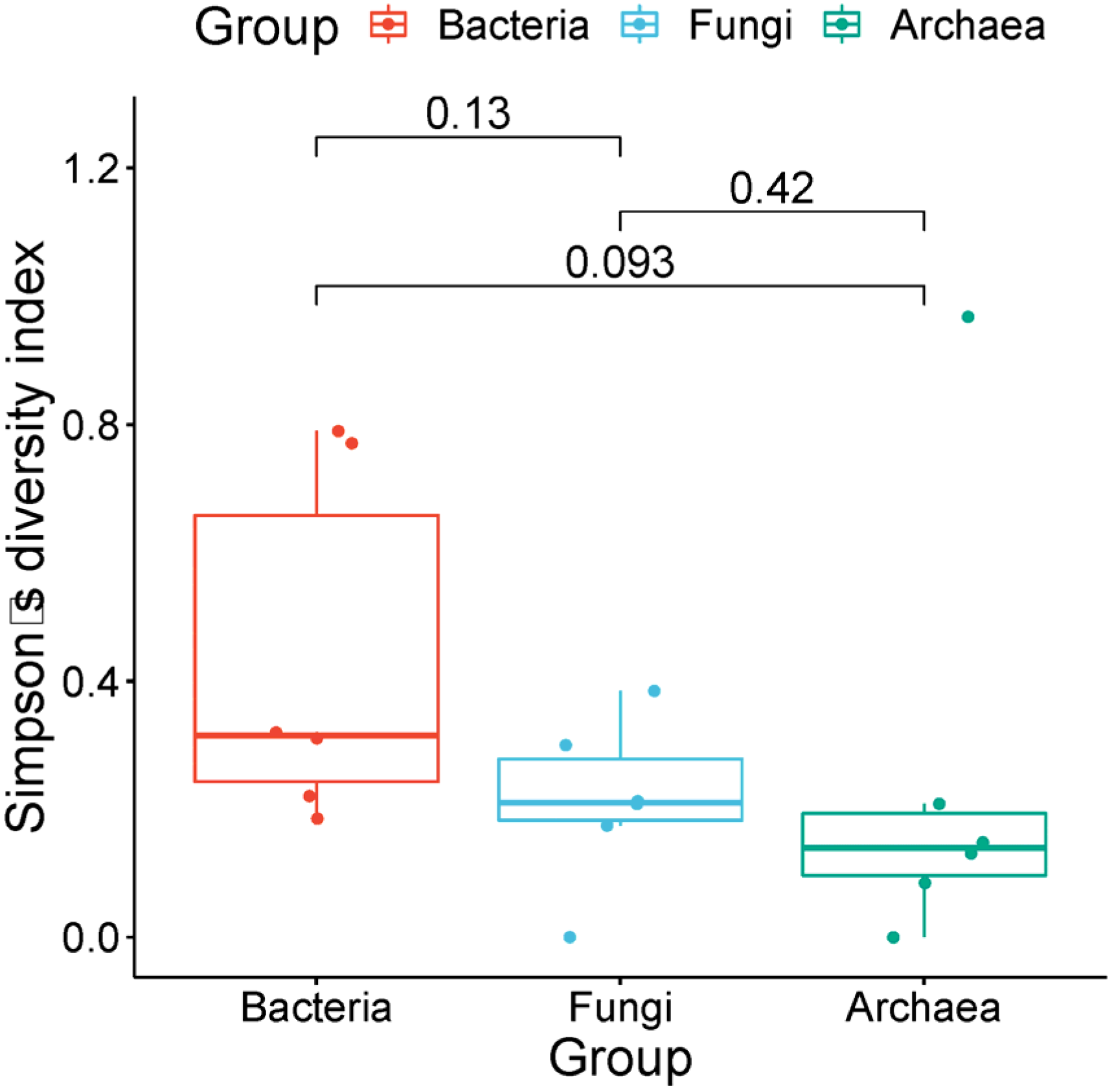
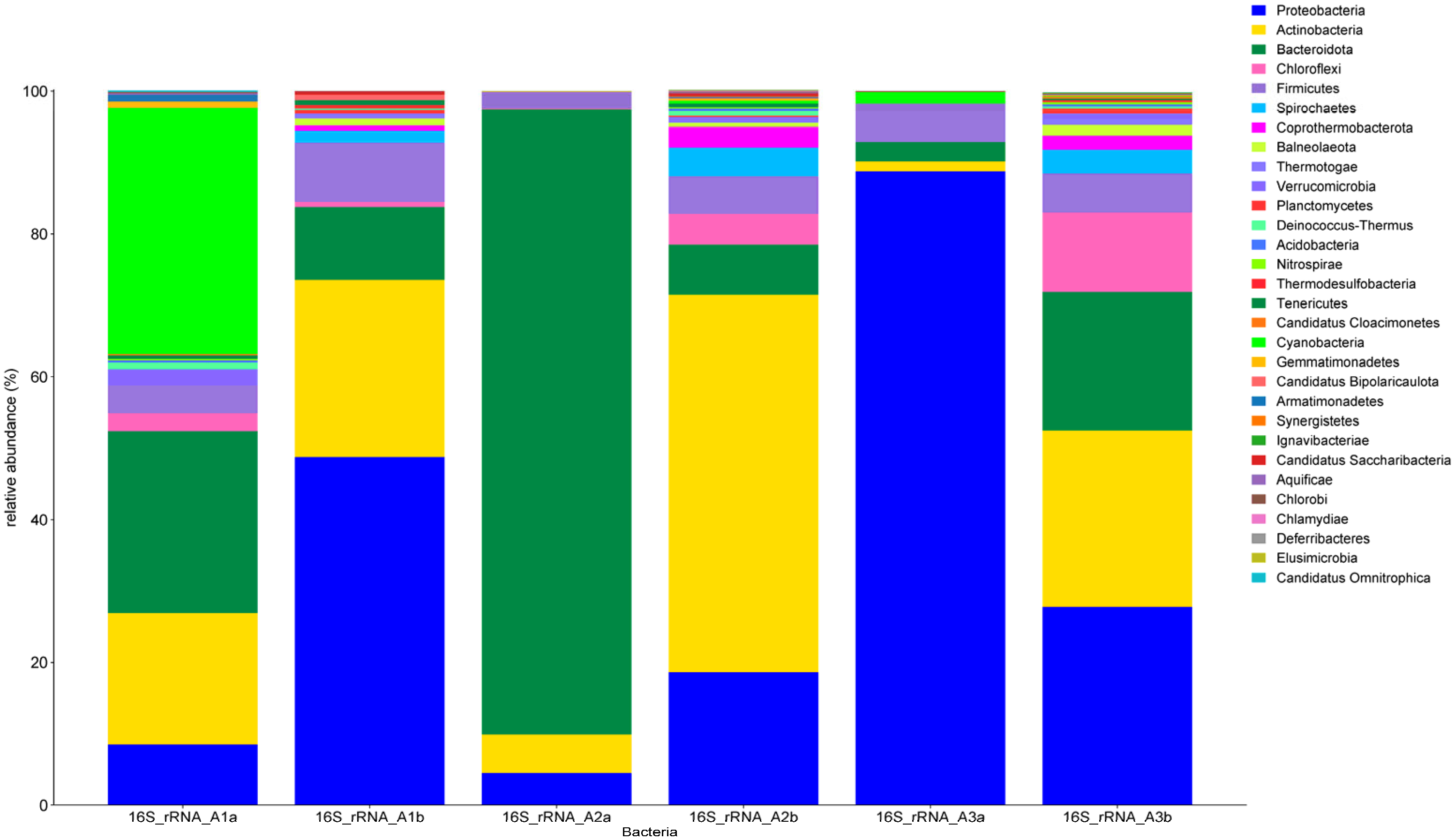
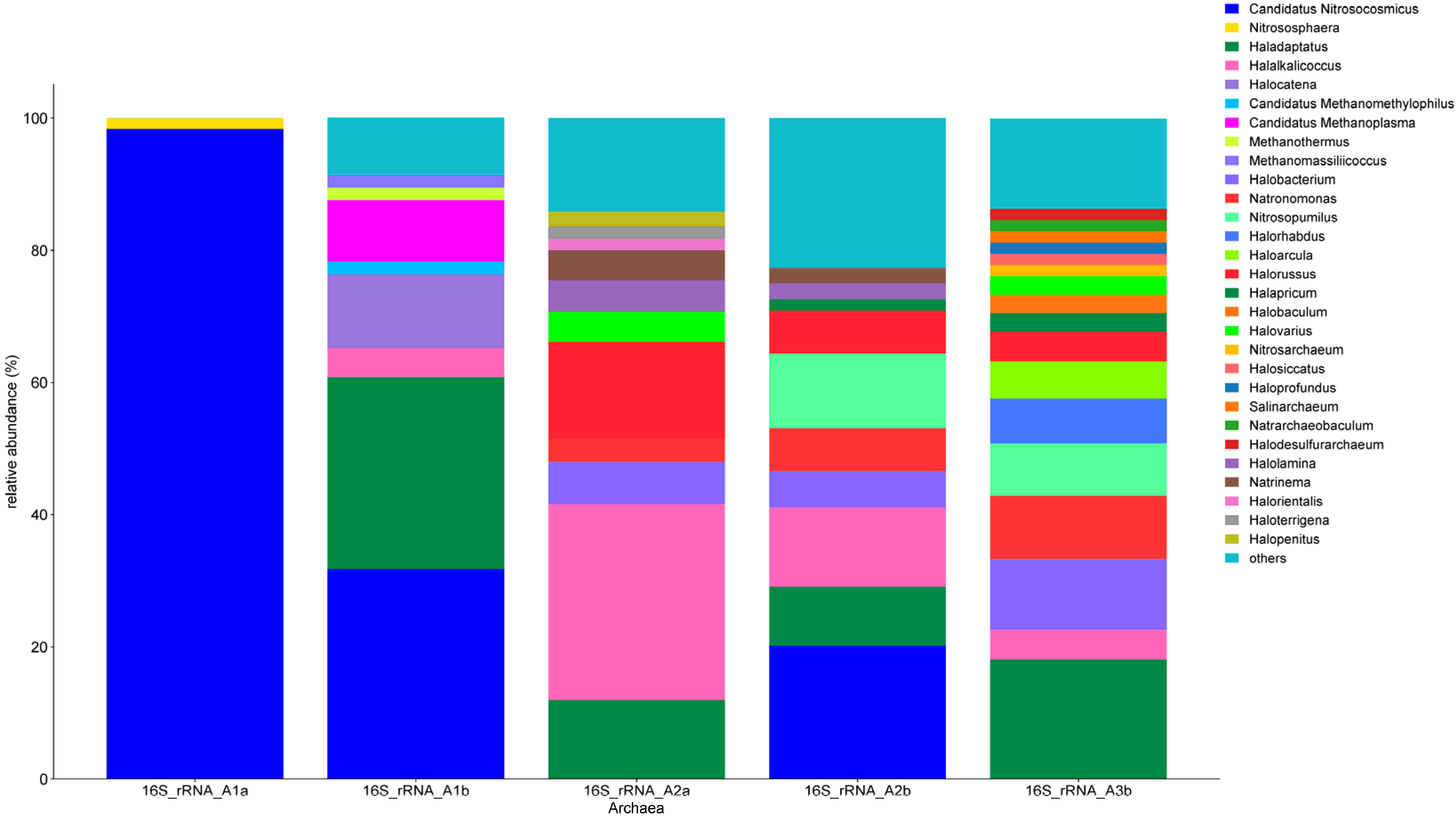
3.4. High-Throughput Sequencing Analysis

To see the differences in alpha-diversity between bacterial fungal and archaeal communities, we performed the Mann–Whitney U test, which did not show significant differences among communities. However, the highest Simpson’s index was found for bacterial communities and the lowest for archaea (Figure 6).

3.4.1. Fungi

The detected fungi belonged mainly to the phylum Ascomycota, namely the classes Saccharomycetes, Eurotiomycetes, Schizosaccharomycetes, Leotiomycetes and Sordariomycetes, while only two classes, Tremellomycetes and Malasseziomycetes, represented Basidiomycota (A3b 0.5%–A3a 5.1%). The PCR of sample A2b did not produce any amplicon; therefore, no data are available.
Nonetheless, 19 fungal genera were identified (Figure 7), of which Aspergillus, represented by Aspergillus oryzae and Aspergillus fumigatus, Colletotrichum higginsianum and Botrytis cinerea, comprised over 50% of all detected taxa (A1a—57%; A1b—73%; A2a—71%; A3b—82%). While sample A3a was uniquely dominated by Saccharomyces cerevisiae (59%), followed by Schizosaccharomyces pombe (16%), the latter was also detected in samples A1a (20%) and A1b (9%). Other species were also detected, such as Neurospora crassa (not found in A1a) and Malassezia restricta (not found in A3b), which belonged to the widely spread fungi.
Practically speaking, only a few links occurred between the cultivation and sequencing analysis. The correlation regarded only the high presence of Aspergillus species, and some members belonged to the order Sordariales (Neurospora/Chaetomium).
Considering the most abundant genera detected by high-throughput sequencing, new taxa were added to the isolated ones. In fact, the well-known phytopathogens Colletotrichum and Botrytis [70] were revealed. Members of the genera Saccharomyces and Schizosaccharomyces are not usually considered as halophilic microorganisms, and perhaps their detection is more associated with their ability to grow in the presence of toxic contaminants [71].

3.4.2. Bacteria

A total of 30 different bacterial phyla were identified (Figure 8), of which Pseudomonadota (A1b—49%, A3a—89%, A3b—28%), Bacteroidota (A2a—88%) and Actinomycetota (A2b—53%) were the most abundant, followed by Cyanobacteria (A1a—34%) and Bacillota (2–8%). The highest bacterial diversity at the phylum level was detected in samples A2b (26 phyla) and A3b (23 phyla).
The most abundant bacterial family of the A1a sample, Hymenobacteraceae (8%), was also identified in A2a (11%), while Chitinophagaceae, Rubrobacteraceae, Cytophagaceae and Oscillatoriaceae (from 7% to 5%) were unique. For each sample, a different profile of the most abundant bacterial families was typical: A1b (Marinobacteraceae 45%, Iamiaceae 9%), A2a (Flavobacteriaceae 57%, Hymenobacteraceae 11%, Fulvivirgaceae 9%), A2b (Iamiaceae 23%, Egicoccaceae 17.5%, Borreliaceae 5%), A3a (Comamonadaceae 33%, Burkholderiaceae 27%, Pseudomonadaceae 14%) and A3b (Flavobacteriaceae 17.5%, Egicoccaceae 11.3%, Iamiaceae 7.5%).
Focusing on the genus level, Rubrobacter tropicus, Oscillatoria nigro-viridis, Rhodocytophaga rosea and mainly Hymenobacter oligotrophus (4–6%) dominated from the 139 bacterial genera in sample A1a, while Marinobacter (46%; M. sp. Arc7-DN-1, M. salarius, M. sp. LPB0319), the oversized Actinomarinicola tropica (6.5%) and another 75 bacterial genera dominated in the A1b sample. Antarcticibacterium (32%; A. flavum, A. sp. 1MA-6-2, A. arcticum) dominated over Fulvivirga (11%; F. sp. W9P-11, F. sp. SS9-22) and another 80 genera with an abundance of less than 10% in the A3a sample, while of the 214 bacterial genera observed in the A2b sample, Egicoccus halophilus, Actinomarinicola tropica and Aquihabitans sp. Kera 3 altogether represented over 40% of all bacteria. Sample A3a was very scarce in terms of bacterial diversity, since “only” 47 bacterial genera were detected, mainly representatives of the phylum Pseudomonadota (Acidovorax sp. YS12, Lautropia mirabilis, Rubrivivax gelatinosus, Schlegelella thermodepolymerans and Pseudomonas spp.), which formed almost 70% of all classified sequences. In contrast, the environment represented by the A3b sample was dominated by Egicoccus halophilus and Aquihabitans sp. Kera 3 (14% and 6%; Actinomycetota), Antarcticibacterium sp. 1MA-6-2 (7%; Bacteroidota) and the Candidatus Promineofilum breve (7%, Chloroflexota), followed by another 174 bacterial genera.
Comparing the culture-dependent analysis with the high-throughput sequencing makes it clear that the two approaches are complementary. There is no link between the genera isolated by cultivation and those detected by Illumina sequencing. An example is given by Bacillus species which were isolated from each sample but which were not among the most abundant bacteria according to sequencing. Some small similarity between the two strategies was seen only at the phylum level regarding Pseudomonadota and Actinomycetota.
It is difficult to compare our results with the previous studies related to Aral Sea samples. Other authors oriented their taxonomical analysis at the phylum or maximally at the class level [65,72]. Correlations with earlier investigations only regarded the genera Rubrobacter, Hymenobacter, Marinobacter and Pseudomonas [63,68].
However, considering the most abundant detected genera, Antarcticibacterium, Egicoccus, Actinomarinicola and Aquihabitans, they are typical of marine environments, and they can tolerate a high concentration of salt [73].

3.4.3. Archaea

Almost all of the analyzed samples were found to be positive for Archaea, mostly belonging to the phylum Euryarchaeota (Figure 9). While the A1a sample was exclusively inhabited by Thaumarchaeota, especially with Candidatus Nitrosocosmicus franklandus (class Nitrososphaeria), the A1b sample was also represented by Halocatena (11%) and Haladaptatus (29%), in addition to Candidatus Nitrosocosmicus (32%). The latter, together with Halalkalicoccus and Halorussus, was also found to be the main representative of the A2a and A2b environments. At the species level, A2a and A2b were found to be dominated by Halalkalicoccus jeotgali (A2a 35.2%; A2b 22%), followed by Halorussus halophilus (A2a 10%; A2b 6%), while Natronomonas salina was present both in A2b (8%) and A3b (12%). The highest diversity could be observed in the A3b sample, which was represented by several abundant genera belonging to the order Halobacteriales (Haladaptatus, Halobacterium, Haloarcula, Halapricum, Halorhabdus, Natronomonas and Halorussus), as well as a representative of Nitrosopumilaceae—Nitrosopumilus. We were not able to detect any archaea in the A3a sample, because the PCR did not work with this sample.
It is evident that the archaeal community in all the analyzed samples is composed mainly of halophilic genera. Members of the order Halobacteriales are the most widespread; they were previously detected in samples of water [74] and soil [68] from the Aral Sea. These archaea in this habitat are involved in denitrification processes [68].
Archaea of the class Nitrososphaeria have also previously been detected in this kind of environment [72]. Members of this class harbored genes encoding a methane/ammonia monooxygenase and are involved in denitrification, dissimilatory nitrate reduction to ammonium and ammonia oxidation [68].

4. Conclusions

The environmental changes in the Aral Sea and also Muynak Lake have led to considerable transformation of all ecosystem elements. From the analyzed soil samples, 15 genera and 31 species of soil microscopic filamentous fungi were identified. The microfungal composition is characterized by a big group of melanized species of the genera Alternaria, Cladosporium, Epicoccum and Stachybotrys, from which most are also phytopathogenic. The isolated bacteria were typical of an arid environment, and several of them can be used for biotechnological applications exploiting their hydrolytic properties (Bacillus and Massilia), bioremediation activities (Rhodococcus) and their ability to produce bioactive compounds (Nocardiopsis).
The high-throughput sequencing approach displayed different microorganisms with respect to the cultivation strategy. The most abundant taxa detected by this method were Colletotrichum, Botrytis, Saccharomyces, Schizosaccharomyces, Antarcticibacterium, Egicoccus, Actinomarinicola and Aquihabitans. Moreover, the archaeal community mainly evidenced its halophilic character including the genera Haladaptatus, Halobacterium, Haloarcula, Halapricum, Halorhabdus, Halorussus and Halalkalicoccus.
The ecological crisis of the Aral Sea bottom caused by human activities has created the conditions for a change in biodiversity.

Supplementary Materials

The following supporting information can be downloaded at: https://www.mdpi.com/article/10.3390/soilsystems8020058/s1.

Author Contributions

A.Š. and S.N.—processing and analyses of soil samples and isolation, cultivation of soil microscopic fungi and their photo documentation. E.P. and M.O.—soil collection, data processing and cartographic images. N.K.—bacterial identification; F.M.—fungal identification; L.K.—sequencing analysis; D.P.—coordination of molecular analyses of isolated microfungi, data processing; K.Š.—processing and evaluation of prokaryotic communities. J.P.—bioinformatic data analysis. All authors have read and agreed to the published version of the manuscript.

Funding

This study was also supported by the European Regional Development Fund project: 313011V578 and the Slovak Research and Development Agency under the Contract no. APVV-21-0099: Effective management of innovation-oriented territorial clusters.

Institutional Review Board Statement

Not applicable.

Informed Consent Statement

Not applicable.

Data Availability Statement

Data are available through a reasonable request made to the corresponding author.

Conflicts of Interest

The authors declare no conflict of interest. The company Caravella, s.r.o. had no role in the design of the study; in the collection, analyses, or interpretation of data; in the writing of the manuscript; or in the decision to publish the results.

References

  1. Matzhanova, K.K.; Orel, M.M.; Matzhanov, T.K. The Aral Sea crisis and its impact on the current state of water bodies and higher aquatic vegetation. ESJ 2021, 2, 82–86. Available online: https://scholarzest.com/index.php/esj/article/view/269 (accessed on 15 November 2023).
  2. Turdimambetov, I.R.; Madreymov, A.; Pauditsova, E.; Oteuliev, M.O.; Bekanov, K.K. Influence of harmful environmental factors on the rate of incidence of children in Karakalpakstan. Cent. Asian J. Geogr. Res. 2021, 3–4, 55–63. [Google Scholar]
  3. Rahi, K.A.; Halihan, T. Changes in the salinity of the Euphrates river system in Iraq. Reg. Environ. Chang. 2010, 10, 27–35. [Google Scholar] [CrossRef]
  4. Siyu, X.; Zhixun, X.; Pingwu, W. Soil salinity in the irrigated area of the Yellow river in Ningxia, China. Arid Soil Res. Rehabil. 1996, 10, 95–101. [Google Scholar] [CrossRef]
  5. Liu, W.; Ma, L.; Abuduwaili, J. Historical change and ecological risk of potentially toxic elements in the lake sediments from North Aral Sea, Central Asia. Appl. Sci. 2020, 10, 5623. [Google Scholar] [CrossRef]
  6. Matsapaeva, I.V.; Osinskaya, N.S.; Danilova, E.A. Concentrations of heavy metals in bottom sediments of Lake Dautkul as an indicator of anthropogenic impact in the area south of the Aral Sea. Water Res. 2010, 37, 586–590. [Google Scholar] [CrossRef]
  7. Ataniyazova, O.A.; Baumann, R.A.; Liem, A.K.D.; Mukhopadhyay, U.A.; Vogelaar, E.F.; Boersma, E.R. Levels of certain metals, organochlorine pesticides and dioxins in cord blood, maternal blood, human milk and some commonly used nutrients in the surroundings of the Aral Sea (Karakalpakstan, Republic of Uzbekistan). Acta Paediatr. 2001, 90, 801–808. [Google Scholar] [CrossRef] [PubMed]
  8. Oren, A. Halophilic microbial communities and their environments. Curr. Opin. Biotechnol. 2015, 33, 119–124. [Google Scholar] [CrossRef] [PubMed]
  9. Belozerskaya, T.; Aslanidi, K.; Ivanova, A.; Gessler, N.; Egorova, A.; Karpenko, Y.; Olishevskaya, S. Characteristics of extremophilic fungi from Chernobyl nuclear power plant. Curr. Res. Technol. Educ. Top. Appl. Microbiol. Microb. Biotechnol. 2010, 1, 88–94. [Google Scholar]
  10. Šimonovičová, A.; Kraková, L.; Pauditšová, D.; Pangallo, D. Occurrence and diversity of cultivable autochthonous microscopic fungi in substretes of old environmental loads from mining acitivities in Slovakia. Ecotoxicol. Environ. Saf. 2019, 172, 194–202. [Google Scholar] [CrossRef]
  11. Andronov, E.E.; Petrova, S.N.; Pinaev, A.G.; Pershina, E.V.; Rakhimgalieva, S.Z.; Akhmedenov, K.M.; Sergaliev, N.K. Analysis of the structure of microbial community in soils with different degrees of salinization using T-RFL Pandreal-time PCR techniques. Eurasian Soil Sci. 2012, 45, 147–156. [Google Scholar] [CrossRef]
  12. Yuan, B.C.; Li, Z.Z.; Liu, H.; Gao, M.; Zhang, Y.Y. Microbial biomass and activity in salt affected soils under arid conditions. Appl. Soil Ecol. 2007, 35, 319–328. [Google Scholar] [CrossRef]
  13. Yan, N.; Marschner, P.; Cao, W.H.; Zuo, C.Q.; Qin, W. Influence of salinity and water content on soil microorganisms. Int. Soil Water Conserv. Res. 2015, 3, 316–323. [Google Scholar] [CrossRef]
  14. Tavoosi, N.; Akhavan Sepahi, A.; Amoozegar, M.A.; Kiarostami, V. Toxic heavy metal/oxyanion tolerance in haloarchaea from some saline and hypersaline ecosystems. J. Basic Microbiol. 2023, 63, 558–569. [Google Scholar] [CrossRef] [PubMed]
  15. Naitam, M.G.; Kaushik, R. Archaea: An agro-ecological perspective. Curr. Microbiol. 2021, 78, 2510–2521. [Google Scholar] [CrossRef] [PubMed]
  16. Ayangbenro, A.S.; Babalola, O.O. Reclamation of arid and semi-arid soils: The role of plant growth-promoting archaea and bacteria. Curr. Plant Bio. 2021, 25, 100173. [Google Scholar] [CrossRef]
  17. Khan, S.A.; Akbar, A.; Permpornsakul, P.; Yanwisetpakdee, B.; Chen, X.; Anwar, M.; Ali, I. Molecular diversity of halophilic fungi isolated from mangroves ecosystem of Miani Hor, Balochistan, Pakistan. Pak. J. Bot. 2020, 52, 1823–1829. [Google Scholar] [CrossRef] [PubMed]
  18. Gunde-Cimerman, N.; Plemenitaš, A.; Oren, A. Strategies of adaptation of microorganisms of the three domains of life to high salt concentrations. FEMS Microbiol. Rev. 2018, 42, 353–375. [Google Scholar] [CrossRef] [PubMed]
  19. Fotedar, R.; Kolecka, A.; Boekhout, T.; Fell, J.W.; Al-Malki, A.; Zeyara, A.; Al Mari, M. Fungal diversity of the hypersaline inland Sea in Qatar. Bot. Mar. 2018, 61, 595–609. [Google Scholar] [CrossRef]
  20. Kladwang, W.; Bhumirattana, A.; Hywel-Jones, N. Alkaline-tolerant fungi from Thailand. Fungal Div. 2003, 13, 69–83. [Google Scholar]
  21. Edbeib, M.F.; Wahab, R.A.; Huyop, F. Halophiles. Biology, adaptation, and their role in decontamination of hypersaline environments. World J. Microbiol. Biotechnol. 2016, 32, 135. [Google Scholar] [CrossRef] [PubMed]
  22. Gostinčar, C.; Grube, M.; de Hoog, S.; Zalar, P.; Gunde-Cimerman, N. Extremotolerance in fungi: Evolution on the edge. FEMS Microbiol. Ecol. 2009, 71, 2–11. [Google Scholar] [CrossRef] [PubMed]
  23. Gonḉalves, M.F.M.; Vicente, T.L.F.; Esteves, A.C.; Alves, A. Novel halotolerant species of Emericellopsis and Parasarocladium associated with macroalgae in an estuarine environment. Mycologia 2020, 112, 154–171. [Google Scholar] [CrossRef] [PubMed]
  24. Zhang, X.; Li, S.J.; Li, J.J.; Liang, Z.Z.; Zhao, C.Q. Novel natural products from extremophilic fungi. Mar. Drugs 2018, 16, 194. [Google Scholar] [CrossRef] [PubMed]
  25. Musa, H.; Kasim, F.H.; Gunny, A.A.N.; Gopinath, S.C.B. Salt-adapted moulds and yeasts: Potentials in industrial and environmental biotechnology. Process. Biochem. 2018, 69, 33–41. [Google Scholar] [CrossRef]
  26. Bano, A.; Hussain, J.; Akbar, A.; Mehmood, K.; Anwar, M.; Hasn, M.S.; Ullah, S.; Sajid, S.; Ali, I. Biosorption of heavy metals by obligate halophilic fungi. Chemosphere 2018, 199, 218–222. [Google Scholar] [CrossRef] [PubMed]
  27. Kreuzberg-Mukhina, E.A. The Aral Sea basin: Changes in migratory and breeding waterbird populations due to major human-induced changes to the region’s hydrology. In Waterbirds around the World; Boere, G.C., Galbraith, C.A., Stroud, D.A., Eds.; The Stationery Office: Edinburgh, UK, 2006; pp. 283–284. [Google Scholar]
  28. Aladin, N.V.; Plotnikov, I.S.; Micklin, P.; Ballatore, T. Aral Sea: Water level, salinity and long-term changes in biological communities of an endangered ecosystem-past, present and future. NREI 2009, 15, 36. Available online: https://digitalcommons.usu.edu/nrei/vol15/iss1/36 (accessed on 20 January 2024).
  29. de Hoog, G.S.; Guarro, J.; Gené, J.; Ahmed, S.A.; Al-Hatmi, A.M.S.; Figueras, M.J.; Vitale, R.G. Atlas of Clinical Fungi The ultimate benchtool for diagnostics. In Part α: Introductions, Lower Fungui, Basidiomycetes, Yeasts, Filamentous Ascomycetes A-B, 4th ed.; Foundation Atlas of Clinical Fungi: Hilversum, The Netherlands, 2020; ISBN 978-94-93226-12-8. [Google Scholar]
  30. Pavlović, J.; Puškárová, A.; Planý, M.; Farkas, Z.; Rusková, M.; Kvalová, K.; Kraková, L.; Bučková, M.; Pangallo, D. Colored stains: Microbial survey of cellulose-based and lignin rich papers. Int. J. Biol. Macromol. 2023, 241, 124456. [Google Scholar] [CrossRef] [PubMed]
  31. Hubka, V.; Kolařík, M.; Kubátová, A.; Peterson, S.W. Taxonomic revision of Eurotium and transfer of species to Aspergillus. Mycologia 2013, 105, 912–937. [Google Scholar] [CrossRef]
  32. Visagie, C.M.; Houbraken, J.; Frisvad, J.C.; Hong, S.B.; Klaassen, C.H.W.; Perrone, G.; Seifert, K.A.; Varga, J.; Yaguchi, T.; Samson, R.A. Identification and nomenclature of the genus Penicillium. Stud. Mycol. 2014, 78, 343–371. [Google Scholar] [CrossRef]
  33. Grivalský, T.; Bučková, M.; Puškárová, A.; Kraková, L.; Pangallo, D. Water-related environments: A multistep procedure to assess the diversity and enzymatic properties of cultivable bacteria. World J. Microbiol. Biotechnol. 2016, 32, 42. [Google Scholar] [CrossRef] [PubMed]
  34. Ramírez, J.S.; Hoyos, V.C.; Guido Plaza, T. Phytosociology of weeds associated with rice crops in the department of Tolima, Colombia. Agron. Colomb. 2015, 33, 64–73. [Google Scholar] [CrossRef]
  35. Kraková, L.; Šoltys, K.; Budiš, J.; Grivalský, T.; Ďuriš, F.; Pangallo, D.; Szemes, T. Investigation of bacterial and archaeal communities: Novel protocols using modern sequencing by Illumina MiSeq and traditional DGGE-cloning. Extremophiles 2016, 20, 795–808. [Google Scholar] [CrossRef] [PubMed]
  36. Šimonovičová, A.; Kraková, L.; Piecková, E.; Planý, M.; Globanová, M.; Pauditšová, E.; Šoltys, K.; Budiš, J.; Szemes, T.; Gáfriková, J.; et al. Soil Microbiota of Dystric Cambisol in the High Tatra Mountains (Slovakia) after Windthrow. Sustainability 2019, 11, 6851. [Google Scholar] [CrossRef]
  37. Wood, D.E.; Lu, J.; Langmead, B. Improved metagenomic analysis with Kraken 2. Genome Biol. 2019, 20, 257. [Google Scholar] [CrossRef] [PubMed]
  38. Čurlík, J.; Bedrna, Z.; Hanes, J.; Holobradý, K.; Hrtánek, B.; Kotvas, F.; Masaryk, Š.; Paulen, J. Soil Reaction and Its Adjustment; Bratislava, Jaroslav Suchoň, Soma print Bratislava: Slovak, NY, USA, 2003; 249p. [Google Scholar]
  39. Nishikawa, J.; Nakashima, C. Japanese species of Alternaria and their species boundaries based on host range. Fungal Syst. Evol. 2020, 5, 197–281. [Google Scholar] [CrossRef] [PubMed]
  40. Durán, P.; Barra, P.J.; Jorquera, M.A.; Viscardi, S.; Fernandez, C.; Paz, C.; de la Luz Mora, M.; Bol, R. Occurrence of soil fungi in Antarctic Pristine environments. Front. Bioeng. Biotechnol. 2019, 7, 28. [Google Scholar] [CrossRef] [PubMed]
  41. Günyar, O.A.; Kirac, S.; Aldi, B.; Ergin, C. Isolation and identification of keratinophilic fungi in soil samples from excavation area of ancient city of Stratonikeia, Turkey and determination of its enzyme potentials. J. Environ. Biol. 2020, 41, 1521–1525. [Google Scholar] [CrossRef]
  42. Klapec, T.; Cholewa, G.; Cholewa, A.; Dutkiewicz, J.; Wójcik-Fatla, A. Fungal diversity of root vegetables and soil rhizosphere collected from organic and conventional farms in Eastern Poland. Ann. Agric. Environ. Med. 2018, 25, 374–381. [Google Scholar] [CrossRef]
  43. Jaouani, A.; Neifar, M.; Prigione, V.; Ayari, A.; Sbissi, I.; Amor, S.B.; Tekaya, S.B.; Varese, G.C.; Cherif, A.; Gtari, M. Diversity and enzymatic profiling of halotolerant micromycetes from Sebkha El Melah, a Saharan salt flat in Southern Tunisia. Bio. Med. Res. Int. 2014, 2014, 439197. [Google Scholar] [CrossRef]
  44. Hashimoto, K.; Oda, H.; Saito, Y.; Akimoto, M.; Nojiri, T.; Kawakami, Y. Isolation od Simplicillium sympodiophorum and Toxicocladosporium irritans from the blowout air of houshold air conditioners. Biocontrol. Sci. 2021, 26, 105–111. [Google Scholar] [CrossRef]
  45. Pethybridge, S.J.; Jones, S.J.; Shivas, R.G.; Hay, F.S.; Wilson, C.R.; Groom, T. Tan spot: A new disease of pyrethrum caused by Microsphaeropsis tanaceti sp. nov. Plant Pathol. 2008, 57, 1058–1065. [Google Scholar] [CrossRef]
  46. Soytong, K.; Kahonokmedhakul, S.; Song, J.; Tongon, R. Chaetomium Application in Agriculture. In Technology in Agriculture; IntechOpen Limited: London, UK, 2021; Open access peer. reviewed chapter. [Google Scholar] [CrossRef]
  47. Chen, A.J.; Hubka, V.; Frisvad, J.C.; Visagie, C.M.; Houbraken, J.; Meijer, M.; Varga, J.; Demirel, R.; Jurjević, Ž.; Kubátová, A.; et al. Polyphasic taxonomy of Aspergillus section Aspergillus (formerly Eurotium), and its occurrence in indoor environments and food. Stud. Mycol. 2017, 88, 37–135. [Google Scholar] [CrossRef] [PubMed]
  48. López, A.L.; Amador, G.B.T.; Tovar, L.J.M.; Ramírez, B.S.H.; Hernández, F.H. Aspergillus proliferans Onychomycossis forming fungal masses. Dermatol. CMQ 2021, 19, 174–179. [Google Scholar]
  49. Rodriguez-Andrade, E.; Cano-Lira, J.F.C.; Wiederhold, N.; Pérez-Cantero, A. A revision of amlbranchea-like fungi from clinical specimens in the United States of America reveals unexpected novelty. IMA Fungus 2021, 12, 25. [Google Scholar] [CrossRef] [PubMed]
  50. Shubert, K.; Groenewald, J.Z.; Braun, U.; Dijksteruis, J.; Starink, M.; Hill, C.F.; Zalar, P.; de Hoog, G.S.; Crous, P.W. Biodiversity in the Cladospoium herbarum complex (Davidiellaceae, Capnodiales), with standartisation of methods for Cladosporium taxonomy and diagnostics. Stud. Mycol. 2007, 58, 105–156. [Google Scholar] [CrossRef] [PubMed]
  51. Kochhar, S.; Gupta, V.S.; Sethi, H.S.; Capoor, M.R. A rare case of keratinomycosis due to Myriodontium keratinophilum. DJO 2018, 29, 54–56. [Google Scholar] [CrossRef]
  52. Deshmukh, S.K.; Verekar, S.A. prevalence of keratinophilic fungi in public park soils of Mumbai, India. Microbiol. Res. 2012, 3, e6. [Google Scholar] [CrossRef]
  53. Pangallo, D.; Kraková, L.; Chovanová, K.; Bučková, M.; Puškárová, A.; Šimonovičová, A. Disclosing a crypt. Microbial diversity and degradatiobn activity of the microflora isolated from funeral clothes of Cardinal Peter Pázmany. Microbiol. Res. 2013, 168, 289–299. [Google Scholar] [CrossRef]
  54. Kirichuk, N.N.; Pivkin, M.V.; Matveeva, T.V. Three new Penicillium species from marine subaqueous soils. Mycol. Prog. 2017, 16, 15–26. [Google Scholar] [CrossRef]
  55. Takahashi, K.; Sakai, K.; Nagano, Y.; Sakaguchi, S.O.; Lima, A.O.; Pellizari, V.H.; Iwatsuki, M.; Takishita, K.; Nonaka, K.; Fujikura, K.; et al. Cladomarine, a new anti-saprolegniasis compound isolared from the deep-sea fungus, Penicillium coralligerum YK-247. J. Antibiot. 2017, 70, 911–914. [Google Scholar] [CrossRef] [PubMed]
  56. Trabelsi, H.; Neji, S.; Hadrich, I.; Sellami, M.; Khemakhem, N.; Sellami, H.; Makni, F.; Hammami, B.; Ayadi, A. Unusual case of otomycosis caused by Saksenaea vasiformis. Med. Mycol. Case. Rep. 2020, 27, 68–71. [Google Scholar] [CrossRef] [PubMed]
  57. Jurjevic, Z.; Peterson, S.W.; Horn, B.W. Aspergillus section Versicolores: Nine new species and multilocus DNA sequences based phylogeny. IMA Fungus 2012, 3, 59–79. [Google Scholar] [CrossRef] [PubMed]
  58. Oetari, A.; Susetyo-Salim, T.; Sjamsuridzal, W.; Suherman, E.A.; Monica, M.; Wongso, R.; Fitri, R.; Nurlaili, D.G.; Ayu, D.C.; Teja, T.P. Occurrence of fungi on deriororated old dluwang manuscript from Indonesia. Int. Biodeter. Biodegr. 2016, 114, 94–103. [Google Scholar] [CrossRef]
  59. Haas, D.; Lesch, S.; Buzina, W.; Galler, H.; Gutschi, A.M.; Habib, J.; Pfeifer, B.; Luxner, J.; Reinthaler, F.F. Culturable fungi in potting soils and compost. Medical Mycol. 2016, 54, 825–834. [Google Scholar] [CrossRef] [PubMed]
  60. Parsi, F. Entomopathogenic fungi collected from Sunn Pest, Eurygaster integriceps Puton (Hemiptera: Scutelleridae), overwintering sites in central Iran. J. Biol. Today’s World 2019, 8, 210. [Google Scholar]
  61. Kondrasheva, K.V.; Umruzokov, A.A.; Kalenov, S.V.; Merkel, A.Y.; Chernyh, N.A.; Slobodkin, A.I.; Gavrilov, S.N.; Davranov, K.D. Calcinating Bacteria in Extreme Ecosystems of the Southern Aral Region. Microbiology 2023, 92, 473–480. [Google Scholar] [CrossRef]
  62. Gao, L.; Ma, J.; Liu, Y.; Huang, Y.; Mohamad, O.A.A.; Jiang, H.; Egamberdieva, D.; Li, W.; Li, L. Diversity and biocontrol potential of cultivable endophytic bacteria associated with halophytes from the West Aral Sea basin. Microorganisms 2021, 9, 1448. [Google Scholar] [CrossRef] [PubMed]
  63. Osman, J.R.; Fernandes, G.R.; Kamilova, E.; DuBow, M.S. Genomic microbiome analyses of surface sand samples from the Kyzyl-Kum Desert (Uzbekistan): Characterization and comparative study. Arch. Microbiol. 2023, 205, 90. [Google Scholar] [CrossRef]
  64. Manni, A.; Filali-Maltouf, A. Diversity and bioprospecting for industrial hydrolytic enzymes of microbial communities isolated from deserted areas of south-east Morocco. AIMS Microbiol. 2022, 8, 5. [Google Scholar] [CrossRef]
  65. Alexyuk, M.; Bogoyavlenskiy, A.; Alexyuk, P.; Moldakhanov, Y.; Berezin, V.; Digel, I. Epipelagic microbiome of the Small Aral Sea: Metagenomic structure and ecological diversity. MicrobiologyOpen 2021, 10, e1142. [Google Scholar] [CrossRef] [PubMed]
  66. Pátek, M.; Grulich, M.; Nešvera, J. Stress response in Rhodococcus strains. Biotechnol. Adv. 2021, 53, 107698. [Google Scholar] [CrossRef] [PubMed]
  67. Bennur, T.; Kumar, A.R.; Zinjarde, S.; Javdekar, V. Nocardiopsis species: Incidence, ecological roles and adaptations. Microbiol. Res. 2015, 174, 33–47. [Google Scholar] [CrossRef] [PubMed]
  68. Wicaksono, W.A.; Egamberdieva, D.; Berg, C.; Mora, M.; Kusstatscher, P.; Cernava, T.; Berg, G. Function-based rhizosphere assembly along a gradient of desiccation in the former Aral Sea. mSystems 2022, 20, e00739-22. [Google Scholar] [CrossRef] [PubMed]
  69. Murray, T.D.; Schroeder, B.K.; Schneider, W.L.; Luster, D.G.; Sechler, A.; Rogers, E.E.; Subbotin, S.A. Rathayibacter toxicus, other Rathayibacter species inducing bacterial head blight of grasses, and the potential for livestock poisonings. Phytopathol. 2017, 107, 804–815. [Google Scholar] [CrossRef] [PubMed]
  70. Malarczyk, D.G.; Panek, J.; Frąc, M. Triplex Real-Time PCR approach for the detection of crucial fungal berry pathogens—Botrytis spp., Colletotrichum spp. and Verticillium spp. Int. J. Mol. Sci. 2020, 21, 8469. [Google Scholar] [CrossRef] [PubMed]
  71. Moraes, D.; Rodrigues, J.G.C.; Silva, M.G.; Soares, L.W.; de Almeida Soares, C.M.; Bailão, A.M.; Silva-Bailão, M.G. Copper acquisition and detoxification machineries are conserved in dimorphic fungi. Fungal Biol. Rev. 2023, 44, 100296. [Google Scholar] [CrossRef]
  72. Jiang, H.; Huang, J.; Li, L.; Huang, L.; Manzoor, M.; Yang, J.; Wu, G.; Sun, X.; Wang, B.; Egamberdieva, D.; et al. Onshore soil microbes and endophytes respond differently to geochemical and mineralogical changes in the Aral Sea. Sci. Total Environ. 2021, 765, 142675. [Google Scholar] [CrossRef] [PubMed]
  73. Chen, D.D.; Fang, B.Z.; Manzoor, A.; Liu, Y.H.; Li, L.; Mohamad, O.A.A.; Shu, W.S.; Li, W.J. Revealing the salinity adaptation mechanism in halotolerant bacterium Egicoccus halophilus EGI 80432 T by physiological analysis and comparative transcriptomics. Appl. Microbiol. Biotechnol. 2021, 105, 2497–2511. [Google Scholar] [CrossRef]
  74. Shurigin, V.; Hakobyan, A.; Panosyan, H.; Egamberdieva, D.; Davranov, K.; Birkeland, N.K. A glimpse of the prokaryotic diversity of the Large Aral Sea reveals novel extremophilic bacterial and archaeal groups. Microbiol. Open 2019, 8, e00850. [Google Scholar] [CrossRef]
Figure 1. Localization of Aral Sea (origin area) in the Central Asian deserts. (https://mapcruzin.com/free-uzbekistan-maps.htm, accessed on 10 January 2024).
Soilsystems 08 00058 g001
Figure 2. Reduction in the water surface of the Aral Sea in the period 1973–2020 ((© GoolgeEarth, Landsat/Copernicus, accessed on 10 January 2024). Legend: (A) 1973; (B) 1984; (C) 1993; (D) 2003; (E) 2013; (F) 2020).
Soilsystems 08 00058 g002
Figure 3. Localization of research sites (the distance between sites is approximately 8–9 km) in the south Aral Sea—places of soil samples near by Muynak lake (© GoolgeEarth, Landsat/Copernicus, accessed on 10 January 2024).
Soilsystems 08 00058 g003
Figure 4. Microscopic filamentous fungi isolated from study soil samples. Rhizopus microsporus ((A) sporangiophores with collumela and rhizoides); Aspergillus niger ((B) conidial head); Aspergillus proliferans ((C) conidial head); Cladosporium iridis ((D) conidia of various shapes and magnifications); Penicillium coralligerum ((E) rough structure of aerial mycelium; (F) revers of Petri dish with dark green pigment; (G) cleistothecia as sexually reproducing forms; (H) detail of cleistothecium with asexually form Penicillium coralligerum).
Soilsystems 08 00058 g004
Figure 5. Microscopic filamentous fungi isolated from study soil samples. Alternaria atra ((A) conidia), Neomicrosphaeropsis italica ((B) conidia), Auxarthron umbrinum ((C) growth of aerial mycelium in orange colours; (D) rough-walled reticuloperidium terminated by spines), Myriodontium keratinophilum ((E) fertile hyphae and conidia).
Soilsystems 08 00058 g005
Figure 6. Simpson’s diversity index for bacterial fungal and archaeal communities.
Soilsystems 08 00058 g006
Figure 7. Fungi: Graphical visualization of the main representatives of fungi. All detected fungal genera were included.
Soilsystems 08 00058 g007
Figure 8. Bacteria: Graphical visualization of identified bacterial phyla. All detected bacterial phyla were included.
Soilsystems 08 00058 g008
Figure 9. Archaea. Graphical visualization of the main representatives of Archaea. Representatives were selected according to their relative abundance with a minimum of 1.5% of all archaeal OTUs at genus level.
Soilsystems 08 00058 g009
Table 1. Basic chemical characteristic of analyzed soil samples.
SamplespHH20%
TCTNC Org.HumusCaCO3
A1a8.41.60.030.10.1714
A1b8.61.00.010.10.178.5
A2a8.12.10.040.20.3416.5
A2b8.52.60.040.20.3419.3
A3a8.21.60.020.50.868.6
A3b8.63.20.040.61.0322.3
Table 2. Salt content in analyzed soil samples.
SamplesCa2+Na2+Mg2+K+Al3+Mn2+Fe2+H+Salinity
g/kg%
A1a1.10.060.40.10.020.0050.560.0020.22
A1b3.10.91.30.10.020.0050.560.0020.60
A2a3.31.31.60.40.020.0050.560.0020.72
A2b2.14.83.30.50.020.0050.560.0021.13
A3a26.05.63.60.30.020.0050.560.0023.61
A3b2.55.90.70.50.020.0050.560.0021.02
Table 3. Soil microscopic filamentous fungi isolated from study soil samples.
Genera/Species/Sequence SimilarityStudy Soil Samples
A1aA1bA2aA2bA3aA3b
KF747365 Alternaria alternata 100%+
MG780401 Alternaria atra 100%++
MF462308 Alternaria japonica 100%+
MH345962 Alternaria tenuissima 100%+
MT645322 Aspergillus flavus 100%++
MT487775 Aspergillus fumigatus 100%+++
KX258805 Aspergillus pseudoglaucus 100%+
LN898704 Aspergillus jensenii 100%+
KJ701548 Aspergillus niger 100%+++
MW058064 Aspergillus oryzae 99%+
KX696377 Aspergillus proliferans 100%+
MH857026 Auxarthron umbrinum 100%+
MT529803 Chaetomium globosum 100%+
MT131338 Cladosporium cladosporioides 100%++
MF472979 Cladosporium floccosum 100%+
OR243878 Cladosporium herbarum 100%+
OQ608649 Cladosporium iridis 100%++
MG787259 Cladosporium sphaerospermum 100%+
ON229430 Epicoccum nigrum 100%+
MH191137 Isaria farinosa 100%+
MK719910 Neomicrosphaeropsis italica 99%+
MH861337 Myriodontium keratinophilum 100%+
MN947607 Parengyodontium album 100%+
MT103060 Penicillium chrysogenum 100%+++
KP016836 Penicillium coralligerum 99%+
KP411588 Penicillium echinulatum 100%+
MN643064 Penicillium expansum 99%+
GU566230 Penicillium rugulosum 100%+
KC206538 Rhizopus microsporus 100%++
FR687323 Saksenaea vasiformis 100%+
NR_111027 Simplicillium sympodiophorum 100%+
MK802874 Stachybotrys chlorohalonata 100%+
∑ 15 genera/32 species7471267
Table 4. Similarity of mycocoenosis according to Sörensen and Jaccard (S/J) [34].
A1aA1bA2aA2bA3aA3b
A1a-S/J = 0.64S/J = 0.47S/J = 0.35S/J = 0.54S/J = 0.47
A1bS/J = 0.64-S/J = 0.36S/J = 0.25S/J = 0.36S/J = 0.33
A2aS/J = 0.36S/J = 0.36-S/J = 0.37S/J = 0.54S/J = 0.5
A2bS/J = 0.35S/J = 0.25S/J = 0.37-S/J = 0.5S/J = 0.57
A3aS/J = 0.47S/J = 0.36S/J = 0.54S/J = 0.63-S/J = 0.37
A3bS/J = 0.47S/J = 0.33S/J = 0.5S/J = 0.57S/J = 0.37-
Table 5. Soil bacteria isolated from study soil samples.
Genera/Species/Sequence SimilarityStudy Soil Samples
A1aA1bA2aA2bA3aA3b
KF747044 Massilia sp. 99%+
MF077219 Massilia varians 99%+
JQ977399 Arthrobacter sp. 100%++
OR539574 Arthrobacter humicola 100%++
KY753214 Arthrobacter crystallopoietes 100%+
MN931288 Bacillus coreaensis 100%+
MG705966 Bacillus flexus 100%+++
LN995471 Bacillus firmus 100%++
MH819519 Bacillus sp. 100%++++++
MH806388 Bacillus cereus 100%+++
MF620082 Bacillus paralicheniformis 100%+
MT804101 Pontibacter sp. 98%+
KC354466 Agrococcus citreus 100%+
KU204869 Pseudarthrobacter siccitolerans 99%+
NR_113620 Oxalicibacterium horti 100%+
MG254794 Kocuria sp. 99%+
KT922050 Rhodococcus erythropolis 100%++
KF040418 Rhodococcus sp. 99%++
KT003514 Pseudomonas sp. 100%+++
MW089200 Microbacterium sp. 100%+
LC565814 Paraliobacillus sp. 100%+
KF876899 Nocardiopsis flavescens 100%++
KC493983 Nocardiopsis aegyptia 100%+
JQ885684 Nocardiopsis sp. 99%+
KC336307 Streptomyces sp. 100%+
KY952739 Halomonas sp. 99%+
MH392690 Sphingomonas sp. 99%+
MH813363 Rathayibacter sp. 99%+
FR727710 Paenisporosarcina sp. 99%+
∑ 18 genera/28 species1587943
Table 6. Similarity of bacteriocenosis according to Sörensen and Jaccard (S/J) [34].
A1aA1bA2aA2bA3aA3b
A1a-S/J = 0.54S/J = 0.65S/J = 0.62S/J = 0.75S/J = 0.79
A1bS/J = 0.54-S/J = 0.47S/J = 0.40S/J = 0.61S/J = 0.67
A2aS/J = 0.65S/J = 0.47-S/J = 0.35S/J = 0.58S/J = 0.64
A2bS/J = 0.62S/J = 0.40S/J = 0.35--S/J = 0.69
A3aS/J = 0.75S/J = 0.61S/J = 0.58S/J = 0.58-S/J = 0.44
A3bS/J = 0.79S/J = 0.67S/J = 0.64S/J = 0.69S/J = 0.44-
Disclaimer/Publisher’s Note: The statements, opinions and data contained in all publications are solely those of the individual author(s) and contributor(s) and not of MDPI and/or the editor(s). MDPI and/or the editor(s) disclaim responsibility for any injury to people or property resulting from any ideas, methods, instructions or products referred to in the content.

