Case Report

Early Summer Meningoencephalitis: Unusual yet Usual Diagnostic Challenge in a Geriatric Patient—A Case Report


1
Geriatric Clinic, Cantonal Hospital of St. Gallen, Health Ostschweiz Ostschweiz, 9007 St. Gallen, Switzerland
2
Regional Hospital La Carità, 6600 Locarno, Switzerland
3
Blood Donation Foundation Stiftung Blutspende Schweizerische Rote Kreuz Ostschweiz, 9000 St. Gallen, Switzerland
4
Medbase Friedtalweg, 9500 Wil, Switzerland
*
Author to whom correspondence should be addressed.
Reports 2026, 9(3), 205; https://doi.org/10.3390/reports9030205
Submission received: 25 May 2026 / Revised: 16 June 2026 / Accepted: 25 June 2026 / Published: 28 June 2026
(This article belongs to the Section Geriatrics)

Abstract

Background and Clinical Significance: When diverse clinical presentations coincide with complex laboratory findings, particularly in older adults, the diagnostic process can be especially challenging. Case Presentation: We report the case of a geriatric patient who was hospitalized with initial gastrointestinal and respiratory symptoms, followed by progressive chest pain and profound weakness, accompanied by elevated transaminases, troponin elevation, and hyponatremia, initially suggesting multiple competing diagnostic entities. During the clinical course, the patient developed neurological symptoms. Ultimately, careful history-taking, including detailed exposure assessment, raised suspicion for tick-borne encephalitis, which was subsequently confirmed by serological testing. Conclusions: This case highlights the diagnostic complexity of tick-borne encephalitis in older adults, where atypical and multisystem presentations may obscure the underlying etiology and delay recognition of a neuroinfectious disease.

1. Introduction and Clinical Significance

Tick-borne encephalitis (TBE) is a viral zoonotic infection caused by the tick-borne encephalitis virus (TBEV), a member of the Flavivirus genus, which is transmitted through the bite of infected Ixodes ticks. The disease is endemic in large parts of Europe and Asia, with marked geographical variation in incidence. In Switzerland, TBE risk areas encompass nearly the entire country, with the exception of the canton of Ticino. Germany is also considered endemic, particularly in the southern federal states of Bavaria and Baden-Württemberg, where the Black Forest region represents one of the most important TBE risk areas. The incidence is highest during the warm season, typically from April to November, reflecting tick activity patterns. Patients over 50 years of age are most frequently affected and often present with atypical symptoms and variable disease courses, which may contribute to diagnostic challenges [1,2,3].
Here, we present the case of a 71-year-old woman with a complex, multisystem prodrome encompassing gastrointestinal, respiratory, cardiac, and neurological symptoms, accompanied by overlapping, dynamic laboratory abnormalities.
Clinical Significance: This case highlights the diagnostic challenges of Tick-Borne Encephalitis (TBE) in geriatric patients, demonstrating how a complex, multisystem prodrome can mask the typical presentation. It underscores the critical necessity of meticulous exposure history, repeated clinical reassessments, and a high index of suspicion to avoid diagnostic delays when initial laboratory and clinical findings are misleading.

2. Case Presentation

A 71-year-old woman was referred to a tertiary care hospital with a three-day history of cough, interscapular pain, and intermittent diarrhea. Upon presentation, she was afebrile, and the physical examination revealed no significant abnormalities. Initial laboratory investigations were unremarkable, except for mildly elevated transaminases. Her medical history was notable for epilepsy of unclear etiology, arterial hypertension, and mild cognitive impairment. Home medications included aspirin, amlodipine, candesartan, carbamazepine, and lamotrigine, with no recent changes in dosage or treatment regimen. Her symptoms were initially interpreted as a nonspecific viral infection, and she was discharged.
Over the following days, the patient developed progressive weakness, nausea, vomiting, and upper abdominal pain, prompting readmission. Repeat laboratory testing demonstrated a further increase in transaminases (Table 1) without evidence of cholestasis, accompanied by a mild elevation in troponin levels.
Abdominal ultrasound and abdominal computed tomography imaging revealed no pathological findings. However, the patient was experiencing severe pain and shortness of breath. Given the differential diagnoses of pulmonary embolism and aortic dissection, a computed tomography angiography was performed, which showed no significant pathology; subsequent echocardiography also revealed no relevant abnormalities.
Given the clinical presentation and laboratory abnormalities, a broad differential diagnosis was explored during the diagnostic work-up (Table 2).
The patient remained stable and was transferred to our clinic with suspected acute gastroenteritis, associated hepatitis, and potential myocarditis. Shortly thereafter, she developed fever, symptomatic orthostatic hypotension with vertigo, and headache. Physical examination revealed new-onset diffuse sensory disturbances in the lower extremities and photophobia. Blood cultures remained negative, and SARS-CoV-2, influenza A/B, and urinary tract infections were excluded. Concurrently, euvolemic hypotonic hyponatremia (124 mmol/L), consistent with syndrome of inappropriate secretion of antidiuretic hormone, was detected and managed with fluid restriction.
During hospitalization, the patient reported a decline in memory and concentration. Her known mild cognitive impairment appeared to have slightly worsened, evidenced by a 3-point drop on the Montreal Cognitive Assessment (MoCA) scale. Upon repeated history-taking, she recalled intermittent headaches and vertigo starting one week prior to her initial presentation. Notably, she had gone hiking in the Black Forest (Germany) approximately two weeks before symptom onset. She did not recall a tick bite and was unvaccinated against tick-borne encephalitis (TBE).
The emergence of neurological symptoms prompted a reassessment of the differential diagnosis, with particular consideration of infectious causes of meningoencephalitis and other tick-borne diseases (Table 3).
The meningoencephalitic symptoms and the overall clinical course, combined with the absence of significantly elevated inflammatory markers, pointed toward viral meningitis as the most likely diagnosis. Based on the clinical presentation and exposure history, tick-borne encephalitis (TBE) was strongly suspected. The patient maintained a Glasgow Coma Scale (GCS) score of 15 without focal neurological deficits. Over the following days, she recovered spontaneously, with initial resolution of fever followed by improvement in sensory disturbances and fewer episodes of vertigo. In light of the clinical course, spontaneous clinical improvement, and the patient’s preferences, a lumbar puncture was not performed. Serological testing ultimately confirmed TBE infection, with positive IgM (>150 U/L) and IgG (>3000 U/L) titers.
Although the patient exhibited the typical biphasic pattern of TBE, her clinical course and laboratory evolution presented several unusual and noteworthy features. At follow-up, a gradual recovery of neurocognitive function back to her baseline level was observed.
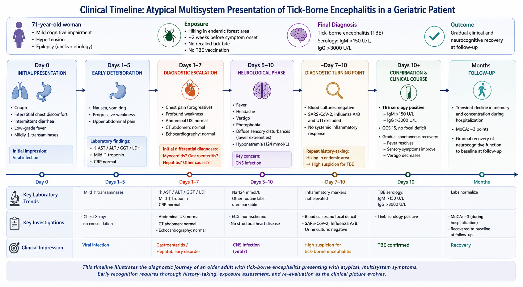
A timeline of the clinical course is shown in Figure 1.

3. Discussion

Clinically, tick-borne encephalitis (TBE) classically follows a biphasic course. It begins with a non-specific flu-like prodrome, followed—after a symptom-free interval—by neurological involvement ranging from meningitis to meningoencephalitis and, in severe cases, encephalomyelitis. However, this typical presentation occurs in only about 10% of cases globally, though it is reported more frequently in Europe (approximately 75%). While many cases, particularly in older adults, exhibit atypical, oligosymptomatic, or diagnostically misleading courses, potential complications include persistent balance disorders, cognitive impairment, paresis, and dysphagia. Rare instances of autonomic dysfunction (e.g., tachycardia, orthostatic hypotension) have also been described, as observed in our patient [4,5,6]. Furthermore, a positive correlation exists between increasing age and a poorer prognosis, with several studies investigating genetic predispositions to severe forms of meningoencephalitis [7,8].
In geriatric patients, the diagnostic challenge is further compounded by multimorbidity, pre-existing cognitive impairment, and the frequent coexistence of non-specific systemic symptoms and laboratory abnormalities. Initial findings such as transaminase elevation, hyponatremia, or troponin elevation may strongly mimic alternative hepatobiliary, cardiac, or gastrointestinal diseases, thereby delaying the consideration of neuroinfectious etiologies.
The importance of travel and exposure history is fundamental in the diagnostic evaluation of suspected tick-borne encephalitis (TBE), as it directly informs pre-test probability and guides clinical reasoning. The epidemiological context was particularly relevant in the present case. Although the patient reported a hiking trip in the Black Forest approximately two weeks before symptom onset, she is a resident of Switzerland, where TBE is endemic across most of the country. The Black Forest region is likewise recognized as one of the major TBE-endemic areas in Central Europe [3]. Given that the travel history represented a single identifiable exposure event, consideration of both the Swiss and German epidemiological settings was essential when assessing the likelihood of TBE.
Consequently, maintaining a broad differential diagnosis is essential, especially when diffuse clinical and laboratory findings initially suggest separate entities—such as hepatitis, myocarditis, or, more commonly, gastroenteritis. Conversely, once neurological symptoms raise suspicion of neuroinflammation, the full spectrum of infectious etiologies must be considered. This includes bacterial meningitis, enteroviruses, and herpes virus infections as critical diagnoses that must not be missed.
Diagnostic protocols for TBE are not yet fully standardized. In clinical practice, diagnosis relies primarily on serology. By the onset of neurological symptoms, viral RNA is typically no longer detectable in the blood or cerebrospinal fluid (CSF), rendering PCR useful only during the very early stages of infection. In contrast, specific IgM and IgG antibodies are reliably detectable in both serum and CSF during the meningoencephalitic phase [5]. Other laboratory anomalies are often non-specific, including moderately elevated transaminases (up to 80% of cases), hyponatremia (~30%), and elevated LDH (~12%), all of which typically normalize alongside clinical improvement [5,6].
Hyponatremia is a recognized laboratory abnormality in tick-borne encephalitis (TBE) and is most commonly attributed to the syndrome of inappropriate antidiuretic hormone secretion (SIADH), likely triggered by central nervous system inflammation and hypothalamic involvement. In some patients, hyponatremia may additionally reflect a broader stress-related neuroendocrine response during acute infection. The temporal association between the onset of neurological symptoms and the development of hyponatremia compatible with SIADH in our patient supports this pathophysiological link and suggests that the electrolyte disturbance was related to TBE. Although non-specific, hyponatremia may represent an additional supportive clue in the diagnostic work-up of TBE when interpreted within the appropriate epidemiological and clinical context.
Furthermore, mild cardiac involvement in tick-borne encephalitis (TBE) has been reported, although it is considered uncommon and is often underrecognized. Proposed mechanisms include direct viral involvement of the myocardium as well as indirect autonomic dysregulation during the acute neuroinfectious phase. These processes may lead to transient electrocardiographic abnormalities and, in rare cases, elevation of cardiac biomarkers such as troponin, even in the absence of clinically overt myocarditis. In our patient, the observed troponin elevation could therefore be interpreted in the context of systemic inflammatory response and possible neurocardiogenic involvement associated with TBE. Similar findings have been described in the literature, supporting the notion that TBE may occasionally be accompanied by reversible cardiac involvement [9].
Lumbar puncture is generally recommended in patients presenting with neurological symptoms, fever, and elevated inflammatory markers. The CSF profile in tick-borne encephalitis (TBE) typically reveals an initial granulocytic pleocytosis that subsequently shifts to a lymphocytic pleocytosis, accompanied by normal glucose levels and mildly elevated protein concentrations. CSF antibody testing, particularly when combined with calculation of the CSF/serum antibody index, can be especially valuable in previously vaccinated patients, as it may help confirm intrathecal antibody production and distinguish true infection from potential serological cross-reactivity with other flaviviruses [5].
In our case, an important limitation is that cerebrospinal fluid (CSF) examination was not performed. Consequently, the diagnosis in our patient relied primarily on serum serology, which carries inherent limitations, including the possibility of flavivirus cross-reactivity. However, interpretation of the serological findings was supported by the patient’s vaccination and exposure history, which were considered compatible with TBE infection.
The decision not to pursue lumbar puncture was made after careful consideration of the potential risks and benefits in this frail geriatric patient. The patient remained clinically stable throughout hospitalization, and her advanced age, known history of epilepsy, and expressed preference to avoid invasive diagnostic procedures were important factors in the shared decision-making process. In this context, the potential diagnostic yield of CSF examination was weighed against the procedural risks and the likelihood that the results would not substantially alter clinical management. This highlights the importance of individualizing diagnostic strategies in older adults, taking into account both clinical circumstances and patient preferences.
Another limitation is the concomitant use of antiepileptic medication, which may have influenced the neurological presentation. However, antiepileptic therapy remained unchanged throughout hospitalization, and renal function remained stable, making clinically relevant alterations in drug pharmacokinetics unlikely. Therefore, the observed neurological manifestations are unlikely to be attributable to changes in antiepileptic drug exposure.
Treatment remains purely supportive, as no specific antiviral therapy is currently available. Although corticosteroids are occasionally used to reduce cerebral edema in viral encephalitis, robust clinical evidence supporting their efficacy in TBE is still lacking [6,8].
Recent evidence suggests that viral neuroinflammation may accelerate cognitive decline in patients with pre-existing mild cognitive impairment (MCI) [10]. In our case, however, the patient exhibited only a transient worsening of neurocognitive function, with subsequent complete recovery to baseline. This transient cognitive decline may not be fully representative of the typical geriatric population, in whom TBE is more frequently associated with persistent and sometimes permanent neurological sequelae. At the same time, it is plausible that the observed impairment was multifactorial and related to the overall clinical context, including dizziness and systemic illness, rather than reflecting isolated direct viral neurocognitive injury. Indeed, the literature also describes a broad clinical spectrum of TBE, ranging from mild and self-limited forms to severe cases with long-term sequelae. Nevertheless, in older individuals affected by TBE, close monitoring of long-term neurological outcomes is warranted. Particular attention should be paid to the risk of persistent cognitive decline, secondary MCI, and other neurocognitive sequelae that can profoundly impair functional status and patient autonomy.

4. Conclusions

This case underscores how a multifaceted and atypical initial presentation can easily mask tick-borne encephalitis in older adults. It highlights that maintaining a high index of suspicion, performing meticulous exposure history-taking, and conducting continuous clinical reassessments are paramount to ensuring a timely and accurate diagnosis in geriatric patients.

Author Contributions

Conceptualization, G.C. and D.P.; methodology, G.C.; software, G.C.; validation, B.H. and T.M.; formal analysis, B.H. and T.M.; investigation, G.C.; resources, G.C. and D.P.; data curation, G.C.; writing—original draft preparation, G.C. and D.P.; writing—eview and editing, G.C.; supervision, B.H. and T.M. All authors have read and agreed to the published version of the manuscript.

Funding

This research received no external funding.

Institutional Review Board Statement

This study was conducted in accordance with the Declaration of Helsinki. The need for ethical approval has been waived by the Institutional Review Board of Canton of St. Gallen-Hospital for reporting individual cases or case series when informed consent is provided by the patient.

Informed Consent Statement

Written informed consent has been obtained from the patient to publish this paper.

Data Availability Statement

The data presented in this study are available on request from the corresponding author due to privacy and ethical restrictions.

Acknowledgments

We would like to thank the patient for allowing us to publish her case. During the preparation of this manuscript, ChatGPT (OpenAI GPT-5.5) was used as an assistive tool for the development of Figure 1. The authors have critically reviewed and edited all generated content and assume full responsibility for the accuracy and integrity of the final manuscript.

Conflicts of Interest

The authors declare no conflicts of interest.

Abbreviations

The following abbreviations are used in this manuscript:
TBETick-borne Encephalitis
CTComputed Tomography
MCIMild Cognitive Impairment
MoCAMontreal Cognitive Assessment
ASTAspartate Aminotransferase
ALTAlanine Aminotransferase
GGTGamma-Glutamyl Transferase
ALPAlkaline Phosphatase
LDHLactate Dehydrogenase
CKCreatine Kinase
CRPC-Reactive Protein
hs Troponin high-sensitivity Troponin

References

  1. Bundesamt für Gesundheit BAG. Official Website—Frühsommer-Meningoenzephalitis. Available online: https://www.bag.admin.ch/bag/de/home/krankheiten/krankheiten-im-ueberblick/fsme.html (accessed on 16 June 2026).
  2. Hasbun, R.; Rosenthal, N.; Balada-Llasat, J.M.; Chung, J.; Duff, S.; Bozzette, S.; Zimmer, L.; Ginocchio, C.C. Epidemiology of Meningitis and Encephalitis in the United States, 2011–2014. Clin. Infect. Dis. 2017, 65, 359–363. [Google Scholar] [CrossRef] [PubMed]
  3. Robert Koch Institut. Official Website—Tick-Borne Encephalitis. Available online: https://gesund.bund.de/en/fruehsommer-meningoenzephalitis-fsme (accessed on 16 June 2026).
  4. Kleiter, I.; Steinbrecher, A.; Flügel, D.; Bogdahn, U.; Schulte-Mattler, W. Autonomic involvement in tick-borne encephalitis (TBE): Report of five cases. Eur. J. Med. Res. 2006, 11, 261–265. [Google Scholar] [PubMed]
  5. Barp, N.; Trentini, A.; Di Nuzzo, M.; Mondardini, V.; Francavilla, E.; Contini, C. Clinical and laboratory findings in tick-borne encephalitis virus infection. Parasite Epidemiol. Control 2020, 10, e00160. [Google Scholar] [CrossRef] [PubMed]
  6. Bogovic, P.; Strle, F. Tick-borne encephalitis: A review of epidemiology, clinical characteristics, and management. World J. Clin. Cases 2015, 3, 430–441. [Google Scholar] [CrossRef] [PubMed]
  7. Logar, M.; Arnez, M.; Kolbl, J.; Avsic-Zupanc, T.; Strle, F. Comparison of the epidemiological and clinical features of tick-borne encephalitis in children and adults. Infection 2000, 28, 74–77. [Google Scholar] [CrossRef] [PubMed]
  8. Eikeland, R.; Henningsson, A.J.; Lebech, A.-M.; Kerlefsen, Y.; Mavin, S.; Vrijlandt, A.; Hovius, J.W.; Lernout, T.; Lim, C.; Dobler, G.; et al. Tick-borne diseases in the North Sea region-A comprehensive overview and recommendations for diagnostics and treatment. Ticks Tick-Borne Dis. 2024, 15, 102306. [Google Scholar] [CrossRef] [PubMed]
  9. Seneghini, M.; Lugano, F.; Maeder, M.T.; Garcia-Boy, R.; Strahm, C. Extraneurological Presentations of Tick-Borne Encephalitis Virus: A Rare Case of TBEV-Associated Myocarditis with Fever and Bicytopenia and a Systematic Literature Review. Case Rep. Infect. Dis. 2026, 2026, 3051689. [Google Scholar] [CrossRef] [PubMed]
  10. Bohmwald, K.; Andrade, C.A.; Gálvez, N.M.; Mora, V.P.; Muñoz, J.T.; Kalergis, A.M. The Causes and Long-Term Consequences of Viral Encephalitis. Front. Cell. Neurosci. 2021, 15, 75587. [Google Scholar] [CrossRef] [PubMed]
Figure 1. Clinical timeline and most prominent laboratory findings (↑: increased).
Reports 09 00205 g001
Table 1. Laboratory findings in the emergency department (first and second presentation).
ParameterReferenceFirst PresentationSecond Presentation
Natrium (mmol/L)136–144137133
Bilirubin total (umol/L)<2034
AST (U/L)<5584330
ALT (U/L)<5569201
GGT (U/L)<35444754
ALP (U/L)30–1209596
Total Proteine (g/L)63–836562
Albumin (g/L)34–4839.137.2
LDH (U/L)<265197366
CK (U/L)<1454175
Pancreatic Amylase (U/L)<461571
CRP (mg/L)<82<1
hs Troponin (ng/L)<1817.545.5
AST (Aspartate Aminotransferase), ALT (Alanine Aminotransferase), GGT (Gamma-Glutamyl Transferase), ALP (Alkaline Phosphatase), Total Protein, Albumin, LDH (Lactate Dehydrogenase), CK (Creatine Kinase), Pancreatic Amylase, CRP (C-Reactive Protein), and hs Troponin (high-sensitivity Troponin).
Table 2. Differential diagnoses considered during the initial diagnostic work-up of the patient, with findings supporting and arguing against each diagnosis in the present case.
Differential DiagnosisTypical SymptomsFindings Supporting the Diagnosis in This CaseFindings Arguing Against the Diagnosis in This Case
Acute viral gastroenteritis with reactive hepatitisDiarrhea, nausea, vomiting, abdominal pain, malaiseInitial gastrointestinal symptoms; elevated transaminasesBiphasic course; subsequent fever, headache, photophobia, sensory disturbances, and dizziness not adequately explained
Acute viral hepatitis (HAV/HEV)Nausea, vomiting, abdominal pain, fatigue, elevated liver enzymes, occasionally feverMarked transaminase elevation; positive HAV IgG and HEV IgG serologyNo evidence of acute infection (negative HAV IgM, HEV IgM, and HEV RNA); neurological symptoms unexplained
MyocarditisChest pain, dyspnea, fatigue, arrhythmiasMild troponin elevation; viral prodromeEchocardiography and cardiac work-up unremarkable; no evidence of myocardial dysfunction
Acute coronary syndrome (ACS)Chest pain, dyspnea, autonomic symptoms, elevated troponinElevated hs-troponin; interscapular/epigastric painNo ischemic ECG changes; cardiac imaging negative; symptoms atypical
Pulmonary embolismDyspnea, chest pain, tachycardia, syncopeElevated troponin may occur; thoracic painTriple-rule-out CT negative
Aortic dissectionSudden chest or interscapular pain, neurological symptoms, elevated cardiac biomarkers occasionallyPain between the shoulder blades prompted evaluationTriple-rule-out CT excluded dissection
Table 3. Differential diagnoses considered at the onset of neurological symptoms, with findings supporting and arguing against each diagnosis in the present case (final diagnosis in bold).
Differential DiagnosisTypical SymptomsFindings Supporting the Diagnosis in This CaseFindings Arguing Against the Diagnosis in This Case
Herpes simplex encephalitisFever, altered mental status, seizures, focal neurological deficitsHeadache, photophobia, history of epilepsyNo altered consciousness, seizures, or rapidly progressive encephalopathy
Pneumococcal meningitisFever, headache, meningism, altered consciousness, photophobia, nausea/vomitingFever, headache, photophobia, neurological symptomsSubacute biphasic course; absence of severe meningeal syndrome and impaired consciousness; negative blood cultures
Enteroviral meningoencephalitisFever, headache, photophobia, gastrointestinal symptoms, meningitis/encephalitisBiphasic viral illness with neurological manifestationsLess consistent epidemiology; absence of leukopenia frequently reported in severe adult cases
NeuroborreliosisHeadache, radicular pain, cranial nerve palsies (especially facial nerve), sensory symptomsTick exposure in endemic region; neurological symptomsNo facial palsy, radiculitis, or typical Bannwarth syndrome
Tick-borne encephalitis (TBE)Biphasic illness with initial flu-like and gastrointestinal symptoms followed by fever, headache, dizziness, photophobia, and neurological manifestationsRecent hiking in endemic area, biphasic disease course, hyponatremia, elevated transaminases, neurological symptoms, positive TBEV IgM and IgG serologyAbsence of CSF confirmation (diagnostic limitation)
Disclaimer/Publisher’s Note: The statements, opinions and data contained in all publications are solely those of the individual author(s) and contributor(s) and not of MDPI and/or the editor(s). MDPI and/or the editor(s) disclaim responsibility for any injury to people or property resulting from any ideas, methods, instructions or products referred to in the content.