Case Report

Inguinal Hernia Containing the Bladder and Postoperative Appearance: A Multimodality Case Report


Department of Radiology, University of Texas Southwestern Medical Center, Dallas, TX 75390, USA
*
Author to whom correspondence should be addressed.
Reports 2026, 9(2), 193; https://doi.org/10.3390/reports9020193
Submission received: 19 May 2026 / Revised: 15 June 2026 / Accepted: 17 June 2026 / Published: 20 June 2026

Abstract

Background and Clinical Significance: Many diagnostic radiopharmaceuticals are excreted in the urine. This can pose a diagnostic challenge when urine-containing structures are in atypical locations, particularly in review of planar imaging without anatomical details from cross-sectional imaging. This case highlights a challenging 99mTc-methylene diphosphonate (99mTc-MDP) bone scan in a patient with an inguinal hernia containing a portion of the urinary bladder. Subsequently, we review diagnostic challenges on conventional and molecular imaging following surgical repair of the inguinal hernia. Case Presentation: A 79-year-old man with prostate cancer underwent initial staging prior to prostatectomy with 99mTc-MDP bone scintigraphy. Anterior and posterior images showed focal uptake overlying the pubic symphysis. Lateral views showed that the activity was extraosseous. Follow-up CT urography showed a bladder hernia as the cause of the abnormality on bone scan. Prostatectomy and inguinal hernia repair were performed as a combination case. Four years postoperatively, follow-up 68Ga-PSMA-11 positron emission tomography/computed tomography (PET/CT) showed no recurrence. The CT component of the exam showed an intermediate-density focus at the right inguinal hernia repair site, corresponding to a plugoma related to a polypropylene mesh plug, and a hyperattenuating Gore-Tex mesh repair of the left inguinal hernia. Conclusions: This case highlights the importance of lateral projections in resolving scintigraphic pitfalls and recognizing mesh-related imaging appearances to prevent misinterpretation.

1. Introduction and Clinical Significance

Bladder herniation into the inguinal canal is rare, occurring in 0.4–3% of the population and accounting for 1–4% of all inguinal hernias [1]. Elderly men are most affected. This condition creates a diagnostic challenge for nuclear medicine tests that rely on radiotracers excreted into the urine. The atypical location and possible discontinuity of urinary activity in the hernia neck require recognition. Otherwise, activity overlying the groin could be misinterpreted as osteoblastic disease. Inguinal hernias can be treated with multiple surgical techniques. The patient in this case report was ultimately treated with two types of surgical mesh. Postoperative appearances of the different mesh materials are reviewed.
Surgery is the definitive therapy for inguinal hernias [2]. Operative intervention is required urgently in the setting of complications like strangulation. However, many patients may receive elective surgical repair, allowing for planning of treatment timing and method. Most inguinal hernia mesh repairs are performed with polypropylene mesh, which induces an inflammatory response that is important in the repair of inguinal hernias [3]. Expanded polytetrafluoroethylene (ePTFE) mesh produces a lesser inflammatory response and is more often used for the repair of incisional hernias, but may be used for inguinal hernia repairs in some instances at surgeon’s discretion.

2. Case Presentation

2.1. Clinical Context

A 79-year-old man with a history of deep venous thrombosis and pulmonary embolism requiring long-term anticoagulation, bilateral inguinal hernias treated surgically, and prostate cancer treated with radical prostatectomy required evaluation for metastatic disease after biochemical recurrence.
Initial workup for his prostate cancer had shown up to Gleason score 4 + 4 (grade group 4) disease. The decision was made to proceed with radical prostatectomy after a negative bone scan. Prostatectomy and inguinal hernia repair were performed as a combination surgical case with urologic and general surgeons completing their respective procedures during the same anesthesia event. Pathology from prostatectomy showed the highest Gleason score of 3 + 5 (grade group 4) disease and positive posterolateral margins from the base to apex. Seminal vesicle invasion was present. Bilateral pelvic nodal dissections were negative for metastatic prostate cancer. The general surgery team approached their procedure transabdominally and found a large right-sided medial hernia and a moderate left-sided lateral hernia. Polypropylene mesh was used for repair of the right-sided hernia. Dissection to expose the left hernia resulted in several peritoneal defects and the decision was made to use ePTFE mesh to avoid direct contact of inflammatory polypropylene mesh with intraperitoneal contents such as bowel.
The patient had a PSA recurrence four years after prostatectomy to 0.31 ng/mL, prompting additional workup for metastatic or recurrent disease. We present imaging done before surgical treatment (bone scan and CT urography) and after biochemical recurrence (PET/CT) to demonstrate the appearance of the bladder containing inguinal hernia and appearance of the mesh hernia repair.

2.2. Bone Scan

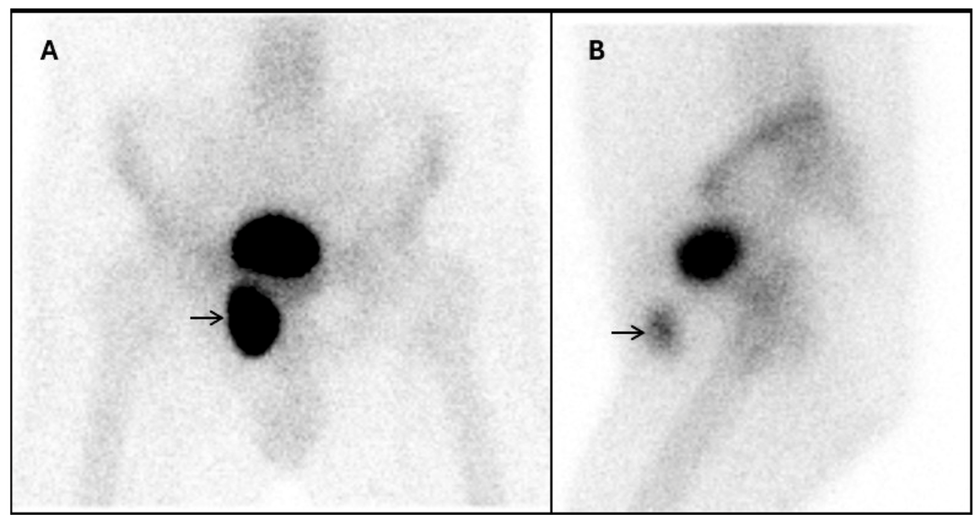
Initial staging whole-body 99mTc-MDP bone scintigraphy showed focal intense activity projecting over the right pubic symphysis (Figure 1A). However, the lateral projection (Figure 1B) localized the activity anterior to the expected location of the pubic bones, suggesting an extraosseous location. Planar bone scintigraphy lacks anatomic detail, limiting differentiation between osseous and extraosseous uptake, whereas single-photon emission computed tomography/computed tomography enables precise localization. This highlights the importance of obtaining lateral or oblique views to rule out skeletal involvement in the pelvis [4]. False-positive uptake may occur when tracer-containing urine accumulates within a herniated bladder segment or a focal bladder diverticulum, mimicking metastasis if the connection to the bladder is obscured by a narrow neck [5].

2.3. CT Urogram

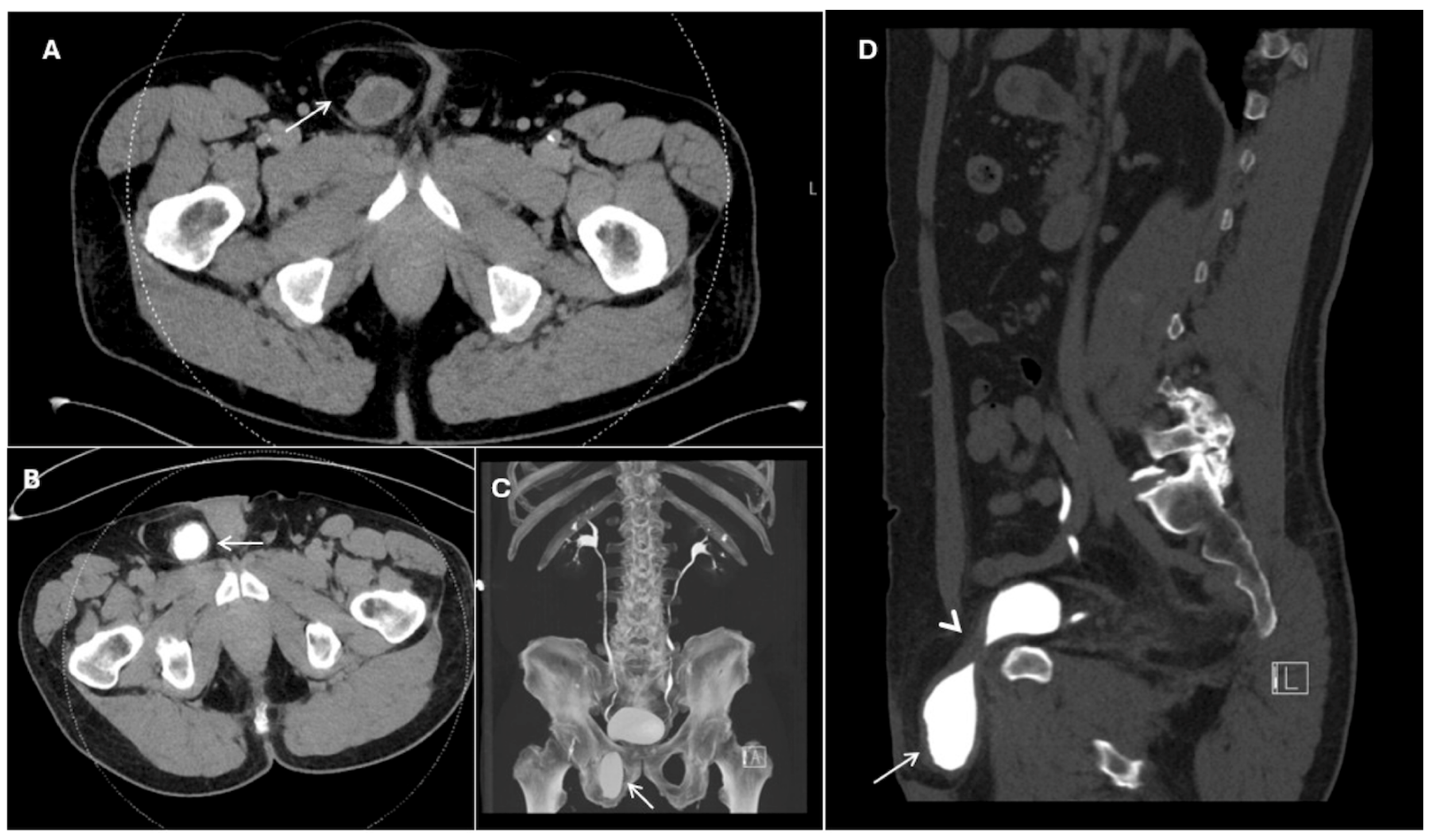
Subsequent CT urography demonstrated a right inguinal hernia containing fat and a portion of the bladder. The herniated bladder only filled on the delayed prone phase (Figure 2B, image flipped), confirming bladder communication. The sagittal reconstruction (Figure 2D) demonstrates the course of the narrowed bladder through the hernia neck. CT urography allows evaluation of fine anatomic details and assessment of the urinary tract with excreted contrast. Patients may be repositioned to assess nonopacified portions of the urinary tract in typical supine positioning.

2.4. PSMA PET/CT

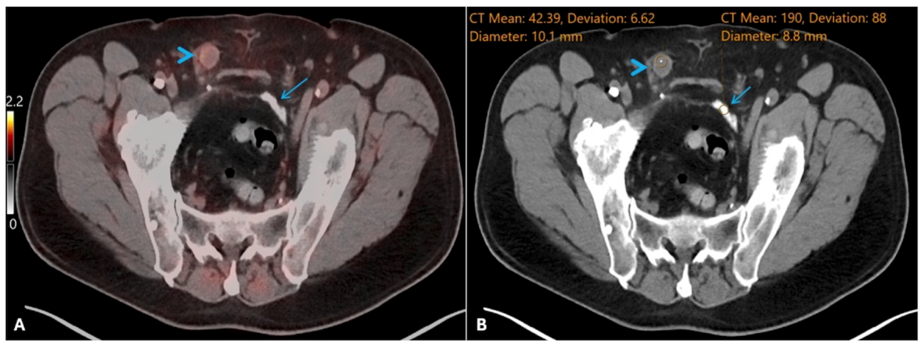
Follow-up 68Ga-PSMA-11 PET/CT four years after prostatectomy did not demonstrate radiotracer-avid recurrent or metastatic disease. Inclusion of a localizer CT allows superior assessment of anatomy compared to planar bone scintigraphy. Axial localizer CT through the pelvis (Figure 3B) demonstrated a rounded soft-tissue density focus at the right hernia-repair site representing a plugoma following use of a polypropylene mesh plug. The appearance is caused by a combination of connective tissue and the mesh material itself [3,6,7,8]. This was initially thought to represent residual herniated bladder before more thorough investigation of the patient’s surgical history. Dense material at a left inguinal hernia repair site represents ePTFE mesh, commonly known as Gore-Tex. ePTFE appears hyperattenuating due to its inert, non-adhesive coating [9] and was selected at surgery for laparoscopically confirmed contact with peritoneal surfaces on the left side. Polypropylene mesh is isoattenuating and may produce an inflammatory response that appears as a rounded density. Although non-avid in this case, PSMA-ligand uptake has been reported in association with mesh-related neovascularization and increased ligand expression in chronic inflammation [10]. 18F-fluorodeoxyglucose uptake can also occur years after mesh hernia repair because of inflammation [11].

3. Discussion

Awareness of possible abnormal bladder configurations is essential for interpreting nuclear medicine studies with radiotracers that are excreted in the urine. Urinary activity can overlap osseous structures on bone scintigraphy and make assessment challenging. Recognition of this pitfall on planar imaging can involve identifying (1) atypical pelvic radiotracer location, (2) activity level similar to bladder, (3) continuity with the bladder, and (4) a teardrop- or tongue-shaped contour [4,12,13,14]. Hybrid imaging can assist in challenging cases. Follow-up cross-sectional imaging, including CT urography, can help define anatomy.
Several reports of inguinal hernias containing bladder detected on bone scintigraphy have been published. Iagaru and Siegel presented a case showing a bladder diverticulum in an inguinal hernia detected on bone scintigraphy [15]. Ilgan et al. demonstrated a case where the entire bladder and portion of the distal ureters herniated into the scrotum via the right inguinal canal, resulting in distal left ureteral obstruction [16]. Other reports have shown portions of the anterior bladder entering inguinal hernias, as was the case in our report [17].
Following hernia repair, it is essential to recognize that nodular soft tissue densities may be post-surgical plugomas. Plugomas can be radiotracer avid with tracers that frequently have inflammatory uptake and should not be confused with metastatic disease. There are reports of potential false positive radiotracer uptake by herniorrhaphy mesh on PSMA [18] and FDG [19] PET/CT. 68Ga-DOTATATE is known to have false positive uptake with inflammatory processes and could conceivably show activity at hernia repair mesh, although there are no reported cases in the literature.

4. Conclusions

In summary, herniation of the urinary bladder can cause abnormal configuration of urinary activity found with many diagnostic radiopharmaceuticals. Following repair of inguinal hernias, the radiologist must also be aware of the range of postoperative appearances of mesh repairs and possible inflammatory radiotracer uptake. Our case provides a unique teaching opportunity to highlight the challenge posed by atypical bladder configuration on planar imaging with radiopharmaceuticals excreted in the urine and the expected postoperative appearance following hernia repair with different types of mesh.

Author Contributions

H.J. retrieved relevant imaging, conducted the literature review, and was the primary author. O.K.Ö. reviewed and edited the manuscript and served as a senior mentor. J.F. reviewed and edited the manuscript, was a significant contributor to writing the manuscript, and aided in image preparation. All authors have read and agreed to the published version of the manuscript.

Funding

This research received no external funding.

Institutional Review Board Statement

The study was conducted in accordance with the Declaration of Helsinki and approved by the Institutional Review Board of the University of Texas Southwestern (protocol code STU-2019-161, approved 16 August 2024).

Informed Consent Statement

Written informed consent has been obtained from the patient to publish this paper.

Data Availability Statement

The original contributions presented in this study are included in the article. Further inquiries can be directed to the corresponding author.

Conflicts of Interest

The authors declare no conflicts of interest.

Abbreviations

The following abbreviations are used in this manuscript:
ePTFEExpanded polytetrafluoroethylene
PET/CTPositron emission tomography/computed tomography
99mTc-MDP99mTc-methylene diphosphonate

References

  1. Shahlaei, S.; Shakeri, S.; Farahmandfar, F.; Emami, F.; Sadeghi, R. Inguino-scrotal bladder hernia: An unexpected finding on 68Ga-PSMA-11 PET/CT. Asia Ocean. J. Nucl. Med. Biol. 2020, 8, 141–144. [Google Scholar] [CrossRef]
  2. Rosenberg, J.; Bisgaard, T.; Kehlet, H.; Wara, P.; Asmussen, T.; Juul, P.; Strand, L.; Andersen, F.H.; Bay-Nielsen, M.; Database, D.H. Danish Hernia Database recommendations for the management of inguinal and femoral hernia in adults. Dan. Med. Bull. 2011, 58, C4243. [Google Scholar] [PubMed]
  3. Novik, B.; Sandblom, G.; Ansorge, C.; Thorell, A. Association of Mesh and Fixation Options with Reoperation Risk after Laparoscopic Groin Hernia Surgery: A Swedish Hernia Registry Study of 25,190 Totally Extraperitoneal and Transabdominal Preperitoneal Repairs. J. Am. Coll. Surg. 2022, 234, 311–325. [Google Scholar] [CrossRef] [PubMed]
  4. Goel, S.; Gadiraju, R. Left inguinal hernia mimicking metastasis on bone scan. Radiol. Case Rep. 2015, 8, 559. [Google Scholar] [CrossRef] [PubMed][Green Version]
  5. Farmer, K.D.; Gellett, L.R.; Vivian, G.C.; Franklin, K. Bladder diverticulum simulating a pelvic metastasis on a Tc-99m HDP bone scan. Clin. Nucl. Med. 2001, 26, 60–61. [Google Scholar] [CrossRef] [PubMed]
  6. Klinge, U.; Klosterhalfen, B.; Müller, M.; Schumpelick, V. Foreign body reaction to meshes used for the repair of abdominal wall hernias. Eur. J. Surg. 1999, 165, 665–673. [Google Scholar] [CrossRef] [PubMed]
  7. Cronin, C.G.; Harisinghani, M.G.; Catalano, O.; Blake, M.A. Multitechnique Imaging Findings of Prolene Plug Hernia Repair. Am. J. Roentgenol. 2010, 195, 701–706. [Google Scholar] [CrossRef] [PubMed]
  8. Yeung, V.H.W.; Pearl, J.M.; Coakley, F.V.; Joe, B.N.; Westphalen, A.C.; Yeh, B.M. Computed tomographic appearance of Prolene Hernia System and polypropylene mesh plug inguinal hernia repair. J. Comput. Assist. Tomogr. 2008, 32, 529–532. [Google Scholar] [CrossRef] [PubMed]
  9. Tonolini, M.; Ippolito, S. Multidetector CT of expected findings and early postoperative complications after current techniques for ventral hernia repair. Insights Imaging 2016, 7, 541–551. [Google Scholar] [CrossRef] [PubMed][Green Version]
  10. de Galiza Barbosa, F.; Queiroz, M.A.; Nunes, R.F.; Costa, L.B.; Zaniboni, E.C.; Marin, J.F.G.; Cerri, G.G.; Buchpiguel, C.A. Nonprostatic diseases on PSMA PET imaging: A spectrum of benign and malignant findings. Cancer Imaging 2020, 20, 23. [Google Scholar] [CrossRef] [PubMed]
  11. Bahçeci, T.; Nursal, G.N.; Aydın, M. Intense FDG Uptake around the Inguinal Surgical Mesh 5 Years after Operation: Case Report and Review of the Literature. Mol. Imaging Radionucl. Ther. 2012, 21, 35–37. [Google Scholar] [CrossRef] [PubMed]
  12. Usmani, S.; Marafi, F.; Ahmed, N.; Kazem, N. Inguinal Herniation of Urinary Bladder on F-18 Sodium Fluoride (NaF) PET-CT. Nucl. Med. Mol. Imaging 2017, 51, 368–370. [Google Scholar] [CrossRef] [PubMed]
  13. Ji, T.; Gao, D.; Chen, B.; Wang, R.; Gao, S. 99mTc-MDP uptake in SPECT/CT by a bladder hernia simulating inguinal metastasis: A case report. Oncol. Lett. 2016, 11, 1398–1400. [Google Scholar] [CrossRef] [PubMed]
  14. Sezgin, C.; Duzgun, F.; Mutevelizade, G.; Gumuser, G.; Sayit, E. Inguinoscrotal Bladder Hernia Mimicking Testicle Tumor. Clin. Med. Insights Case Rep. 2023, 16, 11795476231151329. [Google Scholar] [CrossRef] [PubMed]
  15. Iagaru, A.; Siegel, M.E. Demonstration of a right inguinal hernia containing urinary bladder diverticulum on whole-body bone scan and pelvic CT. Eur. J. Nucl. Med. Mol. Imaging 2006, 33, 234. [Google Scholar] [CrossRef] [PubMed]
  16. Ilgan, S.; Ozguven, M.; Emer, M.O.; Karacalioglu, A.O. Massive inguinoscrotal herniation of the bladder with ureter: Incidental demonstration on bone scan. Ann. Nucl. Med. 2007, 21, 371–373. [Google Scholar] [CrossRef] [PubMed]
  17. Thamsborg, A.K.M.; Krakauer, M.; Fahrenkrug, L. Scintigraphy showing bladder in the scrotum by coincidence. Ugeskr. Laeger 2014, 176, V02140095. [Google Scholar] [PubMed]
  18. Muñoz, G.C.; Fraum, T.J. Plug inguinal hernia repair mimicking nodal spread of prostate cancer on PSMA-PET/CT. Radiol. Case Rep. 2023, 18, 57–59. [Google Scholar] [CrossRef] [PubMed]
  19. Seo, M.; Ha, N.D.; Jo, J.C.; Park, S.H. Chronic Vasitis After Mesh Herniorrhaphy: A Potential Cause of False Positive Results in 18F-FDG PET/CT Studies. Clin. Nucl. Med. 2019, 44, e104–e106. [Google Scholar] [CrossRef] [PubMed]
Figure 1. Planar images from 99mTc-MDP bone scintigraphy. Abnormal activity (arrow) projects over the right pubic symphysis on the anterior projection (A) and is shown to be anterior to the expected location of the pubic bones on the lateral projection (B).
Reports 09 00193 g001
Figure 2. CT urography. The precontrast phase (A) shows a soft tissue density with central fluid density in the right inguinal canal. On delayed prone imaging (B), excreted contrast is seen within the abnormality, indicating it communicates with the bladder. Please note these images are flipped vertically. A maximum intensity projection (C) shows the herniated bladder’s location in relationship to the pelvic bones ([A] for anterior). A delayed sagittal reconstruction (D) shows the thin neck (arrowhead) as the hernia enters the inguinal canal ([L] for left). Contrast is again seen in the herniated bladder (arrow).
Reports 09 00193 g002
Figure 3. PSMA PET/CT following bilateral inguinal hernia repair. Fused (A) and localizer CT (B) images are shown. A soft tissue density in the right inguinal canal represents a plugoma associated with polypropylene mesh (arrowhead). Linear high-density material at the entrance of the left inguinal canal is ePTFE mesh (arrow). Minimal activity is seen at the plugoma despite aggressive windowing.
Reports 09 00193 g003
Disclaimer/Publisher’s Note: The statements, opinions and data contained in all publications are solely those of the individual author(s) and contributor(s) and not of MDPI and/or the editor(s). MDPI and/or the editor(s) disclaim responsibility for any injury to people or property resulting from any ideas, methods, instructions or products referred to in the content.