Case Report

A Rare Case of Reversible Pulmonary Hypertension Phenotype in a Child with Scurvy: Aetiologies Insights

by
Mattia Pasquinucci
1,2,*,
Luisa Bonsembiante
1,
Sofia Mezzalira
1,
Martina Locallo
3 and
Davide Meneghesso
1
1
Department of Pediatrics, AULSS 7 Pedemontana-San Bassiano Hospital, 36061 Bassano del Grappa, Italy
2
Department of Neuroscience, Rehabilitation, Ophthalmology, Genetics, Maternal and Child Health, IRCCS Istituto Giannina Gaslini, 16100 Genoa, Italy
3
Department of Pediatrics, University of Padua, 35100 Padua, Italy
*
Author to whom correspondence should be addressed.
Reports 2026, 9(1), 44; https://doi.org/10.3390/reports9010044
Submission received: 17 December 2025 / Revised: 19 January 2026 / Accepted: 28 January 2026 / Published: 30 January 2026
(This article belongs to the Section Paediatrics)

Abstract

Background and Clinical Significance: Scurvy, caused by chronic vitamin C deficiency, is re-emerging in Western countries, particularly among pediatric patients with highly selective diets. While its musculoskeletal and mucocutaneous manifestations are well-known, its association with pulmonary arterial hypertension (PAH) is rare and poorly understood. Ascorbic acid and iron are essential cofactors for prolyl hydroxylases (PHD), which regulate Hypoxia-Inducible Factors. Their combined deficiency may trigger a “pseudohypoxic” state, leading to pulmonary vascular remodeling and vasoconstriction. Case Presentation: A 30-month-old female presented with a one-month history of limping, lower limb pain, and gingival hypertrophy. Dietary history revealed an almost exclusive cow’s milk-based intake. Physical examination showed diffuse petechiae, pallor, and right knee edema. Laboratory findings confirmed scurvy (undetectable vitamin C), severe iron-deficiency anemia (Hb: 72 g/L; ferritin: 22 mcg/L; RDW: 30%), folate deficiency, and hyperhomocysteinemia. Notably, elevated copper and vitamin B12 levels suggested a state of metabolic dysregulation. Echocardiography revealed moderate PAH phenotype (estimated sPAP: 47–50 mmHg) and a hyperdynamic contractility. A “perfect storm” mechanism was hypothesized, involving iron–ascorbate-dependent PHD impairment, high-output state, and oxidative-stress-induced hepcidin dysregulation (suggested by elevated copper). Following intravenous vitamin C and multivitamin supplementation, pulmonary pressures normalized within one week. Conclusions: PAH phenotype in scurvy represents a reversible metabolic disruption of pulmonary vascular tone rather than a structural disease. This case underscores the synergistic role of vitamin C, iron, and folate in vascular homeostasis. Clinicians should maintain high suspicion for scurvy in children with selective diets and unexplained PAH, as nutritional restoration is curative.

1. Introduction and Clinical Significance

Scurvy is a disease of historical significance, traditionally known as “the disease of sailors.” It results from chronic ascorbic acid deficiency and remains relatively frequent, though underdiagnosed, in developing countries where access to a balanced diet is limited. However, its incidence has been increasing in Western countries in recent years [1]. While it can affect adults, the pediatric population is generally more susceptible. It typically presents with cutaneous, mucosal, and musculoskeletal signs; early diagnostic markers include gingival hypertrophy with bleeding, limping or refusal to walk, and lower limb pain [2]. In cases of malabsorption, global malnutrition, or highly selective diets, scurvy is often associated with other micronutrient deficiencies [3]. Historically, there was no established link between scurvy and cardiovascular manifestations.
Clinical Significance: Recently, however, increasing attention has been paid to this correlation, and associated pulmonary arterial hypertension (PAH) has been rarely reported [4,5,6,7]. Nevertheless, its true incidence in patients with scurvy and its exact etiology remains incompletely understood.

2. Case Presentation

A 30-month-old female presented to our Emergency Department following several visits to other centers for a one-month history of monoarticular swelling, limping, and lower limb pain. The patient had sustained a fall approximately two weeks prior to the onset of symptoms. Clinical evaluation revealed periarticular edema and rubor of the right knee, a maculopapular rash on the same limb, diffuse petechiae, haemorrhagic gingival hypertrophy, and pallor (Figure 1).
Vital signs showed tachycardia and a systemic blood pressure of 90/55 mmHg (below normal limits). Dietary history disclosed a selective cow’s milk-based intake, accounting for over 95% of the child’s caloric intake.
Laboratory findings revealed severe hypochromic microcytic anemia without thrombocytopenia but with severe iron deficiency, along with mild coagulation abnormalities (increased Antithrombin III). Renal and hepatic functions were within normal limits, while electrolytes showed mild hyponatremia (Na+ 132 mmol/L). The metabolic–nutritional profile indicated global malnutrition with low triglycerides and HDL cholesterol and reduced prealbumin; blood glucose was normal. The initial differential diagnosis included malignant lymphoproliferative disease, which was reasonably excluded via peripheral blood smear; the latter instead showed neutrophil hypersegmentation (consistent with malnutrition/folate deficiency). Further biochemical analysis revealed low folate levels and increased homocysteine. High levels of D-dimer, copper, and vitamin B12 were also noted, initially interpreted as markers of systemic hyperinflammation (Table 1).
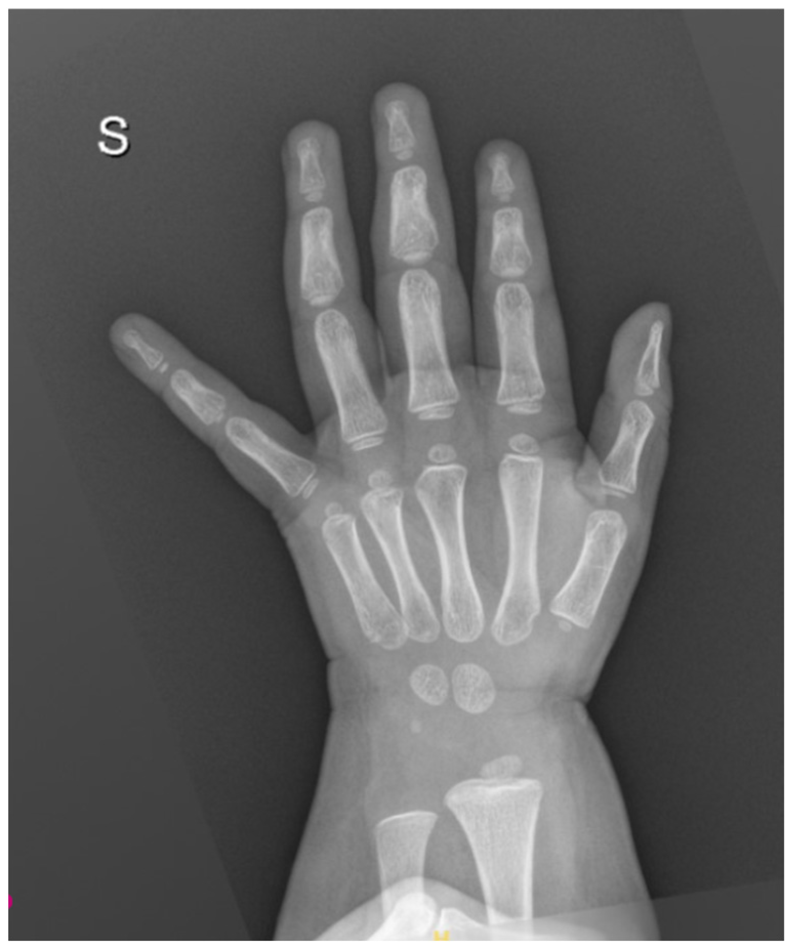
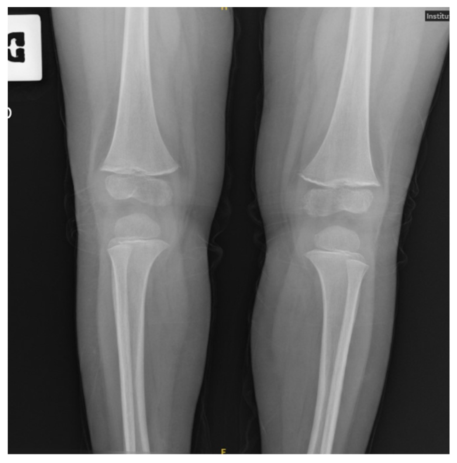
The absence of significant lactic acidosis rendered a concomitant Thiamine deficiency less likely. Celiac disease was ruled out. Vitamin C was undetectable, confirming the diagnosis of scurvy. X-rays of the wrist showed slightly delayed bone age (Figure 2), while knee X-rays revealed typical skeletal changes pathognomonic for scurvy (Figure 3).
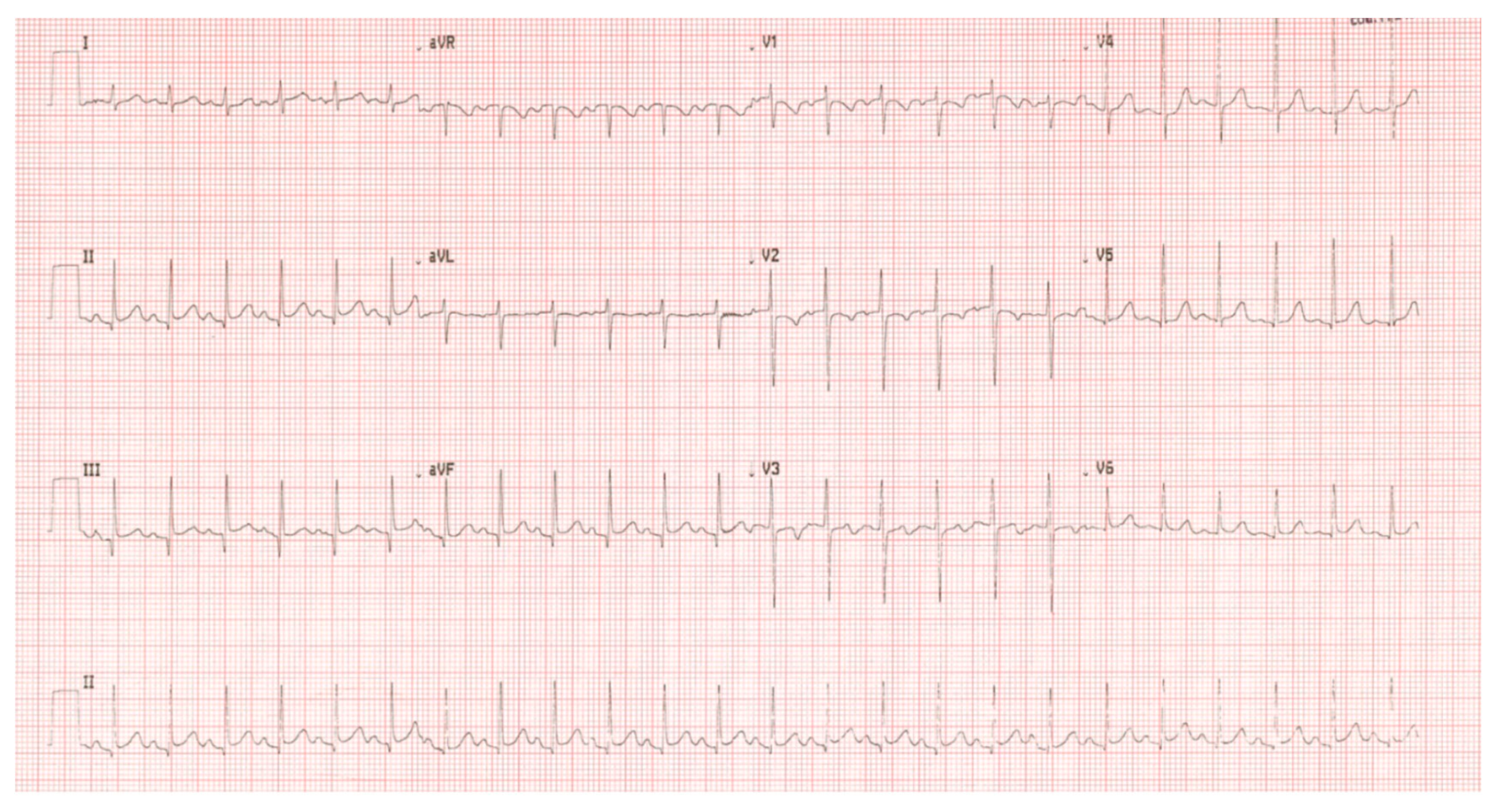
Given the severity of the malnutrition, a cardiological evaluation was performed to rule out malnutrition-related cardiomyopathy before initiating high-volume fluids. Electrocardiography (ECG) showed diffuse repolarization abnormalities (Figure 4).
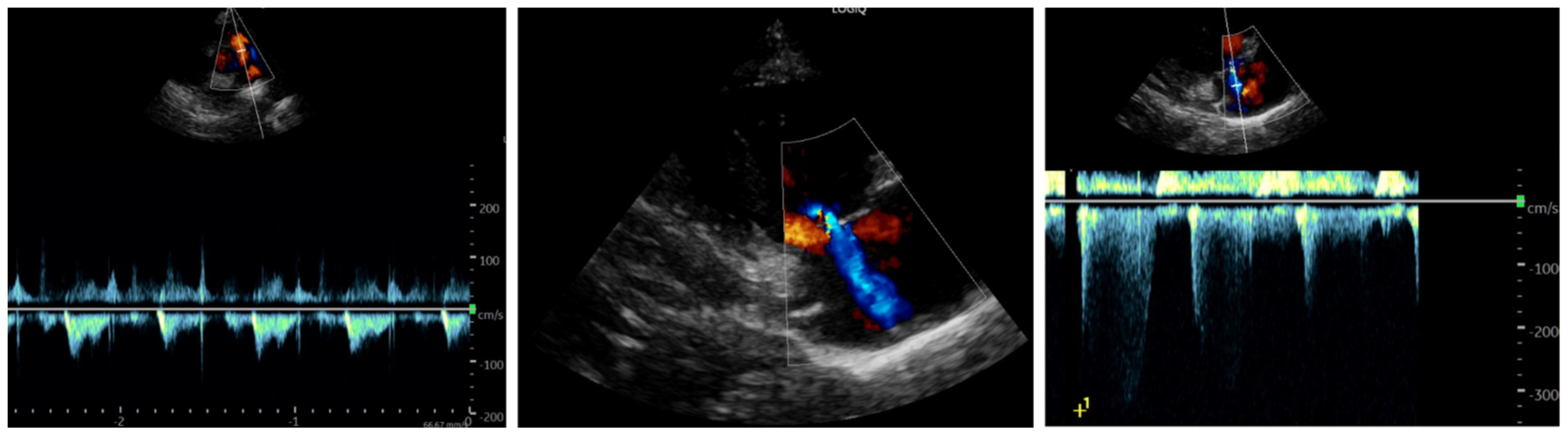
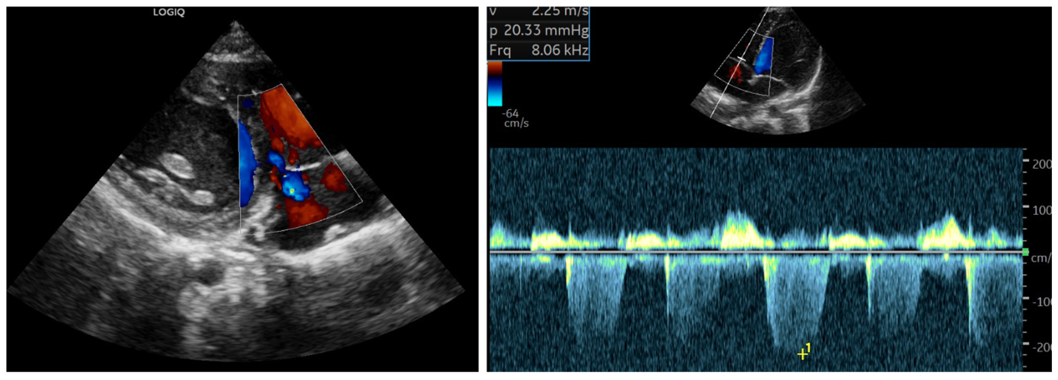
Echocardiography, performed under midazolam sedation due to psychomotor agitation (but with stable hemodynamics), showed findings suggestive of PAH, including midsystolic notching of the pulmonary artery flow, moderate tricuspid regurgitation, a measurable tricuspid regurgitant jet >2.8 m/s with an estimated systolic pulmonary artery pressure (sPAP) of approximately 47–50 mmHg, assuming tricuspid regurgitation velocity 42–45 mmHg and right atrial pressure 5 mmHg, and slight end-diastolic flattening of the interventricular septum (Figure 5). The left heart appeared hyperdynamic. Despite these findings, the patient remained hemodynamically stable with normal resting oxygen saturation.
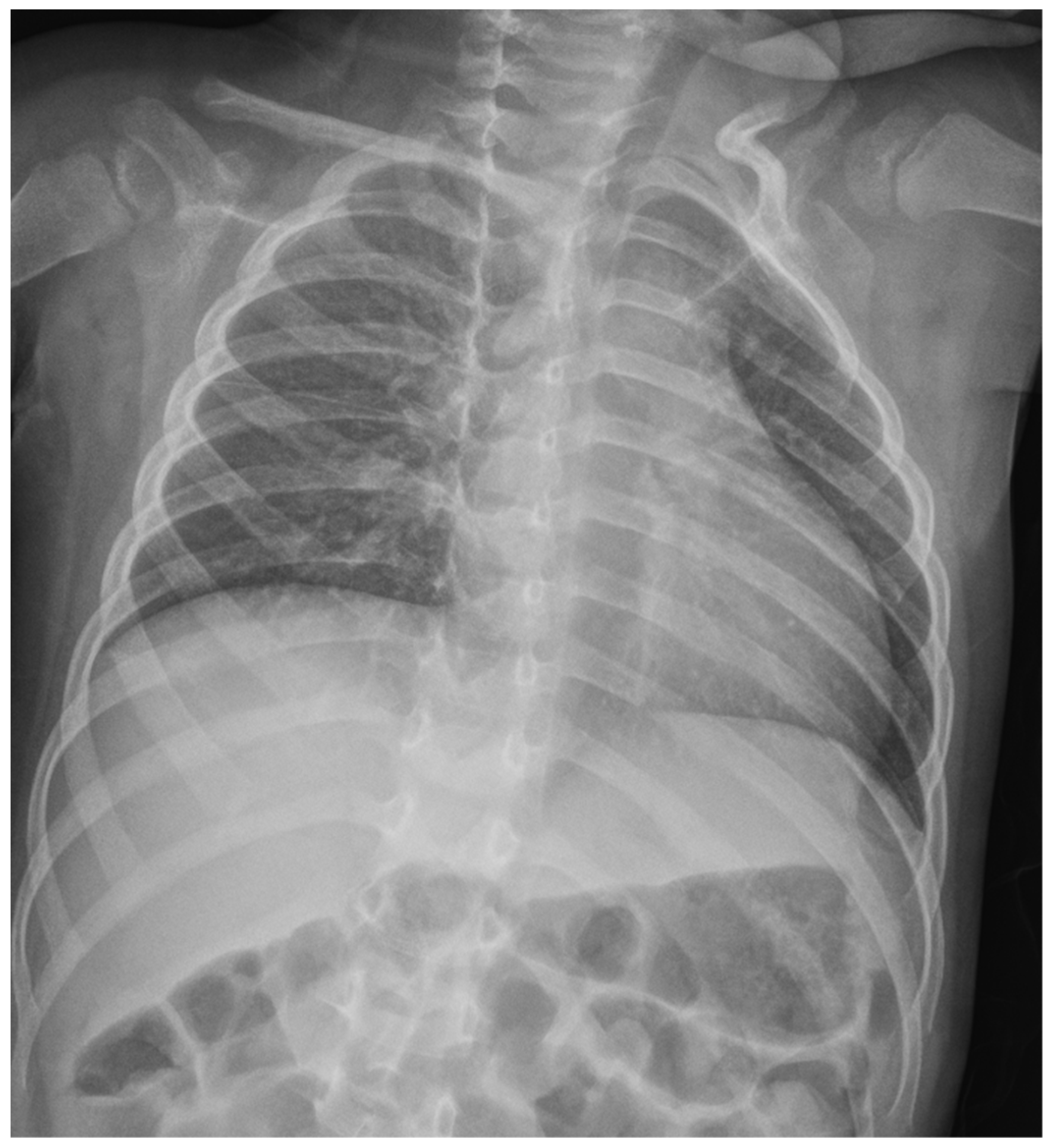
Chest X-ray was performed to screen for pleuroparenchymal involvement or gross cardiomegaly (Figure 6). The exam revealed clear lung fields with normal vascular markings and a cardiac silhouette within the normal limits for age. These findings were instrumental in excluding primary lung diseases (Group 3 PH) and pulmonary venous congestion, thereby supporting the hypothesis of a functional, pre-capillary pulmonary hypertension phenotype.
Suspecting mixed malnutrition (deficiencies in vitamin C, vitamin K, and B-complex vitamins), intravenous vitamin C and multivitamin supplementation were initiated (Table 2).
Follow-up ECG and echocardiography demonstrated complete normalization of pulmonary pressures after one week (Figure 7), which was confirmed one month after discharge along with the resolution of laboratory abnormalities.
The rapid reversibility of the PAH phenotype was documented through sequential echocardiographic assessments. A comprehensive comparison of the hemodynamic and functional parameters from admission to the 7th day of treatment is provided in Table 3.
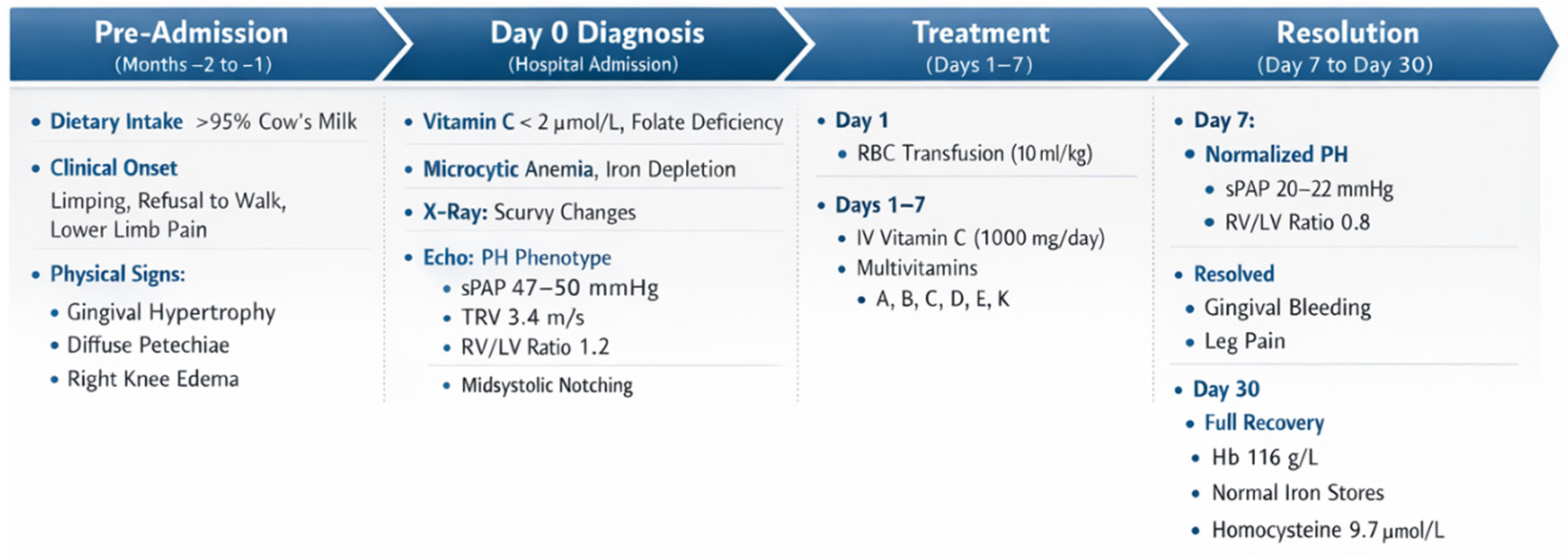
For a comprehensive overview of the clinical sequence—spanning from dietary habits and symptom onset to the resolution of the PH phenotype following treatment—please refer to the detailed timeline provided in Appendix A (Figure A1).

3. Discussion

PAH in scurvy has been rarely described, and it has been defined as pre-capillary (PVRi > 2 WU) in rare cases when cardiac cateterism was performed [8]. Table 4 summarizes the most frequent causes of PAH and their diagnostic tools that we used for differential diagnosis in the absence of a right heart cateterism.
Its pathophysiology in the context of scurvy is likely to be recognized as a multifactorial “perfect storm,” where acute vitamin C depletion acts as the final trigger on a pre-existing substrate of profound metabolic and nutritional derangement [9].
As established in previous reports, the most compelling mechanism involves the impairment of the endothelial–vascular homeostasis. Ascorbic acid and ferrous iron (Fe2+) are essential cofactors for prolyl hydroxylases (PHD), enzymes responsible for the degradation of Hypoxia-Inducible Factors (HIFs) [10,11]. In our patient, the simultaneous absence of vitamin C and severe iron deficiency (suggested by ferritin of 22 mcg/L and transferrin saturation of 5%) likely led to the pathological stabilization of HIF-1α and HIF-2α [12]. This “pseudohypoxia” state promotes pulmonary vasoconstriction and vascular remodeling, mimicking the effects of chronic altitude exposure despite normal oxygen saturation.
Building upon this established framework, we propose a further layer of complexity involving a disrupted iron-trafficking system. Although the C-reactive protein (CRP) was only mildly elevated (1.5 mg/dL), the scurvy-induced state of massive oxidative stress may have stimulated hepcidin production through non-canonical, ROS-dependent pathways. Elevated hepcidin levels, potentially reinforced by iron sequestration via lactoferrin in hypertrophic gingival tissues, would degrade ferroportin, leading to functional iron deficiency. In this framework, the observed elevation in serum copper (in the absence of significant systemic inflammation) becomes highly significant. It may reflect a compensatory, albeit futile, up-regulation of ceruloplasmin (a copper-dependent ferroxidase) in a desperate attempt to mobilize intracellular iron stores [13]. In our hypothesis, this “metabolic lock” would further starve the pulmonary vasculature of the iron necessary for PHD function, reinforcing the hypertensive cycle.
A different explanation may lie beyond systemic inflammation. The observed hypercupremia may be interpreted through the lens of impaired copper trafficking. As suggested by Harris et al., ascorbic acid is essential for labilizing copper bound to ceruloplasmin and facilitating its trans-membrane transport. In a state of profound scurvy, the absence of vitamin C may lead to a paradoxical accumulation of copper in the vascular compartment due to impaired cellular uptake, potentially exacerbating the functional iron deficiency and the subsequent HIF-mediated pulmonary vasoconstriction [14].
The finding of homocysteine levels twice the upper limit of normal serves as a crucial metabolic marker of this global derangement. While primarily interpreted as a signature of folate deficiency (consistent with the high RDW of 30% and neutrophil hypersegmentation), we suggest that hyperhomocysteinemia may have acted as a direct vascular toxin [15]. By impairing nitric oxide bioavailability and inducing oxidative stress, it likely narrowed the vasodilatory reserve of the pulmonary bed, making it more susceptible to the hemodynamic stress of an anemia-induced high-output state.

4. Conclusions

The rapid normalization of pulmonary pressures within one week of multivitamin supplementation, well before the correction of anemia or iron stores, strongly suggests that the recovery of ascorbate-dependent nitric oxide production and the resetting of the PHD/HIF axis were the primary drivers of resolution. This case highlights that PAH phenotype in scurvy might be considered not as a fixed structural disease but as a reversible metabolic disruption of the pulmonary vascular tone. Clinicians should consider scurvy in the differential diagnosis of unexplained echo-suspected PAH in children with selective diets, emphasizing that the cure lies not in vasodilators, but in targeted nutritional restoration.

Author Contributions

Conceptualization, M.P. and L.B.; Methodology, D.M. and M.P.; Validation, S.M., M.P., and D.M.; Investigation, M.L.; Writing—Original Draft Preparation, M.L. and M.P.; Writing—Review and Editing, L.B. and D.M.; Visualization, S.M. All authors have read and agreed to the published version of the manuscript.

Funding

This research received no external funding.

Institutional Review Board Statement

Ethical review and approval of this study were not required by Comitato Etico per la Pratica Clinica (Ethics Committee) of AULSS 7 because case reports are not considered research. The patient’s information has been de-identified.

Informed Consent Statement

Written informed consent has been obtained from the patient’s parent to publish this paper

Data Availability Statement

The raw data supporting the conclusions of this article will be made available by the authors on request.

Conflicts of Interest

The authors declare no conflicts of interest.

Abbreviations

The following abbreviations are used in this manuscript:
PAHPulmonary artery hypertension
ECGElectrocardiography
sPAPSystolic pulmonary artery pressure
PHDProlyl hydroxylases
HIFsHypoxia-Inducible Factors
CRPC-reactive protein
RDWRed cells distribution width

Appendix A

Figure A1. The sequence illustrates the progression from chronic dietary restriction (almost exclusive cow’s milk intake) to the symptomatic presentation of infantile scurvy. The diagnostic “perfect storm” at admission is marked by severe anemia, undetectable vitamin C levels, and a significant pulmonary hypertension phenotype. The timeline concludes with the rapid normalization of clinical signs and pulmonary pressures (within 7 days) following targeted vitamin C and multi-nutrient supplementation.
Reports 09 00044 g0a1

References

  1. Di Nora, A.; Finocchiaro, M.C.; Pizzo, F.; Gian Luca, T.; Cucuzza, M.E.; Di Stefano, A.; Spampinato, S.; Marino, S.; Ruggieri, M.; Pavone, P. Scurvy, a not obsolete disorder: Clinical report in eight young children and literature review. Open Med. 2025, 20, 20241086. [Google Scholar] [CrossRef] [Scilit] [PubMed]
  2. Ayyad, M.; Tran, L.; Ansari, S.; Levy, D.; Matassa, D. Forgotten Deficiency: A Case Series Highlighting Atypical Presentations of Scurvy in the 21st Century. Case Rep. Med. 2025, 2025, 2118907. [Google Scholar] [CrossRef] [Scilit] [PubMed]
  3. Toscano, F.; Zirilli, G.; Foti Randazzese, S.; Carlino, M.; Gallizzi, R.; Wasniewska, M.; Valenzise, M. Scurvy, all the faces you can see: Our experience and review of the literature. Ital. J. Pediatr. 2025, 51, 159. [Google Scholar] [CrossRef] [Scilit] [PubMed]
  4. Hemilä, H.; de Man, A.M.E. Vitamin C deficiency can lead to pulmonary hypertension: A systematic review of case reports. BMC Pulm. Med. 2024, 24, 140. [Google Scholar] [CrossRef] [Scilit] [PubMed]
  5. Berra, G.; Genecand, L.; Juillet, A.; Deux, J.F.; Prada, P.; Müller, H.; Roffi, M.; Beghetti, M. Reversible pulmonary hypertension in a patient diagnosed with scurvy. BMJ Case Rep. 2025, 18, e266143. [Google Scholar] [CrossRef] [Scilit] [PubMed]
  6. Silberkasten, M.; San Martin, E.; Cuello, J.V.; Britto, M.V.; Andrés, M.E. Pulmonary hypertension secondary to vitamin C deficiency: A case report. Arch. Argent. Pediatr. 2025, 123, e202510643. (In English) [Google Scholar] [CrossRef] [Scilit] [PubMed]
  7. Flanagan, L.S.; Sadek, A.; Oliveros, E. Vitamin C Deficiency as a Readily Reversible Cause of Pulmonary Hypertension. JACC Case Rep. 2025, 30, 103424. [Google Scholar] [CrossRef] [Scilit] [PubMed]
  8. Satawiriya, M.; Khongphatthanayothin, A.; Limsuwan, A. Reversible severe pulmonary hypertension related to scurvy in children. BMC Cardiovasc. Disord. 2024, 24, 24. [Google Scholar] [CrossRef] [Scilit] [PubMed]
  9. Jin, Y.; Ding, L.; Fan, M.; Kong, N. Association of Vitamin Deficiency with Pulmonary Arterial Hypertension: A Review. Nutr. Rev. 2025, Jul 11, 119. [Google Scholar] [CrossRef] [Scilit] [PubMed]
  10. Smith, T.G.; Talbot, N.P. Prolyl hydroxylases and therapeutics. Antioxid. Redox Signal. 2010, 12, 431–433. [Google Scholar] [CrossRef] [Scilit] [PubMed]
  11. Kurnick, A.; Zaveri, S.; Tadayoni, A.; Chandrakumar, H.P.; John, S. Reversible severe pulmonary hypertension and right heart failure with cardiogenic shock due to scurvy: A case report. Eur. Heart J. Case Rep. 2023, 7, ytad404. [Google Scholar] [CrossRef] [Scilit] [PubMed]
  12. Kupari, M.; Rapola, J. Reversible pulmonary hypertension associated with vitamin C deficiency. Chest 2012, 142, 225–227. [Google Scholar] [CrossRef] [Scilit] [PubMed]
  13. Qian, Z.M.; Ma, H.; Chen, Y.J.; Guo, Q. Nicotinamide Mononucleotide Reduces Iron in BV-2 Cell by Up-Regulating Ferroportin 1 via Inhibiting the IL-6/STAT3/Hepcidin Pathway. Rejuvenation Res. 2025, in press. [Google Scholar] [CrossRef] [Scilit] [PubMed]
  14. Harris, E.D.; Percival, S.S. A role for ascorbic acid in copper transport. Am. J. Clin. Nutr. 1991, 54, 1193S–1197S. [Google Scholar] [CrossRef] [Scilit] [PubMed]
  15. Wang, M.T.; Chi, P.L.; Cheng, C.C.; Huang, W.C.; Chen, L.W. Application of homocysteine as a non-invasive and effort-free measurements for risk assessment of patients with pulmonary hypertension. Cardiol. J. 2024, 31, 285–299. [Google Scholar] [CrossRef] [Scilit] [PubMed]
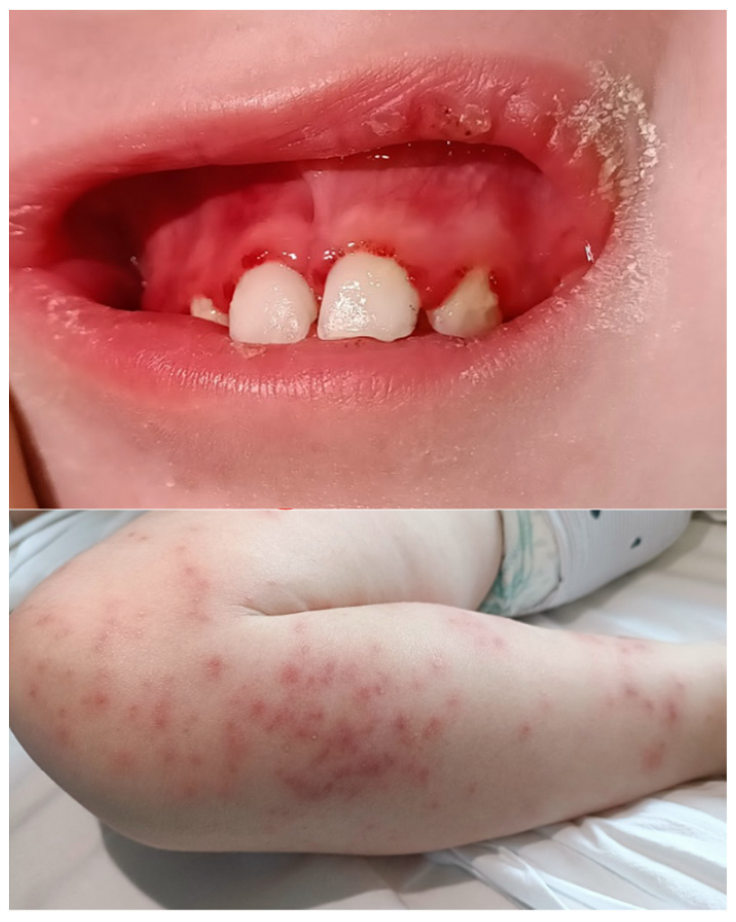
Figure 1. Haemorrhagic gingival hypertrophy and maculopapular rash on admission to the emergency department.
Reports 09 00044 g001
Figure 2. Anteroposterior view of the wrist showing slightly delayed bone age (bone age: 24 months according to Greulich and Pyle; chronological age: 30 months).
Reports 09 00044 g002
Figure 3. Imaging reveals pathognomonic skeletal changes in infantile scurvy. Key findings include the Trümmerfeld zone (a radiolucent band proximal to the dense zone of provisional calcification, representing a site of microfractures and disorganized matrix) and the white line of Fraenkel. Additionally, mild Pelkan spurs are visible at the metaphyseal margins.
Reports 09 00044 g003
Figure 4. Twelve-lead electrocardiogram on admission, showing sinus tachycardia with mild alterations of the ventricular repolarisation phase (DII, DIII, avF), which were no longer evident after approximately one month of adjunctive therapy, reflecting the myocardial metabolic stress associated with global malnutrition and severe anemia.
Reports 09 00044 g004
Figure 5. Findings suggestive of pulmonary hypertension can be observed, including midsystolic notching of the pulmonary artery flow, moderate tricuspid regurgitation, a measurable tricuspid regurgitant jet >2.8 m/s with an estimated systolic pulmonary artery pressure (sPAP) of approximately 47−50 mmHg (tricuspid regurgitation velocity: 42−45 mmHg; right atrial pressure: 5 mmHg) in, and slight end-diastolic flattening of the interventricular septum in the PSAX
Reports 09 00044 g005
Figure 6. Anteroposterior chest X-ray at admission. The lung fields are clear with normal vascular markings and no evidence of parenchymal consolidation or pleural effusion. The cardiac silhouette is within normal limits for age.
Reports 09 00044 g006
Figure 7. Follow-up echocardiography (7 days post−treatment): complete resolution of the midsystolic notching and normalization of the estimated systolic pulmonary artery pressure (<25 mmHg) following one week of intravenous vitamin C and multivitamin supplementation.
Reports 09 00044 g007
Table 1. The table summarizes the main biochemical findings at admission and after one month of effective therapy with vitamin D. The Hb value at 7 days of therapy, after blood transfusion, is also reported. An important microcytic iron-deficiency anemia is highlighted, along with a significant increase in D-dimer and ATIII. Additionally, an increase in copper levels and vitamin B12, likely of inflammatory origin, is noted, with a deficiency in folates and vitamin C. All biochemical findings normalized after approximately one month of therapy. Hb = hemoglobin; PT = Prothrombin Time; aPTT = Activated Partial Thromboplastin Time; ATIII = Antithrombin III.
VariableOn Admission7th Day30th DayReference Values
Hb (g/L)7295116107–144
MCV (fL)63.673.182.580–95
RDW (%)302517.5<15
Serum iron (mcg/dL)32 5716–128
Transferrin saturation (%)5 1110–43
Ferritin (mcg/L)22.1 35.14.6–204.0
PT ratio1.18 1.15 0.8–1.20
aPTT ratio0.74  0.910.8–1.20
D-Dimer (ug/L)49563765 <500 
ATIII (%)135 12480–125
Vitamin B6 (nmol/L)192  51–183
Vitamin B9 (mcg/L)1.70 8.33.1–20.0
Vitamin B12 (ng/L)1035 666187–883
Vitamin C (umol/L)<2 2823–114
Vitamin A (nmol/L)1053  800–2600
Vitamin E (umol/L)15  10.1–21.0
Homocysteine (umol/L)23.8 9.74.4–13.6
Serum copper (umol/L)27.9 17.4 14.1–25.1 
Lactate (mmol/L)<2 <2<2
Table 2. This table summarizes the main integrative therapies administered to the patient, along with their corresponding dosages and duration.
TreatmentPosologyDuration
Vitamin C iv1000 mg/die (80 mg/kg/die)7 days
Followed by Vit C po300 mg/die (24 mg/kg/die)ongoing (60 days)
Vitamin B 6 po4 mg/dieongoing (60 days)
Vitamin B 9 po275 mcg/dieongoing (60 days)
Vitamin B12 po7 mcg/dieongoing (60 days)
Vitamin A po3 mg/dieongoing (60 days)
Vitamin D po500–1000 UI/dieongoing (60 days)
Vitamin E po40 mg/dieongoing (60 days)
Vitamin K im 2 mg/week2 weeks
Red blood cell transfusion10 mL/kg1st day
Table 3. Comparison of echocardiographic parameters at admission and after 7 days of treatment.
EchocardiographyAt admissionFollow-Up (7th Day)
Systolic PAP (mmHg)47−5020–22
Tricuspidal regurgitation (m/s)3.402.25
TAPSE (mm)1114
TAPSE/sPAP (mm/mmHg)0.220.56
Midsystolic NotchingPresentAbsent
RV/LV ratio1.20.8
LVEF75%65%
Table 4. Systematic differential diagnosis workup for the observed echo-suspected pulmonary hypertension (PH) phenotype, illustrating the exclusion of primary cardiac, pulmonary, and thromboembolic etiologies.
ClassificationSubgroupDiagnostic ToolReason for Exclusion
Group 1Congenital heart diseaseEchocardiographyNormal cardiac anatomy 
Group 1Idiopathic or heritableClinical courseImmediate resolution after nutritional therapy
Group 2Left heart diseaseEchocardiographyNo valvular disease
Group 3Chronic lung disease or alveolar hypoxiaChest X-ray
O2 saturation
Clear lung fields on CXR; stable resting O2 saturation
Group 4Pulmonary artery obstructionsD-dimer
clinical course
Rapid resolution without anticoagulation
Group 5Miscellaneous (nutritional)BiochemicalsNormal lactate; no metabolic acidosis; undetectable Vit. C
Disclaimer/Publisher’s Note: The statements, opinions and data contained in all publications are solely those of the individual author(s) and contributor(s) and not of MDPI and/or the editor(s). MDPI and/or the editor(s) disclaim responsibility for any injury to people or property resulting from any ideas, methods, instructions or products referred to in the content.