Article

Test of Diamond sCVD Detectors at High Flux of Fast Neutrons

SARAF, Soreq Nuclear Research Center, Yavne 818000, Israel
*
Author to whom correspondence should be addressed.
Particles 2025, 8(3), 75; https://doi.org/10.3390/particles8030075
Submission received: 27 May 2025 / Revised: 21 July 2025 / Accepted: 24 July 2025 / Published: 7 August 2025
(This article belongs to the Section Experimental Physics and Instrumentation)

Abstract

We have tested the performance of spectroscopic single-crystal Chemical Vapor-Deposited (sCVD) diamond detectors with radioactive sources and with a pulsed deuterium-tritium neutron generator. The tests demonstrate that the detectors could provide good timing and spectroscopic information at high neutron fluxes. The spectroscopic information can be obtained at a 14 MeV neutron rate as high as 1010 n/cm2/s, despite some limitations associated with pulse character of the used neutron generator. Monte-Carlo simulations were performed in order to achieve better understanding of neutron interaction with the detector material. Possible applications for the use of the detectors at Soreq Applied Research Accelerator Facility (SARAF) are considered. The detectors could be used as reliable neutron rate monitors in the vicinity of a strong accelerator-based source of energetic neutrons. The detectors could also be utilized as time-of-flight tagging counters in nuclear physics experiments under condition of high neutron fluxes during short beam pulses. In particular, measurement of the 12C(n,n′)3α cross-section is discussed.

1. Introduction

The SARAF Phase II [1] will provide capabilities for neutron-induced reactions, offering intense primary beams for high neutron flux, and tunable beams allowing for selectable neutron energy. The time-of-flight (TOF) capabilities currently developed at SARAF [2] will allow accurate neutron energy tagging, providing unique opportunities for measurements of neutron-induced reactions. In this respect, it is important to acquire expertise on detector instrumentation possessing the best timing and energy resolution under conditions of high neutron fluxes.
Doped diamond is a semiconducting material that can be used as solid-state spectroscopic detector for radiation monitoring. Until recently, only poly-crystalline Chemical Vapor Deposition (CVD) diamonds were available. The main problem of these detectors was defects at grain boundaries and chemical impurities in the diamond which served as charge trapping centers, thus reducing the charge-collection efficiency. Single crystal, sCVD diamond detectors, developed recently [3], have much better charge collection properties and, hence, exhibit very good spectroscopic characteristics, along with excellent time (less than ns) and energy (~10 keV) resolution and superior radiation hardness (10 MGy) [4]. It was also demonstrated that the sCVD detectors exhibit a good response to high-energy neutrons [5,6,7,8,9]. The best reported energy and time resolution for fast neutrons (FWHM) was 56 keV [5] and 160 ps [10], respectively.
Two diamond detectors and the associated electronics were acquired from Cividec [11]. The original purpose of this acquisition was to perform neutron-induced charge particle cross-section measurements in the vicinity of the strong LIquid LIthium Target (LILIT) neutron source during SARAF Phase I [12]. Several tests of the detectors were performed recently. In particular, the possibility of use of the detectors under the conditions of high neutron flux was evaluated. This was performed in the context of the upcoming second phase of the SARAF.
The acquired equipment included the following:
  • two 140 μm thick, 10 mm2 B3 diamond detectors;
  • two CX-L spectroscopic amplifiers (1.2 μs shaping time, 9 MeV dynamic range);
  • two CX-L spectroscopic amplifiers (1.2 μs shaping time, 150 MeV dynamic range);
  • two C6 fast spectroscopic amplifiers (10 ns shaping time, 50 MeV dynamic range).
This combination allows us to use the detectors in all possible scenarios for light charged particles and for fission fragments, as well as for applications requiring a high neutron rate.

2. Measurements

2.1. Tests with Radioactive Sources

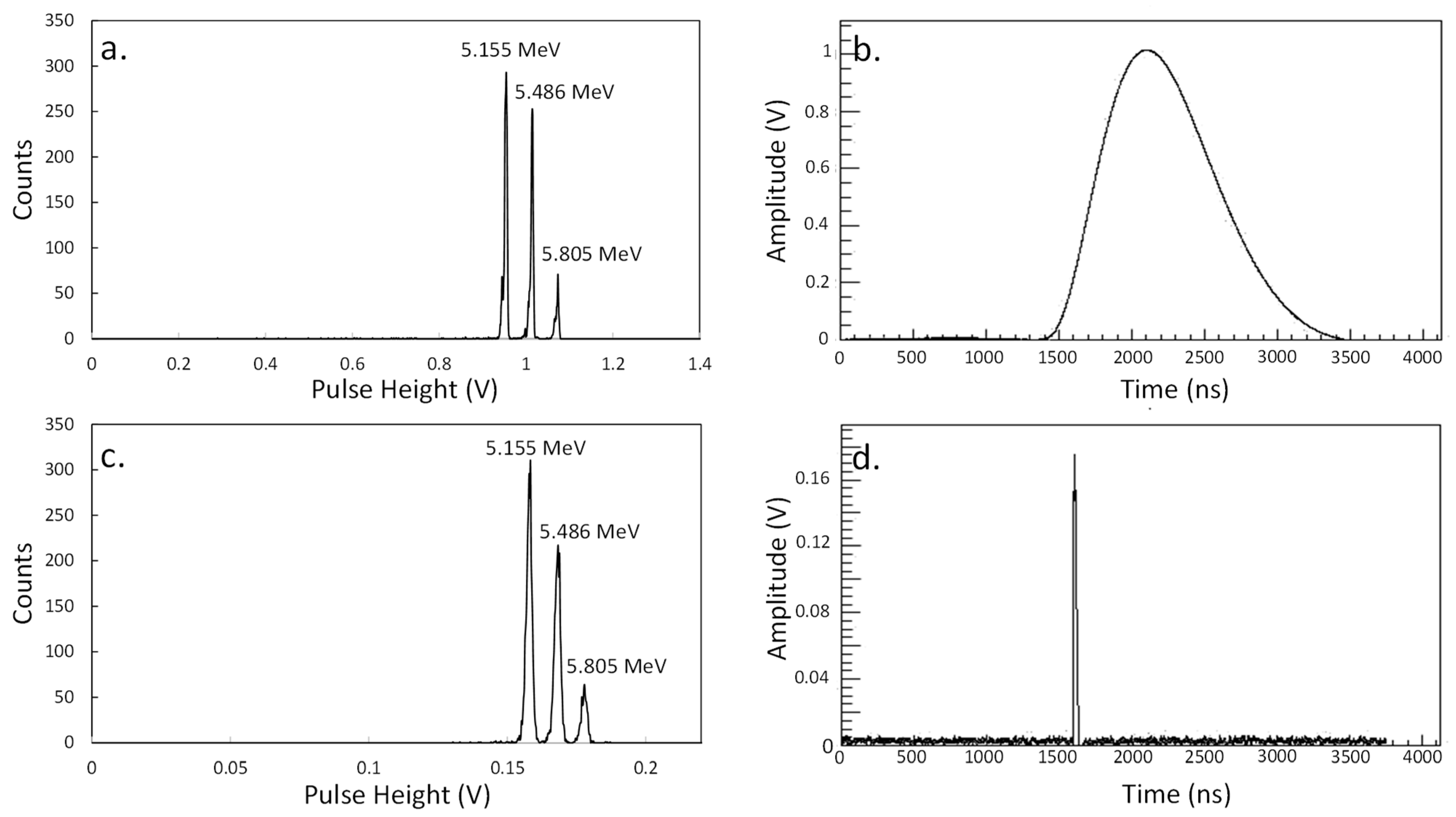
Initial tests were performed with a standard spectroscopic triple alpha source (239Pu, 241Am, 244Cm isotopes with the main alpha lines of 5155, 5486, and 5805 keV). The detector and the source were placed in a test vacuum chamber at a distance of approximately 10 cm. The vacuum pressure in the chamber was of the order of 3 × 10⁻⁵ mbar. The detector bias was +120 V and the dark leakage current was a few nA. The detector output was connected via a short coaxial cable to a vacuum feedthrough. The amplifier was connected directly to the air side of the feedthrough. The amplifier signal was fed to the input of a PC-based digitizer card (Acqiris_U5303A, Acqiris SA, Switzerland) with the maximum sampling rate of 1.6 GHz. The digitizer was self-triggered when the signal was above a predefined threshold (~5 mV). The waveforms were recorded and analyzed on an event-by-event basis and the energy spectra were reconstructed. The energy spectra and examples of a waveform are presented for the case of CX-L (shaping time of 1.2 μs) and C6 (shaping time of 10 ns) amplifiers in Figure 1a,b and Figure 1c,d, respectively. The typical collection times of the energy spectra were of the order of two hours. Th energy resolution obtained with the CX-L amplifier is of the order of 13 keV (FWHM for ~5.5 MeV alpha), a value comparable to those of spectroscopic silicon detectors. It is interesting to note that the data collected with the very fast time amplifier also carry good spectroscopic information (Figure 1c). The energy resolution for this amplifier was approximately 35 keV.
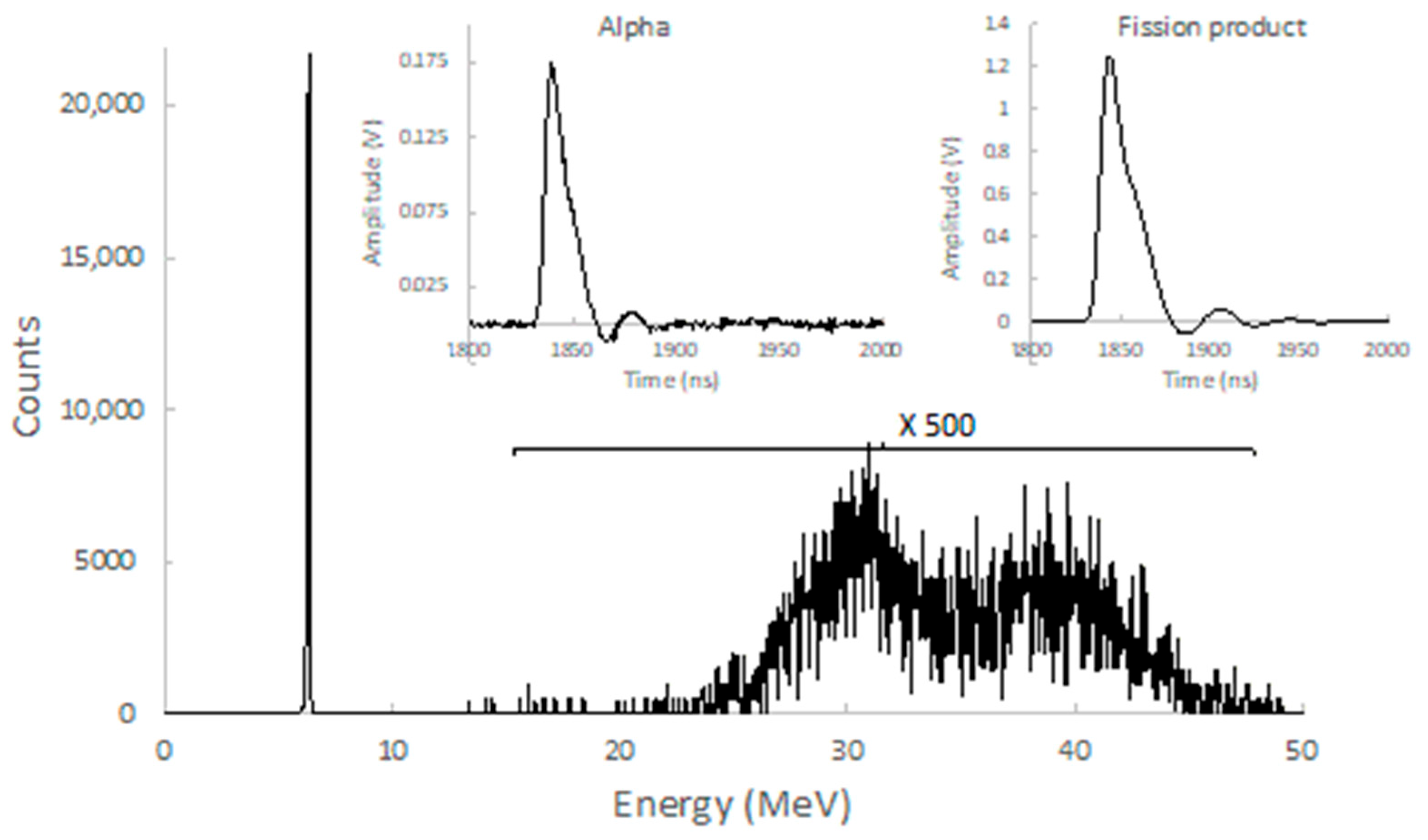
Since the original plans for the detector at Phase I included neutron irradiation of fissile targets, it was also interesting to check the detector response to a fission source. As in the case of the alpha source, an open 252Cf source was placed in the vacuum chamber in front of the detector. The distance between the source and detector was about 3 cm. The energy spectrum obtained with an open 252Cf source and the fast amplifier is presented in Figure 2. The spectrum was collected across approximately 12 h. The spectrum is similar to that presented in [13]. One can clearly observe two bumps corresponding to heavy and light groups of fissile fragments. A 6.1 MeV alpha peak from 252Cf alpha decay is also seen in the spectrum. According to Cividec, the detector has a 100 nm titanium electrode, which is also the detector dead layer. This dead layer does not degrade significant energy of the fission products. It is worth noting that the obtained average energies of heavy and light fragments are 30 and 38.5 MeV, as opposed to the expected 79 and 103 MeV. This is due to the so called strong pulsed-height defect observed for heavy ions in sCVD detectors [14]. The effect is associated with a high electron–hole recombination rate under conditions of high ionization density along fission product tracks. As is seen in Figure 2, the dynamic range of the C6 amplifier was chosen well for this application. The contribution of the neutrons emitted from the 252Cf source is negligible compared to the charged particle contribution. Examples of waveforms collected for an alpha and a fission fragment are shown in the inserts of Figure 2. It is interesting to note that the falling time of the fission fragment waveform is somewhat longer than that of the alpha particle.

2.2. Tests with the Neutron Generator

An ING-031 pulsed neutron generator (NG) [15] was available for tests with neutrons. The t(d,n) neutron generator generates approximately 109 neutrons per second over 4π. The average neutron yield was confirmed on many occasions by performing activation measurements of pure aluminum samples. Generation of 14 MeV neutrons takes place during short pulses with typical a frequency of 30 Hz. Thus, approximately 3.3 × 10⁷ neutrons are emitted per neutron burst. According to the manufacturer, the duration of the neutron pulse above 10% of the maximum amplitude is of the order of 0.5 μs. This corresponds to the effective peak neutron rate of 6.6 × 10¹³ n/s during a 0.5 μs pulse. Such an intense neutron flux makes this neutron generator a unique tool for testing detectors at high rate applications. Similar studies were performed in the literature, albeit at much lower neutron fluxes [5,6,7,8,9,10]. Neutron interaction with diamond detectors corresponds to the number of nuclear reactions with carbon isotopes listed in Table 1. It is worth noticing that the 12C(n,n′)3α reaction includes several reaction channels, the main of which are 12C(n,α)9Be*→n+8Be→2α and 12C(n,n′)12C*→α+8Be→2α [16,17]. It was shown in [18] that the 12C(n,α)9Be*→n+8Be→2α channel plays the dominant role. Nonetheless, measurement with diamond detectors does not allow one to distinguish between these channels. Only information on the total 12C(n,n′)3α cross-section can be obtained.
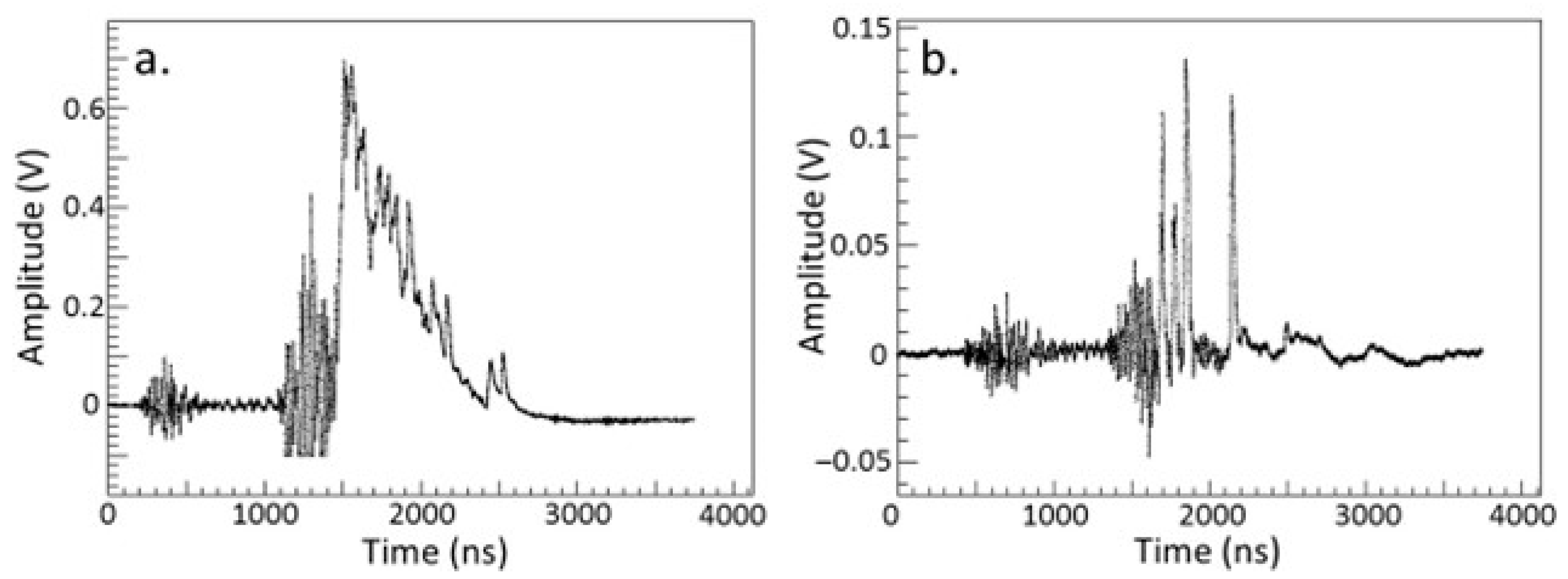
We have collected waveforms of signals from neutron bursts using the detector placed at two distances from the neutron generator frontal surface: 5 cm and 25 cm. The detectors were placed on the neutron generator axis. This would correspond to neutron energy in the range 14.1–15 MeV depending on the neutron generator properties (energy of the deuteron beam, thickness of the tritium target). However, this information is not readily available. As in the case of measurements with radioactive sources, the detector was placed in the grounded stainless-steel chamber. The chamber had a 2 mm stainless-steel thick window on the neutron axis. The detector was connected via a short cable and feedthrough to the fast C6 amplifier. The output signal was fed via a coaxial cable to the digitizer, placed at a remote location (~20 m from NG). Examples of the waveforms for the two geometries are presented in Figure 3. In the figure, one can immediately observe strong electronic noise in the time regions of 300 and 1200 ns. Some measures were performed in an attempt to reduce this noise: rearranging the grounds, placing the amplifier in metallic foil, and feeding the electronics via an insulating transformer. None of these measures led to noise reduction. This noise is associated with the impulse character of the neutron generator. Due to the closer distance to the detector and the amplifier, the noise signal at 5 cm geometry (Figure 3a,b) is much stronger. One can also observe very peculiar time behavior of the base line, also associated with an electronic response to the generator pulse.
The signals from the neutrons are observed in the 1500–2500 ns time range of the waveform. It appears that most of the neutrons are emitted within a 0.5 μs window, as specified by the manufacturer. As seen in the figure, the rate of neutron events is too high at the close geometry. One can observe strong summing and pileup of the signals, making it impossible to obtain any spectroscopic information. At the more distant geometry, however, the individual events are resolved comfortably.
The neutron flux density can be evaluated roughly from the estimated neutron number per pulse (3.3 × 10⁷) and a pulse duration (0.5 μs) in assumption of the point-like geometry of the neutron source. The point-like neutron source geometry assumption is justified, as according the manufacturer [15] the neutron source diameter is only 2 mm. The source is situated at approximately a 5 mm depth from the device frontal surface. The obtained neutron flux density is 1.9 × 10¹¹ and 8.9 × 10⁹ n/s/cm2 for the close (5 cm) and far (25 cm) geometries, respectively. Thus, one can state with confidence that the detector can perform well at a neutron rate density as high as 1010 n/s/cm2.
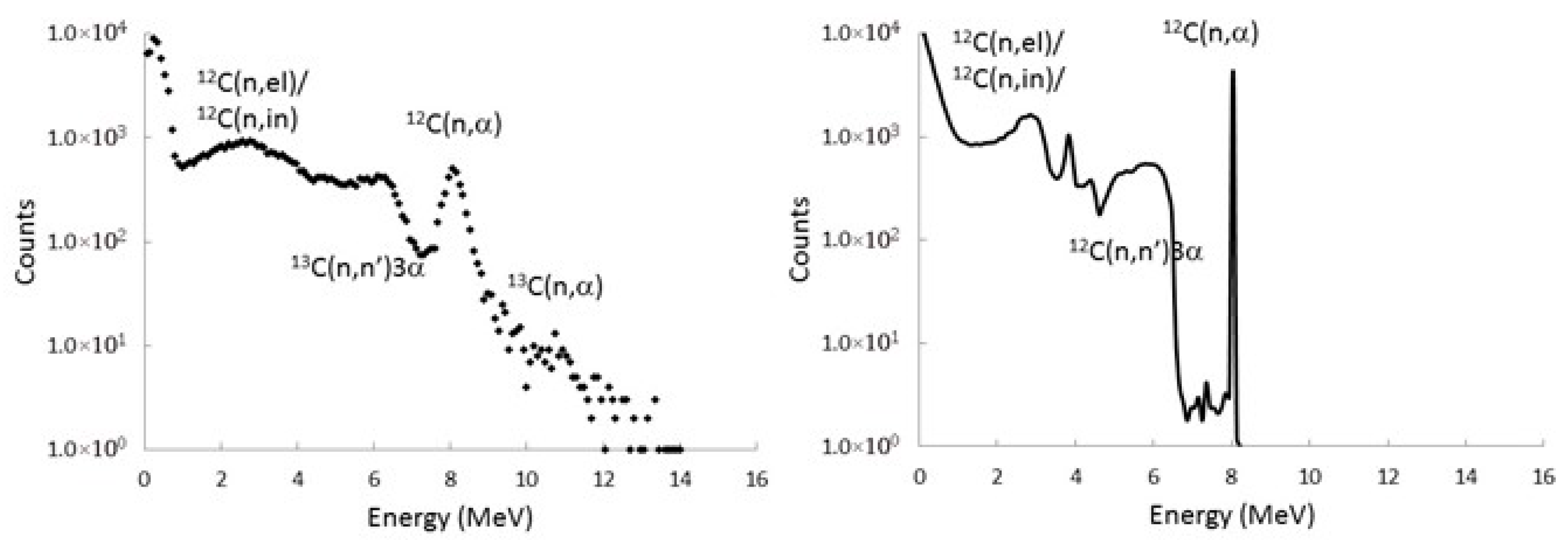
The collected data were analyzed on an event-by-event basis, and the signals from individual peaks were calibrated using measurements with a triple alpha source. The collected spectrum is presented in Figure 4. The data were taken over one hour of irradiation at the 25 cm distance. One can clearly observe the 12C(n,α)9Be peak and a weak sign of the 13C(n,α)10Be peak in the spectrum. The spectrum is somewhat similar to that obtained in [5,6,7,8,9]. Our spectrum, however, suffers from the electronic noise and base-line modulation associated with the pulsed operation of the neutron generator. A much better spectrum quality is expected from a DC neutron generator or from an accelerator-based neutron source. The detector was placed at approximately a 40 cm distance from the concrete floor, so neutron scattering from the environment also has an effect on the spectrum quality.

3. GEANT4 Simulations

3.1. General Simulations

GEANT4 [19] Monte-Carlo simulations were performed in order to achieve a better understanding of neutron interaction with the detector material. The diamond detector was modeled in GEANT4 version 4.11.0.0, as a simple 0.14 mm thick and 3 mm diameter cylindrical sensitive volume. The material properties of the diamond detector model were set to natural carbon abundance (98.93% 12C and 1.07% 13C) with a density of 3.51 g/cm3. The neutron response, NRESP71 [17], model was used for the neutron interaction with carbon. No surrounding material and no effects of electronic noise were taken into account in this simple simulation. The obtained simulated spectrum is shown in Figure 4. The main interaction processes, elastic and inelastic scattering, as well as 12C(n,n′)3α and 12C(n,α) reactions, are seen in the simulations. It is interesting to note that the current version of GEANT4 does not reproduce the 13C(n,α) channel. This problem has to be investigated further. This, however, is not of great importance for the purpose of this preliminary study.
After acquiring the first experimental and simulation expertise with the diamond detectors, the possibility of using the detectors in neutron research in future SARAF applications, in particular for the measurement of the 12C(n,n′)3α reaction, can be considered.

3.2. The Case of the 12C(n,n′)3α Measurements at the Future TOF Facility

Stellar production of 12C takes place via triple alpha reactions populating the resonance Hoyle state [20]. This reaction is, probably, the most important one for nuclear astrophysics. Recently, it was suggested that the triple alpha reaction rate can be significantly modified by so called neutron upscattering [21]. Inelastic neutron scattering on carbon can populate the Hoyle state, following immediate disintegration to three alphas 12C(n,n′)3α. This is the reverse reaction of triple alpha fusion with neutron upscattering as catalysis. Study of the reverse 12C(n,n′)3α reaction is important for understanding the neutron upscattering contribution in stellar carbon synthesis and has attracted much interest recently [22]. In addition, the reaction is interesting for nuclear reactor design, since it corresponds to one of the processes leading to the production of helium gas in a reactor’s carbon rich components [23].
A diamond detector can be used for monitoring neutron flux and measuring neutron energy via TOF at SARAF Phase II. Furthermore, it is interesting that such a detector could be used in parallel as an active target for studying the 12C(n,n′)3α reaction. This already was demonstrated in [24], where diamond detectors were irradiated with monochromatic neutrons. An advantage of SARAF Phase II is the ability of operation in a pulsed regime, where short bursts of protons on a thick target will generate sharp pulses of neutrons at a broad energy range. This neutron spectrum could be measured by a diamond detector using the TOF technique. High neutron intensity would allow us to place the detector at a long enough distance to optimize the energy resolution and counting rate. Thus, the 12C(n,n′)3α cross-section will be measured as a function of neutron energy. These measurements could be performed in a parasitic mode while performing other experiments, where diamond detectors will be used as a neutron monitor.
An important question is whether the 12C(n,n′)3α signal can be disentangled from the neutron scattering background under conditions of the broad neutron energy spectrum in a typical SARAF TOF experiment. To address this question, we performed GEANT4 simulations. The neutron spectrum was taken to be homogeneous in the range of 5–15 MeV. The detector was placed at a distance of 5 m from a production target. Two-dimensional correlation between neutron energy (TOF) and energy deposited by reaction products in the 140 μm detector is shown in Figure 5. The simulation results are shown for the main reaction channels separately and for the total interaction. The results for the 12C(n,n′)3α reaction were generated according to the final products (3α) without differentiation between the two main channels (Table 1). As expected, the dominant background to the 12C(n,n′)3α in the low energy region are the elastic and inelastic scattering channels. However, the high energy part of the 12C(n,n′)3α distribution, denoted by red dashed line, is practically free from background and can be unambiguously measured. Further cleanup from neutron scattering events in the data may be achieved with the pulse shape analysis technique reported in [6,7,8]. Thus, the measurement seems to be feasible in the broad energy range.

4. Summary

Tests of the diamond detector were performed with radioactive sources and the ING-031 pulsed neutron generator. The detector performs well and allows us to obtain time and energy information at fast neutron flux densities as high as 1010 n/s/cm2. The pulsed nature of the available neutron generator resulted in some limitations in the energy resolution due to electronic noise and an unstable base line. However, we managed to collect the energy spectrum with meaningful spectroscopic information at such a high neutron rate. To our knowledge, the spectroscopic performance of diamond detectors has never been demonstrated at such high neutron fluxes using a pulsed generator. The previously reported neutron spectrum measurement was performed at the TAPIRO fast research reactor (ENEA, Casaccia) with fluxes of 109 n/s/cm2 [25]. This and our results validate interest towards these detectors as prospective neutron diagnostic instruments for fusion reactors ([25] and references therein). Single crystal CVD diamond detectors can be used as neutron flux monitoring devices at very high neutron rates at SARAF Phase II in the vicinity of the production target. One has to take into account, however, the reported modification of the detector energy calibration [25] and general deterioration of the detector performance at very high fast neutron fluences above 1016 n/cm2 ([18] and references therein). More detailed studies will be performed in the future.
It has been shown in the literature that the inherent time resolution of the diamond detector is very fast [10]. In our measurements with available fast electronics, we demonstrate the fast rise time of the signal waveforms. Therefore, we assume that a sub-nanosecond time resolution is achievable with diamond detectors at the future TOF facility. We plan to perform these measurements in the future and demonstrate the use of the detectors as time-of-flight tagging counters in nuclear physics experiments under conditions of high neutron fluxes during short beam pulses. An especially interesting application of diamond detectors could be the measurement of 12C(n,n′)3α reaction cross-sections at SARAF Phase II, where the detector is also a reaction target.

Author Contributions

Conceptualization, L.W.; methodology, L.W. and A.S.; analysis, A.S.; simulations S.V.; writing, L.W.; editing L.W., A.S. and S.V. All authors have read and agreed to the published version of the manuscript.

Funding

This research received no external funding.

Data Availability Statement

The data could be provided by the corresponding author upon request.

Acknowledgments

We would like to acknowledge Ch. Weiss from CIVIDEC for providing technical assistance in procurement and operation of the detectors and electronics and H.O.U. Fynbo for discussion of the subject of the 12C(n,n′)3α reaction.

Conflicts of Interest

The authors declare no conflicts of interest.

References

  1. Pichoff, N.; Chance, A.; Dumas, J.; Ferrand, G.; Gougnaud, F.; Cohen, S.; Isakov, H.; Luner, J.; Mardor, I.; Perry, A.; et al. The SARAF linac project status 2023. In Proceedings of the 14th International Particle Accelerator Conference, Venice, Italy, 7–12 May 2023; p. TUPM053. [Google Scholar]
  2. Mardor, I.; Wilsenach, H.; Dickel, T.; Eliyahu, I.; Friedman, M.; Hirsh, T.Y.; Kreisel, A.; Sharon, O.; Tessler, M.; Vaintraub, S.; et al. Opportunities for high-energy neutrons and deuterons induced measurements for fusion technology at the Soreq applied research accelerator facility (SARAF). Front. Phys. 2023, 11, 1248191. [Google Scholar] [CrossRef]
  3. Frais-Kolbl, H.; Griesmayer, E.; Kagan, H.; Pernegger, H. A Fast Low-Noise Charged-Particle CVD Diamond Detector. IEEE Trans. Nucl. Sci. 2004, 51, 3833–3837. [Google Scholar] [CrossRef]
  4. Adam, W.; de Boer, W.; Borchi, E.; Bruzzi, M.; Colledani, C.; D’aNgelo, P.; Dabrowski, V.; Dulinski, W.; van Eijk, B.; Eremin, V.; et al. Radiation hard diamond sensors for future tracking applications. Nucl. Inst. Meth. A 2006, 565, 278–283. [Google Scholar] [CrossRef]
  5. Pillon, M.; Angelone, M.; Krása, A.; Plompen, A.J.M.; Schillebeeckx, P.; Sergi, M.L. Experimental response functions of a single-crystal diamond detector for 5–20.5 MeV neutrons. Nucl. Inst. Meth. A 2011, 640, 185–191. [Google Scholar] [CrossRef]
  6. Weiss, C.; Schuhmacher, H.; Griesmayer, E.; Zimba, A.; Weierganz, M.; Gagnon-Moisan, F.; Kasper, A. Response of CVD Diamond Detectors to 14 MeV Neutrons, CERN-ATS-Note-2012-093 TECH. Available online: https://cds.cern.ch/record/1494167?ln=en (accessed on 1 April 2025).
  7. Weiss, C.; Frais-Kölbl, H.; Griesmayer, E.; Kavrigin, P. Ionization signals from diamond detectors in fast neutron fields. Eur. Phys. J. A 2016, 52, 269. [Google Scholar] [CrossRef]
  8. Kavrigin, P.; Griesmayer, E.; Belloni, F.; Plompen, A.J.M.; Schillebeeckx, P.; Weiss, C. 13C(n,α0)10Be cross section measurement with sCVD diamond detector. Eur. Phys. J. A 2016, 52, 179. [Google Scholar] [CrossRef]
  9. Liu, J.; Jiang, H.; Cui, Z.; Hu, Y.; Bai, H.; Fan, T.; Chen, J.; Gao, Y.; Yang, X.; Zhang, G. Simultaneous measurement of energy spectrum and fluence of neutrons using a diamond detector. Sci. Rep. 2022, 12, 12022. [Google Scholar] [CrossRef] [PubMed]
  10. Osipenko, M.; Bellucci, A.; Ceriale, V.; Corsini, D.; Gariano, G.; Gatti, F.; Girolami, M.; Minutoli, S.; Panza, F.; Pillon, M.; et al. Upgrade of the compact neutron spectrometer for high flux environments. Nucl. Inst. Meth. A 2018, 883, 14–19. [Google Scholar] [CrossRef]
  11. Available online: http://www.cividec.at (accessed on 1 April 2025).
  12. MPaul, M.; Tessler, M.; Friedman, M.; Halfon, S.; Weissman, L. The Liquid-Lithium Target at the Soreq Applied Research Accelerator Facility. Eur. Phys. J. A 2022, 58, 207. [Google Scholar] [CrossRef]
  13. Frégeau, M.O.; Oberstedt, S.; Brys`, T.; Gamboni, T.; Geerts, W.; Hambsch, F.-J.; Oberstedt, A.; Vidali, M. First use of single-crystal diamonds as fission-fragment detector. Nucl. Instr. Meth. A 2015, 791, 58–64. [Google Scholar] [CrossRef]
  14. Beliuskina, O.; Strekalovsky, A.O.; Aleksandrov, A.A.; Aleksandrova, I.A.; Devaraja, H.M.; Heinz, C.; Heinz, S.; Hofmann, S.; Ilich, S.; Imai, N.; et al. Pulse-height defect in single-crystal CVD diamond detectors. Eur. Phys. J. 2017, 53, 32. [Google Scholar] [CrossRef]
  15. Available online: http://www.vniia.ru/eng/production/neitronnie-generatory/neytronnye-generatory.php (accessed on 1 April 2025).
  16. Antolković, B.; Dolenec, Z. The neutron-induced 12C(n, n′)3α reaction at 14.4 MeV in a cinematically complete experiment. Nucl. Phys. A 1975, 237, 235–252. [Google Scholar] [CrossRef]
  17. Garcia, A.R.; Mendoza, E.; Cano-Ott, D.; Nolte, R.; Martinez, T.; Algora, A.; Tain, J.L.; Banerjee, K.; Bhattacharya, C. New physics model in GEANT4 for the simulation of neutron interactions with organic scintillation detectors. Nucl. Instrum. Meth. A 2017, 868, 73–81. [Google Scholar] [CrossRef]
  18. Osipenko, M.; Ripani, M.; Ricco, G.; Caiffi, B.; Pompili, F.; Pillon, M.; Verona-Rinati, G.; Cardarelli, R. Response of a diamond detector sandwich to 14MeV neutrons. Nucl. Instr. Meth. A 2016, 817, 19–25. [Google Scholar] [CrossRef]
  19. Agostinelli, K.A.; Apostolakis, J.; Araujo, H.; Arce, P.; Asai, M.; Axen, D.; Banerjee, S.; Barrand, G.J.N.I.; Behner, F.; Bellagamba, L.; et al. GEANT4—A simulation toolkit. Nucl. Instr. Meth. A 2003, 506, 250–303. [Google Scholar] [CrossRef]
  20. Freer, M.; Fynbo, H.O.U. The Hoyle state in 12C. Prog. Part. Nucl. Phys. 2014, 78, 1–23. [Google Scholar] [CrossRef]
  21. Beard, M.; Austin, S.M.; Cyburt, R. Enhancement of the triple alpha rate in a hot dense medium. Phys. Rev. Lett. 2017, 119, 112701. [Google Scholar] [CrossRef] [PubMed]
  22. Bishop, J.; Parker, C.E.; Rogachev, G.V.; Ahn, S.; Koshchiy, E.; Brandenburg, K.; Brune, C.R.; Charity, R.J.; Derkin, J.; Dronchi, N.; et al. Neutron-upscattering enhancement of the triple-alpha process. Nat. Commun. 2022, 13, 2151. [Google Scholar] [CrossRef]
  23. Pigni, M.; DeBoer, R.; Dimitriou, P. Summary Report of the IAEA Consultants’ Meetings; IEEA: Vienna, Austria, 2022; Available online: https://www-nds.iaea.org/publications/indc/indc-nds-0853.pdf (accessed on 1 April 2025).
  24. Liu, J.; Cui, Z.; Hu, Y.; Bai, H.; Chen, Z.; Xia, C.; Fan, T.; Chen, J.; Zhang, G.; Ruan, X.; et al. 12C(n,n+ 3α) and 12C(n, α0)9Be cross sections in the MeV neutron energy region. Phys. Lett. B 2023, 842, 137985. [Google Scholar] [CrossRef]
  25. Osipenko, M.; Pompili, F.; Ripani, M.; Pillon, M.; Ricco, G.; Caiffi, B.; Verona-Rinati, G.; Argiro, S. Test of a prototype neutron spectrometer based on diamond detectors in a fast reactor. In Proceedings of the 4th International Conference on Advancements in Nuclear Instrumentation Measurement Methods and Their Applications (ANIMMA), Lisbon, Portugal, 20–24 April 2015. [Google Scholar] [CrossRef]
Figure 1. The energy spectra collected from the triple alpha source and example of waveform obtained with CX-L (a,b) and C6 (c,d) amplifiers.
Particles 08 00075 g001
Figure 2. The energy spectrum from 252Cf source obtained with the C6 amplifier. The waveforms from alphas and fission products are shown in the inserts.
Particles 08 00075 g002
Figure 3. Examples of neutron burst waveforms, corresponding to the detector placed at 5 cm (a) and 25 cm (b) from the NG front surface. The measurements are taken with a C6 amplifier.
Particles 08 00075 g003
Figure 4. (Left) Neutron energy spectrum obtained at the 25 cm distance over an hour of irradiation. (Right) GEANT simulations of the detector response to 14 MeV neutrons.
Particles 08 00075 g004
Figure 5. Results of the TOF vs. deposited energy correlations for the individual reaction channels (top) and total interaction (bottom). Note that the top insert plots are with x-y scales identical to the larger plot.
Particles 08 00075 g005
Table 1. Neutron-induced reactions in the diamond detector material (carbon) including Q-values, threshold energy, and approximate cross-sections values at 14 MeV.
ReactionQ-Value (MeV)Threshold (MeV)Cross-Section (mb)
12C(n,el)12C 00~850
13C(n,a)10Be−3.8364.134~11
12C(n,inl)12C −4.4394.81~200
12C(n,a)9Be−5.7026.182~65
12C(n,n′)3α−7.2757.886~200
12C(n,p)12B−13.73214.887~1
Disclaimer/Publisher’s Note: The statements, opinions and data contained in all publications are solely those of the individual author(s) and contributor(s) and not of MDPI and/or the editor(s). MDPI and/or the editor(s) disclaim responsibility for any injury to people or property resulting from any ideas, methods, instructions or products referred to in the content.