Review

Current Knowledge in Planarian Glia and Its Future Implications in Modeling Neurodegenerative Diseases

by
David Gonzalez
1,2,*,
Víctor Alarcón
3 and
Constanza Vásquez-Doorman
2,4,*
1
Escuela de Terapia Ocupacional, Facultad de Ciencias de la Salud, Universidad Bernardo O’Higgins, Santiago 8370854, RM, Chile
2
Departamento de Ciencias Químicas y Biológicas, Facultad de Ciencias de la Salud, Universidad Bernardo O’Higgins, Santiago 8370854, RM, Chile
3
Escuela de Medicina, Facultad de Ciencias Médicas, Universidad Bernardo O’Higgins, Santiago 8370854, RM, Chile
4
Escuela de Kinesiología, Facultad de Ciencias de la Salud, Universidad Bernardo O’Higgins, Santiago 8370854, RM, Chile
*
Authors to whom correspondence should be addressed.
Neuroglia 2025, 6(4), 37; https://doi.org/10.3390/neuroglia6040037
Submission received: 28 August 2025 / Revised: 22 September 2025 / Accepted: 23 September 2025 / Published: 24 September 2025

Abstract

Neurodegenerative diseases are characterized by progressive loss of neurons and remain largely incurable. Numerous mammalian models have been developed to study the mechanisms underlying their physiopathology; however, their high cost, complexity and time requirements highlight the need for alternative systems. Glial cells are increasingly recognized as key contributors to neurodegenerative disease progression through non-cell autonomous mechanisms. Planarians possess a nervous system with diverse neuronal subtypes and glial cells, offering an attractive combination of evolutionary conservation and remarkable regenerative capacity. Unlike mammalian glia, planarian glia originate from phagocytic progenitors and exhibit distinctive molecular markers, including if-1, cali and cathepsin. Emerging evidence suggests that planarian glia may contribute to neurotransmitter homeostasis, neuron–glia interactions and phagocytic activity. Additionally, planarians display robust and quantifiable behavioral responses, making them well suited for modeling neurodegenerative disease. In this review, we summarize the current findings regarding neuronal subtypes and glial cells in planaria, emphasizing their relevance as a model system. Further research into planarian glia will be crucial for understanding their roles in pathological contexts and for exploring their potential applications in neurodegenerative diseases research. Planarian simplicity, regenerative capacity, and compatibility with high-throughput approaches position planarians as a powerful model for investigating the cellular and molecular mechanisms underlying neurodegenerative diseases and for identifying potential therapeutic targets.

1. Introduction

Neurodegenerative diseases are characterized by the progressive loss of specific populations of neurons and can be classified based on clinical features (such as dementia, parkinsonism, or motor neuron involvement), the anatomical regions affected (including frontotemporal, extrapyramidal, or spinocerebellar areas), or underlying molecular abnormalities. They can affect neurons from the brain, brainstem, spinal cord and retina at all ages, but aging is a risk factor [1]. These disorders range from common conditions like Alzheimer’s disease (AD), Parkinson’s disease (PD), multiple sclerosis (MS), and age-related macular degeneration (AMD) to rarer diseases, such as Huntington’s disease (HD), frontotemporal dementia (FTD), corticobasal syndrome (CBS), multiple system atrophy (MSA), amyotrophic lateral sclerosis (ALS), progressive supranuclear palsy (PSP), and spinal muscular atrophy (SMA), among others [2].
Although neurodegenerative diseases are primarily characterized by neuronal loss, glial cells such as astrocytes, microglia and oligodendrocytes also play a critical role in disease progression. Beyond their structural and supportive functions, glial cells exhibit remarkable diversity and functional complexity. However, under pathological conditions, astrocytes and microglia can become reactive, driving neuroinflammation and contributing to disease progression [3,4]. Besides intrinsic neuronal defects, many pathological mechanisms operate in a non-cell autonomous manner, whereby signals and responses from surrounding glial cells affect neuronal survival. For instance, studies using chimeric mice expressing either a wild-type or mutant form of superoxide dismutase 1 (Sod1), a gene associated with familial ALS, have shown that wild type motor neurons surrounded by glial cells expressing mutant Sod1 develop ubiquitin-positive protein aggregates, a hallmark of neuronal damage in ALS [5,6]. Furthermore, transgenic mice in which the mutant Sod1 gene is selectively deleted from motor neurons exhibit a milder ALS phenotype, though the disease is not completely prevented. This finding demonstrates that glial cells contribute to disease progression and highlights the importance of targeting non-neuronal cells as a potential therapeutic strategy [7]. Similar non-cell autonomous mechanisms have been implicated in other neurodegenerative diseases including AD, PD, and HD where astrocytes and microglia are believed to contribute to disease progression through a neuroinflammatory process, a common pathological feature across these conditions [8,9,10].
To date, neurodegenerative diseases remain incurable, and most FDA-approved drugs merely alleviate symptoms, with only a few offering modest effects on slowing disease progression. Consequently, significant efforts have been invested in developing animal models, particularly mammalian ones, that successfully recapitulate key pathological features of these disorders [11,12,13,14]. While these models have been invaluable for advancing our understanding of disease mechanisms and for testing therapeutic strategies, they also present important limitations: they are expensive and time-consuming to maintain, require highly specialized infrastructure, and are often constrained in their suitability for large-scale or high-throughput analyses. These challenges underscore the need for complementary systems that balance biological relevance with experimental accessibility, thereby expanding the biological models available for studying the mechanisms of neurodegenerative diseases and accelerating therapeutic discovery.
Consequently, invertebrate models have been developed to investigate the pathogenic mechanisms underlying neurodegenerative diseases. Models such as Drosophila melanogaster and Caenorhabditis elegans offer valuable insights due to several key features. As reviewed by Pasko et al. these features include: (i) the presence of specialized cells and tissues, including the glia-neuron interactions that are crucial for understanding non-cell autonomous mechanisms in certain neurodegenerative diseases; (ii) their evolutionary relationships, which include conservation of numerous molecular mechanisms involved in cellular processes such as autophagy, cell death, synaptic formation, axonal remodeling, among others; and (iii) measurable nervous system activity and its correlation with behavior [15].
In this context, flatworm planarians belonging to the freshwater family Dugesiidae arise as a complementary model, with the most commonly used species including Girardia tigrina, Girardia dorotocephala, Dugesia japonica, and Schmidtea mediterranea [16,17]. Planarians have long been used as a model system due to their extraordinary regenerative capacity, being able to regenerate all tissues, including a fully functional nervous system after decapitation [17,18,19]. Notably, many of the signals that regulate planarian regeneration are similar to factors related to human brain development [17,20,21]. This regenerative ability is driven by a population of pluripotent adult stem cells known as neoblasts, which are distributed throughout the planarian body and are capable of giving rise to all cell types, including both neurons and glia [22]. Such properties provide a unique opportunity to study fundamental processes such as, neuronal death, survival, plasticity and regrowth in vivo [23,24,25]. Importantly, the simplicity of the planarian nervous system, coupled with its evolutionary conservation and robust regenerative potential, makes planarians a powerful model to dissect mechanisms of nervous system repair that are often difficult to study in mammalian models. These features position planarians as an attractive complementary system for exploring the cellular and molecular basis of neurodegenerative diseases, with potential applications in identifying novel therapeutic targets and strategies.

2. Planarian Nervous System and Neuronal Subtypes

The planarian central nervous system (CNS) consists of a pair of bilobed cerebral ganglia, often considered as a primitive brain, from which two ventral nerve cords (VNCs) extend along the organism. These nerveways are interconnected by transverse commissures, forming a characteristic ladder-like structure. Despite this simplicity, the planarian CNS shares fundamental organization and cell types with the vertebrate CNS, making it a valuable model to investigate conserved mechanisms of neurobiology [26].
Several neuronal subtypes have been identified in planarians, including dopaminergic, cholinergic, GABAergic, and serotonergic neurons, along with conserved neurotransmitters [15,19,26]. These populations are distributed throughout the nervous system and contribute to functions like locomotion, sensory processing, feeding behavior and responses to environmental stimuli. Moreover, some of these neurotransmitter systems are the same ones disrupted in human neurodegenerative disorders, such as dopaminergic neurons in PD, highlighting the translational relevance of studying them in this organism [15]. Besides its neuronal diversity, the accessibility of the planarian CNS, combined with its regenerative capacity, provides an interesting opportunity to investigate how neuronal subtypes are replaced, integrated and functionally restored after injury.
Dopaminergic neurons are among the best characterized in planarians. In D. japonica, the tyrosine hydroxylase gene (DjTH) and an aromatic amino acid decarboxylase-like gene (DjAADCA) have been identified as key enzymes for dopamine biosynthesis [19]. These enzymes are expressed in the cephalic ganglia and ventral cords, forming a distinctive “dopaminergic tiara” [19]. In head amputation experiments, this dopaminergic tiara first reappears in the regenerating brain by day 3 and is fully reestablished between days 5 and 7 [19,27], indicating the robust capacity of planarians to regenerate specific neuronal subtypes with temporal precision. Interestingly, functional studies have further demonstrated that RNAi-mediated silencing of DjTH abolished dopamine production, reduces locomotor activity, and attenuates methamphetamine-induced hyperkinesia, highlighting the conserved role of dopamine in motor regulation [26]. Given that selective vulnerability of dopaminergic neurons is a hallmark of PD, the ability of planarians to regenerate this neuronal subtype and restore its functional circuitry provides a powerful model to study both degeneration and regeneration of dopaminergic pathways in vivo, as will be discussed further.
Cholinergic neurons have also been studied in D. japonica. The gene DjChAT, encoding choline acetyltransferase, is essential for acetylcholine synthesis. These neurons are distributed throughout the planarian nervous system including the brain, ventral and optic nerves, and pharyngeal nerve plexus [26]. Pharmacological assays have shown that inhibition of acetylcholinesterase with physostigmine causes dose-dependent muscular contractions, while pretreatment with the nicotinic antagonist tubecurarine delayed these effects. These findings indicate that both nicotinic and muscarinic receptors contribute to cholinergic regulation of planarian movement, supporting the idea that planarian cholinergic neurons perform functions analogous to those observed in vertebrates [26].
Although less characterized in planarians, GABAergic and serotoninergic neurons have been identified by immunohistochemistry and by the activity of aromatic decarboxylase. These neuronal populations participate in the modulation of locomotor rhythm and behavioral responses to chemical stimuli [15]. In D. japonica, the gene encoding for glutamic acid decarboxylase (DjGAD), required for gamma-aminobutyric acid (GABA) synthesis, is expressed in GABAergic neurons within the brain and around the pharynx [28]. Functional knockdown of DjGAD reduced GABA levels and disrupted negative phototaxis in planarians, further supporting a conserved role for GABA in sensory-motor integration [28]. In 2013, Currie and Pearson demonstrated the critical role of two transcription factors, pitx and lhx1/5-1, in the maintenance and regeneration of serotonergic neurons in planarians. Through RNAi knockdown, the authors showed that these transcription factors are essential for regulating the expression of multiple serotonergic markers, thus maintaining the identity of this specific neuronal subtype. By ablating these neurons, the researchers found that they are required for the coordinated movement of motile cilia on the ventral epidermis, which is responsible for the planarian’s gliding locomotion [29].
Taken together, these findings indicate that planarians possess a diverse and functionally specialized nervous system, where distinct neuronal subtypes not only regulate specific behaviors, but also display outstanding regenerative capacity. These properties make planarians ideal to further study the mechanisms of neuronal specification, maintenance and degeneration.

3. Origin and Putative Functions of Planarian Glia

Planarian glia share some features with mammalian glia. It has been proposed that planarian glia envelop neuronal processes and regulate their microenvironment, because glia-like cells have been found between brain neurons and along the VNCs sheathing axons and commissural fibers within the neuropil of planarians using electron microscopy [18]. Planarian glia are localized in the neuropil next to hedgehog positive (hh+) neurons, and express intermediate filament-1 (if-1), calamari (cali), and estrella, that serve as molecular markers [30,31]. hh signaling is required to maintain cholinergic neurogenesis, which might be essential for supporting axonal growth and synaptic activity of neurons [20]. Glial cells also express glutamine synthetase-1 (GS-1) and excitatory amino acid transporters (slc1a-5/EAAT, GAT), suggesting a role in neurotransmitter homeostasis [30,31,32], although direct experimental evidence for this function is still lacking. Planarian glia are also thought to possess phagocytic properties, as cathepsin+ cells have been shown to engulf Escherichia coli, with their specification occurring in a foxF-1-dependent manner [33]. The transcription factor-encoding gene ets-1 has been identified as essential for maintenance and regeneration of glia, as RNAi-mediated knockdown of ets-1 resulted in impaired locomotion, disorganized nervous system architecture, and loss of cathepsin+ glial cells within the CNS. This silencing also alters neuronal gene expression, indicating that ets-1 is critical for the maintenance of both glia and neurons [32].
Unlike most mammalian glia, which arise from neural progenitors, planarian glia originate from mesodermal notch-1+-phagocytic progenitors, a non-neuronal cell type [30]. Recent findings show that inhibiting notch-1 or delta-2 in S. mediterranea reduced the number of glia cells without affecting the specification of other phagocytic cell types. These notch-1+ phagocytic cells regenerate after neurons, and the loss of delta-2-expressing neurons disrupts glial differentiation [34]. These results suggest that delta-2-expressing neurons promote the differentiation of notch-1-expressing phagocytic progenitors into glial cells through a process dependent on the Notch signaling pathway. As summarized in Table 1, these findings suggest that some functions of planarian glia may be similar to those already well-described in mammalian models [35,36,37].
Glia (cali+) cell number increases in older planarians while GABAergic and dopaminergic neurons decrease in the sexual lineage of S. mediterranea [38]; however, the significance of this remains to be studied.
Table 1. Comparison between main features of mammalian glia vs. planarian glia.
FeatureMammalian GliaPlanarian Glia
Main TypesAstrocytes, oligodendrocytes, microglia, ependymal cells [39]Glia-like cells (not fully diversified; mostly astrocyte-like and ependymal-like) [40]
OriginDerived from neural progenitors (radial glia), yolk sac precursors [35,41]Specified from notch-1-expressing mesoderm-like phagocytic progenitors [34]
Functions in CNS SupportMetabolic and trophic support, ion and neurotransmitter balance, myelin synthesis, immune response, CNS metabolism [35,36,37,42]Envelop neuronal somata/processes; regulate neuronal microenvironment [18]
Role in Synaptic FunctionModulate synaptic transmission, neurotransmitter uptake (e.g., glutamate, GABA) [43]Express orthologs of astrocytic transporters (slc1a-5/EAAT, GAT, glutamine synthetase/GS-1) [30]
Immune ResponseMicroglia as CNS-resident immune cells [35,44]No microglia; innate immunity only [45]
RegenerationLimited (mainly in PNS, poor in CNS) [46]Extensive: glia-like cells participate in CNS repair [40]
Molecular MarkersAstrocytes: GFAP, EAAT1-2, S100β and others.
Oligodendrocytes: MBP, MAG, Olig2 and others.
Microglia: Iba1, iNOS, TNF-α and others [35,37,47,48]
IF-1 (cytoskeletal component), cali (unknown function), cathepsin (lysosomal protease), estrella (unknown function), GS-1, slc1a-5/EAAT, GAT (neurotransmitter homeostasis) [30,31]
Role in NeurodegenerationGlial dysfunction implicated in AD, PD, ALS, MS and others [49]Unknown
Current evidence shows that planarian glia possess a distinctive developmental origin, unique molecular signatures and neuron–glia regulatory mechanisms through Notch signaling, offering valuable insights into the evolutionary diversity of glial biology, as shown in Figure 1. As a result, planarians emerge as a promising biological model for investigating glial function, neuron–glia interactions, and their potential translational applications. Thus, the development of novel tools and approaches are necessary to assess the proposed functions recently attributed to planarian glia. Such efforts will be crucial for elucidating their specific roles and to characterize neuron–glia interactions occurring within the planarian nervous system.

4. Planarians as a Model for Studying Neurodegenerative Diseases

Planarians display a wide repertoire of behavioral responses to environmental stimuli that can be quantified to investigate neural function [50,51,52]. They respond to diverse cues such as light, chemicals, textures, and temperature, making them amenable to simple and robust behavioral assays. Morphological manifestations of these responses include pharynx protrusion, body contraction or “walnut” posture, C-shaped position, screw-like hyperkinesia, snake-like locomotion, bridge-like positioning, and head lifting [52,53]. These phenotypes have been interpreted as convulsion-like behavior in response to neuroactive compounds such as apomorphine, nomifensine, sulpiride haloperidol, physostigmine, nicotine, caffeine, cocaine, and atropine [26,52,53]. It has been shown that C-like shape, screw-like hyperkinesia, and snake-like locomotion are a consequence of impaired neuromuscular control [52,53]. Furthermore, the acetylcholinesterase inhibitor physostigmine induces sustained muscular contraction to a bridge-like posture. RNAi-mediated silencing of genes required for cholinergic and GABAergic neuron maintenance causes abnormal motor phenotypes including excessive contractions and inability to flip back onto the dorsal side [20], as well as phototaxis.
Given these measurable outputs, planarians have been successfully employed as pharmacological models of PD, a disorder characterized by the degeneration of dopaminergic neurons. Exposure to mitochondrial toxins such as rotenone or MPTP (1-methyl-4-phenyl-1,2,3,6-tetra-hydropyridine) in D. japonica induces Parkinsonian-like symptoms, including C-shape postures and immobility, which can be ameliorated by antiparkinsonian drugs like talipexole and pramipexole, or caspase inhibitors [23,24]. Similarly, L-3,4-dihydroxyphenylalanine (L-DOPA) treatment inhibits motility in D. dorotocephala, and this effect is counteracted by a DOPA decarboxylase inhibitor [25]. Genetic approaches further highlight conserved dopaminergic mechanisms: silencing of D. japonica dopamine transporter gene DjDAT led to increased spontaneous locomotion, a hyperactivity suppressed by pretreatment with D1 or D2 receptor antagonists [54]. Furthermore, pharmacological inhibition of MEK (PD98059 or U0126) causes a decrease in dopaminergic neurons, suggesting that MEK/ERK pathway regulates dopaminergic circuits in planarians [55]. Recently, planarians of D. dorotocephala species were used to model acute seizures when exposed to a common chemoconvulsant, pilocarpine, showing several distinct behavioral phenotypes. Based on their results, the authors propose that pilocarpine-induced seizures in planaria can serve as a model to evaluate antiseizure medication to aid treatment of human epilepsy [56]. Collectively, these findings prove that planarians recapitulate key aspects of neuronal dysfunction, positioning them as a valuable model for testing potential therapeutic interventions.
Planarians have also emerged as a powerful tool for neurotoxicological studies, largely due to their high sensitivity to a wide range of compounds and rapid onset of measurable behavioral and morphological changes [52,57]. For instance, one study established a platform to assess the toxicity of neurotoxic agents, such as detergents and pesticides, by evaluating the behavioral responses, regeneration capacity, and brain structure under exposure, thereby positioning planarians as a promising system for developmental neurotoxicology [57]. Building upon this, the same group expanded their screening methodology using an 87-compound library consisting of known and suspected neurotoxic compounds, demonstrating that planarians were sensitive to all 16 pesticide compounds, known to be toxic for mammals [58]. These findings reinforce the translational potential of planarians for identifying neurotoxic compounds.
Importantly, these studies also highlight the possibility of quantifiable behavioral readouts that are directly applicable to modeling neurodegenerative diseases. A commonly used readout corresponds to the planarian locomotor velocity (pLMV) method, analogous to rodent locomotor assay, where a single planaria is placed on a grid and the number of lines crossed within a defined time is recorded. This assay has been used to study the stimulant effects of taurine, a common dietary component, showing a mild effect making use of the dopamine receptor D1 [59]. Thus, this assay allows for the comparison of locomotor activity across experimental groups of planarians. However, a key limitation lies in the lack of standardization, as variations in grid size and total assay time used by different laboratories affect the reproducibility and complicate direct comparison across studies. More sophisticated approaches include the real-time center of mass (COM) tracking, which measures instantaneous velocity and distinguishes between locomotion modalities, such as swimming and gliding [52,56,60]. Additionally, planarian “scrunching”, which is a conserved gait in flatworm species and can be induced by certain noxious stimuli, such as heat shock and amputation, has been characterized as another robust behavioral output [52,61]. Furthermore, behaviors like thermotaxis, phototaxis, and chemotaxis have also been shown to be dependent on neural activity and are quantifiable [50,52].
Together, these assays provide a versatile behavioral toolkit that not only strengthens planarians as neurotoxicology models, but also supports their use in probing motor and sensory dysfunctions relevant to neurodegenerative diseases. Therefore, although planarians have been used for studying brain function for a long time, they are now re-emerging as promising models, particularly for their high-throughput and rapid experimental potential [62,63].

5. Future Directions

In this review, we have highlighted diverse features of the planarian nervous system that make these organisms a valuable model for investigating the cellular and molecular mechanisms underlying neurodegenerative and neuromuscular diseases, including AD, PD, ALS, SMA, among others. Planarians possess neuronal subtypes comparable to those in mammals (cholinergic, GABAergic, and serotoninergic neurons). Although planarian glia differ from mammalian glia in their developmental origin, arising from mesodermal phagocytic progenitors, they share essential functions, such as supporting neurons and controlling neurotransmitter homeostasis [64,65]. These similarities suggest that planarians could potentially enable the study of neuron–glia interactions with translational relevance to mammalian models. While current evidence hints at the potential for planarian glia to model aspects of neurodegenerative diseases, more research is needed to fully understand their specific roles. We believe that one crucial area for future research is to determine how planarian glia respond to and participate in neuronal injury and degeneration;, i.e., investigating how inhibition of specific planarian glial gene affects the outcome of a Parkinson’s-like neurotoxin exposure, such as rotenone or MPTP. This would provide direct evidence of their contribution to neurodegeneration and help clarify the non-cell autonomous mechanisms observed in mammals conserved in planarians. Additionally, investigating the molecular signatures of planarian glia in response to injury or diseases could uncover novel markers or pathways involved in their pathophysiology.
On the other hand, the simplicity, small size, and compatibility with imaging and behavioral assays, make planarians an ideal system for high-throughput drug screening aimed at identifying compounds with therapeutic effects. Current models of neurodegenerative diseases, particularly mammalian ones, are often limited by their high cost and time-consuming maintenance, which makes large-scale drug screening a significant challenge.
The broad spectrum of quantifiable behavioral responses in planarians offers a valuable platform for advancing neurobiological and pharmacological research. These organisms display consistent and measurable reactions to diverse stimuli including light, chemicals and temperature. Their morphological readouts or “scrunching” gait serve as reliable indicators of altered neuromuscular function. Complementing these observations with pLMV or COM tracking could provide valuable outcomes when modeling neurodegenerative diseases.
Future research could potentiate the quantifiable behavioral responses of planarians, such as changes in locomotion or body posture, to rapidly screen vast libraries of compounds. For example, a high-throughput screen could be designed to identify small molecules that rescue the motor deficits in a given planarian disease model.
In addition to their neurobiological and toxicological relevance, planarians also align with the principles of the 3Rs (replacement, reduction, and refinement) [66], making them an ethically advantageous model for studying neurodegenerative diseases. As invertebrates, planarians can serve as a partial replacement for mammalian models in early-stage drug discovery and mechanistic studies, helping to reduce the dependency on vertebrate animals. Their small size, ease of culture, and ability to be assayed in large numbers simultaneously enable a significant reduction in the number of mammals required for screening and preclinical validation. Moreover, their behavioral and regenerative assays represent a refinement over traditional vertebrate approaches, as they minimize animal suffering while still generating reproducible and translatable data. Importantly, planarians also facilitate medium- and high-throughput testing under standardized conditions with computational planarian behavioral profiling [67], further enhancing reproducibility and accelerating the discovery pipeline. These features position planarians as a cost-effective system model for addressing the growing ethical concerns associated with high-order biological systems, thereby complementing mammalian studies within the framework of responsible biomedical research.
Ultimately, expanding our understanding of planarian glial biology will consolidate their role as a powerful complementary system to mammalian models, accelerating the discovery of conserved mechanisms and therapeutic targets that can be validated in more complex organisms. Instead of replacing mammalian models, planarians can serve as an efficient first step in a research workflow. This synergistic approach would allow researchers to benefit from the strengths of each model system to accelerate the pace of neurodegenerative disease research and the discovery of effective treatments. Thus, by integrating planarians into translational pipelines, future preclinical research could be reshaped toward faster and ethically responsible approaches.

Author Contributions

Conceptualization, D.G. and C.V.-D.; investigation, D.G., C.V.-D. and V.A.; resources, C.V.-D. and D.G.; writing—original draft preparation, D.G., C.V.-D. and V.A.; writing—review and editing, D.G. and C.V.-D.; funding acquisition, C.V.-D. and D.G. All authors have read and agreed to the published version of the manuscript.

Funding

This research was funded by Agencia Nacional de Investigación y Desarrollo (ANID), grant number INGE-220020/2024-04.

Acknowledgments

We acknowledge the support given by the “Programa de Formación de Ayudantes de Investigación” from Universidad Bernardo O’Higgins. Figure 1 was created in https://BioRender.com.

Conflicts of Interest

The authors declare no conflicts of interest.

Abbreviations

The following abbreviations are used in this manuscript:
ADAlzheimer’s disease
ALSAmyotrophic lateral sclerosis
AMDAge-related macular degeneration
caliCalamari
CBSCorticobasal syndrome
CNSCentral nervous system
COMCenter of mass tracking
DjAADCAAromatic amino acid descarboxylase-like gene in Dugesia japonica
DjChATCholine acetyltransferase gene in Dugesia japonica
DjDATDopamine transporter gene in Dugesia japonica
DjGADGlutamic acid decarboxylase gene in Dugesia japonica
DjTHTyrosine hydroxylase gene in Dugesia japonica
EAATExcitatory amino acid transporter
FTDFrontotemporal dementia
GABAGamma-aminobutyric acid
GATGABA transporter
GFAPGlial fibrillary acidic protein
GS-1Glutamine synthetase-1
HDHuntington’s disease
hhHedgehog
if-1Intermediate filament-1
L-DOPAL-3,4-dihydroxyphenylalanine
MAGMyelin associated glycoprotein
MBPMyelin basic protein
MEK/ERK pathwayMitogen-activated protein kinase/Extracellular signal-regulated kinase pathway
MPTP1-methyl-4-phenyl-1,2,3,6-tetra-hydropyridine
MSMultiple sclerosis
MSAMultiple system atrophy
PDParkinson’s disease
pLMVPlanarian locomotor velocity
PNSPeripheral nervous system
PSPProgressive supranuclear palsy
RNAiRibonucleic acid interference
SMASpinal muscular atrophy
SOD1Superoxide dismutase 1
VNCsVentral nerve cords

References

  1. Dugger, B.N.; Dickson, D.W. Pathology of Neurodegenerative Diseases. Cold Spring Harb. Perspect. Biol. 2017, 9, a028035. [Google Scholar] [CrossRef]
  2. Temple, S. Advancing Cell Therapy for Neurodegenerative Diseases. Cell Stem Cell 2023, 30, 512–529. [Google Scholar] [CrossRef]
  3. Kwon, H.S.; Koh, S.H. Neuroinflammation in Neurodegenerative Disorders: The Roles of Microglia and Astrocytes. Transl. Neurodegener. 2020, 9, 42. [Google Scholar] [CrossRef]
  4. Ettle, B.; Schlachetzki, J.C.M.; Winkler, J. Oligodendroglia and Myelin in Neurodegenerative Diseases: More Than Just Bystanders? Mol. Neurobiol. 2016, 53, 3046–3062. [Google Scholar] [CrossRef]
  5. Clement, A.M.; Nguyen, M.D.; Roberts, E.A.; Garcia, M.L.; Boillée, S.; Rule, M.; McMahon, A.P.; Doucette, W.; Siwek, D.; Ferrante, R.J.; et al. Domains, Intracellular Domain Associations Would Be Required to Bring These Groups Together into an Adhesive Patch with a Suf-Ficient Number of Intercellular Bonds to Resist Shear Forces. Science 2003, 302, 113–117. [Google Scholar] [CrossRef] [PubMed]
  6. Van Harten, A.C.M.; Phatnani, H.; Przedborski, S. Non-Cell-Autonomous Pathogenic Mechanisms in Amyotrophic Lateral Sclerosis. Trends Neurosci. 2021, 44, 658–668. [Google Scholar] [CrossRef]
  7. Boillée, S.; Yamanaka, K.; Lobsiger, C.S.; Copeland, N.G.; Jenkins, N.A.; Kassiotis, G.; Kollias, G.; Cleveland, D.W. Onset and Progression in Inherited ALS Determined by Motor Neurons and Microglia. Science 2006, 312, 1389–1392. [Google Scholar] [CrossRef] [PubMed]
  8. Bartels, A.L.; Leenders, K.L. Neuroinflammation in the Pathophysiology of Parkinson’s Disease: Evidence from Animal Models to Human in Vivo Studies with [11C]-PK11195 PET. Mov. Disord. 2007, 22, 1852–1856. [Google Scholar] [CrossRef] [PubMed]
  9. Panicker, N.; Ge, P.; Dawson, V.L.; Dawson, T.M. The Cell Biology of Parkinson’s Disease. J. Cell Biol. 2021, 220, e202012095. [Google Scholar] [CrossRef]
  10. Acioglu, C.; Li, L.; Elkabes, S. Contribution of Astrocytes to Neuropathology of Neurodegenerative Diseases. Brain Res. 2021, 1758, 147291. [Google Scholar] [CrossRef]
  11. Gurney, M.E.; Pu, H.; Chiu, A.Y.; Dal Canto, M.C.; Polchow, C.Y.; Alexander, D.D.; Caliendo, J.; Hentati, A.; Kwon, Y.W.; Deng, H.X.; et al. Motor Neuron Degeneration in Mice That Express a Human Cu, Zn Superoxide Dismutase Mutation. Science 1994, 264, 1772–1775. [Google Scholar] [CrossRef]
  12. Borchelt, D.R.; Ratovitski, T. Accelerated Amyloid Deposition in the Brains of Transgenic Mice Coexpressing Mutant Presenilin 1 and Amyloid Precursor Proteins. Neuron 1997, 19, 939–945. [Google Scholar] [CrossRef]
  13. Jankowsky, J.L.; Slunt, H.H.; Gonzales, V.; Jenkins, N.A.; Copeland, N.G.; Borchelt, D.R. APP Processing and Amyloid Deposition in Mice Haplo-Insufficient for Presenilin 1. Neurobiol. Aging 2004, 25, 885–892. [Google Scholar] [CrossRef] [PubMed]
  14. Giasson, B.I.; Duda, J.E.; Quinn, S.M.; Zhang, B.; Trojanowski, J.Q.; M-Y Lee, V. Neuronal-Synucleinopathy with Severe Movement Disorder in Mice Expressing A53T Human-Synuclein. Neuron 2002, 34, 521–533. [Google Scholar] [CrossRef]
  15. Pasko, V.I.; Churkina, A.S.; Shakhov, A.S.; Kotlobay, A.A.; Alieva, I.B. Modeling of Neurodegenerative Diseases: ‘Step by Step’ and ‘Network’ Organization of the Complexes of Model Systems. Int. J. Mol. Sci. 2023, 24, 604. [Google Scholar] [CrossRef]
  16. De Vries, E.J.; Sluys, R. Phylogenetic Relationships of the Genus Dugesia (Platyhelminthes, Tricladida, Paludicola). J. Zool. 1991, 223, 103–116. [Google Scholar] [CrossRef]
  17. Reho, G.; Lelièvre, V.; Cadiou, H. Planarian Nociception: Lessons from a Scrunching Flatworm. Front. Mol. Neurosci. 2022, 15, 733–748. [Google Scholar] [CrossRef] [PubMed]
  18. Cebrià, F. Regenerating the Central Nervous System: How Easy for Planarians! Dev. Genes. Evol. 2007, 217, 733–748. [Google Scholar] [CrossRef] [PubMed]
  19. Nishimura, K.; Kitamura, Y.; Inoue, T.; Umesono, Y.; Sano, S.; Yoshimoto, K.; Inden, M.; Takata, K.; Taniguchi, T.; Shimohama, S.; et al. Reconstruction of Dopaminergic Neural Network and Locomotion Function in Planarian Regenerates. Dev. Neurobiol. 2007, 67, 1059–1078. [Google Scholar] [CrossRef]
  20. Currie, K.W.; Molinaro, A.M.; Pearson, B.J. Neuronal sources of hedgehog modulate neurogenesis in the adult planarian brain. eLife 2016, 5, e19735. [Google Scholar] [CrossRef]
  21. Reddien, P.W. The Cellular and Molecular Basis for Planarian Regeneration. Cell 2018, 175, 327–345. [Google Scholar] [CrossRef] [PubMed]
  22. Ge, X.Y.; Han, X.; Zhao, Y.L.; Cui, G.S.; Yang, Y.G. An Insight into Planarian Regeneration. Cell Prolif. 2022, 55, e13276. [Google Scholar] [CrossRef]
  23. Yoshihisa, K.; Jun-ichi, K.; Takashi, T. Protective Effect of Talipexole on MPTP-Treated Planarian, a Unique Parkinsonian Worm Model. Jpn. J. Pharmacol. 1998, 78, 23–29. [Google Scholar] [CrossRef]
  24. Kitamura, Y.; Inden, M.; Sanada, H.; Takata, K.; Taniguchi, T.; Shimohama, S.; Orii, H.; Mochii, M.; Agata, K.; Watanabe, K. Inhibitory Effects of Antiparkinsonian Drugs and Caspase Inhibitors in a Parkinsonian Flatworm Model. J. Pharmacol. Sci. 2003, 92, 137–142. [Google Scholar] [CrossRef][Green Version]
  25. Raffa, R.B.; Danah, J.; Tallarida, C.S.; Zimmerman, C.; Gill, G.; Baron, S.J.; Rawls, S.M. Potential of a Planarian Model to Study Certain Aspects of Anti-Parkinsonism Drugs. Adv. Park. Dis. 2013, 2, 70–74. [Google Scholar] [CrossRef]
  26. Nishimura, K.; Kitamura, Y.; Taniguchi, T.; Agata, K. Analysis of Motor Function Modulated by Cholinergic Neurons in Planarian Dugesia Japonica. Neuroscience 2010, 168, 18–30. [Google Scholar] [CrossRef] [PubMed]
  27. Nishimura, K.; Inoue, T.; Yoshimoto, K.; Taniguchi, T.; Kitamura, Y.; Agata, K. Regeneration of Dopaminergic Neurons after 6-Hydroxydopamine-Induced Lesion in Planarian Brain. J. Neurochem. 2011, 119, 1217–1231. [Google Scholar] [CrossRef] [PubMed]
  28. Nishimura, K.; Kitamura, Y.; Umesono, Y.; Takeuchi, K.; Takata, K.; Taniguchi, T.; Agata, K. Identification of Glutamic Acid Decarboxylase Gene and Distribution of GABAergic Nervous System in the Planarian Dugesia Japonica. Neuroscience 2008, 153, 1103–1114. [Google Scholar] [CrossRef]
  29. Currie, K.W.; Pearson, B.J. Transcription Factors Lhx1/5-1 and Pitx Are Required for the Maintenance and Regeneration of Serotonergic Neurons in Planarians. Development 2013, 140, 3577–3588. [Google Scholar] [CrossRef]
  30. Roberts-Galbraith, R.H.; Brubacher, J.L.; Newmark, P.A. A Functional Genomics Screen in Planarians Reveals Regulators of Whole-Brain Regeneration. eLife 2016, 5, e17002. [Google Scholar] [CrossRef][Green Version]
  31. Wang, I.E.; Lapan, S.W.; Scimone, M.L.; Clandinin, T.R.; Reddien, P.W. Hedgehog Signaling Regulates Gene Expression in Planarian Glia. eLife 2016, 5, e16996. [Google Scholar] [CrossRef]
  32. Chandra, B.; Voas, M.G.; Davies, E.L.; Roberts-Galbraith, R.H. Ets-1 Transcription Factor Regulates Glial Cell Regeneration and Function in Planarians. Development 2023, 150, dev201666. [Google Scholar] [CrossRef] [PubMed]
  33. Scimone, M.L.; Wurtzel, O.; Malecek, K.; Fincher, C.T.; Oderberg, I.M.; Kravarik, K.M.; Reddien, P.W. FoxF-1 Controls Specification of Non-Body Wall Muscle and Phagocytic Cells in Planarians. Curr. Biol. 2018, 28, 3787–3801.e6. [Google Scholar] [CrossRef] [PubMed]
  34. Scimone, L.L.; Canales, B.I.I.; Aoude, P.; Atabay, K.D.; Reddien, P.W. Coordinated Neuron-Glia Regeneration through Notch Signaling in Planarians. PLoS Genet. 2025, 21, e1011577. [Google Scholar] [CrossRef]
  35. Lannes, N.; Eppler, E.; Etemad, S.; Yotovski, P.; Filgueira, L. Microglia at Center Stage: A Comprehensive Review about the Versatile and Unique Residential Macrophages of the Central Nervous System. Oncotarget 2017, 8, 114393–114413. [Google Scholar] [CrossRef] [PubMed]
  36. Kuhn, S.; Gritti, L.; Crooks, D.; Dombrowski, Y. Oligodendrocytes in Development, Myelin Generation and Beyond. Cells 2019, 8, 1424. [Google Scholar] [CrossRef]
  37. Sofroniew, M.V.; Vinters, H.V. Astrocytes: Biology and Pathology. Acta Neuropathol. 2010, 119, 7–35. [Google Scholar] [CrossRef]
  38. Dai, X.; Li, X.; Tyshkovskiy, A.; Zuckerman, C.; Cheng, N.; Lin, P.; Paris, D.; Qureshi, S.; Kruglyak, L.; Mao, X.; et al. Regeneration Leads to Global Tissue Rejuvenation in Aging Sexual Planarians. Nat. Aging 2025, 5, 780–798. [Google Scholar] [CrossRef]
  39. Zuchero, J.B.; Barres, B.A. Glia in Mammalian Development and Disease. Development 2015, 142, 3805–3809. [Google Scholar] [CrossRef]
  40. Cowles, M.W.; Brown, D.D.R.; Nisperos, S.V.; Stanley, B.N.; Pearson, B.J.; Zayas, R.M. Genome-Wide Analysis of the BHLH Gene Family in Planarians Identifies Factors Required for Adult Neurogenesis and Neuronal Regeneration. Development 2013, 140, 4691–4702. [Google Scholar] [CrossRef]
  41. Malatesta, P.; Hartfuss, E.; Götz, M. Isolation of Radial Glial Cells by Fluorescent-Activated Cell Sorting Reveals a Neural Lineage. Development 2000, 127, 5253–5263. [Google Scholar] [CrossRef] [PubMed]
  42. Deng, S.; Gan, L.; Liu, C.; Xu, T.; Zhou, S.; Guo, Y.; Zhang, Z.; Yang, G.Y.; Tian, H.; Tang, Y. Roles of Ependymal Cells in the Physiology and Pathology of the Central Nervous System. Aging Dis. 2023, 14, 468–483. [Google Scholar] [CrossRef] [PubMed]
  43. Araque, A.; Carmignoto, G.; Haydon, P.G.; Oliet, S.H.R.; Robitaille, R.; Volterra, A. Gliotransmitters Travel in Time and Space. Neuron 2014, 81, 728–739. [Google Scholar] [CrossRef] [PubMed]
  44. Prinz, M.; Jung, S.; Priller, J. Microglia Biology: One Century of Evolving Concepts. Cell 2019, 179, 292–311. [Google Scholar] [CrossRef]
  45. Kangale, L.J.; Raoult, D.; Fournier, P.E.; Abnave, P.; Ghigo, E. Planarians (Platyhelminthes)—An Emerging Model Organism for Investigating Innate Immune Mechanisms. Front. Cell. Infect. Microbiol. 2021, 11, 619081. [Google Scholar] [CrossRef]
  46. Silver, J.; Miller, J.H. Regeneration beyond the Glial Scar. Nat. Rev. Neurosci. 2004, 5, 146–156. [Google Scholar] [CrossRef]
  47. Jurga, A.M.; Paleczna, M.; Kadluczka, J.; Kuter, K.Z. Beyond the GFAP-Astrocyte Protein Markers in the Brain. Biomolecules 2021, 11, 1361. [Google Scholar] [CrossRef]
  48. Mirarchi, A.; Albi, E.; Arcuri, C. Microglia Signatures: A Cause or Consequence of Microglia-Related Brain Disorders? Int. J. Mol. Sci. 2024, 25, 10951. [Google Scholar] [CrossRef]
  49. Verkhratsky, A.; Ho, M.S.; Zorec, R.; Parpura, V. Physiology of astroglia. In Neuroglia in Neurodegenerative Diseases; Springer: Berlin/Heidelberg, Germany, 2019. [Google Scholar] [CrossRef]
  50. Inoue, T.; Hoshino, H.; Yamashita, T.; Shimoyama, S.; Agata, K. Planarian Shows Decision-Making Behavior in Response to Multiple Stimuli by Integrative Brain Function. Zool. Lett. 2015, 1, 7. [Google Scholar] [CrossRef]
  51. Inoue, T.; Agata, K. Quantification of Planarian Behaviors. Dev. Growth Differ. 2022, 64, 16–37. [Google Scholar] [CrossRef]
  52. Hagstrom, D.; Cochet-Escartin, O.; Collins, E.S. Planarian Brain Regeneration as a Model System for Developmental Neurotoxicology. Regeneration 2016, 3, 65–77. [Google Scholar] [CrossRef]
  53. Buttarelli, F.R.; Pontieri, F.E.; Margotta, V.; Palladini, G. Acetylcholine/Dopamine Interaction in Planaria. Comp. Biochem. Physiol. Part C Pharmacol. Toxicol. Endocrinol. 2000, 125, 225–231. [Google Scholar] [CrossRef] [PubMed]
  54. Tashiro, N.; Nishimura, K.; Daido, K.; Oka, T.; Todo, M.; Toshikawa, A.; Tsushima, J.; Takata, K.; Ashihara, E.; Yoshimoto, K.; et al. Pharmacological Assessment of Methamphetamine-Induced Behavioral Hyperactivity Mediated by Dopaminergic Transmission in Planarian Dugesia Japonica. Biochem. Biophys. Res. Commun. 2014, 449, 412–418. [Google Scholar] [CrossRef]
  55. Hijioka, M.; Ikemoto, Y.; Fukao, K.; Inoue, T.; Kobayakawa, T.; Nishimura, K.; Takata, K.; Agata, K.; Kitamura, Y. MEK/ERK Signaling Regulates Reconstitution of the Dopaminergic Nerve Circuit in the Planarian Dugesia Japonica. Neurochem. Res. 2022, 47, 2558–2567. [Google Scholar] [CrossRef]
  56. Miller, T.; Vu, J.; Tran, C.J.; Ritvik, A.; Cruz-Ham, K.; Haq, K.; Musto, B.; Musto, A.E. Planarian as an Animal Model for Experimental Acute Seizure. J. Vis. Exp. 2025, 216, e67307. [Google Scholar] [CrossRef] [PubMed]
  57. Hagstrom, D.; Cochet-Escartin, O.; Zhang, S.; Khuu, C.; Collins, E.M. Freshwater Planarians as an Alternative Animal Model for Neurotoxicology. Toxicol. Sci. 2015, 147, 270–285. [Google Scholar] [CrossRef]
  58. Zhang, S.; Hagstrom, D.; Hayes, P.; Graham, A.; Collins, E.M. Multi-Behavioral Endpoint Testing of an 87-Chemical Compound Library in Freshwater Planarians. Toxicol. Sci. 2019, 167, 26–44. [Google Scholar] [CrossRef]
  59. Livengood, E.J.; Fong, R.A.M.V.; Pratt, A.M.; Alinskas, V.O.; Van Gorder, G.; Mezzio, M.; Mulligan, M.E.; Voura, E.B. Taurine stimulation of planarian motility: A role for the dopamine receptor pathway. PeerJ 2024, 12, e18671. [Google Scholar] [CrossRef]
  60. Talbot, J.; Schötz, E.M. Quantitative Characterization of Planaria Wild-Type Behavior as a Platform for Screening Locomotion Phenotypes. J. Exp. Biol. 2011, 214, 1063–1067. [Google Scholar] [CrossRef]
  61. Cochet-Escartin, O.; Mickolajczyk, K.; Collins, E.M. Scrunching: A Novel Escape Gait in Planarians. Phys. Biol. 2015, 12, 056010. [Google Scholar] [CrossRef] [PubMed]
  62. Ireland, D.; Collins, E.M. New Worm on the Block: Planarians in (Neuro) toxicology. Curr. Protoc. 2022, 2, e637. [Google Scholar] [CrossRef] [PubMed]
  63. Ireland, D.; Word, L.J.; Collins, E.S. Statistical analysis of multi-endpoint phenotypic screening increases sensitivity of planarian neurotoxicity testing. Toxicol. Sci. 2025, kfaf117. [Google Scholar] [CrossRef] [PubMed]
  64. Patani, R.; Hardingham, G.E.; Liddelow, S.A. Functional roles of reactive astrocytes in neuroinflammation and neurodegeneration. Nat. Rev. Neurol. 2023, 19, 395–409. [Google Scholar] [CrossRef] [PubMed]
  65. Hodebourg, R.; Scofield, M.D.; Kalivas, P.W.; Kuhn, B.N. Nonneuronal contributions to synaptic function. Neuron 2025, 113, 2399–2415. [Google Scholar] [CrossRef]
  66. Collins, E.M.; Hessel, E.V.S.; Hughes, S. How Neurobehavior and Brain Development in Alternative Whole-Organism Models Can Contribute to Prediction of Developmental Neurotoxicity. Neurotoxicology 2024, 102, 48–57. [Google Scholar] [CrossRef]
  67. Ireland, D.; Rabeler, C.; Rao, S.; Richardson, R.J.; Collins, E.S. Distinguishing classes of neuroactive drugs based on computational physicochemical properties and experimental phenotypic profiling in planarians. PLoS ONE 2025, 20, e0315394. [Google Scholar] [CrossRef]
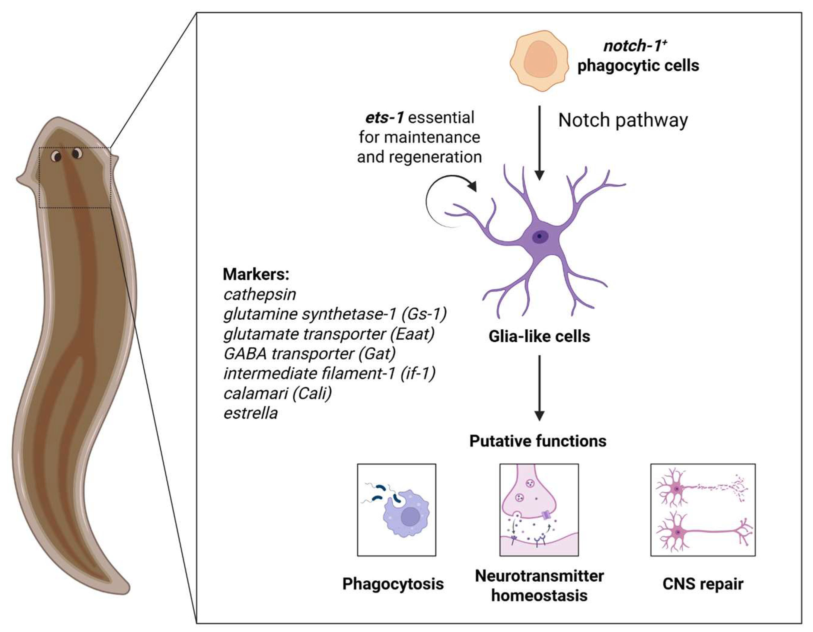
Figure 1. Origin, molecular signatures and putative functions of planarian glia. Notch1+-phagocytic cells differentiate into glial cells, which express cathepsin, glutamine synthetase-1, excitatory amino acid transporters, estrella, if-1, and/or cali. Current evidence suggests that planarian glia could exhibit conserved roles compared to vertebrates including phagocytosis, neurotransmitter homeostasis and CNS repair.
Neuroglia 06 00037 g001
Disclaimer/Publisher’s Note: The statements, opinions and data contained in all publications are solely those of the individual author(s) and contributor(s) and not of MDPI and/or the editor(s). MDPI and/or the editor(s) disclaim responsibility for any injury to people or property resulting from any ideas, methods, instructions or products referred to in the content.