Review

The Neuroimmunological Nexus of Multiple Sclerosis: Deciphering the Microglial Transcriptomic Tapestry

Department of Pharmacology and Toxicology, National Institute of Pharmaceutical Education and Research (NIPER)-Ahmedabad, Gandhinagar 382355, Gujarat, India
*
Author to whom correspondence should be addressed.
Neuroglia 2024, 5(3), 234-253; https://doi.org/10.3390/neuroglia5030017
Submission received: 12 June 2024 / Revised: 11 July 2024 / Accepted: 16 July 2024 / Published: 20 July 2024

Abstract

:
Microglia are poorly understood immune cells of the central nervous system that play a determining role in the progression of multiple sclerosis. With the advent of genomic techniques such as single-cell RNA sequencing and single-nucleus RNA sequencing, a more comprehensive understanding of microglia at the transcriptomic level has uncovered various disease-specific clusters, context-dependent heterogeneity, and region-specific microglia, unlocking the recondite secrets embedded within these glial cells. These techniques have raised questions regarding the conventional and widely accepted categorization of microglia as M1 and M2 phenotypes. The neuroimmune component of multiple sclerosis, which is the microglia, makes it a complex and challenging disease. This review aims to demystify the complexities of microglia in multiple sclerosis, providing a vivid map of different clusters and subclusters of microglia found in multiple sclerosis and outlining the current knowledge of the distinctive roles of microglia. Also, this review highlights the neuroimmune interaction with microglia as the epicenter and how they act as sabotaging agents. Moreover, this will provide a more comprehensive direction toward a treatment approach focusing on local, region-specific microglia.

1. Introduction

Multiple sclerosis (MS) is a challenging, devastating conundrum that challenges our understanding of the neuroimmune orchestra. It is a disease with a neuroimmunological origin, having immune cells (particularly microglia) with the misguided mission of attacking the myelin sheath. Microglia are central nervous system (CNS)-resident immune cells that are embryonically derived and self-renewing [1] and play a central role in neuroinflammation and neurodegeneration. They are the first effector cells responding to any pathological insult and are responsible for generating innate and adaptive immune responses [2] while acting as local interacting partners with infiltrating immune cells [3]. MS is primarily divided into four types—Relapsing–Remitting MS, Secondary-Progressive MS, Primary-Progressive MS, and Primary-Relapsing MS—based on diverse symptomatology and disease duration. The most accepted criteria for the diagnosis of MS are the McDonald criteria, with the most recent incorporation of the magnetic resonance imaging in MS 2016 (MAGNIMS) consensus [4]. The demyelination of or injury to axons occurs due to the complex interplay of microglia with other glial cells (oligodendrocytes and astrocytes), neurons, and peripheral immune cells (trafficked into the CNS) [5], thus forming the neuroimmunological nexus of MS. This intercommunication between the CNS and the immune system leads to a change in the transcriptomic profile of microglia. Traditionally, these immune cells are viewed as a homogeneous population of cells responsible for maintaining the homeostasis of the CNS but lose regulatory function and become detrimental to the CNS in various neuropathologies [6]. The rigid, bifurcating classification of neurotoxic versus neuroprotective microglia, that is, M1 versus M2, is incongruent with the wide repertoire of microglial phenotypes due to their dynamic, diverse, region-specific multidimensional states that exist, depending on their local microenvironment [7]. Various subsets of microglia with intricate transcriptomic profiles have recently been discovered using single-nucleus RNA sequence analysis techniques, including disease-associated microglia (DAM) [6], damage-associated neurodegenerative microglia (MGnD) [8], microglia inflamed in multiple sclerosis (MIMS) [9], injury-responsive microglia 2 (IRM 2) [10], lipid-droplet-accumulating microglia (LDAM) [11], and white matter-associated microglia (WMAM) [12]. Single-cell RNA sequencing-like techniques have elucidated what makes a microglial cell a microglial cell from a genomic and epigenomic standpoint, highlighting their potential neuroinflammatory and neuroprotective roles [7]. With these sequencing techniques integrated with multi-omics, microglia are stealing the spotlight in the realm of neuroimmunology, acting as mediators in orchestrating CNS diseases [13] due to their chronically activated condition and serving as the main drivers of neuroinflammation [14]. However, some studies have also pointed toward the beneficial role of microglia due to their secretion of neurotrophic factors and phagocytosis of myelin debris, which complicates microglial biology [15,16]. Another viewpoint that has been overlooked is the morphological aspect of microglia, which depends on environmental cues [17]. For instance, the activation of adenosine receptors upon the breakdown of ATP prompts microglia to take on an amoeboid morphology [9,18].
These techniques provide a robust rationale for developing novel therapeutics targeting specific subsets of microglia, which, to date, has received little research attention. Adopting a reconfiguring approach, shifting from broad-spectrum anti-inflammatory, immunosuppressive, and comprehensive therapy to active immunomodulation and targeted interventions, has the potential to minimize deleterious off-target effects with the fine-tuning of microglias’ diverse roles. Immunomodulatory therapies that foster neuroprotective and neurodegenerative microglia while preserving the integrity of homeostatic microglia in terms of their specific receptors, mRNA, hold the potential for restoring beneficial microglial signatures. However, although this concept is promising, its translational and practical implementation is still very challenging. Extensive research is needed to elucidate the functional states of all microglial subsets and their interaction with other glial cells. Moreover, noteworthy caveats remain for these techniques, as comprehensive characterization is still required for all microglial genetic and cytokine profiles.
In this review, we will focus on summarizing all of the newly discovered microglial transcriptomic profiles from the toxin-induced demyelination model and the experimental autoimmune encephalomyelitis (EAE) model of MS. The particular focus will be on microglial transcriptomic profiles from MS patients. Also, we will show how microglia can act as sabotaging agents in this debilitating disease and provide a neuroimmunological view in the context of microglia. Furthermore, we will delve into microglias’ morphological and M1 and M2 aspects. While we will not be discussing the pathogenesis and features of MS, as this has already been covered in top-notch reviews [19,20,21], we will highlight the paradigm shift in microglia and the notion of resting and homeostatic microglia in the context of this disease.
For this review, we searched the databases Scopus and Pubmed with keywords like single-cell RNA sequencing, microglia, transcriptomic profile of microglia, and microglia cells in MS.

2. Neuroimmunological Landscape of Multiple Sclerosis: Microglia as a Central Nexus

Neuroimmunology is defined the intercommunication between the immune system and the CNS. A neuroimmune MS component, previously called sclerose en plaque, has an unclear origin [22]. MS has a heterogeneous presentation [19], affecting white and gray matter [23]. The prototypical hallmarks of MS are the spatiotemporal dissemination of lesions on the neuraxis [19], multifocal continuous inflammation [23], which is caused by immune cell infiltration, reactive gliosis [24], synaptopathy [25], the disruption of intricate vasculature barriers (blood–brain barrier and blood–spinal cord barrier) [26], and primary demyelination leading to neuronal and axonal loss [23]. A significant pathological feature in the progression of MS is iron buildup within activated microglia and macrophages, forming a rim [27]. This accumulation has been consistently observed in various forms and stages of the disease [28], and is linked to glutamate excitotoxicity, oxidative stress, protein misfolding, and ferroptosis [29]. Furthermore, it is thus considered a sensitive marker for the progression of the disease [30].
The two systems, immune and CNS, are known to function separately. However, they are closely regulated, with intricate molecular and cellular connections leading to bidirectional interaction. Due to their captivating, multifaceted role as an immune and neuronal regulator, microglia take center stage in neuroimmunology [31,32]. Studies in recent years have unveiled the neuroimmune interactions in MS, whether it is the challenge of immune-privileged sites, the peripheral system’s influence on microglia, or the acute phase response (APR).
Recently, the concept of the brain as an immune-privileged site has been challenged and refined. Indeed, it is a site where the immune system works in a sui generis way. The presence of a functional lymphatic system and the immune surveillance function of immune cells in the meninges play a significant role in synchronizing central and peripheral immune responses [33]. The meningeal blood vessels provide the primary pathway for immune cells to access the cerebrospinal fluid. Microglia are the unique neuroimmune surveillance cells of the CNS [34,35], indispensable for maintaining tissue homeostasis and mitigating neuropathologies [36]. BBB disruption, a prominent hallmark of MS, leads to the infiltration of toxic blood-derived proteins, peripheral immune cells, and pathogens, triggering various inflammatory responses that initiate multiple pathways of neurodegeneration in the brain [37]. The blood protein fibrin induces neurotoxic microglial programming and, thus, neurodegeneration through the CD11b-CD18 integrin (Macrophage-1 antigen) receptor by initiating the outside-in integrin signaling cascade [38]. In 2019, a genome-wide association study revealed that the core emergence of MS lies in the peripheral system and its cells. Every immune cell, whether part of the innate or adaptive immune system, including T cells, B cells, dendritic cells, natural killer cells, and microglia (but not astrocytes or neurons), is enriched for MS susceptibility genes [39].
Another piece of evidence supporting the intercommunication between the immune system and the CNS is the bidirectional gut–brain axis. Different bacteria-derived metabolites like short-chain fatty acids (SCFA; propionate, butyrate) are important molecules in neuroimmune interaction that regulate neuroimmune cell maturation and development, such as microglia [40]. SCFA alleviates EAE, and its mechanism of action involves either binding to specific receptors, GRP41 and GRP43, or inhibiting histone deacetylase activity [41]. Microglia express the aryl hydrocarbon receptor (AhR), which regulates the pathogenic phenotype of astrocytes through molecules such as vascular endothelial growth factor B (VEGF-B) and tumor necrosis factor (TNF-α). Therefore, AhR influences microglial pro-inflammatory transcriptional responses and CNS inflammation in EAE [42]. Moreover, adaptive immune cells in the gut differentiate CD4 effector T cells into helper 1 cells (TH 1 cell) and helper 2 cells (TH17 cell), which migrate to the brain and exacerbate inflammation in MS [43]. Sickness behavior, often accompanying an initial response to an infectious agent, is known as APR [44]. Numerous APR mediators, including cytokines, pentraxins, chemokines, complement proteins, C-reactive proteins, and serum amyloid protein A, induce APR. Chronic inflammation due to the prolonged production of IL-1β is a prominent feature of MS. This prolonged production of IL-1β cytokines leads to elevated APR and chemokine expression, such as CXCL1, a potent neutrophil chemoattractant (equivalent to IL-8 in humans), which subsequently increases migration and the recruitment of leukocytes in the blood. The activation of peripheral APR (leading to peripheral inflammation) contributes to the emergence of central inflammation or may worsen ongoing central inflammation, resulting in a poor clinical outcome [45,46].

3. Microglia as a Sabotaging Agent in Multiple Sclerosis

Many trailblazing genome-wide studies have increasingly pointed to the pivotal role of microglia in ameliorating MS [47]. A concise illustration of the mechanism by which microglia act as a sabotaging agent is depicted in Figure 1. Another study revealed that 48 out of 81 associated risk alleles were particularly expressed in microglia [48]. Microglia nodules, which are small aggregated clusters of microglia found in periplaque, found in normal-appearing white matter (NAWM), have been linked to the early stages of the development of MS. These nodules are also associated with the disruption of paranodal proteins, including neurofascin-155 and contactin-associated protein-1 [49]. These microglia nodules are also called pre-active lesions or newly forming lesions [50], as they express markers for antigen presentation and phagolysosomal activity [51]. These nodules signify the early stage of disruption that can develop into MS lesions [50]. Also, the pro-inflammatory state of microglia contributes to active demyelination and neurodegeneration in MS. They secrete pro-inflammatory cytokines like IL-6, TNF-α, IL-23, IL-1β, and IL-18, in addition to reactive oxygen species and nitrogen species (ROS and RNS) and C1QA, and thus, their coordinated secretion, which induces neurotoxic effects in astrocytic cells [13,14,15,16,17,18,19,20,21,22,23,24,25,26,27,28,29,30,31,32,33,34,35,36,37,38,39,40,41,42,43,44,45,46,47,48,49]. Microglia and astrocytes substantially express miRNAs (microRNAs), regulatory subtypes of noncoded RNA that contribute to microglia-mediated neuroinflammation. miR-155, miR-146a, miR-233, miR-142, miR-34a, miR-326, and miR-23b are highly upregulated in active MS lesions. While miR-23b is associated with IL-17-mediated neuroinflammation, miR-155 promotes the development of neuroinflammation and pro-inflammatory responses in microglia [52]. Another mechanism by which microglia act as a sabotaging agent is through the system of enzymes found in microglia, such as nicotinamide adenine dinucleotide phosphate (NADPH), responsible for producing ROS. This system is predominantly present within and near MS plaques in white matter lesions, as confirmed by the upregulation of NADPH subunits such as gp91phox, p22phox, p47phox, and NADPH oxidase-1 [53]. The production of these free radicals impairs the mitochondrial respiratory chain, leading to even more ROS production. An imbalance in iron homeostasis further amplifies ROS production [54].

3.1. Unsettled Role of Microglial Phagocytosis, Phagoptosis, and Trogocytosis

Another concept gaining momentum is phagoptosis and trogocytosis, in addition to the well-known phagocytosis by microglia. The phagocytosis process carried out by microglia has generally been considered beneficial, as it removes dead and apoptotic neurons and other cells. However, the phagocytosis of viable cells, known as phagoptosis (primary phagocytosis), has been implicated in neurodegenerative disease [55]. Microglia trogocytosis, also known as synaptic nibbling, is the selective partial remodeling of presynaptic axonal compartments [56,57]. Both phagoptosis and trogocytosis are forms of phagocytosis but differ significantly from typical phagocytosis. More research is needed to understand the role of these forms of cell death in neurodegenerative disease. The phagocytosis aspect of microglia becomes detrimental when viable neurons (another term used in many studies is “stressed viable neurons”) are targeted. The same concept applies to phagoptosis. Recent evidence suggests that in models of neurodegeneration, inflamed microglia phagocytose viable brain neurons, thus promoting neuropathology [58]. Phagocytosis depends on three types of phagocytic signaling: ”eat me” signals (which include phosphatidylserine, calreticulin, and UDP), “find me” signals (fractalkine and nucleotides), and “don’t eat me” signals (which include CD47 and sialic acid) [59]. Dysfunctional microglial phagocytosis depends on the aberrant activation and expression of these phagocytic signals [60]. Evidence indicates that neurodegenerative diseases, mainly Alzheimer’s disease (AD), involve the complement cascade C1q and its downstream effector protein C3 in atypical microglial synaptic engulfment, leading to severe cognitive dysfunction [61].

3.2. Involvement of Indirect Pathways in Microglial’s Cascading Effects

Many other indirect pathways make microglial cells a sabotaging agent, such as lysosomal dysfunction, the secretion of proteases, and dysregulation/activation of the complement pathway.
The aberrant release of lysosomal cathepsins (such as cathepsin C [62], H [63], and X [64]), results in lysosomal membrane permeabilization caused by impaired lysosomal acidification within the microglia. These cathepsins have been reported to be the major drivers of neuroinflammation and neurodegeneration. Also, impaired lysosomal dysfunction within the microglial cells leads to the increased release of inflammatory cytokines and the activation of other immune cells [65]. Myelin basic protein and myelin oligodendrocyte glycoprotein are substrates for myelencephalon-specific proteases (MSP), which are secreted by microglia and contribute to demyelination [66,67]. Microglia also secrete a substance known as tissue plasminogen activator (tPA), which is a serine protease [68], and is responsible for inducing extracellular proteolysis in EAE and inflammatory demyelination [69]. Elevated levels of tPA have been reported in MS lesions and the CSF of MS patients [70].
The involvement of the complement system through the alternative pathway induces a microglial priming state, making the brain more vulnerable to neurodegeneration. This phenomenon has been observed in the perilesional white matter of individuals with MS. Specific deletion of the regulatory protein of the complement system, complement receptor 1-related protein y (Crry), and the subsequent activation of alternative pathways through C3 or factor B induce microglial priming [71].

4. Paradigm Shift in Microglia—Reconstructing the Microglia in Multiple Sclerosis

Historically, microglia have been classified based on their polarization state, either as pro-inflammatory or anti-inflammatory. This classification is just an oversimplification, ignoring the fact that microglia show high levels of dynamicity and plasticity and have context-dependent phenotypes. Recent technological advancements, such as single-cell RNA sequencing (scRNA-seq), single nucleus RNA sequencing (snRNA-seq), and cytometry by time of flight spectrometry (CyTOF) with the integration of analytical profiling techniques, have provided the panoramic view of microglia at the gene level and revealed the true meaning of “diversity of microglia”. Different scRNA-seq protocols used to confirm clusters of microglia are Smart-seq, CEL-seq, Quartz-seq, C1-CAGE, RamDa-seq, Drop-seq, and Microwell-seq (Chromium 10× genomics) [72]. Most studies employ t-SNE (t-distributed stochastic neighbor embedding) as statistical dimensionality reduction techniques to visualize clusters of microglia (heterogeneous populations). These techniques have revolutionized the profiling of individual cells and their whole transcriptomes [73], revealing subpopulations with distinct characteristics through the identification of potential markers (more than 40 different surface markers and transcriptional factors at the single-cell level) [74,75]. These techniques are susceptible as they are based on RNA biology, and RNAs are highly susceptible to the influence of micro- and macro-stimuli [76]. New research has shown that human microglia have a more comprehensive diversity range than other mammals [77]. Significantly, microglia have been shown to contain species and clade-specific gene expression pathways associated with the complement system, phagocytosis, and metabolic processes [78]. In the case of MS, this becomes more complicated, as higher interindividual heterogeneity of microglia has been reported, and this heterogeneity depends on the course of the disease (whether it is active, relapsing, or chronic). Microglia exhibit extraordinary heterogeneity during the embryonic stage; this heterogeneity is reduced in the postnatal, juvenile, and adult stages. Microglia in the aging stages are characterized by a higher prevalence of genes associated with susceptibility to MS.

Redefining Physiological Microglia—Homeostatic or Resting Microglia

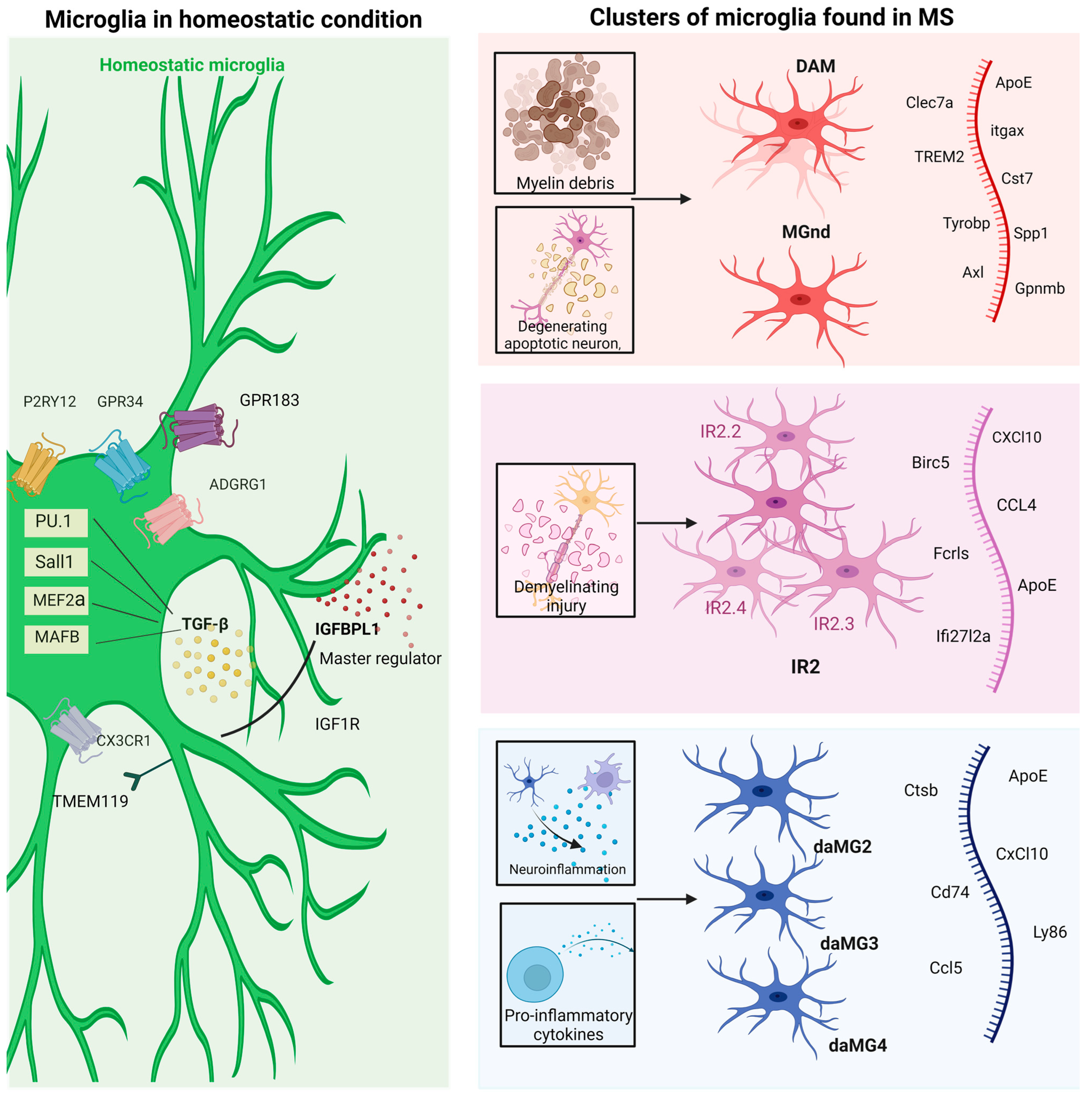
The delineation of homeostatic microglia needs to be better understood, and it cannot be defined simply by a genomics profile. Until recently, homeostatic and resting microglia were thought to be the same. Surveillant microglia and homeostatic microglia are almost the same in terms of function. To better comprehend homeostatic microglia, they should always be delineated in a functional context. Nevertheless, resting microglia is a vague term, as microglia are highly vigilant [79] and continuously survey the CNS microenvironment due to highly motile protrusions [80,81]. Microglias’ motility is due to high amounts of filamentous actin [80]. Thus, resting microglia should be called surveillant/surveilling microglia [7]. Purinergic receptor P2Y12 (P2RY12) and transmembrane protein 119 (TMEM119) are unique microglial transcriptional signatures found in healthy human and murine brains. These two transcriptional signatures are downregulated in MS disease and are exclusive to microglia, not myeloid cells. However, recent evidence regarding TMEM119’s specificity to microglia raises controversy, as TMEM119 is also expressed in peripheral tissues like brown adipose tissue and follicular dendritic cells, and its expression is influenced by the cellular environmental milieu [82]. Another concern when defining pan-microglial markers like P2RY12 and TMEM119 is whether all microglia express these markers or undergo a reduction in expression only under a specific set of conditions, so the validity of these markers as an exclusive homeostatic marker is debatable [83].
One recent study found that insulin-like growth factor-binding protein-1 (IGFBL-1) is an upstream modulator of microglial homeostasis. It resets the transcriptional signature of neurodegeneration and causes microglial homeostasis. Although this gene is present in mouse and human microglia, its function in humans needs further study [84].
The genes C-X3-C motif chemokine receptor 1 (CX3CR1), G-coupled receptors 34 and 183 (GPR34 and 183), P2RY13, and adhesion G protein-coupled receptor (ADGRG1) are involved with homeostasis [85]. ADGRG1 (formerly known as GPR26) was found to be downregulated in microglia obtained from NAWM and normal-appearing gray matter (NAGM) tissue of MS patients [85]. Many studies have compared the transcriptomic profiles of mouse and human microglia [86,87,88]. The microglia of humans and mice exhibited significant conservation in their transcriptomic and epigenomic profiles. However, there were discernible distinctions, which were primarily attributed to the presence of promoters and enhancers (cis-regulatory elements) [48]. Adult microglia express transcription factors, including monocyte enhancer factor 2a (MEF2a), PU.1, spalt-like transcription factor 1 (SALL1), transcription factor Mafb (MAFB), and transforming growth factor-beta (TGF-β), which is a common signaling molecule. These transcription factors play a crucial role in maintaining a homeostatic microglial phenotype. Dysregulation of these transcription factors can disrupt homeostatic microglia as they are pivotal in suppressing pro-inflammatory pathways [89]. The transcriptional profile of homeostatic microglia is shown in Figure 2. Moreover, various cells, primarily astrocytes and neurons, work synergistically to influence the homeostatic gene signature of microglia. Thus, cues provided by the CNS local microenvironment with inter-cell type signaling, including synaptic transmission, play a significant role in determining the physiological state of microglia [90].

5. The Uncharted Territory of Microglia Transcriptomics in Multiple Sclerosis

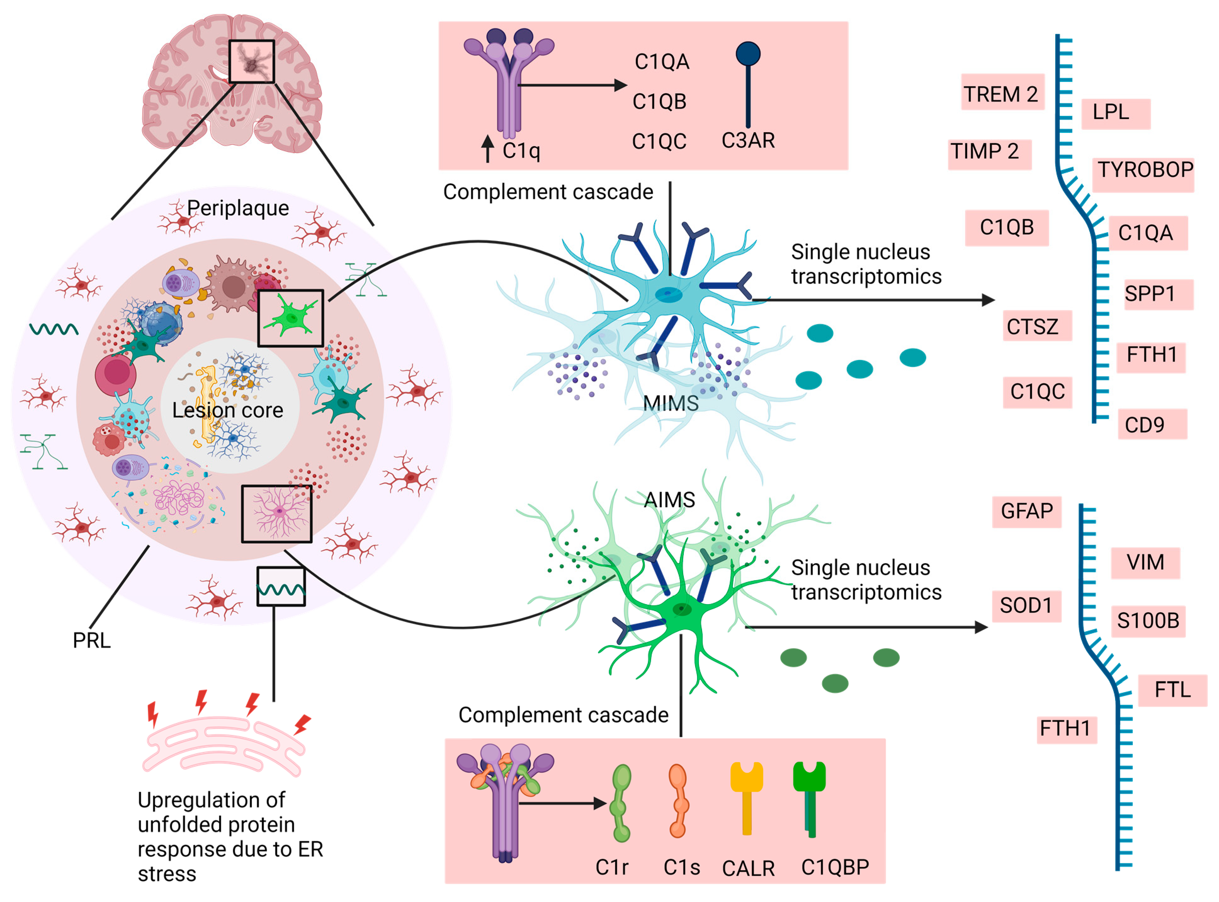
A study published in 2021 found newly discovered glial cells at the edge of chronic active lesions, referred to as MIMS and AIMS [9]. A chronic active lesion, characteristic of progressive MS, also known as a smoldering or slowly expanding lesion, is defined by its distinct regions: the core, lesion edge, and periplaque area. These lesions exhibit ongoing demyelination at their rims [91]. Chronic active lesion edges consist of a higher proportion of immune cells like activated T-cells, plasma cells with short-lived effector cells called plasmablast, limited oligodendrocytes, and few oligodendrocytes precursor cells. These edges include dendritic cells, MIMS, AIMS, a higher proportion of cytotoxic lymphocytes, and complement component C1Q [92]. The MIMS found at the lesion edge is divided into two subclusters: foamy and iron MIMS. Iron MIMS contributes to inflammation propagation through antigen presentation due to their higher proportion of inflammatory genes, MHC, and iron-related markers (SOD1, FTH1, and FTL). In contrast, foamy MIMS are involved in myelin clearance. The transcriptomic profile of MIMS includes TREM 2, LPL, TIMP2, SPP1, FTH1, C1QA, CD68, CD9, C1QB, and C1QC. A summarized transcriptional profile of MIMS and AIMS with complement cascades is shown in Figure 3.
Similarly, in mice with focal demyelination (a model that mimics specific aspects of MS), two distinct clusters of microglia were observed: injury-responsive microglia 1 (IRM1) and injury-responsive microglia 2 (IRM2) [10]. IRM 2 downregulates the typical microglial markers P2RY12 and Cx3cr1. Sub-clustering analysis identified four small subpopulations within IRM 2 clusters: IR2.1, IR2.2, IR2.3, and IR2.4. Specifically, subclusters IR2.2 and IR2.3 showed the upregulation of Ccl4 (C-C motif chemokine ligand 4), subcluster IR 2.2 upregulated Cxcl10 (C-X-C motif chemokine ligand 10), and subcluster IR2.4 upregulated Birc5 (Baculoviral IAP Repeat protein 5). Notably, the transcriptomic signatures of Ccl4 have been found in the brains of MS patients.

6. MS-Associated Microglia versus Neurodegenerative Disorder-Associated Microglia

Disease-associated microglia (DAM) represent a relatively recent characterization within neuroimmunology [1,2,3,4,5,6]. These distinctive microglial subsets are identified in regions affected by neurodegenerative processes like AD, Parkinson’s disease, and other neurodegenerative diseases [93,94]. The notion of shared molecular mechanism has led to extensive comparative analyses of microglial transcriptomic profiles across various neurodegenerative mouse models, including EAE, AD (APP-PS1), and ALS (mSOD1). These analyses have led to two key findings: an increase in the expression of genes responsible for inflammatory molecules such as Apolipoprotein E (APO-E), Integrin alpha X (Itgax), Ccl2, C type lectin domain family 7 (Clec7a), and Receptor protein Tyrosine Kinase (Axl), and the concurrent loss of homeostatic genes, including P2ry12, TMEM119, Csf1r, Hexb, Cx3cr1, and Mertk. However, the term “DAM” should not be used universally for all pathological insults, as it encompasses a diverse population with specific transcriptomic signatures. Moreover, DAM has also been referred to as degeneration-associated microglia [1], adding a layer of discrepancy and sparking questions about whether disease-associated and degenerative-associated microglia are the same.
Another microglial cluster, termed MGnD neurodegenerative-associated microglia, has been identified through transcriptomics studies in degenerative mouse models of ALS, AD, and MS. Several studies have referred to DAM and MGnD as the same type of microglial cells with analogous transcriptomic profiles, but there are apparent differences between them. DAM are proposed to be neuroprotective, while MGnD are neurotoxic [95,96]. The transition from homeostatic microglia to MGnD is orchestrated by the TREM 2-APOE gene pathway. The primary trigger for DAM and MGnD is the apoptotic neuron. These apoptotic neurons expose the “EAT ME” signal of phosphatidylserine on the outer leaflet of their plasma membrane with the help of the enzyme scramblase. This damage-associated lipid is detected by TREM2, which subsequently induces APOE expression.

7. Homeostatic Microglia and Disease-Associated Microglia—Another Transcriptomic Landscape of Microglia in Multiple Sclerosis

Another study published in 2019 identified different subsets of microglia in an EAE model within different CNS compartments. These subsets are termed daMG2, daMG3, and daMG4, reflecting their conditions of homeostasis and neuroinflammation [97]. It is noteworthy that DAM and daMG are two distinct clusters of microglia, reported differently, despite their similar abbreviations. The acronyms used are the same as those reported in the literature. Identifying these subclusters is unique, as they are found in spinal cord lesions. The spinal cord is another major target in MS, and studies on it are limited due to the challenges in diagnosing and imaging spinal cord lesions. These neuroinflammatory-associated microglial subsets exhibit variations in the expression of chemokines, cytokines, and cysteine proteases. daMG 2 has high expression of CD 74 (also known as HLA Class II histocompatibility light antigen gamma chain), Ctsb (cathepsin B), and APOE. da MG3 shows high expression of Cxcl10, Tnf, and Ccl4. In contrast, da MG4 shows high levels of Ccl5, Ctss (Cathepsin S), and Itm2b (Integral membrane protein 2b). daMG2 has a lower proliferative capacity than daMG3 and daMG4 but interacts more with encephalitogenic T cells.
The expression of olfactomedin-like 3 (Olfml3) and secreted protein, acidic and rich in cysteine/osteonectin (Sparc) does not change in the daMG cluster. The expression of myeloid differentiation 1 (MD 1) and proliferation marker protein ki67 (Mki67) is high in all daMG clusters. Gpr34, hexosaminidase subunit beta (hexb), olfml3, P2ry12, P2ry13, spalt-like transcription factor 1 (Sall 1), glia-derived nexin (Serpin2), Sparc, and sialic acid-binding Ig-like lectin H (Siglech H) are expressed by the whole MG population.
A recent study investigated disease-specific microglia in-depth and identified a subpopulation specific to MS, termed MS-associated microglia, and two MS-enriched microglia. MS-associated microglia showed high expression of cathepsin-D, apolipoprotein C1 (APOC1), and transmembrane glycoprotein NMB (GPNMB). The MS-enriched subcluster showed high expression of CD74, HLA Class II histocompatibility antigen, DR alpha chain (HLA-DRA), and HLA-DRB1.
Another study reported the region-specific transcriptomic profile and metabolic changes in human microglia in NAWM and NAGM, isolated from the brains of MS Patients. The transcriptomic signature genes for homeostatic microglia, including P2RY12, P2RY13, and TMEM119, remain unaffected, except ADGRG1 in NAWM and ubiquitin-specific Peptidase 2 (USP2) in NAWM. The NAWM of MS showed the amplification of genes implicated in lipid metabolism, including lipoprotein lipase (LPL) mediated by PPAR Gamma (PPARG), endonuclease exonuclease phosphatase family-containing domain 1 (EEPD1), and chitinase 3-like protein 1 (CHI3L1). The notable signaling pathways altered in microglia include lipid metabolism (lipid catabolism and storage), peroxisome proliferated activated receptor (PPAR), and lysosomal pathways. On the contrary, microglia in NAGM show genes related to iron metabolism and glycolysis. The genes solute carrier family 25 member 3/mitochondrial phosphate transport protein (SLC25A3) and ATP-binding cassette subfamily B member 6 (ABCB6), which play a role in iron metabolism, are present in NAGM [98].

8. Morphological Cues—Microglial Morphological Topology

Another aspect of microglial classification is the variability in their morphology, leading to their categorization as ramified microglia, amoeboid microglia, hyper-ramified microglia, and rod microglia [99,100]. These microglial phenotypes were based on an outdated classification paradigm where ramified morphological microglia were considered resting with limited phagocytic activity, and activated morphological microglia were considered more phagocytic [7,17] . There is evidence linking diminished branching (ramification) and the elevated release of the potent pro-inflammatory cytokine IL-1β through the activation of P2RY12 and the subsequent activation of tandem pore domain halothane-inhibited potassium channel (THIK) [101]. The distinction between the activated and surveillant (often referred to as resting) states of microglia cannot be denied from a morphological and phenotypic perspective. Many cell surface markers are used to assess microglial morphology, such as CD68, CD11b, and IBA1 [102]. Ionized Calcium Binding Adapter Molecule 1 (IBA1) is one of the best markers for morphometric analysis [103]. However, the major caveat with this marker (and other cell surface markers) is its lack of specificity to microglia. Border-associated macrophages and monocyte-derived macrophages also express it. Evidence suggests that microglial morphological changes in response to neuropathological insult are used as parameters to assess the immunological status of the brain [104]. Various methods have been introduced to quantify morphology based on parameters like cell soma size, primary and secondary processes, and length of processes with endpoints [105]. Compared to neurons and astrocytes, quantifying microglial morphology is highly challenging due to their dynamic motility, arborization of processes, and constant surveying of the environment. Automated morphology analysis methods like single-threshold segmentation and multifractal analysis are advanced techniques to quantify microglial morphology [106,107].
Other morphological microglia types reported in different studies include bulbous ending processes, ball-and-chain structures, hyperramification, and jellyfish shapes. However, some of these morphotypes have been reported only in rat, mouse, and zebrafish models and pathological states like traumatic brain injury. Rod microglia, or bipolar microglia, exhibit unique morphology characterized by elongated soma and planar processes extending from the apical to the basal end [108,109]. Their presence is notably associated with injury and disease progression stages, with heightened prominence observed in aging conditions. They have also been reported in clinical cases of MS in the cerebellar cortex [110]. In vitro studies reveal that rod microglia are highly proliferative with increased production of pro-inflammatory cytokines. Another prominent morphotype of microglia in MS is dystrophic microglia, characterized by cytoplasmic fragmentation and discontinuous process with disease-associated microglia-like morphology [111]. John Prineas et al. studied microglial morphology in early and late MS and found walled microglia with CD45 and MHC class II positive immunohistochemical profiles [112]. Subsequently, a dark microglial phenotype has been reported, featuring an electron-dense cytoplasm and nucleoplasm and endoplasmic reticulum dilation, giving them a dark appearance [113], associated with the neurodegenerative state. These different morphotypes have been linked to physiological responses; however, studies have failed to establish a clear correlation between them and their immunological status. Studying and defining microglial morphology is tricky due to challenges like accuracy and subjective interpretation [105,106,107,108,109,110,111,112,113,114]. These morphotypes should be considered significant clues and need deeper exploration into their intricate association of structure and function, as the morphological spectrum of microglia has been overlooked and needs to undergo critical analysis [115]. Identifying a specific morphotype of microglia is challenging due to the lack of specific markers. Novel techniques like MorpOmics and MotiQ are being developed based on automated pipelines to capture the microglial topological morphology [115]. The correlation of transcriptional and proteomic profiles with morphotypes and their impact on microglial function require extensive exploration.

9. Indiscretion of M1 versus M2 Framework

The bifurcation classification of M1 versus M2 macrophages based on T1 helper cells (TH1) and T2 helper cells (TH2) was introduced by Charles Mills [116]. According to him, this classification system represents active and inactive states, each with its own metabolic programming [116]. In 2006, Butovsky et al. introduced pro-inflammatory and anti-inflammatory terms into the domain of microglial research [117]. This Schema was subsequently used for microglial classification. M1 microglia, known as classical microglia, neurodegenerative microglia, and pro-inflammatory microglia, produce inflammatory cytokines and chemokines such as IL-1β, IL-6, IL-12, CCL2, and TNF-α. Surface markers for M1 microglia include B7-1/CD80, B7-2/CD86, and MHC Class 2, whereas M2 microglia are marked by CD163 and CD206. This dichotomous classification represents microglial states at two extremes, providing a one-dimensional view. However, transcriptomic profiles and ex vivo studies reveal a continuum of microglial expression profiles, extending in all directions and presenting a multidimensional view [118,119]. This classification system, based on a singular stimulation paradigm exposing isolated cells to purified stimuli in vitro, fails to align with the complex, multifactorial human CNS ecosystem [120]. In vivo, a multitude of interactions orchestrate microglial behavior. An in vitro microglial culture is grown on a serum-supplemented medium, unlike in vivo microglia, which never come in contact with serum. While solutions have been proposed to grow microglia in a serum-free medium, an astrocyte-conditioned medium with three crucial survival factors—TGF-β, colony-stimulating factor (CSF-1), and cholesterol—when IL-34 is used in place of CSF-1 is called TIC medium [121].
Third, the M1/M2 polarization framework applies to bone marrow-derived macrophages and monocyte-derived macrophages, or nonresident macrophages, which respond to pathological insults (infections and trauma), propagating the inflammatory response. In contrast, microglia are resident macrophages with different ontological lineages [119], making it highly implausible that macrophage polarization pathways in response to isolated cytokine or Toll-like receptor stimuli in vitro are directly applicable to microglia. For instance, TNF-α, mostly regarded as a pro-inflammatory cytokine, critically controls the glutaminergic gliotransmission process, providing stimulatory input to excitatory synapses located in the hippocampal dentate gyrus and regulating synaptic plasticity through “homeostatic synaptic scaling”. Thus, the role of cytokines, well defined in peripheral immunity, cannot be applied to the CNS [122]. Moreover, in MS, different clusters and subclusters of microglial populations are found, as already discussed.
The detailed categorization of M2 into M2a, M2b, and M2c is limited to the periphery due to the differences in marker expression between the CNS and the periphery [123]. However, many studies [124,125,126] have extended this classification to include microglia, covering the diverse range of alternatively activated microglia. Yet, it remains uncertain to what extent this is correct. Intrinsic factors like species, genetic background, sexual dimorphism, spatial and temporal location, and residential environment [127], including intercommunication between the different glial cells and neurons, and extrinsic factors like microbiota, pathogens, and nutrition, influence microglia at phenomic, metabolomic, proteomic, transcriptomic, and epigenomic levels. These factors also include responses to changes such as sickness behavior [128], systemic inflammation, trauma, infection, ischemia, and neurodegenerative diseases, which can lead to the clonal expansion of selective microglial types [7].

10. Discussion

Identifying various microglial states, including MIMS, AIMS, DAM, MGnD, and MS-enriched and MS-associated microglial cells, has provided new insights into the molecular-level understanding of these cells. These studies have elucidated the intricate regulatory pathway signaling that governs the spatial distribution of microglia, offering hope for developing transformative treatments involving microglia-mediated therapies. Such therapies could specifically target proteins, focusing on immunomodulation rather than global immunosuppressive therapies, which have detrimental side effects for MS. However, several limitations exist in these techniques. First, detecting mRNA levels alone cannot signify the functional state of microglia, as the correlation between mRNA expression and protein levels is not direct due to various biological factors and protein turnover [129,130]. Second, techniques like single-cell sequencing can produce artifactual gene expression during the isolation of cells, particularly microglia. Also, protocols for single-cell RNA sequencing have often been neglected, leading to less reproducibility of data [131]. Third, data from single-cell techniques (single-cell RNA and bulk RNA sequencing) often lack spatial information. However, spatial RNA sequencing is gaining attention and could address this challenge [132]. Fourth, microglias’ sensitivity to external environmental cues is noteworthy [133], especially during the enzymatic dissociation and temperature changes involved in sample processing, which can trigger microglial reactions. Cell dissociation, a critical step encompassing mechanical, temperature, and enzymatic (trypsin or collagenase) stress to the cell, can lead to an undesired transcriptomic profile. RiboTag technology, used to isolate ribosome-bound mRNA, can mitigate some pitfalls of scRNA-seq techniques as it does not require mechanical dissociation and investigates solely RNA ready for translation [134]. In 2021, Clark et al. developed a groundbreaking method called RABID-seq, which uses a library of mRNA barcodes using a pseudorabies virus lacking glycoprotein G. This method identified SEMA4D-PlexinB2 and EphrinB3-EphB3 axon guidance molecules as important mediators of crosstalk between microglia and astrocytes, contributing to CNS pathology in EAE [135]. Another aspect that makes MS a challenging disease is that most of the research in this area, whether it is related to disease pathogenesis or microglial isolation, is focused solely on the brain site, neglecting the other imperative organ involved in the disease: the spinal cord. Definite differences exist in the inflammatory response, molecular signature, proliferation, and transcription factors of microglia between the brain and spinal cord. Moreover, the spinal cord is the primary site for inflammation in animals model of MS, and a strong correlation exists between spinal cord lesion extent and ambulatory impairment severity [136,137]. Lesion assessment in the spinal cord is complicated due to its mobility and increased physiological noise compared to the brain.

11. Conclusions

MS is a multifaceted neuroimmunological disease typified by inflammation and neurodegeneration. The involvement of microglia as a significant player in MS pathogenesis adds complexity. With the advancements of techniques like single-cell RNA sequencing and Cy-TOF, numerous aspects of microglia, including their heterogeneity, the context-dependent existence of their subpopulation, and their role in MS, are beginning to unravel, marking just the inception of the journey. Studies on microglial heterogeneity have primarily been conducted on animal models of MS, such as EAE, which mimic specific aspects of the disease. Thus, the findings from these studies may not directly apply to humans. Different studies have identified various subclusters of microglia, leading to discrepancies in the use of acronyms for defining the distinct subpopulation of microglia. To ensure consistency, establishing a universal naming system for the microglia acronym is crucial, as many clusters with comparable transcriptomic profiles have been identified, making standardization essential. Microglia alone can act as a sabotaging agent in the brain, but the consequences become more severe when they interact with other immune cells. This crosstalk amplifies their harmful effects. Another challenge in studying MS is the predominant focus on the brain, neglecting the spinal cord, which is crucially involved in the disease. Workshops or organizations like the Human Leukocyte Differentiation Antigen, and International Protein Nomenclature Guideline Consortium, should focus on microglial nomenclature. Studies have revealed microglial clusters with overlapping transcriptomic profiles, highlighting the need for standardized methodologies for cell-to-cell interaction studies, especially of microglia with other immune cells at the single-cell level. This underscores the need for an innovative pharmacological treatment targeting region-specific microglia. Furthermore, microglia play a dual role in both degenerative and regenerative processes, but a question remains: which role dominates and contributes to worsening disease progression? Evidence suggests that the degenerative role is more closely associated with microglia, but this does not negate the existence of a beneficial role that is yet to be fully understood. Considering all these factors, a precise microglial cell classification framework can be established, aligning with its function, which will be a critical stride in microglial research.

Author Contributions

A.J.: methodology, literature search, writing the original manuscript draft, and illustration preparation; H.K.: conceptualization, supervision, editing, reviewing, and finalizing the manuscript. All authors have read and agreed to the published version of the manuscript.

Funding

This research received no external funding.

Institutional Review Board Statement

Not applicable.

Informed Consent Statement

Not applicable.

Data Availability Statement

All illustrations were prepared with a licensed version of Biorender.

Acknowledgments

Akanksha Jha received a fellowship from the Department of Pharmaceuticals, Ministry of Chemicals and Fertilizers, Govt. of India. The authors thank the Department of Pharmaceuticals, Ministry of Chemicals and Fertilizers, Govt. of India, and NIPER Ahmedabad. The paid version of Biorender.com was utilized to prepare the illustrations.

Conflicts of Interest

The authors declare no conflicts of interest.

References

  1. Song, W.M.; Colonna, M. The identity and function of microglia in neurodegeneration. Nat. Immun. 2018, 19, 1048–1058. [Google Scholar] [CrossRef] [PubMed]
  2. Yang, I.; Han, S.J.; Kaur, G.; Crane, C.; Parsa, A.T. The role of microglia in central nervous system immunity and glioma immunology. J. Clin. Neurosci. 2010, 17, 6–10. [Google Scholar] [CrossRef] [PubMed]
  3. Wolf, S.A.; Boddeke, H.W.G.M.; Kettenmann, H. Microglia in physiology and disease. Annu. Rev. Physiol. 2017, 79, 619–643. [Google Scholar] [CrossRef] [PubMed]
  4. Vattoth, S.; Kadam, G.H.; Gaddikeri, S. Revised McDonald Criteria, MAGNIMS consensus and other relevant guidelines for diagnosis and follow up of MS: What radiologists need to know? Curr. Probl. Diagn. Radiol. 2021, 50, 389–400. [Google Scholar] [CrossRef] [PubMed]
  5. Longo, D.L.J.N.E.J.M.; Daniel, S.; Reich, M.D.; Claudia, F.; Lucchinetti, M.D.; Peter, A.; Calabresi, M.D. Multiple Sclerosis. Eur. J. Neurol. 2018, 378, 169–180. [Google Scholar]
  6. Deczkowska, A.; Keren-Shaul, H.; Weiner, A.; Colonna, M.; Schwartz, M.; Amit, I. Disease-associated microglia: A universal immune sensor of neurodegeneration. Cell 2018, 173, 1073–1081. [Google Scholar] [CrossRef] [PubMed]
  7. Paolicelli, R.C.; Sierra, A.; Stevens, B.; Tremblay, M.-E.; Aguzzi, A.; Ajami, B.; Amit, I.; Audinat, E.; Bechmann, I.; Bennett, M.; et al. Microglia states and nomenclature: A field at its crossroads. Neuron 2022, 110, 3458–3483. [Google Scholar] [CrossRef]
  8. Krasemann, S.; Madore, C.; Cialic, R.; Baufeld, C.; Calcagno, N.; El Fatimy, R.; Beckers, L.; O’Loughlin, E.; Xu, Y.; Fanek, Z.; et al. The TREM2-APOE pathway drives the transcriptional phenotype of dysfunctional microglia in neurodegenerative diseases. Immunity 2017, 47, 566–581.E9. [Google Scholar] [PubMed]
  9. Absinta, M.; Maric, D.; Gharagozloo, M.; Garton, T.; Smith, M.D.; Jin, J.; Fitzgerald, K.C.; Song, A.; Liu, P.; Lin, J.-P. A lymphocyte–microglia–astrocyte axis in chronic active multiple sclerosis. Nature 2021, 597, 709–714. [Google Scholar] [CrossRef]
  10. Hammond, T.R.; Dufort, C.; Dissing-Olesen, L.; Giera, S.; Young, A.; Wysoker, A.; Walker, A.J.; Gergits, F.; Segel, M.; Nemesh, J.; et al. Single-cell RNA sequencing of microglia throughout the mouse lifespan and in the injured brain reveals complex cell-state changes. Immunity 2019, 50, 253–271.E6. [Google Scholar] [CrossRef]
  11. Marschallinger, J.; Iram, T.; Zardeneta, M.; Lee, S.E.; Lehallier, B.; Haney, M.S.; Pluvinage, J.V.; Mathur, V.; Hahn, O.; Morgens, D.W.; et al. Lipid-droplet-accumulating microglia represent a dysfunctional and proinflammatory state in the aging brain. Nat. Neurosci. 2020, 23, 194–208. [Google Scholar] [CrossRef] [PubMed]
  12. Ahn, K.; Lee, S.-J.; Mook-Jung, I. White matter-associated microglia: New players in brain aging and neurodegenerative diseases. Ageing Res. Rev. 2022, 75, 101574. [Google Scholar] [CrossRef] [PubMed]
  13. Prinz, M.; Jung, S.; Priller, J. Microglia biology: One century of evolving concepts. Cell 2019, 179, 292–311. [Google Scholar] [CrossRef] [PubMed]
  14. Wendimu, M.Y.; Hooks, S.B. Microglia phenotypes in aging and neurodegenerative diseases. Cells 2022, 11, 2091. [Google Scholar] [CrossRef] [PubMed]
  15. Sherafat, A.; Pfeiffer, F.; Reiss, A.M.; Wood, W.M.; Nishiyama, A. Microglial neuropilin-1 promotes oligodendrocyte expansion during development and remyelination by trans-activating platelet-derived growth factor receptor. Nat. Commun. 2021, 12, 2265. [Google Scholar] [CrossRef] [PubMed]
  16. Pons, V.; Rivest, S. Beneficial Roles of Microglia and Growth Factors in MS, a Brief Review. Front. Cell. Neurosci. 2020, 14, 284. [Google Scholar] [CrossRef] [PubMed]
  17. Morrison, H.; Young, K.; Qureshi, M.; Rowe, R.K.; Lifshitz, J. Quantitative microglia analyses reveal diverse morphologic responses in the rat cortex after diffuse brain injury. Sci. Rep. 2017, 7, 13211. [Google Scholar] [CrossRef] [PubMed]
  18. Orr, A.G.; Orr, A.L.; Li, X.-J.; E Gross, R.; Traynelis, S.F. Adenosine A2A receptor mediates microglial process retraction. Nat. Neurosci. 2009, 12, 872–878. [Google Scholar] [CrossRef] [PubMed]
  19. Dendrou, C.A.; Fugger, L.; Friese, M.A. Immunopathology of multiple sclerosis. Nat. Rev. Immunol. 2015, 15, 545–558. [Google Scholar] [CrossRef]
  20. Popescu, B.F.G.; Pirko, I.; Lucchinetti, C.F. Pathology of multiple sclerosis: Where do we stand? CONTINUUM Lifelong Learn. Neurol. 2013, 19, 901. [Google Scholar] [CrossRef]
  21. Glass, C.K.; Saijo, K.; Winner, B.; Marchetto, M.C.; Gage, F.H. Mechanisms Underlying Inflammation in Neurodegeneration. Cell 2010, 140, 918–934. [Google Scholar] [CrossRef] [PubMed]
  22. Charabati, M.; Wheeler, M.A.; Weiner, H.L.; Quintana, F.J. Multiple sclerosis: Neuroimmune crosstalk and therapeutic targeting. Cell 2023, 186, 1309–1327. [Google Scholar] [CrossRef] [PubMed]
  23. Bierhansl, L.; Hartung, H.-P.; Aktas, O.; Ruck, T.; Roden, M.; Meuth, S.G. Thinking outside the box: Non-canonical targets in multiple sclerosis. Nat. Rev. Drug Discov. 2022, 21, 578–600. [Google Scholar] [CrossRef] [PubMed]
  24. Baecher-Allan, C.; Kaskow, B.J.; Weiner, H.L. Multiple Sclerosis: Mechanisms and Immunotherapy. Neuron 2018, 97, 742–768. [Google Scholar] [CrossRef] [PubMed]
  25. Mandolesi, G.; Gentile, A.; Musella, A.; Fresegna, D.; De Vito, F.; Bullitta, S.; Sepman, H.; Marfia, G.A.; Centonze, D. Synaptopathy connects inflammation and neurodegeneration in multiple sclerosis. Nat. Rev. Neurol. 2015, 11, 711–724. [Google Scholar] [CrossRef] [PubMed]
  26. Aubé, B.; Lévesque, S.A.; Paré, A.; Chamma, É.; Kébir, H.; Gorina, R.; Lécuyer, M.-A.; Alvarez, J.I.; De Koninck, Y.; Engelhardt, B.; et al. Neutrophils mediate blood–spinal cord barrier disruption in demyelinating neuroinflammatory diseases. J. Immunol. 2014, 193, 2438–2454. [Google Scholar] [CrossRef]
  27. Dal-Bianco, A.; Grabner, G.; Kronnerwetter, C.; Weber, M.; Höftberger, R.; Berger, T.; Auff, E.; Leutmezer, F.; Trattnig, S.; Lassmann, H.; et al. Slow expansion of multiple sclerosis iron rim lesions: Pathology and 7 T magnetic resonance imaging. Acta Neuropathol. 2016, 133, 25–42. [Google Scholar] [CrossRef]
  28. Hamdy, E.; Galeel, A.A.; Ramadan, I.; Gaber, D.; Mustafa, H.; Mekky, J. Iron deposition in multiple sclerosis: Overall load or distribution alteration? Eur. Radiol. Exp. 2022, 6, 49. [Google Scholar] [CrossRef] [PubMed]
  29. Zierfuss, B.; Wang, Z.; Jackson, A.N.; Moezzi, D.; Yong, V.W.; Disorders, R. Iron in multiple sclerosis–Neuropathology, immunology, and real-world considerations. Mult. Scler. Relat. Disord. 2023, 78, 104934. [Google Scholar] [CrossRef] [PubMed]
  30. Elkady, A.M.; Cobzas, D.; Sun, H.; Blevins, G.; Wilman, A.H. Progressive iron accumulation across multiple sclerosis phenotypes revealed by sparse classification of deep gray matter. J. Magn. Reson. Imaging 2017, 46, 1464–1473. [Google Scholar] [CrossRef]
  31. Kipnis, J. Multifaceted interactions between adaptive immunity and the central nervous system. Science 2016, 353, 766–771. [Google Scholar] [CrossRef] [PubMed]
  32. Cryan, J.F.; Dinan, T.G. Microbiota and neuroimmune signalling—Metchnikoff to microglia. Nat. Rev. Gastroenterol. Hepatol. 2015, 12, 494–496. [Google Scholar] [CrossRef]
  33. Louveau, A.; Harris, T.H.; Kipnis, J. Revisiting the mechanisms of CNS immune privilege. Trends Immunol. 2015, 36, 569–577. [Google Scholar] [CrossRef]
  34. Kim, S.-R.; Seong, K.-J.; Kim, W.-J.; Jung, J.-Y. Epigallocatechin gallate protects against hypoxia-induced inflammation in microglia via NF-κB suppression and Nrf-2/HO-1 activation. Int. J. Mol. Sci. 2022, 23, 4004. [Google Scholar] [CrossRef]
  35. Zhang, W.; Jiang, J.; Xu, Z.; Yan, H.; Tang, B.; Liu, C.; Chen, C.; Meng, Q. Microglia-containing human brain organoids for the study of brain development and pathology. Mol. Psychiatry 2022, 28, 96–107. [Google Scholar] [CrossRef]
  36. Galatro, T.F.; Holtman, I.R.; Lerario, A.M.; Vainchtein, I.D.; Brouwer, N.; Sola, P.R.; Veras, M.M.; Pereira, T.F.; Leite, R.E.P.; Möller, T.; et al. Transcriptomic analysis of purified human cortical microglia reveals age-associated changes. Nat. Neurosci. 2017, 20, 1162–1171. [Google Scholar] [CrossRef] [PubMed]
  37. Sweeney, M.D.; Sagare, A.P.; Zlokovic, B.V. Blood–brain barrier breakdown in Alzheimer disease and other neurodegenerative disorders. Nat. Rev. Neurol. 2018, 14, 133–150. [Google Scholar] [CrossRef] [PubMed]
  38. Mendiola, A.S.; Yan, Z.; Dixit, K.; Johnson, J.R.; Bouhaddou, M.; Meyer-Franke, A.; Shin, M.-G.; Yong, Y.; Agrawal, A.; MacDonald, E.; et al. Defining blood-induced microglia functions in neurodegeneration through multiomic profiling. Nat. Immunol. 2023, 24, 1173–1187. [Google Scholar] [CrossRef] [PubMed]
  39. International Multiple Sclerosis Genetics Consortium. Multiple sclerosis genomic map implicates peripheral immune cells and microglia in susceptibility. Science 2019, 365, eaav7188. [Google Scholar] [CrossRef]
  40. Correale, J.; Hohlfeld, R.; Baranzini, S.E. The role of the gut microbiota in multiple sclerosis. Nat. Rev. Neurol. 2022, 18, 544–558. [Google Scholar] [CrossRef]
  41. Huh, J.R.; Veiga-Fernandes, H. Neuroimmune circuits in inter-organ communication. Nat. Rev. Immunol. 2019, 20, 217–228. [Google Scholar] [CrossRef] [PubMed]
  42. Rothhammer, V.; Borucki, D.M.; Tjon, E.C.; Takenaka, M.C.; Chao, C.-C.; Ardura-Fabregat, A.; De Lima, K.A.; Gutiérrez-Vázquez, C.; Hewson, P.; Staszewski, O.; et al. Microglial control of astrocytes in response to microbial metabolites. Nature 2018, 557, 724–728. [Google Scholar] [CrossRef] [PubMed]
  43. Agirman, G.; Yu, K.B.; Hsiao, E.Y. Signaling inflammation across the gut-brain axis. Science 2021, 374, 1087–1092. [Google Scholar] [CrossRef] [PubMed]
  44. Adelman, J.S.; Martin, L.B. Vertebrate sickness behaviors: Adaptive and integrated neuroendocrine immune responses. Integr. Comp. Biol. 2009, 49, 202–214. [Google Scholar] [CrossRef] [PubMed]
  45. Mardiguian, S.; Ladds, E.; Turner, R.; Shepherd, H.; Campbell, S.J.; Anthony, D.C. The contribution of the acute phase response to the pathogenesis of relapse in chronic-relapsing experimental autoimmune encephalitis models of multiple sclerosis. J. Neuroinflamm. 2017, 14, 196. [Google Scholar] [CrossRef] [PubMed]
  46. Barbierato, M.; Borri, M.; Facci, L.; Zusso, M.; Skaper, S.D.; Giusti, P. Expression and differential responsiveness of central nervous system glial cell populations to the acute phase protein serum amyloid A. Sci. Rep. 2017, 7, 12158. [Google Scholar] [CrossRef] [PubMed]
  47. Campbell, S.J.; Meier, U.; Mardiguian, S.; Jiang, Y.; Littleton, E.T.; Bristow, A.; Relton, J.; Connor, T.J.; Anthony, D.C. Sickness behaviour is induced by a peripheral CXC-chemokine also expressed in Multiple Sclerosis and EAE. Brain, Behav. Immun. 2010, 24, 738–746. [Google Scholar] [CrossRef]
  48. Voet, S.; Prinz, M.; van Loo, G. Microglia in central nervous system inflammation and multiple sclerosis pathology. Trends Mol. Med. 2019, 25, 112–123. [Google Scholar] [CrossRef] [PubMed]
  49. Gosselin, D.; Skola, D.; Coufal, N.G.; Holtman, I.R.; Schlachetzki, J.C.M.; Sajti, E.; Jaeger, B.N.; O’Connor, C.; Fitzpatrick, C.; Pasillas, M.P.; et al. An environment-dependent transcriptional network specifies human microglia identity. Science 2017, 356, eaal3222. [Google Scholar] [CrossRef]
  50. Yong, V.W. Microglia in multiple sclerosis: Protectors turn destroyers. Neuron 2022, 110, 3534–3548. [Google Scholar] [CrossRef]
  51. van Noort, J.M.; Baker, D.; Kipp, M.; Amor, S. The pathogenesis of multiple sclerosis: A series of unfortunate events. Clin. Exp. Immunol. 2023, 214, 1–17. [Google Scholar] [CrossRef] [PubMed]
  52. Distéfano-Gagné, F.; Bitarafan, S.; Lacroix, S.; Gosselin, D. Roles and regulation of microglia activity in multiple sclerosis: Insights from animal models. Nat. Rev. Neurosci. 2023, 24, 397–415. [Google Scholar] [CrossRef] [PubMed]
  53. Yang, R.; Yang, B.; Liu, W.; Tan, C.; Chen, H.; Wang, X. Emerging role of non-coding RNAs in neuroinflammation mediated by microglia and astrocytes. J. Neuroinflamm. 2023, 20, 173. [Google Scholar] [CrossRef] [PubMed]
  54. Fischer, M.-T.; Sharma, R.; Lim, J.L.; Haider, L.; Frischer, J.M.; Drexhage, J.; Mahad, D.; Bradl, M.; van Horssen, J.; Lassmann, H. NADPH oxidase expression in active multiple sclerosis lesions in relation to oxidative tissue damage and mitochondrial injury. Brain 2012, 135, 886–899. [Google Scholar] [CrossRef] [PubMed]
  55. Dikalov, S. Cross talk between mitochondria and NADPH oxidases. Free Radic. Biol. Med. 2011, 51, 1289–1301. [Google Scholar] [CrossRef]
  56. Brown, G.C.; Neher, J.J. Microglial phagocytosis of live neurons. Nat. Rev. Neurosci. 2014, 15, 209–216. [Google Scholar] [CrossRef] [PubMed]
  57. Otto, G. Synaptic nibbling. Nat. Rev. Neurosci. 2018, 19, 322. [Google Scholar] [CrossRef] [PubMed]
  58. Lim, T.K.; Ruthazer, E.S. Microglial trogocytosis and the complement system regulate axonal pruning in vivo. Elife 2021, 10, e62167. [Google Scholar] [CrossRef] [PubMed]
  59. Brown, G.C.; Neher, J.J. Eaten alive! Cell death by primary phagocytosis:’phagoptosis’. Trends Biochem. Sci. 2012, 37, 325–332. [Google Scholar] [CrossRef] [PubMed]
  60. Butler, C.A.; Popescu, A.S.; Kitchener, E.J.; Allendorf, D.H.; Puigdellívol, M.; Brown, G.C. Microglial phagocytosis of neurons in neurodegeneration, and its regulation. J. Neurochem. 2021, 158, 621–639. [Google Scholar] [CrossRef]
  61. Wang, K.; Li, J.; Zhang, Y.; Huang, Y.; Chen, D.; Shi, Z.; Smith, A.D.; Li, W.; Gao, Y. Central nervous system diseases related to pathological microglial phagocytosis. CNS Neurosci. Ther. 2021, 27, 528–539. [Google Scholar] [CrossRef]
  62. Hong, S.; Beja-Glasser, V.F.; Nfonoyim, B.M.; Frouin, A.; Li, S.; Ramakrishnan, S.; Merry, K.M.; Shi, Q.; Rosenthal, A.; Barres, B.A.; et al. Complement and microglia mediate early synapse loss in Alzheimer mouse models. Science 2016, 352, 712–716. [Google Scholar] [CrossRef]
  63. Fan, K.; Wu, X.; Fan, B.; Li, N.; Lin, Y.; Yao, Y.; Ma, J. Up-regulation of microglial cathepsin C expression and activity in lipopolysaccharide-induced neuroinflammation. J. Neuroinflamm. 2012, 9, 96. [Google Scholar] [CrossRef]
  64. Fan, K.; Li, D.; Zhang, Y.; Han, C.; Liang, J.; Hou, C.; Xiao, H.; Ikenaka, K.; Ma, J. The induction of neuronal death by up-regulated microglial cathepsin H in LPS-induced neuroinflammation. J. Neuroinflamm. 2015, 12, 54. [Google Scholar] [CrossRef]
  65. Nakanishi, H. Cathepsin regulation on microglial function. Biochim. et Biophys. Acta (BBA) Proteins Proteom. 2020, 1868, 140465. [Google Scholar] [CrossRef]
  66. Quick, J.D.; Silva, C.; Wong, J.H.; Lim, K.L.; Reynolds, R.; Barron, A.M.; Zeng, J.; Lo, C.H. Lysosomal acidification dysfunction in microglia: An emerging pathogenic mechanism of neuroinflammation and neurodegeneration. J. Neuroinflamm. 2023, 20, 1–13. [Google Scholar] [CrossRef]
  67. Scarisbrick, I.A.; Blaber, S.I.; Lucchinetti, C.F.; Genain, C.P.; Blaber, M.; Rodriguez, M. Activity of a newly identified serine protease in CNS demyelination. Brain 2002, 125, 1283–1296. [Google Scholar] [CrossRef] [PubMed]
  68. Xu, Y.; Gao, W.; Sun, Y.; Wu, M. New insight on microglia activation in neurodegenerative diseases and therapeutics. Front. Neurosci. 2023, 17, 1308345. [Google Scholar] [CrossRef]
  69. Siao, C.-J.; Fernandez, S.R.; Tsirka, S.E. Cell type-specific roles for tissue plasminogen activator released by neurons or microglia after excitotoxic injury. J. Neurosci. 2003, 23, 3234–3242. [Google Scholar] [CrossRef]
  70. Teesalu, T.; Hinkkanen, A.E.; Vaheri, A. Coordinated induction of extracellular proteolysis systems during experimental autoimmune encephalomyelitis in mice. Am. J. Pathol. 2001, 159, 2227–2237. [Google Scholar] [CrossRef] [PubMed]
  71. Scarisbrick, I.J.A.i.m.S.; Diseases, E.D. The multiple sclerosis degradome: Enzymatic cascades in development and progression of central nervous system inflammatory disease. Adv. Mult. Scler. Exp. Demyelinating Dis. 2008, 318, 133–175. [Google Scholar]
  72. Ramaglia, V.; Hughes, T.R.; Donev, R.M.; Ruseva, M.M.; Wu, X.; Huitinga, I.; Baas, F.; Neal, J.W.; Morgan, B.P. C3-dependent mechanism of microglial priming relevant to multiple sclerosis. Proc. Natl. Acad. Sci. USA 2012, 109, 965–970. [Google Scholar] [CrossRef] [PubMed]
  73. Ziegenhain, C.; Vieth, B.; Parekh, S.; Reinius, B.; Guillaumet-Adkins, A.; Smets, M.; Leonhardt, H.; Heyn, H.; Hellmann, I.; Enard, W. Comparative Analysis of Single-Cell RNA Sequencing Methods. Mol. Cell 2017, 65, 631–643.e4. [Google Scholar] [CrossRef]
  74. Yuste, R.; Hawrylycz, M.; Aalling, N.; Aguilar-Valles, A.; Arendt, D.; Armañanzas, R.; Ascoli, G.A.; Bielza, C.; Bokharaie, V.; Bergmann, T.B.; et al. A community-based transcriptomics classification and nomenclature of neocortical cell types. Nat. Neurosci. 2020, 23, 1456–1468. [Google Scholar] [CrossRef]
  75. Pettas, S.; Karagianni, K.; Kanata, E.; Chatziefstathiou, A.; Christoudia, N.; Xanthopoulos, K.; Sklaviadis, T.; Dafou, D. Profiling Microglia through Single-Cell RNA Sequencing over the Course of Development, Aging, and Disease. Cells 2022, 11, 2383. [Google Scholar] [CrossRef] [PubMed]
  76. Gao, C.; Jiang, J.; Tan, Y.; Chen, S. Microglia in neurodegenerative diseases: Mechanism and potential therapeutic targets. Signal Transduct. Target. Ther. 2023, 8, 1–37. [Google Scholar] [CrossRef]
  77. Li, X.; Wang, C.-Y. From bulk, single-cell to spatial RNA sequencing. Int. J. Oral Sci. 2021, 13, 36. [Google Scholar] [CrossRef] [PubMed]
  78. Sharma, K.; Bisht, K.; Eyo, U.B. A Comparative Biology of Microglia Across Species. Front. Cell Dev. Biol. 2021, 9. [Google Scholar] [CrossRef]
  79. Geirsdottir, L.; David, E.; Keren-Shaul, H.; Weiner, A.; Bohlen, S.C.; Neuber, J.; Balic, A.; Giladi, A.; Sheban, F.; Dutertre, C.-A.; et al. Cross-Species Single-Cell Analysis Reveals Divergence of the Primate Microglia Program. Cell 2019, 179, 1609–1622.e16. [Google Scholar] [CrossRef]
  80. Benarroch, E.E. Microglia: Multiple roles in surveillance, circuit shaping, and response to injury. Neurology 2013, 81, 1079–1088. [Google Scholar] [CrossRef]
  81. Nimmerjahn, A.; Kirchhoff, F.; Helmchen, F. Resting Microglial Cells Are Highly Dynamic Surveillants of Brain Parenchyma in Vivo. Science 2005, 308, 1314–1318. [Google Scholar] [CrossRef] [PubMed]
  82. Raivich, G. Like cops on the beat: The active role of resting microglia. Trends Neurosci. 2005, 28, 571–573. [Google Scholar] [CrossRef] [PubMed]
  83. Vankriekelsvenne, E.; Chrzanowski, U.; Manzhula, K.; Greiner, T.; Wree, A.; Hawlitschka, A.; Llovera, G.; Zhan, J.; Joost, S.; Schmitz, C.; et al. Transmembrane protein 119 is neither a specific nor a reliable marker for microglia. Glia 2022, 70, 1170–1190. [Google Scholar] [CrossRef] [PubMed]
  84. Kenkhuis, B.; Somarakis, A.; Kleindouwel, L.R.; van Roon-Mom, W.M.; Höllt, T.; van der Weerd, L. Co-expression patterns of microglia markers Iba1, TMEM119 and P2RY12 in Alzheimer’s disease. Neurobiol. Dis. 2022, 167, 105684. [Google Scholar] [CrossRef]
  85. Pan, L.; Cho, K.-S.; Wei, X.; Xu, F.; Lennikov, A.; Hu, G.; Tang, J.; Guo, S.; Chen, J.; Kriukov, E.; et al. IGFBPL1 is a master driver of microglia homeostasis and resolution of neuroinflammation in glaucoma and brain tauopathy. Cell Rep. 2023, 42, 112889. [Google Scholar] [CrossRef] [PubMed]
  86. Hsiao, C.-C.; Sankowski, R.; Prinz, M.; Smolders, J.; Huitinga, I.; Hamann, J. GPCRomics of Homeostatic and Disease-Associated Human Microglia. Front. Immunol. 2021, 12, 674189. [Google Scholar] [CrossRef]
  87. Masuda, T.; Sankowski, R.; Staszewski, O.; Prinz, M. Microglia Heterogeneity in the Single-Cell Era. Cell Rep. 2020, 30, 1271–1281. [Google Scholar] [CrossRef] [PubMed]
  88. Masuda, T.; Sankowski, R.; Staszewski, O.; Böttcher, C.; Amann, L.; Sagar; Scheiwe, C.; Nessler, S.; Kunz, P.; Van Loo, G.; et al. Spatial and temporal heterogeneity of mouse and human microglia at single-cell resolution. Nature 2019, 566, 388–392. [Google Scholar] [CrossRef] [PubMed]
  89. Zia, S.; Rawji, K.S.; Michaels, N.J.; Burr, M.; Kerr, B.J.; Healy, L.M.; Plemel, J.R. Microglia Diversity in Health and Multiple Sclerosis. Front. Immunol. 2020, 11. [Google Scholar] [CrossRef]
  90. Yeh, H.; Ikezu, T. Transcriptional and Epigenetic Regulation of Microglia in Health and Disease. Trends Mol. Med. 2019, 25, 96–111. [Google Scholar] [CrossRef]
  91. Baxter, P.S.; Dando, O.; Emelianova, K.; He, X.; McKay, S.; Hardingham, G.E.; Qiu, J. Microglial identity and inflammatory responses are controlled by the combined effects of neurons and astrocytes. Cell Rep. 2021, 34, 108882. [Google Scholar] [CrossRef] [PubMed]
  92. Kamma, E.; Lasisi, W.; Libner, C.; Ng, H.S.; Plemel, J.R. Central nervous system macrophages in progressive multiple sclerosis: Relationship to neurodegeneration and therapeutics. J. Neuroinflamm. 2022, 19, 45. [Google Scholar] [CrossRef] [PubMed]
  93. Bittner, S.; Zipp, F. A lymphocyte-glia connection sets the pace for smoldering inflammation. Cell 2021, 184, 5696–5698. [Google Scholar] [CrossRef] [PubMed]
  94. Rangaraju, S.; Dammer, E.B.; Raza, S.A.; Rathakrishnan, P.; Xiao, H.; Gao, T.; Duong, D.M.; Pennington, M.W.; Lah, J.J.; Seyfried, N.T.; et al. Identification and therapeutic modulation of a pro-inflammatory subset of disease-associated-microglia in Alzheimer’s disease. Mol. Neurodegener. 2018, 13, 24. [Google Scholar] [CrossRef]
  95. Cowan, M.N.; Sethi, I.; Harris, T.H. Microglia in CNS infections: Insights from Toxoplasma gondii and other pathogens. Trends Parasitol. 2022, 38, 217–229. [Google Scholar] [CrossRef]
  96. García-Revilla, J.; Boza-Serrano, A.; Espinosa-Oliva, A.M.; Soto, M.S.; Deierborg, T.; Ruiz, R.; de Pablos, R.M.; Burguillos, M.A.; Venero, J.L. Galectin-3, a rising star in modulating microglia activation under conditions of neurodegeneration. Cell Death Dis. 2022, 13, 628. [Google Scholar] [CrossRef] [PubMed]
  97. Wei, Y.; Li, X. Different phenotypes of microglia in animal models of Alzheimer disease. Immun. Ageing 2022, 19, 1–16. [Google Scholar] [CrossRef]
  98. Jordão, M.J.C.; Sankowski, R.; Brendecke, S.M.; Sagar; Locatelli, G.; Tai, Y.-H.; Tay, T.L.; Schramm, E.; Armbruster, S.; Hagemeyer, N. Single-cell profiling identifies myeloid cell subsets with distinct fates during neuroinflammation. Science 2019, 363, eaat7554. [Google Scholar]
  99. van der Poel, M.; Ulas, T.; Mizee, M.R.; Hsiao, C.-C.; Miedema, S.S.M.; Adelia, N.; Schuurman, K.G.; Helder, B.; Tas, S.W.; Schultze, J.L.; et al. Transcriptional profiling of human microglia reveals grey–white matter heterogeneity and multiple sclerosis-associated changes. Nat. Commun. 2019, 10, 1139. [Google Scholar] [CrossRef]
  100. Vidal-Itriago, A.; Radford, R.A.W.; Aramideh, J.A.; Maurel, C.; Scherer, N.M.; Don, E.K.; Lee, A.; Chung, R.S.; Graeber, M.B.; Morsch, M. Microglia morphophysiological diversity and its implications for the CNS. Front. Immunol. 2022, 13, 997786. [Google Scholar] [CrossRef]
  101. Torres-Platas, S.G.; Cruceanu, C.; Chen, G.G.; Turecki, G.; Mechawar, N. Evidence for increased microglial priming and macrophage recruitment in the dorsal anterior cingulate white matter of depressed suicides. Brain Behav. Immun. 2014, 42, 50–59. [Google Scholar] [CrossRef] [PubMed]
  102. Madry, C.; Kyrargyri, V.; Arancibia-Cárcamo, I.L.; Jolivet, R.; Kohsaka, S.; Bryan, R.M.; Attwell, D. Microglial ramification, surveillance, and interleukin-1β release are regulated by the two-pore domain K+ channel THIK-1. Neuron 2018, 97, 299–312.e6. [Google Scholar]
  103. Reddaway, J.; Richardson, P.E.; Bevan, R.J.; Stoneman, J.; Palombo, M. Microglial morphometric analysis: So many options, so little consistency. Front. Neurosci. 2023, 17, 1211188. [Google Scholar] [CrossRef] [PubMed]
  104. Green, T.R.F.; Murphy, S.M.; Rowe, R.K. Comparisons of quantitative approaches for assessing microglial morphology reveal inconsistencies, ecological fallacy, and a need for standardization. Sci. Rep. 2022, 12, 18196. [Google Scholar] [CrossRef] [PubMed]
  105. Cătălin, B.; Stopper, L.; Bălşeanu, T.-A.; Scheller, A. The in situ morphology of microglia is highly sensitive to the mode of tissue fixation. J. Chem. Neuroanat. 2017, 86, 59–66. [Google Scholar] [CrossRef]
  106. Abdolhoseini, M.; Kluge, M.G.; Walker, F.R.; Johnson, S.J. Segmentation, Tracing, and Quantification of Microglial Cells from 3D Image Stacks. Sci. Rep. 2019, 9, 8557. [Google Scholar] [CrossRef] [PubMed]
  107. Ding, Y.; Pardon, M.C.; Agostini, A.; Faas, H.; Duan, J.; Ward, W.O.; Easton, F.; Auer, D.; Bai, L. Novel Methods for Microglia Segmentation, Feature Extraction, and Classification. IEEE/ACM Trans. Comput. Biol. Bioinform. 2016, 14, 1366–1377. [Google Scholar] [CrossRef]
  108. Tam, W.Y.; Ma, C.H.E. Bipolar/rod-shaped microglia are proliferating microglia with distinct M1/M2 phenotypes. Sci. Rep. 2014, 4, 7279. [Google Scholar]
  109. Taylor, S.E.; Morganti-Kossmann, C.; Lifshitz, J.; Ziebell, J.M. Rod microglia: A morphological definition. PLoS ONE 2014, 9, e97096. [Google Scholar] [CrossRef]
  110. Giordano, K.R.; Denman, C.R.; Dubisch, P.S.; Akhter, M.; Lifshitz, J. An update on the rod microglia variant in experimental and clinical brain injury and disease. Brain Commun. 2021, 3, fcaa227. [Google Scholar] [CrossRef]
  111. Zhang, X.; Chen, F.; Sun, M.; Wu, N.; Liu, B.; Yi, X.; Ge, R.; Fan, X. Microglia in the context of multiple sclerosis. Front. Neurol. 2023, 14, 1157287. [Google Scholar] [CrossRef]
  112. Prineas, J.W.; Lee, S.J.J.o.N.; Neurology, E. Microglia subtypes in acute, subacute, and chronic multiple sclerosis. J. Neuropathol. Exp. Neurol. 2023, 82, nlad046. [Google Scholar]
  113. Bisht, K.; Sharma, K.; Lacoste, B.; Tremblay, M. Dark microglia: Why are they dark? Commun. Integr. Biol. 2016, 9, e1230575. [Google Scholar] [CrossRef]
  114. Verdonk, F.; Roux, P.; Flamant, P.; Fiette, L.; Bozza, F.A.; Simard, S.; Lemaire, M.; Plaud, B.; Shorte, S.L.; Sharshar, T.; et al. Phenotypic clustering: A novel method for microglial morphology analysis. J. Neuroinflamm. 2016, 13, 153. [Google Scholar] [CrossRef] [PubMed]
  115. Colombo, G.; Cubero, R.J.A.; Kanari, L.; Venturino, A.; Schulz, R.; Scolamiero, M.; Agerberg, J.; Mathys, H.; Tsai, L.-H.; Chachólski, W. Microglial MorphOMICs unravel region- and sex-dependent morphological phenotypes from postnatal development to degeneration. Nat. Neurosci. 2021. [CrossRef]
  116. Mills, C.D.; Kincaid, K.; Alt, J.M.; Heilman, M.J.; Hill, A.M.J.T.J.o.i. M-1/M-2 macrophages and the Th1/Th2 paradigm. J. Immunol. 2000, 164, 6166–6173. [Google Scholar] [CrossRef] [PubMed]
  117. Cho, C.E.; Damle, S.S.; Wancewicz, E.V.; Mukhopadhyay, S.; Hart, C.E.; Mazur, C.; Swayze, E.E.; Kamme, F. A modular analysis of microglia gene expression, insights into the aged phenotype. BMC Genom. 2019, 20, 164. [Google Scholar] [CrossRef] [PubMed]
  118. Ransohoff, R.M. A polarizing question: Do M1 and M2 microglia exist? Nat. Neurosci. 2016, 19, 987–991. [Google Scholar] [CrossRef]
  119. Xue, J.; Schmidt, S.V.; Sander, J.; Draffehn, A.; Krebs, W.; Quester, I.; De Nardo, D.; Gohel, T.D.; Emde, M.; Schmidleithner, L.; et al. Transcriptome-based network analysis reveals a spectrum model of human macrophage activation. Immunity 2014, 40, 274–288. [Google Scholar] [CrossRef] [PubMed]
  120. Sale, A.; Berardi, N.; Maffei, L. Environment and Brain Plasticity: Towards an Endogenous Pharmacotherapy. Physiol. Rev. 2014, 94, 189–234. [Google Scholar] [CrossRef] [PubMed]
  121. Bohlen, C.J.; Bennett, F.C.; Tucker, A.F.; Collins, H.Y.; Mulinyawe, S.B.; Barres, B.A. Diverse Requirements for Microglial Survival, Specification, and Function Revealed by Defined-Medium Cultures. Neuron 2017, 94, 759–773.e8. [Google Scholar] [CrossRef] [PubMed]
  122. Crotti, A.; Ransohoff, R.M. Microglial Physiology and Pathophysiology: Insights from Genome-wide Transcriptional Profiling. Immunity 2016, 44, 505–515. [Google Scholar] [CrossRef]
  123. Cherry, J.D.; Olschowka, J.A.; O’Banion, M.K. Neuroinflammation and M2 microglia: The good, the bad, and the inflamed. J. Neuroinflamm. 2014, 11, 98. [Google Scholar] [CrossRef]
  124. Wang, J.; He, W.; Zhang, J. A richer and more diverse future for microglia phenotypes. Heliyon 2023, 9, e14713. [Google Scholar] [CrossRef]
  125. Akhmetzyanova, E.; Kletenkov, K.; Mukhamedshina, Y.; Rizvanov, A. Different Approaches to Modulation of Microglia Phenotypes After Spinal Cord Injury. Front. Syst. Neurosci. 2019, 13, 37. [Google Scholar] [CrossRef]
  126. Zhang, L.; Zhang, J.; You, Z. Switching of the Microglial Activation Phenotype Is a Possible Treatment for Depression Disorder. Front. Cell. Neurosci. 2018, 12, 306. [Google Scholar] [CrossRef] [PubMed]
  127. Tan, Y.-L.; Yuan, Y.; Tian, L. Microglial regional heterogeneity and its role in the brain. Mol. Psychiatry 2019, 25, 351–367. [Google Scholar] [CrossRef]
  128. Perry, V.H.; Cunningham, C.; Holmes, C. Systemic infections and inflammation affect chronic neurodegeneration. Nat. Rev. Immunol. 2007, 7, 161–167. [Google Scholar] [CrossRef] [PubMed]
  129. Koussounadis, A.; Langdon, S.P.; Um, I.H.; Harrison, D.J.; Smith, V.A. Relationship between differentially expressed mRNA and mRNA-protein correlations in a xenograft model system. Sci. Rep. 2015, 5, srep10775. [Google Scholar] [CrossRef] [PubMed]
  130. Maier, T.; Güell, M.; Serrano, L. Correlation of mRNA and protein in complex biological samples. FEBS Lett. 2009, 583, 3966–3973. [Google Scholar] [CrossRef]
  131. Marsh, S.E.; Kamath, T.; Dissing-Olesen, L.; Hammond, T.R.; de Soysa, T.Y.; Young, A.M.H.; Murphy, S.; Abdulraouf, A.; Nadaf, N.; Dufort, C.; et al. Dissection of artifactual and confounding glial signatures by single-cell sequencing of mouse and human brain. Nat. Neurosci. 2022, 25, 306–316. [Google Scholar] [CrossRef]
  132. Piwecka, M.; Rajewsky, N.; Rybak-Wolf, A. Single-cell and spatial transcriptomics: Deciphering brain complexity in health and disease. Nat. Rev. Neurol. 2023, 19, 346–362. [Google Scholar] [CrossRef]
  133. Hanamsagar, R.; Bilbo, S.D. Environment matters: Microglia function and dysfunction in a changing world. Curr. Opin. Neurobiol. 2017, 47, 146–155. [Google Scholar] [CrossRef]
  134. Acharjee, S.; Gordon, P.M.K.; Lee, B.H.; Read, J.; Workentine, M.L.; Sharkey, K.A.; Pittman, Q.J. Characterization of microglial transcriptomes in the brain and spinal cord of mice in early and late experimental autoimmune encephalomyelitis using a RiboTag strategy. Sci. Rep. 2021, 11, 14319. [Google Scholar] [CrossRef] [PubMed]
  135. Clark, I.C.; Gutiérrez-Vázquez, C.; Wheeler, M.A.; Li, Z.; Rothhammer, V.; Linnerbauer, M.; Sanmarco, L.M.; Guo, L.; Blain, M.; Zandee, S.E.J.; et al. Barcoded viral tracing of single-cell interactions in central nervous system inflammation. Science 2021, 372, 360. [Google Scholar] [CrossRef] [PubMed]
  136. Ransohoff, R.M.; Kivisäkk, P.; Kidd, G. Three or more routes for leukocyte migration into the central nervous system. Nat. Rev. Immunol. 2003, 3, 569–581. [Google Scholar] [CrossRef] [PubMed]
  137. Xuan, F.-L.; Chithanathan, K.; Lilleväli, K.; Yuan, X.; Tian, L. Differences of microglia in the brain and the pinal cord. Front Cell Neurosci. 2019, 13, 504. [Google Scholar]
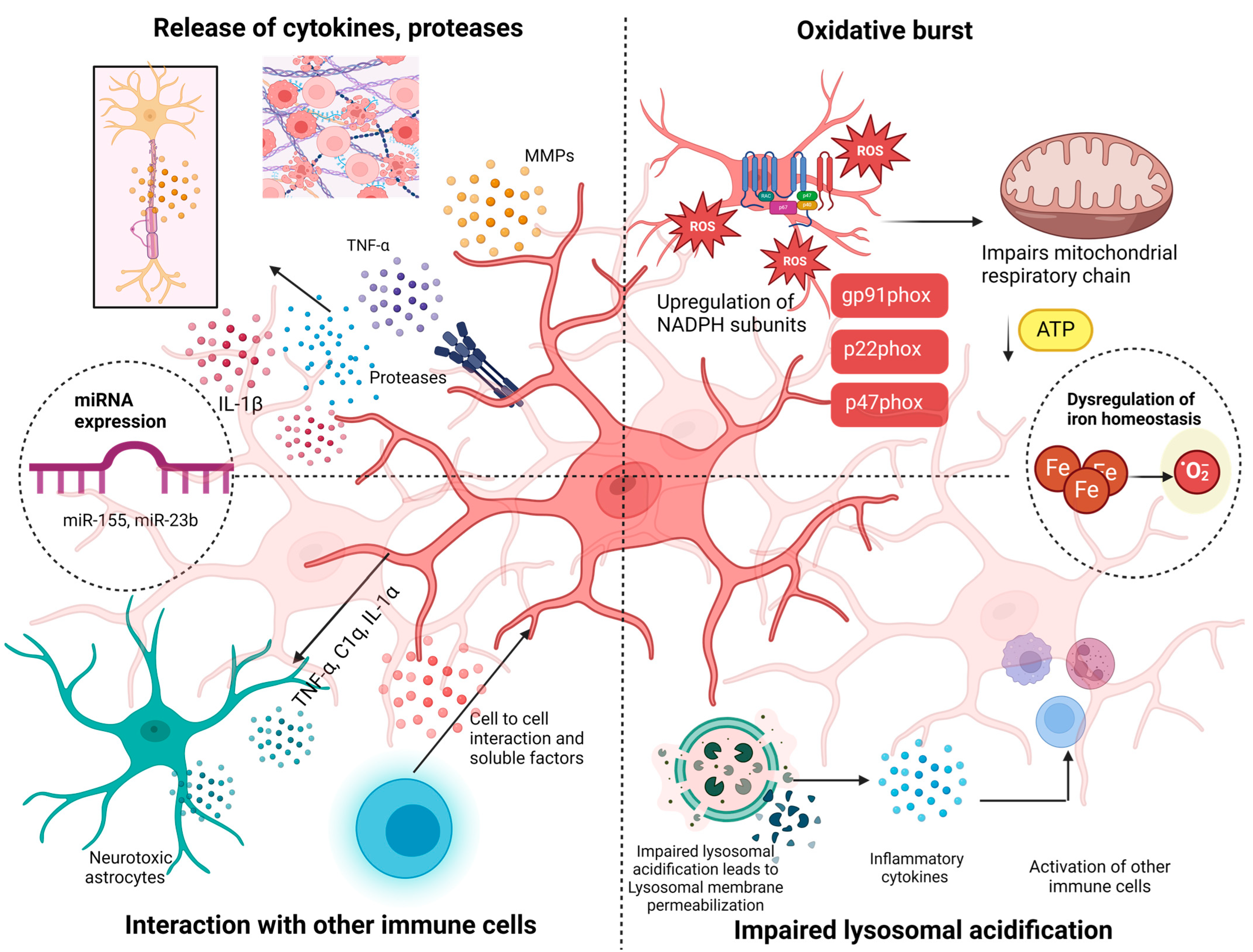
Figure 1. Different ways microglia acts as a sabotaging agent: First, the release of cytokines and proteases like TNF α, IL-β, and MMPs causes ECM and myelin sheath degeneration. Second, the interaction with other immune cells leads to the development of neurotoxic astrocytes and an interaction with T cells, which elevates neuroinflammation. Third, impaired lysosomal acidification leads to lysosomal membrane permeabilization, releasing cathepsins and inflammatory cytokines and leading to the recruitment and activation of other immune cells. Fourth, the upregulation of NADPH subunits leads to oxidative stress, which causes impaired mitochondrial function and decreased ATP production. Other pathways include the expression of microRNA (miRNA), including miR-155 and miR23b, and the dysregulation of iron homeostasis. MMP (matrix metalloproteinase), NADPH (nicotinamide adenine dinucleotide phosphate), ROS (reactive oxygen species), TNF-α (tumor necrosis factor).
Neuroglia 05 00017 g001
Figure 2. Transcriptional profile of homeostatic microglia and clusters of microglia found in MS. Homeostatic microglia with crucial transcription factors like PU.1, Sall1.MEF2a, and MAFB, whose signaling molecule is TGFβ. IGFBL1, also called master regulator, causes microglia to return to homeostasis. Other key homeostasis-associated genes are P2RY12, ADGRG1, P2RY13, CX3CR1, GPR34, and GPR183, which belong to the class of G protein-coupled receptors. Clusters and subclusters of microglia found in MS with their transcriptomic profile are damage-associated microglia (DAM), neurodegenerative-associated microglia (MGnd), injury-responsive microglia-2(IR2), and disease-associated microglia 2, 3, and 4 (daMG2, daMG3, daMG4).
Neuroglia 05 00017 g002
Figure 3. Transcriptomic profile of microglia inflamed in MS (MIMS) and astrocyte inflamed in MS (AIMS). These clusters are analyzed from frozen MS brain tissue samples at the chronic active lesion edge. The chronic active lesion is demarcated by a core, a lesion edge, and a periplaque. One specific feature is the presence of a paramagnetic rim lesion (PRL), a biomarker for smoldering lesions. Chronic active lesion edges consist of MIMS and AIMS. snRNA-seq revealed the transcriptomic profile of MIMS with specific complement cascade genes TREM2, LPL, TIMP2, TYROBOP, C1QB, C1QA, SPP1, CTSZ, FTH1, C1QC, and CD9 with C1QA, C1QB, C1QC, and C3AR (C3 receptor) and AIMS GFAP, VIM, S100B, SOD1, FTL, and FTH1 with C1r, C1s, and C1 receptors (CALR, C1QBP).
Neuroglia 05 00017 g003
Disclaimer/Publisher’s Note: The statements, opinions and data contained in all publications are solely those of the individual author(s) and contributor(s) and not of MDPI and/or the editor(s). MDPI and/or the editor(s) disclaim responsibility for any injury to people or property resulting from any ideas, methods, instructions or products referred to in the content.