1. Introduction
Fine-tuning the optical properties of glass ceramics with embedded optical nanoparticles, as well as synthesizing novel vitreous composite materials, can extend their applicability and functionality and show novel methods for their technical applications [
1,
2]. Transparent glass ceramic materials [
1,
2,
3,
4,
5] are widely used to increase the efficiency of solar cells as near-infrared light sources, in optical glass fibers and sensors and in elements for laser technology [
6,
7]. First of all, this is due to the formation of non-linear optical properties [
2,
8,
9] in glass-based materials. Their chemical and thermal stability, and ability to finely control the optical properties and structural characteristics of nanoparticles during synthesis, were reported [
5,
10,
11].
Rare-earth (RE) ions are optically active elements that are sources of effective luminescence, and nanoparticles doped with these ions are characterized by high quantum yields, wide possibilities of tuning optical properties, and noticeable suppression of the effect of concentration quenching [
12]. Therefore, luminescent glasses and glass ceramics based on rare-earth ions with a stoichiometric or non-stoichiometric composition are a promising replacement for phosphor single crystals [
13].
The formation of predominantly amorphous PbF2 nanoparticles and clusters in the glass matrix is observed [
5,
14]. The nanoparticles form complex fractal-like structures consisting of semi-regular arrangements that resemble concentration bunches inside the glass material. The PbF
2 nanoparticles doped with different RE ions embed well in a glass matrix, which is a source of their optical properties, and greatly enhances the emission yields of the corresponding optical materials. The PbF
2 nanoparticles are clustered in complex aggregates of sizes 10–30 nm [
5]. Recently, it was shown that PbF
2 nanoparticles can be doped with RE ions up to a high doping level of 10 wt.% without luminescence yield losses [
15]. The optically active nanoparticles could be formed based on the density fluctuations in the glasses, which can definitely affect the structural properties of the PbF
2 nanoparticles. Moreover, the glasses and associated glass fibers doped with RE ions have certain advantages, such as a low melting temperature [
5] and high ultraviolet resistance. From this point of view, the up-conversion luminescent glass ceramics with PbF
2 nanoparticles are of particular interest [
15,
16,
17,
18]. These glass materials have potential applications in the fields of emission displays, cathode-ray tubes and solid-state lighters. The phenomenon of up-conversion luminescence is the joint radiation of a multicomponent system through sequential optical transitions between various optically active ions [
14,
15,
16,
17,
18,
19,
20,
21]. Thus, as an example, one can cite the processes of up-conversion luminescence of PbF
2 nanoparticles doped with Yb
3+ and Eu
3+ ions, in which the emission of infrared radiation is observed during optical pumping of Yb
3+ ions from the energy levels of Eu
3+ ions [
18,
19,
20]. During the high-temperature treatment, crystallization of the nanoparticles of PbF
2 from concentration bunches exists [
14]. The crystalline nanoparticles are host systems for rare earth elements that provide the conditions for up-conversion luminescence [
18]. At the same time, the growth and uniform distribution of these nanoparticles inside the glasses are proposed. The uniform distribution of nanoparticles is the reason for the suppression of the concentration luminescence quenching characteristic of large clusters of nanoparticles [
5,
14,
17]. Both the crystallization and uniform distribution of nanoparticles determine the high intensity of luminescence observed in the glasses. The combination of RE ions as joint activators of optical centers with up-conversion luminescence excitation mechanisms enhances the optical properties of the glass ceramics [
15,
18].
For transparent glass ceramic materials, the efficiency of up-conversion luminescence depends not only on the type and concentration of optically active RE ions but also on the type and composition of the glass matrix [
5,
10,
14,
18], the chemical nature of the formed nanoparticles, and thermal treatment modes [
14]. It should be noted that an important problem in the development of glass-nanoceramics is the optimization of the glass composition, which has effects on the spectral–optical properties of the system. Furthermore, the stability of the glass materials is required when introducing fluorides and oxides of RE elements with a molar concentration of a few percent [
3,
16,
17]. Recently, interest in the studies of oxyfluoride and germanium–gallium glasses doped with thulium and holmium ions has grown [
19,
21,
22]. These ions have optical transitions in the infrared region with a high energy transfer efficiency of the Tm
3+-Ho
3+ process during up-conversion luminescence. The presence of holmium ions in glass nanoceramics makes these materials promising for infrared laser sources with a wavelength of 2 µm [
21]. Currently, much attention is paid to optimizing the synthesis of such glass ceramics by selecting the optimal ratio of RE ions for the realization of up-conversion luminescence. Previous studies of germanium–gallium glasses with Tm
3+/Ho
3+ ions indicate a maximum efficiency of up-conversion luminescence of 63% at an initial relative concentration of oxides of 70 Tm
2O
3/15 Ho
2O
3 [
21]. It is known that the formation of luminescent nanoparticles in silicate composite systems is associated with the chemical processes between oxides of RE elements and components of the glass matrix [
5,
14]. However, the structural mechanisms of nanoparticle formation in the glass matrixes are studied less.
It is known that the effectiveness of up-conversion luminescence correlates with the nanoparticle structural characteristics, and the growth of nanoparticles depends on the conditions of glass thermal treatment [
14]. From the position of the broad peak on the small-angle neutron scattering curves, it is possible to roughly estimate a shift in the average characteristic distance between clusters in the glass ceramics. Those distances increase by factors of 2–3, which can indicate an increase in the spacing between clusters in the glasses [
14,
18]. Taking into account the practical aspect of developing glass ceramics based on mixed oxide–fluoride glass matrixes as well as the interest in up-conversion luminescent materials, our work is directed to studying the structural aspects of the formation of nanoparticles containing Tm
3+ and Ho
3+ ions in mixed oxyfluoride glasses using small-angle X-ray scattering and X-ray diffraction methods.
2. Materials and Methods
The main problem in the development of transparent glass ceramic materials is the optimization of the initial glass composition, which, on the one hand, imparts high spectral luminescence properties, and, on the other hand, the stability of the vitreous state upon the introduction of fluorides and oxides of different RE elements with molar contents of several percent [
5,
14,
17]. A small addition of thulium oxide improves the color properties of the up-conversion luminescence. The Ho
3+ and Tm
3+ ions have been chosen because this joint activation may enhance the color characteristics of the luminescence of the glass ceramics due to the diversity of possible mechanisms of excitation of up-conversion luminescence with the participation of pairs of ions Ho
3+–Tm
3+. The parent glass matrix is the mixed oxyfluoride vitreous system 2.2SiO
2–1.3GeO
2–6.9PbO–7.6PbF
2–2CdF
2–xHo
2O
3–y Tm
2O
3. The introduction of Ho
2O
3 and Tm
2O
3 oxides in a certain molar range was performed (
Table 1). The selected concentration range of Ho
2O
3 corresponds to the maximum efficiency of up-conversion luminescence in similar glasses [
21,
23]. Structural studies of glasses doped with a variety of optically active nanoparticles are quite complex tasks due to the possible interaction between the nanoparticle elements and the vitreous material. A lead-containing glass matrix has been chosen to provide the light fusibility of the vitreous system. In particular, the introduction of reagents PbO and PbF
2 as glass-formers is a source of components for PbF
2 nanocrystal formation at lower temperatures of ~400 °C and a reduction of the temperature of glass synthesis to 900 °C. CdF
2 is an additional source of fluorine. It should be noted that in the first step, thoroughly mixed components of glass were fritted to reduce the evaporation of fluorine.
The synthesis of glass was performed using traditional technology by melting a mixture that was prepared from pure chemical reagents. All reagents were mixed in corresponding proportions and homogenized by milling. The thoroughly mixed charge was placed into the corundum crucibles, which were put into an electric furnace. The synthesis of glasses was performed at a temperature of 950 °C, with exposure at maximum temperature for 30 min. The short period of 30 min is sufficient to complete the homogenization and refining of the glass sample. Glass components are fritted to reduce the evaporation of fluorine. We believe that the fluorine evaporation is less because the studied glass is fusible.
The glasses were annealed in the electric muffle furnace at 300 °C for 3 h. The X-ray diffraction data have confirmed the amorphous nature of the obtained glass materials. The glasses were heat-treated at 350 °C for 30 h + 360 °C for 50 h in order to form the PbF
2 nanoparticles [
5,
14].
Small-angle X-ray scattering (SAXS) is a useful technique for nanoscale structural characterization of glass materials [
24,
25]. In SAXS, structural and spatial information is indirectly obtained from the scattering intensity in the spectral domain, known as the reciprocal space [
26]. Therefore, characterizing the structure requires solving the inverse problem of finding a plausible structure model that corresponds to the measured scattering intensity. Small-angle X-ray scattering experiments were performed with a Xeuss 3.0 instrument (XENOCS SAS, Grenoble, France). The radiation was generated by a GeniX3D source (Mo-Kα edge, λ = 0.71078 Å). Small-angle X-ray scattering curves were obtained using an Eiger2 detector at different sample-detector distances from 1 to 4 m. The thickness of the glass samples was 1 mm. The SAXS data were corrected for empty container data. The analysis of small-angle scattering data was performed in the software package SasView (Version 5.0.6) [
27].
The crystalline phase of the up-conversion luminescent nanoparticles in the glass matrix was studied using the X-ray diffraction method with the same Xeuss 3.0 device in diffraction mode, with the detector position at a distance of 0.5 m from the sample. We assumed that the crystal phase relates to the cubic phase of β-PbF
2 with space group
Fmm [
5,
15].
3. Results
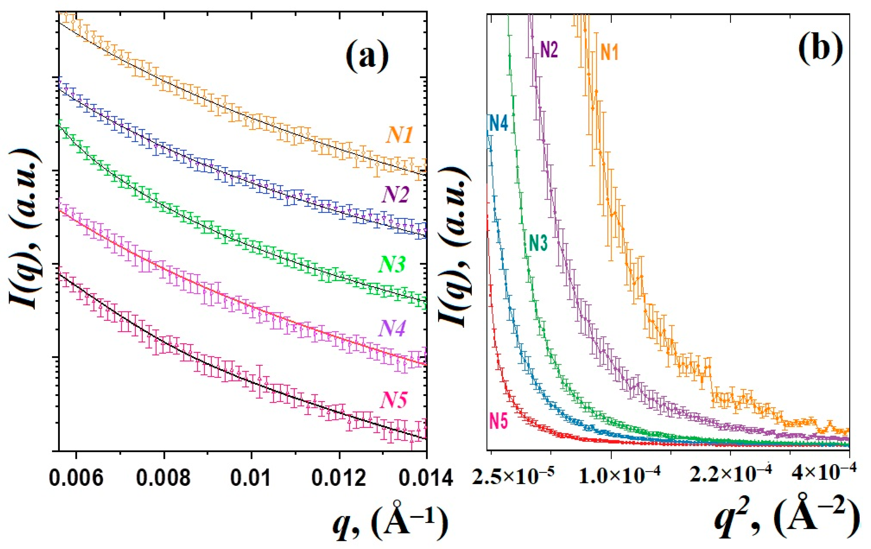
The SAXS curves of the studied glass materials are shown in
Figure 1. The scattered intensity was detected as a function of the momentum transfer modulus
q = (
4π/λ)
sinθ, where
θ is the scattering angle and
λ is the incident X-ray wavelength [
25]. The obtained curves for all samples are similar and have a typical shape for disordered glass systems [
5,
14,
24]. There are small changes in the degree of slope of the curves and their shape, which may correspond to a change in the fractal dimensions of scattering objects.
Figure 1b shows the Guinier plots {
ln(I(q)), q2}, which provide the radius of inertia
Rg of the scatterers [
24,
25]. The glass sample behavior exhibits a non-linear trend towards low
q, where aggregation is detected by an upward curve, whereas a downward curve will be typical of particle repulsion [
24]. It can be seen that, with an increase in the content of thulium oxide Tm
2O
3, there are noticeable changes in the Guinier graphs, which may indicate a clustering or aggregation of smaller particles [
23,
24]. On the other hand, the model of several scatters will be correct [
5,
14]. In this model, when the content of oxides increases, an increase in the average size of large particles or aggregates is expected. Therefore, to analyze the SAXS data, we used a two-particle model, which postulates the contribution to SAXS curves from luminescent nanoparticles, most likely PbF
2:Tm-Ho [
14], and from fluctuations in density inside the glass matrix [
10]. This model has been used previously in studies of other glass systems [
5,
14,
28].
Therefore, the obtained SAXS curves were approximated by using the exponential-power law model of Beaucage [
29,
30]. Those approaches to the analysis of small-angle scattering describe scattering from complex systems that contain multiple levels of related structural features. It should be noted that even in the absence of nanoparticles in the glass matrix, glass density heterogeneities of different natures can act as scattering objects. The scattering intensity from a system of two scatters is represented by the following expression:
where the coefficients
G1,
G2,
B1 and
B2 and the degrees at exponents
P1 and
P2 are the fitted parameters for the first and second structural levels, respectively. The radius of gyration
Rg1 and
Rg2 correspond to the main parameters of the sizes of scattering objects. The functions
and
in a power function are normalized as:
where
k1 and
k2 are empirical coefficients. The values of the gyration radius
Rg1 and
Rg2 are correlated with fluctuations in the density of glass [
10] and with the PbF
2:Tm-Ho nanoparticles, respectively.
The calculated values of the power-law exponents
P1 and
P2 obtained from fitting SAXS data by using Equation (1) are associated with the fractal dimension of the nanostructured system [
24,
31]. A power-law exponent in the range between 1 and 3 corresponds to mass fractals [
24,
25], one between 3 and 4 indicates surface fractals and between 4 and 6 is a diffuse surface. It is evident that the fractal dimensions of the observed nanoparticles PbF
2:Tm-Ho vary slightly within the range
P1 = 3.0 ÷ 3.6, which can correspond to some estimated nanoparticles with a smooth, sharp interface [
24,
31]. At the same time, the slope degree
P2 of the SAXS curve related to the density fluctuations does not exceed 3, which corresponds to the mass fractals. Large regions of density fluctuations of the glass material are formed in the glass matrix and are governed by the essential features of the chemical interaction of the glass components [
10,
14]. The observed density fluctuations of the glass material can serve as nucleation centers [
15] for the nanostructured particles PbF
2:Tm-Ho.
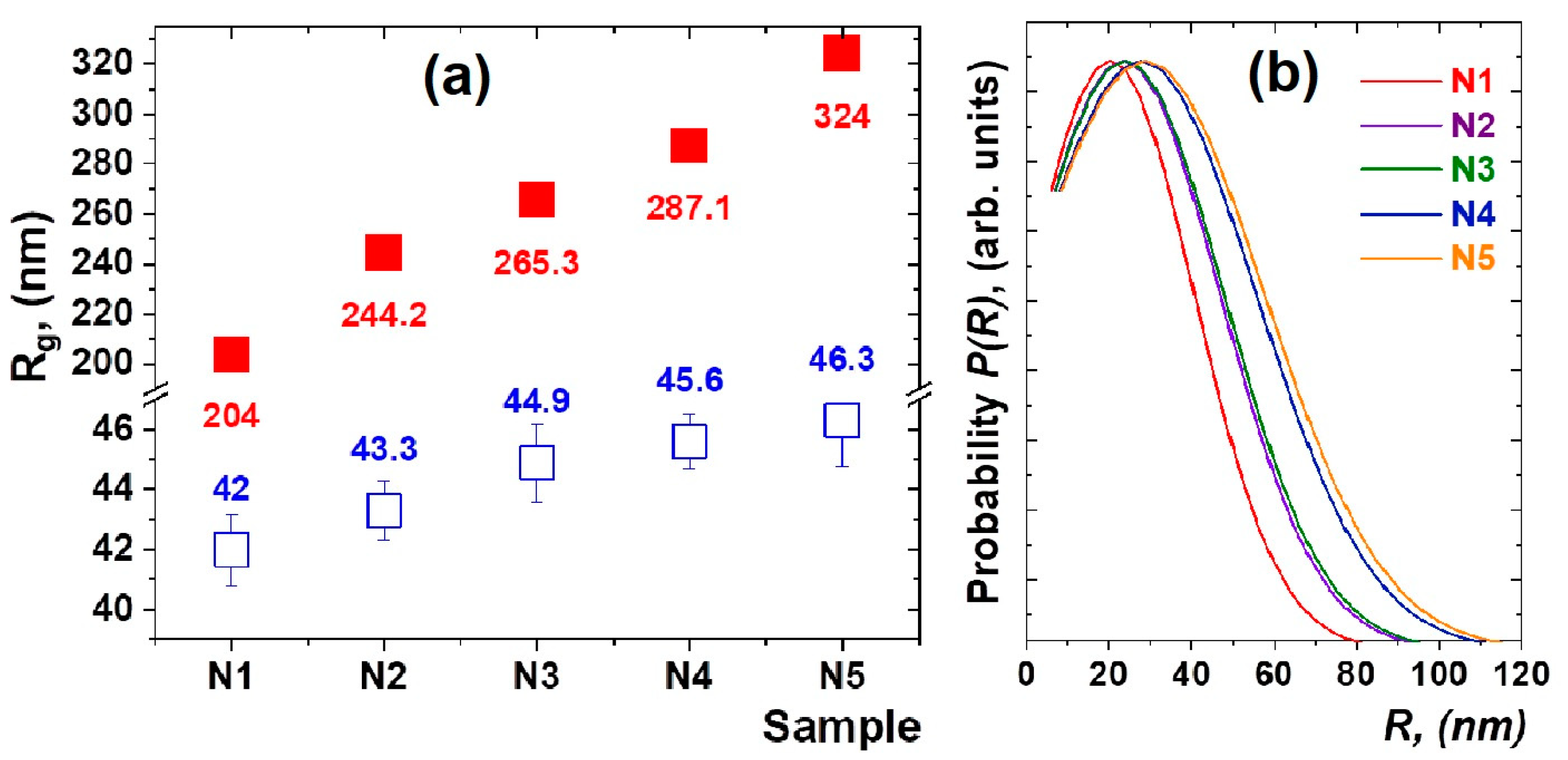
The results of the approximation of the experimental data by the function (1) are shown in
Figure 1a. The calculated size variation of the density fluctuations and formed nanoparticles is shown in
Figure 2a. It can be noted that, in heat-treated glasses doped with thulium and holmium ions, the nanoparticles of 42–46 nm in size (
Figure 2b) are formed in a spherical approximation, where the diameter of the nanoparticles is calculated as
D = 2(5/3)
1/2Rg and the average sizes of density fluctuations in the glass matrix grow from 204(2) nm for sample N1 to 324(3) nm for sample N5.
Interestingly, as the relative concentration of Tm
2O
3/Ho
2O
3 oxides increases, both the average size of nanoparticle clusters and the glass density fluctuations are growing. It can be explained that rare-earth ions are localized not only in nanoparticles but also in the glass matrix in the form of oxides [
5,
14]. In order to estimate the average size ranges of both glass density fluctuations and nanoparticles, approximations of SAXS data using functions (1) and (2) were used. The SAXS technique is much superior when considering the determination of the size distribution on a several-nanometer length scale for opaque solutions and for solid specimens [
31].
Scattering comprises not only contributions from the regularity of the space-filling ordering of particles but also from a single particle. The particle scattering can be mathematically formulated depending on the type of particle shape. In block copolymer microdomain systems, the Gauss distribution of the particle size has been assumed [
24,
31]. Only recently has direct determination of the discrete size distribution been available by fitting the theoretical scattering function to the experimentally obtained SAXS profile [
27].
The paired distribution function of nanoparticles of intermediate size, having a finite maximum size D
max, was approximated by a linear combination of a finite number of N cubic B-splines uniformly distributed in the range from 0 to D
max:
where
ai is the coefficient of the
i-th cubic B-spline,
φi(r) [
31,
32]. The upper limit of the values of the parameter
r, included in the inverse Fourier transform, was chosen in such a way that the function
smoothly tends to 0 for large values of
r. Using the above-mentioned mathematical apparatus, it is possible to estimate the paired distribution function for a system of non-interacting aggregates. Based on the obtained dependencies
, it is possible to determine the radius of gyration
Rg, which characterizes the size of intermediate nanoparticles:
The results of the analysis are shown in
Figure 2b. It can be seen that the average size of nanoparticles in the spherical approximation [
31,
32] shifts to the region of large sizes, although the width of the distribution does not change significantly. Interestingly, the slope of the SAXS curves practically does not change, and its average value is α = −5.1(5). This indicates that the fractal dimensionality and morphology of the nanostructured components of the heat-treated glasses are preserved.
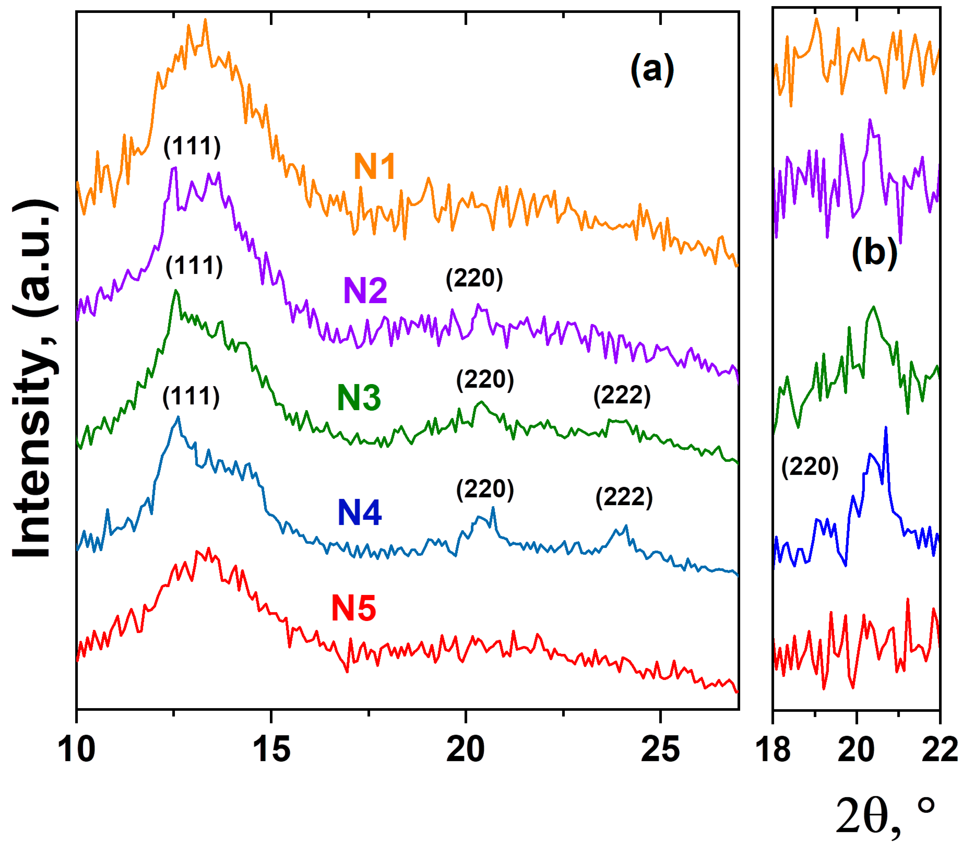
As an important aspect, the structural mechanisms of PbF
2:Tm-Ho nanoparticle formation can also be explained by the detection of the crystalline or amorphous state of the luminescent nanoparticles. X-ray diffraction patterns for the studied glass samples are shown in
Figure 3. All diffraction patterns obtained have a typical shape for scattering from amorphous materials. However, on the X-ray diffraction pattern corresponding to samples N2, N3 and N4, the appearance of several diffraction peaks is observed. The positions of the observed diffraction peaks correspond to the cubic structure with
Fmm symmetry and indicate the PbF
2 phase [
5,
14]. We believe that the rare-earth ions became embedded in crystals of PbF
2 because the unit cell parameter of this phase changes slightly with increasing thulium and holmium oxide concentration, which indicates the entry of Ho
3+ and Tm
3+ ions into the crystal structure of the luminescent nanoparticle PbF
2:Tm-Ho. Here, it is declared that the spectral characteristics of up-conversion luminescence correspond to those related to the cubic crystal structure of PbF
2 crystal [
4,
33].
Based on the obtained experimental data, the following structural mechanism of nanoparticle formation in the heat-treated glass can be proposed. As previously assumed [
5,
10,
19], the density fluctuations in the glass materials can serve as the nucleation centers for the oxide nanoparticles PbF
2:Tm-Ho. At low concentrations of the initial oxides Tm
2O
3 and Ho
2O
3, complex amorphous nanostructured structures, or aggregates, are formed. The nanoparticles form complex branching structures consisting of regular fractal arrangements of clusters inside the glass material. With increasing oxide concentration, the formation of a crystalline phase of PbF
2 nanoparticles with changes in the local environment of the glass matrix is observed. These crystallized PbF
2 nanoparticles are a host system for rare-earth Tm
3+ and Ho
3+ ions, whose entry into the cubic crystal lattice of PbF
2 provides conditions for up-conversion luminescence [
4,
5].


 ,
, 


