
Awake Prone Positioning in Non-Intubated Non-COVID-19 ARDS: A Comprehensive Review


Highlights
- Evidence supporting APP in non-COVID-19 AHRF/ARDS remains scarce and is mainly derived from small observational studies.
- APP may improve oxygenation through enhanced ventilation distribution and V/Q matching, although its impact on clinical outcomes remains uncertain.
- APP appears to be a safe and well-tolerated adjunctive strategy in selected non-intubated patients with AHRF when combined with appropriate respiratory support and close monitoring.
- Future trials should identify patients most likely to benefit and define optimal APP protocols.
Abstract
1. Introduction
2. Methodology
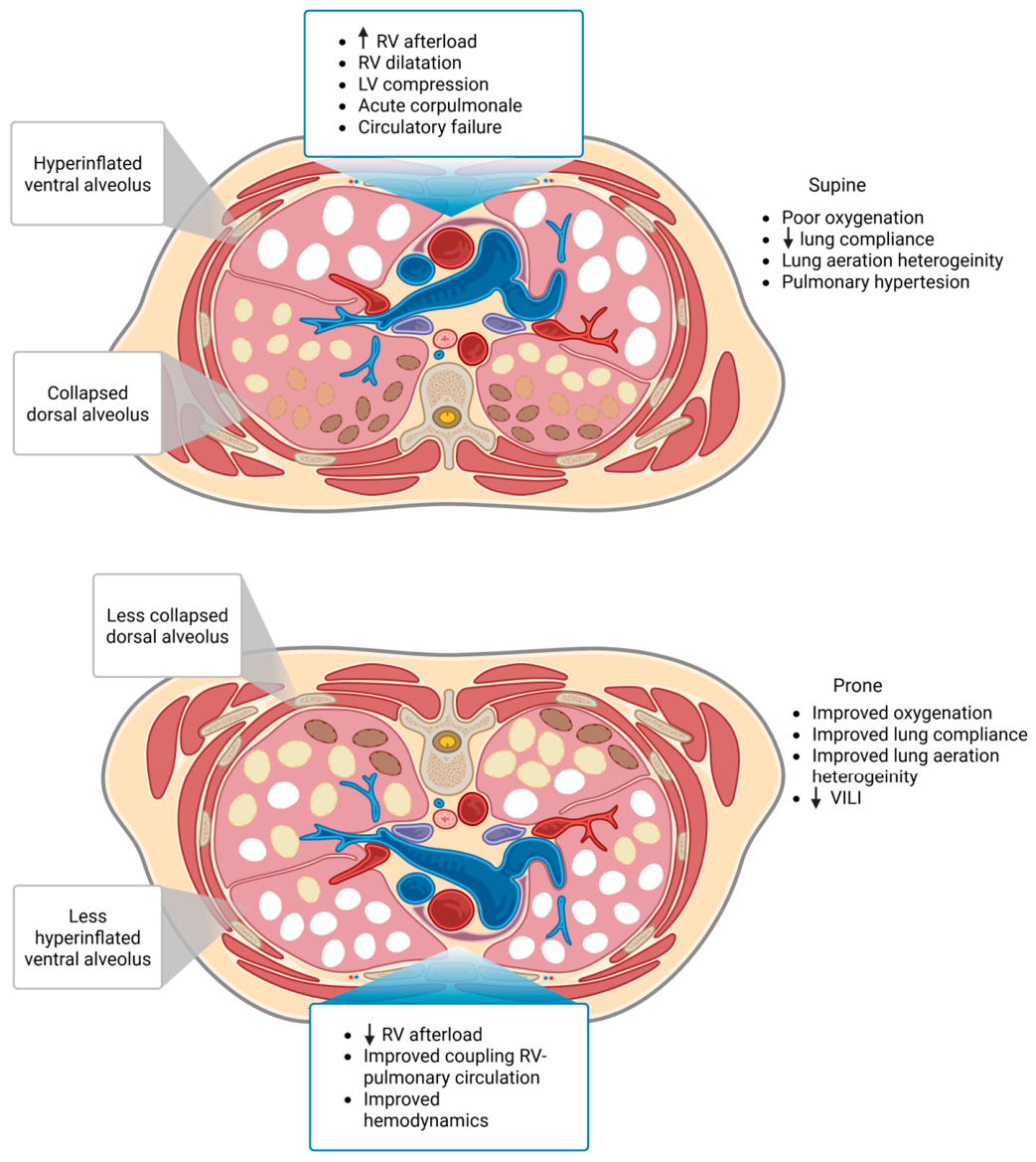
3. Physiological Principles of Prone Positioning
3.1. Pulmonary Effects
3.2. Hemodynamic Effects
4. Impact of Prone Positioning on VILI
5. Impact of Prone Position on P-SILI
6. Existing Data on APP in Non-COVID-19 Respiratory Failure
7. Discussion
8. Limitations and Areas of Future Research
9. Conclusions
| Questions for Future Research |
|
Author Contributions
Funding
Institutional Review Board Statement
Informed Consent Statement
Data Availability Statement
Acknowledgments
Conflicts of Interest
References
- Ziaka, M.; Exadaktylos, A. Fluid management strategies in critically ill patients with ARDS: A narrative review. Eur. J. Med. Res. 2025, 30, 401. [Google Scholar] [CrossRef] [Scilit] [PubMed] [PubMed Central]
- Estenssoro, E.; Dubin, A.; Laffaire, E.; Canales, H.; Sáenz, G.; Moseinco, M.; Pozo, M.; Gómez, A.; Baredes, N.; Jannello, G.; et al. Incidence, clinical course, and outcome in 217 patients with acute respiratory distress syndrome. Crit. Care Med. 2002, 30, 2450–2456. [Google Scholar] [CrossRef] [Scilit] [PubMed]
- Sinha, P.; Bos, L.D. Pathophysiology of the Acute Respiratory Distress Syndrome: Insights from Clinical Studies. Crit. Care Clin. 2021, 37, 795–815. [Google Scholar] [CrossRef] [Scilit] [PubMed] [PubMed Central]
- Ziaka, M.; Exadaktylos, A. Acute Respiratory Distress Syndrome: Pathophysiological Insights, Subphenotypes, and Clinical Implications—A Comprehensive Review. J. Clin. Med. 2025, 14, 5184. [Google Scholar] [CrossRef] [Scilit] [PubMed] [PubMed Central]
- Ding, L.; Wang, L.; Ma, W.; He, H. Efficacy and safety of early prone positioning combined with HFNC or NIV in moderate to severe ARDS: A multi-center prospective cohort study. Crit. Care 2020, 24, 28. [Google Scholar] [CrossRef] [Scilit] [PubMed] [PubMed Central]
- Aswanetmanee, P.; Limsuwat, C.; Maneechotesuwan, K.; Wongsurakiat, P. Noninvasive ventilation in patients with acute hypoxemic respiratory failure: A systematic review and meta-analysis of randomized controlled trials. Sci. Rep. 2023, 13, 8283. [Google Scholar] [CrossRef] [Scilit] [PubMed] [PubMed Central]
- Frat, J.P.; Thille, A.W.; Mercat, A.; Girault, C.; Ragot, S.; Perbet, S.; Prat, G.; Boulain, T.; Morawiec, E.; Cottereau, A.; et al. High-flow oxygen through nasal cannula in acute hypoxemic respiratory failure. N. Engl. J. Med. 2015, 372, 2185–2196. [Google Scholar] [CrossRef] [Scilit] [PubMed]
- Nair, P.R.; Haritha, D.; Behera, S.; Kayina, C.A.; Maitra, S.; Anand, R.K.; Ray, B.R.; Soneja, M.; Subramaniam, R.; Baidya, D.K. Comparison of High-Flow Nasal Cannula and Noninvasive Ventilation in Acute Hypoxemic Respiratory Failure Due to Severe COVID-19 Pneumonia. Respir. Care 2021, 66, 1824–1830. [Google Scholar] [CrossRef] [Scilit] [PubMed]
- Coudroy, R.; Frat, J.P.; Ehrmann, S.; Pène, F.; Decavèle, M.; Terzi, N.; Prat, G.; Garret, C.; Contou, D.; Gacouin, A.; et al. High-flow nasal oxygen alone or alternating with non-invasive ventilation in critically ill immunocompromised patients with acute respiratory failure: A randomised controlled trial. Lancet Respir. Med. 2022, 10, 641–649. [Google Scholar] [CrossRef] [Scilit] [PubMed]
- He, H.; Sun, B.; Liang, L.; Li, Y.; Wang, H.; Wei, L.; Li, G.; Guo, S.; Duan, J.; Li, Y.; et al. A multicenter RCT of noninvasive ventilation in pneumonia-induced early mild acute respiratory distress syndrome. Crit. Care 2019, 23, 300. [Google Scholar] [CrossRef] [Scilit] [PubMed] [PubMed Central]
- Lemiale, V.; Mokart, D.; Resche-Rigon, M.; Pène, F.; Mayaux, J.; Faucher, E.; Nyunga, M.; Girault, C.; Perez, P.; Guitton, C.; et al. Effect of Noninvasive Ventilation vs. Oxygen Therapy on Mortality Among Immunocompromised Patients with Acute Respiratory Failure: A Randomized Clinical Trial. J. Am. Med. Assoc. 2015, 314, 1711–1719. [Google Scholar] [CrossRef] [Scilit] [PubMed]
- Doshi, P.; Whittle, J.S.; Bublewicz, M.; Kearney, J.; Ashe, T.; Graham, R.; Salazar, S.; Ellis, T.W.; Maynard, D.; Dennis, R.; et al. High-Velocity Nasal Insufflation in the Treatment of Respiratory Failure: A Randomized Clinical Trial. Ann. Emerg. Med. 2018, 72, 73–83. [Google Scholar] [CrossRef] [Scilit] [PubMed]
- Scholten, E.L.; Beitler, J.R.; Prisk, G.K.; Malhotra, A. Treatment of ARDS With Prone Positioning. Chest 2017, 151, 215–224. [Google Scholar] [CrossRef] [Scilit] [PubMed] [PubMed Central]
- Kallet, R.H. A Comprehensive Review of Prone Position in ARDS. Respir. Care 2015, 60, 1660–1687. [Google Scholar] [CrossRef] [Scilit] [PubMed]
- Pelosi, P.; D’Andrea, L.; Vitale, G.; Pesenti, A.; Gattinoni, L. Vertical gradient of regional lung inflation in adult respiratory distress syndrome. Am. J. Respir. Crit. Care Med. 1994, 149, 8–13. [Google Scholar] [CrossRef] [Scilit] [PubMed]
- Gattinoni, L.; Pelosi, P.; Vitale, G.; Pesenti, A.; D’Andrea, L.; Mascheroni, D. Body position changes redistribute lung computed-tomographic density in patients with acute respiratory failure. Anesthesiology 1991, 74, 15–23. [Google Scholar] [CrossRef] [Scilit] [PubMed]
- Johnson, N.J.; Luks, A.M.; Glenny, R.W. Gas Exchange in the Prone Posture. Respir. Care 2017, 62, 1097–1110. [Google Scholar] [CrossRef] [Scilit] [PubMed]
- Perez, Y.; Luo, J.; Ibarra-Estrada, M.; Li, J.; Ehrmann, S. Awake prone positioning for patients with COVID-19-induced acute hypoxemic respiratory failure. J. Intensive Med. 2022, 2, 233–240. [Google Scholar] [CrossRef] [Scilit] [PubMed] [PubMed Central]
- Short, B.; Parekh, M.; Ryan, P.; Chiu, M.; Fine, C.; Scala, P.; Moses, S.; Jackson, E.; Brodie, D.; Yip, N.H. Rapid implementation of a mobile prone team during the COVID-19 pandemic. J. Crit. Care 2020, 60, 230–234. [Google Scholar] [CrossRef] [Scilit] [PubMed] [PubMed Central]
- Venus, K.; Munshi, L.; Fralick, M. Prone positioning for patients with hypoxic respiratory failure related to COVID-19. CMAJ Can. Med. Assoc. J. 2020, 23, 192. [Google Scholar] [CrossRef] [Scilit] [PubMed] [PubMed Central]
- Cammarota, G.; Rossi, E.; Vitali, L.; Simonte, R.; Sannipoli, T.; Anniciello, F.; Vetrugno, L.; Bignami, E.; Becattini, C.; Tesoro, S.; et al. Effect of awake prone position on diaphragmatic thickening fraction in patients assisted by noninvasive ventilation for hypoxemic acute respiratory failure related to novel coronavirus disease. Crit. Care 2021, 25, 305. [Google Scholar] [CrossRef] [Scilit] [PubMed] [PubMed Central]
- Valter, C.; Christensen, A.M.; Tollund, C.; Schønemann, N.K. Response to the prone position in spontaneously breathing patients with hypoxemic respiratory failure. Acta Anaesthesiol. Scand. 2003, 47, 416–428. [Google Scholar] [CrossRef] [Scilit] [PubMed]
- Scaravilli, V.; Grasselli, G.; Castagna, L.; Zanella, A.; Isgrò, S.; Lucchini, A.; Patroniti, N.; Bellani, G.; Pesenti, A. Prone positioning improves oxygenation in spontaneously breathing nonintubated patients with hypoxemic acute respiratory failure: A retrospective study. J. Crit. Care 2015, 30, 1390–1394. [Google Scholar] [CrossRef] [Scilit] [PubMed]
- Pérez-Nieto, O.R.; Guerrero-Gutiérrez, M.A.; Deloya-Tomas, E.; Ñamendys-Silva, S.A. Prone positioning combined with high-flow nasal cannula in severe noninfectious ARDS. Crit. Care 2020, 24, 114. [Google Scholar] [CrossRef] [Scilit] [PubMed] [PubMed Central]
- Bellone, A.; Basile, A. Prone positioning in severe acute hypoxemic respiratory failure in the emergency ward. Emerg. Care J. 2018, 14, 22–23. [Google Scholar] [CrossRef] [Scilit]
- Gattinoni, L.; Brusatori, S.; D’Albo, R.; Maj, R.; Velati, M.; Zinnato, C.; Gattarello, S.; Lombardo, F.; Fratti, I.; Romitti, F.; et al. Prone position: How understanding and clinical application of a technique progress with time. Anesthesiol. Perioper. Sci. 2023, 1, 3. [Google Scholar] [CrossRef] [Scilit] [PubMed] [PubMed Central]
- ARDS Definition Task Force; Ranieri, V.M.; Rubenfeld, G.D.; Thompson, B.T.; Ferguson, N.D.; Caldwell, E.; Fan, E.; Camporota, L.; Slutsky, A.S. Acute respiratory distress syndrome: The Berlin Definition. J. Am. Med. Assoc. 2012, 307, 2526–2533. [Google Scholar] [CrossRef] [Scilit] [PubMed]
- Gattinoni, L.; Pesenti, A.; Carlesso, E. Body position changes redistribute lung computed-tomographic density in patients with acute respiratory failure: Impact and clinical fallout through the following 20 years. Intensive Care Med. 2013, 39, 1909–1915. [Google Scholar] [CrossRef] [Scilit] [PubMed]
- Gattinoni, L.; Pesenti, A. The concept of “baby lung”. In Applied Physiology in Intensive Care Medicine; Pinsky, M.R., Brochard, L., Mancebo, J., Eds.; Springer: Berlin/Heidelberg, Germany, 2006; pp. 303–311. [Google Scholar]
- Gattinoni, L.; Marini, J.J.; Pesenti, A.; Quintel, M.; Mancebo, J.; Brochard, L. The “baby lung” became an adult. Intensive Care Med. 2016, 42, 663–673. [Google Scholar] [CrossRef] [Scilit] [PubMed]
- Castellví-Font, A.; Pham, T.; Patel, B.; Fan, E. Lessons from Gattinoni. CHEST Crit. Care 2025, 3, 100153. [Google Scholar] [CrossRef] [Scilit]
- Gattinoni, L.; Taccone, P.; Carlesso, E.; Marini, J.J. Prone position in acute respiratory distress syndrome. Rationale, indications, and limits. Am. J. Respir. Crit. Care Med. 2013, 188, 1286–1293. [Google Scholar] [CrossRef] [Scilit] [PubMed]
- Protti, A.; Santini, A.; Pennati, F.; Chiurazzi, C.; Ferrari, M.; Iapichino, G.E.; Carenzo, L.; Dalla Corte, F.; Lanza, E.; Martinetti, N.; et al. Lung response to prone positioning in mechanically-ventilated patients with COVID-19. Crit. Care 2022, 26, 127. [Google Scholar] [CrossRef] [Scilit] [PubMed] [PubMed Central]
- Hubmayr, R.D. Perspective on lung injury and recruitment: A skeptical look at the opening and collapse story. Am. J. Respir. Crit. Care Med. 2002, 165, 1647–1653. [Google Scholar] [CrossRef] [Scilit] [PubMed]
- Katira, B.H.; Osada, K.; Engelberts, D.; Bastia, L.; Damiani, L.F.; Li, X.; Chen, H.; Yoshida, T.; Amato, M.B.P.; Ferguson, N.D.; et al. Positive End-Expiratory Pressure, Pleural Pressure, and Regional Compliance during Pronation. An Experimental Study. Am. J. Respir. Crit. Care Med. 2021, 203, 1266–1274. [Google Scholar] [CrossRef] [Scilit] [PubMed]
- Terzi, N.; Bayat, S.; Noury, N.; Turbil, E.; Habre, W.; Argaud, L.; Cour, M.; Louis, B.; Guérin, C. Comparison of pleural and esophageal pressure in supine and prone positions in a porcine model of acute respiratory distress syndrome. J. Appl. Physiol. 2020, 128, 1617–1625. [Google Scholar] [CrossRef] [Scilit] [PubMed]
- Kumaresan, A.; Gerber, R.; Mueller, A.; Loring, S.H.; Talmor, D. Effects of Prone Positioning on Transpulmonary Pressures and End-expiratory Volumes in Patients without Lung Disease. Anesthesiology 2018, 128, 1187–1192. [Google Scholar] [CrossRef] [Scilit] [PubMed]
- Hoffman, E.A. Effect of body orientation on regional lung expansion: A computed tomographic approach. J. Appl. Physiol. 1985, 59, 468–480. [Google Scholar] [CrossRef] [Scilit] [PubMed]
- Henderson, A.C.; Sá, R.C.; Theilmann, R.J.; Buxton, R.B.; Prisk, G.K.; Hopkins, S.R. The gravitational distribution of ventilation-perfusion ratio is more uniform in prone than supine posture in the normal human lung. J. Appl. Physiol. 2013, 115, 313–324. [Google Scholar] [CrossRef] [Scilit] [PubMed] [PubMed Central]
- McNicholas, B.A.; Ibarra-Estrada, M.; Perez, Y.; Li, J.; Pavlov, I.; Kharat, A.; Vines, D.L.; Roca, O.; Cosgrave, D.; Guerin, C.; et al. Awake prone positioning in acute hypoxaemic respiratory failure. Eur. Respir. Rev. 2023, 32, 220245. [Google Scholar] [CrossRef] [Scilit] [PubMed] [PubMed Central]
- Amis, T.C.; Jones, H.A.; Hughes, J.M. Effect of posture on inter-regional distribution of pulmonary perfusion and VA/Q ratios in man. Respir. Physiol. 1984, 56, 169–182. [Google Scholar] [CrossRef] [Scilit] [PubMed]
- Jozwiak, M.; Teboul, J.L.; Anguel, N.; Persichini, R.; Silva, S.; Chemla, D.; Richard, C.; Monnet, X. Beneficial hemodynamic effects of prone positioning in patients with acute respiratory distress syndrome. Am. J. Respir. Crit. Care Med. 2013, 188, 1428–1433. [Google Scholar] [CrossRef] [Scilit] [PubMed]
- Albert, R.K.; Hubmayr, R.D. The prone position eliminates compression of the lungs by the heart. Am. J. Respir. Crit. Care Med. 2000, 161, 1660–1665. [Google Scholar] [CrossRef] [Scilit] [PubMed]
- Malbouisson, L.M.; Busch, C.J.; Puybasset, L.; Lu, Q.; Cluzel, P.; Rouby, J.J.; CT Scan ARDS Study Group. Role of the heart in the loss of aeration characterizing lower lobes in acute respiratory distress syndrome. Am. J. Respir. Crit. Care Med. 2000, 161, 2005–2012. [Google Scholar] [CrossRef] [Scilit] [PubMed]
- Mekontso Dessap, A.; Boissier, F.; Charron, C.; Bégot, E.; Repessé, X.; Legras, A.; Brun-Buisson, C.; Vignon, P.; Vieillard-Baron, A. Acute cor pulmonale during protective ventilation for acute respiratory distress syndrome: Prevalence, predictors, and clinical impact. Intensive Care Med. 2016, 42, 862–870. [Google Scholar] [CrossRef] [Scilit] [PubMed]
- Yogeswaran, A.; Kremer, N.C.; Janetzko, P.; Schäfer, S.; Rako, Z.A.; Vadász, I.; Hecker, M.; Tello, K. Right-sided heart failure in acute respiratory distress syndrome. Eur. Respir. Rev. 2025, 34, 250060. [Google Scholar] [CrossRef] [Scilit] [PubMed] [PubMed Central]
- Sylvester, J.T.; Shimoda, L.A.; Aaronson, P.I.; Ward, J.P.T. Hypoxic pulmonary vasoconstriction. Physiol. Rev. 2012, 92, 367–520. [Google Scholar] [CrossRef] [Scilit] [PubMed] [PubMed Central]
- Ryan, D.; Frohlich, S.; McLoughlin, P. Pulmonary vascular dysfunction in ARDS. Ann. Intensive Care 2014, 4, 28. [Google Scholar] [CrossRef] [Scilit] [PubMed] [PubMed Central]
- Pandolfi, R.; Barreira, B.; Moreno, E.; Lara-Acedo, V.; Morales-Cano, D.; Martínez-Ramas, A.; de Olaiz Navarro, B.; Herrero, R.; Lorente, J.Á.; Cogolludo, Á.; et al. Role of acid sphingomyelinase and IL-6 as mediators of endotoxin-induced pulmonary vascular dysfunction. Thorax 2017, 72, 460–471. [Google Scholar] [CrossRef] [Scilit] [PubMed]
- Rialp, G.; Betbesé, A.J.; Pérez-Márquez, M.; Mancebo, J. Short-term effects of inhaled nitric oxide and prone position in pulmonary and extrapulmonary acute respiratory distress syndrome. Am. J. Respir. Crit. Care Med. 2001, 164, 243–249. [Google Scholar] [CrossRef] [Scilit] [PubMed]
- Cavalcanti, A.B.; Suzumura, É.A.; Laranjeira, L.N.; de Paisani, D.M.; Damiani, L.P.; Guimarães, H.P.; Romano, E.R.; Regenga, M.d.M.; Taniguchi, L.N.T.; Teixeira, C.; et al. Effect of Lung Recruitment and Titrated Positive End-Expiratory Pressure (PEEP) vs. Low PEEP on Mortality in Patients with Acute Respiratory Distress Syndrome: A Randomized Clinical Trial. J. Am. Med. Assoc. 2017, 318, 1335–1345. [Google Scholar] [CrossRef] [Scilit] [PubMed] [PubMed Central]
- Xingzheng, L.; Weiguang, G.; Quanqiu, Y.; Huifen, Z.; Zijun, Z.; Qiming, Z.; Suhua, Y.; Fu, Z.; Zhigang, J. The impact of positive end-expiratory pressure on right ventricular function in patients with moderate-to-severe ARDS: A prospective paired-design study. Front. Med. 2024, 11, 1424090. [Google Scholar] [CrossRef] [Scilit] [PubMed] [PubMed Central]
- Jozwiak, M.; Monnet, X.; Teboul, J.L. Optimizing the circulation in the prone patient. Curr. Opin. Crit. Care 2016, 22, 239–245. [Google Scholar] [CrossRef] [Scilit] [PubMed]
- Hering, R.; Vorwerk, R.; Wrigge, H.; Zinserling, J.; Schröder, S.; von Spiegel, T.; Hoeft, A.; Putensen, C. Prone positioning, systemic hemodynamics, hepatic indocyanine green kinetics, and gastric intramucosal energy balance in patients with acute lung injury. Intensive Care Med. 2002, 28, 53–58. [Google Scholar] [CrossRef] [Scilit] [PubMed]
- Hering, R.; Wrigge, H.; Vorwerk, R.; Brensing, K.A.; Schröder, S.; Zinserling, J.; Hoeft, A.; Spiegel, T.V.; Putensen, C. The effects of prone positioning on intraabdominal pressure and cardiovascular and renal function in patients with acute lung injury. Anesth. Analg. 2001, 92, 1226–1231. [Google Scholar] [CrossRef] [Scilit] [PubMed]
- Coxwell Matthewman, M.; Yanase, F.; Costa-Pinto, R.; Jones, D.; Karalapillai, D.; Modra, L.; Radford, S.; Ukor, I.F.; Warrillow, S.; Bellomo, R. Haemodynamic changes during prone versus supine position in patients with COVID-19 acute respiratory distress syndrome. Aust. Crit. Care 2024, 37, 391–399. [Google Scholar] [CrossRef] [Scilit] [PubMed] [PubMed Central]
- Sudheer, P.S.; Logan, S.W.; Ateleanu, B.; Hall, J.E. Haemodynamic effects of the prone position: A comparison of propofol total intravenous and inhalation anaesthesia. Anaesthesia 2006, 61, 138–141. [Google Scholar] [CrossRef] [Scilit] [PubMed]
- Matejovic, M.; Rokyta, R.; Radermacher, P.; Krouzecky, A.; Sramek, V.; Novak, I. Effect of prone position on hepato-splanchnic hemodynamics in acute lung injury. Intensive Care Med. 2002, 28, 1750–1755. [Google Scholar] [CrossRef] [Scilit] [PubMed]
- McAuley, D.F.; Giles, S.; Fichter, H.; Perkins, G.D.; Gao, F. What is the optimal duration of ventilation in the prone position in acute lung injury and acute respiratory distress syndrome? Intensive Care Med. 2002, 28, 414–418. [Google Scholar] [CrossRef] [Scilit] [PubMed]
- Lai, C.; Adda, I.; Teboul, J.L.; Persichini, R.; Gavelli, F.; Guérin, L.; Monnet, X. Effects of Prone Positioning on Venous Return in Patients with Acute Respiratory Distress Syndrome. Crit. Care Med. 2021, 49, 781–789. [Google Scholar] [CrossRef] [Scilit] [PubMed]
- Lai, C.; Monnet, X.; Teboul, J.L. Hemodynamic Implications of Prone Positioning in Patients with ARDS. Crit. Care 2023, 27, 98. [Google Scholar] [CrossRef] [Scilit] [PubMed] [PubMed Central]
- Takata, M.; Wise, R.A.; Robotham, J.L. Effects of abdominal pressure on venous return: Abdominal vascular zone conditions. J. Appl. Physiol. 1990, 69, 1961–1972. [Google Scholar] [CrossRef] [Scilit] [PubMed]
- Kitano, Y.; Takata, M.; Sasaki, N.; Zhang, Q.; Yamamoto, S.; Miyasaka, K. Influence of increased abdominal pressure on steady-state cardiac performance. J. Appl. Physiol. 1999, 86, 1651–1656. [Google Scholar] [CrossRef] [Scilit] [PubMed]
- Wauters, J.; Claus, P.; Brosens, N.; McLaughlin, M.; Hermans, G.; Malbrain, M.; Wilmer, A. Relationship between Abdominal Pressure, Pulmonary Compliance, and Cardiac Preload in a Porcine Model. Crit. Care Res. Pract. 2012, 2012, 763181. [Google Scholar] [CrossRef] [Scilit] [PubMed] [PubMed Central]
- Bloomfield, G.L.; Ridings, P.C.; Blocher, C.R.; Marmarou, A.; Sugerman, H.J. A proposed relationship between increased intra-abdominal, intrathoracic, and intracranial pressure. Crit. Care Med. 1997, 25, 496–503. [Google Scholar] [CrossRef] [Scilit] [PubMed]
- Vieillard-Baron, A.; Charron, C.; Caille, V.; Belliard, G.; Page, B.; Jardin, F. Prone positioning unloads the right ventricle in severe ARDS. Chest 2007, 132, 1440–1446. [Google Scholar] [CrossRef] [Scilit] [PubMed]
- Cornejo, R.; Papazian, L.; Martin Delgado, M.C. Physiological effects of prone positioning during invasive mechanical ventilation: Beyond PaO2. Intensive Care Med. 2026, 9, 574–577. [Google Scholar] [CrossRef] [Scilit] [PubMed]
- Ziaka, M.; Makris, D.; Fotakopoulos, G.; Tsilioni, I.; Befani, C.; Liakos, P.; Zygoulis, P.; Zakynthinos, E. High-Tidal-Volume Mechanical Ventilation and Lung Inflammation in Intensive Care Patients with Normal Lungs. Am. J. Crit. Care 2020, 29, 15–21. [Google Scholar] [CrossRef] [Scilit] [PubMed]
- Sarge, T.; Baedorf-Kassis, E.; Banner-Goodspeed, V.; Novack, V.; Loring, S.H.; Gong, M.N.; Cook, D.; Talmor, D.; Beitler, J.R.; EPVent-2 Study Group. Effect of Esophageal Pressure-guided Positive End-Expiratory Pressure on Survival from Acute Respiratory Distress Syndrome: A Risk-based and Mechanistic Reanalysis of the EPVent-2 Trial. Am. J. Respir. Crit. Care Med. 2021, 204, 1153–1163. [Google Scholar] [CrossRef] [Scilit] [PubMed] [PubMed Central]
- Guérin, C.; Albert, R.K.; Beitler, J.; Gattinoni, L.; Jaber, S.; Marini, J.J.; Munshi, L.; Papazian, L.; Pesenti, A.; Vieillard-Baron, A.; et al. Prone position in ARDS patients: Why, when, how and for whom. Intensive Care Med. 2020, 46, 2385–2396. [Google Scholar] [CrossRef] [Scilit] [PubMed] [PubMed Central]
- Cornejo, R.A.; Díaz, J.C.; Tobar, E.A.; Bruhn, A.R.; Ramos, C.A.; González, R.A.; Repetto, C.A.; Romero, C.M.; Gálvez, L.R.; Llanos, O.; et al. Effects of Prone Positioning on Lung Protection in Patients with Acute Respiratory Distress Syndrome. Am. J. Respir. Crit. Care Med. 2013, 188, 440–448. [Google Scholar] [CrossRef] [Scilit] [PubMed]
- Beitler, J.R.; Guérin, C.; Ayzac, L.; Mancebo, J.; Bates, D.M.; Malhotra, A.; Talmor, D. PEEP titration during prone positioning for acute respiratory distress syndrome. Crit. Care 2015, 19, 436. [Google Scholar] [CrossRef] [Scilit] [PubMed] [PubMed Central]
- Broccard, A.; Shapiro, R.S.; Schmitz, L.L.; Adams, A.B.; Nahum, A.; Marini, J.J. Prone positioning attenuates and redistributes ventilator-induced lung injury in dogs. Crit. Care Med. 2000, 28, 295–303. [Google Scholar] [CrossRef] [Scilit] [PubMed]
- Mauri, T. Personalized Positive End-Expiratory Pressure and Tidal Volume in Acute Respiratory Distress Syndrome: Bedside Physiology-Based Approach. Crit. Care Explor. 2021, 3, e0486. [Google Scholar] [CrossRef] [Scilit] [PubMed] [PubMed Central]
- Gattinoni, L.; Tonetti, T.; Cressoni, M.; Cadringher, P.; Herrmann, P.; Moerer, O.; Protti, A.; Gotti, M.; Chiurazzi, C.; Carlesso, E.; et al. Ventilator-related causes of lung injury: The mechanical power. Intensive Care Med. 2016, 42, 1567–1575. [Google Scholar] [CrossRef] [Scilit] [PubMed]
- Boesing, C.; Krebs, J.; Conrad, A.M.; Otto, M.; Beck, G.; Thiel, M.; Rocco, P.R.M.; Luecke, T.; Schaefer, L. Effects of prone positioning on lung mechanical power components in patients with acute respiratory distress syndrome: A physiologic study. Crit. Care 2024, 28, 82. [Google Scholar] [CrossRef] [Scilit] [PubMed] [PubMed Central]
- Boesing, C.; Graf, P.T.; Schmitt, F.; Thiel, M.; Pelosi, P.; Rocco, P.R.M.; Luecke, T.; Krebs, J. Effects of different positive end-expiratory pressure titration strategies during prone positioning in patients with acute respiratory distress syndrome: A prospective interventional study. Crit. Care 2022, 26, 82. [Google Scholar] [CrossRef] [Scilit] [PubMed] [PubMed Central]
- Valenza, F.; Guglielmi, M.; Maffioletti, M.; Tedesco, C.; Maccagni, P.; Fossali, T.; Aletti, G.; Porro, G.A.; Irace, M.; Carlesso, E.; et al. Prone position delays the progression of ventilator-induced lung injury in rats: Does lung strain distribution play a role? Crit. Care Med. 2005, 33, 361–367. [Google Scholar] [CrossRef] [Scilit] [PubMed]
- Sklienka, P.; Frelich, M.; Burša, F. Patient Self-Inflicted Lung Injury-A Narrative Review of Pathophysiology, Early Recognition, and Management Options. J. Pers. Med. 2023, 13, 593. [Google Scholar] [CrossRef] [Scilit] [PubMed] [PubMed Central]
- Brochard, L.; Slutsky, A.; Pesenti, A. Mechanical Ventilation to Minimize Progression of Lung Injury in Acute Respiratory Failure. Am. J. Respir. Crit. Care Med. 2017, 195, 438–442. [Google Scholar] [CrossRef] [Scilit] [PubMed]
- Balzani, E.; Alcala, G.C.; Bellani, G.; Pesenti, A. Patient self-inflicted lung injury an important phenomenon. Curr. Opin. Crit. Care 2026, 32, 9–16. [Google Scholar] [CrossRef] [Scilit] [PubMed] [PubMed Central]
- Grieco, D.L.; Menga, L.S.; Eleuteri, D.; Antonelli, M. Patient self-inflicted lung injury: Implications for acute hypoxemic respiratory failure and ARDS patients on non-invasive support. Minerva Anestesiol. 2019, 85, 1014–1023. [Google Scholar] [CrossRef] [Scilit] [PubMed]
- Yoshida, T.; Torsani, V.; Gomes, S.; De Santis, R.R.; Beraldo, M.A.; Costa, E.L.V.; Tucci, M.R.; Zin, W.A.; Kavanagh, B.P.; Amato, M.B.P. Spontaneous effort causes occult pendelluft during mechanical ventilation. Am. J. Respir. Crit. Care Med. 2013, 188, 1420–1427. [Google Scholar] [CrossRef] [Scilit] [PubMed]
- Yoshida, T.; Engelberts, D.; Chen, H.; Li, X.; Katira, B.H.; Otulakowski, G.; Fujino, Y. Prone Position Minimizes the Exacerbation of Effort-dependent Lung Injury: Exploring the Mechanism in Pigs and Evaluating Injury in Rabbits. Anesthesiology 2022, 136, 779–791. [Google Scholar] [CrossRef] [Scilit] [PubMed]
- Yoshida, T.; Tanaka, A.; Roldan, R.; Quispe, R.; Taenaka, H.; Uchiyama, A.; Fujino, Y. Prone Position Reduces Spontaneous Inspiratory Effort in Patients with Acute Respiratory Distress Syndrome: A Bicenter Study. Am. J. Respir. Crit. Care Med. 2021, 203, 1437–1440. [Google Scholar] [CrossRef] [Scilit] [PubMed]
- Solverson, K.; Weatherald, J.; Parhar, K.K.S. Tolerability and safety of awake prone positioning COVID-19 patients with severe hypoxemic respiratory failure. Can. J. Anesth. 2021, 68, 64–70. [Google Scholar] [CrossRef] [Scilit] [PubMed] [PubMed Central]
- Feltracco, P.; Serra, E.; Barbieri, S.; Persona, P.; Rea, F.; Loy, M.; Ori, C. Non-invasive ventilation in prone position for refractory hypoxemia after bilateral lung transplantation. Clin. Transplant. 2009, 23, 748–750. [Google Scholar] [CrossRef] [Scilit] [PubMed]
- Thille, A.W.; Balen, F.; Carteaux, G.; Chouihed, T.; Frat, J.P.; Girault, C.; L’Her, E.; Marjanovic, N.; Nay, M.A.; Ray, P.; et al. Oxygen therapy and noninvasive respiratory supports in acute hypoxemic respiratory failure: A narrative review. Ann. Intensive Care 2024, 14, 158. [Google Scholar] [CrossRef] [Scilit] [PubMed]
- Bourke, S.C.; Piraino, T.; Pisani, L.; Brochard, L.; Elliott, M.W. Beyond the guidelines for non-invasive ventilation in acute respiratory failure: Implications for practice. Lancet Respir. Med. 2018, 6, 935–947. [Google Scholar] [CrossRef] [Scilit] [PubMed]
- Munshi, L.; Mancebo, J.; Brochard, L.J. Noninvasive Respiratory Support for Adults with Acute Respiratory Failure. N. Engl. J. Med. 2022, 387, 1688–1698. [Google Scholar] [CrossRef] [Scilit] [PubMed]
- Bellani, G.; Laffey, J.G.; Pham, T.; Madotto, F.; Fan, E.; Brochard, L.; Esteban, A.; Gattinoni, L.; Bumbasirevic, V.; Piquilloud, L.; et al. Noninvasive Ventilation of Patients with Acute Respiratory Distress Syndrome. Insights from the LUNG SAFE Study. Am. J. Respir. Crit. Care Med. 2017, 195, 67–77. [Google Scholar] [CrossRef] [Scilit] [PubMed]
- Monti, G.; Cabrini, L.; Kotani, Y.; Brusasco, C.; Kadralinova, A.; Giardina, G.; Chalkias, A.; Nakhnoukh, C.; Pantazopoulos, I.; Oliva, F.M.; et al. Early noninvasive ventilation in general wards for acute respiratory failure: An international, multicentre, open-label, randomised trial. Br. J. Anaesth. 2025, 134, 382–391. [Google Scholar] [CrossRef] [Scilit] [PubMed]
- Schifino, G.; Vega, M.L.; Pisani, L.; Prediletto, I.; Catalanotti, V.; Comellini, V.; Bassi, I.; Zompatori, M.; Ranieri, M.V.; Nava, S. Effects of non-invasive respiratory supports on inspiratory effort in moderate-severe COVID-19 patients. A randomized physiological study. Eur. J. Intern. Med. 2022, 100, 110–118. [Google Scholar] [CrossRef] [Scilit] [PubMed]
- Grieco, D.L.; Maggiore, S.M.; Roca, O.; Spinelli, E.; Patel, B.K.; Thille, A.W.; Barbas, C.S.V.; De Acilu, M.G.; Cutuli, S.L.; Bongiovanni, F.; et al. Non-invasive ventilatory support and high-flow nasal oxygen as first-line treatment of acute hypoxemic respiratory failure and ARDS. Intensive Care Med. 2021, 47, 851–866. [Google Scholar] [CrossRef] [Scilit] [PubMed]
- Mauri, T.; Turrini, C.; Eronia, N.; Grasselli, G.; Volta, C.A.; Bellani, G.; Pesenti, A. Physiologic Effects of High-Flow Nasal Cannula in Acute Hypoxemic Respiratory Failure. Am. J. Respir. Crit. Care Med. 2017, 195, 1207–1215. [Google Scholar] [CrossRef] [Scilit] [PubMed]
- Mauri, T.; Alban, L.; Turrini, C.; Cambiaghi, B.; Carlesso, E.; Taccone, P.; Bottino, N.; Lissoni, A.; Spadaro, S.; Volta, C.A.; et al. Optimum support by high-flow nasal cannula in acute hypoxemic respiratory failure: Effects of increasing flow rates. Intensive Care Med. 2017, 43, 1453–1463. [Google Scholar] [CrossRef] [Scilit] [PubMed]
- Ehrmann, S.; Li, J.; Ibarra-Estrada, M.; Perez, Y.; Pavlov, I.; McNicholas, B.; Roca, O.; Mirza, S.; Vines, D.; Garcia-Salcido, R.; et al. Awake prone positioning for COVID-19 acute hypoxaemic respiratory failure: A randomised, controlled, multinational, open-label meta-trial. Lancet Respir. Med. 2021, 9, 1387–1395. [Google Scholar] [CrossRef] [Scilit] [PubMed]
- Caputo, N.D.; Strayer, R.J.; Levitan, R. Early Self-Proning in Awake, Non-intubated Patients in the Emergency Department: A Single ED’s Experience During the COVID-19. Acad. Emerg. Med. 2020, 27, 375–378. [Google Scholar] [CrossRef] [Scilit] [PubMed]
- Rosén, J.; Von Oelreich, E.; Fors, D.; Jonsson Fagerlund, M.; Taxbro, K.; Skorup, P.; Eby, L.; Campoccia Jalde, F.; Johansson, N.; Bergström, G.; et al. Awake prone positioning in patients with hypoxemic respiratory failure due to COVID-19: The PROFLO multicenter randomized clinical trial. Crit. Care 2021, 25, 209. [Google Scholar] [CrossRef] [Scilit] [PubMed]
- Slessarev, M.; Cheng, J.; Ondrejicka, M.; Arntfield, R. the Critical Care Western Research Group. Patient self-proning with high-flow nasal cannula improves oxygenation in COVID-19 pneumonia. Can. J. Anesth. 2020, 67, 1288–1290. [Google Scholar] [CrossRef] [Scilit] [PubMed]
- Feltracco, P.; Serra, E.; Barbieri, S.; Milevoj, M.; Michieletto, E.; Carollo, C.; Rea, F.; Zanus, G.; Boetto, R.; Ori, C. Noninvasive High-Frequency Percussive Ventilation in the Prone Position after Lung Transplantation. Transplant. Proc. 2012, 44, 2016–2021. [Google Scholar] [CrossRef] [Scilit] [PubMed]
- Douglas, W.W.; Rehder, K.; Beynen, F.M.; Sessler, A.D.; Marsh, H.M. Improved oxygenation in patients with acute respiratory failure: The prone position. Am. Rev. Respir. Dis. 1977, 115, 559–566. [Google Scholar]
- Wang, F.; Liu, F.; Lu, H. Successful treatment of 1 patient with chlorine-induced ARDS using awake self-prone positioning and nasal high-flow oxygen: A case report. Medicine 2024, 103, e36995. [Google Scholar] [CrossRef] [Scilit] [PubMed] [PubMed Central]
- Grieco, D.L.; Delle Cese, L.; Menga, L.S.; Rosà, T.; Michi, T.; Lombardi, G.; Cesarano, M.; Giammatteo, V.; Bello, G.; Carelli, S.; et al. Physiological effects of awake prone position in acute hypoxemic respiratory failure. Crit. Care 2023, 27, 315. [Google Scholar] [CrossRef] [Scilit] [PubMed]
- Wang, J.; Chen, C.; Zhao, Z.; Deng, P.; Zhang, C.; Zhang, Y.; Lv, H.; Chen, D.; Xie, H.; Wang, R. Awake prone positioning and ventilation distribution as assessed by electric impedance tomography in patients with non-COVID-19 acute hypoxemic respiratory failure: A prospective physiology study. J. Intensive Med. 2025, 5, 43–50. [Google Scholar] [CrossRef] [Scilit] [PubMed]
- Chao, Y.; Yuan, X.; Zhao, Z.; Frerichs, I.; Li, Z.; Sun, Q.; Chen, D.; Zhang, R.; Qiu, H.; Liu, L. Physiologic effects of prone positioning on gas exchange and ventilation-perfusion matching in awake patients with AHRF. BMC Pulm. Med. 2024, 24, 600. [Google Scholar] [CrossRef] [Scilit] [PubMed]
- Pacchiarini, G.; Geraldini, F.; De Cassai, A.; Aviani Fulvio, G.; Boscolo, A.; Zarantonello, F.; Navalesi, P.; Munari, M. Awake prone positioning in a patient with respiratory impairment due to subarachnoid hemorrhage: A case report. J. Med. Case Rep. 2025, 19, 255. [Google Scholar] [CrossRef] [Scilit] [PubMed] [PubMed Central]
- Choudhuri, B.; Patra, S.; Biswas, P.; Dasgupta, A.; Agarwal, N.; Das Thakur, S. Adult Acute Respiratory Distress Syndrome (ARDS) Caused by Human Rhinovirus During Janus Kinase Inhibitor Therapy for Rheumatoid Arthritis: A Case Report and Literature Review. Cureus 2025, 17, e94213. [Google Scholar] [CrossRef] [Scilit] [PubMed] [PubMed Central]
- Jiang, R.; Zhou, H.; Zhou, Z. Avoidance of tracheal intubation using awake prone positioning in a case of severe respiratory failure from lung adenocarcinoma. Medicine 2026, 105, e46989. [Google Scholar] [CrossRef] [Scilit] [PubMed] [PubMed Central]
- Perez-Nieto, O.R.; Escarraman-Martinez, D.; Guerrero-Gutierrez, M.A.; Zamarron-Lopez, E.I.; Mancilla-Galindo, J.; Kammar-García, A.; Martinez-Camacho, M.A.; Deloya-Tomás, E.; Sanchez-Díaz, J.S.; Macías-García, L.A.; et al. Awake prone positioning and oxygen therapy in patients with COVID-19: The APRONOX study. Eur. Respir. J. 2022, 59, 2100265. [Google Scholar] [CrossRef] [Scilit] [PubMed]
- Liu, L.; Xie, J.; Wang, C.; Zhao, Z.; Chong, Y.; Yuan, X.; Qiu, H.; Zhao, M.; Yang, Y.; Slutsky, A.S. Prone position improves lung ventilation–perfusion matching in non-intubated COVID-19 patients: A prospective physiologic study. Crit. Care 2022, 26, 193. [Google Scholar] [CrossRef] [Scilit] [PubMed]
- Brunelle, T.; Prud’homme, E.; Alphonsine, J.E.; Baumstarck, K.; Sanz, C.; Salmi, S.; Peres, N.; Forel, J.M.; Papazian, L.; Hraiech, S.; et al. Awake prone position in COVID-19 acute respiratory failure: A randomised crossover study using electrical impedance tomography. ERJ Open Res. 2023, 9, 00509-2022. [Google Scholar] [CrossRef] [Scilit] [PubMed]
- Gattinoni, L.; Caironi, P.; Cressoni, M.; Chiumello, D.; Ranieri, V.M.; Quintel, M.; Russo, S.; Patroniti, N.; Cornejo, R.; Bugedo, G. Lung Recruitment in Patients with the Acute Respiratory Distress Syndrome. N. Engl. J. Med. 2006, 354, 1775–1786. [Google Scholar] [CrossRef] [Scilit] [PubMed]
- Galiatsou, E.; Kostanti, E.; Svarna, E.; Kitsakos, A.; Koulouras, V.; Efremidis, S.C.; Nakos, G. Prone Position Augments Recruitment and Prevents Alveolar Overinflation in Acute Lung Injury. Am. J. Respir. Crit. Care Med. 2006, 174, 187–197. [Google Scholar] [CrossRef] [Scilit] [PubMed]
- Wang, Y.-X.; Zhong, M.; Dong, M.-H.; Song, J.-Q.; Zheng, Y.-J.; Wu, W.; Tao, J.-L.; Zhu, L.; Zheng, X. Prone positioning improves ventilation–perfusion matching assessed by electrical impedance tomography in patients with ARDS: A prospective physiological study. Crit. Care 2022, 26, 154. [Google Scholar] [CrossRef] [Scilit] [PubMed]
- Dos Santos Rocha, A.; Diaper, J.; Balogh, A.L.; Marti, C.; Grosgurin, O.; Habre, W.; Peták, F.; Südy, R. Effect of body position on the redistribution of regional lung aeration during invasive and non-invasive ventilation of COVID-19 patients. Sci. Rep. 2022, 12, 11085. [Google Scholar] [CrossRef] [Scilit] [PubMed]
- Pellegrini, M.; Gudmundsson, M.; Bencze, R.; Segelsjö, M.; Freden, F.; Rylander, C.; Hedenstierna, G.; Larsson, A.S.; Perchiazzi, G. Expiratory Resistances Prevent Expiratory Diaphragm Contraction, Flow Limitation, and Lung Collapse. Am. J. Respir. Crit. Care Med. 2020, 201, 1218–1229. [Google Scholar] [CrossRef] [Scilit] [PubMed]
- Lamm, W.J.; Graham, M.M.; Albert, R.K. Mechanism by which the prone position improves oxygenation in acute lung injury. Am. J. Respir. Crit. Care Med. 1994, 150, 184–193. [Google Scholar] [CrossRef] [Scilit] [PubMed]
- Yoshida, T.; Fujino, Y.; Amato, M.B.P.; Kavanagh, B.P. Fifty Years of Research in ARDS. Spontaneous Breathing during Mechanical Ventilation. Risks, Mechanisms, and Management. Am. J. Respir. Crit. Care Med. 2017, 195, 985–992. [Google Scholar] [CrossRef] [Scilit] [PubMed]
- Bellani, G.; Grasselli, G.; Teggia-Droghi, M.; Mauri, T.; Coppadoro, A.; Brochard, L.; Pesenti, A. Do spontaneous and mechanical breathing have similar effects on average transpulmonary and alveolar pressure? A clinical crossover study. Crit. Care 2016, 20, 142. [Google Scholar] [CrossRef] [Scilit] [PubMed]
- Akoumianaki, E.; Maggiore, S.M.; Valenza, F.; Bellani, G.; Jubran, A.; Loring, S.H.; Pelosi, P.; Talmor, D.; Grasso, S.; Chiumello, D.; et al. The Application of Esophageal Pressure Measurement in Patients with Respiratory Failure. Am. J. Respir. Crit. Care Med. 2014, 189, 520–531. [Google Scholar] [CrossRef] [Scilit] [PubMed]
- Williams, E.C.; Motta-Ribeiro, G.C.; Vidal Melo, M.F. Driving Pressure and Transpulmonary Pressure: How Do We Guide Safe Mechanical Ventilation? Anesthesiology 2019, 131, 155–163. [Google Scholar] [CrossRef] [Scilit] [PubMed]
- Mauri, T.; Yoshida, T.; Bellani, G.; Goligher, E.C.; Carteaux, G.; Rittayamai, N.; Mojoli, F.; Chiumello, D.; Piquilloud, L.; Grasso, S.; et al. Esophageal and transpulmonary pressure in the clinical setting: Meaning, usefulness and perspectives. Intensive Care Med. 2016, 42, 1360–1373. [Google Scholar] [CrossRef] [Scilit] [PubMed]
- Sabharwal, P.; Singh, V.K.; Tyagi, N.; Kumar, R.; Dhar, D.; Taneja, A.; Chakraborty, S.; Gupta, P.; Dutta, A.; Rao, B.K. Comparison of transpulmonary pressures by esophageal pressure measurement in supine and prone positions in patients with severe acute respiratory distress syndrome: An observational study. In Acute Critical Care; European Respiratory Society: Lausanne, Switzerland, 2021; p. PA3317. [Google Scholar]
- Yoshida, T.; Nakahashi, S.; Nakamura, M.A.M.; Koyama, Y.; Roldan, R.; Torsani, V.; De Santis, R.R.; Gomes, S.; Uchiyama, A.; Amato, M.B.P.; et al. Volume-controlled Ventilation Does Not Prevent Injurious Inflation during Spontaneous Effort. Am. J. Respir. Crit. Care Med. 2017, 196, 590–601. [Google Scholar] [CrossRef] [Scilit] [PubMed]
- Yoshida, T.; Brochard, L. Esophageal pressure monitoring: Why, when and how? Curr. Opin. Crit. Care 2018, 24, 216–222. [Google Scholar] [CrossRef] [Scilit] [PubMed]
- Ferrando, C.; Suarez-Sipmann, F.; Mellado-Artigas, R.; Hernández, M.; Gea, A.; Arruti, E.; Aldecoa, C.; Martínez-Pallí, G.; Martínez-González, M.A.; Slutsky, A.S.; et al. Clinical features, ventilatory management, and outcome of ARDS caused by COVID-19 are similar to other causes of ARDS. Intensive Care Med. 2020, 46, 2200–2211, Correction in Intensive Care Med. 2021, 47, 144–146. https://doi.org/10.1007/s00134-020-06251-8. [Google Scholar] [CrossRef] [Scilit] [PubMed]
- Alhazzani, W.; Parhar, K.K.S.; Weatherald, J.; Al Duhailib, Z.; Alshahrani, M.; Al-Fares, A.; Buabbas, S.; Cherian, S.V.; Munshi, L.; Fan, E.; et al. Effect of Awake Prone Positioning on Endotracheal Intubation in Patients With COVID-19 and Acute Respiratory Failure: A Randomized Clinical Trial. J. Am. Med. Assoc. 2022, 327, 2104. [Google Scholar] [CrossRef] [Scilit] [PubMed]
- Maiolo, G.; Collino, F.; Vasques, F.; Rapetti, F.; Tonetti, T.; Romitti, F.; Cressoni, M.; Chiumello, D.; Moerer, O.; Herrmann, P.; et al. Reclassifying Acute Respiratory Distress Syndrome. Am. J. Respir. Crit. Care Med. 2018, 197, 1586–1595. [Google Scholar] [CrossRef] [Scilit] [PubMed]
- Carvalho, N.C.; Güldner, A.; Beda, A.; Rentzsch, I.; Uhlig, C.; Dittrich, S.; Spieth, P.M.; Wiedemann, B.; Kasper, M.; Koch, T.; et al. Higher Levels of Spontaneous Breathing Reduce Lung Injury in Experimental Moderate Acute Respiratory Distress Syndrome. Crit. Care Med. 2014, 42, 702–715. [Google Scholar] [CrossRef] [Scilit] [PubMed]
- Güldner, A.; Braune, A.; Carvalho, N.; Beda, A.; Zeidler, S.; Wiedemann, B.; Wunderlich, G.; Andreeff, M.; Uhlig, C.; Spieth, P.M.; et al. Higher Levels of Spontaneous Breathing Induce Lung Recruitment and Reduce Global Stress/Strain in Experimental Lung Injury. Anesthesiology 2014, 120, 673–682. [Google Scholar] [CrossRef] [Scilit] [PubMed]
- Ziebart, A.; Hartmann, E.K.; Thomas, R.; Liu, T.; Duenges, B.; Schad, A.; Bodenstein, M.; Thal, S.C.; David, M. Low tidal volume pressure support versus controlled ventilation in early experimental sepsis in pigs. Respir. Res. 2014, 15, 101. [Google Scholar] [CrossRef] [Scilit] [PubMed]
- Prescott, H.C.; Antonelli, M.; Alhazzani, W.; Møller, M.H.; Alshamsi, F.; Azevedo, L.C.P.; Belley-Cote, E.; De Waele, J.; Derde, L.; Dionne, J.C.; et al. Surviving Sepsis Campaign: International Guidelines for Management of Sepsis and Septic Shock 2026. Crit. Care Med. 2026, 54, 725–812. [Google Scholar] [CrossRef] [Scilit] [PubMed]
- Touchon, F.; Trigui, Y.; Prud’homme, E.; Lefebvre, L.; Giraud, A.; Dols, A.M.; Martinez, S.; Bernardi, M.; Begne, C.; Granier, P.; et al. Awake prone positioning for hypoxaemic respiratory failure: Past, COVID-19 and perspectives. Eur. Respir. Rev. 2021, 30, 210022. [Google Scholar] [CrossRef] [Scilit] [PubMed]
- Lu, S.; Huang, X.; Liu, R.; Lan, Y.; Lei, Y.; Zeng, F.; Tang, X.; He, H. Comparison of COVID-19 Induced Respiratory Failure and Typical ARDS: Similarities and Differences. Front. Med. 2022, 9, 829771. [Google Scholar] [CrossRef] [Scilit] [PubMed]
- Chiumello, D.; Froio, S.; Bouhemad, B.; Camporota, L.; Coppola, S. Clinical review: Lung imaging in acute respiratory distress syndrome patients--an update. Crit. Care 2013, 17, 243. [Google Scholar] [CrossRef] [Scilit] [PubMed] [PubMed Central]
- Tomicic, V.; Cornejo, R. Lung monitoring with electrical impedance tomography: Technical considerations and clinical applications. J. Thorac. Dis. 2019, 11, 3122–3135. [Google Scholar] [CrossRef] [Scilit] [PubMed] [PubMed Central]
- Jabaudon, M.; Blondonnet, R.; Ware, L.B. Biomarkers in acute respiratory distress syndrome. Curr. Opin. Crit. Care 2021, 27, 46–54. [Google Scholar] [CrossRef] [Scilit] [PubMed] [PubMed Central]


| Study | Study Design | Patients (n) | Ventilatory Support | Duration of APP | Main Findings |
|---|---|---|---|---|---|
| Valter et al. [22] | Case series | 4 | NIV, CPAP | 1–5 h | Rapid improvement of oxygenation; intubation avoided in 4/4 patients |
| Feltracco et al. [87] | Case series | 2 | NIV | 6–8 h, several days | Improved oxygenation; intubation avoided in 3/3 patients |
| Feltracco et al. [101] | Case series | 3 | HFPV | 1–3 h, 3–6 times daily | Improved oxygenation; decreased respirator effort |
| Scaravilli et al. [23] | Retrospective study | 15 | HFNC, NIV, CPAP | Median duration 3 h daily, 2 days | Improved oxygenation; ICU mortality 20%; intubation avoided in 13/15 patients |
| Ding et al. [5] | Prospective cohort study | 20 | HFNC, NIV | 30 min twice daily, ≥3 days | Improved oxygenation; intubation avoided in 11/20; intubation in 67% of patients with severe ARDS |
| Wang et al. [103] | Case report | 1 | HFNC | Continuous | Improvement of oxygenation |
| Pérez-Nieto et al. [24] | Retrospective case series | 6 | HFNC, NIV | 2–3 h twice daily, 2 days | Improved oxygenation; intubation avoided in 4/6 patients |
| Grieco et al. [104] | Sequential crossover study | 7 | HFNC | 2 h | Improved oxygenation, reduced respiratory rate, improved VT distribution, increased ΔPES |
| Wang et al. [105] | Prospective physiological study | 10 | HFNC | Average duration 2 h (IQR: 2–4.5) | Significant increase in ROX, improvement in ventilation distribution, and homogeneity of lung ventilation |
| Chao et al. [106] | Prospective physiological study | 10 | Nasal tube oxygen, HFNC | 2 h | Improved oxygenation, V/Q matching, shunt reduction in pt with diffuse opacities |
| Pacchiarini et al. [107] | Case report | 1 | Mask | Two cycles of 4 and 8 h | Improvement of gas exchange and respiratory function |
| Choudhuri et al. [108] | Case report | 1 | HFNC, NIV | ≥8 h/day, prolonged | Improvement of oxygenation |
| Jiang et al. [109] | Case report | 1 | HFNC | ≥12 h/day | Improvement of oxygenation |
| Physiological Effect of APP | Reported Findings | References |
|---|---|---|
| Oxygenation | Increased PaO2/FiO2 ratio and improved oxygenation during APP | [5,22,23,24,87,103,106,107,108,109] |
| Ventilation distribution | Improved ventilation distribution with redistribution of tidal ventilation toward dorsal lung regions without changes in overall tidal volume | [104,105] |
| Ventilation homogeneity | Increased ventilatory homogeneity assessed by EIT parameters | [105] |
| Ventilation–perfusion matching | Improved V/Q matching with potential reduction in intrapulmonary shunt and dead space | [106] |
| Respiratory pattern | Reduced respiratory rate and changes in inspiratory effort during APP | [104,107] |
| Non-invasive respiratory support outcomes | Combination of HFNC or NIV with APP associated with avoidance of endotracheal intubation in selected patients | [5,23,24,87] |
| Hemodynamic effects | No significant hemodynamic alterations | [23] |
| Overall physiological response | Increase in ROX index, suggesting improved oxygenation and respiratory status | [105] |
Disclaimer/Publisher’s Note: The statements, opinions and data contained in all publications are solely those of the individual author(s) and contributor(s) and not of MDPI and/or the editor(s). MDPI and/or the editor(s) disclaim responsibility for any injury to people or property resulting from any ideas, methods, instructions or products referred to in the content. |
© 2026 by the authors. Published by MDPI on behalf of the Polish Respiratory Society. Licensee MDPI, Basel, Switzerland. This article is an open access article distributed under the terms and conditions of the Creative Commons Attribution (CC BY) license.
Share and Cite
Ziaka, M.; Exadaktylos, A. Awake Prone Positioning in Non-Intubated Non-COVID-19 ARDS: A Comprehensive Review. Adv. Respir. Med. 2026, 94, 53. https://doi.org/10.3390/arm94040053
Ziaka M, Exadaktylos A. Awake Prone Positioning in Non-Intubated Non-COVID-19 ARDS: A Comprehensive Review. Advances in Respiratory Medicine. 2026; 94(4):53. https://doi.org/10.3390/arm94040053
Chicago/Turabian StyleZiaka, Mairi, and Aristomenis Exadaktylos. 2026. "Awake Prone Positioning in Non-Intubated Non-COVID-19 ARDS: A Comprehensive Review" Advances in Respiratory Medicine 94, no. 4: 53. https://doi.org/10.3390/arm94040053
APA StyleZiaka, M., & Exadaktylos, A. (2026). Awake Prone Positioning in Non-Intubated Non-COVID-19 ARDS: A Comprehensive Review. Advances in Respiratory Medicine, 94(4), 53. https://doi.org/10.3390/arm94040053