Article

Exploring the Feasibility of a Microchip Laser Ablation Method for the Preparation of Biopolymer-Stabilized Gold Nanoparticles: Case Studies with Gelatin and Collagen

1
Division of Applied Chemistry, Graduate School of Engineering, The University of Osaka, 2-1 Yamadaoka, Suita 565-0871, Osaka, Japan
2
Innovative Catalysis Science Division, Institute for Open and Transdisciplinary Research Initiatives (ICS-OTRI), The University of Osaka, 2-1 Yamadaoka, Suita 565-0871, Osaka, Japan
*
Author to whom correspondence should be addressed.
Colloids Interfaces 2025, 9(4), 42; https://doi.org/10.3390/colloids9040042
Submission received: 24 May 2025 / Revised: 17 June 2025 / Accepted: 18 June 2025 / Published: 20 June 2025
(This article belongs to the Special Issue State of the Art of Colloid and Interface Science in Asia)

Abstract

Introducing small-sized metal nanoparticles directly into biopolymers susceptible to thermal and chemical stimulations remains a significant challenge. Recently, we showed a novel approach to fabricating gold nanoparticles through pulsed laser ablation in liquid (PLAL) using a microchip laser (MCL). Despite its lower pulse energy compared to conventional lasers, this technique demonstrates high ablation efficiency, offering the potential to produce composites without compromising the distinctive structure of biopolymers. As a proof of concept, we successfully generated gelatin-stabilized gold nanoparticles with a smaller size (average diameter of approximately 4 nm), while preserving the unchanged circular dichroism (CD) spectra, indicating the retention of gelatin’s unique structure. Extending this technique to the preparation of type I collagen-stabilized gold nanoparticles yielded non-aggregated nanoparticles, although challenges in yield still persist. These results highlight the potential of the microchip laser ablation technique for producing metal nanoparticles within a vulnerable matrix.

Graphical Abstract

1. Introduction

Incorporating small-sized metal nanoparticles (NPs) into biopolymer matrices that are susceptible to chemical and thermal stimuli remains a significant obstacle in the fields of photothermal therapy, drug delivery systems (DDS), bioimaging, and diagnosis [1,2,3]. Traditionally, such NPs are deposited onto biomatrices via in situ chemical methods, which often involve the use of reducing agents, pH control, temperature regulation, and other parameters [4,5,6]. However, these conventional chemical approaches face notable limitations, including NP agglomeration and poor dispersion, which can adversely affect the functionality of the extracellular matrix and potentially lead to its degradation.
To address these issues, a non-chemical strategy—pulsed laser ablation in liquid (PLAL)—offers several advantages for the synthesis of small-sized metal NPs directly on biopolymer matrices. Chief among these is the high purity of the resulting nanoparticles, achieved without the use of chemical additives or surfactants. Additionally, PLAL allows precise control over particle size and shape, resulting in uniform NP distribution [7,8]. Other benefits include the ease of nanoparticle functionalization and the eco-friendly nature of the process [9,10]. Notably, PLAL has been successfully applied to the synthesis of gelatin-stabilized gold nanoparticles, demonstrating its utility for generating biocompatible colloidal systems without chemical reducing agents [11]. Despite these significant advantages, there are few severe limitations, such as the stability of the biomatrix under localized heating and the potential for unwanted chemical changes due to laser irradiation. Moreover, there is the high initial cost for the laser and its setup, as well as ongoing maintenance expenses. Additionally, the yield of produced nanoparticles is lower compared to conventional chemical methods [12].
Based on this background, the PLAL method using a microchip laser (MCL) has attracted attention as a versatile approach to overcoming the limitations of conventional PLAL techniques for metal NPs synthesis. The MCL is a compact and portable device with a cavity length of only 10 mm, which can be easily implemented in a standard synthetic laboratory without the need for vibration isolation tables or complex optical systems—offering significant practical advantages [13]. Moreover, PLAL using MCL exhibits three key features that distinguish it from conventional PLAL setups. First, the system operates at a low pulse energy (1.8 mJ/pulse), minimizing the decomposition of coexisting unstable compounds and enabling the use of highly volatile solvents and additives. Second, it employs a low repetition rate (100 Hz), which, despite the low pulse power, effectively reduces the shielding effect. Third, the short pulse duration (0.9 ns) minimizes energy loss during ablation [13]. These characteristics collectively result in unique NP formation behavior.
Indeed, we previously reported that MCL-PLAL in an aqueous poly(N-vinyl-2-pyrrolidone) (PVP) solution consistently yields AuNPs with an average diameter of approximately 4 nm, regardless of the PVP concentration. This size uniformity is attributed to the generation of small, short-lived cavitation bubbles that govern particle formation independently of the matrix concentration [14]. Additionally, we have demonstrated the applicability of MCL-PLAL in organic solvents [15], and more recently, have shown that using aromatic solvents enables precise control over the core–shell structure of carbon-coated AuNPs [16]. These findings highlight the unique capabilities of MCL-PLAL as a platform for NP synthesis. In particular, its ability to produce uniform particles irrespective of environmental conditions and to minimize matrix damage due to low pulse energy suggests great potential for the direct introduction of AuNPs into biomatrices.
In light of this rationale, we first examined the application of MCL-PLAL to gelatin (Gel), a biopolymer previously used in PLAL-based gold nanoparticle synthesis, which typically yields particles in the 10–15 nm range, to assess the specific impact of MCL parameters. In addition, we attempted the direct synthesis of AuNPs within type I collagen (ColI), a biopolymer matrix considered even more sensitive to external stimuli than gelatin.

2. Materials and Methods

Preparation of different concentrations by wt% Gel solution
Gelatin powder (077-03155, FUJIFILM Wako Pure Chemical Corp., Osaka, Japan) was dissolved in pure water at concentrations of 0.02 wt%, 0.05 wt%, 0.1 wt%, 0.2 wt%, and 0.5 wt%.
Extraction of ColI from the mixture of ColI and ColIII
A total of 1 g of collagen particles (a mixture of ColI and ColIII, Nippon Ham, Osaka, Japan) was placed into a 2 L beaker. Then, 500 mL of water was added and stirred at 500–600 rpm in the ice bath. This mixture of collagens was stirred overnight until all the particles were dissolved. To this were added 0.45 mol/L NaCl (13.15 g) and 5 mmol/L tris-HCl buffer solution (394 mg), and the mixture was stirred for 30 min at room temperature. Then, it was transferred to the refrigerator and stored at 4 °C overnight. Then, 1.2 mol/L NaCl (21.92 g) and 5 mmol/L tris-HCl buffer solution (394 mg) were added, and the mixture was slowly stirred for 30 min at room temperature. Once fully dissolved, it was transferred to the refrigerator and stored at 4 °C overnight. The resulting collagen solution was dispensed into conical tubes (50 mL) and centrifuged for 15 min at 10,000 rpm to afford the ColIII pellet (the centrifugation process should be repeated when no pellet was formed). After that, the supernatant was collected into a 2 L beaker, and the solution was transferred into dialysis membranes (15 kDa, 15 cm in length). The ColI solution in membranes was immersed in water in a 2 L beaker for 7 days. On the first day, the water was changed every hour. From the second day till the seventh day, the water was changed every 3 h to afford a transparent solution. The thus-dialyzed ColI solution was transferred to 50 mL conical tubes, frozen in liquid nitrogen for 30 min, and freeze-dried for 3 d at the pressure under 25 Pa.
Preparation of 0.2 wt% ColI solution
The thus-extracted ColI (50 mg) was placed in a 50 mL conical tube. To this was added pre-cooled PBS (25 mL), and the mixture was homogenized for 2–3 min until the sponge broke into smaller pieces. It was tightly closed and stored in the refrigerator at 4–5 °C until a homogenous solution was obtained (typically 2–3 d). Then, the solution was centrifuged under 4–5 °C for 5–7 h at 4000 rpm to remove bubbles, affording a 0.2 wt% ColI solution. This solution was stored at 4–5 °C.
Procedure for PLAL method using MCL (Figure S1) for Gel/ColI
A Au rod (>99.99% purity, φ 5 mm × 15 mm) was cleaned by ultrasonication in acetone for 5 min and rinsed with deionized water before use. The Au rod was fixed by a self-made holder (PEEK) and placed in a 50 mL vial (Marueme, 30 × 80 mm). Zirconia beads were placed between the holder and the sidewall of the vial to stabilize the position of the gold target during stirring and to maximize the efficiency of laser irradiation. To this was added biopolymer (Gel/ColI) amount in the corresponding solvent (water/PBS, 15 mL). The laser was irradiated to the Au rod with stirring (270 rpm) for 1 h under ambient conditions in the dark. The parameters of the MCL (1064 nm, a mono-lithic Nd–YAG/Cr4+– YAG ceramics) were setup on the monitor of the power supply unit (current: 70 A, pulse repetition rate: 180 Hz, pulse width: 0.9 ns). Under this setting, the laser power of 130–140 mW was obtained, which was confirmed by Nova II OPHIR Power/Energy Meter. The laser head position was adjusted to a height of 14 cm from the surface of the Au rod.

3. Results and Discussions

The primary objective of this study was to synthesize gelatin-stabilized AuNPs using MCL-PLAL without the need for a conventional chemical reducing agent. To achieve this, a bulk gold target was placed in aqueous gelatin solutions of varying concentrations (Figure 1A), and ablation was performed using an MCL. In all concentrations tested, a uniform, red-colored solution was obtained, indicating successful nanoparticle formation (Figure 1B). Watari et al. reported that when synthesizing gelatin-stabilized AuNPs via chemical reduction using sodium ascorbate, particle size varied between 10 and 15 nm depending on gelatin concentration [17]. Darroudi et al., using a high-power laser (360 mJ/pulse, 5 ns pulse duration), observed the formation of larger nanoparticles (7–19 nm), which decreased in size with longer ablation durations [11]. In this study, gelatin concentrations ranging from 0.02 to 0.5 wt% were tested under a fixed laser irradiation time of 1 h. The results are summarized in Table 1, and particle sizes were determined via Transmission Electron Microscopy (TEM) (Figure S2). The AuNPs consistently exhibited uniform sizes ranging from 4.2 to 4.4 nm and maintained a spherical morphology. A representative TEM image for the 0.2 wt% condition is shown in Figure 1C. Additionally, particle size remained constant regardless of laser irradiation time. These findings contrast sharply with previous reports. In Watari’s chemical reduction method, increasing the amount of stabilizer weakened the interaction between the positively charged amino groups of gelatins and the negatively charged AuNP surfaces, leading to smaller metal core sizes. In our case, the consistent nanoparticle size is attributed to the short pulse duration of MCL-PLAL, which produces short-lived, small cavitation bubbles, thereby minimizing the impact of stabilizer concentration on particle size. Moreover, unlike in Darroudi’s report, there was no evidence of secondary ablation of initially formed large particles.
To investigate nanoparticle stability and potential for cellular uptake and toxicity, the colloidal size of the newly formed Au–Gel NPs was measured using dynamic light scattering (DLS). The colloidal size remained consistent across different gelatin concentrations during NP formation, except for Entry 3 (Table 1), where a significantly smaller colloidal size was consistently observed at 0.1 wt%. Pre-measurement of the gelatin colloid size prior to PLAL-MCL revealed immediate colloid formation due to gelatin’s amphiphilic nature. The colloid size decreased linearly with increasing gelatin concentration. To confirm the lack of correlation between colloid size, gelatin concentration, and metal nanoparticle yield, inductively coupled plasma (ICP) measurements were conducted. The results reaffirmed that Au size, yield, and corresponding colloid sizes were independent of gelatin concentration (Table 1).
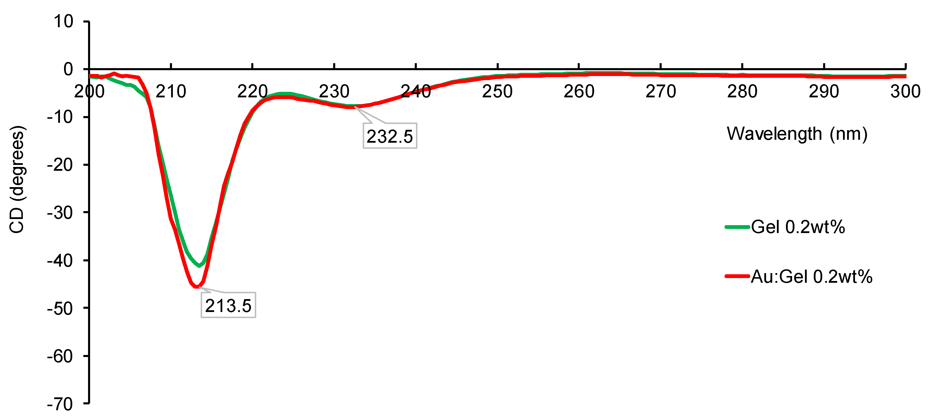
To assess structural degradation, circular dichroism (CD) spectroscopy, an important tool for obtaining information on the secondary structures of biomacromolecule [18], was performed on Au–Gel and pristine gelatin samples prepared at 0.2 wt%. Both samples showed negative peaks at 213 nm and 232 nm [19,20], characteristic of random coil structures in the polypeptide backbone. The Au–Gel sample displayed similar behavior to pristine gelatin with a negative region between 200 and 300 nm (Figure 2). The retention of negative peaks at approximately 213 nm and 232 nm in the CD spectra indicates that the random coil structure of gelatin remains largely preserved following laser ablation at 0.2 wt%, suggesting minimal disruption to its secondary structure under these conditions. CD spectra extended to 600 nm to assess chiral induction on the AuNP surface by the gelatin matrix, but no CD signal corresponding to the surface plasmon resonance (SPR) of AuNPs (500–600 nm) was observed (Figure S4). The absence of CD signals in the 500–600 nm region indicates that the surface of the AuNPs did not experience chiral induction from the surrounding matrix.
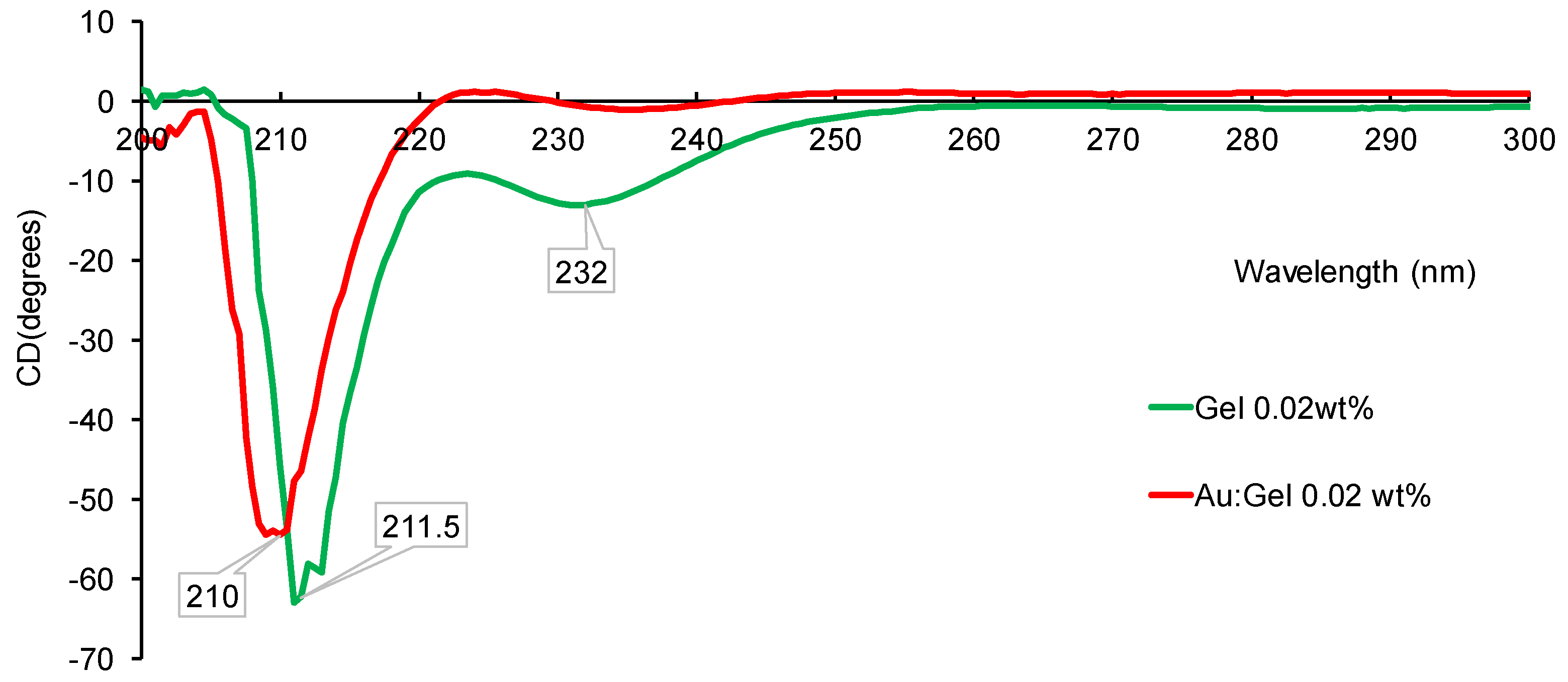
In contrast, Au–Gel prepared at a lower concentration of 0.02 wt% showed different results. While gelatin exhibited two prominent negative peaks at 210 and 230 nm, more pronounced under these conditions, a new positive peak emerged around 225 nm upon laser ablation (Figure 3). This shift from the typical 232 nm signal of pristine gelatin suggests the formation of a triple helical structure, similar to partially denatured native collagen [21,22]. These findings suggest that PLAL-MCL affects the secondary structure of gelatin, promoting a more ordered conformation similar to that of collagen triple helices, which may be beneficial for materials science and biomedical applications. Collectively, these observations indicate that MCL-PLAL enables concentration-dependent fine-tuning of gelatin structure, presenting a promising strategy for designing nanocomposites with tailored bioactive conformations.
Subsequent experiments investigated the influence of different media on reproducibility and Au–Gel NP synthesis. Using the optimized 0.2 wt% gelatin concentration, phosphate-buffered saline (PBS) was tested as a substitute medium to mimic cell culture conditions. The results (Table 2) showed no significant change in pH before and after ablation in pure water (6.57 → 6.81). In contrast, PBS (Entry 2) exhibited a marked increase in pH from 7.16 to 8.33 after one hour of laser irradiation. The resulting solution became visibly turbid, indicating the involvement of inorganic salts in the PBS during the ablation process (Figure S3). Laser irradiation may induce the formation of hydroxyl radicals (•OH), hydrogen atoms (•H), and dissociation of phosphate ions (H2PO4, HPO42−) [23], leading to pH increase, possibly through chloride ion oxidation. Previous studies have shown that chloride ions can reduce the surface charge of AuNPs, weakening electrostatic repulsion and promoting aggregation [24,25]. Accordingly, AuNPs formed in PBS tended to aggregate over time, as stability tests revealed aggregation within one month. In contrast, those formed in water remained stable, confirming water as a more suitable medium for PLAL-based synthesis.
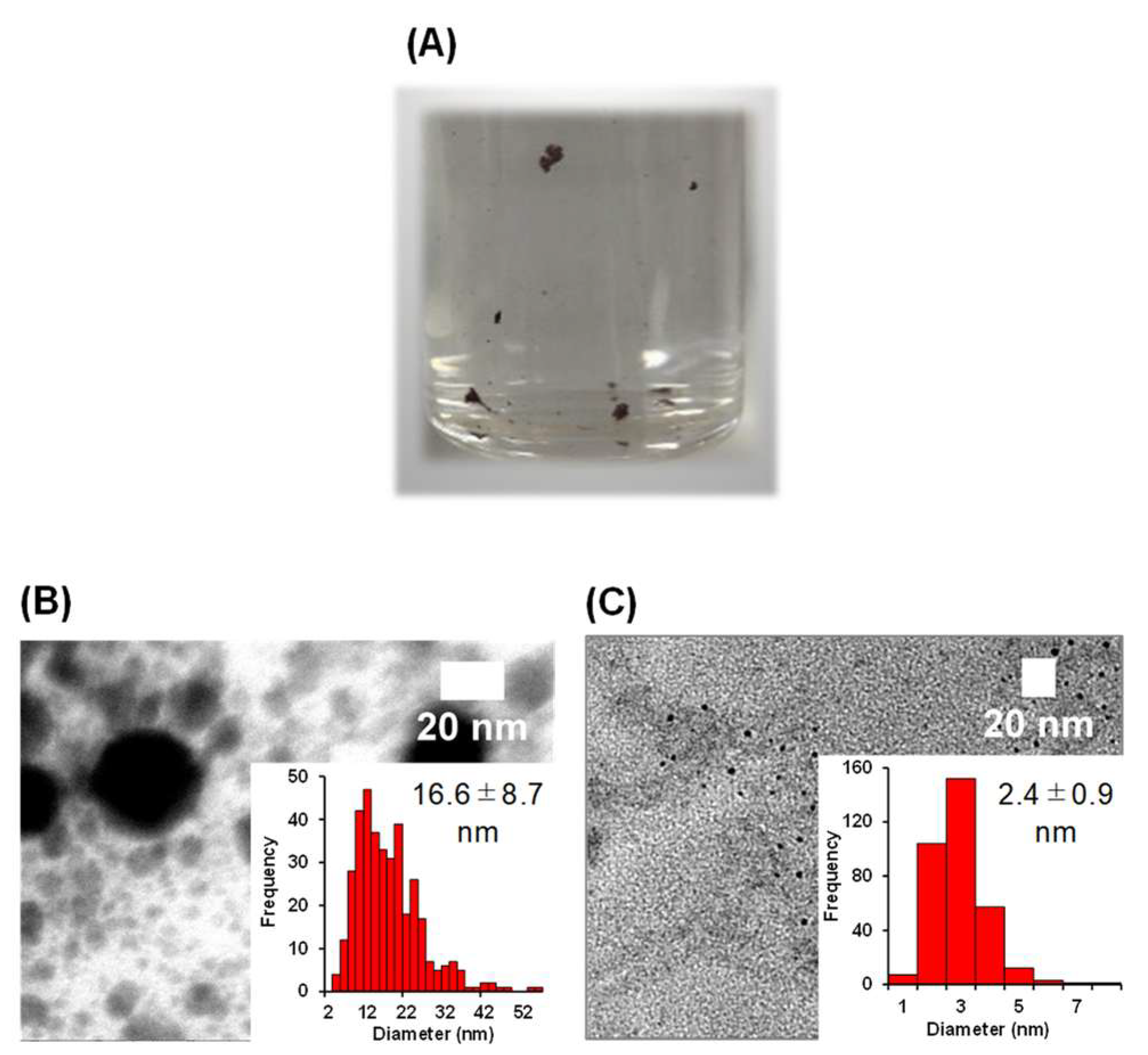
Finally, we attempted direct synthesis of collagen type I (ColI)-protected AuNPs via PLAL-MCL (Table 2, Entry 3). Unlike gelatin, ColI cannot form stable aqueous solutions without additives, necessitating the use of PBS despite its known negative effects. After one hour of irradiation, a faint pink supernatant and red precipitate were obtained, indicating AuNP formation (Figure 4A). Centrifugation was used to separate the precipitate and supernatant for further analysis. ICP-AES revealed that 0.045 µmol of Au was ablated, with 94% present in the precipitate. Due to ColI’s high viscosity (0.082 Pa·s vs. 0.001 Pa·s for water), ablation efficiency was reduced, resulting in lower AuNP yield [26,27,28].
TEM analysis of the red precipitate confirmed AuNP formation with a mean diameter of 16.6 ± 8.7 nm (Figure 4B). However, the precipitates could not be redispersed in any aqueous solution, indicating that significant deformation of ColI might occur during the ablation process. On the other hand, the supernatant, containing 6% of the total Au, showed much smaller and more uniform AuNPs (2.4 ± 0.9 nm) (Figure 4C), the smallest known for ColI matrices [28].

4. Conclusions

In summary, we successfully established a method for the direct incorporation of AuNPs into highly susceptible biopolymer matrices using pulsed laser ablation in liquid (PLAL) with a microchip laser (MCL), without relying on any chemical reduction processes. Despite the low pulse energy of the laser, the method achieved high efficiency. This approach enabled the formation of gelatin-stabilized AuNPs with an average size of approximately 4 nm, without altering the structural integrity of the gelatin—a result that is difficult to achieve with conventional techniques. Moreover, when gelatin was used at low concentrations, there was an indication that the laser process may have influenced the secondary structure of the gelatin.
On the other hand, when the method was extended to type I collagen—an even more sensitive biomatrix—a majority of the gold nanoparticles were entrapped within the aggregated matrix. Nevertheless, we successfully synthesized a small fraction of well-dispersed, non-aggregated collagen-stabilized AuNPs of small size. One possible reason for the low yield in this case may be the degradation of the PBS buffer caused by laser irradiation. If this is indeed the case, the use of PBS may need to be avoided, potentially limiting the applicability of this method to certain biomatrices. Nonetheless, the MCL-PLAL method presented in this study offers a fundamentally distinct and meaningful approach compared to conventional techniques, particularly in enabling NP synthesis under mild conditions within sensitive biological environments.

Supplementary Materials

The following supporting information can be downloaded at: https://www.mdpi.com/article/10.3390/colloids9040042/s1, Figure S1: Photographs of the experimental setup used for microchip laser ablation; Figure S2: TEM images and size distribution histograms of Au:Gel nanoparticles at varying gelatin loadings: 0.02% (A), 0.05% (B), 0.1% (C), and 0.5% (D). Scale bars: 20 nm; Figure S3: Transmission electron microscopy (TEM) images of Au:Gel nanoparticles at low (200 nm) and high (20 nm) magnification, along with corresponding particle size distribution histogram; Figure S4: Circular dichroism (CD) spectra of Au:Gel nanoparticles at 0.02 wt% (red) and 0.2 wt% (purple) gelatin concentrations. Table S1: Zeta potential measurements for Au:Gel (main experiment) and Gel (control experiment).

Author Contributions

Conceptualization, H.S.; methodology, N.A. and T.S.; validation, Y.U., Y.Y., M.M. and H.S.; formal analysis, N.A. and T.S.; investigation, N.A. and T.S.; writing—original draft preparation, N.A.; writing—review and editing, N.A., Y.U., Y.Y. and H.S.; supervision, M.M. and H.S.; project administration, H.S. All authors have read and agreed to the published version of the manuscript.

Funding

JSPS KAKENHI grant JP19K22187, JP24H00460 (H.S.); JP20K15279 (Y.U.); COI-NEXT (JPMJPF2009) from JST (M.M.); JPNP20004 from NEDO (M.M.), the Amada Foundation for the Promotion of Science & Engineering (AF2018234-C2) (Y.Y.), and Special Project by Institute for Molecular Science (IMS program 21-259) (H.S. and Y.Y.).

Data Availability Statement

Data is contained within the article or Supplementary Materials.

Acknowledgments

N.A. acknowledges MEXT for the scholarship.

Conflicts of Interest

The authors declare no conflicts of interest.

References

  1. Kogan, M.J.; Olmedo, I.; Hosta, L.; Guerrero, A.R.; Cruz, L.J.; Albericio, F. Peptides and Metallic Nanoparticles for Biomedical Applications. Nanomedicine 2007, 2, 287–306. [Google Scholar] [CrossRef] [PubMed]
  2. Khlebtsov, N.G.; Dykman, L.A. Optical Properties and Biomedical Applications of Plasmonic Nanoparticles. J. Quant. Spectrosc. Radiat. Transf. 2010, 111, 1–35. [Google Scholar] [CrossRef]
  3. Elahi, N.; Kamali, M.; Baghersad, M.H. Recent Biomedical Applications of Gold Nanoparticles: A Review. Talanta 2018, 184, 537–556. [Google Scholar] [CrossRef] [PubMed]
  4. Chatterjee, S.; Lou, X.Y.; Liang, F.; Yang, Y.W. Surface-Functionalized Gold and Silver Nanoparticles for Colorimetric and Fluorescent Sensing of Metal Ions and Biomolecules. Coord. Chem. Rev. 2022, 459, 214461. [Google Scholar] [CrossRef]
  5. Fatima, Z.; Saleem, R.; Khan, R.R.M.; Liaqat, M.; Pervaiz, M.; Saeed, Z.; Muhammad, G.; Amin, M.; Rasheed, S. Green Synthesis, Properties, and Biomedical Potential of Gold Nanoparticles: A Comprehensive Review. Biocatal. Agric. Biotechnol. 2024, 59, 103271. [Google Scholar] [CrossRef]
  6. Liu, X.; Luo, Y.; Zhang, Y.; Xie, Z.; Xu, C. Gold Nanoparticle-Mediated Fluorescence Resonance Energy Transfer for Analytical Applications in the Fields of Life Health and Safety. Talanta 2025, 282, 127023. [Google Scholar] [CrossRef]
  7. Kim, M.; Osone, S.; Kim, T.; Higashi, H.; Seto, T. Synthesis of Nanoparticles by Laser Ablation: A Review. KONA Powder Part. J. 2017, 2017, 80–90. [Google Scholar] [CrossRef]
  8. Balachandran, A.; Sreenilayam, S.P.; Madanan, K.; Thomas, S.; Brabazon, D. Nanoparticle Production via Laser Ablation Synthesis in Solution Method and Printed Electronic Application—A Brief Review. Results Eng. 2022, 16, 100646. [Google Scholar] [CrossRef]
  9. He, Y.; Wang, L.; Wu, T.; Wu, Z.; Chen, Y.; Yin, K. Facile Fabrication of Hierarchical Textures for Substrate-Independent and Durable Superhydrophobic Surfaces. Nanoscale 2022, 14, 9392–9400. [Google Scholar] [CrossRef]
  10. Yin, K.; Wang, L.; Deng, Q.; Huang, Q.; Jiang, J.; Li, G.; He, J. Femtosecond Laser Thermal Accumulation-Triggered Micro-/Nanostructures with Patternable and Controllable Wettability Towards Liquid Manipulating. Nano-Micro Lett. 2022, 14, 97. [Google Scholar] [CrossRef]
  11. Darroudi, M.; Khorsand Zak, A.; Muhamad, M.R.; Zamiri, R. Preparation of Gelatinous Gold Nanoparticles by Pulsed Laser Ablation. Res. Chem. Intermed. 2015, 41, 4587–4594. [Google Scholar] [CrossRef]
  12. Jiang, Z.; Li, L.; Huang, H.; He, W.; Ming, W. Progress in Laser Ablation and Biological Synthesis Processes: “Top-Down” and “Bottom-Up” Approaches for the Green Synthesis of Au/Ag Nanoparticles. Int. J. Mol. Sci. 2022, 23, 14658. [Google Scholar] [CrossRef] [PubMed]
  13. Lim, H.H.; Taira, T. Sub-Nanosecond Laser Induced Air-Breakdown with Giant-Pulse Duration Tuned Nd:YAG Ceramic Micro-Laser by Cavity-Length Control. Opt. Express 2017, 25, 6302. [Google Scholar] [CrossRef] [PubMed]
  14. Hettiarachchi, B.S.; Takaoka, Y.; Uetake, Y.; Yakiyama, Y.; Lim, H.H.; Taira, T.; Maruyama, M.; Mori, Y.; Yoshikawa, H.Y.; Sakurai, H. Uncovering gold nanoparticle synthesis using a microchip laser system through pulsed laser ablation in aqueous solution. Ind. Chem. Mater. 2024, 2, 340–347. [Google Scholar] [CrossRef]
  15. Hettiarachchi, B.S.; Takaoka, Y.; Uetake, Y.; Yakiyama, Y.; Yoshikawa, H.Y.; Maruyama, M.; Sakurai, H. Mechanistic Study in Gold Nanoparticle Synthesis through Microchip Laser Ablation in Organic Solvents. Metals 2024, 14, 155. [Google Scholar] [CrossRef]
  16. Hettiarachchi, B.S.; Yakiyama, Y.; Sakurai, H. Tailoring Carbon-Encapsulated Gold Nanoclusters via Microchip Laser Ablation in Polystyrene Solution: Controlling Size, Structure, and Photoluminescent Properties. RSC Appl. Interfaces 2025, 2, 772–779. [Google Scholar] [CrossRef]
  17. Neupane, M.P.; Lee, S.J.; Park, I.S.; Lee, M.H.; Bae, T.S.; Kuboki, Y.; Uo, M.; Watari, F. Synthesis of Gelatin-Capped Gold Nanoparticles with Variable Gelatin Concentration. J. Nanopart. Res. 2011, 13, 491–498. [Google Scholar] [CrossRef]
  18. Miles, A.J.; Janes, R.W.; Wallace, B.A. Tools and methods for circular dichroism spectroscopy of proteins: A tutorial review. Chem. Soc. Rev. 2021, 50, 8400–8413. [Google Scholar] [CrossRef]
  19. Gopal, R.; Park, J.S.; Seo, C.H.; Park, Y. Applications of Circular Dichroism for Structural Analysis of Gelatin and Antimicrobial Peptides. Int. J. Mol. Sci. 2012, 13, 3229–3244. [Google Scholar] [CrossRef]
  20. Rub, M.A.; Asiri, A.M.; Khan, J.M.; Khan, R.H.; Din, K.-U. Interaction of Gelatin with Promethazine Hydrochloride: Conductimetry, Tensiometry and Circular Dichroism Studies. J. Mol. Struct. 2013, 1050, 35–42. [Google Scholar] [CrossRef]
  21. Asil, S.M.; Narayan, M. Molecular Interactions between Gelatin-Derived Carbon Quantum Dots and Apo-Myoglobin: Implications for Carbon Nanomaterial Frameworks. Int. J. Biol. Macromol. 2024, 264, 130416. [Google Scholar] [CrossRef] [PubMed]
  22. Lee, S.; Jo, K.; Kim, S.; Woo, M.; Choi, Y.; Jung, S. Food Hydrocolloids. Food Hydrocoll. 2025, 160, 110739. [Google Scholar] [CrossRef]
  23. Correard, F.; Maximova, K.; Estève, M.A.; Villard, C.; Roy, M.; Al-Kattan, A.; Sentis, M.; Gingras, M.; Kabashin, A.V.; Braguer, D. Gold Nanoparticles Prepared by Laser Ablation in Aqueous Biocompatible Solutions: Assessment of Safety and Biological Identity for Nanomedicine Applications. Int. J. Nanomed. 2014, 9, 5415–5430. [Google Scholar] [CrossRef]
  24. Lévy, A.; De Anda Villa, M.; Laurens, G.; Blanchet, V.; Bozek, J.; Gaudin, J.; Lamour, E.; Macé, S.; Mignon, P.; Milosavljević, A.R.; et al. Surface Chemistry of Gold Nanoparticles Produced by Laser Ablation in Pure and Saline Water. Langmuir 2021, 37, 5783–5794. [Google Scholar] [CrossRef]
  25. Kwon, H.; Kim, K.K.; Song, J.K.; Park, S.M. The Effects of Ambient Ions on the Growth of Gold Nanoparticles by Laser Ablation in Liquid. Bull. Korean Chem. Soc. 2014, 35, 865–870. [Google Scholar] [CrossRef]
  26. Costela, A.; Garcia-Moreno, I.; Barroso, J.; Sastre, R. Laser Performance of Coumarin 540A Dye Molecules in Polymeric Host Media with Different Viscosities: From Liquid Solution to Solid Polymer Matrix. J. Appl. Phys. 1998, 83, 650–660. [Google Scholar] [CrossRef]
  27. Takata, T.; Enoki, M.; Chivavibul, P.; Matsui, A.; Kobayashi, Y. Effect of Confinement Layer on Laser Ablation and Cavitation Bubble during Laser Shock Peening. Mater. Trans. 2016, 57, 1776–1783. [Google Scholar] [CrossRef]
  28. Hupfeld, T.; Laurens, G.; Merabia, S.; Barcikowski, S.; Gökce, B.; Amans, D. Dynamics of Laser-Induced Cavitation Bubbles at a Solid-Liquid Interface in High Viscosity and High Capillary Number Regimes. J. Appl. Phys. 2020, 127, 044306. [Google Scholar] [CrossRef]
Figure 1. Setup of PLAL experiment (A), obtained Au NPs for Gel (B) and (C) a TEM image and the histogram in the case of entry 4, Table 1.
Colloids 09 00042 g001
Figure 2. CD spectra of Au–Gel (red) and Gel (green) at 0.2 wt%.
Colloids 09 00042 g002
Figure 3. CD spectra of Au–Gel (red) and Gel (green) at 0.02 wt%.
Colloids 09 00042 g003
Figure 4. (A) Au–ColI dispersion after PLAL showing precipitate and supernatant. (B) TEM and size distribution of nanoparticles from the precipitate (18.6 ± 8.7 nm). (C) TEM and size distribution from the supernatant (2.4 ± 0.9 nm). Scale bars: 20 nm.
Colloids 09 00042 g004
Table 1. Comparison of gelatin concentration, gelatin colloid size before and after formation of NPs by PLAL-MCL.
EntryGel Concentration (wt%)Au–Gel NPs (nm) aYield of Au (mmol) bAu–Gel Colloid Size (nm) cGel Colloid Size (nm) c
10.024.2 ± 2.14.4 × 10−523 ± 411 ± 4
20.053.9 ± 2.05.7 × 10−530 ± 47 ± 2
30.14.5 ± 2.32.2 × 10−510 ± 47 ± 2
40.24.3 ± 0.94.4 × 10−531 ± 27 ± 1
50.54.4 ± 2.57.5 × 10−525 ± 34 ± 2
a measured by TEM, b measured by ICP-AES, c measured by DLS.
Table 2. Effect of different media on bio-matrix stabilized Au NPs formation by PLAL.
EntryMatrixConcentration (wt%)SolventpH Before PLALpH After PLALColor After PLALICP (mmol)
1Gel0.2water6.576.81Clear red4.4 × 10−5
2Gel 0.2PBS7.168.33Cloudy red3.8 × 10−6
3ColI0.2 PBS6.576.81Clear red4.4 × 10−5
Disclaimer/Publisher’s Note: The statements, opinions and data contained in all publications are solely those of the individual author(s) and contributor(s) and not of MDPI and/or the editor(s). MDPI and/or the editor(s) disclaim responsibility for any injury to people or property resulting from any ideas, methods, instructions or products referred to in the content.