Article

Water-Based Bi2S3 Nano-Inks Obtained with Surfactant-Assisted Liquid Phase Exfoliation and Their Direct Processing into Thin Films

1
Dipartimento Scienza Applicata e Tecnologia (DISAT), Politecnico di Torino, Corso Duca degli Abruzzi 24, 10129 Torino, Italy
2
Center for Materials Research (LaMa), Justus Liebig University, Heinrich-Buff-Ring 17, 35392 Giessen, Germany
*
Authors to whom correspondence should be addressed.
Colloids Interfaces 2024, 8(3), 28; https://doi.org/10.3390/colloids8030028
Submission received: 29 February 2024 / Revised: 23 April 2024 / Accepted: 24 April 2024 / Published: 30 April 2024

Abstract

:
Bi2S3 has gained considerable attention as a semiconductor for its versatile functional properties, finding application across various fields, and liquid phase exfoliation (LPE) serves as a straightforward method to produce it in nano-form. Till now, the commonly used solvent for LPE has been N-Methyl-2-pyrrolidone, which is expensive, toxic and has a high boiling point. These limitations drive the search for more sustainable alternatives, with water being a promising option. Nonetheless, surfactants are necessary for LPE in water due to the hydrophobic nature of Bi2S3, and organic molecules with amphoteric characteristics are identified as suitable surfactants. However, systematic studies on the use of ionic surfactants in the LPE of Bi2S3 have remained scarce until now. In this work, we used sodium dodecyl sulfate (SDS), sodium dodecylbenzene sulfonate (SDBS) and sodium hexadecyl sulfonate (SHS) as representative species and we present a comprehensive investigation into their effects on the LPE of Bi2S3. Through characterizations of the resulting products, we find that all surfactants effectively exfoliate Bi2S3 into few-layer species. Notably, SDBS demonstrates superior stabilization of the 2D layers compared to the other surfactants, while SHS becomes the most promising surfactant for obtaining products with high yield. Moreover, the resulting nano-inks are used for fabricating films using spray-coating, reaching a fine tuning of band gap by controlling the number of cycles, and paving the way for the utilization of 2D Bi2S3 in optoelectronic devices.

1. Introduction

Metal chalcogenides cover a large family of 2D materials, which have gained recent attention due to their potential significance in many technological applications [1,2,3,4,5,6]. Among them, Bi2S3 emerges as a potential candidate in thermoelectric applications, energy harvesting, biomedicine, sensors and optoelectronics, due to its low thermal conductivity, strong spin-orbit coupling, direct band structure and high absorption coefficient [7,8,9,10,11,12]. As shown in Figure 1a, Bi2S3 possesses a lamellar structure in which the bismuth and sulfur atoms are bonded through strong covalent bonds inside the layer, and these layers are linked by van der Waals (vdW) forces to one another [13]. This unique layered structure allows the vdW interactions between layers in bulk Bi2S3 to be broken by mechanical force, thus producing 2D layered Bi2S3.
Liquid phase exfoliation (LPE) is a simple and scalable technique used to produce 2D layered materials. This procedure consists of delaminating the layers of the material dispersed in a solvent by mechanical force. In sonication-assisted LPE, the sonication generates the growth and collapse of microbubbles in the solvents, thus resulting in shock waves. These waves can exert shear forces on bulk materials, disrupt the vdW interactions between the layers of the 2D structures and ultimately lead to the formation of layered materials [14,15,16]. The type of solvent plays an important role in the efficiency of the exfoliation, and the solvent typically used for exfoliating Bi2S3 is N-methyl-2-pyrrolidone (NMP) [17,18,19,20]. Considering the high cost, high boiling point and toxicity of NMP, replacing it with water has become a more eco-friendly and sustainable strategy.
Since Bi2S3 is hydrophobic, surfactants are needed to separate and stabilize the nanocrystals in H2O. In LPE, the frequently used surfactants are anionic surfactants that can counteract vdW attraction between the material layers, inhibiting restacking by electrostatic force with each single layer [21,22]. One of the most employed surfactants for metal chalcogenides is the anionic surfactant sodium dodecyl sulfate (SDS), and its structure is shown in Figure 1b [23,24]. The molecule contains an anion with C12 alkyl chain and SO4 group on the terminal of the carbon chain. The carbon chains construct semi-micelles on the basal plane of metal chalcogenides to prevent the restacking of the exfoliated nanosheets, while the terminal SO4 group forms a H-bond with water and stabilizes the exfoliated layers [25]. Our previous work also proves the possibility of exfoliation of Bi2S3 with SDS [26]. Moreover, surfactants with a similar structure to SDS but different functional groups have been reported in LPE, such as sodium dodecylbenzene sulfonate (SDBS). It has been reported that the benzene ring in SDBS improves the colloidal stability of exfoliated MoS2 [27]. Furthermore, it has been reported that by changing the alkyl chain length, the stability of the exfoliated suspension and optoelectronic property of the colloidal nanocrystals can be optimized as well [28]. However, a study regarding the influence of the carbon chain of the surfactant on the LPE process is still missing. In addition to the type of surfactant, the concentration of the surfactant significantly influences the quality and yield of exfoliated metal chalcogenides [21,29]. Therefore, surfactant concentration will be a crucial parameter in optimizing the harvesting of exfoliated Bi2S3 with good quality and high yield.
Based on the above background knowledge, in this work we systematically investigate how surfactant type and concentration impact the quality of exfoliated Bi2S3. Three different surfactants have been chosen, SDS, SDBS and sodium hexadecyl sulfate (SHS), to compare the influence of different functional groups and carbon chain lengths on the LPE of Bi2S3 (Figure 1). It has been reported that controlling the concentration of the surfactants less than their critical micelle concentration (CMC) can result in better-quality exfoliated samples, while other studies reached contradictory conclusions [30,31]. In our study, we set the concentrations from 8.2 mM to 0.5 mM for all the surfactants for better comparison, considering that the CMCs of SDS, SDBS and SHS are 8.2, 2.7 and 0.55 mM, respectively [32,33,34,35]. By assessing the band gap, stability and yield of the exfoliated Bi2S3, it is found that SHS results in the largest band gap and yield, while the colloidal suspension is more stable with SDBS. As to the influence of the concentration, a higher concentration of all the surfactants tends to form exfoliated samples with a higher band gap. Only SHS shows a correlation between the concentration and the sample yield, and the record yield of 1.4% is obtained with 8.2 mM SHS. Moreover, concentration does not influence the stability of the suspension. Eventually, we also show that the suspension can be made into Bi2S3 thin films with a tunable band gap through ultrasonic spray-coating, which is a promising perspective for the use of these nano-inks in the field of eco-friendly, solution-processed optoelectronics.

2. Materials and Methods

2.1. Materials and LPE Process

Fluorine-doped tin oxide (FTO) coated glass slides, Bi2S3 (99%), SDS and SDBS were purchased from Sigma Aldrich and used without further purification. SHS was purchased from TCI Chemicals and used without further purification. All the exfoliations were performed using a Baudelin Sonopuls tip sonicator and the samples were cooled to 0 °C with an ice bath during the process. The tip sonicator operated with 80% power using pulses of 1 s on/1 s off for 4 h.
In all the experiments, the suspension volume was 150 mL with a concentration of the bulk materials of 10 mg/mL. The concentrations of all the surfactants were adjusted to 8.2 mM, 4.1 mM, 2.0 mM, 1.0 mM and 0.5 mM, respectively. The suspensions obtained after the sonication were centrifuged firstly for 30 min at 1500 rpm to reserve the supernatant, then this supernatant was centrifuged for 30 min at a higher speed of 3000 rpm to obtain the final colloidal suspensions. A Universal 320 Hettich centrifuge was employed for the centrifuge treatment.
Thin Bi2S3 films were fabricated using the Nadetech Innovations Ultrasonic Lab Spray Coater on FTO glass substrates measuring 15 × 15 mm, and the suspensions from the previous steps were directly used as inks for the spray-coating. We used N2 pressure with 0.10 bar to get a uniform spray. The speed of the nozzle was 400 mm/min, and the working flow of the suspension was 25 mL/h.

2.2. Characterizations

UV-visible (UV-vis) absorption spectra of the colloidal suspensions were captured using a Goebel Uvikon spectrometer, employing a quartz cuvette with an optical length of 1 cm. The spectra were recorded from 350 to 1000 nm with a scan interval of 0.25 nm. Raman spectra were acquired using a Bruker Senterra instrument equipped with a 532 nm laser excitation source at a power of 2 mW. Integration time was set to 6 s with 60 co-additions. Samples were prepared by drop-casting suspensions onto silicon slides for analysis. Dynamic Light Scattering (DLS) and Zeta potential (ZP) measurements were conducted on a Malvern Zetasizer Nano-ZS device, averaging results from three separate measurements for accuracy. Measurements were performed in Rotilabo precision glass cuvettes with a light path of 10 mm and a volume of 3.5 mL. High-Resolution Transmission Electron Microscopy (HRTEM) imaging was performed using a non-aberration-corrected Transmission Electron Microscope (TALOS F200X, Thermo Scientific, Eindhoven, The Netherlands) operating at 200 kV. Images were captured using a 16Mpxls CMOS camera with an exposure time of 1 s. The fast Fourier transform (FFT) pattern was converted with the Velox software (Thermo Scientific Velox Software, Thermo Fisher Scientific Inc., Waltham, MA, USA).

2.3. Calculation Methods of the Sample Concentration

UV-vis spectra were employed to calculate the final concentration of Bi2S3. A calibration line was obtained using a set of dilutions from a suspension with a known concentration, and this concentration of samples was obtained using filtration of the products. The slope of the calibration line corresponds to the extinction coefficient of the dispersed material. The final concentrations of Bi2S3 samples were obtained with the Lambert-Beer law (Equation (1)):
A = εbc
where A represents the absorbance of the material, ε denotes the extinction coefficient of the dispersed nanosheets, and c signifies the concentration of the suspension. Specifically, we utilized the absorption value at 500 nm as A. The value of ε is derived from calibration lines established using several standard Bi2S3 suspensions with known concentrations. For Bi2S3, the calculated ε is determined to be 7.4 mg/mL·cm.
To compare with the concentration calculated from UV-vis spectra, freeze-drying was used to obtain the final concentration as well. Before freeze-drying, dialysis was performed using a cellulose dialysis bag (Carl Roth) 14 kDa, filling each bag with 20–25 mL of the desired suspension and closing both sides with a plastic pin once filled. The bag was then left in a suitable beaker with Milli-Q water for 3 days, changing the water 3 times per day. Freeze-drying was performed at −10 °C for 16 h and at a pressure of 1 mPa.

3. Results and Discussion

3.1. Characterizations of the Resulting Nanomaterials

3.1.1. TEM Analysis

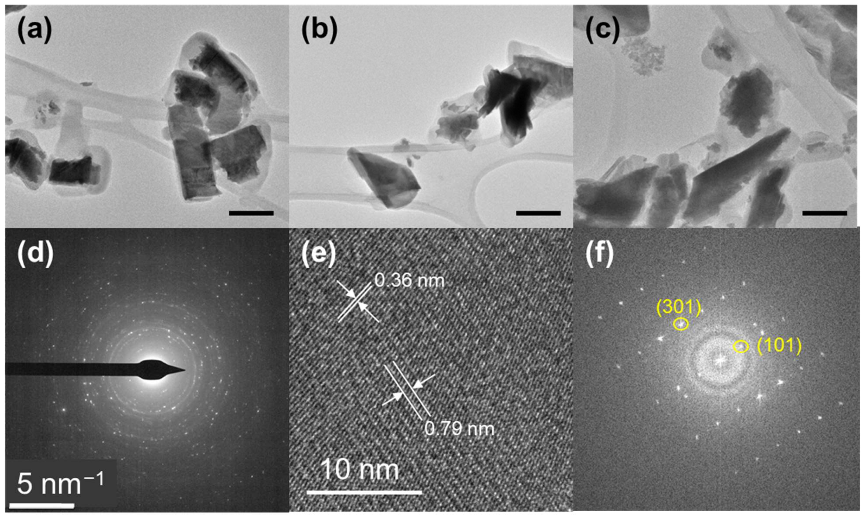
The exfoliated Bi2S3 nano-inks were prepared using the surfactant-assisted LPE method outlined in Section 2.1 and a typical exfoliated Bi2S3 sample was used to characterize the morphology and crystal structure of the colloids. As depicted in Figure 2a–c, nanosheets of irregular shapes are observed, predominantly appearing as a few layers of around 10 nm of thickness rather than single layers. The size distribution of the exfoliated samples ranges from 50 to 350 nm, with the size of around 150–200 nm as the dominate value (Figure S1). Study of the selected area electron diffraction (SAED) illustrates that the crystalline nanosheets can be indexed to orthorhombic structured Bi2S3 (ICSD: 30775) (Figure 2d). Figure 2e,f depict the HRTEM image and its corresponding FFT pattern, revealing lattice planes within the nanosheets. These planes exhibit spacings of 0.79 nm and 0.36 nm, indicative of the 101 and 301 planes, respectively. This small nanosheet shows a [010] orientation, since it is possible to break the bulk Bi2S3 in the direction of [010] to obtain nanoribbons (Figure 1a) [31].

3.1.2. UV-Vis Analysis

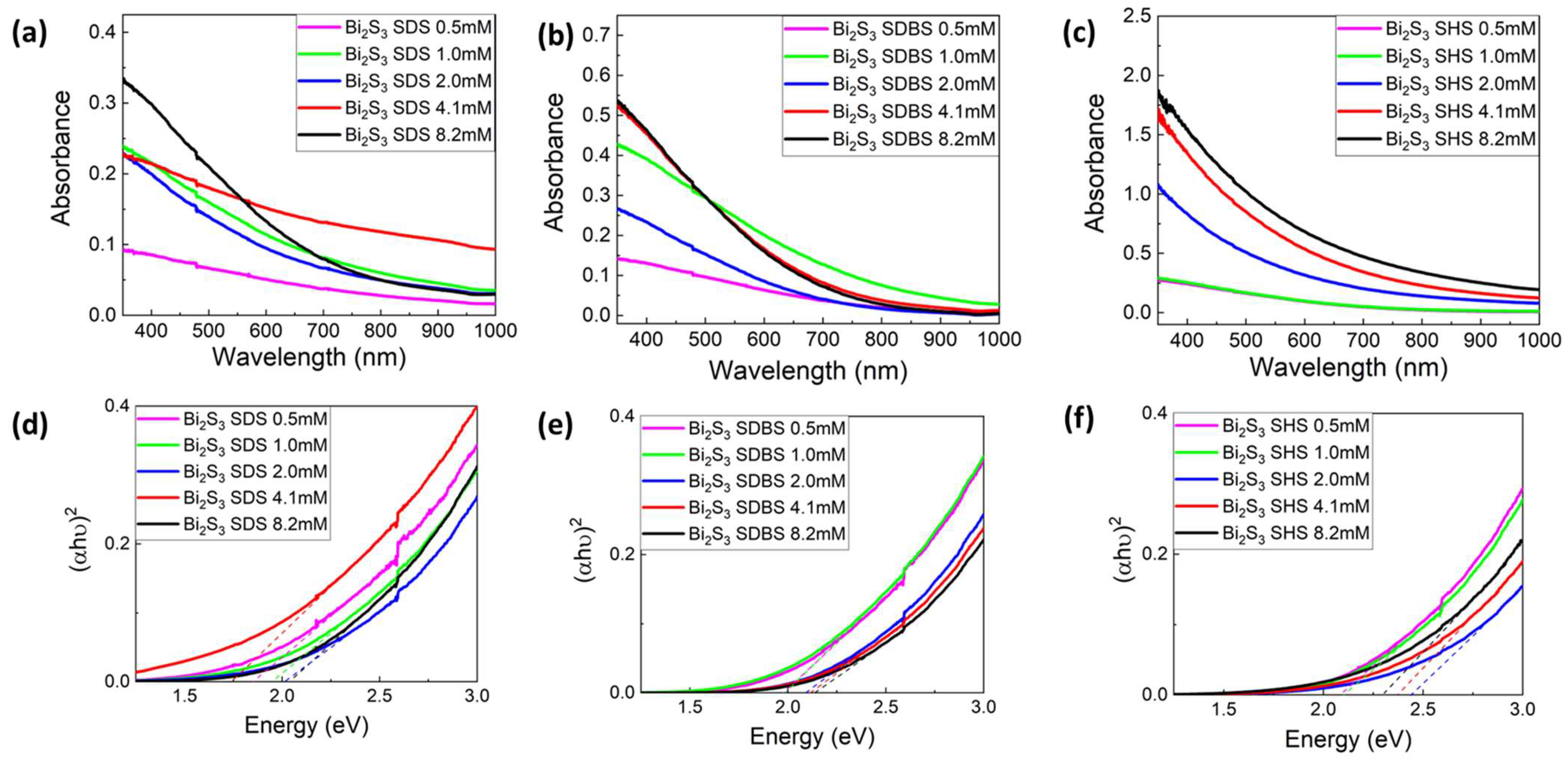
Figure 3a–c show the UV-vis absorption spectra of Bi2S3 samples exfoliated with different types and concentrations of surfactants. It can be observed that there is a growing absorption intensity when the wavelength decreases for all the samples [21,22]. In samples with the highest surfactant concentration, there is a notable rapid decline in absorption intensity beyond 550 nm. This phenomenon could likely be attributed to the strong influence exerted by the elevated concentration of surfactants (Figure S2). As a consequence, this high concentration of surfactant gives less scattering in the spectrum. The band gap of all the samples was calculated with the Tauc Plot equation (Equation (2)):
hv)1/n = A (hv − Eg)
where α is the absorption coefficient, h is the Plank constant, v is the frequency, Eg is the band gap energy and n is 1/2 for Bi2S3 with a direct band gap [36]. The values are shown in Figure 3d–f and Table 1. In general, all the samples have a broader band gap ranging from 1.74 to 2.44 eV in comparison to the band gap of bulk Bi2S3 (1.3 eV) [37]. This is evidence that all the samples were exfoliated to nanoscale. Regarding the impact of surfactant type, SDS yielded nano-inks with a lower band gap ranging from 1.85 to 2.03 eV, whereas samples with SHS exhibited a higher band gap range of 2.08 to 2.3 eV. This indicates that samples derived from SHS possess a smaller size in terms of thickness or particle size. Concerning surfactant concentration, the overarching trend is that higher concentrations lead to broader band gaps. It is worth noting the detected non-linear change in band gap with surfactant concentration. This complexity arises from various factors influencing the band gap: not only is it connected to quantum confinement effects, but it is also significantly impacted by the atomistic arrangement on the nanosheet surface, which is dictated by the nature of the capping surfactant [28]. Though higher concentrations are not strictly related to higher band gap, all these concentrations still result in samples with a widened band gap, which is proof that all the concentrations can exfoliate bulk Bi2S3.

3.1.3. Raman Analysis

Figure 4 displays the Raman spectra of Bi2S3 samples with all the surfactants. It is possible to identify two main active modes, namely the B1g and Ag Raman modes, in all the samples. The peaks located at 188 cm−1 and 237 cm−1 belong to B1g mode, which arises from longitudinal vibrations, while the Ag transversal modes are noticeable at 106 cm−1, 170 cm−1 and 265 cm−1 [38] After the exfoliation of the material, the frequency of the longitudinal optical phonon is higher, so the ratio between the two modes decreases [39]. The two modes with the most identifiable peaks (B1g mode at 237 cm−1 and Ag mode at 265 cm−1) were chosen to characterize the surficial property of the exfoliated samples. Specifically, the intensity ratio of Ag mode and B1g mode is calculated to determine whether the samples were exfoliated [19,40]. In bulk Bi2S3, the Ag/B1g ratio is 1.68 [26], while for all the exfoliated Bi2S3, the Ag/B1g ratio ranges from 1.02 to 1.32 (Table 2), which is clear evidence that all the samples were exfoliated, and the surficial molecular vibration modes are different from the bulk Bi2S3. When comparing band gap values with Ag/B1g ratios in Raman spectra, a linear correlation is not emerging. Initially, a lower ratio of Ag/B1g suggests a higher band gap, yet this association is due to the quantum confinement effect, which is related to layer count, nanosheet anisotropy and average size. In these experiments, the average size of the nanosheets is not controlled, thus it is difficult to build a linear relationship between the average number of layers and the band gap values. However, there is no report in the literature related to the values of Ag/B1g ratio and its connection to any layer property of exfoliated Bi2S3, thus we do not obtain any further information from Raman spectra. Concerning the Raman mode shift in the samples, a distinct shift compared to bulk Bi2S3 was not observed, except for Bi2S3 SDBS 8.2 mM. This is likely due to the fact that the frequency of the Raman mode does not vary significantly whether Bi2S3 is in bulk form or exfoliated. Another prominent mode observed in the samples using SDBS appears at around 122 cm−1. This mode corresponds to a Bi-O stretching characteristic of β-Bi2O3, which may be associated with a non-stable oxide phase likely induced by laser irradiation [41].

3.1.4. Zeta Potenzial and DLS

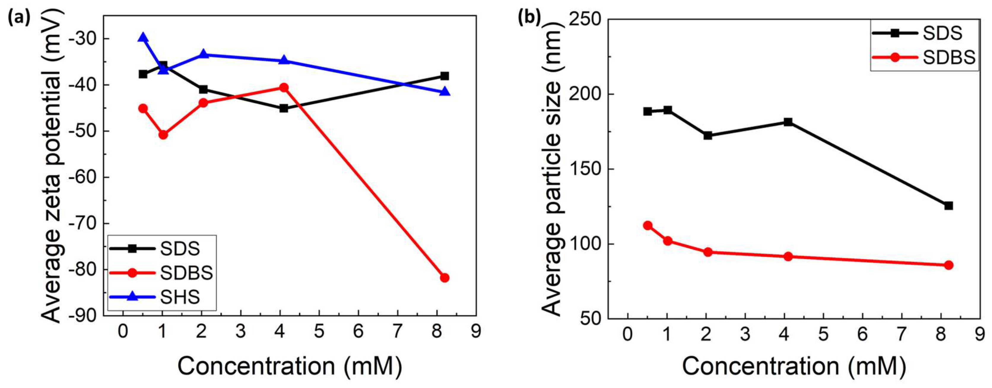
ZP analysis was conducted to evaluate the stability of the Bi2S3 colloidal suspensions from the surfactant-assisted LPE method. Generally, when the ZP values are between −20 and −30 mV, the suspension shows a short-term stability, while samples with −30 mV ZP values show a long-term stability of up to several months [42]. Figure 5a illustrates that all Bi2S3 water-based inks exhibit a ZP value below −30 mV, indicating long-term colloidal stability of more than one month. Specifically, the ZP values of most Bi2S3 samples are in the range between −30 and −50 mV, and the surfactant concentration appears to have minimal impact on them. An exceptional value of −80 mV is observed for the ink produced using 8.2 mM of SDBS, indicating that the colloidal suspension obtained under these experimental conditions exhibits outstanding stability.
The average particle size of each sample was estimated by DLS and the results are shown in Figure 5b. The average particle size of Bi2S3 remains approximately 100 nm for SDBS, and the particle size shows a slight decrease when the surfactant concentration increases. Upon comparing the concentration with the obtained band gap values, a positive correlation becomes evident. Higher band values correspond to increased surfactant concentration, resulting in a slight reduction in the average particle size. The sizes range from approximately 110 nm to 90 nm, spanning surfactant concentrations of 0.5 to 8.2 mM, respectively. With SDS, the average particle size of Bi2S3 ranges from around 200 nm to 125 nm from the lowest to highest surfactant concentration and it drops dramatically when increasing the concentration of SDS. It is apparent that some inconsistencies emerge in band gap values versus average particle size calculated from DLS. This discrepancy may arise from the fact that for 2D and 1D materials, DLS primarily estimates the lateral length of the nanosheet rather than the thickness [43]. Since both small thickness and small lateral length of the nanosheets correspond to higher band gap values, because of the quantum confinement, the influence of layer thickness is not considered in the values of DLS, thus causing the mismatch between band gap values and average particle size, as measured from DLS. Additionally, surface chemistry can influence band gap values as well, and this factor has not been well studied for exfoliated Bi2S3 with surfactants yet. Meanwhile, the trend in particle size using SHS shows a rough range mostly under 200 nm, but little correlation with the concentration of SHS and band gap values, as shown in Figure S3. This trend in particle size agrees with the result from the band gap calculation in Table 1, in which we expect samples with SDS to have roughly larger size than samples with SDBS.

3.2. Yield and Final Concentration of the Nanomaterials

The yield of the exfoliated material is another crucial metric used to evaluate the effects of the surfactants and the associated experimental variables. As described in Section 2.3, UV-vis spectra are employed to quantify the final concentration of the samples. All the results are listed in Table 3 and shown in Figure 6. Apparently, samples with SHS demonstrate superior values compared to those treated with the other two surfactants. Especially with surfactant concentrations higher than 2.0 mM, the concentration of SHS reaches more than 0.05 mg/mL and yields more than 0.5%, while all the samples obtained with SDS and SDBS have a concentration lower than this value. Moreover, higher SHS concentration can result in higher yield, and we are able to obtain the maximum sample concentration of 0.14 mg/mL and yield of 1.4% in this work with 8.2 mM of SHS.
To validate this data, selected samples with higher concentrations were subjected to freeze-drying after removing the surfactant through dialysis, and different values compared to the calculated concentration from UV-vis spectra were obtained. This is probably due to the residual presence of surfactant in the dispersion or the loss of samples during the operation.

3.3. Production of Thin Films with Ultrasonic Spray-Coating

To test the potential for solution-processing of the prepared, water-based nano-inks, we produced thin films of Bi2S3 using the ultrasonic spray-coating method, which is very suitable for deposition of nanomaterial inks and might pave the way to potential applications of these colloids in the sustainable fabrication of optoelectronic devices. With the Bi2S3 nano-ink exfoliated with 2.0 mM of SDBS, we sprayed for multiple steps on transparent substrates, to be able to detect the change in band gap as a function of the number of layers deposited. The colloidal ink was directly used without further dilution, since the concentration of the product was already very low (0.02 mg/mL). Figure 7 shows the UV-vis spectra of the thin films prepared from spraying exfoliated Bi2S3. From the corresponding Tauc plots, the band gap values of the Bi2S3 thin films are found to be 1.43 and 1.26 eV for 30 and 50 spray-steps, respectively (Figure 7b). The intense absorption is consistent with the remarkable extinction coefficient of Bi2S3 [44]. Furthermore, the absorption spectrum is clearly characterized by fringes. These fringes are evidently due to the FTO film between the glass substrate and Bi2S3 thin films. In fact, it is remarkable that, for the two different Bi2S3 thin films, the spectral position of the fringes is the same. It is evident that by adjusting the number of sprayed layers, the tuning of the band gap of the Bi2S3 film is possible. These values are smaller than the band gaps of the nanosheets in the suspension (Table 1), which is likely due to the nanosheets aggregation at the solid state, within the film. Figure 7c,d display the SEM images of the as-synthesized thin films. In Figure 7d, after 50 layers of spray coating, the sample shows a uniform Bi2S3 surface, while the sample with 30 layers is not fully covering the transparent conductive oxide substrate. The full-view images of Figure 7c,d are shown in Figure S4.

4. Conclusions

In conclusion, we conducted a systematic investigation into the surfactant-assisted LPE of Bi2S3. Utilizing three distinct ionic surfactants (SDS, SDBS and SHS), we performed exfoliation processes in water to generate stable colloidal suspensions, through a green method that is a good alternative to the use of toxic organic solvents. Through comprehensive characterization employing various techniques, we compared the quality of the exfoliated 2D Bi2S3 nanosheets present in the inks in terms of layer size, colloidal stability and product yield. It is found that with SHS, an exfoliated Bi2S3 with smaller particle size and higher yield can be obtained, but SDBS can lead to more stable colloidal suspensions compared to the other two surfactants investigated. Moreover, SHS with higher concentrations results in a higher yield in the exfoliated product, with the 8.2 mM concentration providing the optimal value of 1.4%. Considering that the CMC of SHS is only 0.55 mM, our results surprisingly indicate that the best concentration is high above the value of CMC. Moreover, we found that uniform Bi2S3 films with a tunable band gap can be obtained with ultrasonic spray-coating, while further optimization of the film fabrication process is needed to obtain films for practical applications in optoelectronic devices.

Supplementary Materials

The following supporting information can be downloaded at: https://www.mdpi.com/article/10.3390/colloids8030028/s1, Figure S1: Histogram of the distribution of particle size Bi2S3; Figure S2: UV-vis spectrum of SDS 8.2mM; Figure S3: DLS Bi2S3 SHS; Figure S4: SEM images of the Bi2S3 thin films with (a) 30 (b) 50 layers. Table S1: Parameters of LPE process.

Author Contributions

Conceptualization, T.G. and M.W.; methodology, M.P., F.B., S.D. and M.C.; software, M.P.; validation, M.P. and M.W.; formal analysis, M.P., M.C., F.S. and F.B; investigation, M.W.; resources, B.S. and T.G.; data curation, M.P.; writing—original draft preparation, M.P.; writing—review and editing, M.W., T.G. and F.S.; supervision, M.W.; project administration, B.S. and T.G.; funding acquisition, T.G. and M.W. All authors have read and agreed to the published version of the manuscript.

Funding

This research was funded by the ERC StG project JANUS BI (grant agreement No. [101041229]) and by the Starting Grant ERC program of Compagnia di San Paolo.

Data Availability Statement

The data from this study are available upon request made to the corresponding author.

Acknowledgments

M.W. and T.G. thank Fondazione Compagnia di San Paolo for financial support through the “Bando TRAPEZIO—Paving the way to research excellence and talent attraction”. M.C. and F.B. acknowledge the support of the European Commission through the H2020 FET-PROACTIVE-EIC-07-2020 project LIGHT-CAP (grant number 101017821).

Conflicts of Interest

The authors declare no conflicts of interest.

References

  1. Shawky, A.; Alahmadi, N.; Mohamed, R.M.; Zaki, Z.I. Bi2S3-Sensitized TiO2 Nanostructures Prepared by Solution Process for Highly Efficient Photoreduction of Hexavalent Chromium Ions in Water Under Visible Light. Opt. Mater. 2022, 124, 111964. [Google Scholar] [CrossRef]
  2. Ott, S.; Wolff, N.; Rashvand, F.; Rao, V.J.; Zaumseil, J.; Backes, C. Impact of the MoS2 Starting Material on the Dispersion Quality and Quantity after Liquid Phase Exfoliation. Chem. Mater. 2019, 31, 8424–8431. [Google Scholar] [CrossRef]
  3. Han, M.; Jia, J. The Interlace of Bi2S3 Nanowires with TiO2 Nanorods: An Effective Strategy for High Photoelectrochemical Performance. J. Colloid Interface Sci. 2016, 481, 91–99. [Google Scholar] [CrossRef] [PubMed]
  4. Zhang, X.; Shi, Y.; Shi, Z.; Xia, H.; Ma, M.; Wang, Y.; Huang, K.; Wu, Y.; Gong, Y.; Fei, H.; et al. High-Pressure Synthesis of Single-Crystalline SnS Nanoribbons. Nano Lett. 2023, 23, 7449–7455. [Google Scholar] [CrossRef] [PubMed]
  5. Barraza-Lopez, S.; Fregoso, B.M.; Villanova, J.W.; Parkin, S.S.P.; Chang, K. Colloquium: Physical Properties of Group-IV Monochalcogenide Monolayers. Rev. Mod. Phys. 2021, 93, 011001. [Google Scholar] [CrossRef]
  6. Villanova, J.W.; Kumar, P.; Barraza-Lopez, S. Theory of Finite-Temperature Two-Dimensional Structural Transformations in Group-IV Monochalcogenide Monolayers. Phys. Rev. B 2020, 101, 184101. [Google Scholar] [CrossRef]
  7. Xie, Y.; Zhou, Y.; Gao, C.; Liu, L.; Zhang, Y.; Chen, Y.; Shao, Y. Construction of AgBr/BiOBr S-Scheme Heterojunction Using Ion Exchange Strategy for High-Efficiency Reduction of CO2 to CO under Visible Light. Sep. Purif. Technol. 2022, 303, 122288. [Google Scholar] [CrossRef]
  8. Bai, Y.; Ouyang, T.; Li, X.; Yan, Y.; Kong, Z.; Ma, X.; Li, Z.; Li, Z.; Cai, X.; Cai, J.; et al. Boosting the Thermoelectric Performance of n-Type Bi2S3 by Compositing rGO. J. Alloys Compd. 2023, 933, 167814. [Google Scholar] [CrossRef]
  9. Zhao, F.; Sheng, H.; Sun, Q.; Wang, J.; Liu, Q.; Hu, Z.; He, B.; Wang, Y.; Li, Z.; Liu, X. Harvesting the Infrared Part of Solar Light to Promote Charge Transfer in Bi2S3/WO3 Photoanode for Enhanced Photoelectrochemical Water Splitting. J. Colloid Interface Sci. 2022, 621, 267–274. [Google Scholar] [CrossRef] [PubMed]
  10. Xu, Y.; Chen, B.; Xu, L.; Zhang, G.; Cao, L.; Liu, N.; Wang, W.; Qian, H.; Shao, M. Urchin-like Fe3O4@Bi2S3 Nanospheres Enable the Destruction of Biofilm and Efficiently Antibacterial Activities. ACS Appl. Mater. Interfaces 2023, 16, 3215–3231. [Google Scholar] [CrossRef] [PubMed]
  11. Zamani, M.; Jamali-Sheini, F.; Cheraghizade, M. Visible-Range and Self-Powered Bilayer p-Si/n-Bi2S3 Heterojunction Photodetector: The Effect of Au Buffer Layer on the Optoelectronics Performance. J. Alloys Compd. 2022, 905, 164119. [Google Scholar] [CrossRef]
  12. Yang, Z.; Wang, Y.; Zhang, D.; Chen, C. A Sensitizing Photoelectrochemical Sensing Platform Strategy Based on Bio-Etching Preparation of Bi2S3/BiOCl p–n Heterojunction. Talanta 2018, 190, 357–362. [Google Scholar] [CrossRef] [PubMed]
  13. Smith, R.J.; King, P.J.; Lotya, M.; Wirtz, C.; Khan, U.; De, S.; O’Neill, A.; Duesberg, G.S.; Grunlan, J.C.; Moriarty, G.; et al. Large-Scale Exfoliation of Inorganic Layered Compounds in Aqueous Surfactant Solutions. Adv. Mater. 2011, 23, 3944–3948. [Google Scholar] [CrossRef] [PubMed]
  14. Li, Z.; Young, R.J.; Backes, C.; Zhao, W.; Zhang, X.; Zhukov, A.A.; Tillotson, E.; Conlan, A.P.; Ding, F.; Haigh, S.J.; et al. Mechanisms of Liquid-Phase Exfoliation for the Production of Graphene. ACS Nano 2020, 14, 10976–10985. [Google Scholar] [CrossRef] [PubMed]
  15. Xu, Y.; Cao, H.; Xue, Y.; Li, B.; Cai, W. Liquid-Phase Exfoliation of Graphene: An Overview on Exfoliation Media, Techniques, and Challenges. Nanomaterials 2018, 8, 942. [Google Scholar] [CrossRef] [PubMed]
  16. Sarkar, A.S.; Konidakis, I.; Gagaoudakis, E.; Maragkakis, G.M.; Psilodimitrakopoulos, S.; Katerinopoulou, D.; Sygellou, L.; Deligeorgis, G.; Binas, V.; Oikonomou, I.M.; et al. Liquid Phase Isolation of SnS Monolayers with Enhanced Optoelectronic Properties. Adv. Sci. 2023, 10, 2201842. [Google Scholar] [CrossRef] [PubMed]
  17. Jawaid, A.; Nepal, D.; Park, K.; Jespersen, M.; Qualley, A.; Mirau, P.; Drummy, L.F.; Vaia, R.A. Mechanism for Liquid Phase Exfoliation of MoS2. Chem. Mater. 2016, 28, 337–348. [Google Scholar] [CrossRef]
  18. Hu, C.X.; Shin, Y.; Read, O.; Casiraghi, C. Dispersant-Assisted Liquid-Phase Exfoliation of 2D Materials beyond Graphene. Nanoscale 2021, 13, 460–484. [Google Scholar] [CrossRef] [PubMed]
  19. Guo, Y.; Zhao, Q.; Yao, Z.; Si, K.; Zhou, Y.; Xu, X. Efficient Mixed-Solvent Exfoliation of Few-Quintuple Layer Bi2S3 and Its Photoelectric Response. Nanotechnology 2017, 28, 335602. [Google Scholar] [CrossRef] [PubMed]
  20. Sarkar, A.S.; Stratakis, E. Dispersion Behaviour of Two Dimensional Monochalcogenides. J. Colloid Interface Sci. 2021, 594, 334–341. [Google Scholar] [CrossRef] [PubMed]
  21. Griffin, A.; Nisi, K.; Pepper, J.; Harvey, A.; Szydłowska, B.M.; Coleman, J.N.; Backes, C. Effect of Surfactant Choice and Concentration on the Dimensions and Yield of Liquid-Phase-Exfoliated Nanosheets. Chem. Mater. 2020, 32, 2852–2862. [Google Scholar] [CrossRef]
  22. Ying, G.G. Fate, Behavior and Effects of Surfactants and their Degradation Products in the Environment. Environ. Int. 2006, 32, 417–431. [Google Scholar] [CrossRef] [PubMed]
  23. Gupta, A.; Arunachalam, V.; Vasudevan, S. Water Dispersible, Positively and Negatively Charged MoS2 Nanosheets: Surface Chemistry and the Role of Surfactant Binding. J. Phys. Chem. Lett. 2015, 6, 739–744. [Google Scholar] [CrossRef] [PubMed]
  24. Domínguez, H. Self-Aggregation of the SDS Surfactant at a Solid-Liquid Interface. J. Phys. Chem. B 2007, 111, 4054–4059. [Google Scholar] [CrossRef]
  25. Yu, H.; Zhu, H.; Dargusch, M.; Huang, Y. A Reliable and Highly Efficient Exfoliation Method for Water-Dispersible MoS2 Nanosheet. J. Colloid Interface Sci. 2018, 514, 642–647. [Google Scholar] [CrossRef] [PubMed]
  26. Wang, M.; Crisci, M.; Pavan, M.; Liu, Z.; Gallego, J.; Gatti, T. New Insights into the Surfactant-Assisted Liquid-Phase Exfoliation of Bi2S3 for Electrocatalytic Applications. Catalysts 2023, 13, 551. [Google Scholar] [CrossRef]
  27. Guan, Z.; Wang, C.; Li, W.; Luo, S.; Yao, Y.; Yu, S.; Sun, R.; Wong, C.P. A Facile and Clean Process for Exfoliating MoS2 Nanosheets Assisted by a Surface Active Agent in Aqueous Solution. Nanotechnology 2018, 29, 425702. [Google Scholar] [CrossRef] [PubMed]
  28. Zhang, B.; Wang, M.; Ghini, M.; Melcherts, A.E.M.; Zito, J.; Goldoni, L.; Infante, I.; Guizzardi, M.; Scotognella, F.; Kriegel, I.; et al. Colloidal Bi-Doped Cs2Ag1- XNaxInCl6 Nanocrystals: Undercoordinated Surface Cl Ions Limit Their Light Emission Efficiency. ACS Mater. Lett. 2020, 2, 1442–1449. [Google Scholar] [CrossRef]
  29. Lotya, M.; King, P.J.; Khan, U.; De, S.; Coleman, J.N. High-Concentration, Surfactant-Stabilized Graphene Dispersions. ACS Nano 2010, 4, 3155–3162. [Google Scholar] [CrossRef]
  30. Abreu, B.; Almeida, B.; Ferreira, P.; Fernandes, R.M.F.; Fernandes, D.M.; Marques, E.F. A Critical Assessment of the Role of Ionic Surfactants in the Exfoliation and Stabilization of 2D Nanosheets: The Case of the Transition Metal Dichalcogenides MoS2, WS2 and MoSe2. J. Colloid Interface Sci. 2022, 626, 167–177. [Google Scholar] [CrossRef] [PubMed]
  31. Wang, S.; Yi, M.; Shen, Z. The Effect of Surfactants and Their Concentration on the Liquid Exfoliation of Graphene. RSC Adv. 2016, 6, 56705–56710. [Google Scholar] [CrossRef]
  32. Markarian, S.A.; Harutyunyan, L.R.; Harutyunyan, R.S. The Properties of Mixtures of Sodium Dodecylsulfate and Diethylsulfoxide in Water. J. Solution Chem. 2005, 34, 361–368. [Google Scholar] [CrossRef]
  33. Yang, K.; Zhu, L.; Xing, B. Enhanced Soil Washing of Phenanthrene by Mixed Solutions of TX100 and SDBS. Environ. Sci. Technol. 2006, 40, 4274–4280. [Google Scholar] [CrossRef] [PubMed]
  34. Muzzalupo, R.; Gente, G.; La Mesa, C.; Caponetti, E.; Chillura-Martino, D.; Pedone, L.; Saladino, M.L. Micelles in Mixtures of Sodium Dodecyl Sulfate and a Bolaform Surfactant. Langmuir 2006, 22, 6001–6009. [Google Scholar] [CrossRef]
  35. Antonioli Júnior, R.; Poloni, J.d.F.; Pinto, É.S.M.; Dorn, M. Interdisciplinary Overview of Lipopeptide and Protein-Containing Biosurfactants. Genes 2023, 14, 76. [Google Scholar] [CrossRef] [PubMed]
  36. Messalea, K.A.; Zavabeti, A.; Mohiuddin, M.; Syed, N.; Jannat, A.; Atkin, P.; Ahmed, T.; Walia, S.; McConville, C.F.; Kalantar-Zadeh, K.; et al. Two-Step Synthesis of Large-Area 2D Bi2S3 Nanosheets Featuring High In-Plane Anisotropy. Adv. Mater. Interfaces 2020, 7, 2001131. [Google Scholar] [CrossRef]
  37. Dhar, N.; Syed, N.; Mohiuddin, M.; Jannat, A.; Zavabeti, A.; Zhang, B.Y.; Datta, R.S.; Atkin, P.; Mahmood, N.; Esrafilzadeh, D.; et al. Exfoliation Behavior of van Der Waals Strings: Case Study of Bi2S3. ACS Appl. Mater. Interfaces 2018, 10, 42603–42611. [Google Scholar] [CrossRef] [PubMed]
  38. Yang, D.; Lu, C.; Ma, J.; Luo, M.; Zhao, Q.; Jin, Y.; Xu, X. Enhanced Nonlinear Saturable Absorption from Type III van Der Waals Heterostructure Bi2S3/MoS2 by Interlayer Electron Transition. Appl. Surf. Sci. 2021, 538, 147989. [Google Scholar] [CrossRef]
  39. Zumeta-Dubé, I.; Ortiz-Quiñonez, J.L.; Díaz, D.; Trallero-Giner, C.; Ruiz-Ruiz, V.F. First Order Raman Scattering in Bulk Bi2S3 and Quantum Dots: Reconsidering Controversial Interpretations. J. Phys. Chem. C 2014, 118, 30244–30252. [Google Scholar] [CrossRef]
  40. Clark, R.M.; Kotsakidis, J.C.; Weber, B.; Berean, K.J.; Carey, B.J.; Field, M.R.; Khan, H.; Ou, J.Z.; Ahmed, T.; Harrison, C.J.; et al. Exfoliation of Quasi-Stratified Bi2S3 Crystals into Micron-Scale Ultrathin Corrugated Nanosheets. Chem. Mater. 2016, 28, 8942–8950. [Google Scholar] [CrossRef]
  41. Ni, J.; Bi, X.; Jiang, Y.; Li, L.; Lu, J. Bismuth Chalcogenide Compounds Bi2×3 (X=O, S, Se): Applications in Electrochemical Energy Storage. Nano Energy 2017, 34, 356–366. [Google Scholar] [CrossRef]
  42. Kim, J.; Kwon, S.; Cho, D.H.; Kang, B.; Kwon, H.; Kim, Y.; Park, S.O.; Jung, G.Y.; Shin, E.; Kim, W.G.; et al. Direct Exfoliation and Dispersion of Two-Dimensional Materials in Pure Water via Temperature Control. Nat. Commun. 2015, 6, 8294. [Google Scholar] [CrossRef] [PubMed]
  43. Ni, P.; Dieng, M.; Vanel, J.C.; Florea, I.; Bouanis, F.Z.; Yassar, A. Liquid Shear Exfoliation of MoS2: Preparation, Characterization, and NO2-Sensing Properties. Nanomaterials 2023, 13, 2502. [Google Scholar] [CrossRef] [PubMed]
  44. Medles, M.; Benramdane, N.; Bouzidi, A.; Nakrela, A.; Tabet-Derraz, H.; Kebbab, Z.; Mathieu, C.; Khelifa, B.; Desfeux, R. Optical and Electrical Properties of Bi2S3 Films Deposited by Spray Pyrolysis. Thin Solid Film. 2006, 497, 58–64. [Google Scholar] [CrossRef]
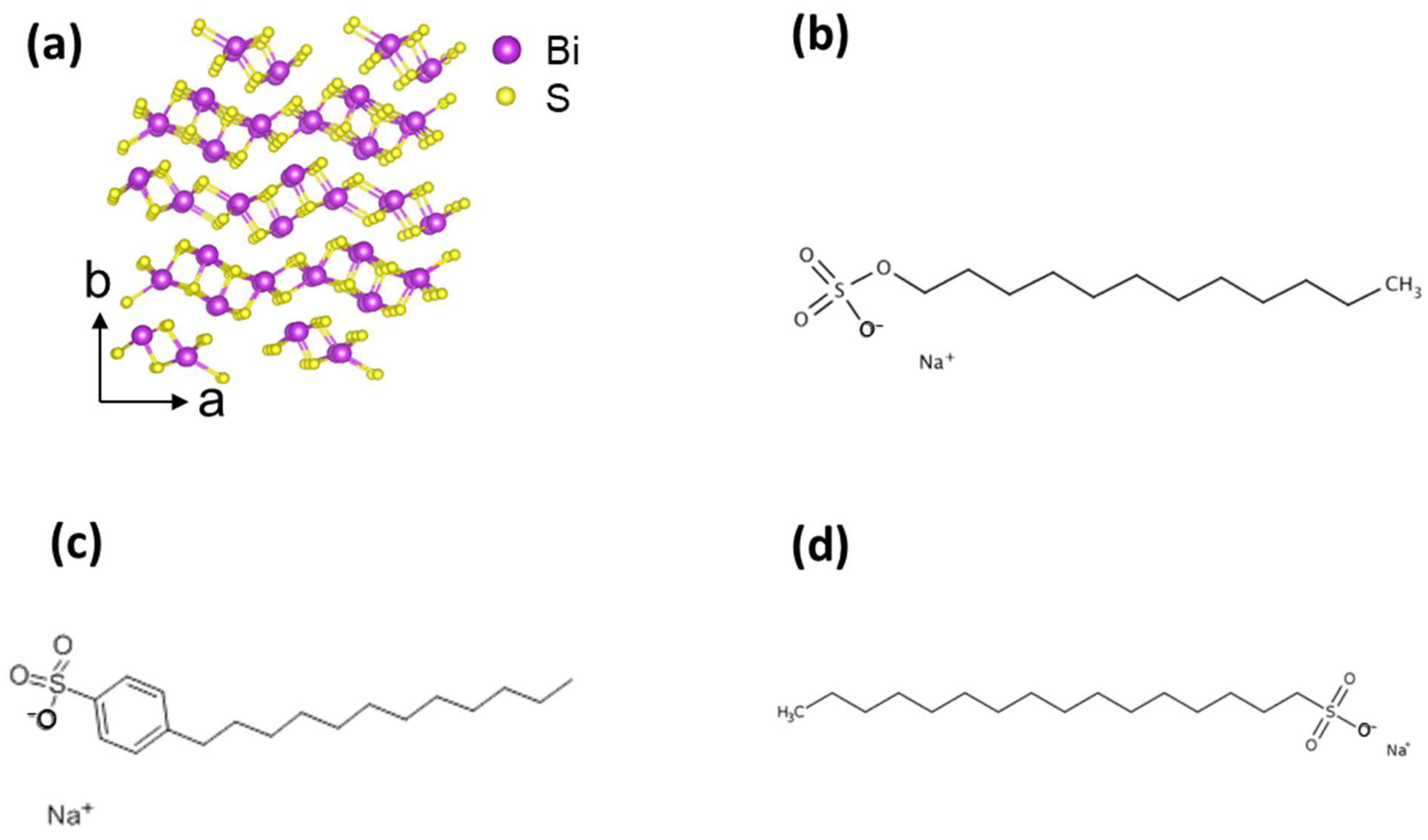
Figure 1. (a) Crystalline structure of Bi2S3 along the c-axis highlighting the vdW architecture and molecular structures of (b) SDS, (c) SDBS and (d) SHS.
Colloids 08 00028 g001
Figure 2. TEM image of exfoliated Bi2S3 with (a) SDS, (b) SDBS and (c) SHS. The scale bar in each panel is 200 nm. (d) SAED pattern and (e) HRTEM of typical Bi2S3 nanosheets. (f) is the FFT pattern of region (e).
Colloids 08 00028 g002
Figure 3. UV-vis absorption spectra and corresponding Tauc plots of exfoliated Bi2S3 nano-inks with (a,d) SDS, (b,e) SDBS and (c,f) SHS.
Colloids 08 00028 g003
Figure 4. Raman spectra of Bi2S3 (a) SDS, (b) SDBS and (c) SHS.
Colloids 08 00028 g004
Figure 5. Zeta potential (a) and DLS (b) trends for the exfoliated Bi2S3 nano-inks.
Colloids 08 00028 g005
Figure 6. Final concentration exfoliated Bi2S3 nano-inks.
Colloids 08 00028 g006
Figure 7. (a) UV-vis spectra and (b) Tauc Plot of Bi2S3 thin films produced from multiple ultrasonic spray-coating steps from the 2 mM SDBS nano-ink. Top-view SEM images of the Bi2S3 thin films with (c) 30 and (d) 50 sprayed layers.
Colloids 08 00028 g007
Table 1. Optical band gap values extrapolated from Tauc plots of all exfoliated Bi2S3 nano-inks.
SurfactantSurf. Concentration (mM)Band Gap (eV)
SDS8.22.03
4.11.74
2.02.01
1.01.95
0.51.85
SDBS8.22.13
4.12.11
2.02.10
1.01.99
0.51.97
SHS8.22.30
4.12.37
2.02.44
1.02.10
0.52.08
Table 2. Ag/B1g ratio of all Bi2S3 samples.
2D MaterialSurfactantSurf. Concentration (mM)Ag/B1g Ratio
Bi2S3 8.21.20
4.11.18
SDS2.01.15
1.01.07
0.51.32
Bi2S3 8.21.32
4.11.25
SDBS2.01.19
1.01.17
0.51.02
Bi2S3 8.21.30
4.11.12
SHS2.01.30
1.01.25
0.51.25
Table 3. Product concentration and yield for LPE Bi2S3 calculated from Lambert-Beer law and from weighted freeze-dried samples.
SurfactantSurfactant Concentration (mM)Product Concentration with
UV-Vis Absorption
(mg/mL)
Product Concentration with Freeze-Drying (mg/mL) Yield (%)
8.20.03 0.3
4.10.02 0.2
SDS2.00.02 0.2
1.00.02 0.2
0.50.01 0.1
8.20.040.040.4
4.10.04 0.4
SDBS2.00.02 0.2
1.00.04 0.4
0.50.01 0.1
8.20.140.081.4
4.10.110.441.1
SHS2.00.07 0.7
1.00.02 0.2
0.50.02 0.2
Disclaimer/Publisher’s Note: The statements, opinions and data contained in all publications are solely those of the individual author(s) and contributor(s) and not of MDPI and/or the editor(s). MDPI and/or the editor(s) disclaim responsibility for any injury to people or property resulting from any ideas, methods, instructions or products referred to in the content.