Article

Amplification of SERS Signal of Methotrexate Using Beta-Cyclodextrin Modified Silver Nanoparticles

by
Natalia E. Markina
,
Irina Yu. Goryacheva
and
Alexey V. Markin
*
Institute of Chemistry, Saratov State University, Astrakhanskaya Street 83, 410012 Saratov, Russia
*
Author to whom correspondence should be addressed.
Colloids Interfaces 2023, 7(2), 42; https://doi.org/10.3390/colloids7020042
Submission received: 12 March 2023 / Revised: 15 May 2023 / Accepted: 19 May 2023 / Published: 26 May 2023
(This article belongs to the Special Issue Biocolloids and Biointerfaces)

Abstract

:
The paper describes the use of native β-cyclodextrin (CD) for the modification of silver nanoparticles (AgNPs) in order to improve the determination of the anticancer drug methotrexate (MTX) using surface-enhanced Raman spectroscopy (SERS). A control experiment with unmodified AgNPs showed that the strong SERS signal of MTX can only be achieved in alkaline media. However, competitive interactions and the strong background signal of human body fluid components significantly challenge MTX determination in real samples. While previous reports propose the use of thorough sample pretreatment (e.g., solid phase extraction), the application of CD-modified AgNPs increases the SERS signal of MTX in neutral media by seven times which enables simplifying the analysis and improving its accuracy by reducing the influence of endogenous components of body fluids. A detailed study of the synthesis conditions (CD concentration and reaction time) and SERS registration conditions (pH, NaCl concentration, dilution of urine samples) was performed to maximize the analytical signal and signal-to-noise ratio. The final assay was tested for MTX determination in artificially spiked samples of real human urine. The results demonstrated that MTX can be determined within the concentration range suitable for therapeutic drug monitoring (20–300 μg mL−1) with satisfactory precision (6–15% RSD), accuracy (95–111% apparent recovery), and limit of detection (0.3 μg mL−1).

Graphical Abstract

1. Introduction

The effect of surface-enhanced Raman scattering (SERS) is observed for molecules adsorbed on the surface of metal nanostructures (SERS substrate) fabricated using silver, gold, or copper [1,2]. However, SERS as a phenomenon lacks selectivity, and a SERS-active surface can enhance the signal of any molecule located near it. This fact often complicates the use of SERS for accurate analysis of objects with complex compositions. For example, SERS-based analysis of human body fluids becomes very difficult if the analyte molecules possess weak (or even moderate) interaction with the SERS substrate surface compared to numerous components of body fluids, or if the analyte has an inherently weak intensity of SERS signal [3,4,5].
To enhance the sensitivity and selectivity of SERS-based analysis, modification of the SERS substrate surface is widely used to improve (1) the adsorption of the target analyte onto the SERS substrate, which leads to better SERS signal enhancement, and (2) the control over the adsorption of admixtures on the SERS substrate. For example, the substrate modification with monolayers of low molecular weight compounds, such as surfactants or amino acids [6,7], protects the surface from interaction with undesirable components (e.g., proteins of human body fluids), which can compete with target analyte molecules and block SERS-active sites. Additionally, SERS substrates can be modified by molecules that chemically react with the target analyte leading either to the formation of a product with an intense SERS signal [8,9] or to a decrease in the SERS signal of the initial modifier molecule (Raman marker) [10]. This approach increases the selectivity of the SERS analysis, but a limited number of reactions can be carried out on the surface of the SERS substrate, for example, the reaction conditions should not lead to the aggregation of colloidal SERS substrates or the loss of their SERS activity in the case of reactions requiring high temperatures.
Other reports have shown that the incorporation of metal nanoparticles (SERS-active elements) into a matrix of (in)organic sorbents enables (i) to concentrate the analyte by increasing the sorption capacity of the SERS substrate and (ii) to partially separate the target molecules from other components of the mixture [11,12,13]. Some papers have shown that the separation of an analyte from admixtures can also be based on differences in molecular charges and the removal of the admixtures with a charge opposite to the target analyte [12,14]. As can be seen from the examples listed above, the modification of the SERS substrate surface can have different efficiency to improve selectivity by working against certain parameters of the admixture molecules (e.g., size or charge). Better improvement of the selectivity can be achieved using molecular recognition approaches that take into account several molecular parameters such as size, geometry, polarity, and position of functional groups, e.g., coating the SERS substrate surface with molecularly imprinted polymers [15]. However, the best results are obtained by modifying the substrate with natural recognition molecules such as antibodies and aptamers which also led to the formation of a separate area of SERS analysis–label-based SERS [16].
Finally, another interesting direction is the use of recognition molecules which provide only a moderate improvement in the selectivity of SERS analysis due to the formation of inclusion complexes, e.g., cyclodextrins (CDs) [17]. In this case, the selectivity is improved due to the presence of a hydrophobic cavity inside the CD molecules. Therefore, the interaction between CD-modified SERS-active surface and analytes is possible only if the analyte molecules or their moieties fit a certain size range corresponding to the cavity size and have the proper polarity and spatial arrangement of functional groups. Additionally, the dimensions of the CD cavity are well-known and uniform (compared to pore size in (in)organic sorbents), which increases the reproducibility and precision of SERS measurements. A very important advantage of CDs as modifiers of SERS substrates is a very low contribution to the background signal due to the low Raman activity of CD molecules. Additionally, the use of CDs as recognition elements is a more cost-effective solution compared to other natural receptors and more environmentally friendly compared to molecularly imprinted polymers, the synthesis of which requires many reagents and solvents.
However, despite the increase in the number of articles, the results of our literature analysis show that, unfortunately, the number of works on the use of CDs in SERS analysis is in general extremely small [17]. In particular, there are very few reports on the use of such assays for the analysis of really complex objects such as food [18] and body fluids [19,20]. There are also not enough reports on the study of the effect of the conditions used during the synthesis of CD-modified SERS substrates. Therefore, in this report, we investigated the dependence of the analytical signal on the reaction time and CD concentration used to prepare the SERS substrate–silver nanoparticles modified with β-CD molecules (AgNP-CD). Native β-CD was chosen for our study because its molecules provide the best interaction with the MTX molecules compared to α- and γ-CD [21]. The effect of pH and NaCl concentration on the analytical SERS signal was also studied. Finally, AgNP-CD samples obtained using optimized conditions were tested for the determination of the analyte molecules in a complex mixture–artificially spiked samples of real human urine. In order to make the study more relevant for practical applications, the anticancer drug methotrexate (MTX) was used as the analyte of interest. Such drugs are subject to therapeutic drug monitoring in order to achieve the best therapeutic effect while minimizing side effects caused by the high toxicity of the drug. It was shown that proper selection of synthesis and signal registration conditions enabled the development of a simple SERS assay that is suitable for MTX determination in human urine at concentrations required for therapeutic drug monitoring.

2. Materials and Methods

2.1. Reagents and Equipment

Silver nitrate, hydroxylamine hydrochloride, and MTX were purchased from Sigma Aldrich. Powder of β-CD was purchased from Acros Organics. Sodium hydroxide, hydrochloric acid, and sodium chloride were purchased from Vekton Ltd. (Saint Petersburg, Russia). Double-distilled water was used for the preparation of silver nanoparticles (AgNPs); distilled water was used for the preparation of all other solutions.
SERS spectra were measured using a portable Raman setup (i-Raman, B&WTek, USA) equipped with a 532 nm laser (26 mW power at the sample) and connected to a microscope module equipped with objective (20×, 0.4 N.A.). Zetasizer Ultra (Malvern, UK) was used to perform dynamic light scattering (DLS) measurements and analyze the hydrodynamic diameter and surface charge (zeta potential) of AgNP-CD. Absorbance measurements in the UV–visible range were carried out using an SF-2000 spectrophotometer (OKB Spectr, Saint Petersburg, Russia). The nanoparticle solutions were diluted 10 times before DLS and absorbance measurements. The level of pH of the samples was measured using pH-indicator strips.

2.2. Urine Samples

Three healthy volunteers (two men and one woman) provided six urine samples, which were stored at 4 °C. Each volunteer provided two urine samples: (1) collected at the first urination in the morning after waking up, and (2) taken at the third urination during the day. Aqueous stock solutions of MTX (2–30 mg mL−1) were used for spiking of the urine samples: 10 μL of stock solution per 990 μL of the urine sample. Blank urine samples were used as reference samples. Urine samples (both blank and spiked) were diluted 1000 times before SERS measurements. The analysis of urine samples was approved by the Ethical Committee of the Saratov State Medical University (protocol No. 8, 2 March 2021).

2.3. Synthesis of SERS Substrates

The basic scheme of the synthesis of silver nanoparticles coated with β-cyclodextrin molecules (AgNP-CD) was taken from our previous report [20]. The synthesis optimized for the determination of MTX includes the following steps. First, 50 μL of NaOH (1 M) was mixed with 9.9 mL of an aqueous solution of β-CD (1 mM), and the mixture was heated to 60 °C with vigorous stirring. Then, 100 μL of AgNO3 (0.1 M) was added, and the mixture was kept at 60 °C for 3 h with stirring. Samples of final AgNP-CD were cooled and stored at room temperature in closed glass vials.
Five samples of AgNP-CD were prepared using various concentrations of CD molecules (0.1, 0.5, 1, 5, 10 mM) and used to study the effect of CD concentration on the SERS signal. The kinetics of AgNP-CD synthesis was investigated by taking 200 µL portions of a sample from the reaction mixture every 20 min and using these portions for UV-visible absorbance and SERS studies immediately.
AgNPs without CD coating were prepared according to the well-known protocol of Leopold and Lendl [22] and used as a control SERS substrate for comparison. The concentration of Ag ions in both synthesis protocols was fixed at 1 mM. Additionally, the pH value of all as-prepared samples of AgNPs was 6.5.

2.4. SERS Measurements

A pure aqueous solution of MTX (0.1 μg mL−1) and urine samples spiked with MTX (20 μg mL−1) were used to determine the optimal conditions for SERS signal registration and to optimize the final analysis protocol, respectively. The SERS measurements were performed using the mixture of MTX-containing solution (120 μL of pure solution or spiked urine), a portion of the SERS substrate solution (40 µL of AgNP-CD or control AgNPs), and an activating agent (10 μL of NaCl, 1 M). Solutions of NaOH or HCl were used as activating agents to study the effect of pH on the SERS signal of MTX. The mixture was placed in a disposable plastic cuvette (300 μL), and a set of 30 spectra was collected; 1 s was used to record a single SERS spectrum. SERS measurements were repeated three times for each sample in order to study the effect of the solution pH and the analyte concentration on the SERS signal, i.e., 90 spectra in total were collected for each sample. SERS measurements during the investigation of the kinetics of AgNP-CD synthesis were carried out by collecting one set of 20 spectra for each sample. Baseline-corrected intensities of SERS peaks of MTX at 1364, 1345 and 1600 cm−1 were used to analyze the signal changes in neutral, alkaline, and acidic media, respectively. The SERS peak at 1598 cm−1 was used in the final assay for the determination of MTX concentration in urine samples.

3. Results and Discussion

3.1. Effect of pH on SERS Signal of MTX

In order to achieve the best analytical performance of the analysis, a detailed study of various experimental conditions was carried out to determine the optimal ones for the preparation of AgNP-CD and registration of the analytical signal. The correction of pH is a good tool to control the SERS signal (both intensity and profile) which is critical for further control of analytical and background signals during analysis. Performing (de)protonation of analyte molecules by pH changing influences (i) spectral profiles and intensities by changing the molecular polarizability (i.e., Raman activity), (ii) the analyte adsorption on the SERS-active surface, and (iii) the mechanism of Raman enhancement (charge-transfer processes).
A preliminary study with a pure MTX solution demonstrated that the best intensity of the SERS signal was observed in an alkaline media for both AgNPs with and without surface modification with CD molecules (Figure 1a,d and Figure S1a,b). This result for control AgNPs is in agreement with those from previous studies of the SERS signal of MTX [13,23]. In the case of control AgNPs, we explain the positive effect of high pH values on the signal intensity by deprotonation of the carboxyl groups of the glutamic acid residue in MTX molecules (Figure 1c) as well as by protection of the nitrogen atoms of the 2,4-diaminopteridine part from protonation. As a result, free lone pairs of electrons of oxygen and nitrogen atoms of these moieties are able to form multiple strong donor-acceptor (coordination) bonds with silver atoms on the surface of AgNPs. In the case of AgNP-CD, the non-polar CD cavity poorly interacts with charged moieties of MTX such as deprotonated carboxyl or protonated amino groups. Therefore, the interaction between AgNP-CD and MTX in alkaline solutions should occur only through non- or low-polar parts, and 2,4-diaminopteridine is the most suitable one (Figure 1b). This part is also sterically favorable for such interaction due to its outermost position. The results of the IR study of Singh et al. [24] also showed that MTX molecules interact with the CD cavity through this group.
The use of acidic media leads to the protonation of the electron pairs of carboxyl and amino groups, the inhibition of the donor-acceptor interaction between MTX and the surface of control AgNPs, and, consequently, a reduction in the signal intensity. The protonation of several nitrogen atoms of 2,4-diaminopteridine also explains the deterioration of the MTX interaction with the CD-modified SERS-active surface and the very weak intensity of the MTX signal. Although protonation leads to a neutral charging of carboxyl groups of MTX, the inherently high polarity of these groups does not allow for a strong interaction between them and the non-polar CD cavity and makes them unsuitable for interaction with the surface of AgNP-CD.
Although the MTX signal is maximal in an alkaline medium, our previous results have shown that the MTX signal in body fluids is completely suppressed by the creatinine signal in this medium [13]. Additionally, according to previous results, a neutral medium is a more favorable one for preventing the effect of interferences in urine during SERS analysis. Unfortunately, MTX does not have a SERS signal in the pH range of 3–11 for both types of SERS substrates (Figure 1d and Figure S1a,b). We explain this fact by the association of MTX molecules analogously to the association processes reported by Khaled and Krumdieck [25] for the molecules of folic acid. Such an analogy is possible because MTX is an antimetabolite of folic acid, and these molecules have a very similar structure. This association leads to the formation of dimeric and polymeric complexes that leads to steric restriction of the interaction between the CD cavity and MTX associates. In the case of control AgNPs, the electron pairs intended for interaction with the SERS-active surface are involved in the formation of intermolecular bonds in MTX associates.
The addition of a standard neutral activating agent (NaCl) leads to the appearance of the MTX signal in the neutral medium (Figure S1c). Moreover, the use of AgNP-CD results in a significant improvement of the signal intensity compared to control AgNPs: a seven-fold increase at 60 mM of NaCl and pH 6.5 (Figure 1e). Analogously to the behavior of the folic acid associates in KCl solutions [25], we explain the appearance of MTX signal by the destruction of the MTX associates at an increased ionic strength that makes it possible for separated MTX molecules to interact with the surface of control AgNPs or AgNP-CD. Additional results regarding the influence of NaCl concentration on the MTX signal are detailed in Section 3.2.
MTX molecules in a neutral media are in the zwitterionic form [26] with deprotonated carboxyl groups and protonated 2,4-diaminopteridine. Thus, the interaction of MTX with control AgNPs is possible only through the electron pairs of carboxyl groups, and the MTX signal, in this case, is the weakest because this part of the MTX molecule has poor Raman activity. In contrast, it is known that the formation of inclusion complexes with CDs can change the ionization constants of molecules in solutions leading to easier deprotonation of amino groups [27]. Therefore, a stronger MTX signal in neutral media for AgNP-CD can be attributed to (i) the reduction in the protonation degree of 2,4-diaminopteridine due to complexation with the CD molecules and (ii) the location of the most Raman active part of the MTX molecule to a close proximity to the SERS-active surface. These explanations are in excellent agreement with the results of the theoretical studies of Castillo et al. [28]. The calculations also showed that the peaks of the SERS spectra of MTX are attributed to 2,4-diaminopteridine, and the closer this part is to the SERS substrate, the stronger the signal. Changes in the protonation degree can also explain the large changes in the spectral profiles observed for the MTX molecules in solutions with different pH (Figure 1a and Figure S1). For example, we suppose that the changes in the maximum of the large peak in the range of 1300–1400 cm−1 are caused by different contributions of the protonated and neutral forms of 2,4-diaminopteridine to the final SERS spectrum.
Therefore, in order to increase the accuracy of the final analysis procedure, a neutral medium with activation of the SERS signal by the addition of NaCl was subsequently used for recording the SERS signal of MTX during the optimization of AgNP-CD synthesis and the development of the analysis protocol. Moreover, additional control experiments also showed that the simple addition of CD molecules to the control AgNPs does not affect the intensity of the MTX signal at any pH and NaCl concentration. Thus, we can state that the modification of the AgNP surface with CD molecules directly during the synthesis of AgNPs (in situ modification) is a required process for obtaining AgNP-CD and achieving the influence of CD molecules on the SERS signal.

3.2. Study of AgNP-CD Synthesis and SERS Measurement Conditions

Because CD molecules serve for the reduction and stabilization of AgNPs, their concentration will affect the speed and completeness of the synthesis and determine the colloidal stability of AgNP-CD. Moreover, it strongly influences the interaction between the final AgNP-CD and analyte molecules and, consequently, will determine the efficiency of the final SERS analysis. Additionally, previously proposed procedures for the AgNP-CD synthesis describe significantly different times of the synthesis (from 0.5 to 3 h [20,29,30]) without detailed explanations. Therefore, studies of the dependence of the SERS signal on the CD concentration and the reaction time used for AgNP-CD synthesis are important steps for preparing an effective SERS substrate. Measurements of the SERS signal of MTX were used to study the effect of these factors on the enhancement properties of AgNP-CD (Figure 2a and Figure S2). Changes in the surface plasmon resonance (SPR) of AgNP-CD during the synthesis were also measured by recording absorbance spectra in the UV–visible range (Figure 2b and Figure S3). The DLS method was used to study the effect of CD concentration on the hydrodynamic diameter (HDD) and surface charge (zeta potential) of AgNP-CD because this method can provide reliable results for native AgNP-CD in an environment used for SERS analysis (i.e., in aqueous solutions).
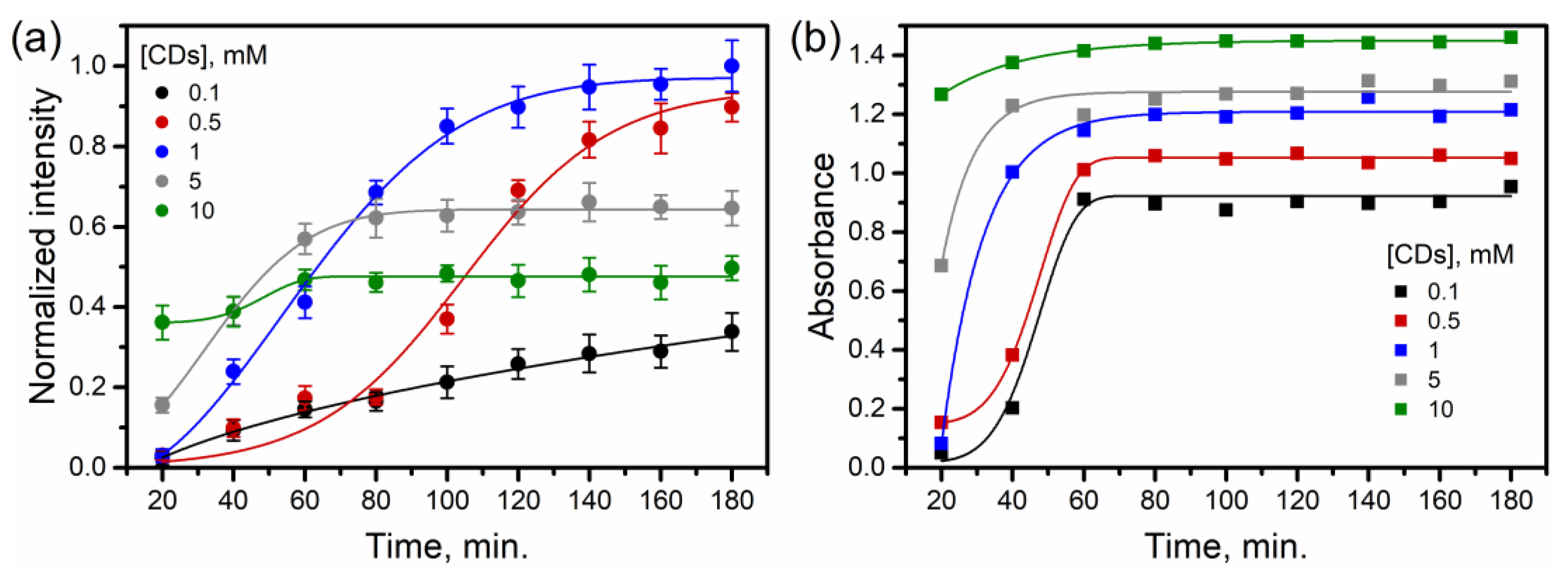
First of all, the study of the reaction kinetics shows a trivial effect: the reaction rate (estimated as a change in the absorbance and SERS intensity) is significantly increased with the growth in CD concentration (Figure 2). This is clearly seen from the signal intensities at 20 min of the reaction: the use of 10 mM of CD results in the fastest signal growth. Interestingly, the absorbance of AgNP-CD stops changing one hour after the start of the synthesis, regardless of the used CD concentration (Figure 2b). On the other hand, the growth in the SERS signal intensity strongly depends on the used CD concentration: an increase in the concentration speeds up the growth of the SERS activity and shortens the time when the intensity reaches its maximum (Figure 2a). This result is important because it clearly demonstrates the need for mandatory use of the SERS signal during the synthesis optimization instead of absorbance. Therefore, a longer time of AgNP-CD synthesis is preferable in order to ensure complete synthesis (180 min for the described system). However, at the lowest CD concentration (0.1 mM) the intensity of the SERS signal did not reach its maximum even after 180 min of the reaction.
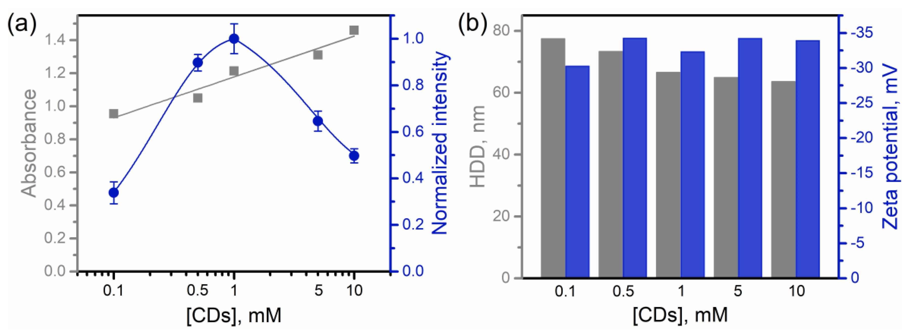
Analysis of the effect of CD concentration at fixed reaction time (180 min) on the Raman enhancement and SPR intensity shows a monotonic increase in the intensity of the SPR peak of AgNP-CD (Figure 3a). However, the SERS activity of AgNP-CD is non-linearly dependent on the CD concentration having a maximum of 1 mM of CDs. The low level of SERS activity at low CD concentrations (≤0.1 mM) we mainly associate with poor coverage of the AgNP surface with CD molecules. Although lower absorbance values can also indicate an incomplete reduction of AgNPs at low CD concentrations, the difference in the intensity of SERS signals is much larger compared to the difference in the concentration of the SERS substrate nanoparticles (according to absorbance measurements). Therefore, we assume that the reduction in AgNPs can proceed effectively at low CD concentrations, but non-reacted CD molecules are not enough to cover the surface of AgNPs, providing adsorption sites for MTX molecules.
On the other hand, the use of CD concentrations above 1 mM also leads to a decrease in the SERS signal. We explain this signal reduction by competition between CD molecules adsorbed on the SERS-active surface and CD molecules dissolved in the bulk solution for interaction with MTX molecules. Therefore, an excess of CDs leads to the formation of the MTX-CD inclusion complex in solution preventing MTX interaction with the CD-modified SERS substrate. The signal reduction at high CD concentrations can be additionally explained by the multilayer adsorption of CD on AgNP. Indeed, numerous hydroxyl groups facilitate the association of CD molecules via intramolecular hydrogen bonds [31]. Consequently, the formation of CD agglomerates on the AgNP surface can prevent the access of MTX molecules to the SERS-active sites. Quite similar non-linear (with a maximum) influence of a stabilizer on the SERS signal intensity was also observed in one of our previous studies, where an excess of sodium citrate, used to prepare SERS substrate, caused almost 40% decrease in the intensity of analyte signal [32].
Importantly, the presence of CD molecules and changes in their concentration does not affect the profile of analytical and background signals (Figure S4). The spectral changes observed for AgNP-CD samples prepared with 0.5 and 10 mM of CDs (Figure S4f) are associated with the poor signal intensity of the analyte. Summing up and comparing the results of SPR and Raman enhancement measurements, we can state that they do not correlate with each other. Thus, SPR results on their own cannot be used to predict and improve SERS activity. This fact suggests that the electromagnetic mechanism of Raman enhancement does not work reliably and should be used very carefully in the case of systems such as those described in this report.
According to the literature analysis [17] and our previous results [20], CD molecules promote the formation of spherical AgNPs. In this study, we additionally observed that AgNP-CD has only one SPR peak and its position is independent of CD concentration (Figure S3f). Thus, we conclude that the shape of AgNPs also does not depend on changes in the CD concentration. However, the study of the effect of CD concentration at fixed reaction time (180 min) on HDD of AgNP-CD shows that an increase in CD concentration leads to a moderate decrease in the size of AgNP-CD from ~80 nm to ~65 nm (Figure 3b). We associate this result with a faster synthesis reaction and better stabilization of AgNPs by an excess of CD molecules. On the other hand, measurements of zeta potential demonstrated that the surface charge is independent of the CD concentration and has negative values.
As shown in Section 3.1 and Figure 1e, the addition of a neutral activating agent (NaCl) is the mandatory condition to obtain the SERS signal of MTX using AgNP-CD in a neutral medium. A more detailed study of the effect of NaCl concentration on the MTX signal also showed that it has a non-linear influence with a maximum SERS signal enhancement at 60 mM (Figure 4a). We associate the observed increase in SERS intensity with the triggering of artificial aggregation of AgNP-CD and the formation of agglomerates with additionally enhanced SERS activity [2]. A DLS study of the NaCl effect on HDD of AgNP-CD revealed the formation of aggregates at 60 mM of NaCl, which are ~50% larger compared to the particles of native AgNP-CD samples (Figure 4b). Moreover, the aggregation of AgNP-CD also leads to changes in their extinction spectra, so that the used excitation wavelength (532 nm laser) starts to be absorbed by the SERS substrate more efficiently compared to non-aggregated AgNP-CD (Figure S5). Interestingly, absorbance spectra change strongly upon addition of NaCl up to 60 mM, and the light absorption at 532 nm at this concentration reaches a maximum. However, a further increase in the NaCl concentration above 60 mM leads to a decrease in the SERS signal intensity (while light absorption remains unchanged), which we associate with an increased competition between MTX and chloride ions. A negative effect of alkali metal chlorides (NaCl and KCl) on the stability of inclusion complexes between CD and molecular dyes was also reported previously [33,34].
According to DLS, an increase in CD concentration improves the colloidal stability of AgNP-CD making them less sensitive to the growth of NaCl concentration (Figure 4b). Thus, considering the positive effect of aggregation on the signal intensity, the highest intensity was expected for AgNP-CD prepared with 0.1 mM of CDs because they form the largest aggregates. However, insufficient coverage of the AgNP surface with CD molecules leads only to a moderate signal enhancement with increasing NaCl concentration (see discussions for Figure 2 and Figure 3a). On the other hand, the improvement of the colloidal stability at high CD concentrations (5 and 10 mM) is actually not as big as the signal reduction at these CD concentrations (compared to the optimal CD concentration). Therefore, the improvement of colloidal stability should play a minor role in the intensity reduction compared to (1) the competition between CD molecules on the AgNP surface and CD molecules dissolved in the bulk solution for interaction with MTX molecules, and (2) multilayer adsorption of CD molecules on the SERS-active surface.

3.3. Determination of Methotrexate in Urine Samples Using AgNP-CD

Based on the results of the studies in this report, a SERS-based assay for the determination of MTX in human urine using AgNP-CD was developed. Three healthy volunteers provided urine samples (two portions from each volunteer): (1) collected at the first urination in the morning after waking up, and (2) taken at the third urination during the day. All six urine samples were artificially spiked with MTX molecules and used to obtain a calibration plot and calculate figures of merit. Because urine collected in the morning has a quite high level of complexity, these samples were used to pre-optimize the analysis procedure in order to maximize the accuracy and precision of the final assay. Lastly, AgNP-CD samples prepared using 1 mM of CD and 3 h of the reaction time and the neutral activating agent (NaCl, 60 mM in measured solution) were used in the analysis since these conditions provide the maximum signal intensity.

3.3.1. Effect of Sample Dilution on Background Signal

Dilution of urine samples was used as a simple sample pretreatment in order to additionally facilitate analysis by reducing the concentration of endogenous body fluid components (i.e., interferences). For example, as shown above for the activating agent, the negative effect of excessive concentrations of NaCl in body fluids can uncontrollably deteriorate the analysis. Because the use of AgNP-CD enables to obtain an analytical signal in neutral media, pure water was used for the urine dilution; since control AgNPs do not give an intense MTX signal at this condition (Figure 1d,e) they were not used for comparison. The study of the dilution effect on the signal of MTX-spiked urine showed that the analyte signal intensity increases at dilution rates up to 750 times leading to suppression of the background signal (Figure 5a). We attribute this increase to a reduction in competitive interactions between urine components and analyte molecules for SERS-active sites. However, further dilution (≥1000) logically leads to a reduction in the MTX signal due to too strong a decrease in the MTX concentration. On the other hand, the background signal monotonically decreases with increasing the dilution rate. Despite a remarkable reduction in the intensity of the background signal after dilution, the results also showed that the most intense peak of MTX (1364 cm−1) is the most sensitive to the presence of interferences. Therefore, a 1000-fold dilution and SERS peak of MTX at 1598 cm−1 were chosen for the final analysis procedure.
Because MTX has the best intensity of SERS signal in alkaline media (Figure 1d), additional control measurements of the MTX-spiked urine were performed with control AgNPs and AgNP-CD in alkaline solutions (Figure S6). The results showed that both control AgNP and AgNP-CD do not allow detecting MTX in real samples in alkaline media due to the very strong interference effect of endogenous urine components (mainly creatinine [13]). Importantly, the interfering effect is so strong that the sample dilution does not improve the signal even in the case of AgNP-CD.

3.3.2. Analytical Performance

The calibration plot (Figure 5b) was obtained by averaging the results for six urine samples, regardless of the time of urine collection. Remarkably, the signal has a linear dependence on concentration, while non-linear calibration plots were usually reported for SERS assays of MTX [13,23,35]. We associate this effect namely with the modification of the SERS substrate with CD molecules that leads to a more homogeneous interaction between the CD-modified surface and the analyte molecules [20]. The analysis of figures of merit shows that the final assay has good analytical performance and is suitable for application in therapeutic drug monitoring [36]: linear range 20–300 μg mL−1, RSD 6–15%, apparent recovery 95–111%, and LOD 0.3 μg mL−1 (according to the 3σ/S approach recommended by IUPAC). Additionally, important advantages of CD-modified AgNPs for analytical applications are the synthesis reproducibility and the long-term stability of Raman enhancement of the samples (>1.5 months), which have been proven previously [20,30].

3.3.3. Comparison with Other Reports

In contrast to previous analysis protocols proposed for SERS-based determination of MTX in urine, the new one enables MTX determination without complicated sample pretreatment, requiring only sample dilution with pure water. For example, Subaihi et al. [35] used HPLC separation before the identification of MTX and its metabolite in urine using SERS. Although the protocol has excellent accuracy and precision, the use of HPLC requires labor and time-consuming sample treatment, including protein removal with organic solvents and a two-fold concentration of urine by evaporation. In another report, we developed a SERS-active sorbent based on AgNPs embedded into a porous alumina matrix [13] and used it for MTX determination in urine. The application of the SERS substrate with a porous matrix makes it easy to separate, purify, and concentrate MTX molecules from a complex mixture by performing solid-phase extraction. However, the need for centrifugation to separate the sorbent from the sample and the need to further dissolve the alumina matrix to release the trapped MTX molecules require additional time and reduce the analysis precision (RSD is 11–19% vs. 6–15% for current assay).
Regarding the use of CD-modified SERS substrates for biomedical analysis, only two reports are currently available. Cao et al. [19] used artificial urine and a commercial SERS substrate modified with a thiol derivative of β-CD in order to develop an assay for the detection of acetyl amantadine, which is an exogenous cancer biomarker. Unfortunately, a critical evaluation of the results by the authors showed the presence of a strong interfering effect of some metabolites (creatinine and corticosterone) that does not allow the use of the assay for biomedical analysis. More positive results were obtained when AgNP-CD was used to improve SERS-based determination of fluoroquinolone antibiotics in human body fluids [20]. The authors developed an assay suitable for the analyte-class-specific analysis and showed that it allows the determination of four widely used and structurally related fluoroquinolones in urine and blood plasma samples. However, the treatment of samples with an organic solvent was identified as a mandatory preliminary step to remove endogenous components of body fluids. Therefore, the assay proposed in this report demonstrates competitive results with good analytical performance combined with very simple sample pretreatment.

4. Conclusions

Thus, this paper reports physicochemical studies dedicated to the investigation of the conditions used for the preparation of the CD-modified SERS substrate, which influences the efficiency of the analysis. Besides others, the most important finding is the poor correlation between plasmonic and Raman enhancement properties for AgNP-CD prepared using various concentrations of CD. This result clearly demonstrates the need for very careful application of the electromagnetic mechanism of Raman enhancement for evaluation of the enhancement capability of SERS substrates in general. Additionally, it shows that the optimization of SERS substrate preparation should be conducted using only the SERS signal and not absorbance measurements. The next important result is the negative influence of excess concentrations of activating agent (NaCl) on the analytical signal which is important to account for the objects with high content of this compound (e.g., urine). Finally, AgNP-CD were used to analyze real samples of urine and the proposed assay demonstrates competitive results. The results can be extrapolated to the analysis of other analytes with a good affinity for CD molecules in urine or other objects of complex composition.

Supplementary Materials

The following supporting information can be downloaded at: https://www.mdpi.com/article/10.3390/colloids7020042/s1. Figure S1: Influence of pH in acidic and alkaline media and NaCl concentration in a neutral medium on the SERS spectra of MTX obtained using AgNPs without and with modification with CD molecules; Figure S2: Dependence of the SERS signal of MTX on the reaction time and CD concentration used for the synthesis of AgNP-CD; Figure S3: Dependence of the absorbance spectra of AgNP-CD on the reaction time and CD concentration used for the synthesis of AgNP-CD; Figure S4: Dependence of the SERS signal of MTX on CD concentration used for the synthesis of AgNP-CD and the concentration of NaCl used to increase the intensity of the SERS signal; Figure S5: Influence of the NaCl concentration used to increase intensity of the SERS signal of MTX on the extinction spectra of AgNP-CD and extinction of AgNP-CD at fixed wavelengths; Figure S6: Effect of the degree of sample dilution on the SERS spectra of morning urine without (blank) and with (spiked) the addition of MTX; Figure S7: Averaged SERS spectra of urine samples spiked with MTX and diluted 1000 times.

Author Contributions

Conceptualization, N.E.M.; methodology, A.V.M.; validation, N.E.M.; investigation, N.E.M.; resources, I.Y.G.; writing—original draft preparation, A.V.M.; writing—review and editing, I.Y.G. and A.V.M.; visualization, N.E.M.; supervision, I.Y.G. and A.V.M.; funding acquisition, N.E.M. All authors have read and agreed to the published version of the manuscript.

Funding

The work was supported by Russian Science Foundation (project 21-73-00098).

Institutional Review Board Statement

The study was conducted in accordance with the Declaration of Helsinki, and approved by the Institutional Ethics Committee of the Saratov State Medical University (protocol No. 8, 2 March 2021).

Informed Consent Statement

Informed consent was obtained from all volunteers involved in the study.

Data Availability Statement

Data will be made available on reasonable request.

Conflicts of Interest

The authors declare no conflict of interest.

References

  1. Markin, A.V.; Markina, N.E.; Popp, J.; Cialla-May, D. Copper nanostructures for chemical analysis using surface-enhanced Raman spectroscopy. Trends Anal. Chem. 2018, 108, 247–259. [Google Scholar] [CrossRef]
  2. Zong, C.; Xu, M.; Xu, L.J.; Wei, T.; Ma, X.; Zheng, X.S.; Hu, R.; Ren, B. Surface-enhanced Raman spectroscopy for bioanalysis: Reliability and challenges. Chem. Rev. 2018, 118, 4946–4980. [Google Scholar] [CrossRef] [PubMed]
  3. Bonifacio, A.; Cervo, S.; Sergo, V. Label-free surface-enhanced Raman spectroscopy of biofluids: Fundamental aspects and diagnostic applications. Anal. Bioanal. Chem. 2015, 407, 8265–8277. [Google Scholar] [CrossRef]
  4. Jaworska, A.; Fornasaro, S.; Sergo, V.; Bonifacio, A. Potential of surface enhanced Raman spectroscopy (SERS) in therapeutic drug monitoring (TDM). A critical review. Biosensors 2016, 6, 47. [Google Scholar] [CrossRef]
  5. Markina, N.E.; Goryacheva, I.Y.; Markin, A.V. Surface-enhanced Raman spectroscopy for the determination of medical and narcotic drugs in human biofluids. J. Anal. Chem. 2022, 77, 930–947. [Google Scholar] [CrossRef]
  6. Sun, F.; Hung, H.C.; Sinclair, A.; Zhang, P.; Bai, T.; Galvan, D.D.; Jain, P.; Li, B.; Jiang, S.; Yu, Q. Hierarchical zwitterionic modification of a SERS substrate enables real-time drug monitoring in blood plasma. Nat. Commun. 2016, 7, 13437. [Google Scholar] [CrossRef]
  7. Panikar, S.S.; Ramírez-García, G.; Sidhik, S.; Lopez-Luke, T.; Rodriguez-Gonzalez, C.; Ciapara, I.H.; Castillo, P.S.; Camacho-Villegas, T.; De la Rosa, E. Ultrasensitive SERS substrate for label-free therapeutic-drug monitoring of paclitaxel and cyclophosphamide in blood serum. Anal. Chem. 2019, 91, 2100–2111. [Google Scholar] [CrossRef] [PubMed]
  8. Litti, L.; Ramundo, A.; Biscaglia, F.; Toffoli, G.; Gobbo, M.; Meneghetti, M. A surface enhanced Raman scattering based colloid nanosensor for developing therapeutic drug monitoring. J. Colloid Interface Sci. 2019, 533, 621–626. [Google Scholar] [CrossRef]
  9. Zhang, Y.; Li, L.; Gao, Y.; Wang, X.; Sun, L.; Ji, W.; Ozaki, Y. Nitrosonaphthol reaction-assisted SERS assay for selective determination of 5-hydroxyindole-3-acetic acid in human urine. Anal. Chim. Acta 2020, 1134, 34–40. [Google Scholar] [CrossRef]
  10. Yang, H.; Xiang, Y.; Guo, X.; Wu, Y.; Wen, Y.; Yang, H. Diazo-reaction-based SERS substrates for detection of nitrite in saliva. Sens. Actuators B Chem. 2018, 271, 118–121. [Google Scholar] [CrossRef]
  11. Farquharson, S.; Gift, A.; Shende, C.; Inscore, F.; Ordway, B.; Farquharson, C.; Murren, J. Surface-enhanced Raman spectral measurements of 5-fluorouracil in saliva. Molecules 2008, 13, 2608–2627. [Google Scholar] [CrossRef] [PubMed]
  12. Yue, S.; Sun, X.T.; Wang, Y.; Zhang, W.S.; Xu, Z.R. Microparticles with size/charge selectivity and pH response for SERS monitoring of 6-thioguanine in blood serum. Sens. Actuators B Chem. 2018, 273, 1539–1547. [Google Scholar] [CrossRef]
  13. Markina, N.E.; Zakharevich, A.M.; Markin, A.V. Determination of methotrexate in spiked human urine using SERS-active sorbent. Anal. Bioanal. Chem. 2020, 412, 7757–7766. [Google Scholar] [CrossRef] [PubMed]
  14. Markina, N.E.; Markin, A.V.; Zakharevich, A.M.; Goryacheva, I.Y. Calcium carbonate microparticles with embedded silver and magnetite nanoparticles as new SERS-active sorbent for solid phase extraction. Microchim. Acta 2017, 184, 3937–3944. [Google Scholar] [CrossRef]
  15. Ma, J.; Yan, M.; Feng, G.; Ying, Y.; Chen, G.; Shao, Y.; She, Y.; Wang, M.; Sun, J.; Zheng, L.; et al. An overview on molecular imprinted polymers combined with surface-enhanced Raman spectroscopy chemical sensors toward analytical applications. Talanta 2021, 225, 122031. [Google Scholar] [CrossRef] [PubMed]
  16. Wang, Z.; Zong, S.; Wu, L.; Zhu, D.; Cui, Y. SERS-activated platforms for immunoassay: Probes, encoding methods, and applications. Chem. Rev. 2017, 117, 7910–7963. [Google Scholar] [CrossRef]
  17. Markina, N.E.; Cialla-May, D.; Markin, A.V. Cyclodextrin-assisted surface-enhanced Raman spectroscopy: A critical review. Anal. Bioanal. Chem. 2022, 414, 923–942. [Google Scholar] [CrossRef] [PubMed]
  18. Ma, P.; Liang, F.; Sun, Y.; Jin, Y.; Chen, Y.; Wang, X.; Zhang, H.; Gao, D.; Song, D. Rapid determination of melamine in milk and milk powder by surface-enhanced Raman spectroscopy and using cyclodextrin-decorated silver nanoparticles. Microchim. Acta 2013, 180, 1173–1180. [Google Scholar] [CrossRef]
  19. Cao, G.; Hajisalem, G.; Li, W.; Hof, F.; Gordon, R. Quantification of an exogenous cancer biomarker in urinalysis by Raman spectroscopy. Analyst 2014, 139, 5375–5378. [Google Scholar] [CrossRef]
  20. Markina, N.E.; Markin, A.V.; Cialla-May, D. Cyclodextrin-assisted SERS determination of fluoroquinolone antibiotics in urine and blood plasma. Talanta 2023, 254, 124083. [Google Scholar] [CrossRef]
  21. Kritskiy, I.; Kumeev, R.; Volkova, T.; Shipilov, D.; Kutyasheva, N.; Grachev, M.; Terekhova, I. Selective binding of methotrexate to monomeric, dimeric and polymeric cyclodextrins. New J. Chem. 2018, 42, 14559–14567. [Google Scholar] [CrossRef]
  22. Leopold, N.; Lendl, B. A new method for fast preparation of highly surface-enhanced Raman scattering (SERS) active silver colloids at room temperature by reduction of silver nitrate with hydroxylamine hydrochloride. J. Phys. Chem. B 2003, 107, 5723–5727. [Google Scholar] [CrossRef]
  23. Hidi, I.J.; Mühlig, A.; Jahn, M.; Liebold, F.; Cialla, D.; Weber, K.; Popp, J. LOC-SERS: Towards point-of-care diagnostic of methotrexate. Anal. Methods 2014, 6, 3943–3947. [Google Scholar] [CrossRef]
  24. Singh, U.V.; Aithal, K.S.; Udupa, N. Physicochemical and biological studies of inclusion complex of methotrexate with β-cyclodextrin. Pharm. Sci. 1997, 3, 573–577. [Google Scholar]
  25. Abu Khaled, M.; Krumdieck, C.L. Association of folate molecules as determined by proton NMR: Implications on enzyme binding. Biochem. Biophys. Res. Commun. 1985, 130, 1273–1280. [Google Scholar] [CrossRef]
  26. Poe, M. Acidic dissociation constants of folic acid, dihydrofolic acid, and methotrexate. J. Biol. Chem. 1977, 252, 3724–3728. [Google Scholar] [CrossRef] [PubMed]
  27. Jelić, R.; Tomović, M.; Stojanović, S.; Joksović, L.; Jakovljević, I.; Djurdjević, P. Study of inclusion complex of β-cyclodextrin and levofloxacin and its effect on the solution equilibria between gadolinium(III) ion and levofloxacin. Monatsh. Chem. 2015, 146, 1621–1630. [Google Scholar] [CrossRef]
  28. Castillo, J.J.; Rindzevicius, T.; Wu, K.; Rozo, C.E.; Schmidt, M.S.; Boisen, A. Silver-capped silicon nanopillar platforms for adsorption studies of folic acid using surface enhanced Raman spectroscopy and density functional theory. J. Raman Spectrosc. 2015, 46, 1087–1094. [Google Scholar] [CrossRef]
  29. Ouyang, L.; Zhu, L.; Ruan, Y.; Tang, H. Preparation of a native β-cyclodextrin modified plasmonic hydrogel substrate and its use as a surface-enhanced Raman scattering scaffold for antibiotics identification. J. Mater. Chem. C 2015, 3, 7575–7582. [Google Scholar] [CrossRef]
  30. Yang, L.; Chen, Y.; Li, H.; Luo, L.; Zhao, Y.; Zhang, H.; Tian, Y. Application of silver nanoparticles decorated with β-cyclodextrin in determination of 6-mercaptopurine by surface-enhanced Raman spectroscopy. Anal. Methods 2015, 7, 6520–6527. [Google Scholar] [CrossRef]
  31. Loftsson, T.; Másson, M.; Brewster, M.E. Self-association of cyclodextrins and cyclodextrin complexes. J. Pharm. Sci. 2004, 93, 1091–1099. [Google Scholar] [CrossRef] [PubMed]
  32. Markina, N.E.; Ustinov, S.N.; Zakharevich, A.M.; Markin, A.V. Copper nanoparticles for SERS-based determination of some cephalosporin antibiotics in spiked human urine. Anal. Chim. Acta 2020, 1138, 9–17. [Google Scholar] [CrossRef] [PubMed]
  33. Mochida, K.; Kagita, A.; Matsui, Y.; Date, Y. Effects of inorganic salts on the dissociation of a complex of β-cyclodextrin with an azo dye in an aqueous solution. Bull. Chem. Soc. JPN 1973, 46, 3703–3707. [Google Scholar] [CrossRef]
  34. Buvári, Á.; Barcza, L. Complex formation of inorganic salts with β-cyclodextrin. J. Incl. Phenom. Macrocycl. Chem. 1989, 7, 379–389. [Google Scholar] [CrossRef]
  35. Subaihi, A.; Trivedi, D.K.; Hollywood, K.A.; Bluett, J.; Xu, Y.; Muhamadali, H.; Ellis, D.I.; Goodacre, R. Quantitative online liquid chromatography–surface-enhanced Raman scattering (LC-SERS) of methotrexate and its major metabolites. Anal. Chem. 2017, 89, 6702–6709. [Google Scholar] [CrossRef] [PubMed]
  36. Bratlid, D.; Moe, P.J. Pharmacokinetics of high-dose methotrexate treatment in children. Eur. J. Clin. Pharmacol. 1978, 14, 143–147. [Google Scholar] [CrossRef]
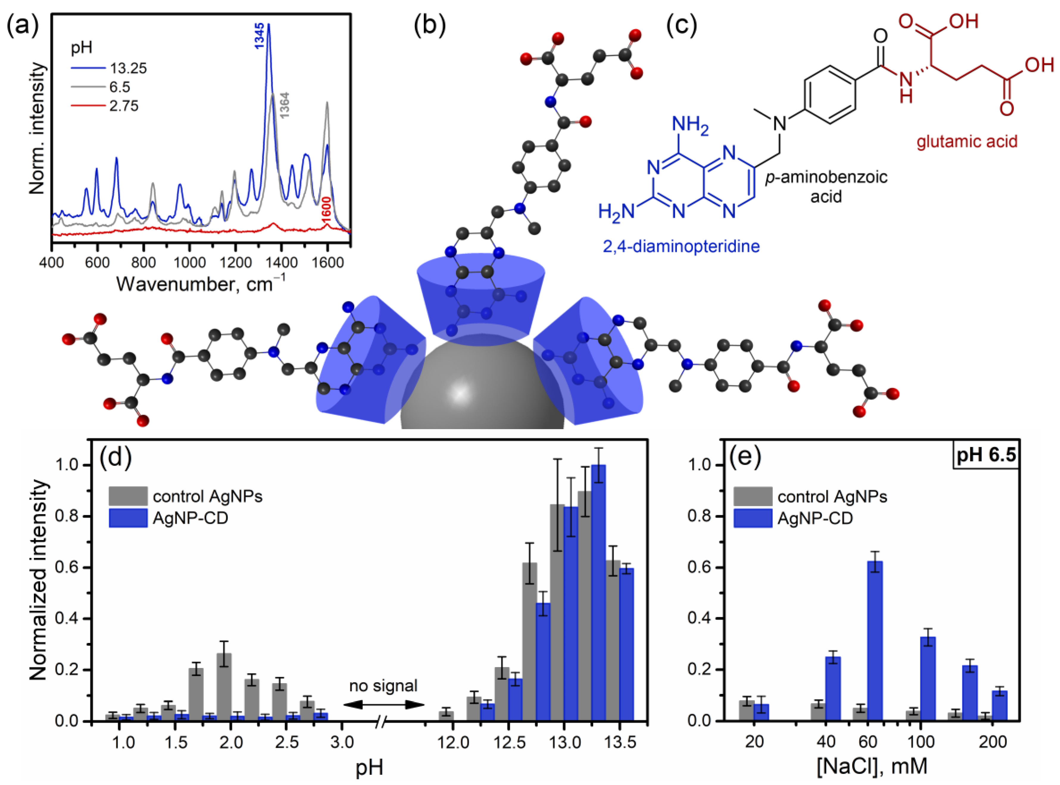
Figure 1. (a) Examples of SERS spectra of methotrexate (MTX) obtained using silver nanoparticles coated with CD molecules (AgNP-CD) and measured in media with different pH values. (b) Schematic illustration of the complex between AgNP-CD and MTX molecules. (c) Chemical structure of MTX molecule with highlighted main constituents. (d,e) Effect of pH (d) and concentration of NaCl (at 6.5 pH) (e) on the intensity of SERS signal of MTX (0.1 μg mL−1) obtained using AgNP-CD and AgNPs without CD modification (control AgNPs). Baseline-corrected intensities of SERS peaks at 1364, 1345 and 1600 cm−1 were used to analyze the signal changes in neutral, alkaline, and acidic media, respectively. The signal intensity at pH 13.25 was used to normalize signal intensities obtained at other pH values. Full spectra are shown in Supplementary Material (Figure S1).
Colloids 07 00042 g001
Figure 2. Effect of CD concentration used for the synthesis of AgNP-CD on the reaction kinetics according to (a) intensity of SERS signal of MTX and (b) absorbance measurements (peak of AgNPs at 410 nm). Conditions used to obtain the SERS signal: peak at 1364 cm−1, 0.25 μg mL−1 of MTX, 60 mM of NaCl, pH 6.5. Full SERS and absorbance spectra are shown in Figures S2 and S3, respectively.
Colloids 07 00042 g002
Figure 3. (a) Effect of CD concentration used for the synthesis of AgNP-CD on intensity of SERS signal of MTX and absorbance peak of AgNP-CD. The plots are based on the results for the substrates synthesized during 180 min and shown in Figure 2. (b) Effect of CD concentration used for the synthesis of AgNP-CD on hydrodynamic diameter (HDD) and zeta potential of AgNP-CD synthesized during 180 min.
Colloids 07 00042 g003
Figure 4. Influence of the CD concentration used for synthesis of AgNP-CD and the concentration of NaCl used for additional enhancement of SERS signal on (a) the signal intensity of MTX (0.1 μg mL−1, pH 6.5, peak at 1364 cm−1) and (b) HDD of AgNP-CD. AgNP-CD samples were prepared using 180 min reaction time. Full spectra are shown in Figure S4.
Colloids 07 00042 g004
Figure 5. (a) Effect of the degree of sample dilution on the SERS spectra of morning urine without (blank) and with (spiked) addition of MTX (20 μg mL−1). The spectra were obtained using CD-modified AgNPs with the addition of an activating agent (60 mM of NaCl). (b) Calibration plot for MTX (peak at 1598 cm−1) in spiked urine (6 samples) obtained using the assay with optimized measurement conditions. Full spectra are shown in Figure S7.
Colloids 07 00042 g005
Disclaimer/Publisher’s Note: The statements, opinions and data contained in all publications are solely those of the individual author(s) and contributor(s) and not of MDPI and/or the editor(s). MDPI and/or the editor(s) disclaim responsibility for any injury to people or property resulting from any ideas, methods, instructions or products referred to in the content.