Article

Automated Single-Slice Lumbar QCT HU Value Measurement with Clinical Workflow

Faculty of Health Sciences, Hokkaido University, Sapporo 060-0812, Japan
*
Author to whom correspondence should be addressed.
Mach. Learn. Knowl. Extr. 2026, 8(3), 77; https://doi.org/10.3390/make8030077
Submission received: 10 January 2026 / Revised: 9 March 2026 / Accepted: 15 March 2026 / Published: 19 March 2026

Abstract

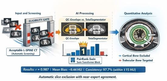
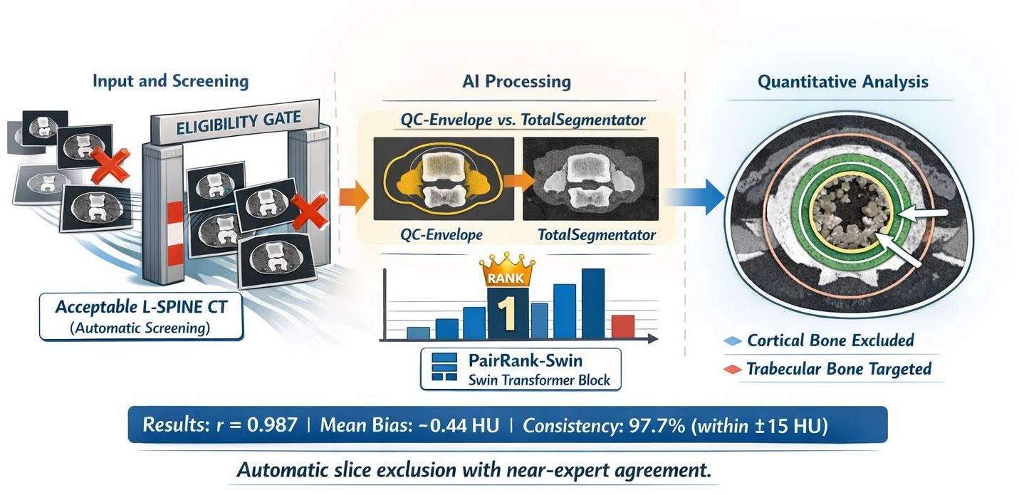
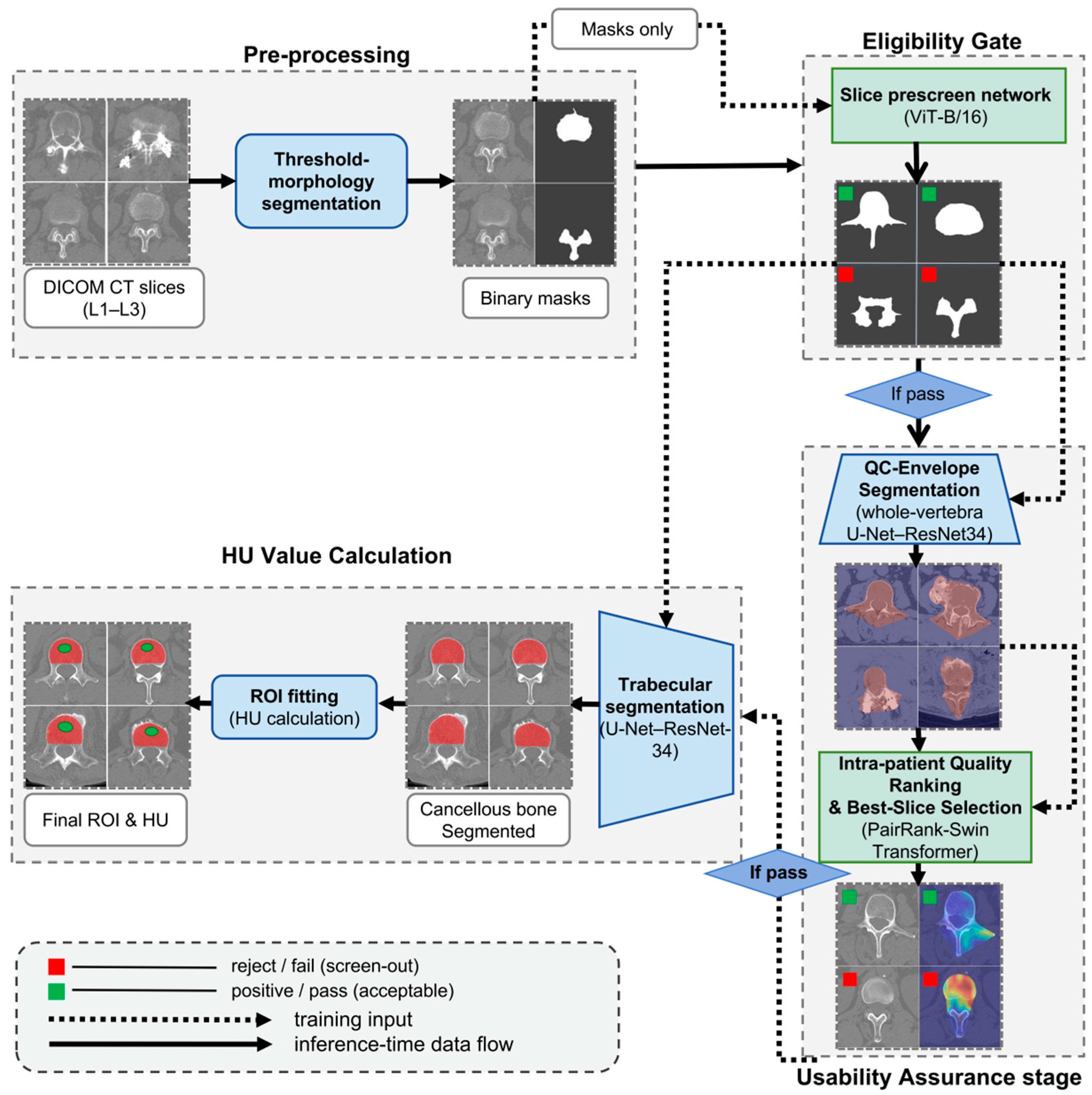
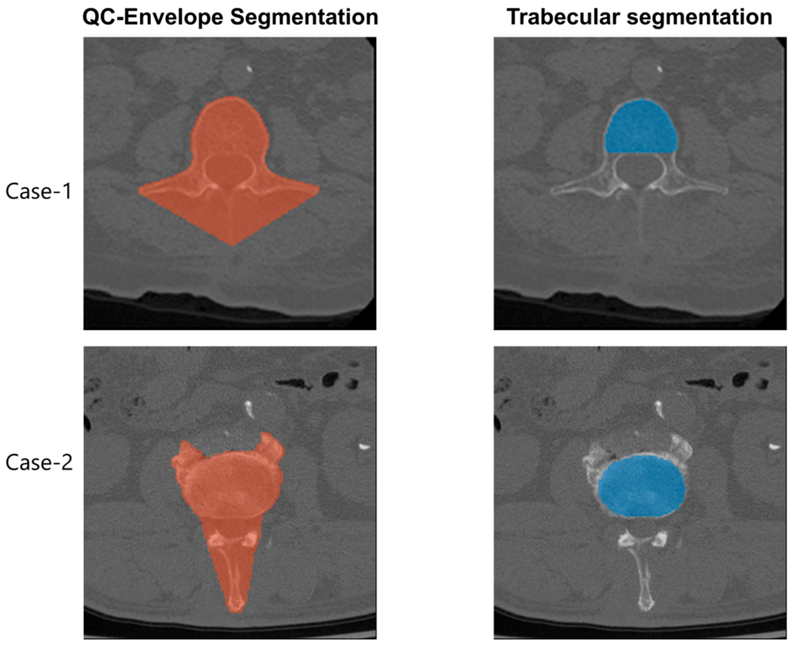
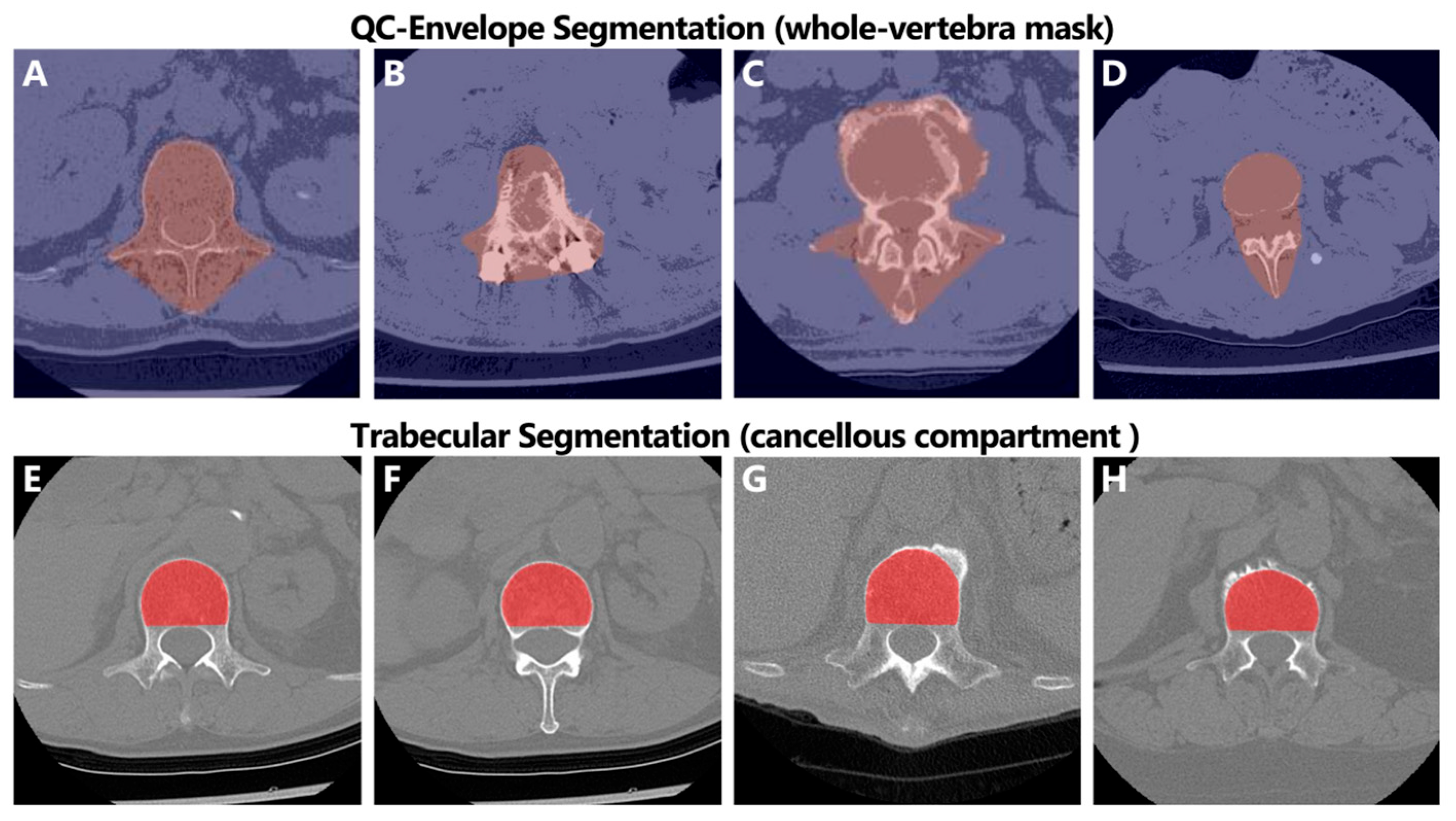
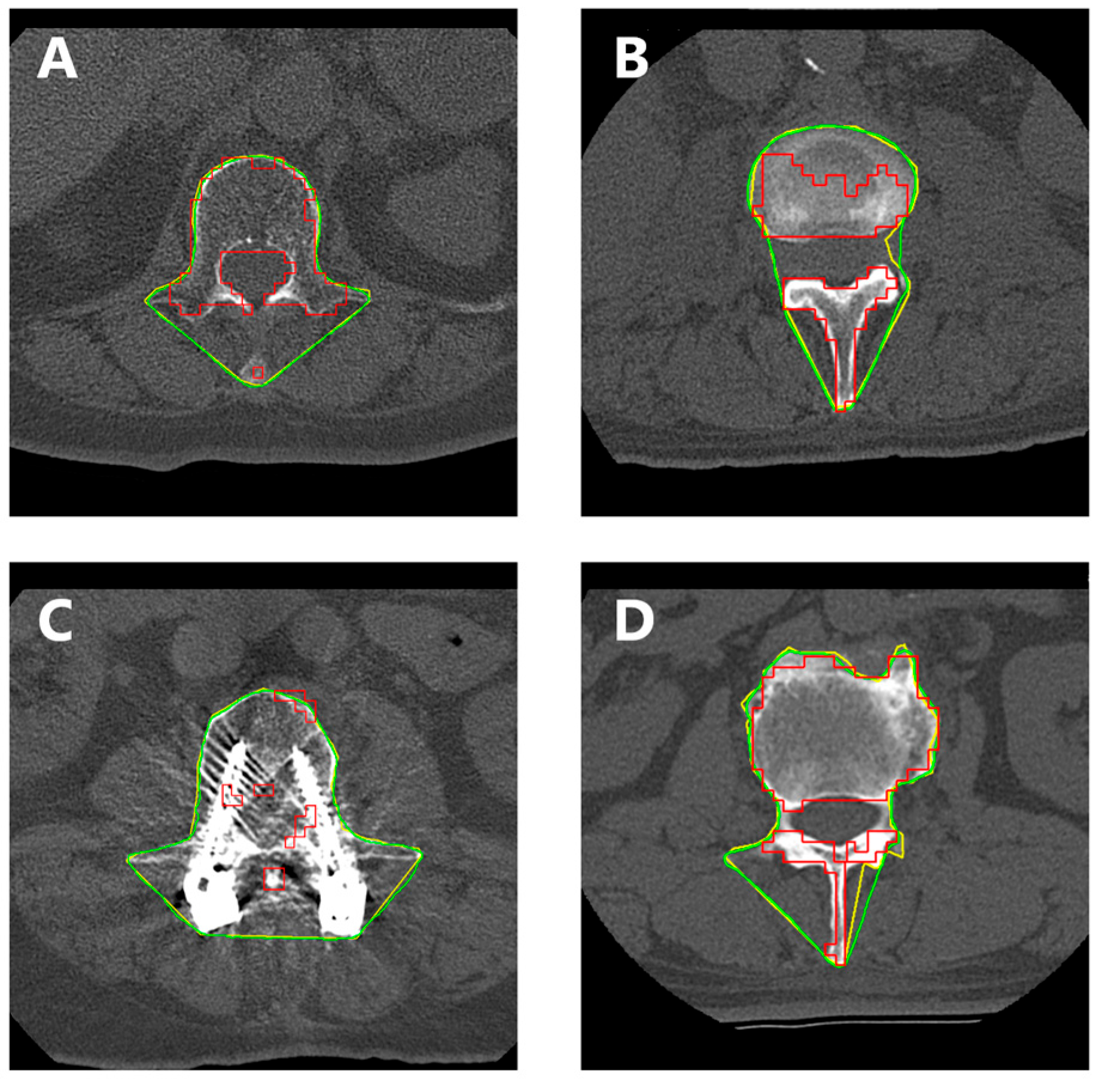
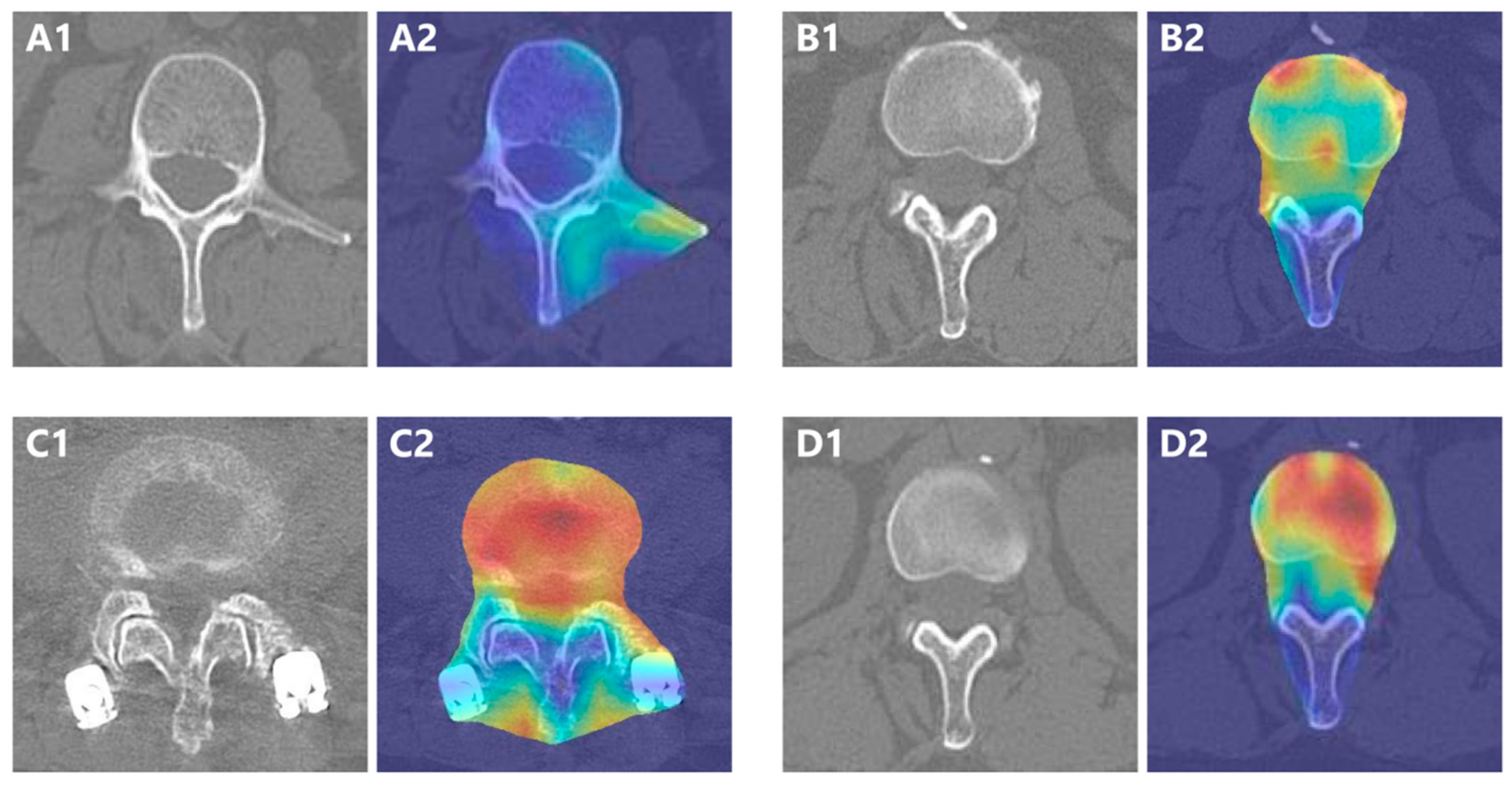
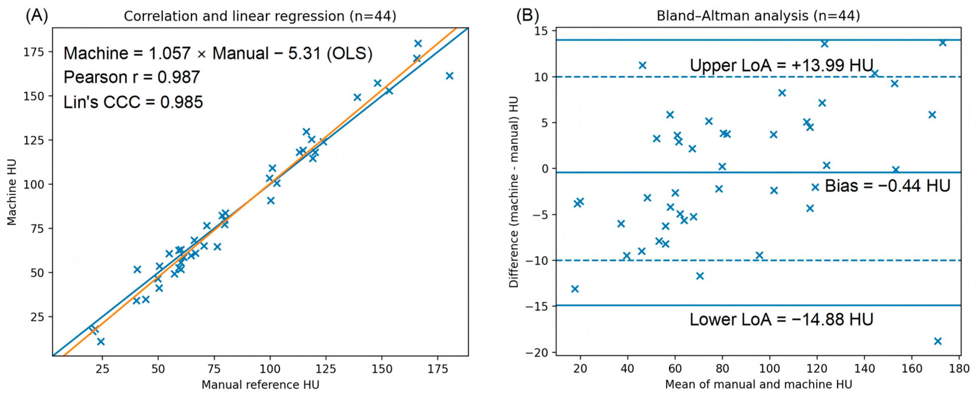
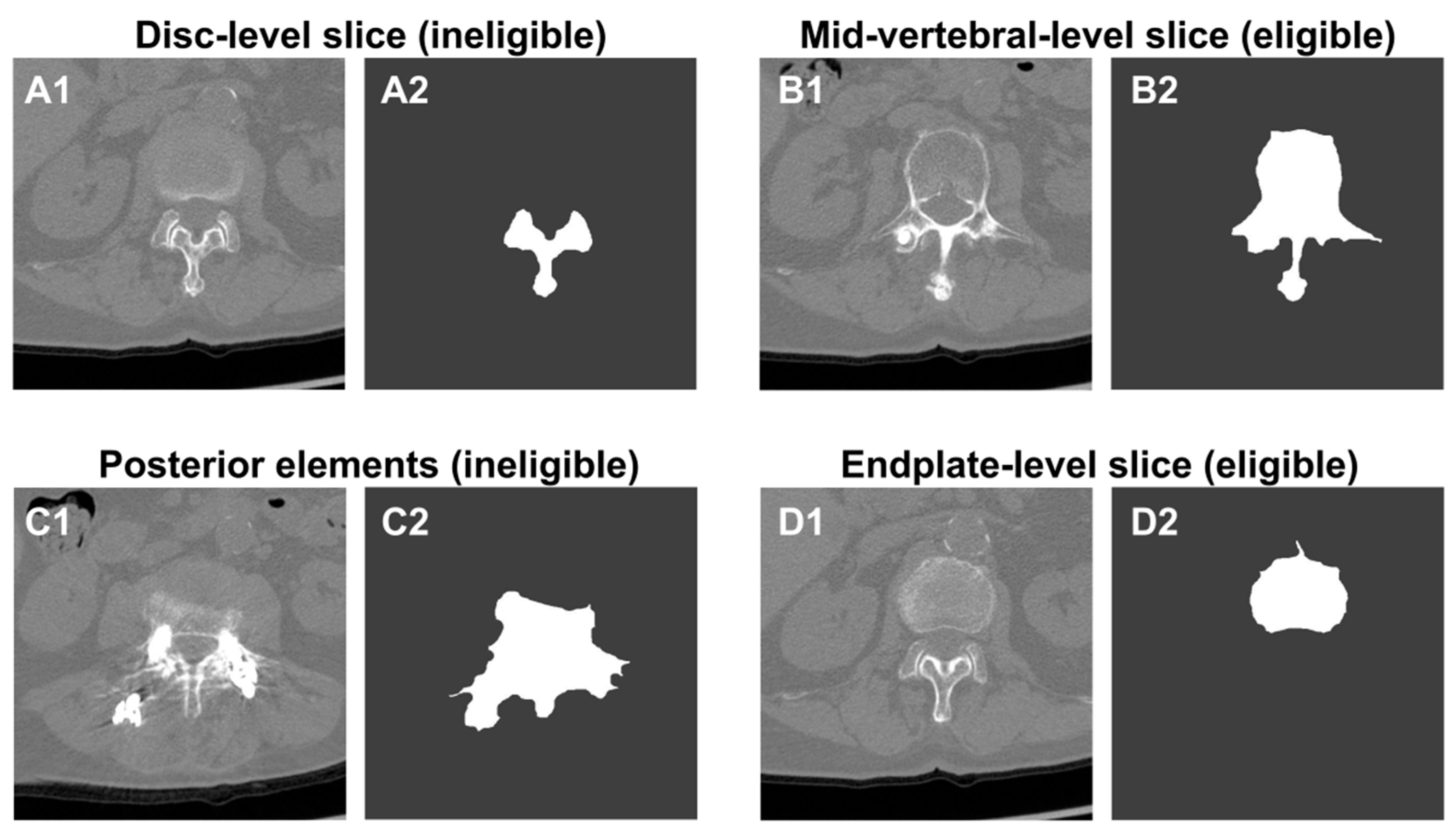
Manual single-slice lumbar quantitative computed tomography (QCT) depends on operator-driven slice selection and trabecular region-of-interest (ROI) placement. We developed a fully automated single-slice workflow for vertebral trabecular Hounsfield unit (HU) measurement that combines unsuitable-slice prescreening, dual-purpose segmentation, intra-patient slice-quality ranking, and a deterministic inner ROI rule. The pipeline includes an Eligibility Gate, QC-Envelope segmentation for broad, vertebral- and usability-preserving delineation, PairRank-Swin for best-slice selection, and dedicated trabecular segmentation for final quantitative analysis. In the independent external cohort, 4 cases were considered non-evaluable by both manual review and the pipeline, and 2 additional borderline-quality cases were manually measured but rejected by the pipeline; therefore, paired HU agreement analysis included 44 evaluable cases. Agreement remained high, with Pearson’s r = 0.987, Lin’s CCC = 0.985, mean bias −0.44 HU, and limits of agreement from −14.88 to +13.99 HU. Coverage was 84.1% within ±10 HU and 97.7% within ±15 HU. Ablation analysis showed that slice ranking and ROI erosion were the most critical components. In an open module-level baseline comparison, QC-Envelope segmentation substantially outperformed TotalSegmentator. This workflow provides high agreement with expert HU measurement while preserving reviewable intermediate outputs.
Keywords: lumbar spine; opportunistic osteoporosis screening; quantitative computed tomography; Hounsfield unit; deep learning; Swin transformer; picture archiving and communication system (PACS) lumbar spine; opportunistic osteoporosis screening; quantitative computed tomography; Hounsfield unit; deep learning; Swin transformer; picture archiving and communication system (PACS)
Graphical Abstract