Article

Cell Viability of Wharton’s Jelly-Derived Mesenchymal Stem Cells (WJ-MSCs) on 3D-Printed Resins for Temporary Dental Restorations

by
Mónica Antonio-Flores
1,*,
Andrés Eliú Castell-Rodríguez
2,
Gabriela Piñón-Zárate
2,
Beatriz Hernández-Téllez
2,
Abigailt Flores-Ledesma
1,
Enrique Pérez-Martínez
1,
Carolina Sámano-Valencia
1,
Gerardo Quiroz-Petersen
1 and
Katia Jarquín-Yáñez
2,*
1
Faculty of Stomatology, Meritorious Autonomous University of Puebla (BUAP), Av. 31 Pte. 1304, Los Volcanes, Heroica Puebla de Zaragoza 72410, Mexico
2
Laboratory of Immunotherapy and Tissue Engineering, Department of Cellular and Tissue Biology, Faculty of Medicine, National Autonomous University of México, Av. Universidad 3000, Copilco Universidad, Coyoacán, Ciudad de México 04510, Mexico
*
Authors to whom correspondence should be addressed.
J. Compos. Sci. 2025, 9(8), 404; https://doi.org/10.3390/jcs9080404 (registering DOI)
Submission received: 22 June 2025 / Revised: 22 July 2025 / Accepted: 29 July 2025 / Published: 1 August 2025
(This article belongs to the Section Biocomposites)

Abstract

There is insufficient evidence regarding the cytotoxicity of restorative 3D-printing resins, used as part of the digital workflow in dentistry. This study presents a novel comparative evaluation of cell viability and adhesion using human Wharton’s jelly-derived mesenchymal stem cells (WJ-MSCs), a less commonly used but clinically relevant cell line in dental biomaterials research. The aim of this study was to evaluate the cell viability of WJ-MSCs seeded on 3D-printed resins intended for temporary restorations. Resin discs of three commercial 3D-printing resins (NextDent C&B, Leaf Dental C&B, and UNIZ Temp) and a conventional self-curing acrylic resin (NicTone) were used. WJ-MSCs were cultured on the specimens for 1, 4, and 10 days. Cell viability was assessed using the PrestoBlue assay, Live/Dead immunofluorescence staining, and 7AAD/Annexin V staining. Cell adhesion was evaluated using scanning electron microscopy. Direct exposure to the 3D-printed resins and the self-curing acrylic caused slight reductions in cell viability compared to the control group in both microscopic analyses. 7AAD/Annexin V showed the highest percentage of viable WBCs for the conventional acrylic (34%), followed by UNIZ (35%), NextDent (42%), and Leaf Dental (36%) (ANOVA p < 0.05 Tukey’s post-hoc test p < 0.05). These findings suggest that 3D-printed resins could be considered safe for use in temporary restorations.

1. Introduction

In recent years, there has been a significant shift in the use of 3D-printed materials in dentistry, moving beyond their initial role in provisional restorations toward broader applications in permanent treatments [1,2,3]. Advances in resin chemistry, photopolymerization technology, and the resolution capabilities of 3D printers have enabled the development of novel materials with enhanced mechanical strength, wear resistance, and aesthetic properties suitable for long-term clinical use [4]. As a result, 3D-printed resins are increasingly being adopted for permanent restorations, including single crowns, multi-unit bridges, inlays, onlays, and implant-supported prostheses [5]. These materials offer distinct advantages, such as cost-effectiveness, reduced fabrication time, and seamless integration into digital workflows, which support mass customization and improved treatment planning. Additionally, the potential to fabricate complex geometries with high precision enables clinicians to meet both functional and aesthetic demands in restorative and prosthetic dentistry [6].
Parallel to these developments, the use of 3D-printed resins for provisional restorations continues to evolve. Materials designed for temporary use now demonstrate favorable mechanical and physical properties, such as high flexural strength and fracture resistance [7,8,9], which contribute to their clinical reliability during the provisional phase. Their low water sorption and solubility [10] help prevent degradation, discoloration, and bacterial infiltration—factors critical for maintaining tissue health and the success of the definitive restoration. Moreover, the ease of intraoral adjustment and chairside repair makes them highly practical for clinicians during interim periods. The primary purpose of temporary restorations is to protect the prepared tooth structure and the pulp from thermal, mechanical, and biological stimuli, as well as to help stabilize tooth position, restore aesthetics, and ensure phonetic and masticatory function [11,12]. They are commonly used throughout the fabrication process of final restorations [13].
Currently, temporary restorations can be fabricated using either conventional methods, by mixing self-curing acrylic resins and liquid monomers poured into molds, or by digital additive manufacturing techniques using 3D printing materials [14,15,16]. In 3D printing, liquid resins are polymerized layer by layer through the projection of an image using digital light processing (DLP) and photo-initiators, resulting in a three-dimensional object [17]. After printing, restorations undergo immersion and centrifugation in 90% isopropyl alcohol to remove residual resin, followed by post-curing in a light-curing chamber to complete polymerization [18,19].
These advancements reflect a growing trend toward the broader clinical acceptance of 3D-printed materials, not only as temporary solutions but as viable long-term alternatives to conventional restorative materials. However, further research is required to validate their long-term biological performance and mechanical stability under intraoral conditions [20,21,22]. However, studies have reported that dental 3D printing resins may contain residual unpolymerized monomers [23,24,25], which are potentially responsible for cytotoxic effects when in direct contact with oral soft tissues [26,27,28,29,30,31,32,33,34]. This can lead to inflammatory responses and damage to surrounding cells [35,36,37,38,39].
Mesenchymal stem cells (MSCs) are multipotent stromal cells capable of differentiating into various lineages such as fibroblasts, osteoblasts, chondrocytes, and adipocytes [40,41]. Wharton’s jelly-derived MSCs (WJ-MSCs), obtained from the umbilical cord matrix, are particularly attractive due to their non-invasive collection, immunoprivileged phenotype, and high proliferation potential [42,43,44,45,46,47,48]. Unlike cells derived from adult tissues, WJ-MSCs do not elicit a strong immune response, making them ideal for biocompatibility and regenerative medicine applications [49,50]. Clinically, MSCs have demonstrated promising outcomes in bone and periodontal tissue regeneration [51,52]. Compared to other commonly used cell lines such as human dental pulp stem cells or fibroblasts obtained from periodontal ligament or gingival connective tissue, WJ-MSCs offer greater accessibility and yield [53,54,55]. Pulpal stem cells require extraction of healthy, non-carious teeth, which limits their availability, while fibroblasts from gingiva or periodontal ligament are typically harvested in small quantities through invasive procedures [56,57]. Given that provisional restorations fabricated from 3D-printed resins remain in direct contact with these soft tissues, the use of WJ-MSCs offers a practical and clinically relevant model for evaluating cellular responses to these materials. The novelty of this study lies in the application of WJ-MSCs for direct contact assays with temporary restorative resins fabricated via additive manufacturing, providing insight into their biological safety and compatibility within the oral environment [58,59].
Despite increasing interest in 3D-printed dental resins, comparative biocompatibility studies using human-derived WJ-MSCs remain limited. Therefore, this study aims to evaluate the cell viability, proliferation, and adhesion of WJ-MSCs in response to direct exposure to three commercial 3D-printed resins and one conventional self-curing acrylic resin, commonly used for temporary dental restorations. Cell viability was assessed using the PrestoBlue assay with spectrophotometric reading, Live/Dead immunofluorescence staining, and 7AAD/Annexin V staining, while cell adhesion was evaluated by scanning electron microscopy.

2. Materials and Methods

2.1. Sample Preparation

Three 3D printing resins indicated for the fabrication of temporary crowns and fixed partial dentures were evaluated: C&B MFH (NextDent, Soesterberg, The Netherlands), Seed Print C&B (Leaf Dental, California, CA, USA), and zDental C&B (Uniz, Beijing, China). These were compared with a conventional self-curing acrylic resin, NicTone (MDC Dental, Zapopan, Jal, México). The main characteristics of the materials are summarized in Table 1 [4,5,6,10].
Disc-shaped samples (20 mm in diameter ×1.5 mm in height) were formed. For the 3D printing resins (n = 16), the designs were created using Meshmixer software V 3.5 (Autodesk Inc., Californa, CA, USA) as shown in (Figure 1) and then converted to STL files processed in Chitubox software V1.8.0 Beta (Chitubox, Shenzhen, Guangdong, China). Printing was performed using a 3D printer (Phrozen Mini 4K, Hsinchu, Taiwan) with parameters specified by each manufacturer. After printing, each sample was washed for 5 min in a washing unit (Phrozen Mini 4K, Hsinchu, Taiwan) with 90% isopropyl alcohol. Although a 40 min post-curing cycle was applied for all printed resins in a curing unit (Phrozen Mini 4K, Hsinchu, Taiwan), this duration was selected based on pre-testing protocols designed to optimize polymerization depth and reduce residual monomer content, in alignment with manufacturer recommendations. This ensured a standardized and complete curing process across all experimental groups [60,61,62]. All samples were polished using the same protocol with rubber wheels and brushes from the Laboratory Kit No. 1906 (Jota, Rüthi, Switzerland) and a polishing paste (Polyshine, MDC Dental, Zapopan, Jal, México). Conventional acrylic resin samples were fabricated using a cylindrical metal mold with the same dimensions (20 mm diameter × 1.5 mm height) and polished using the same protocol as the printed specimens.

2.2. Cell Isolation

Wharton’s jelly-derived mesenchymal stem cells (WJ-MSCs) were used, approved by the Ethics Committee of the School of Medicine, UNAM (No. FM/DI/112/2022) on 13 April 2023. Cells were expanded in Dulbecco’s Modified Eagle Medium–Ham’s F12 (DMEM-F12) supplemented with 10% fetal bovine serum (FBS, Biowest, Bradenton, FL, USA) and 1% antibiotic (penicillin–streptomycin, Gibco, Grand Island Biological Company, New York, NY, USA). Cultures were maintained at 37 °C in a humidified atmosphere with 5% CO2, with medium changes every third day until reaching 90% confluence for use in Presto Blue (Thermo Fisher Scientific, California, CA, USA) and Live/Dead assays (Thermo Fisher Scientific, California, CA, USA) [23,24,25].
For the Annexin V/7-AAD assays (Thermo Fisher Scientific, California, CA, USA), spleen cells from three male Balb-C mice, aged two months, were used, approved by the Ethics Committee of the School of Medicine, UNAM (No. FM/DI/112/2022). The spleens were excised and manually homogenized to obtain the cells, which were immediately cultured on the resin discs in RPMI medium (Biowest, Bradenton, FL, USA) supplemented with 10% FBS and 1% antibiotic at a density of 1 × 10⁷ cells per well.

2.3. Annexin V and 7-AAD Assay

For the Annexin V (FITC, BioLegend, San Diego, CA, USA) and 7-AAD (Pacific Blue, BioLegend, San Diego, CA, USA) assays, spleen cells in contact with NicTone, NextDent, Leaf Dental, and UNIZ discs were collected and centrifuged to obtain a pellet. Five microliters of each reagent were incubated with the cells according to the manufacturer’s instructions for 15 min at room temperature in the dark. Samples were then analyzed using an Attune NxT Flow Cytometer (Thermo Fisher Scientific, Waltham, MA, USA) at the National Flow Cytometry Laboratory (LabNalCit UNAM). All experiments were performed in triplicate, and data were analyzed using FlowJo software version 8.8.7 (BD, Franklin Lakes, NJ, USA). The purpose of performing the Annexin V/7AAD assay using spleen-derived cells was to evaluate a potential systemic response rather than a localized one.

2.4. Live/Dead Assay

Twenty-four-well plates containing poly-L-lysine-coated coverslips (Sigma Aldrich, St. Louis, MO, USA) were seeded with 5 × 10³ WJ-MSCs. The resin discs (NicTone, NextDent, Leaf Dental, UNIZ) were then placed on top of the cells; for the control, cells were cultured without any material. Samples were incubated at 37 °C in a humidified atmosphere with 5% CO2 in DMEM-F12 medium supplemented with 10% FBS and 1% antibiotic. Evaluations were performed on days 1, 4, and 10. The Live/Dead Kit (Invitrogen, Waltham, MA, USA) was applied to each sample for 15 min at 37 °C, staining live cells green with calcein and dead cells red with ethidium homodimer. Observations were made using an ECLIPSE 80i epifluorescence microscope (Nikon Instruments Inc., Melville, NY, USA) [26,27]. The quantitative analysis of fluorescence was performed with Image J software V 1.54k (Image Processing and Analysis in Java, NIH, USA) [63].

2.5. Scanning Electron Microscopy (SEM) Analysis

To evaluate the attachment of WJ-MSCs on NicTone, NextDent, Leaf Dental, and UNIZ discs, 1 × 10⁴ cells were seeded on each experimental group under the same culture conditions as described above. A positive control group consisting of WJ-MSCs seeded directly onto a fibrin clot—a biocompatible, medical-grade material—was included for reference [64,65]. After 10 days, the samples were fixed with 2% glutaraldehyde for an hour, washed with cacodylate buffer, dehydrated, and coated with gold using plasma-assisted physical vapor deposition (PAPVD, HummerVI-A, Anatech Ltd., Alexandria, VA, USA). Samples were observed using a scanning electron microscope (SEM, JSM-7800F, JEOL Ltd., Akishima, Tokyo, Japan).

2.6. Presto Blue Assay

WJ-MSCs were seeded at a density of 1 × 10⁴ cells on each experimental group (NicTone, NextDent, Leaf Dental, UNIZ) and on a 96-well culture plate for the control. Cultures were maintained in DMEM-F12 medium supplemented with 10% FBS and 1% antibiotic at 37 °C in 5% CO2, with evaluations on days 1, 4, and 10. All experiments were performed in triplicate. The Presto Blue resazurin-based kit (Thermo Fisher Scientific, Massachusetts, USA) was used according to the manufacturer’s instructions to assess mitochondrial activity of viable cells. Absorbance intensity was measured at a wavelength of 570 nm using a Thermo Labsystems 354 Multiskan Ascent Microplate Reader (Thermo Fisher Scientific, Waltham, MA, USA), and readings were obtained using Ascent software version 2.6 for multiple layers.

2.7. Statistical Analysis

Sample size calculation was performed using G-Power Calculator (Heinrich-Heine-Universität Düsseldorf, Germany), assuming an effect size of d = 1.26, resulting in a sample size of n = 16 per experimental and control group. The significance level was set at p < 0.05. Differences among groups were analyzed using one-way ANOVA followed by Tukey’s post hoc test for multiple comparisons. All data was analyzed and graphed using GraphPad software Prism V 10.5.0 (Dotmatics, Boston, MA, USA). For fluorescence quantification, the Games–Howell post hoc test was selected for pairwise comparisons following one-way ANOVA, due to differences in sample sizes and potential heterogeneity of variances among the experimental groups, obtained from the calcein images.

3. Results

3.1. Annexin V and 7-AAD Assay

Figure 2 shows the Annexin V/7-AAD analysis dot plots of splenic cells seeded on NicTone, Next Dental, Leaf Dental, and Uniz resins; as a control, cells were seeded directly onto culture plates. As expected, the unstained group did not show defined parameters to determine quadrants for viable (Q4), early apoptotic (Q1), late apoptotic (Q2), or necrotic cells (Q3). The results indicate that after 24 h of culture, the control group showed an average viability of 61%, followed by Next Dental with 42%, Leaf Dental with 36%, Uniz with 35%, and NicTone with 34%; no significant differences among the experimental groups were observed (ANOVA, p > 0.05). This suggests that the evaluated resins are biocompatible under in vitro cell culture conditions.

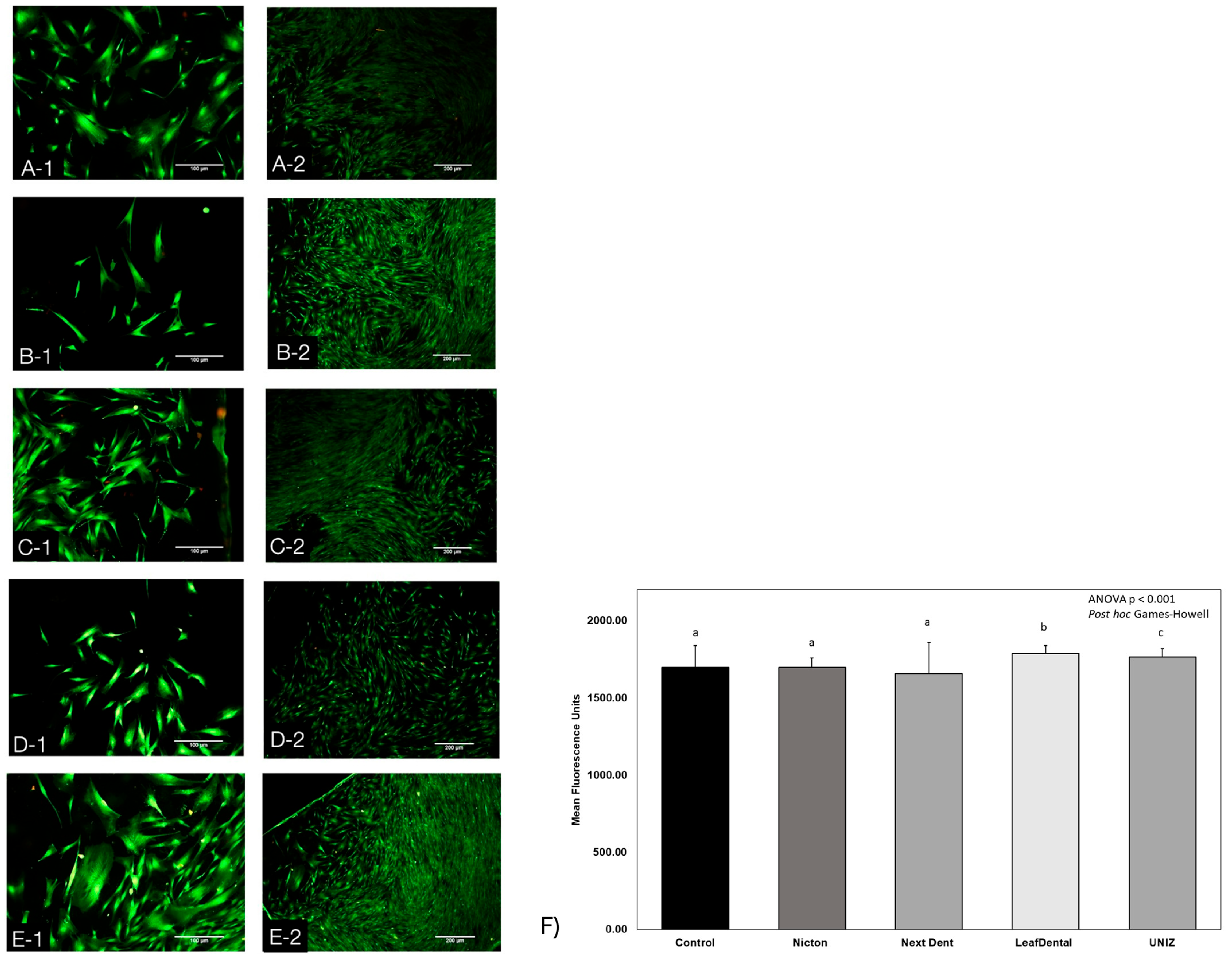
3.2. Live/Dead Assay

Calcein-AM staining enabled the visualization of homogeneous green fluorescence in the NextDent (Figure 3C-1, 3C-2) and Leaf Dental (Figure 3D-1, 3D-2) groups, comparable to the control group without material exposure. In contrast, NicTone (Figure 3B-1) showed moderate staining, while UNIZ (Figure 3D-1, 3D-2) exhibited the highest cell confluence and proliferation. Ethidium homodimer binding to nuclear DNA indicates minimal membrane integrity loss in dead cells within the Next Dental group, resulting in red fluorescence. Cytoplasmic extensions appeared elongated, with good adherence to resin surfaces and well-defined oval nuclei, as shown in Figure 3.
Quantitative fluorescence analysis based on calcein staining revealed statistically significant differences among the experimental groups (ANOVA p < 0.001). The results indicated that the UNIZ group exhibited significantly higher fluorescence intensity compared to all other groups (post hoc Games–Howell p < 0.001). Similarly, the Leaf Dental group also demonstrated significantly greater fluorescence than the remaining materials, suggesting enhanced cell viability and surface colonization in these two resin types.

3.3. Scanning Electron Microscopy (SEM) Analysis

To study cell adhesion to the different resin discs, samples were analyzed using SEM, as shown in Figure 4. Differences in surface structure and topography were observed. After 10 days, Figure 4A (positive control) showed enhanced cellular activity and spreading, serving as a baseline for optimal adhesion. In contrast, thinner WJ-MSCs were observed on the NicTone surface (Figure 4B). In Figure 4C–E, larger cells adhered to the surface of the resin discs, exhibiting broader cytoplasmic extensions and filopodial projections. In Figure 4D, increased extracellular matrix production and fibers in the intercellular space were evident, correlating with surface roughness and suggesting that this microtopography provided a favorable stimulus for extracellular matrix deposition. Elongated cells connected to one another were also clearly visible.
Compared to the 3D-printed resins, the self-curing NicTone acrylic (Figure 4A) exhibited the lowest cell adhesion and reduced number of filopodial extensions, this result is supported by the findings from the PrestoBlue assay with spectrophotometric reading, Live/Dead immunofluorescence staining, and 7AAD/Annexin V staining. Overall, the SEM images revealed that the quality of the surface finish directly influenced cell adhesion and proliferation, as roughened surfaces facilitated cell spreading and attachment.

3.4. Presto Blue Assay

Exposure of WJ-MSCs to the tested provisional restoration materials for the specified time points resulted in a favorable impact on cell viability. Regardless of the group, an increase in cell proliferation was observed compared to the control group, which was not exposed to any resin.
According to the estimated cell count derived from metabolic activity (Figure 5A), all materials showed a significant increase in proliferation on day 10 compared to day 1. Specifically, the 3D-printed resins (Leaf Dental, NextDent and UNIZ) promoted increased cell viability over time, indicating that cells adapted to the microenvironment created by the resins. Furthermore, the absorbance analysis shown in Figure 5B revealed a statistically significant difference between the UNIZ and NextDent groups on day 4 (ANOVA p = 0.036; post hoc Tukey p = 0.041), suggesting that the interaction between these materials and the cells differed in their early proliferation behavior.
When comparing the different resin materials at each individual time point, no statistically significant differences in cell viability were observed among the groups (Table 2). On day 1, all materials exhibited similarly high levels of cell viability, with values ranging from 84.76% to 91.42%, and the differences between them were not statistically significant (ANOVA, p = 0.517). This suggests that immediately after polymerization, the cytotoxic effects of the resins were comparable across the groups. By day 4, although a general decline in cell viability was observed in all materials, the intergroup differences remained statistically non-significant (ANOVA, p = 0.908), indicating a uniform biological response to the materials at this time point. On day 10, cell viability values varied more noticeably between groups—ranging from 68.48% in NextDent to 78.17% in NicTone—yet these differences also did not reach statistical significance (ANOVA, p = 0.466). Overall, these findings indicate that, despite temporal changes in cell viability within some individual materials, the biocompatibility profiles among the tested resins remained statistically comparable at each evaluation period.
When evaluating changes in cell viability over time within each resin group, different patterns were observed. For NicTone, no statistically significant differences were found between days 1, 4, and 10 (ANOVA, p = 0.494), indicating a relatively stable biocompatibility profile throughout the testing period. Although a numerical decline was observed on day 4, followed by a slight recovery on day 10, these variations did not reach statistical significance, suggesting minimal time-dependent cytotoxicity. Similarly, Leaf Dental exhibited no significant differences across the three time points (ANOVA, p = 0.095). In this group, cell viability decreased from day 1 to day 4 and remained low on day 10, but the changes were not statistically conclusive.
In contrast, NextDent showed a significant reduction in cell viability over time (ANOVA, p = 0.032). The viability decreased markedly from 90.23% on day 1 to 68.48% on day 10, suggesting a progressive decline in biocompatibility, potentially associated with the release of residual monomers or degradation products. A similar trend was observed in the Uniz group, where a significant decrease was also detected (ANOVA, p = 0.015). Although cell viability in Uniz remained relatively high on day 4, it declined by day 10, which may reflect delayed cytotoxic effects. These results indicate that while NicTone and Leaf Dental maintained consistent biocompatibility across the testing period, both NextDent and Uniz displayed significant time-dependent cytotoxicity, highlighting the importance of evaluating not only initial biocompatibility but also the material’s stability and behavior over time.

4. Discussion

In this study, the cell viability, proliferation, and adhesion of mesenchymal stem cells derived from Wharton’s jelly (WJ-MSC) were evaluated on 3D-printed resins intended for temporary restorations. Currently, computer-aided design and manufacturing (CAD/CAM) technology is widely applied in the fabrication of additive-type temporary restorations, producing three-dimensional objects through a digital workflow aimed at replacing conventional materials such as self-cure acrylic resin [66,67,68].
Among the advantages of these biomaterials are their physical properties, which provide stability to temporary restorations due to their high flexural strength [69,70]. Clinically, this translates into a material capable of withstanding masticatory forces, reducing the probability of fracture during the period prior to placement of the definitive restoration. They also exhibit low solubility and sorption, which helps prevent the absorption of fluids that could contaminate, stain, or compromise the physical and mechanical properties of the restoration [4,10,12,14].
However, there is still insufficient evidence regarding the biological characteristics of these materials to ensure their use does not cause harmful adverse effects on patient health [26,27,28,29,71,72]. Recent studies have demonstrated that 3D-printed resins for dental use may retain residual unpolymerized monomers after processing, which are likely responsible for certain cytotoxic reactions when these materials are in direct contact with soft oral tissues [35,36]. This can lead to inflammatory processes, which should be carefully considered when selecting materials for clinical use [73,74].
Recent studies have evaluated the biocompatibility of 3D-printed dental resins, particularly their effects on soft tissues, using various dental-related cell lines such as human gingival fibroblasts, periodontal ligament cells, and keratinocytes [26,27,28,29,30,31,32,33,34,35,36]. In this study, was assessed the viability of Wharton’s jelly-derived mesenchymal stem cells (WJ-MSCs) when in direct contact with 3D-printed resins intended for temporary restorations. This cellular model offers a broader perspective of soft tissue response due to its multilineage differentiation potential and regenerative relevance.
Several studies have reported the presence of unpolymerized residual monomers in 3D-printed resins used for temporary dental restorations, which may contribute to cytotoxic effects upon direct contact with oral tissues. Among the most common monomers identified are urethane dimethacrylate (UDMA), tri-ethylene glycol dimethacrylate (TEGDMA), and bisphenol A-glycidyl methacrylate (Bis-GMA). These components can leach out from incompletely polymerized resins and interact with surrounding soft tissues, potentially inducing inflammatory responses, oxidative stress, and even apoptosis. The release rate and concentration of these monomers can vary depending on the resin composition, degree of polymerization, and post-curing protocols. Therefore, understanding the type and behavior of residual monomers is essential when assessing the biocompatibility of dental materials, particularly for temporary restorations that remain in the oral cavity for extended periods [75,76].
NicTone self-curing acrylic resin contains monomers, esters, and glycols that may exert cytotoxic effects upon direct contact with oral soft tissues. This was evident in our analysis, where NicTone demonstrated the greatest surface irregularities and residual cell debris, consistent with lower polish quality. In contrast, the 3D-printed resins evaluated here incorporate different monomers and photo-initiators such as ethylene dimethacrylate, HEMA, MEHQ, 4-methoxyphenol, and hydroquinone monomethyl ether—materials that exhibit improved viscosity and hydrophilicity compared to traditional monomers like Bis-GMA or UDMA. In addition to resin composition, surface ultrastructure also plays a key role in influencing cell behavior.
The present study demonstrated that surface irregularities and the presence of an extracellular matrix can favor cell adhesion and proliferation, as they provide a more suitable microenvironment for the anchorage of filopodial extensions—despite all experimental groups having undergone the same polishing protocol. Annexin V/7AAD analysis revealed that samples with rougher surfaces exhibited higher cell confluence and more extensive filopodial attachment to the substrate. Among the studied groups, UNIZ exhibited the smoothest surface compared to the other 3D-printed resins, which correlated with lower cell adhesion relative to those resins, but it still showed greater cell adhesion than the self-cure acrylic resin (NicTone) under the same polishing conditions.
These observations align with findings from Atria P. (2021) [12], where NextDent showed higher cytotoxicity in LDH assays, and Park J. (2020) [29], who found no significant differences in viability among bis-acrylic and 3D-printed resins. In our Live/Dead fluorescence assay and calcein staining, WJ-MSCs displayed homogeneous proliferation across experimental groups. Notably, cells seeded on UNIZ resin exhibited strong adhesion, with complete calcein staining throughout the observed area and multilayered overgrowth.
In the present study, the Live/Dead fluorescence assay and calcein staining revealed that cell proliferation was homogeneous among the experimental groups compared to the control group, like findings reported by Oberoi G. et al. in 2021 [36], who used periodontal ligament cells and gingival fibroblasts in direct contact with LAY-FOMM 40, LAY-FOMM 60, and PLA resins. Notably, in the current study, WJ-MSCs in direct contact with the UNIZ resin discs exhibited increased proliferation and surface adhesion, with complete calcein staining throughout the observed area and with multilayered cell overgrowth observed.
Annexin V/7-AAD staining further allowed discrimination between viable, apoptotic, and necrotic cells, providing important insights into potential resin-induced cytotoxicity [10]. This methodology remains underexplored in dental resin research but has proven useful, as demonstrated by Guerrero Gironés J. (2022) [28], who applied this technique to orthodontic resin materials, demonstrating a homogeneous distribution of cells in early apoptosis, late apoptosis, and necrosis using Annexin V-FITC and 7-AAD reagents.
The present study presented certain limitations that should be acknowledged. Pre-conditioning procedures such as mechanical aging or simulated salivary degradation were not incorporated. Also, the evaluation was restricted to short-term exposure. However, compared to previous studies by Kurt A. (2018) and Bandarra S. (2020) [77,78], whose methodologies typically involved test periods of only 24–120 h and exposure exclusively to supernatants obtained after 5 days of material contact, in the present investigation, cells were exposed directly to the material surfaces for 10 days. The culture medium was replaced every 3 days, simulating a physiological renewal process that closely resembles salivary flow under clinical conditions.
Although no statistically significant differences were found among the tested 3D-printed resins, all showed slightly better biological performance than the conventional acrylic resin. Leaf Dental and UNIZ demonstrated the most favorable behavior in terms of cell viability, proliferation, and adhesion. This finding supports their potential selection as preferred materials for temporary dental applications.
While in vitro studies offer a valuable and controlled framework to evaluate cytotoxicity and cellular responses, the clinical environment introduces multiple interacting factors that may influence outcomes differently. In the oral cavity, materials are constantly subjected to salivary flow, enzymatic degradation, microbial colonization, immune responses, and mechanical forces generated by mastication and occlusion [20,24]. These dynamic conditions can accelerate material degradation, affect monomer elution rates, and modulate tissue responses in ways not fully replicated in vitro [12]. As such, the observed biocompatibility in cell culture settings may not always directly translate to clinical scenarios.
In particular, the low but measurable reductions in cell viability observed over time in our experiments could carry biological significance in vivo, especially for patients with predisposing factors such as inflammation, compromised healing capacity, or immunological sensitivity [63]. While the magnitude of these changes does not indicate acute cytotoxicity, their potential cumulative effect during the clinical use of provisional restorations warrants further investigation. Understanding whether these subtle cellular alterations influence peri-restorative tissue health in the short- or medium-term is critical for validating the safety of these materials in real-world applications.
Despite the minor reductions in viability observed over time, the results appear within clinically acceptable limits for short-term provisional use. Nevertheless, even slight declines may become significant in patients with compromised healing capacity or underlying inflammatory conditions. Therefore, additional in vivo investigations—including cytokine profiling and immune response analysis—are warranted.

5. Conclusions

Based on the results obtained in this study, Leaf Dental and UNIZ 3D-printed resins demonstrated the most favorable biological performance, showing higher cell viability, proliferation, and adhesion of Wharton’s jelly-derived mesenchymal stem cells (WJ-MSCs). These findings suggest that these two materials could be considered the preferred choice for the fabrication of temporary restorations in clinical practice, offering reliable biocompatibility and functional performance.
Furthermore, the incorporation of WJ-MSCs as the cell model represents a novel approach, as previous studies have primarily focused on fibroblasts or dental pulp cells, which are more limited in quantity and availability. The use of WJ-MSCs provides a broader, more clinically relevant perspective, particularly considering their regenerative potential and biological proximity to periodontal and gingival tissues. This approach opens new avenues for future research aimed at evaluating longer exposure times and more complex biological interactions, ultimately contributing to a more comprehensive understanding of the biocompatibility of dental restorative materials.

Author Contributions

Conceptualization, M.A.-F., A.F.-L., E.P.-M., G.Q.-P. and K.J.-Y.; Methodology, M.A.-F., A.E.C.-R., B.H.-T., G.P.-Z., E.P.-M., C.S.-V., G.Q.-P. and K.J.-Y.; Data curation M.A.-F., K.J.-Y., A.F.-L., G.P.-Z. and C.S.-V.; Formal analysis, M.A.-F., A.E.C.-R., G.P.-Z., B.H.-T., E.P.-M., A.F.-L., C.S.-V., G.Q.-P. and K.J.-Y.; Investigation, M.A.-F. and K.J.-Y.; Supervision, K.J.-Y., A.E.C.-R., A.F.-L., B.H.-T. and G.P.-Z.; Validation, A.E.C.-R.; Software, B.H.-T. and G.P.-Z.; Resources, E.P.-M., C.S.-V. and G.Q.-P. All authors have read and agreed to the published version of the manuscript.

Funding

This work was supported by PAPIIT IN216723, IA203025 and IT200525 (1 January 2025), CONAHCYT CF-2023-I-2388 (1 January 2024).

Institutional Review Board Statement

The study was conducted in accordance with the Declaration of Helsinki and approved by the Institutional Review Board of The National Autonomous University of México (No. FM/DI/112/2022 on 13 April 2023). The animal study protocol was approved by the Ethics Committee of The Meritorious Autonomous University of Puebla (FESIEP/CIFE/151/2023 on 19 September 2023), and the Ethics Committee of The National Autonomous University of México (No. FM/DI/112/2022 on 13 April 2023).

Informed Consent Statement

Not applicable.

Data Availability Statement

Data will be made available upon request to the authors.

Acknowledgments

The authors would like to thank the biologist Armando Zepeda from the Department of Cell and Tissue Biology, Faculty of Medicine (UNAM), for their assistance with SEM; Blanca Esther Blancas Luciano from the Department of Microbiology and Parasitology, Faculty of Medicine (UNAM), for their support with the spectrophotometer; Sara Judith Álvarez Pérez and Miguel A. Herrera Enríquez for their support in the cell culture; the National Flow Cytometry Laboratory (LabNalCit UNAM) for their cytometry equipment; and BA (Hons) Gretta Walsh for her support with the revision of the English translation.

Conflicts of Interest

The authors declare no conflicts of interest.

Abbreviations

The following abbreviations are used in this manuscript:
WJ-MSCMesenchymal stem cells derived from Wharton’s jelly
WBCsSplenic white blood cells
CAD/CAMComputer-aided design and manufacturing
DMEM-F12Dulbecco’s Modified Eagle Medium–Ham’s F12
DSDisc sample

References

  1. Haidar, Z.S. Digital dentistry: Past, present, and future. Digit. Med. Healthc. Technol. 2023, 2, 1–16. [Google Scholar]
  2. Balestra, D.; Lowther, M.; Goracci, C.; Mandurino, M.; Cortili, S.; Paolone, G.; Louca, C.; Vichi, A. 3D Printed Materials for Permanent Restorations in Indirect Restorative and Prosthetic Dentistry: A Critical Review of the Literature. Materials 2024, 17, 1380. [Google Scholar] [CrossRef]
  3. Vitali, J.; Cheng, M.; Wagels, M. Utility and cost–effectiveness of 3D-printed materials for clinical use. J. 3D Print. Med. 2019, 3, 209–218. [Google Scholar] [CrossRef]
  4. de Gois Moreira, F.G.; da Silva, N.R.; Bezerra, M.G.P.G.; da Silva, S.E.G.; Butler, S.; Souza, K.B.; de Assunção E Souza, R.O. Influence of 3D printing system, postpolymerization and aging protocols on resin flexural strength and dimensional stability for printing occlusal splints, models and temporary restorations. Clin. Oral. Investig. 2024, 28, 604. [Google Scholar] [CrossRef] [PubMed]
  5. Alammar, A.; Att, W.; Beuer, F. The Accuracy of 3D-Printed Fixed Dental Restorations. J. Esthet. Restor. Dent 2025, 37, 1047–1061. [Google Scholar] [CrossRef] [PubMed]
  6. Anadioti, E.; Musharbash, L.; Blatz, M.B.; Papavasiliou, G.; Kamposiora, P. 3D printed complete removable dental prostheses: A narrative review. BMC Oral. Health 2020, 20, 343. [Google Scholar] [CrossRef]
  7. NextDent. NextDent C&B MFH (Micro Filled Hybrid). 2025. Available online: https://nextdent.com/products/cb-mfh-micro-filled-hybrid/ (accessed on 14 November 2024).
  8. UNIZ. Zdental. 2025. Available online: https://uniz--en.oss-us-west-1.aliyuncs.com/pdf/zDental-IDB-TDS.pdf (accessed on 14 November 2024).
  9. LeafDental. LeafDental Resins for 3D Printing. 2025. Available online: https://www.uniz.com/us_en/materials/dental (accessed on 14 November 2024).
  10. Choudhury, M.; Nahar, N.; Yazdi, S.; Choudhury, F.; Sultana, A. Fabrication of provisional restoration on freshly prepared tooth: Indirect and direct technique. Bangladesh J. Med. Sci. 2015, 14, 59–64. [Google Scholar] [CrossRef]
  11. Rogers, H.B.; Zhou, L.T.; Kusuhara, A.; Zaniker, E.; Shafaie, S.; Owen, B.C.; Duncan, F.E.; Woodruff, T.K. Dental resins used in 3D printing technologies release ovo-toxic leachates. Chemosphere 2021, 270, 129003. [Google Scholar] [CrossRef]
  12. Atria, P.J.; Bordin, D.; Marti, F.; Nayak, V.V.; Conejo, J.; Benalcázar Jalkh, E.; Witek, L.; Sampaio, C.S. 3D-printed resins for provisional dental restorations: Comparison of mechanical and biological properties. J. Esthet. Restor. Dent. 2022, 34, 804–815. [Google Scholar] [CrossRef]
  13. American College of Prosthodontists. Glossary of digital dental terms. 2nd edition. J. Prosthodont. 2021, 30, 172–181. [Google Scholar] [CrossRef]
  14. Liang, X.; Yu, B.; Dai, Y.; Wang, Y.; Hu, M.; Zhong, H.-J.; He, J. Three-Dimensional Printing Resin-Based Dental Provisional Crowns and Bridges: Recent Progress in Properties, Applications, and Perspectives. Materials 2025, 18, 2202. [Google Scholar] [CrossRef] [PubMed]
  15. Tigmeanu, C.V.; Ardelean, L.C.; Rusu, L.C.; Negrutiu, M.L. Additive Manufactured Polymers in Dentistry, Current State-of-the-Art and Future Perspectives-A Review. Polymers 2022, 14, 3658. [Google Scholar] [CrossRef] [PubMed]
  16. Azari, A.; Nikzad, S. Computer-assisted implantology: Historical background and potential outcomes—A review. Int. J. Med. Robot. Comput. Assist. Surg. 2008, 4, 95–104. [Google Scholar] [CrossRef] [PubMed]
  17. Prakash, J.; Shenoy, M.; Alhasmi, A.; Al Saleh, A.A.; Shivakumar, G.C.; Shivakumar, S. Biocompatibility of 3D-Printed Dental Resins: A Systematic Review. Cureus 2024, 16, e51721. [Google Scholar] [CrossRef] [PubMed] [PubMed Central]
  18. Alyami, M.H. The Applications of 3D-Printing Technology in Prosthodontics: A Review of the Current Literature. Cureus 2024, 16, e68501. [Google Scholar] [CrossRef]
  19. Kessler, A.; Reymus, M.; Hickel, R.; Kunzelmann, K.H. Three-body wear of 3D printed temporary materials. Dent. Mater. 2019, 35, 1805–1812. [Google Scholar] [CrossRef]
  20. Revilla-León, M.; Özcan, M. Additive Manufacturing Technologies Used for Processing Polymers: Current Status and Potential Application in Prosthetic Dentistry. J. Prosthodont. 2019, 28, 146–158. [Google Scholar] [CrossRef]
  21. Alammar, A.; Kois, J.C.; Revilla-León, M.; Att, W. Additive Manufacturing Technologies: Current Status and Future Perspectives. J. Prosthodont. 2022, 31 (Suppl. S1), 4–12. [Google Scholar] [CrossRef]
  22. Hegedus, T.; Kreuter, P.; Kismarczi-Antalffy, A.A.; Demeter, T.; Banyai, D.; Vegh, A.; Geczi, Z.; Hermann, P.; Payer, M.; Zsembery, A.; et al. User Experience and Sustainability of 3D Printing in Dentistry. Int. J. Environ. Res. Public Health 2022, 19, 1921. [Google Scholar] [CrossRef]
  23. Perea-Lowery, L.; Gibreel, M.; Vallittu, P.K.; Lassila, L.V. 3d-printed vs. Heat-polymerizing and autopolymerizing denture base acrylic resins. Materials 2021, 14, 5781. [Google Scholar] [CrossRef]
  24. Li, W.; Zhou, J.; Xu, Y. Study of the in vitro cytotoxicity testing of medical devices. Biomed. Rep. 2015, 3, 617–620. [Google Scholar] [CrossRef]
  25. Tahayeri, A.; Morgan, M.; Fugolin, A.P.; Bompolaki, D.; Athirasala, A.; Pfeifer, C.S.; Ferracane, J.L.; Bertassoni, L.E. 3D printed versus conventionally cured provisional crown and bridge dental materials. Dent. Mater. 2018, 34, 192–200. [Google Scholar] [CrossRef]
  26. Park, J.-H.; Lee, H.; Kim, J.W.; Kim, J.H. Cytocompatibility of 3D printed dental materials for temporary restorations on fibroblasts. BMC Oral Health 2020, 20, 157. [Google Scholar] [CrossRef]
  27. Folwaczny, M.; Ahantab, R.; Kessler, A.; Ern, C.; Frasheri, I. Cytotoxicity of 3D printed resin materials for temporary restorations on human periodontal ligament (PDL-hTERT) cells. Dent. Mater. 2023, 39, 529–537. [Google Scholar] [CrossRef]
  28. Guerrero-Gironés, J.; López-García, S.; Pecci-Lloret, M.R.; Pecci-Lloret, M.P.; Rodríguez Lozano, F.J.; García-Bernal, D. In vitro biocompatibility testing of 3D printing and conventional resins for occlusal devices. J. Dent. 2022, 123, 104163. [Google Scholar] [CrossRef] [PubMed]
  29. Moon, W.; Kim, S.; Lim, B.-S.; Park, Y.-S.; Kim, R.J.-Y.; Chung, S.H. Dimensional Accuracy Evaluation of Temporary Dental Restorations with Different 3D Printing Systems. Materials 2021, 14, 1487. [Google Scholar] [CrossRef] [PubMed]
  30. Kurzmann, C.; Janjić, K.; Shokoohi-Tabrizi, H.; Edelmayer, M.; Pensch, M.; Moritz, A.; Agis, H. Evaluation of Resins for Stereolithographic 3D-Printed Surgical Guides: The Response of L929 Cells and Human Gingival Fibroblasts. Biomed. Res. Int. 2017, 2017, 4057612. [Google Scholar] [CrossRef]
  31. Kim, G.-T.; Go, H.-B.; Yu, J.-H.; Yang, S.-Y.; Kim, K.-M.; Choi, S.-H.; Kwon, J.-S. Cytotoxicity, Colour Stability and Dimensional Accuracy of 3D Printing Resin with Three Different Photoinitiators. Polymers 2022, 14, 979. [Google Scholar] [CrossRef]
  32. International Organization for Standardization. Biological Evaluation of Medical Devices—Part 5: Tests for In Vitro Cytotoxicity; International Organization for Standardization: Geneva, Switzerland, 2009. [Google Scholar]
  33. Ríos Hernández, M.; Cepero Cañas, J. Citotoxicidad in vitro: Sistema para la evaluación de biomateriales y equipos médicos implantables en Cuba. CENIC Cienc. Biológicas 2006, 37, 173–176. [Google Scholar]
  34. Wulff, J.; Schweikl, H.; Rosentritt, M. Cytotoxicity of printed resin-based splint materials. J. Dent. 2022, 120, 104097. [Google Scholar] [CrossRef]
  35. Frasheri, I.; Aumer, K.; Keßler, A.; Miosge, N.; Folwaczny, M. Effects of resin materials dedicated for additive manufacturing of temporary dental restorations on human gingival keratinocytes. J. Esthet. Restor. Dent. 2022, 34, 1105–1112. [Google Scholar] [CrossRef]
  36. Oberoi, G.; Nitsch, S.; Janjić, K.; Shokoohi-Tabrizi, H.; Moritz, A.; Moscato, F.; Unger, E.; Agis, H. The impact of 3D-printed LAY-FOMM 40 and LAY-FOMM 60 on L929 cells and human oral fibroblasts. Clin. Oral. Investig. 2021, 25, 1869–1877. [Google Scholar] [CrossRef]
  37. Hwangbo, N.K.; Nam, N.E.; Choi, J.H.; Kim, J.E. Effects of the washing time and washing solution on the biocompatibility and mechanical properties of 3d printed dental resin materials. Polymers 2021, 13, 4410. [Google Scholar] [CrossRef]
  38. Sa, L.; Kaiwu, L.; Shenggui, C.; Junzhong, Y.; Yongguang, J.; Lin, W.; Li, R. 3D printing dental composite resins with sustaining antibacterial ability. J. Mater. Sci. 2019, 54, 3309–3318. [Google Scholar] [CrossRef]
  39. Saleh Alghamdi, S.; John, S.; Roy Choudhury, N.; Dutta, N.K. Additive Manufacturing of Polymer Materials: Progress, Promise and Challenges. Polymers 2021, 13, 753. [Google Scholar] [CrossRef] [PubMed]
  40. Huang, G.T.J.; Garcia-Godoy, F. 13—Stem cells and dental tissue reconstruction. In Material-Tissue Interfacial Phenomena; Spencer, P., Misra, A., Eds.; Woodhead Publishing: Sawston, UK, 2017; pp. 325–353. [Google Scholar]
  41. Zhang, Q.; Nguyen, P.D.; Shi, S.; Burrell, J.C.; Cullen, D.K.; Le, A.D. 3D bio-printed scaffold-free nerve constructs with human gingiva-derived mesenchymal stem cells promote rat facial nerve regeneration. Sci. Rep. 2018, 8, 6634. [Google Scholar] [CrossRef] [PubMed]
  42. Simancas-Escorcia, V.; Díaz-Caballero, A.; Vergara-Hernández, C. Aspectos morfológicos in vivo e in vitro de fibroblastos gingivales en pacientes con agrandamiento gingival. Entramado 2020, 16, 276–284. [Google Scholar] [CrossRef]
  43. Baust, J.M.; Buehring, G.C.; Campbell, L.; Elmore, E.; Harbell, J.W.; Nims, R.W.; Price, P.; Reid, Y.A.; Simione, F. Best practices in cell culture: An overview. Vitr. Cell Dev. Biol. Anim. 2017, 53, 669–672. [Google Scholar] [CrossRef]
  44. Coecke, S.; Balls, M.; Bowe, G.; Davis, J.; Gstraunthaler, G.; Hartung, T.; Hay, R.; Merten, O.-W.; Price, A.; Schechtman, L.; et al. Guidance on Good Cell Culture Practice. Altern. Lab. Anim. 2005, 33, 261–287. [Google Scholar] [CrossRef]
  45. Oyeleye, O.O.; Ogundeji, S.T.; Ola, S.I.; Omitogun, O.G. Basics of animal cell culture: Foundation for modern science. Biotechnol. Mol. Biol. Rev. 2016, 11, 6–16. [Google Scholar] [CrossRef]
  46. Bacakova, L.; Zarubova, J.; Travnickova, M.; Musilkova, J.; Pajorova, J.; Slepicka, P.; Kasalkova, N.S.; Svorcik, V.; Kolska, Z.; Motarjemi, H.; et al. Stem Cells: Their Source, Potency and Use in Regenerative Therapies with Focus on Adipose-Derived Stem Cells—A Review. Biotechnol. Adv. 2018, 36, 1111–1126. [Google Scholar] [CrossRef] [PubMed]
  47. Feng, L.; Shi, L.; Lu, Y.; Wang, B.; Tang, T.; Fu, W.; He, W.; Li, G.; Zhang, J. Linc-ROR Promotes Osteogenic Differentiation of Mesenchymal Stem Cells by Functioning as a Competing Endogenous RNA for miR-138 and miR-145. Mol. Ther.–Nucleic Acids 2018, 11, 345–353. [Google Scholar] [CrossRef] [PubMed]
  48. Bassir, S.H.; Wisitrasameewong, W.; Raanan, J.; Ghaffarigarakani, S.; Chung, J.; Freire, M.; Andrada, L.C.; Intini, G. Potential for Stem Cell-Based Periodontal Therapy. J. Cell. Physiol. 2016, 231, 50–61. [Google Scholar] [CrossRef] [PubMed]
  49. Inchingolo, A.M.; Inchingolo, A.D.; Nardelli, P.; Latini, G.; Trilli, I.; Ferrante, L.; Malcangi, G.; Palermo, A.; Inchingolo, F.; Dipalma, G. Stem Cells: Present Understanding and Prospects for Regenerative Dentistry. J. Funct. Biomater. 2024, 15, 308. [Google Scholar] [CrossRef]
  50. Jia, L.; Zhang, Y.; Ji, Y.; Li, X.; Xing, Y.; Wen, Y.; Huang, H.; Xu, X. Comparative Analysis of lncRNA and mRNA Expression Profiles Between Periodontal Ligament Stem Cells and Gingival Mesenchymal Stem Cells. Gene 2019, 699, 155–164. [Google Scholar] [CrossRef]
  51. Raveau, S.; Jordana, F. Tissue Engineering and Three-Dimensional Printing in Periodontal Regeneration: A Literature Review. J. Clin. Med. 2020, 9, 4008. [Google Scholar] [CrossRef]
  52. Abdal-Wahab, M.; Abdel Ghaffar, K.A.; Ezzatt, O.M.; Hassan, A.A.A.; El Ansary, M.M.S.; Gamal, A.Y. Regenerative potential of cultured gingival fibroblasts in treatment of periodontal intrabony defects (randomized clinical and biochemical trial). J. Periodontal Res. 2020, 55, 441–452. [Google Scholar] [CrossRef]
  53. Guo, B.; Tang, C.; Wang, M.; Zhao, Z.; Shokoohi-Tabrizi, H.A.; Shi, B.; Andrukhov, O.; Rausch-Fan, X. In vitro biocompatibility of biohybrid polymers membrane evaluated in human gingival fibroblasts. J. Biomed. Mater. Res. Part. B 2020, 108, 2590–2598. [Google Scholar] [CrossRef]
  54. Bakopoulou, A.; Leyhausen, G.; Volk, J.; Tsiftsoglou, A.; Garefis, P.; Koidis, P.; Geurtsen, W. Comparative analysis of in vitro osteo/odontogenic differentiation potential of human dental pulp stem cells (DPSCs) and stem cells from the apical papilla (SCAP). Arch. Oral. Biol. 2011, 56, 709–721. [Google Scholar] [CrossRef]
  55. Dave, J.R.; Chandekar, S.S.; Behera, S.; Desai, K.U.; Salve, P.M.; Sapkal, N.B.; Mhaske, S.T.; Dewle, A.M.; Pokare, P.S.; Page, M.; et al. Human gingival mesenchymal stem cells retain their growth and immunomodulatory characteristics independent of donor age. Sci. Adv. 2022, 8, eabm6504. [Google Scholar] [CrossRef]
  56. Grawish, M.E.; Saeed, M.A.; Sultan, N.; Scheven, B.A. Therapeutic Applications of Dental Pulp Stem Cells in Regenerating Dental, Periodontal and Oral-Related Structures. World J. Meta-Anal. 2021, 9, 176–192. [Google Scholar] [CrossRef]
  57. Ma, L.; Liu, A.Q.; Guo, H.; Xuan, K. Dental pulp stem cells in tooth regeneration: Advancement and emerging directions. Chin. J. Stomatol. 2024, 59, 496–501. [Google Scholar]
  58. Cai, H.; Xu, X.; Lu, X.; Zhao, M.; Jia, Q.; Jiang, H.-B.; Kwon, J.-S. Dental Materials Applied to 3D and 4D Printing Technologies: A Review. Polymers 2023, 15, 2405. [Google Scholar] [CrossRef]
  59. Zeng, W.Y.; Ning, Y.; Huang, X. Advanced technologies in periodontal tissue regeneration based on stem cells: Current status and future perspectives. J. Dent. Sci. 2021, 16, 501–507. [Google Scholar] [CrossRef]
  60. Hassanpour, M.; Narongdej, P.; Alterman, N.; Moghtadernejad, S.; Barjasteh, E. Effects of Post-Processing Parameters on 3D-Printed Dental Appliances: A Review. Polymers 2024, 16, 2795. [Google Scholar] [CrossRef]
  61. Alharbi, S.; Alshabib, A.; Algamaiah, H.; Aldosari, M.; Alayad, A. Influence of Post-Printing Polymerization Time on Flexural Strength and Microhardness of 3D Printed Resin Composite. Coatings 2025, 15, 230. [Google Scholar] [CrossRef]
  62. Bayarsaikhan, E.; Gu, H.; Hwangbo, N.-K.; Lim, J.-H.; Shim, J.-S.; Lee, K.-W.; Kim, J.-E. Influence of different postcuring parameters on mechanical properties and biocompatibility of 3D printed crown and bridge resin for temporary restorations. J. Mech. Behav. Biomed. Mater. 2022, 128, 105127. [Google Scholar] [CrossRef]
  63. Rasband, W.S. ImageJ. U.S. National Institute of Health, Bethesda, MD, USA, 1997–2018. Available online: http://imagej.net/ij/ (accessed on 14 November 2024).
  64. Alami, S.Y.; Hampton, J.W.; Race, G.J.; Speer, R.J. Fibrin stabilizing factor (factor XIII). Am. J. Med. 1968, 44, 1. [Google Scholar] [CrossRef]
  65. Anilkumar, T.V.; Muhamed, J.; Jose, A.; Jyothi, A.; Mohanan, P.V.; Krishnan, L.K. Advantages of hyaluronic acid as a component of fibrin sheet for care of acute wound. Biologicals 2011, 39, 81–88. [Google Scholar] [CrossRef] [PubMed]
  66. Mellor, S.; Hao, L.; Zhang, D. Additive manufacturing: A framework for implementation. Int. J. Prod. Econ. 2014, 149, 194–201. [Google Scholar] [CrossRef]
  67. Barazanchi, A.; Li, K.C.; Al-Amleh, B.; Lyons, K.; Waddell, J.N. Additive technology: Update on current materials and applications in dentistry. J. Prosthodont. 2017, 26, 156–163. [Google Scholar] [CrossRef] [PubMed]
  68. Revilla-León, M.; Meyers, M.J.; Zandinejad, A.; Özcan, M. A review on chemical composition, mechanical properties, and manufacturing work flow of additively manufactured current polymers for interim dental restorations. J. Esthet. Restor. Dent. 2019, 31, 51–57. [Google Scholar] [CrossRef] [PubMed]
  69. Srinivasan, M.; Kalberer, N.; Kamnoedboon, P.; Mekki, M.; Durual, S.; Özcan, M.; Müller, F. CAD-CAM complete denture resins: An evaluation of biocompatibility, mechanical properties, and surface characteristics. J. Dent. 2021, 114, 103785. [Google Scholar] [CrossRef] [PubMed]
  70. Konidena, A. 3D printing: Future of dentistry? J. Indian. Acad. Oral. Med. Radiol. 2016, 28, 109. [Google Scholar] [CrossRef]
  71. Alshamrani, A.; Alhotan, A.; Kelly, E.; Ellakwa, A. Mechanical and biocompatibility properties of 3D-printed dental resin reinforced with glass silica and zirconia nanoparticles: In vitro study. Polymers 2023, 15, 2523. [Google Scholar] [CrossRef]
  72. Piedra-Cascón, W.; Krishnamurthy, V.R.; Att, W.; Revilla-León, M. 3D printing parameters, supporting structures, slicing, and post-processing procedures of vat-polymerization additive manufacturing technologies: A narrative review. J. Dent. 2021, 109, 103630. [Google Scholar] [CrossRef]
  73. Fouassier, J.-P.; Lalevée, J. Photoinitiators for Polymer Synthesis: Scope, Reactivity and Efficiency; John Wiley & Sons: Hoboken, NJ, USA, 2012. [Google Scholar]
  74. Lin, C.-H.; Lin, Y.-M.; Lai, Y.-L.; Lee, S.-Y. Mechanical properties, accuracy, and cytotoxicity of UV-polymerized 3D printing resins composed of Bis-EMA, UDMA, and TEGDMA. J. Prosthet. Dent. 2020, 123, 349–354. [Google Scholar] [CrossRef]
  75. Almeida, S.M.; Meereis, C.T.; Leal, F.B.; Carvalho, R.V.; Boeira, P.O.; Chisini, L.A.; Cuevas-Suárez, C.E.; Lima, G.S.; Piva, E. Evaluation of alternative photoinitiator systems in two-step self-etch adhesive systems. Dent. Mater. 2020, 36, e29–e37. [Google Scholar] [CrossRef]
  76. Zeng, B.; Cai, Z.; Lalevée, J.; Yang, Q.; Lai, H.; Xiao, P.; Liu, J.; Xing, F. Cytotoxic and cytocompatible comparison among seven photoinitiators-triggered polymers in different tissue cells. Toxicol. Vitr. 2021, 72, 105103. [Google Scholar] [CrossRef]
  77. Kurt, A.; Altintas, S.H.; Kiziltas, M.V.; Tekkeli, S.E.; Guler, E.M.; Kocyigit, A.; Usumez, A. Evaluation of residual monomer release and toxicity of self-adhesive resin cements. Dent. Mater. J. 2018, 37, 40–48. [Google Scholar] [CrossRef] [PubMed]
  78. Bandarra, S.; Mascarenhas, P.; Luís, A.R.; Catrau, M.; Bekman, E.; Ribeiro, A.C.; Félix, S.; Caldeira, J.; Barahona, I. In vitro and in silico evaluations of resin-based dental restorative material toxicity. Clin. Oral. Investig. 2020, 24, 2691–2700. [Google Scholar] [CrossRef] [PubMed]
Figure 1. Design and fabrication of sample discs. (A) Digital design of 3D-printed resin discs using Meshmixer software. (B) Conventional acrylic resin discs fabricated using a cylindrical metal mold. All discs were standardized to 20 mm in diameter and 1.5 mm in height.
Jcs 09 00404 g001
Figure 2. Flow cytometry analysis of cell apoptosis and necrosis induced by temporary resins. Assessment of cell viability following 24 h exposure to resin extracts using Annexin V-FITC and 7-AAD staining in WBCs. (A) Unstained control. (B) Untreated control. (C–F) Cells exposed to NicTone, NextDent, Leaf Dental, and UNIZ, respectively. (G) Quantitative summary of viable cell percentages across groups. Quadrant Q4 indicates viable cells (Annexin V/7-AAD); Q1, early apoptotic (Annexin V+/7-AAD); Q2, late apoptotic (Annexin V+/7-AAD+); and Q3, necrotic cells (Annexin V/7-AAD+). Values are presented as mean ± SD. Groups with identical lowercase letters show no significant differences (ANOVA, p > 0.05; Tukey’s post hoc test p < 0.05).
Jcs 09 00404 g002
Figure 3. Live/Dead staining of WJ-MSCs in contact with temporary resins. Representative fluorescence micrographs of WJ-MSCs cultured on different resins for 10 days. Cells were stained with Calcein-AM (green, viable) and Ethidium homodimer-1 (red, nonviable). (A-1) Control group; (B-1) NicTone; (C-1) NextDent; (D-1) Leaf Dental; (E-1) UNIZ (scale bars 100 µm). (A-2) Control group; (B-2) NicTone; (C-2) NextDent; (D-2) Leaf Dental; (E-2) UNIZ (scale bars 200 µm). (F) Quantitative summary of fluorescence across groups. Values are presented as mean ± SD. Groups with identical lowercase letters show no significant differences (ANOVA, p > 0.05; Games-Howell’s post hoc test p < 0.05).
Jcs 09 00404 g003
Figure 4. Scanning electron microscopy of WJ-MSC adhesion on resin surfaces. SEM images showing the morphology and adhesion of WJ-MSCs on different temporary resin discs after 10 days of culture. (A) Positive control group -WJ-MSCs in direct contact with a fibrin clot; (B) NicTone; (C) NextDent; (D) Leaf Dental; (E) UNIZ. Cells showed varying degrees of spreading, filopodial extension, and extracellular matrix deposition depending on surface topography. Magnification: ×1000; scale bar = 10 μm. DS: Disc surface; WJ-MSC: Wharton’s jelly-derived mesenchymal stem cells.
Jcs 09 00404 g004
Figure 5. Metabolic activity and proliferation of WJ-MSCs exposed to temporary resins. Quantitative analysis of WJ-MSC viability and proliferation over 1, 4, and 10 days using the Presto Blue assay. Results presented as mean ± standard deviation. (A) Cell number estimation derived from metabolic activity. (B) Absorbance values over time for each material. Significant differences are indicated (ANOVA, p < 0.05). Groups: NicTone; NextDent; Leaf Dental; UNIZ).
Jcs 09 00404 g005
Table 1. Materials tested in this study and their name, manufacturer, type, matrix composition (according to manufacturer), and processing method.
Brand NameManufacturerType* MatrixProcessing Method
NicToneMDC DentalPMMA-basedLiquid: methyl methacrylate;
powder: methacrylate copolymers, initiators, and pigments.
Conventional: auto polymerization
C&B MFHNextDentMethacrylate-basedMethacrylates 7,7,9(or 7,9,9)-trimethyl-4,13-dioxo-3,14-dioxa-5,12-diazahexadecane-1,16-diyl bismethacrylate; ethylene dimethacrylate; HEMA; TPO; E-BPA; titanium dioxide; mequinol; 4-methoxyphenol; hydroquinone monomethyl ether.3D printing:
photopolymerization
Seed C&BLeaf DentalMethacrylate-basedMethyl methacrylate, ethyl methacrylate, urethane dimethacrylate (UDMA), Bisphenol A glycidyl methacrylate (BisGMA); photo initiators: camphor quinone and benzophenone; additives: diphenyl phthalate, aluminum hydroxide, and titanium dioxide.3D printing:
photopolymerization
zDental C&BUNIZMethacrylate-basedOnly data available: Acrylate monomer, acrylate oligomers, and photo initiators.3D printing:
photopolymerization
* The chemical composition and manufacturing information for each resin were sourced from the respective official websites of NextDent, Leaf Dental, UNIZ, and MDC Dental.
Table 2. Cell viability percentage.
1d4d10dANOVA p
Nic Tone84.76 ± 7.67 a,£75.67 ± 5.25 a,£78.17 ± 12.81 a,£0.494
NextDent90.23 ± 7.60 a,£74.35 ± 6.14 a,£,¥68.48 ± 9.15 a,¥0.032 *
Leaf Dental90.08 ± 9.24 a,£73.63 ± 14.34 a,£69.41 ± 3.23 a,£0.095
Uniz91.42 ± 1.86 a,£78.41 ± 5.34 a,¥75.98 ± 5.99 a,¥0.015 *
ANOVA p0.5170.9080.466
* The percentage of live cells and standard deviation are indicated. Statistical analysis per column (between experimental groups) is indicated in lower case letters; the same letters indicate no statistical difference. Statistical analysis per row (compared by time) is indicated with a, £ and ¥ symbols; the same symbols indicate no statistical difference.
Disclaimer/Publisher’s Note: The statements, opinions and data contained in all publications are solely those of the individual author(s) and contributor(s) and not of MDPI and/or the editor(s). MDPI and/or the editor(s) disclaim responsibility for any injury to people or property resulting from any ideas, methods, instructions or products referred to in the content.

