Article

Antibacterial UV-Curable Gel with Hydroxyapatite Nanoparticles for Regenerative Medicine in the Field of Orthopedics

by
Julia A. Burunkova
1,*,
Valeria V. Semykina
1,*,
Vera E. Sitnikova
1,
Dmitry M. Dolgintsev
1,
Faliya F. Zaripova
1,
Alina A. Ponomareva
1,
Diana R. Mizina
1,
Attila Csick
2,
Sandor Kokenyesi
3 and
Anton Zhilenkov
4
1
Centre for Chemical Engineering, Laboratory of Quantum Processes and Measurements, ITMO University, 197101 Saint Petersburg, Russia
2
Institute for Nuclear Research (ATOMKI), 4026 Debrecen, Hungary
3
Department of Electrical Engineering, Debrecen University, 4032 Debrecen, Hungary
4
Institute of Robotics and Intelligent Systems, Saint-Petersburg State Marine Technical University, Lotsmanskaya St., 3, 190121 Saint Petersburg, Russia
*
Authors to whom correspondence should be addressed.
J. Compos. Sci. 2025, 9(2), 65; https://doi.org/10.3390/jcs9020065
Submission received: 25 December 2024 / Revised: 15 January 2025 / Accepted: 20 January 2025 / Published: 1 February 2025
(This article belongs to the Section Biocomposites)

Abstract

:
The development and analysis of the properties of a new material based on UV-curable acrylate monomers with silicon-containing hydroxyapatite and zinc oxide nanoparticles as an antibacterial component and gelatin was carried out. Using this material in orthopedics and dentistry is very convenient because it covers any surface geometry of metal implants and hardens under ultraviolet light. In this work, sorption properties, changes in porosity, and mechanical properties of the material were investigated. The conditions for obtaining hydroxyapatite (HA) nanoparticles and the presence of silicon oxide nanoparticles and organic for the shell in an aqueous medium were studied for the pH of the medium, the sequence of administration and concentration of the material components, as well as antibacterial properties. This polymer material is partially resorbable. That supports not only the growth of bone cells but also serves as a protective layer. It reduces friction between organic tissues and a metal implant and can be a solution to the problem of the aseptic instability of metal implants. The material can also be used to repair damaged bones and cartilage tissues, especially in cases where the application and curing procedure is performed using laparoscopic methods. In this work, the authors propose a simple and quite cheap method for obtaining material based on photopolymerizable acrylates and natural gelatin with nanoparticles of HA, zinc oxide, and silicon oxide. The method allows one to obtain a composite material with different nanoparticles in a polymer matrix which retain the requisite properties needed such as active-sized HA, antibacterial ZnO, and structure-forming and stability-improving SiO2 nanoparticles.

1. Introduction

Traumatism and the long life of an aging population raise the problem of new functional materials development. It is needed both to restore damaged bones’ function and to produce implants or metal implant coatings that are in demand in dentistry and orthopedics [1]. Modern materials based on metals, ceramics, or polymers are used for bone tissue regeneration. An orthopedic material must be able to withstand high mechanical stresses while being elastic, in accordance with the properties of natural bone, and be biochemically compatible with the body and stimulate natural regeneration processes. Metals and alloys (titanium, nickel, their alloys, gold, etc.) are usually used for heavily loaded implants, but the surface of such materials is not osteoconductive; that is, bone cells do not proliferate, do not multiply, and do not differentiate with the formation of an extracellular matrix, so there is poor integration in the implant site, especially over time [2]. One solution to this problem is to apply bioactive coatings to the metal surface, which should allow the normal functioning of bone cell growth and reduce fibrous encapsulation. To stimulate bone cell growth, calcium phosphate (CaP) bioactive additives are used in different stoichiometries. Hydroxyapatite (HA), which is a bioactive and osteoconductive material with a crystallographic and chemical composition close to that of bone, is considered the most applicable [3]. The application of CaP and HA to the surface of orthopedic and dental implants by different methods has been proven to improve their fixation, especially in the early stage of healing. Bioactive glass coatings (Bioglass R), developed by Lacefield and Hench [4], or biological agents, such as bone morphogenetic proteins, which create conditions for rapid bone tissue formation in CaP coatings, are also used [5]. Currently, hydroxyapatites with various ionic substitutions such as magnesium, silicon, fluorine, and carbonate [6] are being used, which have been reported to improve the rate and quality of bone repair [7]. It can be considered an established fact that nanosized hydroxyapatite (20–200 nm) contributes more effectively to regenerative bioprocesses [8]. Spherical hydroxyapatite nanostructures with dimensions of 5–200 µm, rod-shaped nanostructures with a thickness of 5–150 µm, and needle-shaped nanostructures with shapes from 40 nm to 150 µm can be obtained by various methods. The structure and porosity of the implant material have an important influence on the formation of bone and cartilage tissue. Microporosity is responsible for osteoinduction processes and promotes cartilage formation before osteogenesis, while smaller pores (1–10 μm) induce direct osteogenesis [9], pore sizes of 100 μm enhance cell growth and migration, and, finally, pores larger than 300 μm promote bone and capillary formation [10].
The main problem is how to place the active agent (hydroxyapatite) on the metal surface of the implant in a way that preserves its functional activity. Plasma, magnetron, and other types of sputtering [2], as well as sol–gel technology are used. They use materials in the form of dense HA ceramics; however, this is a low-activity material in which the resorption and growth rate of bone and cartilage tissue are slowed. Recently, several HA cements have been developed that exhibit too high resorption rates but actively promote bone growth. However, the most promising is the use of a composite based on hydroxyapatite obtained by different techniques (deposition, hydrolysis, the hydrothermal method, the high-temperature solid-phase method, the sol/gel method) in a biocompatible polymer material. The composite is used to coat implants or for other orthopedic purposes. Numerous hydroxyapatite composites are based on natural (collagen, gelatin, silk, chitosan, alginate) and/or synthetic polymers (ethylene glycol derivatives, lactones, their copolymers). An electrophoretic deposition [11], solution casting [12], electrospinning [13], electrochemical deposition [14], freeze-drying [15], and pulsed laser deposition [16] are actively used to create coatings based on such materials. However, most of these techniques require expensive and complex equipment, and solvent toxicity is the main drawback of solution deposition methods [17,18,19]. Recently, biocompatible polymeric materials that are suitable for 3D implant printing have been developed [20]. However, despite the enormous interest in such materials, there are still very few studies. It is important to note, although 3D printing has significant advantages in producing scaffolds with complex shapes and structures and a high degree of precision, issues such as ensuring scaffold sterility, maintaining cell viability during printing, and achieving biomimetic mechanical properties, in addition to the cost of 3D printers, are challenges that must be considered and overcome [21]. Thus, despite numerous studies, there are currently no well-functioning self-healing materials [22]. This determines the relevance and prospects of ongoing research.
In this paper, the technology and properties of a porous polymeric material based on gelatin and UV-cured acrylate monomers, with silicon-substituted hydroxyapatite used for metal implant coatings as well as for other orthopedic and dental applications, were investigated. The material can coat arbitrarily shaped surfaces and then be hardened by UV irradiation. The material is also suitable for use in lithography to form a structure in the volume. The peculiarity of the material is that the partially resorbable porous polymer coating will most likely be able to ensure not only the proliferation of bone cells, but it will also serve as a damping layer against the abrasion of organic tissue by the implant metal. That will probably solve the problem of aseptic loosening when using metal implants.
The material can also be used to repair the thinned cartilage tissue of the bone in the articular bag, when application and curing is carried out by the laparoscopic method.
The authors propose a technique for a simple way to obtain a new type of biocompatible material for orthopedics and dentistry. The potential of the material is determined by the fact that this type of composite allows us to solve some of the main problems that all modern types of materials have and which were described in [17,18,19,20,21,22]. Metallic implants create problems of aseptic loosening, which requires a second replacement operation [23]. Also, because of their structure, metals do not allow tissue cells to grow into the implant and the processes of tissue proliferation are disrupted. Similar problems are observed when using ceramic materials [24], which are brittle in structure. Polymer composites are characterized by rapid resorption; in a short period of time the tissues do not have time to create the required volume, or conditions for recovery are not created due to aseptic loosening. The novelty of the developed material is that it is a new way of solving problems. It is assumed that material is partially resorbed at the same time pores form. Therefore, conditions for the formation of an intermediate layer between the metal implant and the natural growing tissue (bone or cartilage) are created. The intermediate layer allows for continuous tissue regeneration processes. In addition, if the material is not used as a cover for a metal implant, then the partially resorbable material increases the time for the regeneration of organic tissue.

2. Materials and Methods

2.1. Materials and Methods

In our work, we used the following monomers: linear acrylic monomer 2-Carboxyethyl acrylate (2Carboxylate, Aldrich, Product No. 552348, St. Louis, MO, USA), bifunctional acrylate (Bisphenol A glycerolate dimethacrylate, -Bisphenol A glycerolate dimethacrylate (BisA, Aldrich Product No. 41116-7, St. Louis, MO, USA)), and polymerisation initiator 2,2-dimethoxy-2-phenylacetophenone (In, Aldrich Product No. 196118, St. Louis, MO, USA). As shown in our work [25], nanocomposites based on these monomers and silicon oxide nanoparticles are non-toxic and biocompatible, so these monomers were used in the development of nanocomposites for metal implant coatings. Calcium chloride, orthophosphoric acid, gelatin, zinc oxide, and silicon oxide nanoparticles (Aldrich Product No. 637238, St. Louis, MO, USA, nanopowder, 10–20 nm particle size (BET)) were also used.
Nanocomposites are formed on the basis of the Bis and carboxylate monomer. The monomer Bis has two acrylate groups, i.e., it has two double bonds. During photopolymerization, a cross-linked structure is formed in volume. The carboxylate monomer has one C=C bond, so a linear structure is formed during photopolymerization (Figure 1). Different ratios of the monomer concentrations affect the percentage of crosslinking of the structure.
Films of acrylate–gelatin nanocomposite with HA nanoparticles were formed by UV curing. A drop of the solution was placed on a substrate (glass or polyethylene terephthalate (PET)) and covered with glass or PET. To adjust the layer thickness, spacers of a certain thickness and a press were used. UV irradiation treatment was performed for 40 min using a diode COMTECH FL1608 black (λ = 380 nm) (Shenzhen, China).
Studies of the size of HA particles were carried out under normal conditions in solutions using the method of dynamic light scattering on Horiba LB-550 (Kyoto, Japan). Refractive indices for the medium were set as the refractive index of water—1.33, and for hydroxyapatite particles—1.67, which corresponds to the literature data [26].
To identify the obtained HA nanoparticles, elemental composition was investigated using the FEI scanning electron microscope Inspect SEM with the energy dispersion attachment EDAX. The samples were placed on silicon substrates.
The phase composition was determined by using X-ray diffraction using CuKα radiation. The processes of structure formation in the nanocomposite during synthesis were studied by FTIR spectroscopy using a Bruker Fourier spectrometer TENSOR37 (Billerica, MA, USA).
The obtained nanocomposite films were examined using an Olympus optical microscope (Shinjuku City, Japan), and the wettability angle and water sorption rate of nanocomposite films were determined using a DSA 100 edge angle measuring unit from the German company KRUSS (Hamburg, Germany).
TMA analyses were carried out on a TMA 402 F1 Hyperion from NETZSCH. Creep curves were obtained on this instrument. Real-time creep, recoverable strain, and stress relaxation tests were carried out in penetration mode at 37 °C using the TMA 402 F1 Hyperion (NETZSCH, Selb, Germany). Creep, recovery strain, and stress relaxation were determined as a function of time (tcreep = trecovery = tSR = 30 min). The applied stress was fixed at 1 MPa so that the creep measurements remained in the linear viscoelastic strain regime.

2.2. Synthesis of HA—Acrylate–Gelatin Nanocomposite

In this paper, we propose a method of obtaining acrylate–gelatin composite with hydroxyapatite nanoparticles in the presence of silicon oxide nanoparticles and an organic substance that forms a shell. The hydroxyapatite nanoparticles were obtained by precipitation from aqueous solutions according to the following reaction:
10CaCl2 + 6H3PO4 + 20NH4OH → Ca10(PO4)6(OH)2 + 20NH4Cl + 18H2O
Silicon oxide nanoparticles (0.24 g) were added to water (20 mL), with pH = 9 (for these purposes we use 15% ammonia solution) and thorough mixing, 30 min, 25 °C (RT). Then, 2Carboxylate monomer (quantity—according to the given monomer weight ratios from Table 1) was added in solution and thorough mixing took place—60 min, RT. After that, calcium chloride (0.180 g) was added while mixing—30 min, RT. The orthophosphoric acid (0.160 g in 5 mL water) was added dropwise with stirring—10 min, RT. Under these conditions, calcium phosphate particles are formed. The monomer 2-Carboxylate has an acidic group, so it is able to interact and to form a shell on the surface of CaP nanoparticles. Thus, the acrylic monomer 2Carboxylate forms a shell on the surface of the formed CaP-nanoparticles, which prevents their growth and agglomeration.
Then, in order to impart antibacterial properties to the nanocomposite material, zinc oxide nanoparticles (0.015 g) were added to the solution thorough mixing—5 min, RT. Then, gelatin (0.600 g, used 10 wt% gelatin in water) was gradually added to the solution while thorough mixing—60 min, RT.
The solution was kept for 48 h at 40 °C. The formation of GA particles from calcium phosphate occurs under such conditions.
Then, evaporation was carried out at 40 °C, until a constant weight of the composition was reached. Maintaining pH = 9–10, bifunctional acrylate BisA (quantity—according to the given monomer weight ratios from Table 1) and UV-curing initiator (0.015 g) were added in portions, with thorough mixing—120 min, RT.
The solution was stored for a long time in the absence of light.
Table 1 shows the component concentrations for all types of nanocomposites.

3. Results and Discussion

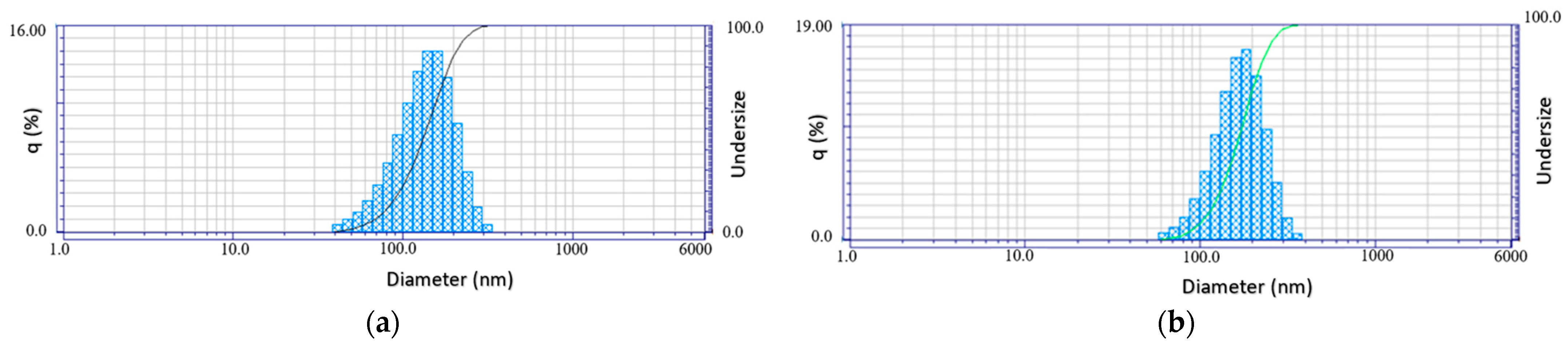
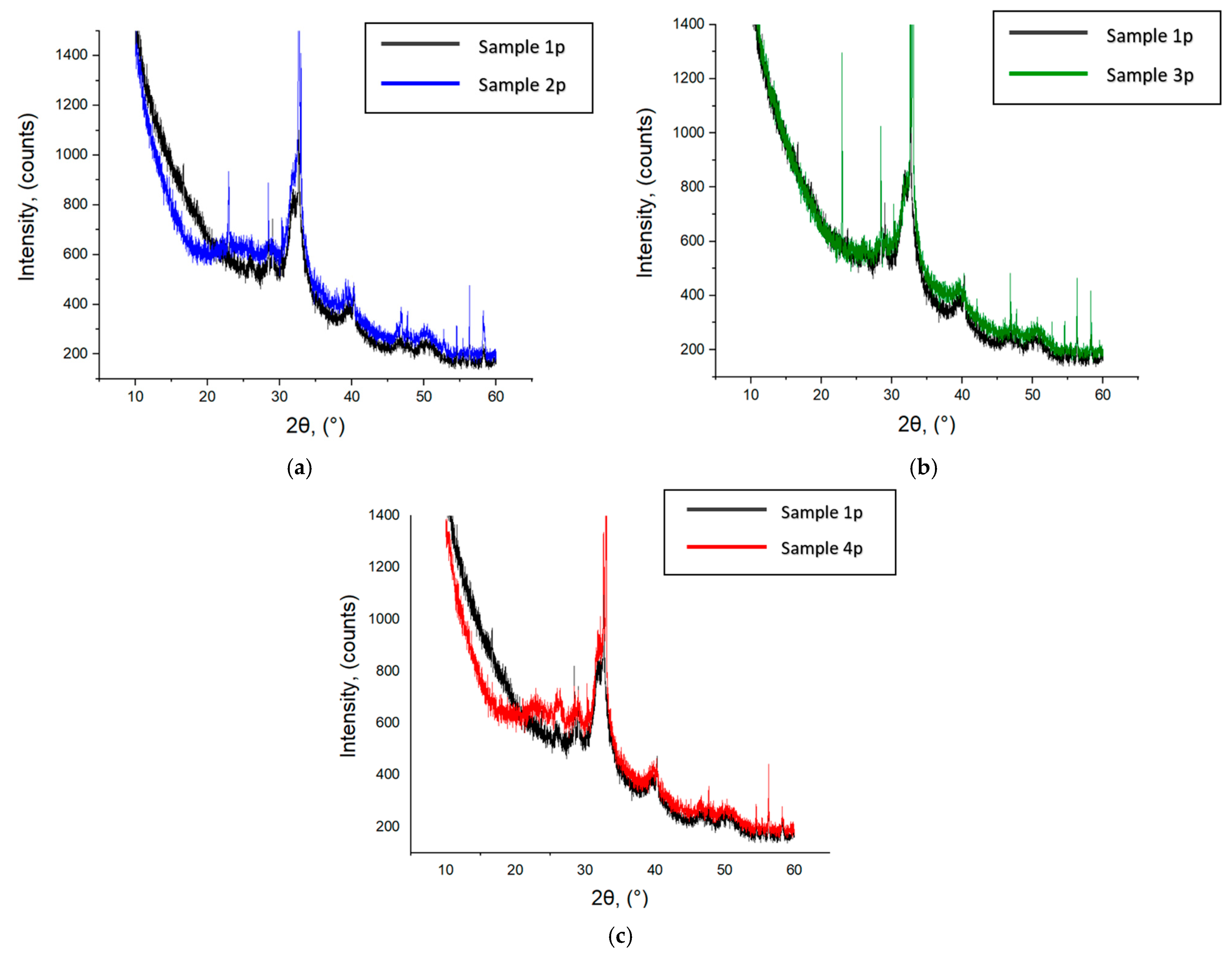
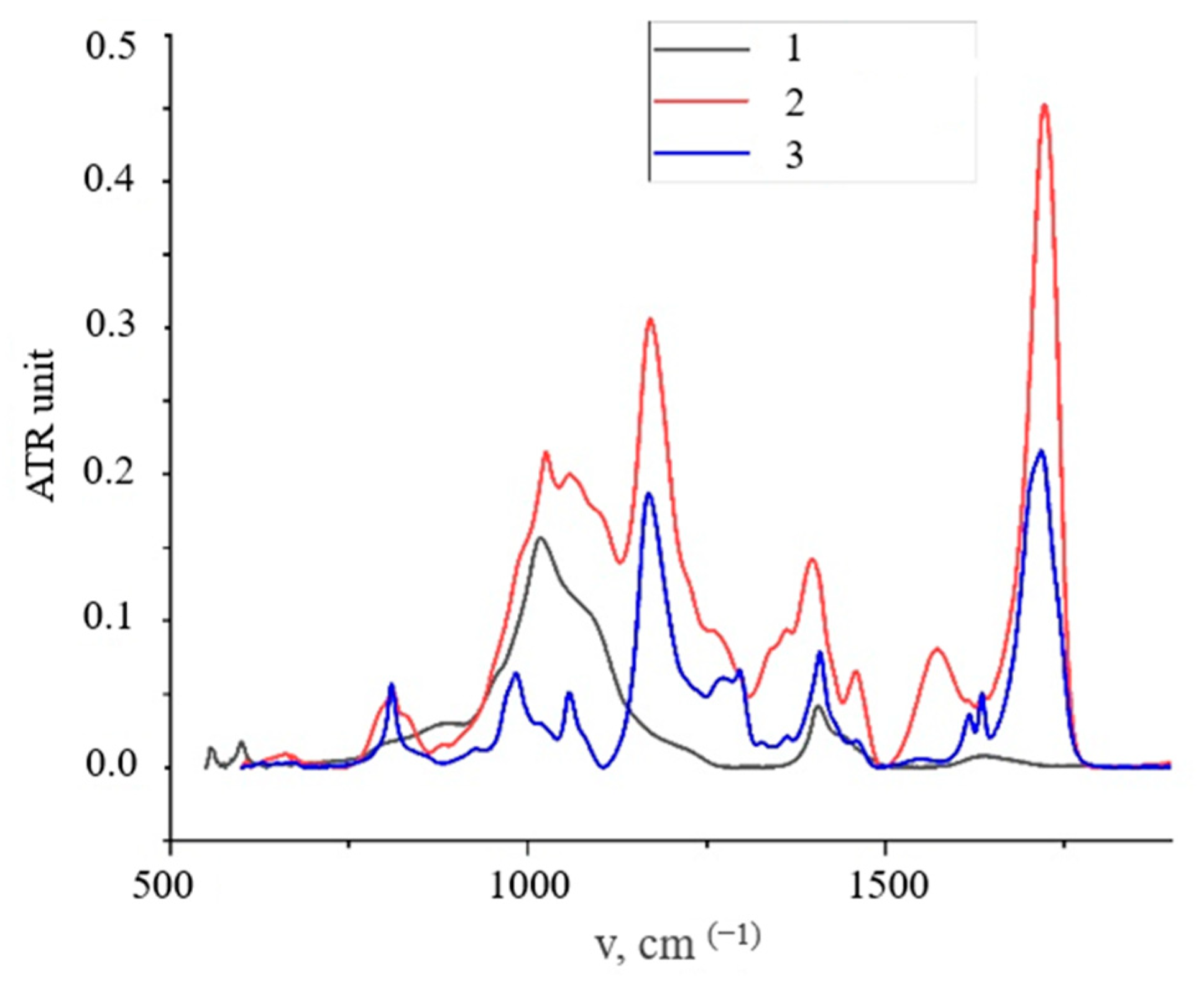
3.1. HA Nanoparticles

Studies of particle sizes in solutions were carried out using the dynamic light scattering method on the Horiba LB-550 installation. The refractive index of the medium is equal to the value of the refractive index for water—1.33. For hydroxylapatite particles, a value of 1.63 was set. The temperature in solutions was 25 °C.
Measurements were carried out at two stages of synthesis, when they were formed only in solution with nanoparticles of carboxylate and silicon oxide, and for the second time, after the addition of gelatin. Figure 2 shows histograms of the distribution of HA nanoparticles by size.
As can be seen, the size of the obtained particles in solution with carboxylate and silicon oxide nanoparticles is 135 nm, and after the adding of gelatin, it became 174 nm. The monomer 2-Carboxyethyl acrylate has carboxylate groups (–COO). The carboxylate groups (–COO) of the 2-Carboxyethyl acrylate monomer are capable of forming bonds with calcium ions on the surface of hydroxyapatite particles (Ca10(PO4)6(OH)2). In this way, an organic shell is formed on the surface of the HA particles. The shell can prevent the growth of HA particles and their agglomeration. It also improves the compatibility of inorganic HA particles with monomers and the formation of more stable composites. Some more details about the interaction between the carboxylate shell and hydroxyapatite nanoparticles are described below, in the IR spectroscopy data section.
The size distribution of the obtained HA nanoparticles, as well as the distribution for biological HA, is localized in a narrow range of 0–200 nm, and differs in that for biological HA, the maximum values are in the range of 50–100 nm. It is important to clarify that the size of silicon oxide nanoparticles is 7 nm, which does not make a significant difference.
The effect of the components on the Ca/P ratio in Si-HA nanoparticles was investigated by EDX method. Figure 3 shows the energy-dispersive X-ray spectra of the HA particles obtained in the presence of carboxylate and silicon oxide nanoparticles before and after the introduction of gelatin. The stoichiometric ratio data for the synthesized pure HA, as well as for HA with silicon nanoparticles only, with 2Carboxylate only, and together with SiO2 and 2Carboxylate, are presented in Table 2.
In the spectra, the element peaks of calcium, phosphorus, and silicon as well as carbon and oxygen elements can be observed since 2Carboxylate and gelatin are present. When examining the elemental composition of the particle’s atomic ratio, a value of Ca/P = 1.58 was obtained in the presence of 2Carboxylate and silicon oxide nanoparticles only, and Ca/P = 1.66 after the introduction of 2Carboxylate and silicon oxide nanoparticles together, which is close to the values of natural hydroxyapatite.
It can be seen that the introduction of silicon oxide increases the Ca/P ratio, but for all syntheses, the value is in the range acceptable according to modern scientific requirements, while it is known that the atomic Ca/P ratio for natural HA is 1.67.
The phase composition of HA obtained in solution with different components was determined by X-ray diffraction (XRD) analysis. Figure 4 shows the XRD results for HA nanoparticle dry powder: HA powder obtained in solution without additives (sample 1p); HA particle powder formed in solution with silicon oxide nanoparticles (sample 2p); HA nanoparticle powder formed in solution with 2Carboxylate (sample 3p); HA particle powder formed in solution with silicon oxide nanoparticles and with 2Carboxylate (sample 4p).
For all samples, only the formation of the HA phase corresponding to biological hydroxyapatite was recorded [27]. For samples with silicon oxide nanoparticles, a small blurred peak characteristic of the amorphous phase was observed.
The crystallite size for HA was calculated from the intensity of the X-ray diffractogram’s strong reflections by measuring the full width at half maximum (FWHM). The Scherrer equation for calculating the crystallite size is given as follows:
D = K λ β h k l cos θ h k l ,
where K is the Scherrer constant, λ is the wavelength of the X-ray for diffraction, βhkl is the FWHM of the corresponding plane, and θhkl is the angle measured of the same plane. The crystallite size calculated using XRD data (Figure 4) for HA grains in synthesized samples is presented in Table 3.
As can be seen from Table 3, the HA crystalline size strongly depends on the obtaining technique. The 2Carboxylate polymer matrix influences on the HA formation process due to the spatial separation of components. This results in the higher values of the HA grain size. No clear effect of silicon oxide on the formation of HA was found.
The processes of organic 2Carboxylate shell formation on the surface of HA nanoparticles were investigated by ATR (IR spectroscopy). As can be seen (Figure 5) in the spectrum of SiO2-HA particles modified with 2Carboxylates, the most notable peaks were at 1019 cm−1 (corresponding to PO43−, v3 stretching), 962 cm−1 (associated with PO43−, v1 stretching), and 600, 560 cm−1 (related to PO43−, v bending) [28]. The presence of the 1560 cm−1 band can be attributed as the valence vibration band of the carboxylate anion. In the spectrum of pure 2Carboxylate monomer, we observe a weak intensity band at 1550 cm−1. This band is enhanced and has a long-wave shift in the spectrum of HA nanoparticles with the organic 2carboxylate shell. In [29], it is proved by numerous examples that HA nucleation is initiated through a 2Carboxyl group if it is present in the reaction medium. This fact confirms the bonding of the 2Carboxylate monomer by its carboxylate group to the surface of HA nanoparticles. In our case, we observe a band at 1550 cm−1, which has a weak intensity in the spectra of pure 2Carboxylate. This fact confirms the bonding of the 2Carboxylate monomer by its carboxylate group to the surface of HA nanoparticles. The authors of [30] presented IR studies confirming the interactions of HA nanoparticles with the functional groups of polymers [28]. It has also been indicated [31] that the COOH and OH groups on the polymer surface can apparently act as initiation centers for HA growth.
Nanocomposite films were studied by optical spectroscopy and TEM (Figure 6). Studies using optical microscopy and TEM confirmed the homogeneity of the structure of the composite material after photocuring. As you can see, there are no micron-sized inclusions in the volume of composites.
Thus, the composite is a homogeneous mixture based on UV-curable acrylates, gelatin, and HA particles up to 400 nm in size. Photopolymerization results in the formation of a strong polymer matrix.

3.2. Wettability Angle and Water Sorption Rate of Nanocomposite Films

For good biocompatibility and proliferation of bone tissue cells, the implant coating material must be hydrophilic and have good sorption properties. Wettability angle changes for nanocomposite films were investigated and the sorption rate was calculated according to the data obtained. Based on the slope angle of the straight lines approximating the sorption graphs, sorption rate can be estimated for each composition (Table 4).
As can be seen, all the obtained nanocomposite materials are hydrophilic, and the addition of gelatin to acrylates always lowers the contact angle, which makes the nanocomposite more hydrophilic. Since the wettability angle decreases rapidly with time for all nanocomposites, this indicates a good sorption capacity of the materials.
The sorption rate varies considerably depending on the composition of the material. It is directly affected by the addition of gelatin and the monomer ratio. It can be seen that the addition of gelatin leads to an increase in the sorption rate, with a higher rate for the 1/1 monomer ratio than for 4/6.
Possible changes in the composition and structure of nanocomposites when in the aqueous medium are important characteristics for metal implant coating materials. Since the polymer material is formed by UV polymerization, the nanocomposite may contain residual monomers and other substances which are not embedded in the structure. Therefore, examining the materials while soaking them in an aqueous environment makes it possible to investigate the removal process of such components and determine the porosity of the material. For this reason, the material samples were kept in distilled water while the change in mass was monitored. On the 36th day of the study, lyophilic drying was carried out and then the samples were placed back into distilled water. On the 74th day of the experiment, the samples were taken out of the water and dried in the thermostat at 30 °C for a week, after which the weight loss was weighed and assessed using the following formula:
ΔM = (M0 − Mκ)/M0 × 100%,
where M0 is the initial weight of films and Mk is the weight of films after drying in the thermostat.
As can be seen from Figure 7, a non-monotonic change in weight was observed in Stage 1 for all nanocomposite samples when soaked in water. The increase in weight of the samples was due to the sorption of water, while the decrease in weight was due to the washing out of water-soluble components from the volume of the material. IR spectroscopic studies of the aqueous medium in which the films were kept showed the presence of traces of AL monomer (carboxylate), which shows good solubility in water but is poorly polymerizable.
It may be noted that the introduction of gelatin leads to a more monotonic change in the weight of the material while in water. This is particularly evident for formulations with a monomer ratio of 1/1.
The 2nd step was a lyophilization which was carried out after the weight of the samples kept in aqueous medium had ceased to change significantly. Examination of the samples with an optical microscope after lyophilic drying revealed no changes.
Stage 3—the lyophilically dried films were again placed in water. Figure 7 shows that for the samples with gelatin only, a consistent increase in weight is observed until constant values are reached; for the samples without gelatin, especially at a ratio of 4/6, an increase and decrease in weight is observed again.
Step 4—the drying of nanocomposite film samples at 30 °C in the thermostat.
As a result, the reduction in weight of the films as compared to the initial weight (ΔM %, Table 2) was evaluated. The weight loss ranges from 10% to 26% for the different compositions of nanocomposites. For nanocomposites without gelatin with an increased amount of a poorly polymerizing and water-soluble component (carboxylate)—Bis/Al = 1/1—the weight loss is half as much as for material with a 4/6 monomer ratio. It is important to note that the introduction of gelatin modifies the structure of nanocomposites. It can be seen that for the nanocomposite (Bis/Al = 1/1 + Gel), the mass loss compared to the materials without gelatin is twice as much, while for (Bis/Al = 4/6 + Gel), the mass loss is less. There is a significant degradation of acrylate–gelatin nanocomposites in aqueous medium by at least 20%, which is indicative of structural changes that create conditions for good bone tissue proliferation. It is important to point out that after being in an aqueous environment, the samples remained solid, not gel-like, and retained their shape.

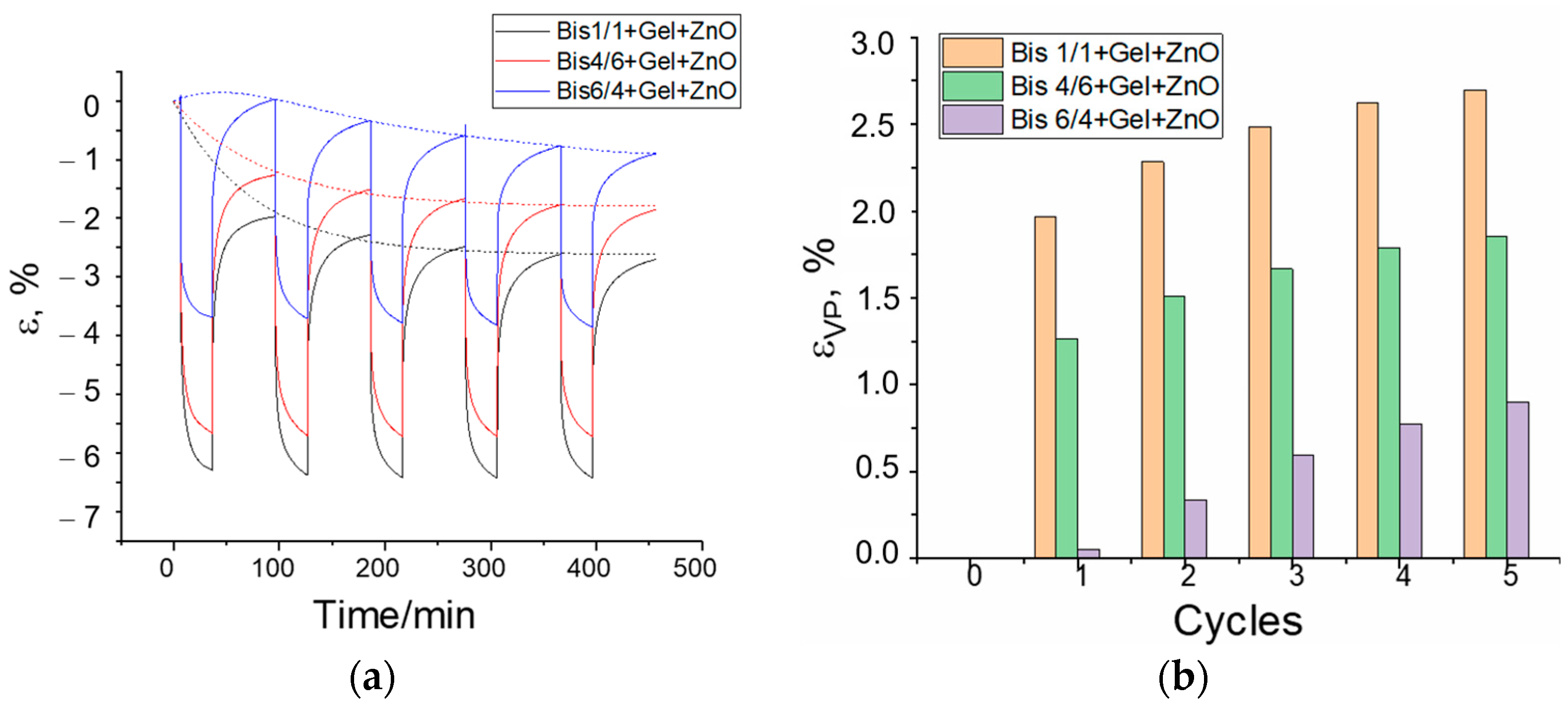
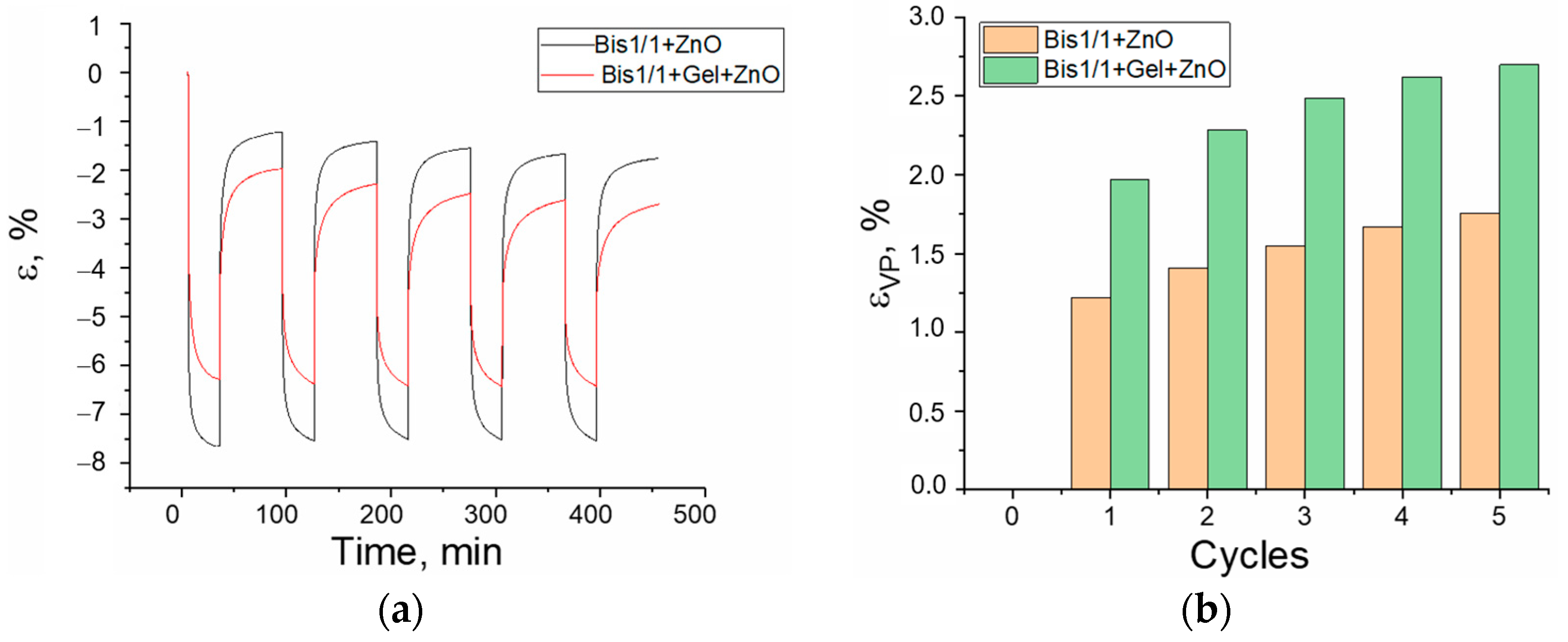
3.3. Creep/Recovery Test

The ability of polymeric materials to withstand loading is one of their most important performance characteristics. Most polymeric materials exhibit time-dependent deformation or creep under loading. Since this property is determined by the forces of intermolecular interaction between the chain segments, factors such as crystallinity, the number of low-molecular-weight additives, frequency of cross-linking of macromolecules, and other factors may significantly influence the creep rate. The value of creep can be estimated if the sample is subjected to mechanical action and the change in a strain is recorded over time. Firstly, the creep of a material should be measured by a single load and, therefore, the creep should be measured by a single deformation–relaxation cycle. Secondly, the creep of the same material should be studied under several load cycles; when at the beginning, samples are deformed rapidly to constant values and the subsequent stress relaxation is monitored over time.
Cyclic loading effects on the samples of BisA-2Car-HA/ZnO/ and BisA-2Car/HA/ZnO/Gel composites before and after incubation in phosphate buffer were performed at a constant stress level of 1 MPa and 37 °C using the penetration mode and the deformation values were recorded as a function of time. The results obtained with one exposure cycle are shown in Figure 8a,b.
For samples of BisA-2Car/Si HA/ZnO composites with no treatment in water, the effect of the addition of gelatin on the creep phase of the material can be clearly seen in Figure 8a. BisA-2Car/SiO2_HA/ZnO composites without gelatin addition (samples 1 and 2) show larger instantaneous deformation and larger residual strain or viscous flow deformation ε(VP) compared to composites with a different 2Ccarboxylate fraction containing gelatin. This means that matrices without gelatin show more plastic properties, while composites with gelatin show more elastic properties. This may be due to the structuring effect of gelatin for the acrylate matrix, where a decrease in the mobility of the polymer chains leads to an increase in the stiffness of the whole material [32,33]. Moreover, the low instantaneous and low residual deformation of a composite containing 40% 2Carboxylate characterizes it as a material with a more net-like structure compared to other materials.
Creep-recovery properties of nanocomposites have been investigated by simulating the conditions of their residence in the human body. After incubation in phosphate buffer for 14 days, the swollen polymer composites show close values of instantaneous deformation—5 to 7%, plastic deformation (elastic deformation ε(VI)) for all materials decreases, and residual deformation for the materials is 0.1 to 0.5%, i.e., the materials show typical behavior of polymer mesh materials (Figure 8b). These differences are probably related to the processes of residual monomer washout and gelatin swelling in phosphate buffer.
Figure 8 and Figure 9 show load–relaxation diagrams for composites subjected to several successive force cycles. As can be seen in Figure 8a, after several load cycles the accumulated strain increases in the untreated nanocomposites, with lower values also present for the sample with 40% 2Carboxylate content (Figure 9b).
For the same samples after conditioning in phosphate buffer medium (Figure 10), the gelatin-free polymer matrix has a significantly lower value of accumulated strain compared to the gelatin-filled composite, which may also indicate a more porous structure of the BisA-2Car/HA/ZnO/Gel composite.
After being in the aquatic environment for more than 70 days, the degradation is 20–26% (Table 4); the samples remained solid rather than gel-like. The material also demonstrated good mechanical hardness in the model medium after being in phosphate buffer for 14 days (FBS). This material is promising in terms of resorption. At the moment, the result can only be considered preliminary, but these results are better than were demonstrated in [34], as, for example, for PCL-based materials which have one of the degradation steps associated with the hydrolysis of an ester group. Further quantitative analyses are planned to study the effect of mechanical stability in more detail and will be published in the next paper.
The nanocomposite monomer compositions were applied to smooth titanium plates and to plates with surface relief in the form of parallel strips 30 μm deep with a spacing of 100 μm. After UV irradiation, polymer film coatings are formed which do not peel from the metal, both in air and in water or in phosphate buffer.
This material can be used to solve the following problems of regenerative medicine. The first is the repair of thinned bone cartilage in the joint bag. It is possible to carry out laparoscopic application and the curing of protective material on the bone surface instead of degraded cartilage tissue. The proliferation of biological tissues is ensured in the porous partially resorbable composite layer. The second direction is the coating of the surface of metal implants. In this case, the problems with mechanical loading are solved due to the properties of the implant metal, and the development of biotissues occurs in a porous composite polymer material. For both directions of use, both the primary application of layers and secondary repairs are possible, as needed. These application possibilities are due to the fact that the obtained composites have a set of positive properties for medical use:
-
The materials are liquid, but they are used without solvents, and they can be applied to a surface of any shape;
-
Materials are cured by a UV irradiation source;
-
Materials are biocompatible and, most importantly, partially resorbable. This property ensures that when composites are applied to a biological object or implant, there will always be a damping protective layer in which normal processes of bone and cartilage tissue development can take place.

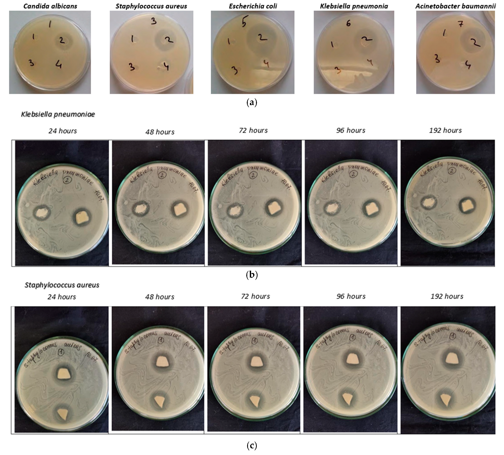
3.4. Antimicrobial Properties

The study for antimicrobial activity was carried out on five cultures of microorganisms: Candida albicans, Staphylococcus aureus, Escherichia coli, Klebsiella pneumonia, and Acinetobacter baumannii. The nanocomposite films were previously treated with UV rays in a laminar flow cabinet. The duration of the UFO was 40 min. The nutrient medium was prepared in advance and poured into Petri dishes. Preliminary studies of the antimicrobial activity of materials with and without zinc oxide nanoparticles have been conducted (Figure 11a). As you can see, one fact was observed for all microorganisms. Without zinc oxide nanoparticles, the material has no antimicrobial activity. The authors also investigated the dynamics of antimicrobial activity for films with zinc oxide nanoparticles for five types of microorganisms (Figure 11b,c). The two pieces of film were placed in one cup and placed in a thermostat.
Figure 11 shows the results of studies for two cultures, which show areas of inhibition of microbial growth. The diameters of the suppression zones are from 13 to 18 mm for Klebsiella pneumoniae and from 12 to 16 mm for Staphylococcus aureus. For other crops, the results are similar; the diameters of the suppression zones are shown in Table 5.
Table 5 shows the dynamics of the suppression of the growth of microorganisms; measurements were carried out for 196 h. The suppression zone decreased slightly over time. The results of the analysis showed that the films have antimicrobial properties, as they inhibit the growth of cultures.
To date, many materials for regenerative medicine in the field of orthopedics, dentistry, or maxillofacial surgery (implants, scaffolds, or other applications) have been developed [34,35,36,37]. Some modern composites are formed on the basis of nanoparticles and polymers. As a rule, all these materials have a fine-tuned biodegradability and are replaced with native tissues over time. However, orthopedic or dental implants require the ability to withstand high loads; that is why metals or alloys are used. This type of material does not provide tissue cell proliferation and creates problems of aseptic loosening. We offer a different way to solve the problem. We proposed a polymer composite that is a lining between metal and native tissues. It does not resorb completely, but pores are formed. In this way, conditions are created for an optimal microenvironment for cellular adhesion, proliferation, and differentiation. In this study, a new type of polymer composite was proposed. This can serve as a lining between metal and native tissues. As our investigation has shown, this composite has good mechanical characteristics, can partially and slowly be soluble in aqua solutions, and has good antibacterial properties. Thus, this material has several advantages and good prospects for regenerative medicine applications.

4. Conclusions

In this study, new material for the regeneration of bone or cartilage tissue was developed, and a simple and inexpensive method for obtaining it was proposed. The material is synthesized on the basis of photopolymerizable acrylates and natural polymer gelatin with nanoparticles of HA, zinc oxide, and silicon oxide. Nanoparticles can improve the biocompatibility of the material. Biocompatible natural polymer gelatin provides the formation of the porosity of the material when in the body’s aqueous environment. Photo-curable acrylates allow any shape of implant or coating layers to be obtained in a simple way. Therefore, it is possible to use viscous biomaterial in traditional applications of orthopedics and dentistry, as well as for laparoscopy. Since these acrylates are practically not resorbed in the body after photocuring, this could increase the lifetime of the implants covered by the material based on them. The authors managed to synthesize the composite material with improved properties. It is important that the conditions for the compatibility of all components have been found in order to obtain a homogeneous viscous composite, which after photocuring forms a homogeneous solid material. The advantage of that method is that no solvents are required for the further use of the viscous composite material. HA nanoparticles are formed in a viscous composite medium. The initial viscous composite can be applied to a surface of almost any shape. The properties and size of HA nanoparticles obtained in a water–organic medium in the presence of silicon oxide nanoparticles were studied. The nanocomposites are hydrophilic. The process of the formation of a porous structure in water was also studied. The porosity is 20–25% depending on the composition of the monomers. Thermomechanical studies of the material have shown that under cyclic loading, there are no significant residual deformations, which confirms the possibility of the long-term use of devices based on this material. The nanocomposite monomer compositions were applied to smooth titanium plates and to plates with surface relief. After UV irradiation, polymer film coatings are formed. The polymer film coatings exhibit good adhesion to titanium and retain integrity in phosphate buffer. The materials have antimicrobial properties, as they inhibit the growth of microorganism cultures (Candida albicans, Staphylococcus aureus, Escherichia coli, Klebsiella pneumoniae, Acinetobacter baumannii). In general, the material is a promising option for use in regenerative medicine. Such viscous biomaterial can be applied in traditional orthopedic and dental applications as well as for laparoscopy.

Author Contributions

Conceptualization, J.A.B., A.A.P. and S.K.; methodology, V.V.S., V.E.S., D.M.D., F.F.Z., A.A.P. and A.Z.; software, J.A.B.; validation, V.E.S. and A.C.; formal analysis, V.V.S., D.M.D. and D.R.M.; investigation, V.V.S. and A.C.; data curation, D.M.D.; funding acquisition, A.Z. All authors have read and agreed to the published version of the manuscript.

Funding

The research is partially funded by the Ministry of Science and Higher Education of the Russian Federation as part of the World-Class Research Center program Advanced Digital Technologies (contract No. 075-15-2022-312, dated 20 April 2022).

Data Availability Statement

The original contributions presented in the study are included in the article; further inquiries can be directed to the corresponding authors.

Acknowledgments

The XRD measurements were carried out in the frame of the projects TKP2021-NKTA-42 financed by the National Research, Development and Innovation Fund of the Ministry for Innovation and Technology, Hungary.

Conflicts of Interest

The authors declare no conflicts of interest.

References

  1. Ricotti, L.; Ciofani, G.; Mattoli, V.; Menciassi, A. Nano-Doped Matrices for Tissue Regeneration. In Advances in Regenerative Medicine; IntechOpen: London, UK, 2011; p. 295. [Google Scholar]
  2. Sun, L.; Berndt, C.C.; Gross, K.A.; Kucuk, A. Material fundamentals and clinical performance of plasma-sprayed hydroxyapatite coatings: A review. J. Biomed. Mater. Res. 2001, 58, 570–592. [Google Scholar] [CrossRef] [PubMed]
  3. De Goot, K.; Wolke, J.G.C.; Jancen, J.A. Calcium phosphate coatings for medical implants. Proc. Instn. Mech. Engrs. 1998, 212, 137–147. [Google Scholar] [CrossRef] [PubMed]
  4. Lacefield, W.R.; Hench, L.L. The bonding of Bioglass® to a cobalt-chromium surgical implant alloy. Biomaterials 1986, 7, 104–108. [Google Scholar] [CrossRef]
  5. Vehof, J.W.; Mahmood, J.; Takita, H.; Hof, M.V.; Kuboki, Y.; Spauwen, P.H.; Jansen, J.A. Ectopic bone formation in titanium mesh loaded with bone morphogenetic protein and coated with calcium phosphate. Plast. Reconstr. Surg. 2001, 108, 434–443. [Google Scholar] [CrossRef]
  6. Porter, A.E.; Patel, N.; Skepper, J.N.; Best, S.M.; Bonfield, W. Comparison of in vivo dissolution processes in hydroxyapatite and silicon-substituted hydroxyapatite bioceramics. Biomaterials 2003, 24, 4609–4620. [Google Scholar] [CrossRef]
  7. Porter, A.E.; Patel, N.; Skepper, J.N.; Best, S.M.; Bonfield, W. Effect of sintered silicate-substituted hydroxyapatite on remodelling processes at the bone–implant interface. Biomaterials 2004, 25, 3303–3314. [Google Scholar] [CrossRef] [PubMed]
  8. Pepla, E.; Besharat, L.K.; Palaia, G.; Tenore, G.; Migliau, G. Nano-hydroxyapatite and its applications in preventive, restorative and regenerative dentistry: A review of literature. Ann. Stomatol. 2014, 5, 108–114. [Google Scholar] [CrossRef]
  9. Kundu, B.; Ghosh, D.; Sinha, M.K.; Sen, P.S.; Balla, V.K.; Das, N.; Basu, D. Doxorubicin-inter-calated nano-hydroxyapatite drug-delivery system for liver cancer: An animal model. Ceram. Int. 2013, 39, 9557–9566. [Google Scholar] [CrossRef]
  10. Dorozhkin, S.V. Nanosized and nanocrystalline calcium orthophosphates. Acta Biomater. 2010, 6, 715–734. [Google Scholar] [CrossRef]
  11. Pang, X.; Zhitomirsky, I. Electrophoretic deposition of composite hydroxyapatite-chitosan coatings. Mater. Charact. 2007, 57, 339–348. [Google Scholar] [CrossRef]
  12. Kim, S.H.; Lim, B.K.; Sun, F.; Koh, K.; Ryu, S.C.; Kim, H.S.; Lee, J. Preparation of high flexible composite film of hydroxyapatite and chitosan. Polym. Bull. 2009, 62, 111–118. [Google Scholar] [CrossRef]
  13. Sun, F.; Zhou, H.; Lee, J. Various preparation methods of highly porous hydroxyapatite/polymer nanoscale biocomposites for bone regeneration. Acta Biomater. 2011, 7, 3813–3828. [Google Scholar] [CrossRef] [PubMed]
  14. Redepenning, J.; Venkataraman, G.; Chen, J.; Stafford, N. Electrochemical preparation of chitosan/hydroxyapatite composite coatings on titanium substrates. J. Biomed. Mater. Res. Part A 2003, 66, 411–416. [Google Scholar] [CrossRef]
  15. Liuyun, J.; Yubao, L.; Chengdong, X. Preparation and biological properties of a novel composite scaffold of nanohydroxyapatite/chitosan/carboxymethyl cellulose for bone tissue engineering. J. Biomed. Sci. 2009, 16, 65. [Google Scholar] [CrossRef]
  16. Zhou, W.Y.; Wang, M.; Cheung, W.L.; Ip, W.Y. Chapter 9—Selective laser sintering of poly (L-Lactide)/carbonated hydroxyapatite nanocomposite porous scaffolds for bone tissue engineering. In Tissue Engineering; InTech: London, UK, 2010; pp. 179–204. [Google Scholar]
  17. Wang, X.; Zhang, Y.; Chen, S. Recent Advances in Biocompatible Polymeric Materials for Tissue Engineering. J. Mater. Chem. B 2023, 11, 258–271. [Google Scholar]
  18. Liu, L.; Wang, J.; Li, H. Design and Characterization of Biocompatible Polymeric Materials for Drug Delivery. Polym. Chem. 2024, 21, 367–380. [Google Scholar]
  19. Huang, W.; Chen, H.; Wu, Q. Biocompatible Polymeric Materials for Implantable Medical Devices. Mater. Sci. Eng. C 2024, 42, 189–202. [Google Scholar]
  20. O’Brien, C.M.; Holmes, B.; Faucett, S.; Zhang, L.G. Three-Dimensional Printing of Nanomaterial Scaffolds for Complex Tissue Regeneration. Tissue Eng. Part B 2014, 21, 103–114. [Google Scholar] [CrossRef]
  21. Álvarez-Chimal, R.; Arenas-Alatorre, J.Á.; Álvarez-Pérez, M.A. Nanoparticle-polymer composite scaffolds for bone tissue engineering. Eur. Polym. J. 2024, 213, 113093. [Google Scholar] [CrossRef]
  22. Turnbull, G.; Clarke, J.; Picard, F.; Riches, P.; Jia, L.; Han, F.; Li, B.; Shu, W. 3D bioactive composite scaffolds for bone tissue engineering. Bioact. Mater. 2018, 3, 278–314. [Google Scholar] [CrossRef]
  23. Maglio, M.; Salamanna, F.; Brogini, S.; Borsari, V.; Pagani, S.; Nicoli Aldini, N.; Giavaresi, G.; Fini, M. Histological, histomorphometrical, and biomechanical studies of bone-implanted medical devices: Hard resin embedding. BioMed Res. Int. 2020, 2020, 1804630. [Google Scholar] [CrossRef] [PubMed]
  24. Dorozhkin, S.V. Calcium Orthophosphate-Based Bioceramics. Materials 2013, 6, 3840–3942. [Google Scholar] [CrossRef] [PubMed]
  25. Csarnovics, I.; Burunkova, J.; Sviazhina, D.; Oskolkov, E.; Alkhalil, G.; Orishak, E.; Nilova, L.; Szabo, I.; Rutka, P.; Bene, K.; et al. Development and study of biocompatible polyurethane-based polymer-metallic nanocomposites. J. Nanotechnol. Sci. Appl. 2020, 13, 11–22. [Google Scholar] [CrossRef]
  26. Kulyashova, K.S.; Sharkeyev, Y.P. Preparation of synthetic hydroxyapatite for the formation of bio-coatings on medical implants. Chem. Sustain. Dev. 2011, 19, 447–453. [Google Scholar]
  27. Jongwattanapisan, P.; Charoenphandhu, N.; Krishnamra, N.; Thongbunchoo, J.; Tang, I.M.; Hoonsawat, R.; Smith, S.M.; Pon-On, W. In vitro study of the SBF and osteoblast-like cells on hydroxyapatite/chitosan–silica nanocomposite. Mater. Sci. Eng. C 2011, 31, 290–299. [Google Scholar] [CrossRef]
  28. Dorozhkin, S.V.J. Calcium Orthophosphate-Containing Biocomposites and Hybrid Biomaterials for Biomedical Applications. Funct. Biomater. 2015, 6, 708–832. [Google Scholar] [CrossRef]
  29. Ahmad Hariza, A.M.A.; Mohd Yunus, M.H.; Fauzi, M.B.; Murthy, J.K.; Tabata, Y.; Hiraoka, Y. The Fabrication of Gelatin–Elastin–Nanocellulose Composite Bioscaffold as a Potential Acellular Skin Substitute. Polymers 2023, 15, 779. [Google Scholar] [CrossRef] [PubMed]
  30. Soradech, S.; Nunthanid, J.; Sriamornsak, P.; Limmatvapirat, S.; Luangtana-anan, M. Factors affecting on the enhancement of mechanical properties of composite edible film based on Shellac and Gelatin. Thai J. Agric. Sci. 2011, 4, 263–269. [Google Scholar]
  31. Afewerki, S.; Sheikhi, A.; Kannan, S.; Ahadian, S.; Khademhosseini, A. Gelatin-polysaccharide composite scaffolds for 3D cell culture and tissue engineering: Towards natural therapeutics. Bioeng. Transl. Med. 2018, 4, 96–115. [Google Scholar] [CrossRef]
  32. Lee, K.Y.; Shim, J.; Lee, H.G. Mechanical properties of gellan and gelatin composite film. Carbohydr. Polym. 2004, 56, 251–254. [Google Scholar] [CrossRef]
  33. Li, B.; Webster, T. (Eds.) Progress in Biology, Manufacturing, and Industry Perspectives; Orthopedic Biomaterials; Springer: Berlin/Heidelberg, Germany, 2018. [Google Scholar] [CrossRef]
  34. Habibzadeh, F.; Sadraei, S.M.; Mansoori, R.; Chauhan, N.P.S.; Sargazi, G. Nanomaterials supported by polymers for tissue engineering applications: A review. Heliyon 2022, 8, e12193. [Google Scholar] [CrossRef]
  35. Filippi, M.; Born, G.; Chaaban, M.; Scherberich, A. Natural polymeric scaffolds in bone regeneration. Front. Bioeng. Biotechnol. 2020, 8, 474. [Google Scholar] [CrossRef]
  36. Kumari, S.; Katiyar, S.; Darshna Anand, A.; Singh, D.; Singh, B.N.; Mallick, S.P.; Mishra, A.; Srivastava, P. Design strategies for composite matrix and multifunctional polymeric scaffolds with enhanced bioactivity for bone tissue engineering. Front. Chem. 2022, 10, 1051678. [Google Scholar] [CrossRef] [PubMed]
  37. Amiryaghoubi, N.; Fathi, M.; Pesyan, N.N.; Samiei, M.; Barar, J.; Omidi, Y. Bioactive polymeric scaffolds for osteogenic repair and bone regenerative medicine. Med. Res. Rev. 2020, 40, 1833–1870. [Google Scholar] [CrossRef] [PubMed]
Figure 1. The structural formula of the Bis—GMA acrylate monomer.
Jcs 09 00065 g001
Figure 2. Size of HA particles: (a) in the presence of carboxylate and silicon oxide nanoparticles; (b) in the presence of 2-carboxylate, silicon nanoparticles, and gelatin (q (%)—percentage of particles by diameter; undersize—total particle content).
Jcs 09 00065 g002
Figure 3. Energy-dispersive spectra of hydroxyapatite samples: (a) in presence of carboxylate and silicon oxide nanoparticles; (b) after addition of gelatin.
Jcs 09 00065 g003
Figure 4. XRD results of HA powder samples (Table 2) obtained in aqueous solution with different accompanying components: (a) HA (sample 1) and HA particles formed in the presence of silicon oxide nanoparticles only (sample 2); (b) HA (sample 1) and HA nanoparticles formed in solution with 2Ccarboxylate (sample 3); (c) HA (sample 1) and HA particles formed in the simultaneous presence of silicon oxide nanoparticles and 2Carboxylate (sample 1).
Jcs 09 00065 g004
Figure 5. IR spectra: 1—silica hydroxyapatite nanoparticles (black, sample 2 in Table 2 or Table 3); 2—silica hydroxyapatite nanoparticles surface-modified with 2Carboxylate (red, sample 4 in Table 2 or Table 3); 3—monomer 2Carboxylate (blue).
Jcs 09 00065 g005
Figure 6. Composite image 1. (a)—Optical microscope; (b)—TEM.
Jcs 09 00065 g006
Figure 7. Changes in relative mass of film samples (Table 1) when soaking in water: (a) nanocomposites 2 (Bis1/1 + ZnO) and 4 (Bis4/6 +ZnO) based on monomers only; (b) nanocomposites 1 (Bis1/1 + Gel + ZnO) and 3 (Bis4/6 + Gel + ZnO) based on monomers with gelatin.
Jcs 09 00065 g007
Figure 8. Creep curves of dry polymeric materials (a) and wet polymeric materials (after staying in phosphate buffer solution—PBS, wet); (b) at a constant stress level of 1 MPa and 37 °C (ε-creep [%]).
Jcs 09 00065 g008
Figure 9. Dry sample: Bis1/1 + Gel + ZnO, Bis4/6 + Gel + ZnO, Bis6/4 + Gel + ZnO; 1 multi-step creep response of polymer samples at 37 °C and at 30 min steps. (a)—Strain–relaxation cycles; (b)—residual strain (∆L/L0—slope of relative change in length; ε-creep [%]).
Jcs 09 00065 g009
Figure 10. Multi-step creep response of samples at 37 °C and body conditions for wet state after 14 days in PBS (phosphate buffer): (a)—deformation–relaxation cycles; (b)—accumulation of permanent deformation.
Jcs 09 00065 g010
Figure 11. Antimicrobial activity test results (on solid nutrient medium BisA-2Car/SiO2_HA/Gel with and without ZnO): (a) preliminary results. Sample 2 in the Petri dish has ZnO; other samples have a similar composition but are without zinc oxide. Photos of samples after 10 days: 1—Candida albicans; 3—Staphylococcus aureus; 5—Escherichia coli; 6—Klebsiella pneumonia; 7—Acinetobacter baumannii; (b,c) studies of the antimicrobial activity of the composite over time: (b)—Klebsiella pneumoniae; (c)—Staphylococcus aureus.
Jcs 09 00065 g011
Table 1. Compositions of nanocomposites.
NoNanocompositeBisA/2Carboxylate
[wt/wt]
SiO2
[wt%]
CaCl2 + H3PO4
[wt%]
Gelatin
[wt%]
DMPA
[wt%]
ZnO
[wt%]
1Bis1/1 + Gel + ZnO1/1810200.50.5
2Bis1/1 +ZnO1/1810-0.50.5
3Bis4/6 + Gel + ZnO4/6810200.50.5
4Bis4/6 + ZnO4/6810-0.50.5
5Bis6/4 + Gel + ZnO6/4810200.50.5
Table 2. Weight percentages of Ca/P.
NoComposition of the SynthesisCa/P [wet %]
1pCaCl2 + H3PO41.67
2pSiO2 + CaCl2 + H3PO42.19
3pCaCl2 + H3PO4 + 2Car1.58
4pSiO2 + CaCl2 + H3PO4 + 2Car1.66
Table 3. Crystallite size of HA grains in synthesized samples using different methodology.
NoSampleAverage Crystallite Size, [nm]
1pHA powder4.05
2pHA + SiO2 powder5.93
3pHA particles in carboxylate12.63
4pHA + SiO2 particles in carboxylate8.15
Table 4. Wettability angle, sorption rate, and mass loss of nanocomposite films in water.
NoNanocompositeContact Angle
[degree]
Sorption Rate
[deg/min]
Weight Loss Due to Soaking
[%]
1Bis1/1 + Gel + ZnO54.64.225
2Bis1/1 +ZnO67.52.410
3Bis4/6 + Gel + ZnO52.32.320
4Bis4/6 + ZnO62.21.826
Table 5. Dynamics of suppression of growth of microorganisms by films of BisA-2Car/SiO2_HA/ZnO/Gel.
MicroorganismsRadius of Growth Suppression, mm
24 h48 h72 h96 h192 h
Candida albicans17 × 1916.5 × 18.515.5 × 1814 × 16.514 × 15
Staphylococcus aureus14 × 1613 × 14.512 × 1412 × 14-
Escherichia coli22 × 2320 × 2018 × 1917 × 17.517 × 17.5
Klebsiella pneumoniaeLeft 15 × 17
Right 18 × 18
15 × 17
18 × 18
14 × 16
16.5 × 17
13 × 16
16.5 × 17
-
16.5 × 17
Acinetobacter baumanniiLeft 20 × 21
Right 21 × 21
19 × 19.5
20 × 20
19 × 19.5
20 × 20
19 × 19.5
20 × 20
-
20 × 20
Disclaimer/Publisher’s Note: The statements, opinions and data contained in all publications are solely those of the individual author(s) and contributor(s) and not of MDPI and/or the editor(s). MDPI and/or the editor(s) disclaim responsibility for any injury to people or property resulting from any ideas, methods, instructions or products referred to in the content.

