Article

Characterization and Antibacterial Properties of Centrifugally Spun Polyvinylpyrrolidone/Copper(II) Acetate Composite Fibers

1
School of Integrative Biological and Chemical Sciences, University of Texas, Rio Grande Valley, Edinburg, TX 78539, USA
2
Mechanical Engineering Department, University of Texas, Rio Grande Valley, Edinburg, TX 78539, USA
3
School of Integrative Biological and Chemical Sciences, University of Texas, Rio Grande Valley, 1 West University Blvd, Brownsville, TX 78521, USA
4
School of Earth, Environmental and Marine Sciences, University of Texas, Rio Grande Valley, 1 West University Blvd, Brownsville, TX 78521, USA
*
Authors to whom correspondence should be addressed.
These authors contributed to this work equally.
J. Compos. Sci. 2025, 9(11), 590; https://doi.org/10.3390/jcs9110590 (registering DOI)
Submission received: 25 September 2025 / Revised: 22 October 2025 / Accepted: 24 October 2025 / Published: 1 November 2025
(This article belongs to the Special Issue Polymer Composites and Fibers, 3rd Edition)

Abstract

The demand for effective antibacterial materials is growing rapidly in today’s world. Both metallic and metal oxide nanoparticles have been widely used as antibacterial agents against various bacterial species due to their unique mechanisms of destroying bacterial membrane cells. The current study explores the antibacterial activity of centrifugally spun fibers prepared from copper acetate polyvinylpyrrolidone (PVP) ethanol precursor solutions against both Gram-negative and Gram-positive bacteria. During the synthesis of the composite fibers, the physical and chemical conditions were optimized. The structure and morphology of the PVP/Cu-Ac fibers were analyzed using scanning electron microscopy (SEM), X-ray diffraction (XRD), energy-dispersive X-ray spectroscopy (EDS), and thermogravimetric analysis (TGA). The antibacterial activity of PVP/copper acetate fibers was tested against Gram-positive Staphylococcus aureus and Gram-negative Escherichia coli. The PVP/Copper acetate fibers demonstrated bactericidal activity against both bacterial strains, making the PVP/copper acetate composite fibers an effective material for biomedical applications.

1. Introduction

Over the last few decades, there has been an increasing interest in new materials with high antibacterial activity, especially for species that are acquiring new antibiotic resistance. New antibacterial materials have been developed to treat bacteria that have developed new resistance to antibiotics, leaving healthcare with limited options [1]. Different materials, such as antibiotics, disinfectants, and antiseptics, have been used as antibacterial agents against Gram-negative and Gram-positive bacteria [2,3]. Common antibiotics have been shown to be less effective against Staphylococcus aureus (S. aureus); therefore, infection management is becoming a significant challenge for patients in hospitals [4]. Methicillin-resistant Staphylococcus aureus (MRSA) is an epidemic that threatens the community and healthcare systems [5]. Furthermore, Escherichia coli (E. coli) is becoming increasingly resistant to common antibiotics around the world [6]. This has led researchers to focus on developing new antibacterial materials that are not antibiotics to reduce bacterial infections in hospital settings. Nanotechnology is becoming increasingly utilized to produce nanostructured fibers for biomedical purposes, including metal-embedded nanofibers with antibacterial properties.
Multiple transition metals, such as Cu, Zn, Cd, PB, and Co, have been found to be effective antibacterial agents against both Gram-positive and Gram-negative bacteria due to their capacity to effectively cause damage to the cell membrane [7,8]. Copper, in particular, is abundant on Earth and both affordable and sustainable [9]. It is also an essential trace element in living organisms because it serves as a cofactor for key enzymes in the body [10]. Although small levels of copper are typically found in the human body, high blood copper concentrations are harmful and may cause health issues [11]. Copper and copper (II) oxide nanoparticles (NPs) have been found to be an effective antibacterial agent against both Gram-positive and Gram-negative bacteria [12]. Additionally, results reported in the literature have shown that copper is also effective against fungi, algae, and viruses in addition to bacteria [13].
However, researchers have not determined the exact antibacterial mechanism of these metals. Contact killing is thought to be caused by their positive charge, which binds to bacterial cell walls with a negative charge, causing membrane damage [14]. Metal ions can form reactive oxygen species (ROS), which induce cellular stress, affecting transcription and causing downregulation of many genes, particularly those involved in bacterial energy production, ultimately resulting in cellular death [7,14]. Yalcinkaya et al. assessed the effectiveness of several oxidative metals’ antibacterial activity and found that CuO > ZnO = ZnO/TiO2 > AgNO3 > ZrO2 > TiO2 > SrO2 [15]. According to numerous studies, copper and its precursors exhibit the strongest antibacterial properties [15,16]. Various copper composite nanofibers have been tested against both Gram-positive and Gram-negative bacteria, demonstrating good antibacterial activity. The results showed that copper salts outperformed other copper precursors in terms of antibacterial activity due to their capacity to release Cu2+ instantly [17]. Copper (II) acetate, a copper salt, is a greener alternative that can be synthesized by reacting copper (II) oxide with acetic acid [18]. Previous research has proved that Cu is an effective antibacterial agent. The antibacterial activity of copper could be enhanced by embedding Cu NPs in polymer nanofibers. The unique properties of nanofibers, such as their high surface area to volume ratio, high porosity, and mechanical and thermal stability, make them promising candidates for tissue repair, wound dressing, and tissue engineering [19]. Drug delivery for medicinal therapies has been successfully accomplished by the application of nanofibers [19].
Nanofibers can be prepared by multiple methods, including electrospinning [20], phase separation, template synthesis, self-assembly, dry spinning [21], and centrifugal spinning [22,23,24,25,26,27]. Centrifugal spinning has been widely used to prepare polymer/ceramic fibers from conductive and nonconductive precursor solutions to produce fibers with high production rate/almost 500 faster than electrospinning, which significantly can reduce the production cost of fibers/nanofibers [22,23,24,25,26,27]. However, electrospinning is the most widely used method due to its simplicity and low cost [21]. Nanofibers are known for their flexibility; nanofiber mats facilitate easy loading and attachment, enabling control over the bulk mechanical properties [1]. The morphology of fibers can be controlled by monitoring the electrospinning parameters and the solution concentration [28]. Studies have indicated that thicker nanofibers are produced when the polymer concentration is higher and the relative humidity is low [29]. Thicker nanofibers support sustained release properties, which enhance antibacterial activity [29]. Additionally, Hamdan et al. noted that designing nanofibers with a small diameter (300–1000 nm), positively charged surfaces, rough surfaces with a high surface area, hydrophobic surfaces, and large pore diameters can promote better adhesion of nanofibers, thereby improving their antibacterial activity [19]. Generally, nanofibers are made of polymers using electrostatic force. Polyvinylpyrrolidone (PVP), a water-soluble polymer, has been demonstrated to be safe for use on human skin due to its temperature resistance, low toxicity, biological inertness, and non-irritant and non-sensitizing properties [4]. Due to these features, PVP has been utilized as a polymer in various biomedical applications, most notably in the preparation of nanofibers. Previous research has demonstrated that copper precursor NPs embedded in PVP nanofibers are effective against bacteria [30]. Different polymers/nanoparticle precursor solutions, including PVP, were used by our group to prepare centrifugally spun composite fibers as antibacterial agents against both Gram-positive and Gram-negative bacteria [22,30,31,32]. However, to the best of the author’s knowledge, the antibacterial activity of PVP/copper acetate (CuAc/PVP) composite fibers has not been investigated
The objectives of this research are to process and examine the structure and morphology of centrifugally spun CuAc/PVP fibers and assess their inhibition of Gram-positive and Gram-negative bacteria growth, specifically S. aureus and E. coli, which differ in their cell membrane and cell wall compositions. SEM, TGA, EDS, and XRD were used to characterize and assess the morphology of the fibers. CuAc/PVP composite fibers were successfully synthesized through centrifugal spinning and coated with different concentrations of copper acetate. CuAc/PVP composite fibers could be used as a wound dressing on damaged skin to prevent the growth of bacteria. These fibers mimic the skin’s extracellular matrix and provide a gentle application, even on sensitive skin.

2. Experimental

2.1. Materials

All chemicals were used as received without further purification or treatment. Polyvinylpyrrolidone (PVP) (96.8%) with an average molecular weight of 1,300,000 and copper (II) acetate (CuAc, Cu(CH3COO)2 99.99%) were purchased from Sigma-Aldrich (St. Louis, MO, USA). Absolute ethanol was used as the solvent for solution preparation.

2.2. Preparation of CuAc/PVP Composite Fibers

Stock solution of 15% (w/w) PVP was prepared by dissolving PVP in absolute ethanol under stirring. Four precursor solutions with different concentrations of copper (II) acetate (CuAc) 5%, 10%, 25% and 50% relative to the PVP content, were prepared by adding Copper (II) acetate to the PVP solution. The precursor solutions were magnetically stirred overnight at 650 rpm using a Thermo Scientific Cimarec+4 × 4 HP120 stirrer (Thermo Scientific, Waltham, MA, USA) to ensure complete dispersion and homogeneity. The homogeneous CuAc/PVP precursor solutions were then processed by centrifugal spinning using a Cyclone L-1000 M (Fiberio Technology Corporation, McAllen, TX, USA) to prepare the composite fibers. The centrifugal spinning setup system has a spinneret equipped with 30-gauge, half-inch, regular bevel needle (Beckett-Dickerson, Franklin Lakes, NJ, USA). The Cyclone L1000 M can operate at a spinneret rotational speed range from 500 to 22,000 RPM with 1 to 2000 s per spinning run. The rotational speed of the spinneret was checked by using a digital tachometer(Cole-Parmer, Vernon Hills, IL, USA) to ensure the rotational speed was correct. The CuAc/PVP solution was injected into the spinneret and sufficient centrifugal forces were applied to overcome the surface tension of the polymer droplets. The CuAc/PVP composite fibers were then collected, and the process was repeated 5 times to form a fibrous mat. Centrifugal spinning was performed at 8500 rpm for 2 min at room temperature with relative humidity maintained at 55% ± 5%. The spinneret was placed inside the center hole of the Cyclone L1000 M and tightened with a wrench. The spin axis of this spinneret extends centrally and vertically through the hole, perpendicular to the top plate. The fibers were collected on 8 vertical collectors with 13 cm between the columns, which were positioned at a distance of 16 cm from the center of the spinneret. Multiple spinning trials were performed to optimize the fiber formation. The collected fibers were wrapped in aluminum foil and dried in a vacuum oven(MTI Corporation, Richmond, CA, USA) for 24 h.

2.3. Characterization

The morphology of CuAc/PVP composite fibers was analyzed using a field-emission scanning electron microscope (FE-SEM; Sigma VP Carl Zeiss, White Planes, NY, USA). The average fiber diameter was calculated from SEM images taken at 500× magnification by averaging the measurement of 110 fibers using the image analysis software JMicroVision V.1.2.7, (University of Geneva, Geneva, Switzerland) and Origin Pro® 2020 software (OriginLab Corporation, Northampton, MA, USA). Energy dispersive spectroscopy (EDS) (EDAX, Pleasanton, CA, USA) was performed to evaluate the elemental composition of the composite fibers and to confirm the presence of CuAc NPs at varying concentrations. Thermogravimetric analysis (TGA) of the composite fibers was performed using a Perkin Elmer TGA 7 (Perkin Elmer, Shelton CT, USA) under an air atmosphere. Fiber samples of 10 mg were placed into crucibles and heated from 23 to 700 °C at a heating rate of 5 °C/min. Fourier-transformed infrared spectroscopy (FTIR) data were collected using a Perkin Elmer Frontier FTIR spectrometer (Perkin Elmer, Shelton, CT, USA) equipped with a UATR accessory. Spectra were recorded at a resolution of 2 cm−1 over a range of 650–4000 cm−1. X-ray diffraction (XRD) data was collected using a Rigaku Miniflex 600 diffractometer (Rigaku Americas Corporation, Woodlands, TX, USA) on both fiber and powder samples. The XRD measurements were performed with a Cu source (Kα 1.540 Å). Data were collected over a 2θ range from 5 to 60° with a step of 0.05. The fitting of the XRD patterns was performed using the LeBail procedure in the Fullprof Suite (V 5.20, Grenoble, France) [33].

2.4. Antibacterial Testing

The antibacterial testing of the PVP and the CuAc/PVP composite fibers was performed against Gram-positive S. aureus and Gram-negative E. coli. The Kirby-Bauer disk diffusion method was used to evaluate the antibacterial properties of the composite fibers. Agar plates were prepared by inoculating 100 µL of bacterial suspension, which was uniformly spread using a sterile L-shaped glass rod. The bacterial solutions transferred to each Petri dish contained approximately 2 × 10⁷ CFU/mL. The different fibrous mats were cut into circular disks of 12 mm diameter using a punch to allow for consistent and clear visualization of antibacterial performance. The fiber disks were then placed on the agar plates using sterile tweezers. Finally, the plates were incubated at 36° C for 48 h. The antibacterial testing of the CuAc/PVP composite fibers was conducted against Gram-positive S. aureus and Gram-negative E. coli.

3. Results and Discussion

Centrifugally spun PVP and CuAc/PVP composite fibers were produced after several optimization trials involving variations in the process parameters such as rotational speed of the spinneret, spinning time, solution concentration, and relative humidity. For optimal fiber formation, a 15% (w/w) PVP solution was required; lower or higher polymer concentrations did not result in homogeneous fibrous mats. A spinneret rotational speed of 8500 rpm was determined to be optimal for generating fine fibers, considering a relative humidity of 55 ± 5%. Higher rotational speeds led to the formation of beaded fibers.
The SEM images shown in Figure 1 illustrate the morphology and average fiber diameters of composite fibers produced with varying concentrations of CuAc, ranging from 2.266 to 2.663 µm. The corresponding fiber diameter distributions, presented in histograms, Figure 1C,F,I,L,O, indicate that the average fiber diameter does not increase proportionally with the CuAc NPs concentration. The fibers showed an average diameter of 2.380 ± 0.80, 2.656 ± 0.66, 2.663 ± 0.53, 2.659 ± 0.58, and 2.266 ± 0.59 μm, for the PVP, 5% CuAc, 10% CuAc, 25% CuAc, and 50% CuAc, respectively. Notably, fibers containing 10% (w/w) showed the largest average diameter, while those with 50% (w/w) displayed the smallest. This trend may be influenced by variations in spinning parameters, such as rotational speed and relative humidity, which require further investigation.
Figure 2 and Figure 3 present the EDS mapping of the CuAc/PVP composite fibers containing 25 and 50% (w/w) CuAc, respectively. The presence of carbon, oxygen, nitrogen, and copper confirms that the polymer matrix and active material were homogenously distributed. Table 1 presents the elemental composition based on EDS analysis collected during SEM characterization. Table 1 shows that the sample with 25% (w/w) CuAc NPs had a measured copper content of 5.25%, while the 50% (w/w) CuAc NPs sample showed a slightly lower copper content of 5.10%. This slight discrepancy is probably due to surface effects and the distribution of the material. Since EDS is a surface-sensitive technique, copper may appear less abundant if it is more deeply embedded or dispersed unevenly, which can occur at higher concentrations as a result of aggregation or phase separation. Additionally, variations in fiber thickness and the inherent limitations of EDS quantification may contribute to this unexpected result. It can be determined from the EDS mapping that the CuAc NPs were very well distributed in the sample.
Table 2 presents the results of the TGA analysis, which was performed under an air atmosphere, to determine the amount of copper acetate present in the sample. Based on the results, the 5% (w/w) CuAc sample contained approximately 4.4% (w/w) of CuAc, while the 10% (w/w) CuAc sample showed a content of approximately 8% (w/w) of CuAc. Interestingly, the 25 and 50% (w/w) CuAc samples both exhibit a similar copper content of 16.4% (w/w). This observation suggests that there may be a solubility or a loading limit for CuAc in the PVP matrix.
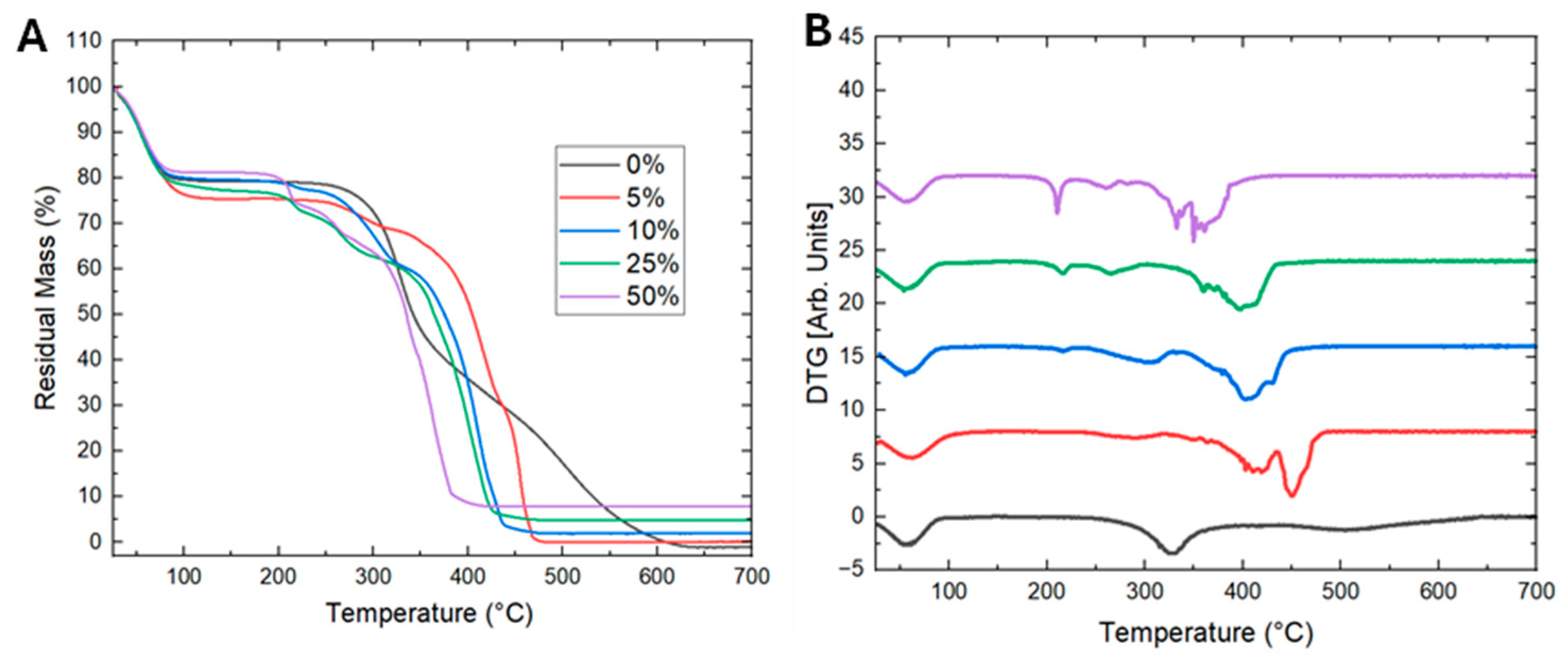
The thermal degradation of PVP fibers and PVP/CuAc composite fibers, containing CuAc (5, 10, 25, and 50 wt.%), was investigated using thermogravimetric analysis (TGA) under an air atmosphere. As shown in Figure 4A, the pristine PVP fibers exhibited a constant degradation during heating up to 600 °C, with almost a negligible residual mass. The TGA results also showed that the first initial weight loss occurred between 50 and 75 °C which was attributed to moisture/water evaporation. The thermal degradation of the composite fibers with different CuAc concentrations exhibited an additional weight loss around 200–320 °C which was attributed to the dehydration of copper acetate [31,32]. A significant mass loss was also observed between 320 and 460 °C for all composite fibers. Notably, samples with 25% and 50 wt.% of CuAc showed higher residual mass, indicating improved thermal stability due to inorganic residue retention [34]. The three peaks with most contribution to mass loss of PVP/CuAc composite fibers are shown in Figure 4B. whereas dehydration, decomposition of the anhydrous acetate, oxidation, and stabilization took place. During the dehydration process, the copper acetate loses its water to form anhydrous acetate in the range of 50–75 °C. Secondly, the decomposition of the anhydrous acetate involves the reduction in the copper (II) to metallic copper (Cu), deriving in volatile products such as carbon dioxide, acetone, and acetic acid. During oxidation (Figure 4A), the copper binds to oxygen resulting in Copper Oxide (CuO) and at the end of this process, the samples were stabilized with a constant residual mass.
Derivative thermogravimetric (DTG) analysis provided further insight into the decomposition kinetics of the PVP and PVP/CuAC fibers (Figure 4B). The PVP fibers exhibited a sharp, single degradation peak between 300 and 350 °C, characteristic of polymer matrix breakdown [32]. In contrast, the incorporation of CuAc introduced additional peaks between ~200–460 °C, corresponding to copper acetate decomposition and its interaction with the polymer [34]. At higher CuAc loadings (25% and 50 wt.% with respect to PVP concentration in the precursor solution), the main degradation peak of CuAc was observed at 200 °C [34], which was increased in intensity at increasing CuAc concentration, suggesting altered degradation pathways [35]. The increased residual mass above 475 °C in these samples confirmed the formation of thermally stable inorganic components, reflecting enhanced structural stability at elevated temperatures [36].
Figure 5 presents the FTIR spectra of pure PVP, pure CuAc, and CuAc/PVP composites. Table 3 shows the peak locations and their corresponding assignments of the polymer-based samples. All major peaks identified in the spectra are listed in Table 3. All samples exhibit the presence of water, as indicated by a broad, weak peak around 3400–3600 cm−1. The fiber samples generally exhibit similar FTIR spectra to those of pure PVP, which is expected due to the high concentration of PVP in the samples. These peaks are located at 1637, 1494, 1462, 1430, 1372, 1318, 1287 cm−1. In the presence of CuAc, some shifts in peak positions are observed. The peak at 1637 cm−1 shifts slightly higher by approximately 8 cm−1 in the CuAc/PVP samples. The peak located at 1430 cm−1 is shifted to 1438 cm−1, indicating a structural change due to the presence of copper. Additionally, a distinct peak appears at approximately 1550 cm−1 in the CuAc/PVP samples, which is absent in the spectrum of pure PVP. This peak is attributed to Cu2+ interacting with O in the pyridine ring or to Cu-O vibration modes, as it is also present as a main peak in the spectrum of the CuAc dihydrate sample.
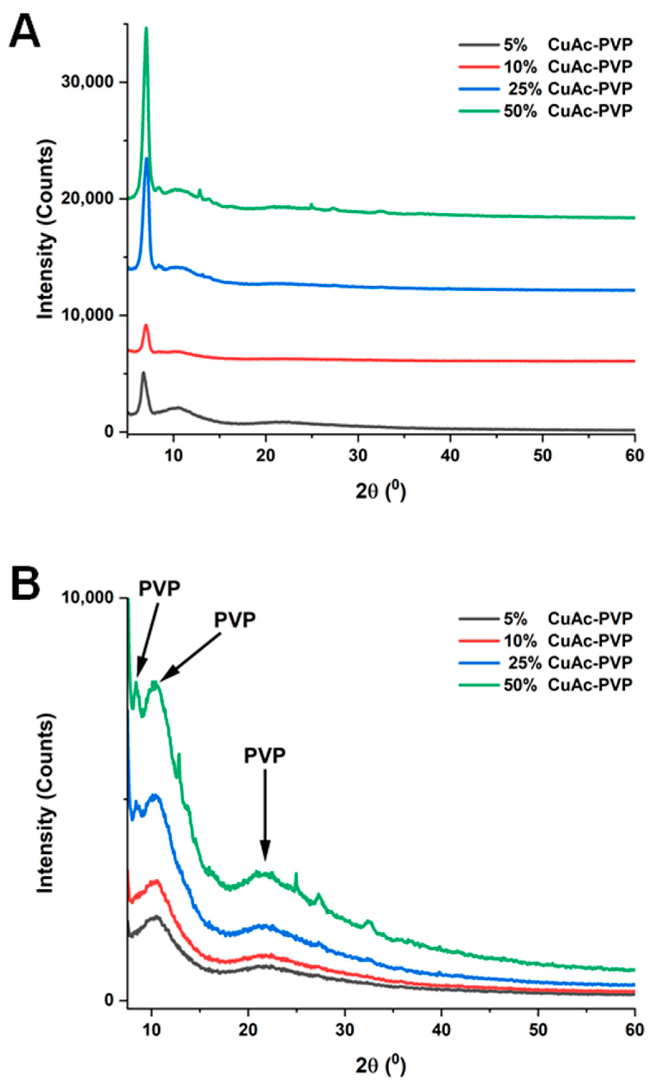
The diffraction patterns for CuAc dihydrate and the samples are shown in Figure 6 and Figure 7, and the LeBail fitting data are presented in Table 4. The diffraction pattern for PVP typically exhibits weak, diffuse peaks located around 11.25° and 21°, as summarized in Table 5 [47,48,49]. The diffraction patterns for the CuAc/PVP composites are shown in Figure 6A, and an expanded view focusing on the PVP peaks is provided in Figure 6B to facilitate comparison. In the present study, the lower-angle diffraction peak of PVP located at approximately 11.2° was observed to shift to lower angles and split into two features at approximately 8.5° and a second one at 10.5° in 2θ. In contrast, the weak diffraction peak at approximately 21° in 2θ was observed to shift to a slightly higher angle as the CuAc content increased. Teng et al. reported a similar change in the relative positions of the PVP diffraction patterns, where the peak at approximately 11.2° shifted to a lower diffraction angle in the presence of water. They observed that the lower peaks shifted to lower scattering angles, while the higher-angle halo shifted to higher scattering angles [47]. Similarly, Timaeva et al. observed a shift and splitting of the diffuse peak in the PVP located at approximately 11°, which was observed to shift down to 9.5° in 2θ, attributed to the presence of half-bound water molecules [48]. Kahn et al. reported diffraction peaks in PVP powder and fibers at 5 and 12°, which were suggested to reflect the degree of crystallinity in PVP [49].
Additional peaks observed in the diffraction patterns located at 6.5, 15.10, 16.0, 18.54, 27.23, and 34.85° correspond to the 200, 002, 400, 113/020, and 421/322 planes, respectively, of the Cu3(OH)4(CH3COO)2 phase [50]. Among all samples, only the 50% (w/w) CuAc composite sample showed additional peaks beyond the 200-reflection associated with the copper-containing phase. In addition, all the CuAc-PVP composite samples showed a diffraction peak at approximately 6.5° in 2θ, which has been shown to be present in the Cu3(OH)4(CH3COO)2 material. The LeBail fitting of the diffraction pattern showed a reduced χ2 of 3.26; in general, a χ2 value below 5 is considered a good fit [51,52]. In addition, the Rp/Rwp factors are below 10 which also indicates a good fitting for both the CuAc and the 50% CuAC-PVP composite sample. The CuAc in the samples was identified as having a C2/c crystal lattice, which is representative of the dehydrated form of CuAc hydrate [50]. A summary of the XRD fitting results is shown in Table 4.

Antibacterial Analysis

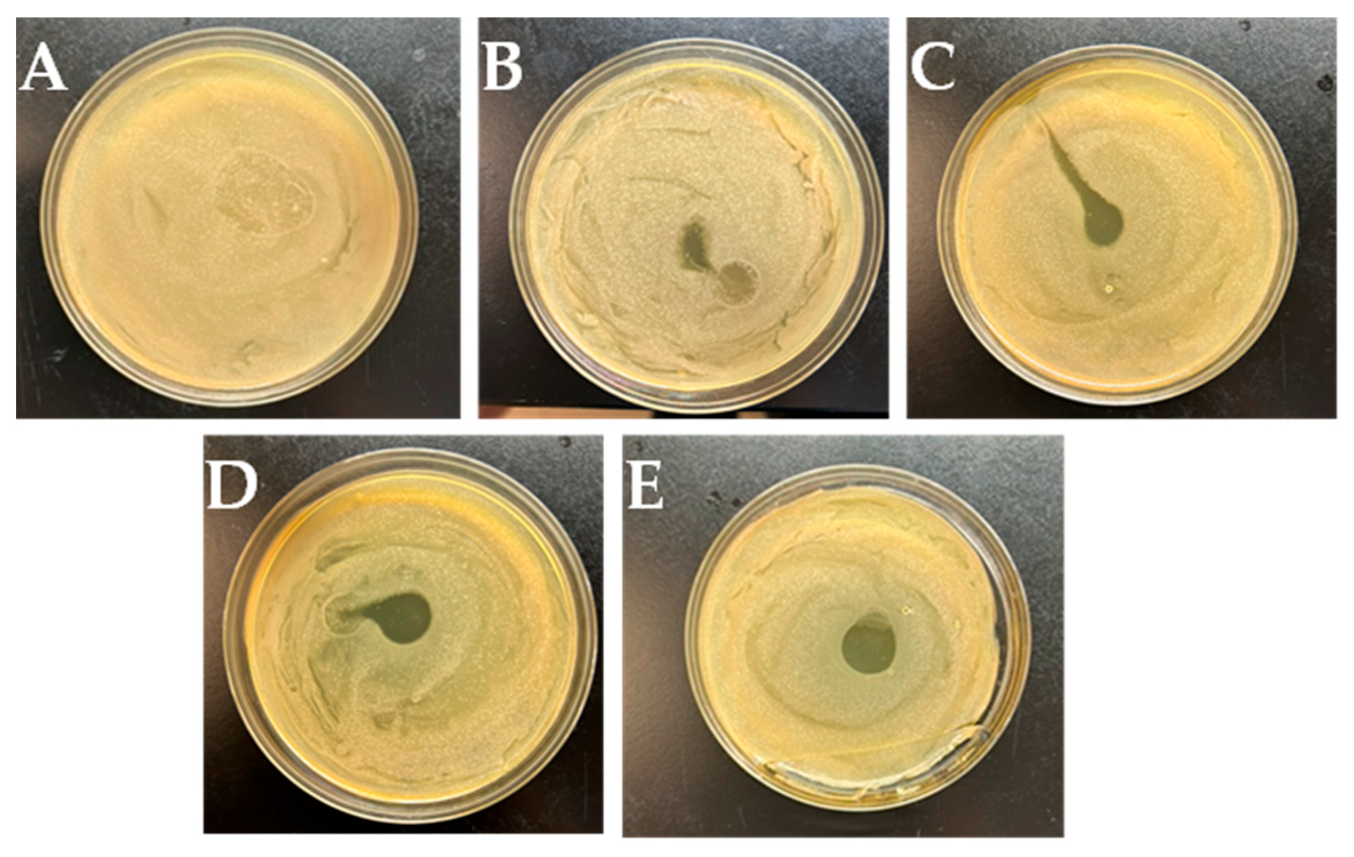
The antibacterial activity of pure PVP fibers and CuAc/PVP composite fibers containing 5, 10, 25, and 50% (w/w) CuAc were tested against S. aureus and E. coli. For the test, membranes with a diameter of 12 mm were prepared for each sample and used in the Kirby-Bauer disk diffusion assay. The fiber mats had an average thickness of 430 ± 90 μm, 5% 400 ± 20 μm, 10% 280 ± 40 μm, 25% 295 ± 20 μm, and 50% 260 ± 70 μm, for the PVP, 5% CuAc/PVP, 10% CuAc/PVP, 25% CuAc/PVP, and 50% CuAc/PVP, respectively. Figure 8 and 9 show the inhibition zones formed by each sample against E. coli and S. aureus after incubation at 36 °C for 48 h.
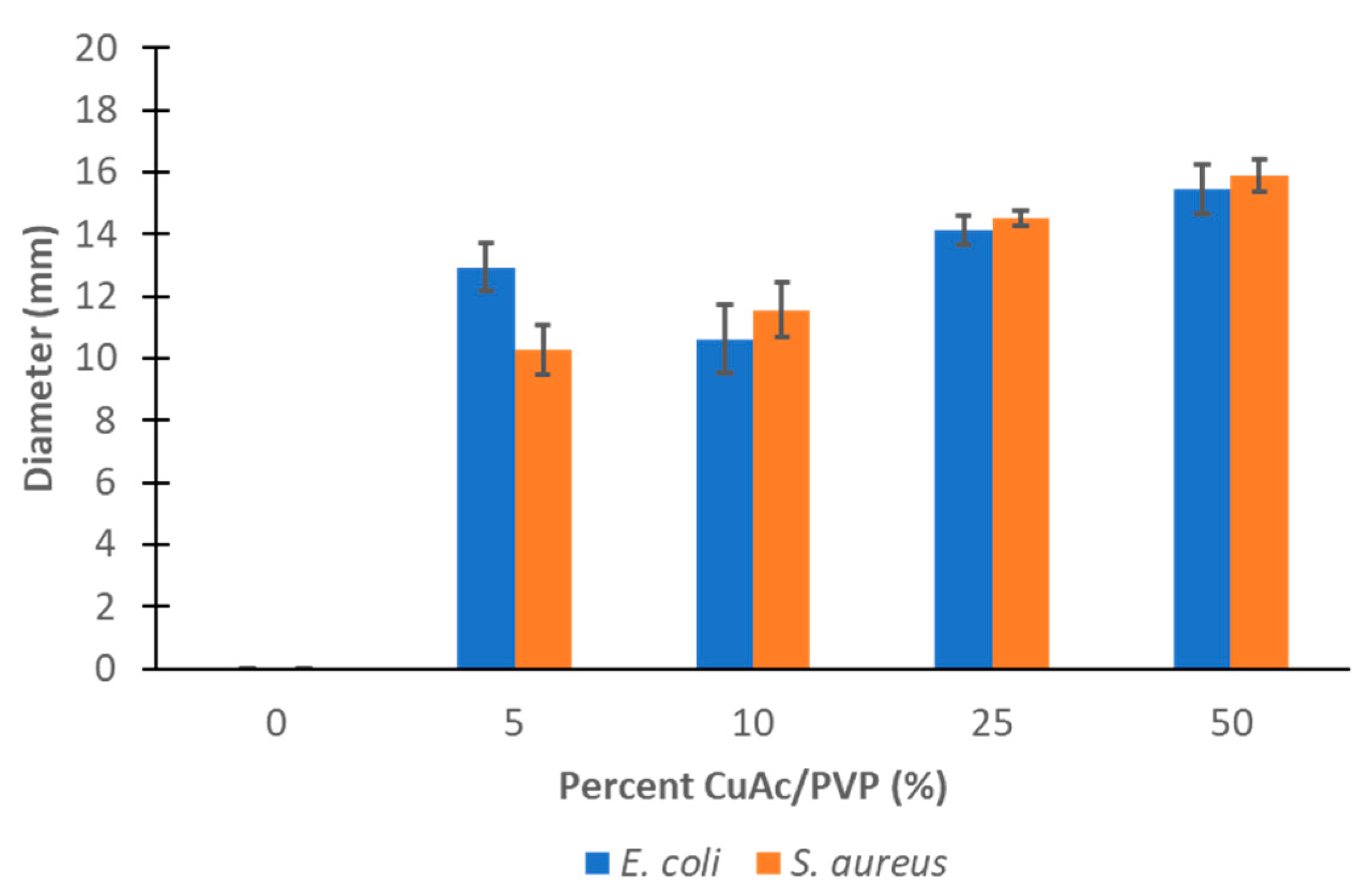
As shown in Figure 8 and Figure 9, inhibition growth zones were observed in all CuAc/PVP composite fibers tested, including those with 5, 10, 15, 25, and 50% (w/w) concentrations against E. coli and S. aureus. The pristine PVP fibers did not show any antibacteria effect against E. coli and S. aureus. The average diameter of the inhibition zone for each sample tested against E. coli was measured using Image-J. The CuAc/PVP fiber membranes demonstrated a maximum inhibition zone of 15 mm out of a total membrane diameter of 12 mm, indicating that minimal bacterial growth occurred within the fibrous region, as can be observed in the images. A trend was observed in which higher CuAc concentrations correspond to larger inhibition zones. The inhibition zone diameters for 5, 10, 25, and 50% (w/w) CuAc samples against E. coli were 12.9, 10.6, 14.1, and 15.5 mm, respectively. The average maximum inhibition zone diameter against S. aureus was observed to be 15.9 mm compared to the 12 mm composite disk used. The corresponding individual measurements were 10.3, 11.6, 14.5, and 15.9 mm for 5, 10, 25, and 50% (w/w) CuAc samples, respectively.
As shown in Figure 10, the fibrous PVP membranes incorporated with CuAc exhibited a slightly higher pronounced antibacterial effect against S. aureus compared to E. coli at lower concentration. However, at higher concentration, the toxicity effects were observed to be very similar. Whereas the PVP was not observed to be toxic to either the S. aureus or E. coli. Among the samples tested, the membrane containing CuAc demonstrated a small disparity in the inhibition zone diameters between the Gram-negative and Gram-positive bacterial strains at lower concentrations. This observation suggests the Gram-positive bacteria is more susceptible to disruption by CuAc. The antimicrobial mechanism is attributed to the ability of CuAc to interfere with bacterial metabolic processes, potentially through the inactivation or destruction of cellular components through the generation of reactive oxygen species, ultimately leading to cell death.
Table 6 shows a comparison by the results reported in the current manuscript with those already published on similar polymer/composite fiber systems studied as antibacterial agents against Gram-negative and Gram-positive bacteria. The current results show similar trends to those previously reported in the literature using Cu and CuO/polymer composite fibers [32,53,54].

4. Conclusions

CuAc/PVP composite fibers were successfully synthesized using centrifugal spinning of a precursor solution. The synthesized composite fibers were characterized using SEM, EDS, and XRD, which confirmed the presence of both CuAc and PVP in the CuAc/PVP composite fibers. The fibers showed an average diameter of 2.380 ± 0.80, 2.656 ± 0.66, 2.663 ± 0.53, 2.659 ± 0.58, and 2.266 ± 0.59 μm for the PVP, 5% CuAc, 10% CuAc, 25% CuAc, and 50% CuAc, respectively. The FTIR results showed the presence of both materials (CuAc and PVP) in the composite fibers. In addition, the XRD analysis results showed that the CuAc/PVP composite fiber were a mixture of CuAc and PVP. The CuAc was present as in the form of Cu3(OH)4(CH3COO)2, and the PVP showed expansion of the lattice as the diffraction peak shifted to lower angles more than likely due to the presence of water. The PVP fibers by themselves did not show any significant antibacterial activity against S. aureus and E. coli bacteria. However, the addition of CuAc to the PVP matrix showed antibacterial activity against both S. aureus and E. coli. The results indicated that CuAc/PVP fibers became more effective with increasing concentration of CuAc. Furthermore, stronger bactericidal effects against E. coli were observed in comparison with the S. aureus. However, at a CuAc concentration in the Fibers, 50% the inhibition zone was the same. These fibers may serve as a potential alternative to conventional antibacterial materials such as wound dressing and skin treatment. PVP/CuAc composite fibers could be a potential treatment based on results from studies using the current of the bacterial strains.

Author Contributions

Conceptualization, M.A.; Methodology, M.A.; Formal analysis, B.I., R.C., S.I., H.M., J.G.P. and M.A.; Investigation, B.I., R.C., S.I., O.E., H.M., J.G.P. and M.A.; Resources, H.M., J.G.P. and M.A.; Data curation, J.G.P. and M.A.; Writing—original draft, B.I. and R.C.; Writing—review and editing, L.M., H.M., J.G.P. and M.A.; Supervision, M.A.; Project administration, M.A.; Funding acquisition, J.G.P. and M.A. All authors have read and agreed to the published version of the manuscript.

Funding

This research was funded by Welch Foundation grant number [BX-0048] and U.S. National Science Foundation grant number [DMR-2122178].

Data Availability Statement

The original contributions presented in this study are included in the article. Further inquiries can be directed to the corresponding authors.

Acknowledgments

This work was supported by the Lloyd M. Bentsen, Jr. Endowed Chair in Engineering Endowment at UTRGV. This work was also supported by the National Science Foundation (NSF) PREM award under grant no. DMR-2122178: UTRGV-UMN Partnership for Fostering Innovation by Bridging Excellence in Research and Student Success. J.G. Parsons and HM Morales acknowledge and are grateful for the support provided by funding from the UTRGV Chemistry Departmental Welch Foundation Grant (Grant No. BX-0048).

Conflicts of Interest

The authors declare no conflicts of interest.

References

  1. Sill, T.J.; von Recum, H.A. Electrospinning: Applications in drug delivery and tissue engineering. Biomaterials 2008, 29, 1989–2006. [Google Scholar] [CrossRef]
  2. Zhao, X. Antibacterial bioactive materials. In Bioactive Materials in Medicine; Woodhead Publishing: Cambridge, UK, 2011; pp. 97–123. [Google Scholar]
  3. McDonnell, G.; Russell, A.D. Antiseptics and disinfectants: Activity, action, and resistance. Clin. Microbiol. Rev. 1999, 12, 147–179. [Google Scholar] [CrossRef]
  4. Kurakula, M.; Rao, G.S.N.K. Pharmaceutical assessment of polyvinylpyrrolidone (PVP): As excipient from conventional to controlled delivery systems with a spotlight on COVID-19 inhibition. J. Drug Deliv. Sci. Technol. 2020, 60, 102046. [Google Scholar] [CrossRef]
  5. Damyanova, T.; Dimitrova, P.D.; Borisova, D.; Topouzova-Hristova, T.; Haladjova, E.; Paunova-Krasteva, T. An Overview of Biofilm-Associated Infections and the Role of Phytochemicals and Nanomaterials in Their Control and Prevention. Pharmaceutics 2024, 16, 162. [Google Scholar] [CrossRef] [PubMed]
  6. Yu, F.; Wang, D.; Zhang, H.; Wang, Z.; Liu, Y. Evolutionary trajectory of bacterial resistance to antibiotics and antimicrobial peptides in Escherichia coli. mSystems 2025, 10, e0170024. [Google Scholar] [CrossRef] [PubMed]
  7. Giner-Lamia, J.; Lopez-Maury, L.; Florencio, F.J. Global transcriptional profiles of the copper responses in the cyanobacterium Synechocystis sp. PCC 6803. PLoS ONE 2014, 9, e108912. [Google Scholar] [CrossRef] [PubMed]
  8. Sanchez, J.A.; Materon, L.; Parsons, J.G.; Alcoutlabi, M. Synthesis, Characterization, and Antibacterial Activity of Graphene Oxide/Zinc Hydroxide Nanocomposites. Appl. Sci. 2024, 14, 6274. [Google Scholar] [CrossRef]
  9. Chemler, S.R. Copper catalysis in organic synthesis. Beilstein J. Org. Chem. 2015, 11, 2252–2253. [Google Scholar] [CrossRef]
  10. Ray, S.S.; Chen, S.S.; Nguyen, N.C.; Nguyen, H.T. Electrospinning: A Versatile Fabrication Technique for Nanofibrous Membranes for Use in Desalination. In Nanoscale Materials in Water Purification; Elsevier: Amsterdam, The Netherlands, 2019; pp. 247–273. [Google Scholar]
  11. Wentworth, B.J.; Stotts, M. Wilson’s Disease: The Copper Connection. Pract. Gastroenterol. 2020, 44, 10–20. [Google Scholar]
  12. Flores-Rábago, K.M.; Rivera-Mendoza, D.; Vilchis-Nestor, A.R.; Juarez-Moreno, K.; Castro-Longoria, E. Antibacterial Activity of Biosynthesized Copper Oxide Nanoparticles (CuONPs) Using Ganoderma sessile. Antibiotics 2023, 12, 1251. [Google Scholar] [CrossRef]
  13. Vincent, M.; Duval, R.E.; Hartemann, P.; Engels-Deutsch, M. Contact killing and antimicrobial properties of copper. J. Appl. Microbiol. 2018, 124, 1032–1046. [Google Scholar] [CrossRef]
  14. Vasiliev, G.; Kubo, A.L.; Vija, H.; Kahru, A.; Bondar, D.; Karpichev, Y.; Bondarenko, O. Synergistic antibacterial effect of copper and silver nanoparticles and their mechanism of action. Sci. Rep. 2023, 13, 9202. [Google Scholar] [CrossRef]
  15. Yalcinkaya, F.; Lubasova, D. Quantitative evaluation of antibacterial activities of nanoparticles (ZnO, TiO2, ZnO/TiO2, SnO2, CuO, ZrO2, and AgNO3) incorporated into polyvinyl butyral nanofibers. Polym. Adv. Technol. 2017, 28, 137–140. [Google Scholar] [CrossRef]
  16. Akhidime, I.D.; Saubade, F.; Benson, P.S.; Butler, J.A.; Olivier, S.; Kelly, P.; Verran, J.; Whitehead, K.A. The antimicrobial effect of metal substrates on far food pathogens. Food Bioprod. Process 2019, 113, 68–76. [Google Scholar] [CrossRef]
  17. Phan, D.N.; Dorjjugder, N.; Saito, Y.; Khan, M.Q.; Ullah, A.; Bie, X.Y.; Taguchi, G.; Kim, I.S. Antibacterial mechanisms of various copper species incorporated in polymeric nanofibers against bacteria. Mater. Today Commun. 2020, 25, 101377. [Google Scholar] [CrossRef]
  18. Mohamadpour, F.; Maghsoodlou, M.T.; Heydari, R.; Lashkari, M. Copper(II) acetate monohydrate: An efficient and eco-friendly catalyst for the one-pot multi-component synthesis of biologically active spiropyrans and 1-pyrazolo[1,2-]phthalazine-5,10-dione derivatives under solvent-free conditions. Res. Chem. Intermed. 2016, 42, 7841–7853. [Google Scholar] [CrossRef]
  19. Hamdan, N.; Yamin, A.; Hamid, S.A.; Khodir, W.K.W.A.; Guarino, V. Functionalized Antimicrobial Nanofibers: Design Criteria and Recent Advances. J. Funct. Biomater. 2021, 12, 59. [Google Scholar] [CrossRef]
  20. Lee, H.; Alcoutlabi, M.; Watson, J.V.; Zhang, X.W. Electrospun nanofiber-coated separator membranes for lithium-ion rechargeable batteries. J. Appl. Polym. Sci. 2013, 129, 1939–1951. [Google Scholar] [CrossRef]
  21. Anusiya, G.; Jaiganesh, R. A review on fabrication methods of nanofibers and a special focus on application of cellulose nanofibers. Carbohydr. Polym. Technol. Appl. 2022, 4, 100262. [Google Scholar] [CrossRef]
  22. De la Garza, D.; De Santiago, F.; Materon, L.; Chipara, M.; Alcoutlabi, M. Fabrication and characterization of centrifugally spun poly(acrylic acid) nanofibers. J. Appl. Polym. Sci. 2019, 136, 47480. [Google Scholar] [CrossRef]
  23. Flores, D.; Villarreal, J.; Lopez, J.; Alcoutlabi, M. Production of carbon fibers through Forcespinning® for use as anode materials in sodium ion batteries. Mater. Sci. Eng. B 2018, 236, 70–75. [Google Scholar] [CrossRef]
  24. Agubra, V.A.; Zuniga, L.; De la Garza, D.; Gallegos, L.; Pokhrel, M.; Alcoutlabi, M. Forcespinning: A new method for the mass production of Sn/C composite nanofiber anodes for lithium ion batteries. Solid State Ion. 2016, 286, 72–82. [Google Scholar] [CrossRef]
  25. Chavez, R.O.; Lodge, T.P.; Alcoutlabi, M. Recent developments in centrifugally spun composite fibers and their performance as anode materials for lithium-ion and sodium-ion batteries. Mater. Sci. Eng. B 2021, 266, 115024. [Google Scholar] [CrossRef]
  26. Chavez, R.O.; Lodge, T.P.; Huitron, J.; Chipara, M.; Alcoutlabi, M. Centrifugally spun carbon fibers prepared from aqueous poly(vinylpyrrolidone) solutions as binder-free anodes in lithium-ion batteries. J. Appl. Polym. Sci. 2021, 138, e50396. [Google Scholar] [CrossRef]
  27. Lopez, J.; Gonzalez, R.; Ayala, J.; Cantu, J.; Castillo, A.; Parsons, J.; Myers, J.; Lodge, T.P.; Alcoutlabi, M. Centrifugally spun TiO2/C composite fibers prepared from TiS2/PAN precursor fibers as binder-free anodes for LIBS. J. Phys. Chem. Solids 2021, 149, 109795. [Google Scholar] [CrossRef]
  28. Pelipenko, J.; Kocbek, P.; Kristl, J. Nanofiber diameter as a critical parameter affecting skin cell response. Eur. J. Pharm. Sci. 2015, 66, 29–35. [Google Scholar] [CrossRef]
  29. Liu, Y.B.; Chen, X.H.; Gao, Y.H.; Liu, Y.Y.; Yu, D.G.; Liu, P. Electrospun Core-Sheath Nanofibers with Variable Shell Thickness for Modifying Curcumin Release to Achieve a Better Antibacterial Performance. Biomolecules 2022, 12, 1057. [Google Scholar] [CrossRef]
  30. Sanchez, J.A.; Ibrahim, A.; Materon, L.; Parsons, J.G.; Alcoutlabi, M. Centrifugally spun PVP/ZnO composite fibers with different surfactants and their use as antibacterial agents. J. Appl. Polym. Sci. 2023, 140, e54314. [Google Scholar] [CrossRef]
  31. Hasan, M.T.; Gonzalez, R.; Chipara, M.; Materon, L.; Parsons, J.; Alcoutlabi, M. Antibacterial activities of centrifugally spun polyethylene oxide/silver composite nanofibers. Polym. Adv. Technol. 2021, 32, 2327–2338. [Google Scholar] [CrossRef]
  32. Hasan, M.T.; Gonzalez, R.; Munoz, A.A.; Materon, L.; Parsons, J.G.; Alcoutlabi, M. Forcespun polyvinylpyrrolidone/copper and polyethylene oxide/copper composite fibers and their use as antibacterial agents. J. Appl. Polym. Sci. 2022, 139, e51773. [Google Scholar] [CrossRef]
  33. Rodriguezcarvajal, J. Recent Advances in Magnetic-Structure Determination by Neutron Powder Diffraction. Phys. B Condens. Matter 1993, 192, 55–69. [Google Scholar] [CrossRef]
  34. Allahverdi, Ç. Synthesis of copper nano/microparticles via thermal decomposition and their conversion to copper oxide film. Turk. J. Chem. 2023, 47, 616–632. [Google Scholar] [CrossRef]
  35. Tamaekong, N.; Liewhiran, C.; Phanichphant, S. Synthesis of Thermally Spherical CuO Nanoparticles. J. Nanomater. 2014, 2014, 507978. [Google Scholar] [CrossRef]
  36. Naderi-Samani, H.; Razavi, R.S.; Mozaffarinia, R. The effects of complex agent and sintering temperature on conductive copper complex paste. Heliyon 2022, 8, e12624. [Google Scholar] [CrossRef] [PubMed]
  37. Heyns, A.M. The low-temperature infrared spectra of the copper(II)acetates. J. Mol. Struct. 1972, 11, 93–103. [Google Scholar] [CrossRef]
  38. Rahma, A.; Munir, M.M.; Khairurrijal; Prasetyo, A.; Suendo, V.; Rachmawati, H. Intermolecular Interactions and the Release Pattern of Electrospun Curcumin-Polyvinyl(pyrrolidone) Fiber. Biol. Pharm. Bull. 2016, 39, 163–173. [Google Scholar] [CrossRef]
  39. Bette, S.; Costes, A.; Kremer, R.K.; Eggert, G.; Tang, C.C.; Dinnebier, R.E. On Verdigris, Part III: Crystal Structure, Magnetic and Spectral Properties of Anhydrous Copper(II) Acetate, a Paddle Wheel Chain. Z. Für Anorg. Und Allg. Chem. 2019, 645, 988–997. [Google Scholar] [CrossRef]
  40. Wang, Z.; Li, K.F. Understanding water vapor sorption hysteresis and scanning behaviors of hardened cement pastes: Experiments and modeling. Cem. Concr. Res. 2024, 177, 107435. [Google Scholar] [CrossRef]
  41. Wu, K.H.; Wang, Y.R.; Hwu, W.H. FTIR and TGA studies of poly(4-vinylpyridine--divinylbenzene)-Cu(II) complex. Polym. Degrad. Stab. 2003, 79, 195–200. [Google Scholar] [CrossRef]
  42. Mireles, L.K.; Wu, M.R.; Saadeh, N.; Yahia, L.; Sacher, E. Physicochemical Characterization of Polyvinyl Pyrrolidone: A Tale of Two Polyvinyl Pyrrolidones. ACS Omega 2020, 5, 30461–30467. [Google Scholar] [CrossRef]
  43. Huang, S.W.; Lin, Y.F.; Li, Y.X.; Hu, C.C.; Chiu, T.C. Synthesis of Fluorescent Carbon Dots as Selective and Sensitive Probes for Cupric Ions and Cell Imaging. Molecules 2019, 24, 1785. [Google Scholar] [CrossRef] [PubMed]
  44. Zidan, H.M.; Abdelrazek, E.M.; Abdelghany, A.M.; Tarabiah, A.E. Characterization and some physical studies of PVA/PVP filled with MWCNTs. J. Mater. Res. Technol. 2019, 8, 904–913. [Google Scholar] [CrossRef]
  45. Ito, K.; Bernstein, H.J. The Vibrational Spectra of the Formate, Acetate, and Oxalate Ions. Can. J. Chem. 1956, 34, 170–178. [Google Scholar] [CrossRef]
  46. Cena, C.R.; Silva, M.J.; Malmonge, L.F.; Malmonge, J.A. Poly(vinyl pyrrolidone) sub-microfibers produced by solution blow spinning. J. Polym. Res. 2018, 25, 238. [Google Scholar] [CrossRef]
  47. Teng, J.; Bates, S.; Engers, D.A.; Leach, K.; Schields, P.; Yang, Y. Effect of water vapor sorption on local structure of poly(vinylpyrrolidone). J. Pharm. Sci. 2010, 99, 3815–3825. [Google Scholar] [CrossRef]
  48. Timaeva, O.; Pashkin, I.; Mulakov, S.; Kuzmicheva, G.; Konarev, P.; Terekhova, R.; Sadovskaya, N.; Czakkel, O.; Prevost, S. Synthesis and physico-chemical properties of poly(N-vinyl pyrrolidone)-based hydrogels with titania nanoparticles. J. Mater. Sci. 2020, 55, 3005–3021. [Google Scholar] [CrossRef]
  49. Khan, M.Q.; Kharaghani, D.; Nishat, N.; Ishikawa, T.; Ullah, S.; Lee, H.; Khatri, Z.; Kim, I.S. The development of nanofiber tubes based on nanocomposites of polyvinylpyrrolidone incorporated gold nanoparticles as scaffolds for neuroscience application in axons. Text. Res. J. 2019, 89, 2713–2720. [Google Scholar] [CrossRef]
  50. Bulfin, B.; Vieten, J.; Agrafiotis, C.; Roeb, M.; Sattler, C. Applications and limitations of two step metal oxide thermochemical redox cycles; a review. J. Mater. Chem. A 2017, 5, 18951–18966. [Google Scholar] [CrossRef]
  51. Morales, H.M.; Vieyra, H.; Sanchez, D.A.; Fletes, E.M.; Odlyzko, M.; Lodge, T.P.; Padilla-Gainza, V.; Alcoutlabi, M.; Parsons, J.G. Synthesis and Characterization of Vanadium Nitride/Carbon Nanocomposites. Int. J. Mol. Sci. 2024, 25, 6952. [Google Scholar] [CrossRef]
  52. Morales, H.M.; Sanchez, D.A.; Fletes, E.M.; Odlyzko, M.; Padilla-Gainza, V.; Alcoutlabi, M.; Parsons, J.G. Synthesis and Characterization of Titanium and Vanadium Nitride-Carbon Composites. J. Compos. Sci. 2024, 8, 485. [Google Scholar] [CrossRef]
  53. Yalcinkaya, F.; Komarek, M.; Daniela Lubasova, D.; Sanetrnik, F.; Maryska, J. Preparation of Antibacterial Nanofibre/Nanoparticle Covered Composite Yarns. J. Nanomater. 2016, 2016, 7565972. [Google Scholar] [CrossRef]
  54. Hashmi, M.; Ullah, S.; Kim, I.S. Copper oxide (CuO) loaded polyacrylonitrile (PAN) nanofiber membranes for antimicrobial breath mask applications. Curr. Res. Biotechnol. 2019, 1, 1–10. [Google Scholar] [CrossRef]
Figure 1. SEM images and histograms of fiber diameter for (AC) PVP, and CuAc/PVP composite fibers at 5% (w/w) (DF), and 10% (w/w) (GI), 25% (w/w) (JL), 50% (w/w) (MO).
Jcs 09 00590 g001
Figure 2. EDS mappings of PVP composite fibers with 10 wt% CuAc (A) SEM of the analyzed area, (B) elemental map of carbon, (C) elemental map of oxygen, (D) elemental map of nitrogen, and (E) elemental map of copper.
Jcs 09 00590 g002
Figure 3. Elemental Mappings of 50% (w/w) CuAC/PVP concentration (A) SEM of the analyzed area, (B) elemental map of carbon, (C) elemental map of oxygen, (D) elemental map of nitrogen, and (E) elemental map of copper.
Jcs 09 00590 g003
Figure 4. TGA plots for the thermal decomposition of the PVP and PVP composites in air (A) and the DTGA plots of the decomposition of the PVP and CuAC/PVP composites in air (B).
Jcs 09 00590 g004
Figure 5. FTIR spectra of pure PVP, pure CuAc, and various CuAc/PVP composite fibers.
Jcs 09 00590 g005
Figure 6. LeBail fitting results of the X-ray diffraction patterns for the CuAc dihydrate starting material (A) and the 50% (w/w) CuAc/PVP sample (B).
Jcs 09 00590 g006
Figure 7. (A) Overlay of the XRD patterns of CuAc/PVP composites and (B) Expanded view of the diffraction patterns in the range of 8° to 60°, highlighting the PVP-related peaks.
Jcs 09 00590 g007
Figure 8. Antibacterial activity of CuAc/PVP composite fibers from 0% (A), 5% CuAc (B), 10% CuAc (C), 25% CuAc (D) and 50% CuAc (E) against E. coli.
Jcs 09 00590 g008
Figure 9. Antibacterial activity of CuAc/PVP composite fibers from 0% (A), 5% CuAc (B), 10% CuAc (C), 25% CuAc (D) and 50% CuAc (E) against S. aureus.
Jcs 09 00590 g009
Figure 10. Relationship between the CuAc/PVP composition and inhibition zone for E. coli and S. aureus.
Jcs 09 00590 g010
Table 1. Elemental composition based on EDS analysis collected from the SEM analysis.
Sample
Element5%SE10%SE25%SE50%SE
C64.070.5762.443.5165.011.5267.902.51
N18.260.5417.741.0915.412.2323.671.59
O15.640.1917.702.4315.570.6914.321.37
Cu1.910.152.100.295.251.295.100.34
(note: SE is the standard error based on 5 independent samples).
Table 2. Weight percentage composition of CuAc/PVP fibers.
SampleCuAc, %PVP, %
PVP0.00100.00
5% (w/w) CuAc/PVP4.3895.62
10% (w/w) CuAc/PVP 7.9492.06
25% (w/w) CuAc/PVP 16.4183.59
50% (w/w) CuAc/PVP 16.4483.56
Table 3. Main FTIR peak location and corresponding assignments for pure PVP, pure CuAc, and various CuAc/PVP composite fibers [37,38,39,40,41,42,43,44,45,46].
FTIR Peaks (cm−1)
CuAc50% (w/w) CuAc/PVP25% (w/w) CuAc/PVP10% (w/w) CuAc/PVP5% (w/w) CuAc/PVPPVPAssignment
3462 O-H
335834003389338933893389OH
32653265326532663266 OH
298629592955295529552955Asymmetric CH2
291829182918291829182918Sym CH2
165016501643164216431637C=O/OH (from H2O)
1594 sym C-O
1546155015521556 Cu2+ = O and Cu2+-N pyridine ring
14941492149114941494C-H, C-N
14581461146514631462C-N
143514381438143814381430sym CH2
142314231423 C-O sym
1417 Sym C-O
1354 CH3 sym
13741373137313711372C-H
13181317131713131318C-H
12871287128612821287CH2 Wag, C-N
12741274 CH2 Wag, C-N
1223122012251224 CH2 Wag, C-N
1169116911691165 CH2 twist of the pyrrole
1047 CH rocking
1031 CH3 Rocking
10181016101210121012C-C, CH2 rock
926926930929937C-C
896891891890888Pyrrolidone ring breathing
842842841842847CH2
732732734734736CH2 rock
683 COO-Cu
Table 4. Crystal structure parameters from LeBail fitting for pure CuAc and 50% (w/w) CuAc/PVP.
SampleSpace Groupa(Å)b(Å)c(Å)α(°)β(°)γ(°)χ2Rp/Rwp
CuAclitC 2/c13.1688.65413.85890.0117.0290.0N/A [42]N/A
CuAcC/2c13.1519.59513.85290.0116.990.01.38.0/6.8
Cu3(OH)4(CH3COO)2 litPbca20.9747.20713.12290.090.090.0N/A [39]N/A
50% (w/w) CuAc/PVPPbca20.9847.21412.91490.090.090.03.266.5/8.6
Table 5. Position of key diffraction peaks from various CuAc/PVP samples.
SamplePeak 1 PositionPeak 2 PositionPeak Position 3
PVPlitN/A10.5021.0 [33]
PVPlitN/A11.2521.21 [38]
5% (w/w) CuAc/PVPN/A10.1521.32
10% (w/w) CuAc/PVP8.5210.2821.80
25% (w/w) CuAc/PVP8.4610.3821.70
50% (w/w) CuAc/PVP8.4610.5521.75
Table 6. Comparison of data from literature with the results reported in the present work.
Fibers/BacteriaLoading
(wt%)
Fiber Dia.
(μm)
Inhibition Zone Diameter (cm)Refs.
PEO/Cu in E. coli0–350.2–0.25 1.64 out of 2.26[32]
PEO/Cu in B. cereus0–350.2–0.251.68 out of 2.26[32]
PVP/Cu in E. coli0–354.9–5.51.56 out of 2.26[32]
PVP/Cu in B. cereus0–354.9–5.51.53 out of 2.26[32]
PVB/CuO in E. coli 0–10>200100% inhibition after 1–4 h[53]
PU/CuO in E. coli0–10>20070% inhibition after 1–4 h[53]
PAN/CuO in E. coli0–1000.14–0.18inhibition not observed[54]
PAN/CuO in B. subtilis0–1000.14–0.18inhibition not observed)[54]
PVP/CuAc in E. coli0–502.26–2.381.327 out of 1.2This work
PVP/CuAc in B. cereus0–502.26–2.381.3 out of 1.2This work
Disclaimer/Publisher’s Note: The statements, opinions and data contained in all publications are solely those of the individual author(s) and contributor(s) and not of MDPI and/or the editor(s). MDPI and/or the editor(s) disclaim responsibility for any injury to people or property resulting from any ideas, methods, instructions or products referred to in the content.