
Green Synthesis of Gold, Silver, and Iron Nanoparticles for the Degradation of Organic Pollutants in Wastewater


Abstract
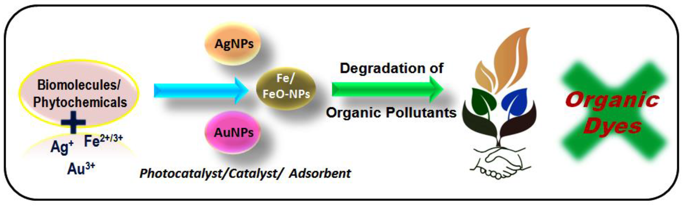
:1. Introduction
2. Surface Plasmon Resonance
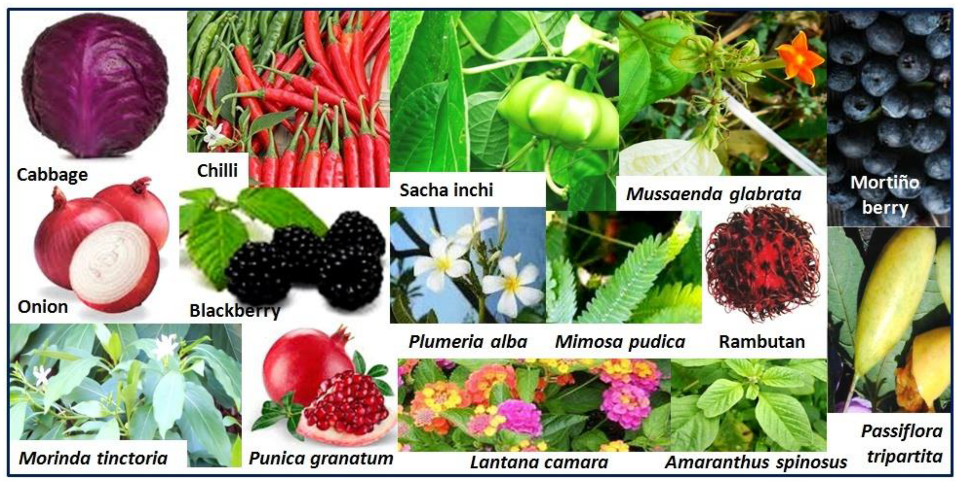
3. Gold Nanoparticles
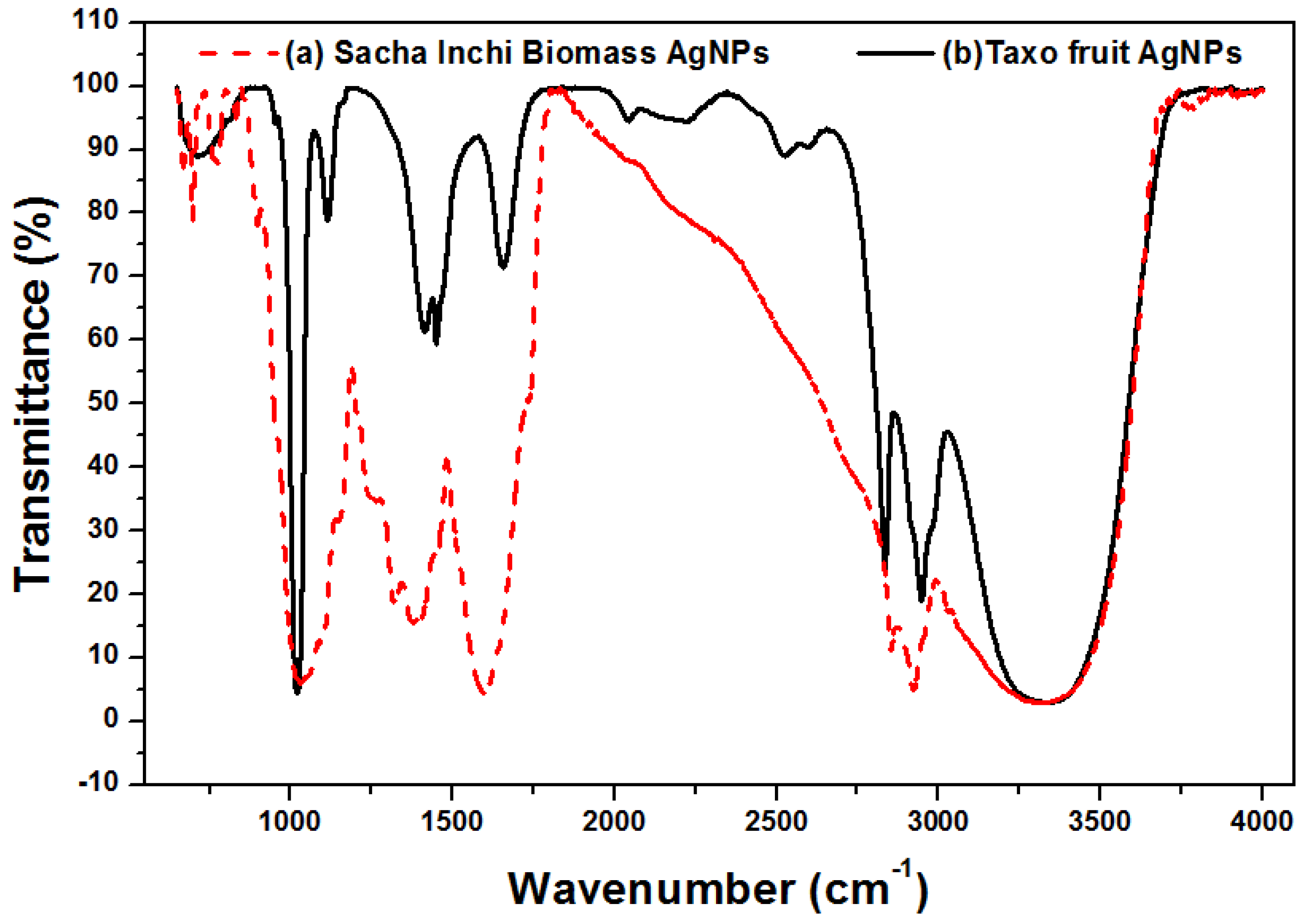
4. Silver Nanoparticles
5. Iron and Iron Oxide Nanoparticles
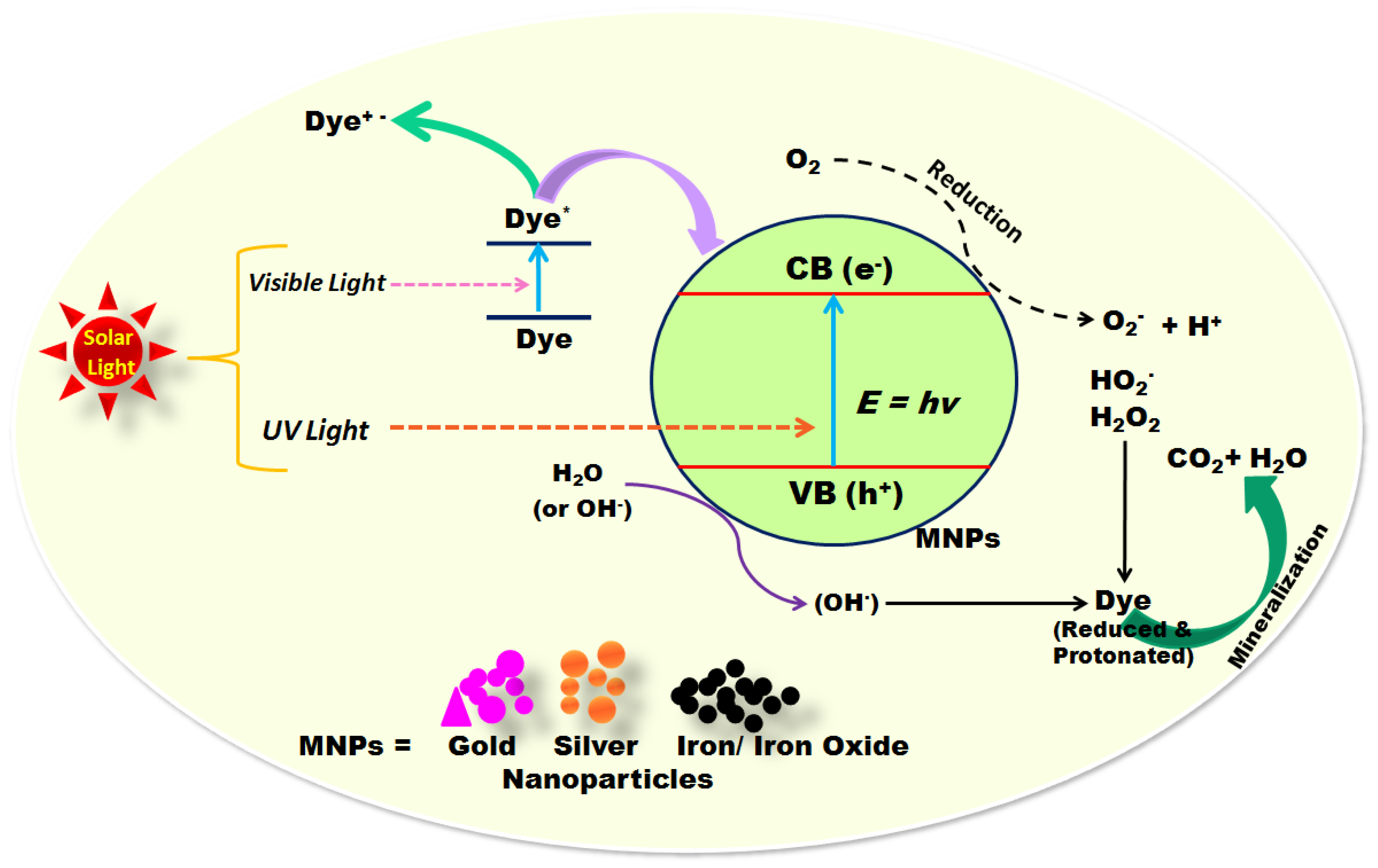
6. Mechanism for Degradation of Organic Dyes Using Nanoparticles
7. Toxicological Issue
8. Conclusions
Funding
Institutional Review Board Statement
Informed Consent Statement
Acknowledgments
Conflicts of Interest
References
- Sharma, V.K.; Filip, J.; Zboril, R.; Varma, R.S. Natural inorganic nanoparticles—Formation, fate, and toxicity in the environment. Chem. Soc. Rev. 2015, 44, 8410–8423. [Google Scholar] [CrossRef] [PubMed] [Green Version]
- Stark, W.J.; Stoesset, P.R.; Wohlleben, W.; Hafner, A. Industrial applications of nanoparticles. Chem. Soc. Rev. 2015, 44, 5793–5805. [Google Scholar] [CrossRef] [PubMed] [Green Version]
- Soenen, S.J.; Parak, W.J.; Rejman, J.; Manshian, B. (Intra)cellular stability of inorganic nanoparticles: Effects on cytotoxicity, particle functionality, and biomedical applications. Chem. Rev. 2015, 115, 2109–2135. [Google Scholar] [CrossRef] [PubMed]
- Talebzadeh, S.; Queffelec, C.; Knight, D.A. Surface modification of plasmonic noble metal–metal oxide core–shell nanoparticles. Nanoscale Adv. 2019, 1, 4578–4591. [Google Scholar] [CrossRef] [Green Version]
- Virkutyte, J.; Varma, R.S. Green synthesis of metal nanoparticles: Biodegradable polymers and enzymes in stabilization and surface functionalization. Chem. Sci. 2011, 2, 837–846. [Google Scholar] [CrossRef]
- Kumar, B.; Smita, K.; Debut, A.; Cumbal, L. Andean Sacha Inchi (Plukenetia volubilis L.) leaf-mediated synthesis of Cu2O nanoparticles: A Low-Cost Approach. Bioengineering 2020, 79, 54. [Google Scholar] [CrossRef]
- Shankar, P.D.; Shobana, S.A.; Karuppusamy, I.; Pugazhendhi, A.; Ramkumar, V.S.; Arvindnarayan, S.; Kumar, G. A review on the biosynthesis of metallic nanoparticles (gold and silver) using bio-components of microalgae: Formation mechanism and applications. Enzym. Microb. Technol. 2016, 95, 28–44. [Google Scholar] [CrossRef]
- Nasrollahzadeh, M.; Yek, S.M.-G.; Motahharifar, N.; Gorab, M.G. Recent Developments in the Plant-Mediated Green Synthesis of Ag-Based Nanoparticles for Environmental and Catalytic Applications. Chem. Rec. 2019, 19, 2436–2479. [Google Scholar] [CrossRef]
- Rahman, A.; Lin, J.; Jaramillo, F.E.; Bazylinski, D.A.; Jeffryes, C.; Dahoumane, S.A. In Vivo Biosynthesis of Inorganic Nanomaterials Using Eukaryotes—A Review. Molecules 2020, 25, 3246. [Google Scholar] [CrossRef]
- Saha, B.; Das, S.; Saikia, J.; Das, G. Preferential and enhanced adsorption of different dyes on iron oxide nanoparticles: A comparative study. J. Phys. Chem. C 2011, 115, 8024–8033. [Google Scholar] [CrossRef]
- Kumar, B.; Smita, K.; Kumar, B. Phytochemical functionalized metal nanocatalyst (Ag, Au, Fe, Zn and Pd) for remediation of organic dyes. In Advances in Chemistry Research; Taylor, J.C., Ed.; Nova Science Publishers: Hauppauge, NY, USA, 2017; Chapter 3; Volume 37, pp. 87–120. [Google Scholar]
- Lam, S.-M.; Sin, J.-C.; Abdullah, A.Z.; Mohamed, A.R. Degradation of wastewaters containing organic dyes photocatalysed by zinc oxide: A review. Desalination Water Treat. 2012, 41, 131–169. [Google Scholar] [CrossRef]
- Sharma, M.; Jain, T.; Singh, S.; Pandey, O.P. Photocatalytic degradation of organic dyes under UV-Visible light using capped ZnS nanoparticles. Sol. Energy 2012, 86, 626–633. [Google Scholar] [CrossRef]
- Safavi, A.; Momeni, S. Highly efficient degradation of azo dyes by palladium/ hydroxyapatite/Fe3O4nanocatalyst. J. Hazard. Mater. 2012, 201–202, 125–131. [Google Scholar] [CrossRef]
- Singla, P.; Sharma, M.; Pandey, O.P.; Singh, K. Photocatalytic degradation of azo dyes using Zn-doped and undoped TiO2 nanoparticles. Appl. Phys. A 2014, 116, 371–378. [Google Scholar] [CrossRef]
- Touati, A.; Hammedi, T.; Najjar, W.; Ksibi, Z.; Sayadi, S. Photocatalytic degradation of textile wastewater in presence of hydrogen peroxide: Effect of cerium doping titania. J. Ind. Eng. Chem. 2016, 35, 36–44. [Google Scholar] [CrossRef]
- Koe, W.S.; Lee, J.W.; Chong, W.C.; Pang, Y.L.; Sim, L.C. An overview of photocatalytic degradation: Photocatalysts, mechanisms, and development of photocatalytic membrane. Environ. Sci. Pollut. Res. 2020, 27, 2522–2565. [Google Scholar] [CrossRef] [PubMed]
- Khalid, N.R.; Majid, A.; Tahir, M.B.; Niaz, N.A.; Khalid, S. Carbonaceous-TiO2 nanomaterials for photocatalytic degradation of pollutants: A review. Ceram. Int. 2017, 43, 14552–14571. [Google Scholar] [CrossRef]
- Wood, R.W. On a remarkable case of uneven distribution of light in a diffraction grating spectrum. Proc. Phys. Soc. Lond. 1902, 18, 269–275. [Google Scholar] [CrossRef]
- Homola, J.; Yee, S.S.; Gauglitz, G. Surface Plasmon Resonance Sensors: Review. Sens. Actuators B 1999, 54, 3–15. [Google Scholar] [CrossRef]
- Raliya, R.; Saharan, V.; Dimkpa, C.; Biswas, P. Nanofertilizer for Precision and Sustainable Agriculture: Current State and Future Perspectives. J. Agric. Food Chem. 2018, 669, 6487–6503. [Google Scholar] [CrossRef]
- Kang, H.; Buchman, J.T.; Rodriguez, R.S.; Ring, H.L.; He, J.; Bantz, K.C.; Haynes, C.L. Stabilization of Silver and Gold Nanoparticles: Preservation and Improvement of Plasmonic Functionalities. Chem. Rev. 2019, 119, 664–699. [Google Scholar] [CrossRef]
- Cortie, M.B.; McDonagh, A.M. Synthesis and Optical Properties of Hybrid and Alloy Plasmonic Nanoparticles. Chem. Rev. 2011, 111, 3713–3735. [Google Scholar] [CrossRef] [Green Version]
- Arinze, E.S.; Qiu, B.; Nyirjesy, G.; Thon, S.M. Plasmonic Nanoparticle Enhancement of Solution-Processed Solar Cells: Practical Limits and Opportunities. ACS Photonics 2016, 3, 158–173. [Google Scholar] [CrossRef]
- Kumar, B.; Smita, K.; Debut, A.; Cumbal, L. Utilization of Persea americana (Avocado) oil for the synthesis of gold nanoparticles in sunlight and evaluation of antioxidant and photocatalytic activities. Environ. Nanotechnol. Monit. Manag. 2018, 10, 231–237. [Google Scholar]
- Kong, F.Y.; Zhang, J.W.; Li, R.F.; Wang, Z.X.; Wang, W.J.; Wang, W. Unique roles of gold nanoparticles in drug delivery, targeting and imaging applications. Molecules 2017, 22, 1445. [Google Scholar] [CrossRef] [PubMed] [Green Version]
- Sarina, S.; Waclawik, E.R.; Zhu, H. Photocatalysis on supported gold and silver nanoparticles under ultraviolet and visible light irradiation. Green Chem. 2013, 15, 1814–1833. [Google Scholar] [CrossRef]
- Kumar, B.; Smita, K.; Cumbal, L.; Debut, A. Extracellular biofabrication of gold nanoparticles by using Lantana camara berry extract. Inorg. Nano-Met. Chem. 2017, 47, 138–142. [Google Scholar] [CrossRef]
- Kumar, B.; Smita, K.; Cumbal, L. Phytosynthesis of gold nanoparticles using Andean Ajı′ (Capsicum baccatum L.). Cogent Chem. 2015, 1, 1120982. [Google Scholar] [CrossRef]
- Paul, B.; Bhuyan, B.l.; Purkayastha, D.D.; Dhar, S.S. Photocatalytic and antibacterial activities of gold and silver nanoparticles synthesized using biomass of Parkia roxburghii leaf. J. Photochem. Photobiol. B Biol. 2016, 154, 1–7. [Google Scholar] [CrossRef] [PubMed]
- Baruah, D.; Goswami, M.; Yadav, R.N.S.; Yadav, A.; Das, A.M. Biogenic synthesis of gold nanoparticles and their application in photocatalytic degradation of toxic dyes. J. Photochem. Photobiol. B Biol. 2018, 186, 51–58. [Google Scholar] [CrossRef]
- Desai, M.P.; Sangaokar, G.M.; Pawar, K.D. Kokum fruit mediated biogenic gold nanoparticles with photoluminescent, photocatalytic and antioxidant activities. Process. Biochem. 2018, 70, 188–197. [Google Scholar] [CrossRef]
- Kumar, B.; Smita, K.; Cumbal, L. Biofabrication of nanogold from the flower extracts of Lantana camara. IET Nanobiotechnol. 2016, 10, 154–157. [Google Scholar] [CrossRef]
- Kumar, B.; Smita, K.; Cumbal, L.; Debut, A. One pot synthesis and characterization of gold nanocatalyst using Sacha inchi (Plukenetia volubilis) oil: Green approach. J. Photochem. Photobiol. B Biol. 2016, 158, 55–60. [Google Scholar] [CrossRef] [PubMed]
- Mythili, R.; Selvankumar, T.; Srinivasan, P.; Sengottaiyan, A.; Sabastinraj, J.; Ameen, F.; Al-Sabri, A.; Kamala-Kannan, S.; Govarthanan, M.; Kim, H. Biogenic synthesis, characterization and antibacterial activity of gold nanoparticles synthesised from vegetable waste. J. Mol. Liq. 2018, 262, 318–321. [Google Scholar] [CrossRef]
- Devi, H.S.; Singh, N.R.; Singh, H.P.; Singh, T.D. Facile synthesis of biogenic gold nanocatalyst for efficient degradation of organic pollutants. J. Environ. Chem. Eng. 2015, 3, 2042–2049. [Google Scholar] [CrossRef]
- Mata, R.; Bhaskaran, A.; Sadras, S.R. Green-synthesized gold nanoparticles from Plumeria alba flower extract to augment catalytic degradation of organic dyes and inhibit bacterial growth. Particuology 2016, 24, 78–86. [Google Scholar] [CrossRef]
- Singh, R.K.; Behera, S.S.; Singh, K.R.; Mishra, S.; Panigrahi, B.; Sahoo, T.R.N.; Parhi, P.J.K.; Mandal, D. Biosynthesized gold nanoparticles as photocatalysts for selective degradation of cationic dye and their antimicrobial activity. J. Photochem. Photobiol. A Chem. 2020, 400, 112704. [Google Scholar] [CrossRef]
- Naik, G.K.; Mishra, P.M.; Parida, K. Green synthesis of Au/TiO2 for effective dye degradation in aqueous system. Chem. Eng. J. 2013, 229, 492–497. [Google Scholar] [CrossRef]
- Umamaheswari, C.; Lakshmanan, A.; Nagarajan, N.S. Green synthesis, characterization and catalytic degradation studies of gold nanoparticles against congo red and methyl orange. J. Photochem. Photobiol. B Biol. 2018, 178, 33–39. [Google Scholar] [CrossRef] [PubMed]
- Bogireddy, N.K.R.; Anand, K.K.H.; Mandal, B.K. Gold nanoparticles-Synthesis by Sterculia acuminata extract and its catalytic efficiency in alleviating different organic dyes. J. Mol. Liq. 2015, 211, 868–875. [Google Scholar] [CrossRef]
- Paul, B.; Bhuyan, B.; Purkayastha, D.D.; Dey, M.; Dhar, S.S. Green synthesis of gold nanoparticles using Pogestemon benghalensis (B) O. Ktz. leaf extract and studies of their photocatalytic activity in degradation of methylene blue. Mater. Lett. 2015, 148, 37–40. [Google Scholar] [CrossRef]
- Francis, S.; Joseph, S.; Koshy, E.P.; Mathew, B. Green synthesis and characterization of gold and silver nanoparticles using Mussaenda glabrata leaf extract and their environmental applications to dye degradation. Environ. Sci. Pollut. Res. 2017, 24, 17347–17357. [Google Scholar] [CrossRef] [PubMed]
- Ali, S.; Ali, H.; Siddique, M.; Gulab, H.; Haleem, M.A.; Ali, J. Exploring the biosynthesized gold nanoparticles for their antibacterial potential and photocatalytic degradation of the toxic water wastes under solar light illumination. J. Mol. Struct. 2020, 1215, 128259. [Google Scholar] [CrossRef]
- Kumar, I.; Mondal, M.; Meyappan, V.; Sakthivel, N. Green one-pot synthesis of gold nanoparticles using Sansevieria roxburghiana leaf extract for the catalytic degradation of toxic organic pollutants. Mater. Res. Bull. 2019, 117, 18–27. [Google Scholar] [CrossRef]
- Vinay, S.P.; Nagaraju, G.; Chandrappa, C.P.; Chandrasekhar, N. Hydrothermal synthesis of gold nanoparticles using spider cobweb as novel biomaterial: Application to photocatalytic. Chem. Phys. Lett. 2020, 748, 137402. [Google Scholar] [CrossRef]
- Chokkalingam, M.; Rupa, E.J.; Huo, Y.; Mathiyalagan, R.; Anandapadmanaban, G.; Ahn, J.C.; Park, J.K.; Lu, J.; Yang, D.C. Photocatalytic degradation of industrial dyes using Ag and Au nanoparticles synthesized from Angelica gigas ribbed stem extracts. Optik 2019, 185, 1213–1219. [Google Scholar] [CrossRef]
- Sastry, M.; Patil, V.; Sainkar, S.R. Electrostatically controlled diffusion of carboxylic acid derivatized silver colloidal particles in thermally evaporated fatty amine films. J. Phys. Chem. B 1998, 102, 1404–1410. [Google Scholar] [CrossRef]
- Kumar, B.; Smita, K.; Cumbal, L.; Debut, A.; Pathak, R.N. Sonochemical Synthesis of Silver Nanoparticles Using Starch: A Comparison. Bioinorg. Chem. Appl. 2014, 2014, 784268. [Google Scholar] [CrossRef]
- Kumar, B.; Smita, K.; Cumbal, L.; Debut, A. Phytosynthesis of silver nanoparticles using andean cabbage: Structural Characterization and its Application. Mater. Today Proc. 2020, 21, 2079–2086. [Google Scholar] [CrossRef]
- Kumar, B.; Vizuete, K.S.; Sharma, V.; Debut, A.; Cumbal, L. Ecofriendly synthesis of monodispersed silver nanoparticles using Andean Mortiño berry as reductant and its photocatalytic activity. Vacuum 2019, 160, 272–278. [Google Scholar] [CrossRef]
- Guzmán, K.; Kumar, B.; Vallejo, M.J.; Grijalva, M.; Debut, A.; Cumbal, L. Ultrasound-assisted synthesis and antibacterial activity of gallic acid-chitosan modified silver nanoparticles. Prog. Org. Coat. 2019, 129, 229–235. [Google Scholar] [CrossRef]
- Kumar, B.; Smita, K.; Cumbal, L.; Debut, A. Sacha inchi (Plukenetia volubilis L.) oil for one pot synthesis of silver nanocatalyst: An ecofriendly approach. Ind. Crop. Prod. 2014, 58, 238–243. [Google Scholar] [CrossRef]
- Sharma, P.; Pant, S.; Rai, S.; Yadav, B.; Dave, V. Green synthesis of silver nanoparticle capped with Allium cepa and their catalytic reduction of textile dyes: An ecofriendlyapproach. J. Polym.Environ. 2018, 26, 1795–1803. [Google Scholar] [CrossRef]
- Kumar, B.; Smita, K.; Cumbal, L. Biosynthesis of silver nanoparticles using Lantana camara flower extract and its application. J. Sol-Gel. Sci. Technol. 2016, 78, 285–292. [Google Scholar] [CrossRef]
- Vizuete, K.S.; Kumar, B.; Guzmán, K.; Debut, A.; Shora, L.C. (Capparis petiolaris) fruit mediated green synthesis and application of silver nanoparticles. Green Process. Synth. 2017, 6, 23–30. [Google Scholar] [CrossRef]
- Arunachalam, R.; Dhanasingh, S.; Kalimuthu, B.; Uthirappan, M.; Rose, C.; Mandal, A.B. Phytosynthesis of silver nanoparticles using Cocciniagrandis leaf extract and its application in the photocatalytic degradation. Colloids Surf. B Biointerfaces 2012, 94, 226–230. [Google Scholar] [CrossRef] [PubMed]
- Kou, J.; Varma, R.S. Beet juice-induced green fabrication of plasmonic AgCl/Ag nanoparticles. ChemSusChem 2012, 5, 2435–2441. [Google Scholar] [CrossRef] [PubMed]
- Kumar, B.; Smita, K.; Cumbal, L.; Debut, A. Sacha inchi (Plukenetia volubilis L.) shell biomass for synthesis of silver nanocatalyst. J. Saudi Chem. Soc. 2017, 21, S293–S298. [Google Scholar] [CrossRef] [Green Version]
- Kumar, B.; Smita, K.; Cumbal, L.; Debut, A.; Camacho, J.; Hernández-Gallegos, E.; Chávez-López, M.d.; Grijalva, M.; Angulo, Y.; Rosero, G. Pomosynthesis and Biological activity of Silver Nanoparticles using Passiflora tripartita fruit extracts. Adv. Mater. Lett. 2015, 6, 127–132. [Google Scholar] [CrossRef]
- Vanaja, M.; Paulkumar, K.; Baburaja, M.; Rajeshkumar, S.; Gnanajobitha, G.; Malarkodi, C.; Sivakavinesan, M.; Annadurai, G. Degradation of methylene blue using biologically synthesized silver nanoparticles. Bioinorg. Chem. Appl. 2014, 2014, 742346. [Google Scholar] [CrossRef]
- Varadavenkatesan, T.; Selvaraj, R.; Vinayagam, R. Phyto-synthesis of silver nanoparticles from Mussaenda erythrophylla leaf extract and their application in catalytic degradation of methyl orange dye. J. Mol. Liq. 2016, 221, 1063–1070. [Google Scholar] [CrossRef]
- Kumar, B.; Smita, K.; Angulo, Y.; Cumbal, L. Valorization of Rambutan peel for the synthesis of silver-doped titanium dioxide (Ag/TiO2) nanoparticles. Green Process. Synth. 2016, 5, 371–377. [Google Scholar] [CrossRef]
- Bordbar, M. Biosynthesis of Ag/almond shell nanocomposite as a cost-effective and efficient catalyst for degradation of 4-nitrophenol and organic dyes. RSC Adv. 2017, 7, 180–189. [Google Scholar] [CrossRef] [Green Version]
- MeenaKumari, M.; Philip, D. Degradation of environment pollutant dyes using phytosynthesized metal nanocatalysts. Spectrochim. Acta Part A Mol. Biomol. Spectrosc. 2015, 135, 632–638. [Google Scholar] [CrossRef] [PubMed]
- Edison, T.N.J.I.; Lee, Y.R.; Sethuraman, M.G. Green synthesis of silver nanoparticles using Terminalia cuneata and its catalytic action in reduction of direct yellow-12 dye. Spectrochim. Acta Part A Mol. Biomol. Spectrosc. 2016, 161, 122–129. [Google Scholar] [CrossRef]
- Kaviya, S.; Prasad, E. Biogenic synthesis of ZnO–Ag nano custard apples for efficient photocatalytic degradation of methylene blue by sunlight irradiation. RSC Adv. 2015, 5, 17179–17185. [Google Scholar] [CrossRef]
- Gupta, N.; Singh, H.P.; Sharma, R.K. Metal nanoparticles with high catalytic activity in degradation of methyl orange: An electron relay effect. J. Mol. Catal. A Chem. 2011, 335, 248–252. [Google Scholar] [CrossRef]
- Veisi, H.; Azizi, S.; Mohammadi, P. Green synthesis of the silver nanoparticles mediated by Thymbra spicata extract and its application as a heterogeneous and recyclable nanocatalyst for catalytic reduction of a variety of dyes in water. J. Clean. Prod. 2018, 170, 1536–1543. [Google Scholar] [CrossRef]
- Khodadadi, B. Hazelnut shell as a valuable bio-waste support for green synthesis of Ag NPs using Origanum vulgare leaf extract: Catalytic activity for reduction of methyl orange and Congo red. Iran. J. Catal. 2017, 7, 111–119. [Google Scholar]
- Wang, L.; Lu, F.; Liu, Y.; Wu, Y.; Wu, Z. Photocatalytic degradation of organic dyes and antimicrobial activity of silver nanoparticles fast synthesized by flavonoids fraction of Psidium guajava L. leaves. J. Mol. Liq. 2018, 263, 187–192. [Google Scholar] [CrossRef]
- Lateef, A.; Akande, M.A.; Ojo, S.A.; Folarin, B.I.; Gueguim-Kana, E.B.; Beukes, L.S. Paper wasp nest-mediated biosynthesis of silver nanoparticles for antimicrobial, catalytic, anticoagulant, and thrombolytic applications. 3 Biotech. 2016, 6, 140. [Google Scholar] [CrossRef] [PubMed] [Green Version]
- Qing, W.; Chen, K.; Wang, Y.; Liu, X.; Lu, M. Green synthesis of silver nanoparticles by waste tea extract and degradation of organic dye in the absence and presence of H2O2. Appl. Surf. Sci. 2017, 423, 1019–1024. [Google Scholar] [CrossRef]
- Raj, S.; Singh, H.; Trivedi, R.; Son, V. Biogenic synthesis of AgNPs employing Terminalia arjuna leaf extract and its efcacy towards catalytic degradation of organic dyes. Sci. Rep. 2020, 10, 9616. [Google Scholar] [CrossRef] [PubMed]
- Vanaamudan, A.; Soni, H.; Sudhakar, P.P. Palm shell extract capped silver nanoparticles—As efficient catalysts for degradation of dyes and as SERS substrates. J. Mol. Liq. 2016, 215, 787–794. [Google Scholar] [CrossRef]
- David, L.; Moldovan, B. Green Synthesis of biogenic silver nanoparticles for efficient catalytic removal of harmful organic dyes. Nanomaterials 2020, 10, 202. [Google Scholar] [CrossRef] [PubMed] [Green Version]
- Patil, S.S.; Mali, M.G.; Tamboli, M.S.; Patil, D.R.; Kulkarni, M.V.; Yoon, H.; Kim, H.; Al-Deyab, S.S.; Yoon, S.S.; Kolekar, S.S.; et al. Green approach for hierarchical nanostructured Ag-ZnO and their photocatalytic performance under sunlight. Catal. Today 2016, 260, 126–134. [Google Scholar] [CrossRef]
- Li, X.-Q.; Elliott, D.W.; Zhang, W.-X. Zero-Valent Iron Nanoparticles for Abatement of Environmental Pollutants: Materials and Engineering Aspects. Crit. Rev. Solid State Mater. Sci. 2006, 31, 111–122. [Google Scholar] [CrossRef]
- Wu, W.; He, Q.; Jiang, C. Magnetic Iron Oxide Nanoparticles: Synthesis and Surface Functionalization Strategies. Nanoscale Res. Lett. 2008, 3, 397–415. [Google Scholar] [CrossRef] [Green Version]
- Nagajyothi, P.C.; Vattikuti, S.V.P.; Devarayapalli, K.C.; Yoo, K.; Shim, J.; Sreekanth, T.V.M. Green synthesis: Photocatalytic degradation of textile dyes using metal and metal oxide nanoparticles-latest trends and advancements. Crit. Rev. Environ. Sci. Technol. 2020, 50, 2617–2723. [Google Scholar] [CrossRef]
- Frost, R.L.; Xi, Y.; He, H. Synthesis, characterization of palygorskite supported zero-valent iron and its application for methylene blue adsorption. J. Colloid Interface Sci. 2010, 341, 153–161. [Google Scholar] [CrossRef] [Green Version]
- Hoag, G.E.; Collins, J.B.; Holcomb, J.L.; Hoag, J.R.; Nadagouda, M.N.; Varma, R.S. Degradation of bromothymol blue by “greener” nano-scale zero-valent iron synthesized using tea polyphenols. J. Mater. Chem. 2009, 19, 8671–8677. [Google Scholar] [CrossRef]
- Kumar, B.; Smita, K.; Cumbal, L.; Debut, A.; Galeas, S.; Guerrero, V.H. Phytosynthesis and photocatalytic activity of magnetite (Fe3O4) nanoparticles using the Andean blackberry leaf. Mater. Chem. Phys. 2016, 179, 310–315. [Google Scholar] [CrossRef]
- Huang, L.; Weng, X.; Chen, Z.; Megharaj, M.; Naidu, R. Green synthesis of iron nanoparticles by various tea extracts: Comparative study of the reactivity. Spectrochim. Acta Part A Mol. Biomol. Spectrosc. 2014, 130, 295–301. [Google Scholar] [CrossRef] [PubMed]
- Ruíz-Baltazar, Á.D.; Reyes-López, S.Y.; Mondragón-Sánchez, M.D.; Robles-Cortés, A.I.; Pérez, R. Eco-friendly synthesis of Fe3O4 nanoparticles: Evaluation of their catalytic, activity in methylene blue degradation by kinetic adsorption models. Results Phys. 2019, 12, 989–995. [Google Scholar] [CrossRef]
- Luo, F.; Yang, D.; Chen, Z.; Megharaj, M.; Naidu, R. The mechanism for degrading Orange II based on adsorption and reduction by ion-based nanoparticles synthesized by grape leaf extract. J. Hazard. Mater. 2015, 296, 37–45. [Google Scholar] [CrossRef]
- Anchan, S.; Pai, S.; Sridevi, H.; Varadavenkatesan, T.; Vinayagam, R.; Selvaraj, R. Biogenic synthesis of ferric oxide nanoparticles using the leaf extract of Peltophorum pterocarpum and their catalytic dye degradation potential. Biocatal. Agric. Biotechnol. 2019, 20, 101251. [Google Scholar] [CrossRef]
- Lin, J.; Weng, X.; Jin, X.; Megharaj, M.; Naidu, R.; Chen, Z. Reactivity of iron-based nanoparticles by green synthesis under various atmospheres and their removal mechanism of methylene blue. RSC Adv. 2015, 5, 70874–70882. [Google Scholar] [CrossRef]
- Muthukumar, H.; Matheswaran, M. Amaranthus spinosus leaf extract mediated FeO nanoparticles: Physicochemical traits, photocatalytic and antioxidant activity. ACS Sustain. Chem. Eng. 2015, 3, 3149–3156. [Google Scholar] [CrossRef]
- Rather, M.Y.; Sundarapandian, S. Magnetic iron oxide nanorod synthesis by Wedelia urticifolia(Blume) DC. leaf extract for methylene blue dye degradation. Appl. Nanosci. 2020, 10, 2219–2227. [Google Scholar] [CrossRef]
- Lohrasbi, S.; Kouhbanani, M.A.J.; Beheshtkhoo, N.; Ghasemi, Y.; Amani, A.M.; Taghizadeh, S. Green synthesis of iron nanoparticles using Plantago major leaf extract and their application as a catalyst for the decolorization of azo dye. BioNanoScience 2019, 9, 317–322. [Google Scholar] [CrossRef]
- Prasad, C.; Yuvaraja, G.; Venkateswarlu, P. Biogenic synthesis of Fe3O4magnetic nanoparticles using Pisum sativum peels extract and its effect on magnetic and Methyl orange dye degradation studies. J. Magn. Magn. Mater. 2017, 424, 376–381. [Google Scholar] [CrossRef]
- Kumar, B.; Smita, K.; Galeas, S.; Sharma, V.; Guerrero, V.H.; Debut, A.; Cumbal, L. Characterization and application of biosynthesized iron oxide nanoparticles using Citrus paradisi peel: A sustainable approach, Inorganic Chemistry Communications. Inorg. Chem. Commun. 2020, 119, 108116. [Google Scholar] [CrossRef]
- Alshehri, A.; Malik, M.A.; Khan, Z.; Al-Thabaitia, S.A.; Hasan, N. Biofabrication of Fe nanoparticles in aqueous extract of Hibiscus sabdariffa with enhanced photocatalytic activities. RSC Adv. 2017, 7, 25149–25159. [Google Scholar] [CrossRef] [Green Version]
- Vasantharaj, S.; Sathiyavimal, S.; Senthilkumar, P.; LewisOscar, F.; Pugazhendh, A. Biosynthesis of iron oxide nanoparticles using leaf extract of Ruellia tuberosa: Antimicrobial properties and their applications in photocatalytic degradation. J. Photochem. Photobiol. B Biol. 2019, 192, 74–82. [Google Scholar] [CrossRef] [PubMed]
- Kumar, B.; Smita, K.; Galeas, S.; Guerrero, V.H.; Debut, A.; Cumbal, L. One-Pot Biosynthesis of Maghemite (γ-Fe2O3) Nanoparticles in Aqueous Extract of Ficus carica Fruit and Their Application for Antioxidant and 4-Nitrophenol Reduction. Waste Biomass Valor. 2021, 12, 3575–3587. [Google Scholar] [CrossRef]
- Adam, R.E.; Pozina, G.; Willander, M.; Nur, O. Synthesis of ZnO nanoparticles by co-precipitation method for solar driven photodegradation of Congo red dye at different pH. Photon. Nanostruct. Fundam. Appl. 2018, 32, 11–18. [Google Scholar] [CrossRef]
- Kamat, P.V. Photochemistry on nonreactive and reactive (semiconductor) surfaces. Chem. Rev. 1993, 93, 267–300. [Google Scholar] [CrossRef]
- Mudhoo, A.; Paliya, S.; Goswami, P.; Singh, M.; Lofrano, G.; Carotenuto, M.; Carraturo, F.; Libralato, G.; Guida, M.; Usman, M.; et al. Fabrication, functionalization and performance of doped photocatalysts for dye degradation and mineralization: A review. Environ. Chem. Lett. 2020, 18, 1825–1903. [Google Scholar] [CrossRef]
- Zhu, X.; Qiu, F.; Li, X.; Rong, X.; Wang, J.; Yang, D. Silver carbonateloaded on activated carbon composite photocatalyst with enhanced photocatalytic activity under visible light irradiation. Mater. Technol. 2017, 32, 38–45. [Google Scholar] [CrossRef]
- Bhattacharjee, A.; Ahmaruzzaman, M. A novel and green process for the production of SnO2 quantum dots and its application as a photocatalyst for the degradation of dyes from aqueous phase. J. Colloid Interface Sci. 2015, 448, 130–139. [Google Scholar] [CrossRef]
- Colvin, V.L. The potential environmental impact of engineered nanomaterials. Nat. Biotechnol. 2003, 21, 1166–1170. [Google Scholar] [CrossRef]
- Syafiuddin, A.; Salim, M.R.; Beng Hong Kueh, A.; Hadibarata, T.; Nur, H. A Review of Silver Nanoparticles: Research Trends, Global Consumption, Synthesis, Properties, and Future Challenges. J. Chin. Chem. Soc. 2017, 64, 732–756. [Google Scholar] [CrossRef]
- Wang, L.; Zhang, T.; Li, P.; Huang, W.; Tang, J.; Wang, P.; Liu, J.; Yuan, Q.; Bai, R.; Li, B.; et al. Use of Synchrotron Radiation-Analytical Techniques To Reveal Chemical Origin of Silver-Nanoparticle Cytotoxicity. ACS Nano. 2015, 9, 6532–6547. [Google Scholar] [CrossRef] [PubMed]
- Takenaka, S.; Karg, E.; Roth, C.; Schulz, H.; Ziesenis, A.; Heinzmann, U.; Schramel, P.; Heyder, J. Pulmonary and systemic distribution of inhaled ultrafine silver particles in rats. Environ. Health Perspect. 2001, 109, 547–549. [Google Scholar] [PubMed] [Green Version]
- Braydich-Stolle, L.; Hussain, S.; Schlager, J.; Hofmann, M.C. In Vitro Cytotoxicity of Nanoparticles in Mammalian Germline Stem Cells. Toxicol. Sci. 2005, 88, 412–419. [Google Scholar] [CrossRef] [Green Version]
- Kim, Y.S.; Kim, J.S.; Cho, H.S.; Rha, D.S.; Kim, J.M.; Park, J.D.; Choi, B.S.; Lim, R.; Chang, H.K.; Chung, Y.H.; et al. Twenty-eight-day oral toxicity, genotoxicity, and gender-related tissue distribution of silver nanoparticles in Sprague-Dawley rats. Inhal. Toxicol. 2008, 20, 575–583. [Google Scholar] [CrossRef]
- Goodman, C.M.; McCusker, C.D.; Yilmaz, T.; Rotello, V.M. Toxicity of gold nanoparticles functionalized with cationic and anionic side chains. Bioconj. Chem. 2004, 15, 897–900. [Google Scholar] [CrossRef]
- Pan, Y.; Neuss, S.; Leifert, A.; Fischler, M.; Wen, F.; Simon, U.; Schmid, G.; Brandau, W.; Jahnen-Dechent, W. Size-Dependent Cytotoxicity of Gold Nanoparticles. Small 2007, 3, 1941–1949. [Google Scholar] [CrossRef]
- Valdiglesias, V.; Fernández-Bertólez, N.; Kilic, G.; Costa, C.; Costa, S.; Fraga, S.; Bessa, M.J.; Pásaro, E.; Teixeira, J.P.; Laffon, B. Are iron oxide nanoparticles safe? Current knowledge and future perspectives. J. Trace Elem. Med. Biol. 2016, 38, 53–63. [Google Scholar] [CrossRef] [Green Version]
- Wang, B.; Yin, J.-J.; Zhou, X.; Kurash, I.; Chai, Z.; Zhao, Y.; Feng, W. Physicochemical origin for free radical generation of iron oxide nanoparticles in biomicroenvironment: Catalytic activities mediated by surface chemical states. J. Phys. Chem. C 2012, 117, 383–392. [Google Scholar] [CrossRef]


| Plant Materials | MNPs | Size and Shape | Organic Dyes | Catalytic Reagents/Sunlight | Degradation (%) or Rate of Reaction (k) | Ref. |
|---|---|---|---|---|---|---|
| Persea americana (Avocado) oil | AuNPs | 48.8 ± 24.8 nm, spherical, decahedron and triangular | MB | Sunlight | >84%, 10 mg/L, 0.0057664 min−1 | [25] |
| Capsicum baccatum fruit | AuNPs | 23.9 ± 9.7 nm, spherical | MB | Sunlight | >50%, 1.9585 × 10⁻³ min−1 | [29] |
| Parkiarox burghii leaf | AuNPs | 5–25 nm, quasi-spherical | MB and Rhodamine B | Sunlight | 0.263 and 0.209 min−1 | [30] |
| Alpinia nigra leaf | AuNPs | 21.52 nm, spherical | MO and Rhodamine B | Sunlight | 83.25% and 87.64% | [31] |
| Garcinia indica Choissy (Kokum) fruit | AuNPs | 20–30 nm, spherical | Methylene violet | visible light, UV light | 89.17%, 0.014 min−1 and 86.02%, k = 0.0025 min−1 | [32] |
| Lantana camara flower | AuNPs | 10.6 ± 2.9 nm, Spherical | MB | Sunlight | >62% | [33] |
| Plukenetia volubilis (Sacha inchi) oil | AuNPs | 5–15 nm, spherical | MB | Sunlight | >75%, 3.263 × 10⁻³ min−1 | [34] |
| Mimosa pudica leaf | AuNPs | 16 nm | Rhodamine B | NaBH4 | 0.6319 min−1 | [36] |
| Plumeria alba leaf | AuNPs | 28 ± 5.6, spherical | MB, Eosin Y, MR, CR, Ethidium bromide | NaBH4 | >80% | [37] |
| Cassytha filiformis | AuNPs | 12 nm, spherical | MB | Sunlight | >87% | [38] |
| Cinnamomum tamala leaf | Au/TiO2 | 8–20 nm, spherical | MO | Sunlight | 0.346 h−1 | [39] |
| Dalbergia coromandeliana root | AuNPs | 10.5 nm, spherical | Congo red and MO | NaBH4 | 4.5 × 10⁻³ s−1 and 1.7 × 10⁻³ s−1 | [40] |
| Sterculia acuminate fruit | AuNPs | 9.37 to 38.12 nm, spherical | MB, MO and Direct blue | Sunlight | 0.04314 min−1, 0.04502 min−1, 0.00606 min−1 | [41] |
| Pogestemon benghalensis leaf | AuNPs | 10–50 nm, spherical and triangular | MB | NaBH4 | 0.1758 min−1 | [42] |
| Mussaenda glabrata leaf | AuNPs | 10.59 nm, spherical and triangular | Rhodamine B, MO and 4-Nitrophenol | NaBH4 | 0.7250 min−1, 3.8617 min−1, and 0.3426 min−1 | [43] |
| Glomus aureum fungus | AuNPs | <250 nm, irregular | MO | Sunlight | 45% | [44] |
| Sansevieria roxburghiana leaf | AuNPs | spherical, triangle, hexagonal, rod and decahedral | Acridine orange, Congo red, Bromothymol blue, Phenol red, and MB | NaBH4 | 40.44%,0.0673 min−1; 93.09%, 0.3320 min−1; 88.16%, 0.2092 min−1; 85.88%, 0.1202 min−1; 49.62%, 0.0118 min−1 | [45] |
| Spider cobweb | AuNPs | 40 nm, spherical, | Rhodamine B andMB | Sunlight | 80.64% and 79.32% | [46] |
| Angelica gigas stem | AuNPs | 20–80 nm, spherical | Eosin Y and Malachite green | UV light | 83% and 65% | [47] |
| Plant Materials | MNPs | Size and Shape | Organic Dyes | Catalytic Reagents/Sunlight | Degradation (%) or Rate of Reaction (k) | Ref. |
|---|---|---|---|---|---|---|
| Andean Mortino berry | AgNPs | 20.5 ± 1.5, spherical | MB | Sunlight | 0.00707788 min−1 | [51] |
| Plukenetia volubilis (Sacha inchi) oil | AgNPs | 60 nm, cubic/square | MB | Sunlight | >65%, 2.776 × 10⁻³ min−1 | [53] |
| Allium cepa (onion) | AgNPs | 50 to 100 nm, spherical | MB, MR, Eosin yellowish, Safranin, direct and reactive dye | 37 °C, visible light | >80% | [54] |
| Lantana camara flower | AgNPs | 33 ± 5 nm, spherical | MB | Sunlight | 70.20%, 3.407 × 10⁻³ min−1 | [55] |
| Angelica gigas stem | AgNPs | spherical | Eosin Y and Malachite green | UV light | 67% and 64% | [47] |
| Capparis petiolaris fruit | AgNPs | 10–30 nm, spherical | MB | Sunlight | >58%, 2.53 × 10⁻³ min−1 | [56] |
| Coccinia grandis leaf | AgNPs | 20–30 nm, spherical | Coomassie Brilliant Blue G-250 | Fluorescent UV light | >30%, 90 min | [57] |
| Mussaenda glabrata leaf | AgNPs | 51.32 nm, spherical | Rhodamine B, MO and 4-Nitrophenol | NaBH4 | 0.4464 min−1, 0.7910 min−1, and 0.0943 min−1 | [43] |
| Beet root | AgCl/ Ag NPs | 100 nm, spherical | MO | Xe arc lamp | >98%, 60 min | [58] |
| Sacha inchi Shell biomass (SISB) | AgNPs | 7.2 nm, spherical | MO | Sunlight | 60%, 0.0008898 min−1 | [59] |
| Morinda tinctoria leaf | AgNPs | 79–96 nm, spherical | MB | Sunlight | 95.3% at 72 h | [61] |
| Mussaenda erythrophylla leaf | AgNPs | 82–88 nm, spherical | MO | NaBH4 | >50%, 45 min | [62] |
| Rambutan Peel | Ag@TiO2 | Spherical | MB | Sunlight | 81.6%, 0.002495 min−1 | [63] |
| Punica granatum fruit | AgNPs, | 36 nm, spherical | MB, MO, Eosin Y | NaBH4 | 83%, 99%, 96% | [65] |
| Terminalia cuneate bark | AgNPs | 25–50 nm | Direct yellow-12 | NaBH4 | >95%, 40 min | [66] |
| Thymbra spicata leaf | AgNPs | 7 nm, spherical | 4-Nitrophenol, Rhodamin B, MB | NaBH4 | 6.45 × 10⁻² s −1; 5.74 × 10⁻² s −1 and 8.64 × 10⁻² s−1 | [69] |
| Psidium guajava leaf | AgNPs | 15–20 nm, spherical | MO and Coomassie brilliant blue G-250 | Sunlight/UV light | >60% | [71] |
| Terminalia arjuna leaf | AgNPs | 10–50 nm, spherical | MO, MB, Congo red, and 4- Nitrophenol | NaBH4 | 86.68%, 0.166 min−1; 93.60%, 0.138 min−1; 92.20%, 0.182 min−1; 88.80%, 0.142 min−1; | [74] |
| Viburnum opulus fruit | AgNPs | 16 nm, spherical | Tartrazine, Carmoisine and Brilliant blue FCF | NaBH4 | 28%, 45%, 38% | [76] |
| Plant Materials | MNPs | Size and Shape | Organic Dyes | Catalytic Reagents/Sunlight | Degradation (%) or Rate of Reaction (k) | Ref. |
|---|---|---|---|---|---|---|
| Camellia sinensis leaf | nZVI/ Fe0 NPs | 5 to 15 nm, spherical | Bromothymol blue | H2O2 | 0.1447 min−1 | [82] |
| Andean blackberry leaf | Fe3O4 NPs | 54.5 ± 24.6 nm, spherical | MB, CR, MO | Sunlight | 0.0105475, 0.0043240, and 0.0028930 min−1 | [83] |
| Green, Oolong, and black teas | FeNPs | 40–50 nm | MG | Adsorption | 81.6%, 75.6%, and 67.1% | [84] |
| Cynara cardunculus leaf | Fe3O4 NPs | 13.5 nm, Semi-spherical, aggregated | MB | Adsorption | >90% | [85] |
| Grape leaf | nZVI/Fe0 NPs | 18–30 nm | Orange II | High temperature | >92% | [86] |
| Peltophorum pterocarpum leaf | γ and α-Fe2O3NPs | 16.99 nm, rod-like | MB | Fenton | 90% | [87] |
| Green tea leaf | FeNPs | 130–270 nm, aggregated | MB | NaBH4 | 0.0404 min−1 | [88] |
| Amaranthus spinosus leaf | FeO-NPs | 91 nm, rhombohedral | MO andMB | Sunlight | 75 ± 2% and 69 ± 2% | [89] |
| Wedelia urticifolia leaf | FeO-NPs | 70 nm, rod | MB | Sunlight | 0.3299 min−1 | [90] |
| Plantago major leaf | FeO-NPs | 4.6–30.6 nm, Spherical | MO | H2O2 | 83.33% | [91] |
| Pisum sativum peel | Fe3O4 NPs | 20–30 nm, spherical | MO | Adsorption | 96.2%,100 mg/L | [92] |
| Citrus paradise peel | Fe3O4, α-Fe2O3 and γ-Fe2O3 NPs | 28–32, spherical aggregating | MR, MB and MO | Adsorption | 96.65%, 50 mg/L; 80.76%, 10 mg/L; and 89.64%, 10 mg/L | [93] |
| Hibiscus sabdariffa flower | FeO-NPs | 10–100 nm, spherical | CR | UV-light | 96.1%, 100 mM, pH 4 | [94] |
| Ruellia tuberose leaf | FeO-NPs | 52.78 nm, hexagonal nanorods with agglomeration | Crystal violet | Sunlight | 80% | [95] |
| Ficus carica fruit | γ-Fe2O3 | 4–6 nm, spherical | 4-Nitrophenol | NaBH4 | 49.975 × 10⁻³ min−1 | [96] |
Publisher’s Note: MDPI stays neutral with regard to jurisdictional claims in published maps and institutional affiliations. |
© 2021 by the author. Licensee MDPI, Basel, Switzerland. This article is an open access article distributed under the terms and conditions of the Creative Commons Attribution (CC BY) license (https://creativecommons.org/licenses/by/4.0/).
Share and Cite
Kumar, B. Green Synthesis of Gold, Silver, and Iron Nanoparticles for the Degradation of Organic Pollutants in Wastewater. J. Compos. Sci. 2021, 5, 219. https://doi.org/10.3390/jcs5080219
Kumar B. Green Synthesis of Gold, Silver, and Iron Nanoparticles for the Degradation of Organic Pollutants in Wastewater. Journal of Composites Science. 2021; 5(8):219. https://doi.org/10.3390/jcs5080219
Chicago/Turabian StyleKumar, Brajesh. 2021. "Green Synthesis of Gold, Silver, and Iron Nanoparticles for the Degradation of Organic Pollutants in Wastewater" Journal of Composites Science 5, no. 8: 219. https://doi.org/10.3390/jcs5080219
APA StyleKumar, B. (2021). Green Synthesis of Gold, Silver, and Iron Nanoparticles for the Degradation of Organic Pollutants in Wastewater. Journal of Composites Science, 5(8), 219. https://doi.org/10.3390/jcs5080219